ocular parasitic infections an overview · ocular parasitic infections – an overview ......
TRANSCRIPT
Chapter 3
Ocular Parasitic Infections – An Overview
Nancy Malla and Kapil Goyal
Additional information is available at the end of the chapter
http://dx.doi.org/10.5772/64137
Abstract
Eyes are said to be the windows of body, by which this beautiful world is visualized. Hu‐man eye has a unique structure and is vulnerable to numerous infections. Whenever ana‐tomical structures are breached, host defenses come into play, but if infection is severeand not treated timely, it could lead to visual impairment or blindness. Parasitic infec‐tions are considered, the significant causes of ophthalmic diseases worldwide. In thischapter, an overview of ocular parasitic infections (OPI) is detailed out, with an initialbrief introduction followed by description of anatomy of the human eye and various de‐fense mechanisms to provide better understanding of the parasitic infections affectingdifferent parts of human eye. The last part includes individual details of various humanocular parasitic infections.
Ocular infections can be classified based on either the etiological agent or according to theanatomical site of infection. The parasitic etiological agents include mainly protozoa, hel‐minths and ectoparasites. Due to the complex life cycles of parasites and their tendencyto cause wide range of pathologic lesions, different parasites/parasitic infections havebeen addressed separately, including brief epidemiology, clinical features, diagnosis andtreatment.
Keywords: Eye, parasitic infections, protozoa, nematodes, cestodes, trematodes, ectopar‐asites
1. Introduction
The ocular parasitic infections (OPI) are considered significant causes of ocular pathologiesworldwide [1]. The common protozoal parasites primarily infecting the ocular tissue(s) areAcanthamoeba species and Toxoplasma gondii [2–7]. In addition, case studies of eye diseasescaused by Leishmania, Trypanosoma cruzi, Entamoeba histolytica, Hartmannella, Plasmodiumfalciparum, Microsporidia and Giardia lamblia have been rarely reported [1, 8, 9]. Among thehelminths, ocular infections are caused primarily by nematode parasites (Onchocerca volvulus,
© 2016 The Author(s). Licensee InTech. This chapter is distributed under the terms of the Creative CommonsAttribution License (http://creativecommons.org/licenses/by/3.0), which permits unrestricted use, distribution,and reproduction in any medium, provided the original work is properly cited.
Loa loa, Toxocara canis and Toxocara cati) [1, 8, 10–12]. In addition, case studies of ocular infectionscaused by other nematodes (Angiostrongylus cantonensis, Dirofilaria repens, Trichinella spiralis,Thelazia callipaeda, Baylisascaris procyonis, Wuchereria bancrofti and B. malayi), cestodes (T.soli‐um cysticercus, Echinococcus granulosus, and Multiceps multiceps larvae) and trematodes (Fasciolahepatica and Schistosoma species) have been reported from different geographical areas [1, 8,13–15]. The ectoparasites infecting the eye include larvae of flies [16] (Oestrus ovis, Rhinoestruspurpureus, Dermatobia hominis, Chrysomia bezziana, Lucilia spp., Cuterebra, Hypoderma, Cochlio‐myia, Wohlfahrtia, Gastrophilus), Phthirus pubis, hard and soft ticks (belonging to class Arach‐nida) [1, 17]. Ocular pentastomiasis caused by the larval stage of Pentastomida, the crustacean-related parasites, is reported to cause permanent loss of vision due to the retinal detachmentor lens subluxation [18]. Further, with the advent of HIV/AIDS (human immunodeficiencyvirus/acquired immune deficiency syndrome), few ocular infections have also been reportedin HIV-infected patients [19, 20].
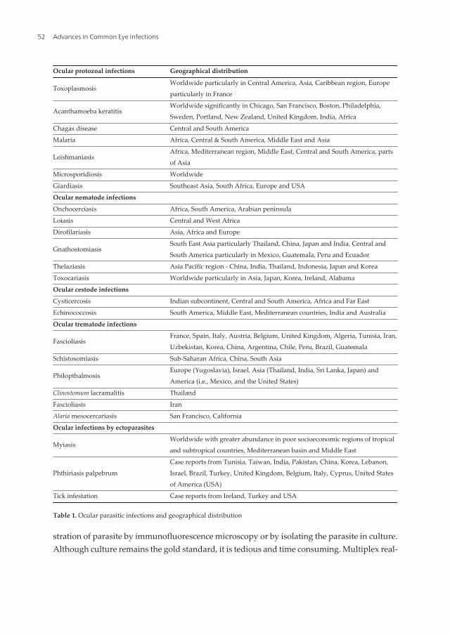
Ocular parasitic infections have been widely reported from different geographical areas (Table1), mainly depending on the endemicity of the parasite(s). The prevalence depends primarilyon the geographical distribution of the parasite, socioeconomic environment and immunestatus of the patient. The common modes of infection are direct contact (blepharoconjunctivitiscaused by Leishmania, Acanthamoeba keratitis, microsporidial infections, infestation caused bylice and mites) [21–23], through blood stream (Toxoplasma chorioretinitis, retinal involvementin malaria, uveitis caused by Toxocara) [1, 23, 24], congenital transmission (Toxoplasmosis) andzoonotic transmission (primarily infectious diseases of animals that can naturally be trans‐mitted to humans) [25]. In addition, few of the helminths that may lead to ocular infection aretransmitted by vectors (onchocerciasis, dirofilariasis and thelaziasis), consumption of conta‐minated food (sparganosis, trichinellosis) and indirectly from the environment (fascioliasis,ascariasis and echinococcosis).
Adult and/or larval stages of the parasites may reside in human ocular tissues externally or inthe ocular globe. The clinical symptoms and signs vary, depending on the etiological agentand the ocular tissue/part involved. However, local defense mechanisms and host immuneresponses play role in establishing the infection. The pathology in the eye can occur due todirect damage by the infecting pathogen, indirectly by toxic products, immune mediated orectopic localization by ectoparasites. The clinical diagnosis usually mimics other pathologiesdue to numerous etiologies both infectious and non-infectious, which can cause conjunctivitis,keratitis, uveitis and endopthalmitis [26]. Thus, a high index of clinician suspicion is requiredfor infective parasite etiology in patients having inflammation in the eye. In addition, eye canbe involved in various systemic disorders and thorough ocular examination along with historyof travel to the endemic area, risk factors and other associated medical illness that help inestablishing the preliminary diagnosis. However, confirmatory diagnosis is usually achievedby direct demonstration of parasite in clinical samples and/or pathological changes observedby either slit lamp or biopsy examination [1, 8, 27, 28]. The antigen and antibody detection inocular fluids and/or serum usually substantiates the clinical diagnosis in few parasiticinfections (Toxoplasmosis, malaria, leishmaniasis, ocular gnathostomiasis, cysticercosis,toxocariasis, echinococcosis) [1, 10, 29, 30]. Molecular techniques including detection ofparasite DNA by polymerase chain reaction (PCR) have added new dimensions in thediagnosis and species identification [31–36]. The treatment of choice is mostly surgical excision,
Advances in Common Eye Infections42
while in few infections, medical treatment is usually advised either in conjunction with surgicalprocedure (onchocerciasis, dirofilariasis [37], cysticercosis [38], echinococcosis [39], myiasis,infections due to ticks and mites) or for inoperable patients. Although surgical excision isusually reserved for worms that are large, it is also recommended for space-occupying lesionsof the orbit. Drug resistance is posing problem for the effective medical treatment, thusnecessitating the discovery of new antiparasitic drugs [32]. Prevention and control measuresdiffer in various infections and usually include proper health education and awareness ofvarious risk factors. The various experimental animal models for few of the ocular infectionshave been successfully established to study the pathogenic mechanisms, drug efficacy andlocal immune responses [40, 41].
Although issues mainly are the timely diagnosis and treatment, yet many challenges need tobe considered/addressed.
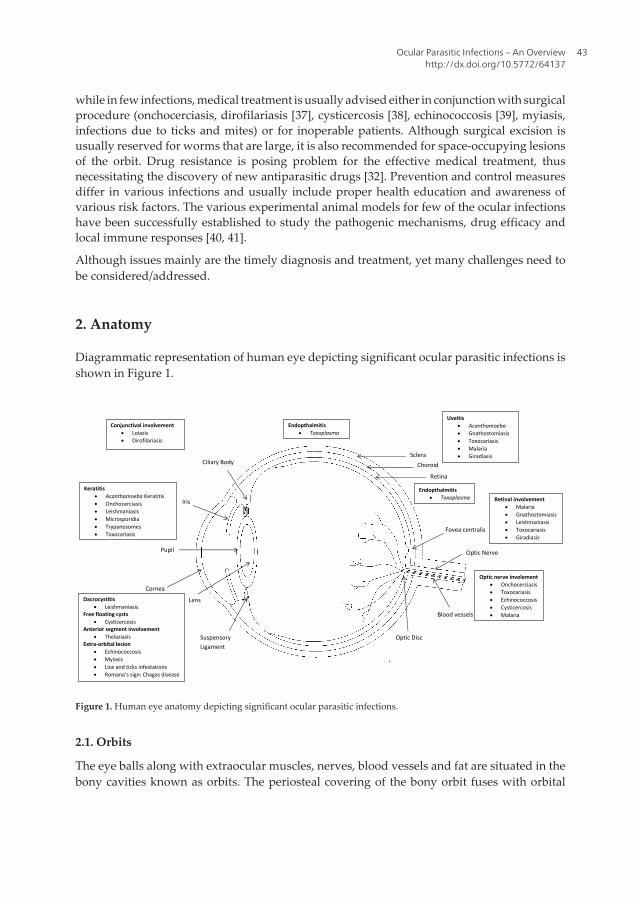
2. Anatomy
Diagrammatic representation of human eye depicting significant ocular parasitic infections isshown in Figure 1.
Pupil
Lens
Cornea
Iris
Ciliary Body Sclera
Choroid
Retina
Optic Disc Suspensory Ligament
Fovea centralis
Optic Nerve
Blood vessels
Keratitis Acanthamoeba Keratitis Onchocerciasis Leishmaniasis Microsporidia Trypanosomes Toxocariasis
Endopthalmitis Toxoplasma Retinal involvement
Malaria Gnathostomiasis Leishmaniasis Toxocariasis Giradiasis
Uveitis Acanthamoeba Gnathostomiasis Toxocariasis Malaria Giradiasis
Endopthalmitis Toxoplasma
Optic nerve involement Onchocerciasis Toxocariasis Echinococcosis Cysticercosis Malaria
Dacrocystitis Leishmaniasis
Free floating cysts Cysticercosis
Anterior segment involvement Thelaziasis
Extra‐orbital lesion Echinococcosis Myiasis Lice and ticks infestations Romana’s sign: Chagas disease
Conjunctival involvement Loiasis Dirofilariasis
Figure 1. Human eye anatomy depicting significant ocular parasitic infections.
2.1. Orbits
The eye balls along with extraocular muscles, nerves, blood vessels and fat are situated in thebony cavities known as orbits. The periosteal covering of the bony orbit fuses with orbital
Ocular Parasitic Infections – An Overviewhttp://dx.doi.org/10.5772/64137
43
septum and duramater. Abscess due to infectious agent can localize in the space beneath theperiostium. The paranasal sinuses are separated from it by the floor, medial wall and roof ofthe orbit and may act as the source of orbital infection. Lamina papyracea are the thinnest bonywalls, which separate orbit from ethmoidal sinuses. Thus, any breach in it causes the ingressof sinus microbiota to orbital tissue leading to infection. Orbital cellulitis can also be causedby direct extension of the infection from the ethmoidal sinuses to the orbital cavity. The lateralwall of the sphenoidal sinus constitutes the medial wall of the optic canal and infection of theformer can percolate to the latter causing optic nerve damage and visual loss. There are variousapertures present in the orbital cavity, which provides the route of communication with theadjacent structures. The superior and inferior orbital fissures, the lacrimal fossa, nasolacrimalduct and the optic canal constitute such important apertures [1, 42–46].
2.2. Blood supply
The ophthalmic artery and its branches constitute main arterial supply of orbit. The majorityof the venous drainage occurs through superior ophthalmic vein, which drains into cavernoussinus that is located just posterior to the orbital apex. Veins from the facial region and manyanterior ophthalmic veins anastomose and drain into cavernous sinus through superior orbitalvein. Thus, cavernous sinus is prone to infection from facial region and also from the orbitalregion through the superior ophthalmic vein leading to a serious complication.
2.3. Eyelids
The eyelids impart two protective anatomical barriers, i.e., orbital septum and conjunctiva.Former divides the orbit from the eyelid into preseptal and postseptal spaces and provides aphysical barrier to infectious agents and latter one is reflected back on itself, which providesprotection by hindering the free movement of the material posteriorly from the anterior surfaceof the globe.
2.4. Lacrimal system
Lacrimal system consists of lacrimal gland, accessory gland and excretory system. Tears aresecreted by lacrimal gland, which flows over the cornea and finally drain into nasal cavity bynasolacrimal duct through lacrimal sac. Any obstruction to the nasolacrimal duct can lead toregurgitation of the accumulated fluid onto the ocular surface leading to increased chances ofinfection.
2.5. Layers of eye ball
The basic structure of eye ball or globe consists of three concentric layers. The outermostcovering is composed of sclera and cornea. The middle covering is composed of uveal tract,consisting of choroid, ciliary body and iris. The inner most covering is retina. The sclera isalmost avascular except for the presence of superficial small blood vessels. The choroid is ahighly vascular structure and provides nutrition and oxygenation to the retina beneath it. Due
Advances in Common Eye Infections44
to these qualities, choroid serves as a fertile area for the proliferation of various pathogens,which spread by hematogenous route.
2.6. Anterior and posterior chambers
Anterior segment of the eye in front of the vitreous humor comprises anterior one-third of theeye and is further divided into anterior chamber and posterior chamber. Anterior chamber isthe space between posterior surface of cornea and the iris, whereas posterior chamber is thespace between iris and the front of vitreous. The aqueous humor is produced by non-pig‐mented ciliary epithelium in the posterior chamber and drains through the pupillary apertureinto the anterior chamber. Cornea is composed of well-organized collagen fibrils, which isavascular in nature. Lens is also an avascular crystalline structure, which continues to growthroughout life. Thus, aqueous humor fills these spaces and provides nutrition to the sur‐rounding structures.
2.7. Vitreous humor
It is a gel-like substance present in front of retina and posterior to the lens in the posteriorsegment of the eye. It is optically clear and is composed of collagen framework interspersedwith hyaluronic acid. During intraocular inflammation, it becomes hazy and may causeimpairment of vision.
2.8. Retina and optic nerve
Retina constitutes the innermost covering of the eye ball and captures the light energy withthe help of rods and cones. The outer half of the retina is supplied by central retinal artery,whereas inner half receives its blood supply from the choroid.
The optic nerve is formed by axons of the inner cell layer that exits the globe. It is covered byall the three meningeal coverings, which are direct extensions of the brain coverings. Thus, itis vulnerable to infections originating from both within cranial vault and within orbits.
3. Ocular defense mechanisms
The surface of the eye is well protected by both mechanical and immunological defensemechanisms. To breach the defense mechanism, some form of trauma is essential. The eyelidsprovide mechanical protection to the surface of eyeball. The eyelashes protect against airborneparticles and trauma by initiating blink reflex. The cornea is also sensitive to tactile sensationand helps in the initiation of blink reflex, which is provided by dense sensory nerve endings.The lids direct the tears, particulate debris, allergens and microbes to the lacrimal excretorysystem by its sweeping action over the anterior surface of the eyeball. Bell’s phenomenon alsoprovides protection to cornea as globe is turned upwards and slightly outwards during eyelidclosure to avoid corneal exposure [47]. Meibomian glands secrete lipids, which providestability to the tear film. The epithelial surface of the cornea and conjunctiva provides ana‐
Ocular Parasitic Infections – An Overviewhttp://dx.doi.org/10.5772/64137
45
tomical barrier to the pathogens. This function is further strengthened by the impermeabilityprovided by the basement and cellular junctional complexes of the cornea. Indigenous floraof the eye also provides protection by creating a competition for colonization by the pathogens.
Immune defense mechanisms are provided by the vascular supply of the eye. Any breach inthe anatomical defense system initiates the ocular inflammatory response, which helps invasodilation and exudation of immunologically active substances and cells [1, 8, 48–52].
3.1. Defenses of the tear film
There are three layers of the tear film: oil, aqueous and mucous. Majority of the tear film iscomposed of aqueous layer and pH of the tear film helps in neutralization of toxic substances.Flow of tears help in mechanical flushing of the foreign particles and allergens into the lacrimalexcretory system. Mucosal layer helps in entrapment of pathogens. Tear film contains variousimmunological active substances such as lactoferrin, lysozyme, β-lysin, ceruloplasmin,complement and immunoglobulins.
3.2. Conjunctival defenses
The conjunctival associated lymphoid tissue lies beneath the conjunctiva. It consists of both Band T lymphocytes. B and T cell precursors mature when exposed to foreign particles orallergens, then migrate to regional lymph nodes for further development, and thereafter returnto the conjunctiva through blood stream to produce specific immunoglobulins and cellulardefense responses.
3.3. Corneal defenses
Although the cornea is avascular, it is provided by limited defense mechanisms in the form ofLangerhans cells (dendritic cells) and immunoglobulins. The surface of the cornea is coveredby mucous glycoprotein, which helps in cross-linkage of the IgA and protects the anteriorsurface of the cornea. Immune defense mechanisms are activated whenever injury occurs,leading to recruitment of the polymorphonuclear cells, lymphocytes and fibroblasts.
3.4. Cellular immune responses
Langerhans cells are situated along the peripheral margin of the cornea and conjunctiva. Thesecells possess receptors, which help in phagocytosis and processing of certain antigens forpresentation. Langerhans cells stimulate B and T cells to elicit a strong cellular immuneresponse. During inflammation Langerhans cells migrate toward the cornea, causing increasedrelease of inflammatory substances.
3.5. Leukocyte defense
Polymorphonuclear leukocytes are the hallmark of acute inflammation and are associated withoxygen-dependent pathways for the generation of free radicals that help in killing of the
Advances in Common Eye Infections46
invading pathogens. Another immune defense mechanism operated by the production ofdefensins is antimicrobial proteins active against wide range of pathogens.
3.6. Defensins
Ocular surface is constantly exposed to environment and foreign bodies, thus there are greaterchances of infection. However, robust innate immune system at ocular surface protects the eyefrom infection. There are several peptides of defensins and cathelicidin families that are presentin tear film and secreted by corneal and conjunctival cells. These are not only antimicrobial innature but also help in the recruitment of immune cells and thus provide a link to adaptiveimmunity. The important defensins present in human eye are hBD-1 (human beta defensins),hBD-2, hBD-3, CAP37 (Cathelicidin-related antimicrobial peptide), LL37 (type of cathelicidin)and HNP-1, 2, 3 (human neutrophil defensins) [53].
4. Protozoan eye infections
4.1. Toxoplasmosis
Toxoplasmosis is caused by obligatory intracellular protozoan parasite known as Toxoplasmagondii. The mode of infection is either by the ingestion of oocysts shed in feces of the cats orother Felidae (definitive host) or by the consumption of tissue cyst present in the raw oruncooked meat. Life cycle of Toxoplasma includes three stages that are oocysts, tachyzoites andbradyzoites. It completes its life cycle in two phases, one as an intestinal phase in its homolo‐gous host, such as cats and another as an extraintestinal phase in its heterologous host, suchas mouse, man and other animals. When cats feed on mouse brain containing tissue cysts ofT. gondii, a large number of oocysts are released in the infected cat’s feces. After 1–5 days,oocysts get matured and become infective to man and other animals. After ingestion, oocystsliberate sporozoites, which penetrate intestinal mucosa and reach to distant organs such asbrain, eyes, liver, spleen, lymph nodes, heart, skeletal muscles and placenta by blood andlymphatic stream. Toxoplasma tissue cysts also occur in the skeletal muscles of the intermediatehost such as sheep and pigs (Figure 2) [54]. In addition, developing fetus can acquire theinfection transplacentally from the mother during pregnancy. Rarely, infection may also resultfrom consumption of drinking water contaminated with oocysts. The ocular infection can beeither congenital or acquired.
Approximately, one-third of the world’s population is thought to be infected by T. gondii. It iscommon in hot and humid climates such as Central America, Asia and the Caribbean region(Table 1, Figure 3). In Europe, toxoplasmosis is common and the highest prevalence rates havebeen reported in France. Various risk factors such as geographical region, meat consumption,personal habits, animal reservoir and climatic conditions play a significant role in the trans‐mission of infection. In recent years, due to indoor keeping of livestock and improvement inhygiene standards, the risk of acquiring infection has decreased tremendously in the devel‐oped nations. However, in the developing nations, risk has increased due to populationgrowth, urbanization trends and increase in meat consumption. Drinking water, seawater and
Ocular Parasitic Infections – An Overviewhttp://dx.doi.org/10.5772/64137
47
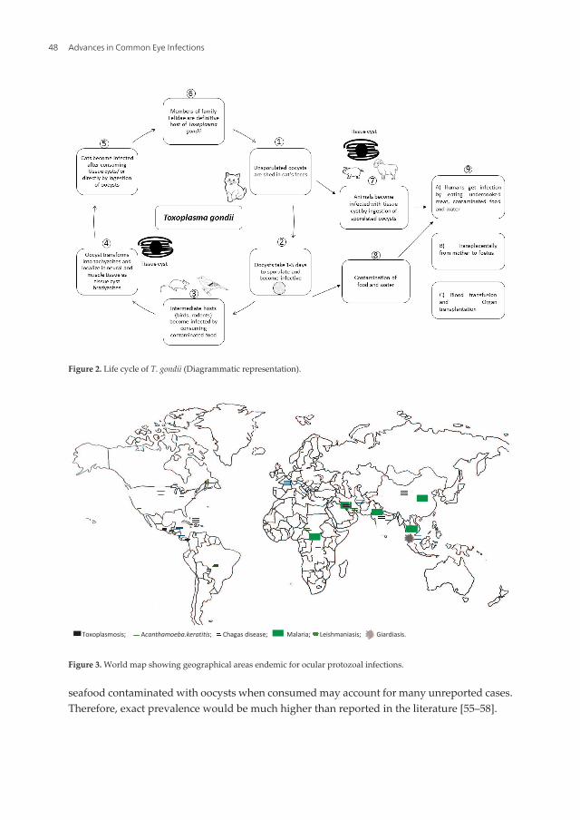
seafood contaminated with oocysts when consumed may account for many unreported cases.Therefore, exact prevalence would be much higher than reported in the literature [55–58].
Figure 2. Life cycle of T. gondii (Diagrammatic representation).
Toxoplasmmosis; A ; Chagas diseease; Malarria; Leishmaniaasis; Giardiasis.
Figure 3. World map showing geographical areas endemic for ocular protozoal infections.
Advances in Common Eye Infections48
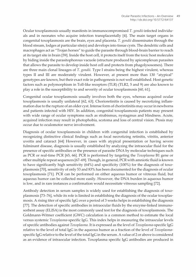
Ocular toxoplasmosis usually manifests in immunocompromised T. gondii-infected individu‐als and in neonates who acquire infection transplacentally [4]. The main target organs incongenital toxoplasmosis are the brain, eyes and placenta. T. gondii disseminates through theblood stream, lodges at particular site(s) and develops into tissue cysts. The dendritic cells andmacrophages act as “Trojan horses” to guide the parasite through blood-brain barrier to reachat its target site in brain [59]. Inside the host cell, it protects itself from the toxic host moleculesby hiding inside the parasitophorous vacuole (structure produced by apicomplexan parasitesthat allows the parasite to develop inside host cell and protects from phagolysosomes). Thereare three main clonal lineages of T. gondii. Type I strains being the highest virulent, whereastypes II and III are moderately virulent. However, at present more than 130 “atypical”genotypes are known, but their exact role in pathogenesis is not well established. Host geneticfactors such as polymorphism in Toll-like receptors (TLR) (TLR2, 5 and 9) are also known toplay a role in the susceptibility to and severity of ocular toxoplasmosis [60, 61].
Congenital ocular toxoplasmosis usually involves both the eyes, whereas acquired oculartoxoplasmosis is usually unilateral [62, 63]. Chorioretinitis is caused by necrotizing inflam‐mation due to the rupture of an older cyst. Intense form of choriretinitis may occur in newbornsand patients infected with HIV. In addition, congenital toxoplasmosis patients may presentwith wide range of ocular symptoms such as strabismus, nystagmus and blindness. Acute,acquired infection may result in photophobia, scotoma and loss of central vision. Ptosis mayoccur due to oculomotor nerve involvement.
Diagnosis of ocular toxoplasmosis in children with congenital infection is established byrecognizing distinctive clinical findings such as focal necrotizing retinitis, vitritis, anterioruveitis and cataract [64] However, in cases with atypical presentation or having severefulminant disease, diagnosis is usually established by analyzing the intraocular fluid for thepresence of specific antibodies or the presence of parasite DNA by molecular techniques suchas PCR or real-time PCR [65, 66]. PCR is performed by targeting the Toxoplasma B1 gene orother multiple repeat sequences [67–69]. Though, in general, PCR with amniotic fluid is knownto have significantly high sensitivity (64%) and specificity (100%) for the diagnosis of toxo‐plasmosis [70], sensitivity of only 53 and 83% has been documented for the diagnosis of oculartoxoplasmosis [71]. PCR can be performed on either aqueous humor or vitreous fluid, butaqueous humor can be collected more easily. However, the DNA burden in aqueous humoris low, and in rare instances a confirmation would necessitate vitreous sampling [72].
Antibody detection in serum samples is widely used for establishing the diagnosis of toxo‐plasmosis [73–76], while its role is limited in establishing the diagnosis of the ocular toxoplas‐mosis. A rising titer of specific IgG over a period of 3 weeks helps in establishing the diagnosis[77]. The detection of specific antibodies in intraocular fluids by the enzyme-linked immuno‐sorbent assay (ELISA) is the most commonly used test for the diagnosis of toxoplasmosis. TheGoldmann-Witmer coefficient (GWC) calculation is a common method to estimate the localversus systemic Toxoplasma-specific IgG. This index helps in measuring the intraocular levelsof specific antibodies against Toxoplasma. It is expressed as the level of Toxoplasma-specific IgGrelative to the level of total IgG in the aqueous humor as a fraction of the level of Toxoplasma-specific IgG relative to the level of the total IgG in the serum. A value of 2 or above is consideredas an evidence of intraocular infection. Toxoplasma specific IgG antibodies are produced in
Ocular Parasitic Infections – An Overviewhttp://dx.doi.org/10.5772/64137
49
response to the actively multiplying tachyzoites at local site of infection [72, 78] The presenceof T. gondii-specific IgM is the hallmark of a recently acquired systemic or, possibly, ocularinfection. However, high rate of false-positive results due to the persistence of antibodies,decreases its utility as a diagnostic marker for recent ocular toxoplasmosis. In patients withreactivated ocular toxoplasmosis, it is not useful as T. gondii-specific IgM antibodies are eitherabsent or present in very low quantity [79]. Saliva samples have also been tested for thedetection of specific antibodies for the diagnosis of toxoplasma encephalitis in immunocom‐promised individuals, but it may play a limited role in ocular toxoplasmosis [74].
An algorithm for the laboratory confirmation of clinically suspected cases of ocular toxoplas‐mosis has been reported [72]. Reactivated form of ocular toxoplasmosis is considered inpatients with typical lesions of toxoplasmic retinochoroiditis, specific IgG seropositive, specificIgM seronegative and responding to anti-Toxoplasma treatment. However, if patients arespecific IgM seropositive, then additional laboratory tests are required. If doubt persists aboutdiagnosis, paired serum and aqueous samples are required to be tested in parallel. The clinicaldiagnosis along with laboratory evidence is documented in 60-85% of cases and thus, labora‐tory evidence is lacking in 15-40% of clinically suspected patients. Analysis of aqueous humoris useful in patients presenting with atypical ocular lesions or not responding to specifictreatment [72].
In immunocompetent individuals, toxoplasma retinochoroiditis usually resolves within 2–3months [80]. Classic therapy or triple therapy with a combination of pyrimethamine, sulfa‐diazine and systemic corticosteroids is recommended for lesions involving or near to fovea,an area critical for vision. Classic therapy is usually associated with significant side effects,therefore other drugs such as trimethoprim-sulfamethoxazole, clindamycin, atovaquone andazithromycin are being evaluated for the treatment of ocular toxoplasmosis [81].
Trimethoprim-sulfamethoxazole (Bactrim) appears to be a safe and effective substitute forsulfadiazine, pyrimethamine and folinic acid for the treatment of ocular toxoplasmosis.
Progressive and recurring necrotizing retinitis, with vision-threatening complications such asretinal detachment, choroidal neovascularization and glaucoma, may occur at any time duringthe clinical course if the infection is not treated on time. Congenital toxoplasmosis can lead tocataract. The aim of the treatment is to arrest parasite multiplication during the active periodof retinochoroiditis and to minimize damage to the retina and optic disc [64].
Animal model(s) can be used to study various aspects of ocular toxoplasmosis [40].
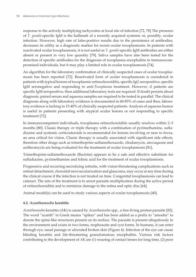
4.2. Acanthamoeba keratitis
Acanthamoeba keratitis (AK) is caused by Acanthamoeba spp., a free-living protest parasite [82].The word “acanth” in Greek means “spikes” and has been added as a prefix to “amoeba” todenote the spine-like structures present on its surface. The parasite is present ubiquitously inthe environment and exists in two forms, trophozoite and cyst forms. In humans, it can enterthrough eye, nasal passage or ulcerated broken skin (Figure 4). Infection of the eye can causeblinding keratitis and life-threatening granulomatous encephalitis. Various risk factorscontributing to the development of AK are (1) wearing of contact lenses for long time, (2) poor
Advances in Common Eye Infections50
personal hygiene, (3) cleaning of lenses with contaminated water and (4) formation of biofilmon contact lenses [82].
Figure 4. Life cycle of Acanthamoeba (Diagrammatic representation).
Acanthamoeba keratitis is common among the contact lens users, and its geographic distributionis depicted in Table 1 and Figure 3. However, in India the infection is reported even in non-contact lens users [7]. The incidence of Acanthamoeba keratitis in developed nations varies from1 to 33 cases per million contact lens wearers. In developing nations where contact lens usersare limited, the other suggested risk factors are trauma, exposure to contaminated water, useof traditional eye medicine, low socioeconomic background, splashing contaminated waterinto the eye following dust fall and corneal injury with mud [7, 22, 83]. The pathogenesis ofAcanthamoeba involves following sequential events, i.e., breach in the epithelial barrier,invasion of stroma by amoeba, depletion of keratocytes, induction of inflammatory response,photophobia and finally necrosis of stroma leading to blindness [82].
The diagnosis of AK is difficult as it is usually confused with symptoms of bacterial, fungal orviral keratitis. However, history of contact lens use together with a history of excruciating painis a strong indication toward the diagnosis of AK. For establishing the clinical diagnosis withhigh sensitivity, in vivo confocal microscopy can be used, which is a non-invasive procedure.The Acanthamoeba cysts appear as hyper-reflective, spherical structures that are well definedbecause of their double wall. However, trophozoites are difficult to distinguish from leuko‐cytes and keratocyte nuclei [84, 85]. Laboratory confirmation is established by direct demon‐
Ocular Parasitic Infections – An Overviewhttp://dx.doi.org/10.5772/64137
51
stration of parasite by immunofluorescence microscopy or by isolating the parasite in culture.Although culture remains the gold standard, it is tedious and time consuming. Multiplex real-
Ocular protozoal infections Geographical distribution
ToxoplasmosisWorldwide particularly in Central America, Asia, Caribbean region, Europeparticularly in France
Acanthamoeba keratitisWorldwide significantly in Chicago, San Francisco, Boston, Philadelphia,Sweden, Portland, New Zealand, United Kingdom, India, Africa
Chagas disease Central and South America
Malaria Africa, Central & South America, Middle East and Asia
LeishmaniasisAfrica, Mediterranean region, Middle East, Central and South America, partsof Asia
Microsporidiosis Worldwide
Giardiasis Southeast Asia, South Africa, Europe and USA
Ocular nematode infections
Onchocerciasis Africa, South America, Arabian peninsula
Loiasis Central and West Africa
Dirofilariasis Asia, Africa and Europe
GnathostomiasisSouth East Asia particularly Thailand, China, Japan and India, Central andSouth America particularly in Mexico, Guatemala, Peru and Ecuador
Thelaziasis Asia Pacific region - China, India, Thailand, Indonesia, Japan and Korea
Toxocariasis Worldwide particularly in Asia, Japan, Korea, Ireland, Alabama
Ocular cestode infections
Cysticercosis Indian subcontinent, Central and South America, Africa and Far East
Echinococcosis South America, Middle East, Mediterranean countries, India and Australia
Ocular trematode infections
FascioliasisFrance, Spain, Italy, Austria, Belgium, United Kingdom, Algeria, Tunisia, Iran,Uzbekistan, Korea, China, Argentina, Chile, Peru, Brazil, Guatemala
Schistosomiasis Sub-Saharan Africa, China, South Asia
PhilopthalmosisEurope (Yugoslavia), Israel, Asia (Thailand, India, Sri Lanka, Japan) andAmerica (i.e., Mexico, and the United States)
Clinostomum lacramalitis Thailand
Fascioliasis Iran
Alaria mesocercariasis San Francisco, California
Ocular infections by ectoparasites
MyiasisWorldwide with greater abundance in poor socioeconomic regions of tropicaland subtropical countries, Mediterranean basin and Middle East
Phthiriasis palpebrumCase reports from Tunisia, Taiwan, India, Pakistan, China, Korea, Lebanon,Israel, Brazil, Turkey, United Kingdom, Belgium, Italy, Cyprus, United Statesof America (USA)
Tick infestation Case reports from Ireland, Turkey and USA
Table 1. Ocular parasitic infections and geographical distribution
Advances in Common Eye Infections52
time PCR assays (multiplex assays targets more than one region and simultaneously can detecttwo or more target regions) have also been developed for the detection of different pathogenicfree-living amoeba and/or different genotypes of Acanthamoeba. Although molecular techni‐ques have high sensitivity and specificity, these are only available at apex laboratories and alsorequire a well-established molecular laboratory [86]. Newer techniques such as Matrix-Assisted Laser Desorption Ionization Time-Of-Flight (MALDI-TOF) and 1H-NMR spectrosco‐py [87] are also being tested for the rapid identification of Acanthamoeba in the clinicalspecimens [88].
Chances of recovery are good if the pathogen is restricted to cornea epithelium but can leadto vision loss, if it invades stroma leading to necrosis and intense inflammation. Medicaltreatment, if started early, can lead to a significant improvement within 2–3 weeks [89].
Preventive measures include thorough and adequate disinfection of contact lenses. It isrecommended to remove contact lenses before any activity involving contact with water,including showering, using a hot tub, or swimming. Hands should be washed with soap andwater and dried before handling contact lenses. Contact lenses should not be rinsed with tapwater and should be cleaned and stored as per manufacturer’s guidelines. It is suggested thatthe increased awareness about the other predisposing factors (corneal injury, fall of foreignbody in eye) among the general public may enable early and frequent recognition and propermanagement of AK in patients other than contact lens wearers [7].
4.3. Chagas disease
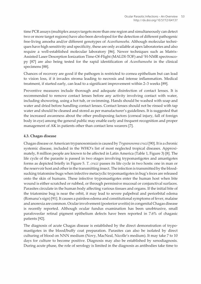
Chagas disease or American trypanosomiasis is caused by Trypanosoma cruzi [90]. It is a chronicsystemic disease, included in the WHO’s list of most neglected tropical diseases. Approxi‐mately, 8 million people are known to be affected in Latin America (Table 1, Figure 3) [8]. Thelife cycle of the parasite is passed in two stages involving trypomastigotes and amastigotesforms as depicted briefly in Figure 5. T. cruzi passes its life cycle in two hosts: one in man orthe reservoir host and other in the transmitting insect. The infection is transmitted by the blood-sucking triatomine bugs when infective metacyclic trypomastigotes in bug’s feces are releasedonto the skin of humans. These infective trypomastigotes enter the human host when bitewound is either scratched or rubbed, or through permissive mucosal or conjunctival surfaces.Parasites circulate in the human body affecting various tissues and organs. If the initial bite ofthe triatomine bug is near the orbit, it may lead to severe palpebral and periorbital edema(Romana’s sign) [91]. It causes a painless edema and constitutional symptoms of fever, malaiseand anorexia are common. Ocular involvement (posterior uveitis) in congenital Chagas diseaseis recently reported. Although ocular fundus examination has been unobtrusive, smallparafoveolar retinal pigment epithelium defects have been reported in 7.6% of chagasicpatients [92].
The diagnosis of acute Chagas disease is established by the direct demonstration of trypo‐mastigotes in the blood/buffy coat preparation. Parasites can also be isolated by directculturing of blood on NNN medium (Novy, MacNeal, Nicolle’s medium). It may take 7 to 10days for culture to become positive. Diagnosis may also be established by xenodiagnosis.During acute phase, the role of serology is limited in the diagnosis as antibodies take time to
Ocular Parasitic Infections – An Overviewhttp://dx.doi.org/10.5772/64137
53
develop and false positive results have also been known to be associated with serological testsdue to cross-reaction of antibodies to non-pathogenic Trypanosoma rangeli [8, 91]. Furthermore,detailed examination by the ophthalmologist may aid in establishing the diagnosis. However,accumulation of retinal pigment epithelium defects have been shown in patients with intra‐ocular involvement of intermediate and chronic Chagas disease in Paraguay/South America,but overall fundus examination has shown to be unobtrusive [92, 93].
Acute cases of Chagas disease are treated by nifurtimox and benznidazole. Benznidazole isgiven as 5–7.5 mg/kg per day orally in two divided doses for 60 days. Nifurtimox is given as8–10 mg/kg per day orally in three or four divided doses for 90 days [91, 94].
Within few weeks, symptoms of acute Chagas disease such as Romana’s sign fade away, butinfection persists. The average life-time risk of developing complications of chronic phase isaround 30%. It may take more than 20 years to develop chronic complications. However,trypanocidal therapy did not significantly reduce cardiac clinical deterioration through 5 yearsof follow-up as documented by randomized trial of benznidazole for chronic Chagas’ cardio‐myopathy [95, 96].
4.4. Leishmaniasis
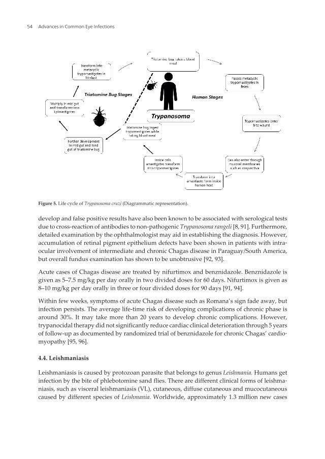
Leishmaniasis is caused by protozoan parasite that belongs to genus Leishmania. Humans getinfection by the bite of phlebotomine sand flies. There are different clinical forms of leishma‐niasis, such as visceral leishmaniasis (VL), cutaneous, diffuse cutaneous and mucocutaneouscaused by different species of Leishmania. Worldwide, approximately 1.3 million new cases
Figure 5. Life cycle of Trypanosoma cruzi (Diagrammatic representation).
Advances in Common Eye Infections54
occur every year with a mortality of 20,000 to 30,000 persons per annum [97]. While taking theblood meal, infected sandfly injects promastigotes into humans. Further in the human body,the promastigotes are transformed into amastigote forms, and these are engulfed by tissuemacrophages. Amastigote forms replicate inside the cells and further spread either systemi‐cally or through cutaneous route, depending on the species of the parasite (Figure 6). Ocularinvolvement due to leishmaniasis has been reported from various countries such as India,Sudan, Italy, Norway, Turkey and Iran (Table 1, Figure 3) [98–103]. Anterior uveitis is the mostcommon ocular manifestation in VL, which can occur during the course of infection and canfurther progress to glaucoma [104, 105]. Focal retinal whitening, cotton wool spots, hemor‐rhages and increased vessel tortuosity have also been reported on fundus examination [106–110]. In severe cases, flame-shaped lesions also appear, which denote hemorrhage from theanterior capillaries of the nerve fiber layer. These findings have also been correlated withanemia and thrombocytopenia as these hemorrhages usually get resolved with treatment,leading to improvement in anemia/thrombocytopenia. Optic neuropathy has been reporteddue to mucosal leishmaniasis. Eyelid involvement has been documented in cutaneous andmucocutaneous leishmaniasis [111, 112]. Severe involvement can progress to ptosis andectropion secondary to cutaneous leishmaniasis leading to keratopathy and altered vision[112]. However, eyelid is rarely involved by leishmaniasis and is reported in approximatelyonly 2.5% of cases with cutaneous leishmaniasis [113]. The most common aspect of eyelidleishmaniasis is chalazion-like lesions, but other forms such as ulcerous, phagedenic, cancer-like forms and unilateral chronic granulomatous blepharitis may be observed. Chronicdacryocystitis has been reported in patients suffering from mucocutaneous leishmaniasis,which can effect formation of tear film, leading to dryness of eyes [114]. Endo-ocular lesionshave been observed in patients having disseminated cutaneous leishmaniasis. A report fromBrazil documented the presence of Leishmania in the aqueous humor along with iridiocyclitis[115]. Although ocular manifestations are not very common, it is suggested that a person withocular manifestation from endemic country should undergo fundus examination for earlydiagnosis [116].
Diagnosis of leishmaniasis can be achieved by the direct demonstration of parasites in thetissue smears and/or biopsy samples, culture technique(s), antigen and/or antibody detectionand molecular technique(s). However, each technique has its own merits and demerits.Amastigotes can be easily identified in the cutaneous and mucocutaneous lesions but are noteasily identified in cases with ocular disease [103, 117, 118]. Molecular techniques such as PCR/real-time PCR can identify the genome of parasite with greater sensitivity (100%) and specif‐icity (100%) [119, 120]. The treatment of leishmaniasis depends on several factors such asclinical form of the disease. The antileishmanial drugs include pentavalent antimony, sodiumstibogluconate, liposomal Amphotericin B, miltefosine and paromomycin [118, 121].
Ocular lesions do not heal without treatment and could lead to vision loss if conjunctiva isinvolved due to severe ulceration. Healing occurs without visual impairment if treatment isinitiated early during the course of infection and vigorous treatment is required to preventblindness [121, 122].
Ocular Parasitic Infections – An Overviewhttp://dx.doi.org/10.5772/64137
55
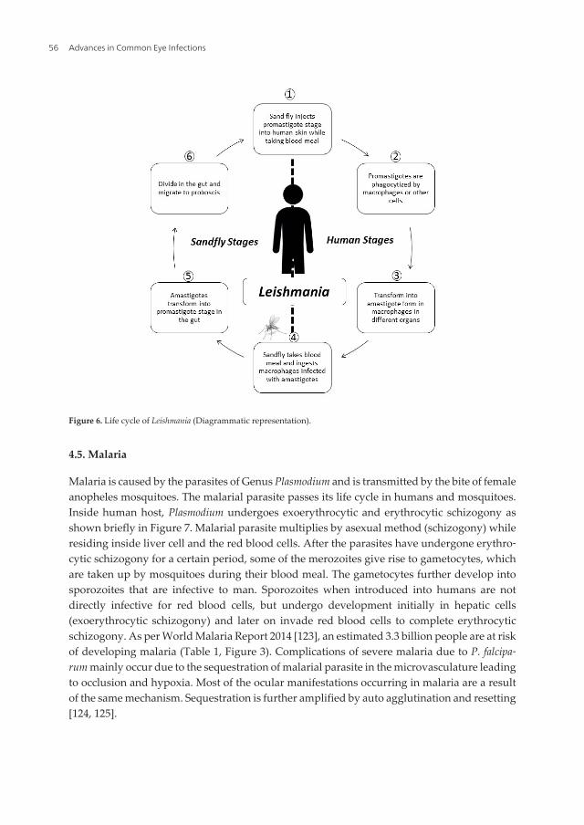
4.5. Malaria
Malaria is caused by the parasites of Genus Plasmodium and is transmitted by the bite of femaleanopheles mosquitoes. The malarial parasite passes its life cycle in humans and mosquitoes.Inside human host, Plasmodium undergoes exoerythrocytic and erythrocytic schizogony asshown briefly in Figure 7. Malarial parasite multiplies by asexual method (schizogony) whileresiding inside liver cell and the red blood cells. After the parasites have undergone erythro‐cytic schizogony for a certain period, some of the merozoites give rise to gametocytes, whichare taken up by mosquitoes during their blood meal. The gametocytes further develop intosporozoites that are infective to man. Sporozoites when introduced into humans are notdirectly infective for red blood cells, but undergo development initially in hepatic cells(exoerythrocytic schizogony) and later on invade red blood cells to complete erythrocyticschizogony. As per World Malaria Report 2014 [123], an estimated 3.3 billion people are at riskof developing malaria (Table 1, Figure 3). Complications of severe malaria due to P. falcipa‐rum mainly occur due to the sequestration of malarial parasite in the microvasculature leadingto occlusion and hypoxia. Most of the ocular manifestations occurring in malaria are a resultof the same mechanism. Sequestration is further amplified by auto agglutination and resetting[124, 125].
Figure 6. Life cycle of Leishmania (Diagrammatic representation).
Advances in Common Eye Infections56
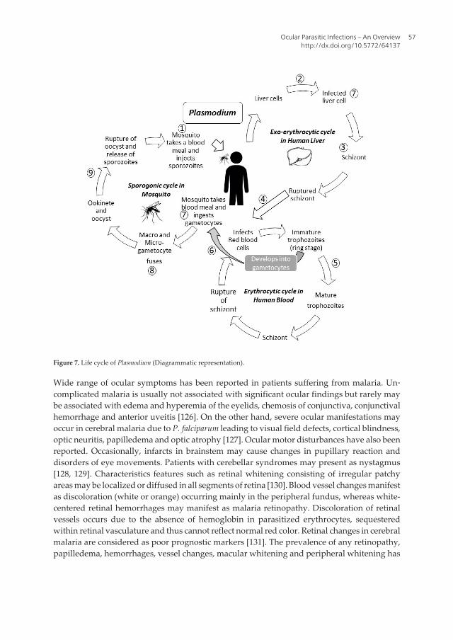
Figure 7. Life cycle of Plasmodium (Diagrammatic representation).
Wide range of ocular symptoms has been reported in patients suffering from malaria. Un‐complicated malaria is usually not associated with significant ocular findings but rarely maybe associated with edema and hyperemia of the eyelids, chemosis of conjunctiva, conjunctivalhemorrhage and anterior uveitis [126]. On the other hand, severe ocular manifestations mayoccur in cerebral malaria due to P. falciparum leading to visual field defects, cortical blindness,optic neuritis, papilledema and optic atrophy [127]. Ocular motor disturbances have also beenreported. Occasionally, infarcts in brainstem may cause changes in pupillary reaction anddisorders of eye movements. Patients with cerebellar syndromes may present as nystagmus[128, 129]. Characteristics features such as retinal whitening consisting of irregular patchyareas may be localized or diffused in all segments of retina [130]. Blood vessel changes manifestas discoloration (white or orange) occurring mainly in the peripheral fundus, whereas white-centered retinal hemorrhages may manifest as malaria retinopathy. Discoloration of retinalvessels occurs due to the absence of hemoglobin in parasitized erythrocytes, sequesteredwithin retinal vasculature and thus cannot reflect normal red color. Retinal changes in cerebralmalaria are considered as poor prognostic markers [131]. The prevalence of any retinopathy,papilledema, hemorrhages, vessel changes, macular whitening and peripheral whitening has
Ocular Parasitic Infections – An Overviewhttp://dx.doi.org/10.5772/64137
57
been reported in 61, 15, 46, 32, 46 and 44%, respectively, among children with cerebral malariain Malawi [132].
Diagnosis of malaria is established by light microscopy or by rapid antigen detection kits. Lightmicroscopic examination of Giemsa-stained peripheral blood smear is considered as goldstandard for the diagnosis of malaria with a threshold of about 50–100 parasites/µL [133].However, in addition, ocular examination may provide clue to the diagnosis as specific retinalchanges can be seen directly [129, 134, 135].
Treatment depends on the species of Plasmodium causing infection. Artemisinin combinationtherapy is recommended for malaria due to P. falciparum. Artemisinin combination therapyincludes short-acting artemisinin derivative and long-acting antimalarial (sulphadoxine-pyrimethamine, lumefantrine). Chloroquine along with primaquine is recommended formalaria due to P. vivax. Ocular toxicity [136] is very well documented with chloroquinetherapy. This includes corneal changes (cornea verticillata) and corneal deposits. Toxicmaculopathy and scotoma has also been reported. Quinine overdose has also been known tocause decreased vision, retinal and macular degeneration, mild scotomas and color visiondefects [136].
If not treated, malarial retinopathy is associated with serious consequences as reports indicatethat the severity of retinopathy is related to prolonged death and coma. After antimalarialtreatment and resolution of coma in severe malaria, malarial retinopathy resolves after sometime [132, 137].
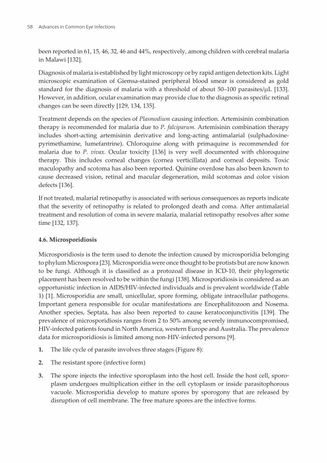
4.6. Microsporidiosis
Microsporidiosis is the term used to denote the infection caused by microsporidia belongingto phylum Microspora [23]. Microsporidia were once thought to be protists but are now knownto be fungi. Although it is classified as a protozoal disease in ICD-10, their phylogeneticplacement has been resolved to be within the fungi [138]. Microsporidiosis is considered as anopportunistic infection in AIDS/HIV-infected individuals and is prevalent worldwide (Table1) [1]. Microsporidia are small, unicellular, spore forming, obligate intracellular pathogens.Important genera responsible for ocular manifestations are Encephalitozoon and Nosema.Another species, Septata, has also been reported to cause keratoconjunctivitis [139]. Theprevalence of microsporidiosis ranges from 2 to 50% among severely immunocompromised,HIV-infected patients found in North America, western Europe and Australia. The prevalencedata for microsporidiosis is limited among non-HIV-infected persons [9].
1. The life cycle of parasite involves three stages (Figure 8):
2. The resistant spore (infective form)
3. The spore injects the infective sporoplasm into the host cell. Inside the host cell, sporo‐plasm undergoes multiplication either in the cell cytoplasm or inside parasitophorousvacuole. Microsporidia develop to mature spores by sporogony that are released bydisruption of cell membrane. The free mature spores are the infective forms.
Advances in Common Eye Infections58
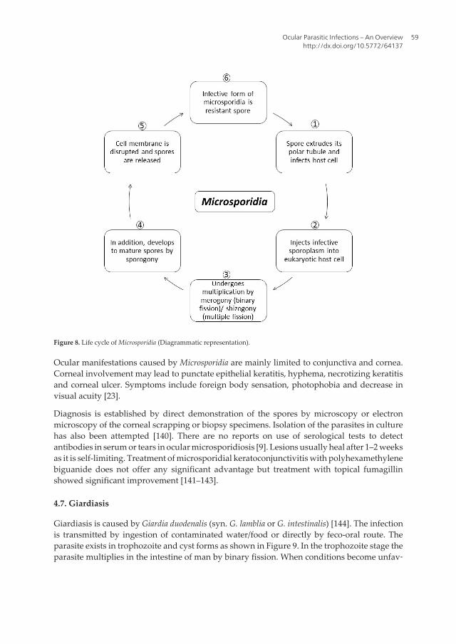
Figure 8. Life cycle of Microsporidia (Diagrammatic representation).
Ocular manifestations caused by Microsporidia are mainly limited to conjunctiva and cornea.Corneal involvement may lead to punctate epithelial keratitis, hyphema, necrotizing keratitisand corneal ulcer. Symptoms include foreign body sensation, photophobia and decrease invisual acuity [23].
Diagnosis is established by direct demonstration of the spores by microscopy or electronmicroscopy of the corneal scrapping or biopsy specimens. Isolation of the parasites in culturehas also been attempted [140]. There are no reports on use of serological tests to detectantibodies in serum or tears in ocular microsporidiosis [9]. Lesions usually heal after 1–2 weeksas it is self-limiting. Treatment of microsporidial keratoconjunctivitis with polyhexamethylenebiguanide does not offer any significant advantage but treatment with topical fumagillinshowed significant improvement [141–143].
4.7. Giardiasis
Giardiasis is caused by Giardia duodenalis (syn. G. lamblia or G. intestinalis) [144]. The infectionis transmitted by ingestion of contaminated water/food or directly by feco-oral route. Theparasite exists in trophozoite and cyst forms as shown in Figure 9. In the trophozoite stage theparasite multiplies in the intestine of man by binary fission. When conditions become unfav‐
Ocular Parasitic Infections – An Overviewhttp://dx.doi.org/10.5772/64137
59
orable in the small intestine, encystment occurs and cysts are released along with feces. Afteringestion, within 30 minutes, cyst hatches out trophozoites that further multiply in the smallintestine. It is found both in developing and developed nations (Table 1, Figure 3). Althoughit mainly causes diarrhea and malabsorption, in one-third of the patients, it can also result inlong-term extra intestinal manifestations [145].
Figure 9. Life cycle of Giardia lamblia (Diagrammatic representation).
Barraquer was the first to report the ocular manifestation (iridiocyclitis, choroiditis and retinalhemorrhages) in patients who were suffering from diarrhea due to G. duodenalis. Retinalchanges in the form of ”salt and pepper” degeneration have been reported in children sufferingfrom giardiasis. Corsi et al. [146] reported salt and pepper retinal changes in 19.9% of thepatients with giardiasis. This occurs due to the damage of the retinal cells and subsequentrelease of pigment granules in retina giving an appearance of blackish dots on a backgroundof light yellow pink retina. The exact mechanism(s) by which giardiasis leads to ocularmanifestations is still unknown, although possibility of direct invasion by the parasite isexcluded (137). Further studies are desired to exactly pinpoint the mechanism by which retinalmanifestations follow the occurrence of intestinal giardiasis. Alterations in the retinal pigmentlayer are most common but do not cause functional changes in retina, and these lesions do notprogress or regress with time [146].
Advances in Common Eye Infections60
The diagnosis is established by direct demonstration of the parasite in the fecal samples bymicroscopy. Concentration techniques of the samples yield higher sensitivity. Nitroimidazolegroup of drugs are highly effective against G. duodenalis. Most commonly used drugs aremetronidazole for 5–7 days or ornidazole/tinidazole in single dose [147]. Treatment ofintestinal infection is recommended if present, but no specific treatment is required for ocularmanifestations related to retina [146].
5. Nematode infections
5.1. Onchocerciasis
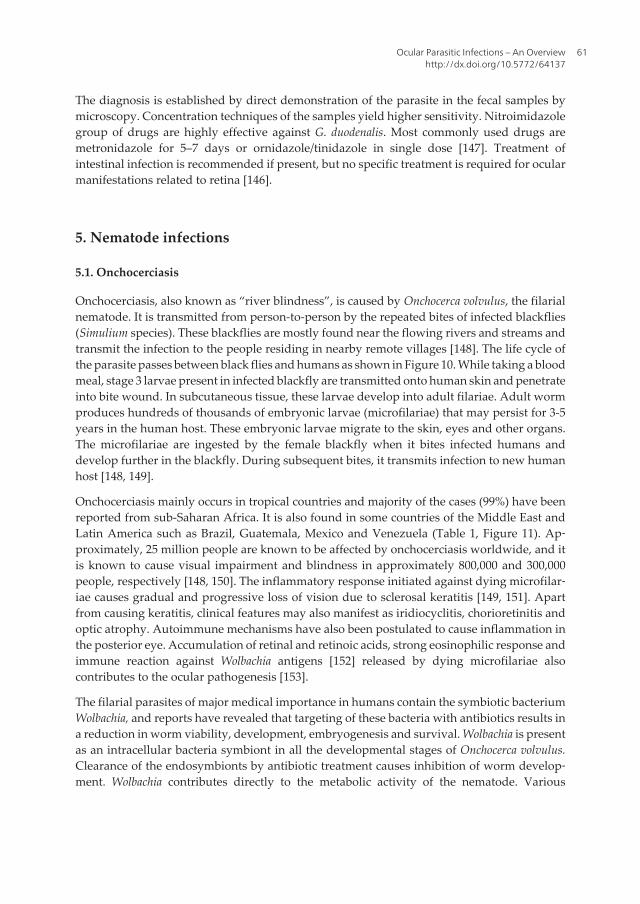
Onchocerciasis, also known as “river blindness”, is caused by Onchocerca volvulus, the filarialnematode. It is transmitted from person-to-person by the repeated bites of infected blackflies(Simulium species). These blackflies are mostly found near the flowing rivers and streams andtransmit the infection to the people residing in nearby remote villages [148]. The life cycle ofthe parasite passes between black flies and humans as shown in Figure 10. While taking a bloodmeal, stage 3 larvae present in infected blackfly are transmitted onto human skin and penetrateinto bite wound. In subcutaneous tissue, these larvae develop into adult filariae. Adult wormproduces hundreds of thousands of embryonic larvae (microfilariae) that may persist for 3-5years in the human host. These embryonic larvae migrate to the skin, eyes and other organs.The microfilariae are ingested by the female blackfly when it bites infected humans anddevelop further in the blackfly. During subsequent bites, it transmits infection to new humanhost [148, 149].
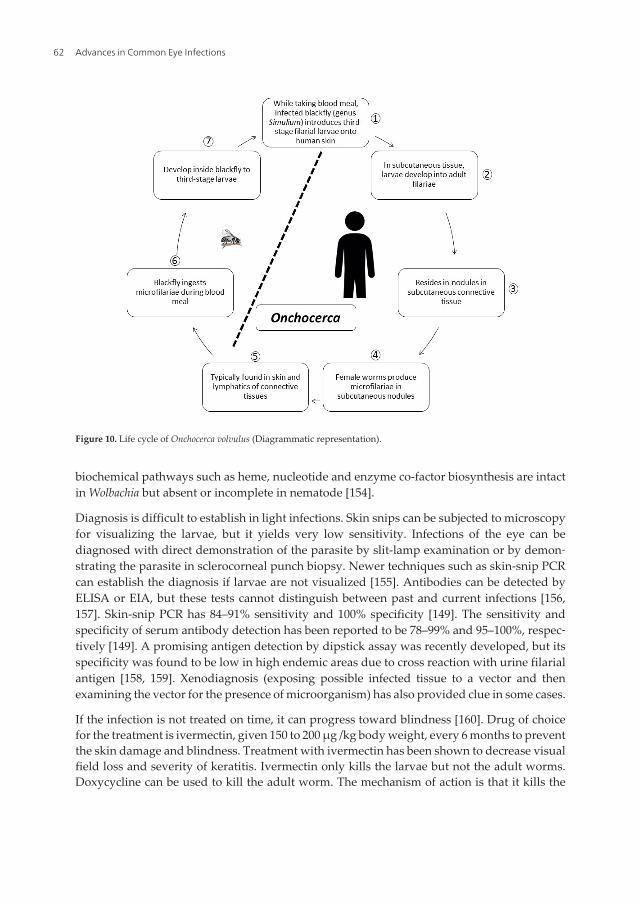
Onchocerciasis mainly occurs in tropical countries and majority of the cases (99%) have beenreported from sub-Saharan Africa. It is also found in some countries of the Middle East andLatin America such as Brazil, Guatemala, Mexico and Venezuela (Table 1, Figure 11). Ap‐proximately, 25 million people are known to be affected by onchocerciasis worldwide, and itis known to cause visual impairment and blindness in approximately 800,000 and 300,000people, respectively [148, 150]. The inflammatory response initiated against dying microfilar‐iae causes gradual and progressive loss of vision due to sclerosal keratitis [149, 151]. Apartfrom causing keratitis, clinical features may also manifest as iridiocyclitis, chorioretinitis andoptic atrophy. Autoimmune mechanisms have also been postulated to cause inflammation inthe posterior eye. Accumulation of retinal and retinoic acids, strong eosinophilic response andimmune reaction against Wolbachia antigens [152] released by dying microfilariae alsocontributes to the ocular pathogenesis [153].
The filarial parasites of major medical importance in humans contain the symbiotic bacteriumWolbachia, and reports have revealed that targeting of these bacteria with antibiotics results ina reduction in worm viability, development, embryogenesis and survival. Wolbachia is presentas an intracellular bacteria symbiont in all the developmental stages of Onchocerca volvulus.Clearance of the endosymbionts by antibiotic treatment causes inhibition of worm develop‐ment. Wolbachia contributes directly to the metabolic activity of the nematode. Various
Ocular Parasitic Infections – An Overviewhttp://dx.doi.org/10.5772/64137
61
biochemical pathways such as heme, nucleotide and enzyme co-factor biosynthesis are intactin Wolbachia but absent or incomplete in nematode [154].
Diagnosis is difficult to establish in light infections. Skin snips can be subjected to microscopyfor visualizing the larvae, but it yields very low sensitivity. Infections of the eye can bediagnosed with direct demonstration of the parasite by slit-lamp examination or by demon‐strating the parasite in sclerocorneal punch biopsy. Newer techniques such as skin-snip PCRcan establish the diagnosis if larvae are not visualized [155]. Antibodies can be detected byELISA or EIA, but these tests cannot distinguish between past and current infections [156,157]. Skin-snip PCR has 84–91% sensitivity and 100% specificity [149]. The sensitivity andspecificity of serum antibody detection has been reported to be 78–99% and 95–100%, respec‐tively [149]. A promising antigen detection by dipstick assay was recently developed, but itsspecificity was found to be low in high endemic areas due to cross reaction with urine filarialantigen [158, 159]. Xenodiagnosis (exposing possible infected tissue to a vector and thenexamining the vector for the presence of microorganism) has also provided clue in some cases.
If the infection is not treated on time, it can progress toward blindness [160]. Drug of choicefor the treatment is ivermectin, given 150 to 200 µg /kg body weight, every 6 months to preventthe skin damage and blindness. Treatment with ivermectin has been shown to decrease visualfield loss and severity of keratitis. Ivermectin only kills the larvae but not the adult worms.Doxycycline can be used to kill the adult worm. The mechanism of action is that it kills the
Figure 10. Life cycle of Onchocerca volvulus (Diagrammatic representation).
Advances in Common Eye Infections62
Wolbachia bacteria residing in the worm, on which the adult worm depends for its survival.Treatment with a 6-week course of doxycycline has been shown to kill more than 60% of adultfemale worms and to sterilize 80–90% of females 20 months after treatment. Thus, treatmentwith ivermectin is advised one week prior to treatment with doxycycline to provide relief topatient [148, 161].
The best method to get the protection from insect bite is the use of insect repellent. Community-directed treatment with ivermectin (CDTI) along with vector control measures is the mainapproach to control onchocerciasis. Ivermectin kills microfilariae and also prevents adultworms from producing more microfilariae for few months following treatment, so reducestransmission [148].
5.2. Loiasis
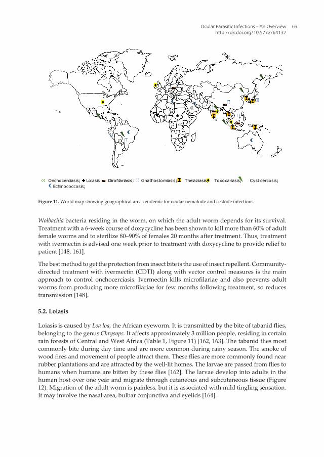
Loiasis is caused by Loa loa, the African eyeworm. It is transmitted by the bite of tabanid flies,belonging to the genus Chrysops. It affects approximately 3 million people, residing in certainrain forests of Central and West Africa (Table 1, Figure 11) [162, 163]. The tabanid flies mostcommonly bite during day time and are more common during rainy season. The smoke ofwood fires and movement of people attract them. These flies are more commonly found nearrubber plantations and are attracted by the well-lit homes. The larvae are passed from flies tohumans when humans are bitten by these flies [162]. The larvae develop into adults in thehuman host over one year and migrate through cutaneous and subcutaneous tissue (Figure12). Migration of the adult worm is painless, but it is associated with mild tingling sensation.It may involve the nasal area, bulbar conjunctiva and eyelids [164].
Onchocerciasis; Loiasis Dirofilariasis; Gnathostomiasis; Thelaziasis; Toxocariasis; Cysticercosis; Echinococcosis;
Figure 11. World map showing geographical areas endemic for ocular nematode and cestode infections.
Ocular Parasitic Infections – An Overviewhttp://dx.doi.org/10.5772/64137
63
Figure 12. Life cycle of Loa loa (Diagrammatic representation).
Ocular manifestations may occur due to the presence of both microfilariae and adult worms.The adult worms may survive up to 15 years and have been found in the conjunctiva, vitreous,eyelid and anterior chamber. Calabar swellings [165] may occur as a result of localizedangioedema due to intense atopic reaction. Retinal hemorrhages may occur due to aneurysmaldilatation of the retinal vessels due to the invasion of the retinal and choroid vessels by themicrofilariae present in blood stream. Perivascular inflammation can also be present, andocular examination under slit lamp examination is useful in establishing the diagnosis.
The diagnosis is usually confirmed by the direct demonstration of the microfilariae in the bloodby visualizing Giemsa-stained slides under the microscope. However, many of the individualshaving visible worm in the eye may test as amicrofilaraemic [166]. Blood should be drawnduring the midday as this time coincides with the periodicity of the microfilariae in the blood.The microfilariae can also be demonstrated in unstained blood smear. Adult worm extractionestablishes the diagnosis in patients having conjunctival involvement [167]. Antibody detec‐tion [168] may aid in establishing the diagnosis, but its presence cannot differentiate betweenrecent and past infection. Eosinophilia and high IgE also indicate active infection [169].
Eye worm if not treated causes very little damage to eye as it lasts less than one week (oftenjust hours). Surgical removal relieves eye symptoms, in addition medical treatment is required
Advances in Common Eye Infections64
for treating loiasis [170]. Therapy involves manual removal of adult worms and administrationof diethylcarbamazine (DEC), which kills both adult worms and microfilariae.
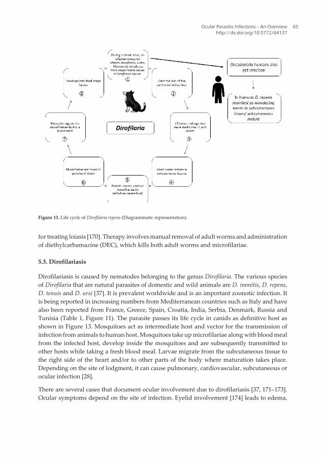
5.3. Dirofilariasis
Dirofilariasis is caused by nematodes belonging to the genus Dirofilaria. The various speciesof Dirofilaria that are natural parasites of domestic and wild animals are D. immitis, D. repens,D. tenuis and D. ursi [37]. It is prevalent worldwide and is an important zoonotic infection. Itis being reported in increasing numbers from Mediterranean countries such as Italy and havealso been reported from France, Greece, Spain, Croatia, India, Serbia, Denmark, Russia andTunisia (Table 1, Figure 11). The parasite passes its life cycle in canids as definitive host asshown in Figure 13. Mosquitoes act as intermediate host and vector for the transmission ofinfection from animals to human host. Mosquitoes take up microfilariae along with blood mealfrom the infected host, develop inside the mosquitoes and are subsequently transmitted toother hosts while taking a fresh blood meal. Larvae migrate from the subcutaneous tissue tothe right side of the heart and/or to other parts of the body where maturation takes place.Depending on the site of lodgment, it can cause pulmonary, cardiovascular, subcutaneous orocular infection [28].
There are several cases that document ocular involvement due to dirofilariasis [37, 171–173].Ocular symptoms depend on the site of infection. Eyelid involvement [174] leads to edema,
Figure 13. Life cycle of Dirofilaria repens (Diagrammatic representation).
Ocular Parasitic Infections – An Overviewhttp://dx.doi.org/10.5772/64137
65
pain, pruritus and congestion of conjunctiva, whereas intraocular [175] involvement leads toforeign body sensation, diplopia, photophobia and floaters.
Diagnosis can be established by the direct demonstration and identification of the adult worm.Intraocular presence of the parasite can be confirmed by ophthalmoscopy. Serologicaltechniques are not useful in establishing the diagnosis due to the cross reaction with otherparasitic helminths, particularly Toxocara canis. Recombinant proteins proved to exhibit 100%sensitivity and 90% specificity by ELISA for the diagnosis of pulmonary dirofilariasis [176].
Without treatment, worm remains in eye causing symptoms due to its presence [177]. Surgicalexcision is the treatment of choice; however use of diethylcarbamazine (DEC) has also beenreported with some success [37, 178].
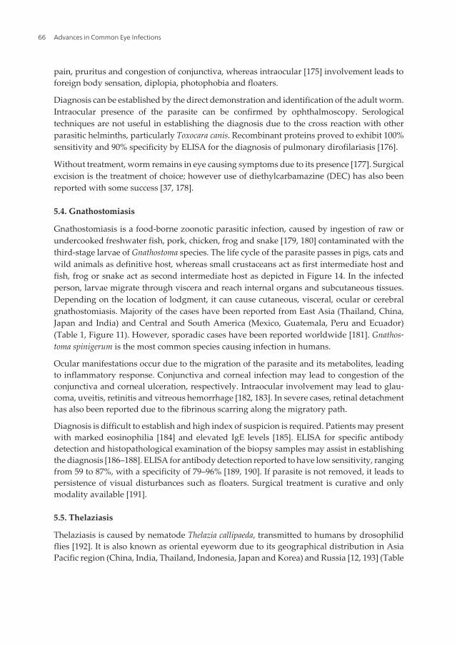
5.4. Gnathostomiasis
Gnathostomiasis is a food-borne zoonotic parasitic infection, caused by ingestion of raw orundercooked freshwater fish, pork, chicken, frog and snake [179, 180] contaminated with thethird-stage larvae of Gnathostoma species. The life cycle of the parasite passes in pigs, cats andwild animals as definitive host, whereas small crustaceans act as first intermediate host andfish, frog or snake act as second intermediate host as depicted in Figure 14. In the infectedperson, larvae migrate through viscera and reach internal organs and subcutaneous tissues.Depending on the location of lodgment, it can cause cutaneous, visceral, ocular or cerebralgnathostomiasis. Majority of the cases have been reported from East Asia (Thailand, China,Japan and India) and Central and South America (Mexico, Guatemala, Peru and Ecuador)(Table 1, Figure 11). However, sporadic cases have been reported worldwide [181]. Gnathos‐toma spinigerum is the most common species causing infection in humans.
Ocular manifestations occur due to the migration of the parasite and its metabolites, leadingto inflammatory response. Conjunctiva and corneal infection may lead to congestion of theconjunctiva and corneal ulceration, respectively. Intraocular involvement may lead to glau‐coma, uveitis, retinitis and vitreous hemorrhage [182, 183]. In severe cases, retinal detachmenthas also been reported due to the fibrinous scarring along the migratory path.
Diagnosis is difficult to establish and high index of suspicion is required. Patients may presentwith marked eosinophilia [184] and elevated IgE levels [185]. ELISA for specific antibodydetection and histopathological examination of the biopsy samples may assist in establishingthe diagnosis [186–188]. ELISA for antibody detection reported to have low sensitivity, rangingfrom 59 to 87%, with a specificity of 79–96% [189, 190]. If parasite is not removed, it leads topersistence of visual disturbances such as floaters. Surgical treatment is curative and onlymodality available [191].
5.5. Thelaziasis
Thelaziasis is caused by nematode Thelazia callipaeda, transmitted to humans by drosophilidflies [192]. It is also known as oriental eyeworm due to its geographical distribution in AsiaPacific region (China, India, Thailand, Indonesia, Japan and Korea) and Russia [12, 193] (Table
Advances in Common Eye Infections66
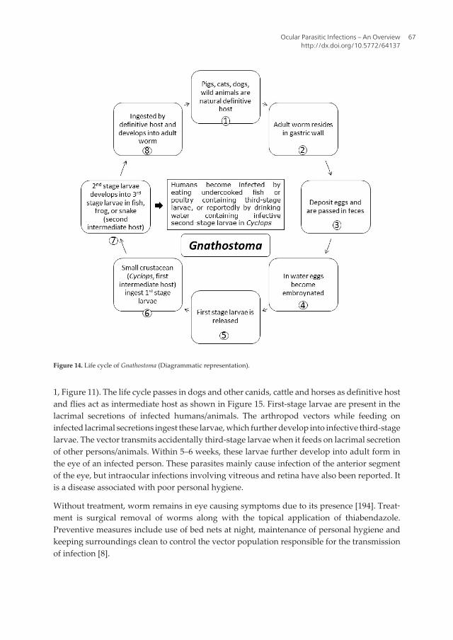
1, Figure 11). The life cycle passes in dogs and other canids, cattle and horses as definitive hostand flies act as intermediate host as shown in Figure 15. First-stage larvae are present in thelacrimal secretions of infected humans/animals. The arthropod vectors while feeding oninfected lacrimal secretions ingest these larvae, which further develop into infective third-stagelarvae. The vector transmits accidentally third-stage larvae when it feeds on lacrimal secretionof other persons/animals. Within 5–6 weeks, these larvae further develop into adult form inthe eye of an infected person. These parasites mainly cause infection of the anterior segmentof the eye, but intraocular infections involving vitreous and retina have also been reported. Itis a disease associated with poor personal hygiene.
Without treatment, worm remains in eye causing symptoms due to its presence [194]. Treat‐ment is surgical removal of worms along with the topical application of thiabendazole.Preventive measures include use of bed nets at night, maintenance of personal hygiene andkeeping surroundings clean to control the vector population responsible for the transmissionof infection [8].
Figure 14. Life cycle of Gnathostoma (Diagrammatic representation).
Ocular Parasitic Infections – An Overviewhttp://dx.doi.org/10.5772/64137
67
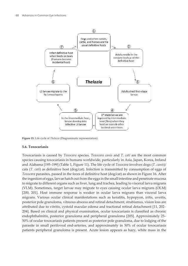
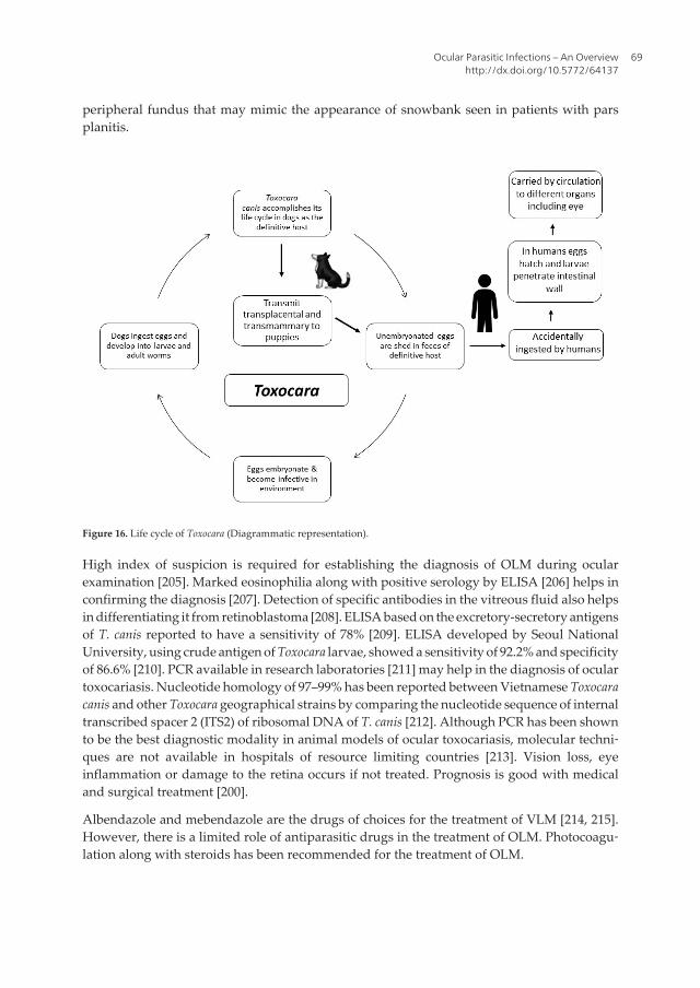
5.6. Toxocariasis
Toxocariasis is caused by Toxocara species. Toxocara canis and T. cati are the most commonspecies causing toxocariasis in humans worldwide, particularly in Asia, Japan, Korea, Irelandand Alabama [195–199] (Table 1, Figure 11). The life cycle of Toxocara involves dogs (T. canis)/cats (T. cati) as definitive host (dog/cat). Infection is transmitted by consumption of eggs ofToxocara parasites, passed in the feces of definitive host (dog/cat) as shown in Figure 16. Afterthe ingestion of eggs, larvae hatch out from the eggs in the small intestine and penetrate mucosato migrate to different organs such as liver, lung and trachea, leading to visceral larva migrans(VLM). Sometimes, target larvae may migrate to eyes causing ocular larva migrans (OLM)[200, 201]. Host immune response is weaker in ocular larva migrans than visceral larvamigrans. Various ocular clinical manifestations such as keratitis, hypopyon, iritis, uveitis,posterior pole granuloma, vitreous abscess and retinal detachment, strabismus, vision loss areattributed due to vitritis, cystoid macular edema and tractional retinal detachment [11, 202–204]. Based on clinical and physical examination, ocular toxocariasis is classified as chronicendophthalmitis, posterior granuloma and peripheral granuloma [205]. Approximately 25–50% of ocular toxocariasis patients present as posterior pole granuloma, due to lodging of theparasite in small perifoveal end-arteries, and approximately in 50% of ocular toxocariasispatients peripheral granuloma is present. Acute lesion appears as hazy, white mass in the
Figure 15. Life cycle of Thelazia (Diagrammatic representation).
Advances in Common Eye Infections68
peripheral fundus that may mimic the appearance of snowbank seen in patients with parsplanitis.
Figure 16. Life cycle of Toxocara (Diagrammatic representation).
High index of suspicion is required for establishing the diagnosis of OLM during ocularexamination [205]. Marked eosinophilia along with positive serology by ELISA [206] helps inconfirming the diagnosis [207]. Detection of specific antibodies in the vitreous fluid also helpsin differentiating it from retinoblastoma [208]. ELISA based on the excretory-secretory antigensof T. canis reported to have a sensitivity of 78% [209]. ELISA developed by Seoul NationalUniversity, using crude antigen of Toxocara larvae, showed a sensitivity of 92.2% and specificityof 86.6% [210]. PCR available in research laboratories [211] may help in the diagnosis of oculartoxocariasis. Nucleotide homology of 97–99% has been reported between Vietnamese Toxocaracanis and other Toxocara geographical strains by comparing the nucleotide sequence of internaltranscribed spacer 2 (ITS2) of ribosomal DNA of T. canis [212]. Although PCR has been shownto be the best diagnostic modality in animal models of ocular toxocariasis, molecular techni‐ques are not available in hospitals of resource limiting countries [213]. Vision loss, eyeinflammation or damage to the retina occurs if not treated. Prognosis is good with medicaland surgical treatment [200].
Albendazole and mebendazole are the drugs of choices for the treatment of VLM [214, 215].However, there is a limited role of antiparasitic drugs in the treatment of OLM. Photocoagu‐lation along with steroids has been recommended for the treatment of OLM.
Ocular Parasitic Infections – An Overviewhttp://dx.doi.org/10.5772/64137
69
6. Cestodes infections
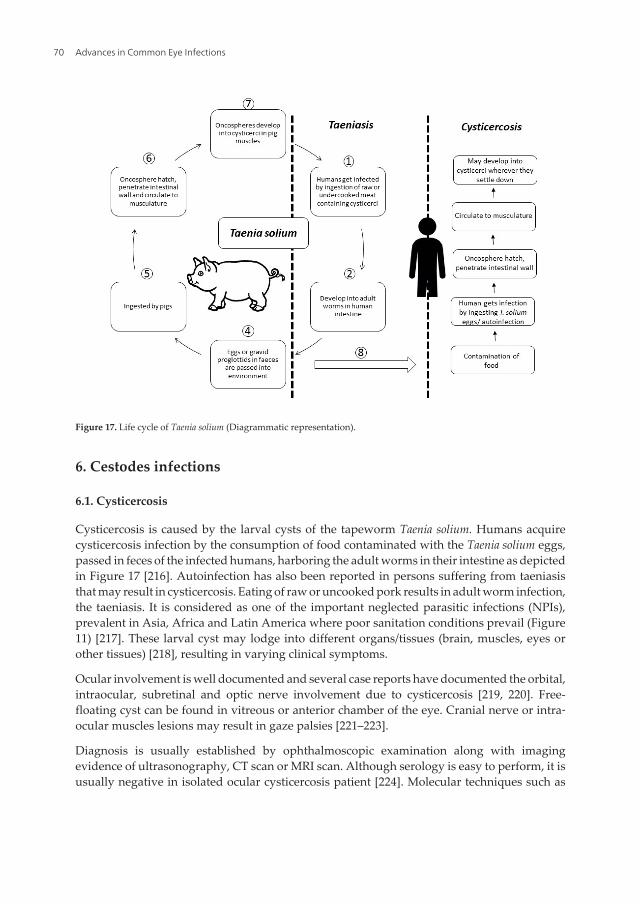
6.1. Cysticercosis
Cysticercosis is caused by the larval cysts of the tapeworm Taenia solium. Humans acquirecysticercosis infection by the consumption of food contaminated with the Taenia solium eggs,passed in feces of the infected humans, harboring the adult worms in their intestine as depictedin Figure 17 [216]. Autoinfection has also been reported in persons suffering from taeniasisthat may result in cysticercosis. Eating of raw or uncooked pork results in adult worm infection,the taeniasis. It is considered as one of the important neglected parasitic infections (NPIs),prevalent in Asia, Africa and Latin America where poor sanitation conditions prevail (Figure11) [217]. These larval cyst may lodge into different organs/tissues (brain, muscles, eyes orother tissues) [218], resulting in varying clinical symptoms.
Ocular involvement is well documented and several case reports have documented the orbital,intraocular, subretinal and optic nerve involvement due to cysticercosis [219, 220]. Free-floating cyst can be found in vitreous or anterior chamber of the eye. Cranial nerve or intra‐ocular muscles lesions may result in gaze palsies [221–223].
Diagnosis is usually established by ophthalmoscopic examination along with imagingevidence of ultrasonography, CT scan or MRI scan. Although serology is easy to perform, it isusually negative in isolated ocular cysticercosis patient [224]. Molecular techniques such as
Figure 17. Life cycle of Taenia solium (Diagrammatic representation).
Advances in Common Eye Infections70
conventional PCR, real-time PCR [218] and loop-mediated isothermal amplification (LAMP)[225] can be utilized for establishing the diagnosis of ocular cysticercosis and for genotyping[30, 226]. However, it requires a sophisticated molecular laboratory setup, which is notavailable widely in developing nations.
Without treatment, symptoms related to visual disturbances persist. Symptoms resolve withsurgical and medical treatment [227]. Albendazole along with steroids are the main drugs usedin the treatment. Steroid treatment decreases the inflammatory response associated with theantihelminthic therapy around the lesions. Surgical removal of large cysts is recommendedwhere there is an impairment of the vision [224].
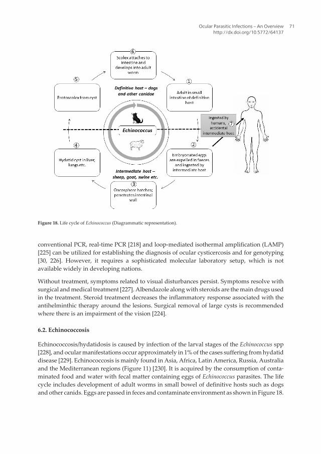
6.2. Echinococcosis
Echinococcosis/hydatidosis is caused by infection of the larval stages of the Echinococcus spp[228], and ocular manifestations occur approximately in 1% of the cases suffering from hydatiddisease [229]. Echinococcosis is mainly found in Asia, Africa, Latin America, Russia, Australiaand the Mediterranean regions (Figure 11) [230]. It is acquired by the consumption of conta‐minated food and water with fecal matter containing eggs of Echinococcus parasites. The lifecycle includes development of adult worms in small bowel of definitive hosts such as dogsand other canids. Eggs are passed in feces and contaminate environment as shown in Figure 18.
Figure 18. Life cycle of Echinococcus (Diagrammatic representation).
Ocular Parasitic Infections – An Overviewhttp://dx.doi.org/10.5772/64137
71
The symptoms and signs depend on the location of the cyst in the target organ. Most commonocular finding is the development of proptosis due to the presence of intraorbital spaceoccupying lesion. This may further lead to exposure to keratitis and ulceration of the cornea.Other complications due to the local invasion of the expanding cyst may lead to erosion oforbital wall, optic atrophy and optic neuritis. Subretinal hydatid cyst has been reported. Insevere cases, blindness may also occur [231].
The diagnosis depends on the clinical findings suggestive of hydatid cyst on ocular examina‐tion and confirmed by radiological techniques such as ultrasonography, CT scan and/or MRI[232, 233]. “Double wall” sign is a characteristic of orbital hydatid cyst seen by ultrasonography[232]. Serology may also aid in diagnosis. However, in majority of the commercially and in-house serological assays, hydatid fluid is the main antigenic component and sensitivity of IgG-ELISA reported in various studies varies from 64.8 to 100%, while specificity varies from 87.5to 100%. Purified and recombinant antigens are also being tried for developing ELISA withhigh sensitivity and specificity [234]. Fine needle aspiration cytology can also be performedfor establishing the diagnosis [235].
Symptoms persist if not treated [236]. Surgical removal of the cyst is the treatment of choice.Medical therapy includes administration of albendazole or mebendazole to prevent therecurrences due to the contents of the cyst leaking into the surgical sites [237]. If the cyst isaccidently ruptured, in situ irrigation with hypertonic saline should be performed. However,it causes local inflammatory reaction that may lead to atrophy of optic nerve [238].
7. Trematodes infections
7.1. Fascioliasis
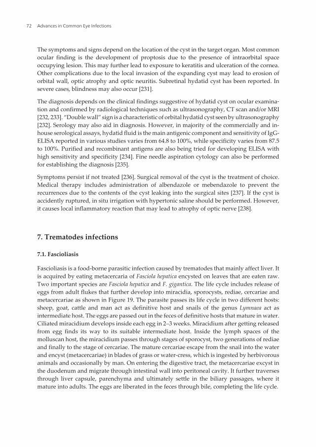
Fascioliasis is a food-borne parasitic infection caused by trematodes that mainly affect liver. Itis acquired by eating metacercaria of Fasciola hepatica encysted on leaves that are eaten raw.Two important species are Fasciola hepatica and F. gigantica. The life cycle includes release ofeggs from adult flukes that further develop into miracidia, sporocysts, rediae, cercariae andmetacercariae as shown in Figure 19. The parasite passes its life cycle in two different hosts:sheep, goat, cattle and man act as definitive host and snails of the genus Lymnaea act asintermediate host. The eggs are passed out in the feces of definitive hosts that mature in water.Ciliated miracidium develops inside each egg in 2–3 weeks. Miracidium after getting releasedfrom egg finds its way to its suitable intermediate host. Inside the lymph spaces of themolluscan host, the miracidium passes through stages of sporocyst, two generations of rediaeand finally to the stage of cercariae. The mature cercariae escape from the snail into the waterand encyst (metacercariae) in blades of grass or water-cress, which is ingested by herbivorousanimals and occasionally by man. On entering the digestive tract, the metacercariae excyst inthe duodenum and migrate through intestinal wall into peritoneal cavity. It further traversesthrough liver capsule, parenchyma and ultimately settle in the biliary passages, where itmature into adults. The eggs are liberated in the feces through bile, completing the life cycle.
Advances in Common Eye Infections72
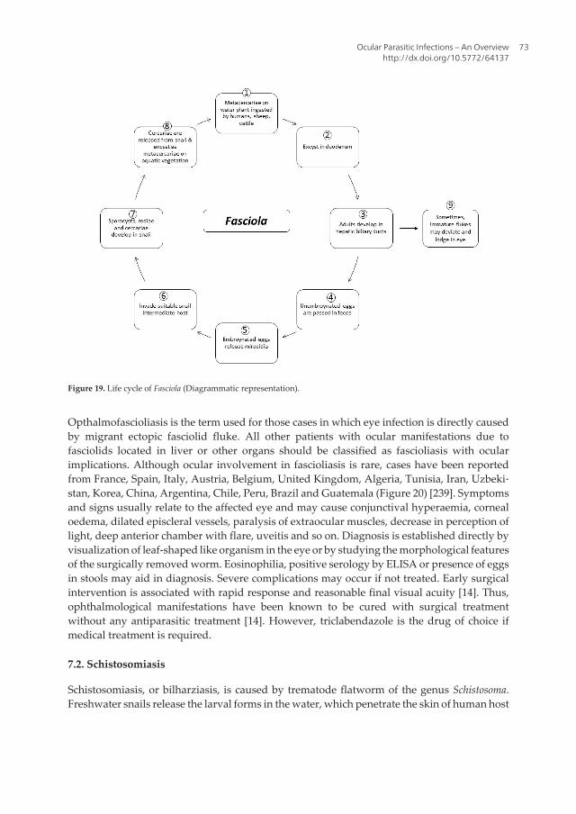
Opthalmofascioliasis is the term used for those cases in which eye infection is directly causedby migrant ectopic fasciolid fluke. All other patients with ocular manifestations due tofasciolids located in liver or other organs should be classified as fascioliasis with ocularimplications. Although ocular involvement in fascioliasis is rare, cases have been reportedfrom France, Spain, Italy, Austria, Belgium, United Kingdom, Algeria, Tunisia, Iran, Uzbeki‐stan, Korea, China, Argentina, Chile, Peru, Brazil and Guatemala (Figure 20) [239]. Symptomsand signs usually relate to the affected eye and may cause conjunctival hyperaemia, cornealoedema, dilated episcleral vessels, paralysis of extraocular muscles, decrease in perception oflight, deep anterior chamber with flare, uveitis and so on. Diagnosis is established directly byvisualization of leaf-shaped like organism in the eye or by studying the morphological featuresof the surgically removed worm. Eosinophilia, positive serology by ELISA or presence of eggsin stools may aid in diagnosis. Severe complications may occur if not treated. Early surgicalintervention is associated with rapid response and reasonable final visual acuity [14]. Thus,ophthalmological manifestations have been known to be cured with surgical treatmentwithout any antiparasitic treatment [14]. However, triclabendazole is the drug of choice ifmedical treatment is required.
7.2. Schistosomiasis
Schistosomiasis, or bilharziasis, is caused by trematode flatworm of the genus Schistosoma.Freshwater snails release the larval forms in the water, which penetrate the skin of human host
Figure 19. Life cycle of Fasciola (Diagrammatic representation).
Ocular Parasitic Infections – An Overviewhttp://dx.doi.org/10.5772/64137
73
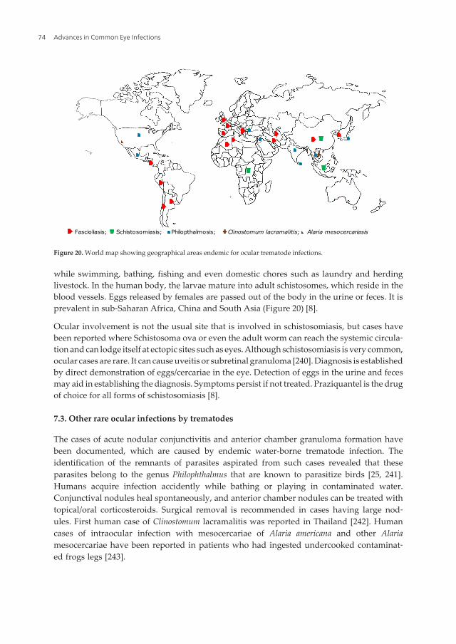
while swimming, bathing, fishing and even domestic chores such as laundry and herdinglivestock. In the human body, the larvae mature into adult schistosomes, which reside in theblood vessels. Eggs released by females are passed out of the body in the urine or feces. It isprevalent in sub-Saharan Africa, China and South Asia (Figure 20) [8].
Ocular involvement is not the usual site that is involved in schistosomiasis, but cases havebeen reported where Schistosoma ova or even the adult worm can reach the systemic circula‐tion and can lodge itself at ectopic sites such as eyes. Although schistosomiasis is very common,ocular cases are rare. It can cause uveitis or subretinal granuloma [240]. Diagnosis is establishedby direct demonstration of eggs/cercariae in the eye. Detection of eggs in the urine and fecesmay aid in establishing the diagnosis. Symptoms persist if not treated. Praziquantel is the drugof choice for all forms of schistosomiasis [8].
7.3. Other rare ocular infections by trematodes
The cases of acute nodular conjunctivitis and anterior chamber granuloma formation havebeen documented, which are caused by endemic water-borne trematode infection. Theidentification of the remnants of parasites aspirated from such cases revealed that theseparasites belong to the genus Philophthalmus that are known to parasitize birds [25, 241].Humans acquire infection accidently while bathing or playing in contaminated water.Conjunctival nodules heal spontaneously, and anterior chamber nodules can be treated withtopical/oral corticosteroids. Surgical removal is recommended in cases having large nod‐ules. First human case of Clinostomum lacramalitis was reported in Thailand [242]. Humancases of intraocular infection with mesocercariae of Alaria americana and other Alariamesocercariae have been reported in patients who had ingested undercooked contaminat‐ed frogs legs [243].
Fascioliasis; Schistosomiasis; Philopthalmosis; Clinostomum lacramalitis; Alaria mesocercariasis
Figure 20. World map showing geographical areas endemic for ocular trematode infections.
Advances in Common Eye Infections74
8. Eye infections caused by ectoparasites
8.1. Myiasis
Myiasis is an infection caused by larvae of flies. It is common in tropical and subtropical areas.It is known as ophthalmomyiasis when ocular structures are involved. Ophthalmomyiasis iscategorized into three clinical categories (ophthalmomyiasis externa, ophthalmomyiasisinterna and orbital myiasis), depending on the location of larvae in the eyes. Several generahave been reported to cause myiasis such as Dermatobia, Gasterophilus, Oestra, Cordylobia,Chrysomyia, Wohlfahrtia, Cochliomyia and Hypoderma [1]. Significantly, larvae causing ophthal‐momyiasis belong to the genus Hypoderma [16]. Three cases of external ophthalmomyiasis, twodue to Oestrus ovis and one due to Cochliomyia hominivorax were reported earlier from NorthIndia [17]. Oestra ovis also known as sheep nasal botfly is responsible for causing ophthalmo‐myiasis externa in shepherding areas [244–247]. It mainly involves eyelids, conjunctiva,lacrimal sac and nasolacrimal ducts. Most common clinical feature is the foreign bodysensation and may be associated with conjunctivitis and keratitis.
Diagnosis is established by the identification of the maggots. Treatment usually involves thesurgical removal of the maggots. Medical treatment involves just one oral dose (150 to 200 µg/kg of body weight) of ivermectin [16]. However, the use of ivermectin for the treatment ofmyiasis is an off-label treatment in many countries and should be used for selected cases. Theside effects such as dermal eruptions, fever, dizziness, migraines and muscular pains arecommon. Antibiotics and steroids may also be required to prevent the inflammation andsuperadded bacterial infection. Opthalmomyiasis interna [248] is caused by the invasion of theocular structures leading to uveitis, lens dislocation and retinal detachment. Diagnosis isestablished by visualizing the migratory tracks along subretina by the ophthalmoscopy.Symptoms persist if not treated. Serious complications may also occur such as lens dislocationand retinal detachment due to invasion of tissue [1]. Steroid therapy is advocated if there issevere inflammation, and surgical removal is performed in severe cases. Orbital myiasis is seenin patients who are not able to maintain good personal hygiene [16]. Treatment is directed atremoval of maggots and control of secondary infection. Preventive measures include mainte‐nance of good sanitation conditions and proper disposal of waste material to control the fliesin surrounding areas.
8.2. Lice
Important genera of the lice causing human infestation belong to Pediculus and Phthirus.Geographical areas where Phthiriasis palpebrarum is commonly found have been depicted inTable 1 and Figure 21. Eggs or nits laid down by lice glue themselves to body hairs or clothingfibers. Nymphs emerge from eggs and feed on the host, causing pruritis. Eyebrows andeyelashes are most commonly, involved. Excoriation marks along with small erythematouspapules aid in diagnosis. Nits can be found at the base of eyelashes, substantiating the clinicaldiagnosis. Symptoms persist if not treated. Eyelid disease is treated by petrolatum and non-eyelid involvement may be treated with lindane, permethrin, pyrethrin or malathion [1].
Ocular Parasitic Infections – An Overviewhttp://dx.doi.org/10.5772/64137
75
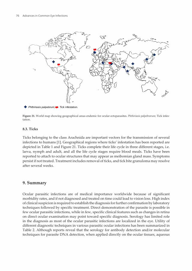
8.3. Ticks
Ticks belonging to the class Arachnida are important vectors for the transmission of severalinfections to humans [1]. Geographical regions where ticks’ infestation has been reported aredepicted in Table 1 and Figure 21. Ticks complete their life cycle in three different stages, i.e.larva, nymph and adult, and all the life cycle stages require blood meals. Ticks have beenreported to attach to ocular structures that may appear as meibomian gland mass. Symptomspersist if not treated. Treatment includes removal of ticks, and tick bite granuloma may resolveafter several weeks.
9. Summary
Ocular parasitic infections are of medical importance worldwide because of significantmorbidity rates, and if not diagnosed and treated on time could lead to vision loss. High indexof clinical suspicion is required to establish the diagnosis for further confirmation by laboratorytechniques followed by specific treatment. Direct demonstration of the parasite is possible infew ocular parasitic infections, while in few, specific clinical features such as changes in retinaon direct ocular examination may point toward specific diagnosis. Serology has limited rolein the diagnosis as most of the ocular parasitic infections are localized in the eye. Utility ofdifferent diagnostic techniques in various parasitic ocular infections has been summarized inTable 2. Although reports reveal that the serology for antibody detection and/or moleculartechniques for parasite DNA detection, when applied directly on the ocular tissues, aqueous
Phthiriasis palpebrum; Tick infestation.
Figure 21. World map showing geographical areas endemic for ocular ectoparasites. Phthiriasis palpebrarum; Tick infes‐tation.
Advances in Common Eye Infections76
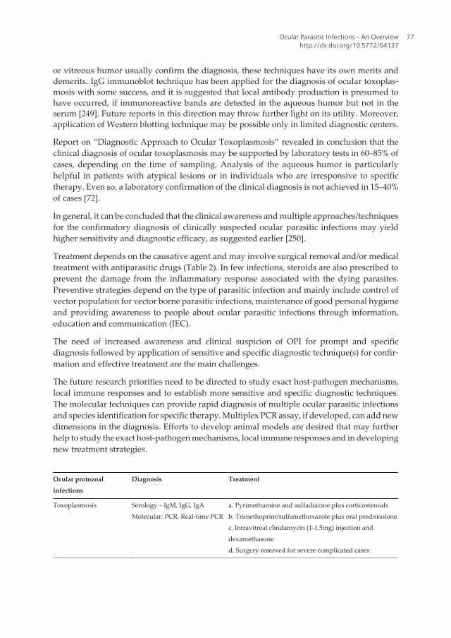
or vitreous humor usually confirm the diagnosis, these techniques have its own merits anddemerits. IgG immunoblot technique has been applied for the diagnosis of ocular toxoplas‐mosis with some success, and it is suggested that local antibody production is presumed tohave occurred, if immunoreactive bands are detected in the aqueous humor but not in theserum [249]. Future reports in this direction may throw further light on its utility. Moreover,application of Western blotting technique may be possible only in limited diagnostic centers.
Report on “Diagnostic Approach to Ocular Toxoplasmosis” revealed in conclusion that theclinical diagnosis of ocular toxoplasmosis may be supported by laboratory tests in 60–85% ofcases, depending on the time of sampling. Analysis of the aqueous humor is particularlyhelpful in patients with atypical lesions or in individuals who are irresponsive to specifictherapy. Even so, a laboratory confirmation of the clinical diagnosis is not achieved in 15–40%of cases [72].
In general, it can be concluded that the clinical awareness and multiple approaches/techniquesfor the confirmatory diagnosis of clinically suspected ocular parasitic infections may yieldhigher sensitivity and diagnostic efficacy, as suggested earlier [250].
Treatment depends on the causative agent and may involve surgical removal and/or medicaltreatment with antiparasitic drugs (Table 2). In few infections, steroids are also prescribed toprevent the damage from the inflammatory response associated with the dying parasites.Preventive strategies depend on the type of parasitic infection and mainly include control ofvector population for vector borne parasitic infections, maintenance of good personal hygieneand providing awareness to people about ocular parasitic infections through information,education and communication (IEC).
The need of increased awareness and clinical suspicion of OPI for prompt and specificdiagnosis followed by application of sensitive and specific diagnostic technique(s) for confir‐mation and effective treatment are the main challenges.
The future research priorities need to be directed to study exact host-pathogen mechanisms,local immune responses and to establish more sensitive and specific diagnostic techniques.The molecular techniques can provide rapid diagnosis of multiple ocular parasitic infectionsand species identification for specific therapy. Multiplex PCR assay, if developed, can add newdimensions in the diagnosis. Efforts to develop animal models are desired that may furtherhelp to study the exact host-pathogen mechanisms, local immune responses and in developingnew treatment strategies.
Ocular protozoalinfections
Diagnosis Treatment
Toxoplasmosis Serology – IgM, IgG, IgAMolecular: PCR, Real-time PCR
a. Pyrimethamine and sulfadiazine plus corticosteroidsb. Trimethoprim/sulfamethoxazole plus oral prednisolonec. Intravitreal clindamycin (1-1.5mg) injection anddexamethasoned. Surgery reserved for severe complicated cases
Ocular Parasitic Infections – An Overviewhttp://dx.doi.org/10.5772/64137
77
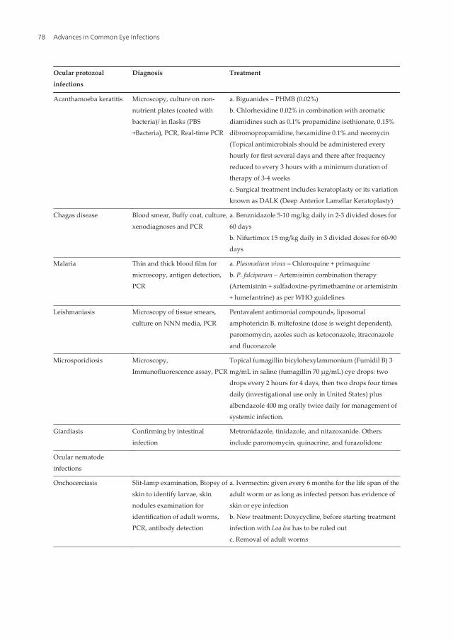
Ocular protozoalinfections
Diagnosis Treatment
Acanthamoeba keratitis Microscopy, culture on non-nutrient plates (coated withbacteria)/ in flasks (PBS+Bacteria), PCR, Real-time PCR
a. Biguanides – PHMB (0.02%)b. Chlorhexidine 0.02% in combination with aromaticdiamidines such as 0.1% propamidine isethionate, 0.15%dibromopropamidine, hexamidine 0.1% and neomycin(Topical antimicrobials should be administered everyhourly for first several days and there after frequencyreduced to every 3 hours with a minimum duration oftherapy of 3-4 weeksc. Surgical treatment includes keratoplasty or its variationknown as DALK (Deep Anterior Lamellar Keratoplasty)
Chagas disease Blood smear, Buffy coat, culture,xenodiagnoses and PCR
a. Benznidazole 5-10 mg/kg daily in 2-3 divided doses for60 daysb. Nifurtimox 15 mg/kg daily in 3 divided doses for 60-90days
Malaria Thin and thick blood film formicroscopy, antigen detection,PCR
a. Plasmodium vivax – Chloroquine + primaquineb. P. falciparum – Artemisinin combination therapy(Artemisinin + sulfadoxine-pyrimethamine or artemisinin+ lumefantrine) as per WHO guidelines
Leishmaniasis Microscopy of tissue smears,culture on NNN media, PCR
Pentavalent antimonial compounds, liposomalamphotericin B, miltefosine (dose is weight dependent),paromomycin, azoles such as ketoconazole, itraconazoleand fluconazole
Microsporidiosis Microscopy,Immunofluorescence assay, PCR
Topical fumagillin bicylohexylammonium (Fumidil B) 3mg/mL in saline (fumagillin 70 µg/mL) eye drops: twodrops every 2 hours for 4 days, then two drops four timesdaily (investigational use only in United States) plusalbendazole 400 mg orally twice daily for management ofsystemic infection.
Giardiasis Confirming by intestinalinfection
Metronidazole, tinidazole, and nitazoxanide. Othersinclude paromomycin, quinacrine, and furazolidone
Ocular nematodeinfections
Onchocerciasis Slit-lamp examination, Biopsy ofskin to identify larvae, skinnodules examination foridentification of adult worms,PCR, antibody detection
a. Ivermectin: given every 6 months for the life span of theadult worm or as long as infected person has evidence ofskin or eye infectionb. New treatment: Doxycycline, before starting treatmentinfection with Loa loa has to be ruled outc. Removal of adult worms
Advances in Common Eye Infections78
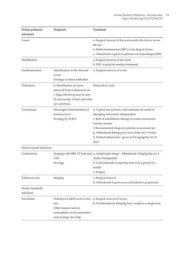
Ocular protozoalinfections
Diagnosis Treatment
Loiasis a. Surgical removal of the worm under the skin or acrossthe eyeb. Diethylcarbamazine (DEC) is the drug of choicec. Albendazole is given to patients not responding to DEC
Dirofilariasis a. Surgical removal of the wormb. DEC is given for medical treatment
Gnathostomiasis Identification of the removedwormSerology to detect antibodies
a. Surgical removal of worm
Thelaziasis b. Identification of wormremoved from conjunctival sacc. Eggs and larvae may be seenby microscopy of tears and othereye secretions
Removal of worm
Toxocariasis Histological demonstration oftoxocara larvaSerology by ELISA
d. Topical and systemic corticosteroids are useful inmanaging intraocular inflammatione. Role of anthelmintic therapy in ocular toxocariasisremains unclearf. Recommended drugs for systemic toxocariasis are:g. Albendazole 400mg given twice daily for 7-14 daysh. Diethylcarbamazine - given at 3-4 mg/kg/day for 21days
Ocular cestode infections
Cysticercosis Imaging with MRI, CT scan andUSGSerology
a. Antiparasitic drugs – Albendazole 15mg/kg/day for 4weeks, Praziquantelb. Corticosteroids in tapering dose over a period of 1monthc. Surgery
Echinococcosis Imaging a. Surgical removalb. Albendazole is given as an anti-infective prophylaxis
Ocular trematodeinfections
Fascioliasis Detection of adult worm in theeyeOther features such aseosinophilia, stool examinationand serology may help
a. Surgical removal of wormb. Triclabendazole 10mg/kg body weight as a single dose
Ocular Parasitic Infections – An Overviewhttp://dx.doi.org/10.5772/64137
79
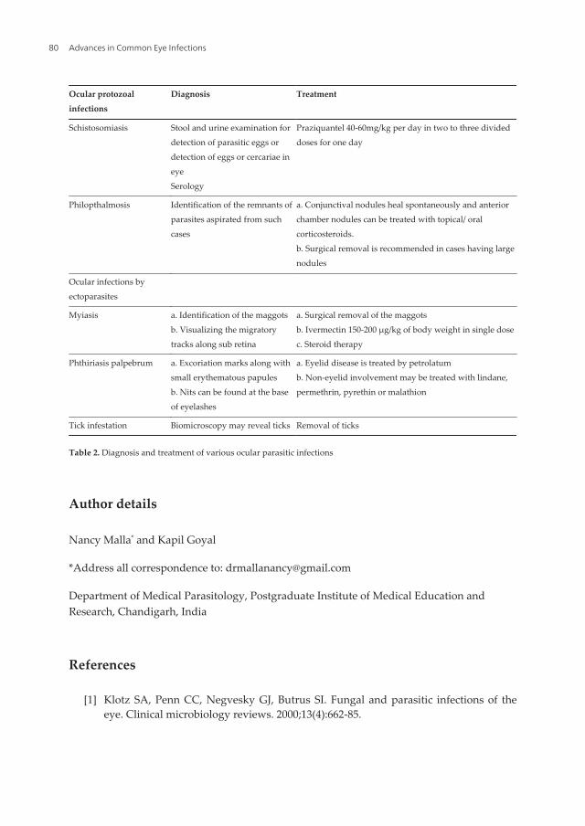
Ocular protozoalinfections
Diagnosis Treatment
Schistosomiasis Stool and urine examination fordetection of parasitic eggs ordetection of eggs or cercariae ineyeSerology
Praziquantel 40-60mg/kg per day in two to three divideddoses for one day
Philopthalmosis Identification of the remnants ofparasites aspirated from suchcases
a. Conjunctival nodules heal spontaneously and anteriorchamber nodules can be treated with topical/ oralcorticosteroids.b. Surgical removal is recommended in cases having largenodules
Ocular infections byectoparasites
Myiasis a. Identification of the maggotsb. Visualizing the migratorytracks along sub retina
a. Surgical removal of the maggotsb. Ivermectin 150-200 µg/kg of body weight in single dosec. Steroid therapy
Phthiriasis palpebrum a. Excoriation marks along withsmall erythematous papulesb. Nits can be found at the baseof eyelashes
a. Eyelid disease is treated by petrolatumb. Non-eyelid involvement may be treated with lindane,permethrin, pyrethin or malathion
Tick infestation Biomicroscopy may reveal ticks Removal of ticks
Table 2. Diagnosis and treatment of various ocular parasitic infections
Author details
Nancy Malla* and Kapil Goyal
*Address all correspondence to: [email protected]
Department of Medical Parasitology, Postgraduate Institute of Medical Education andResearch, Chandigarh, India
References
[1] Klotz SA, Penn CC, Negvesky GJ, Butrus SI. Fungal and parasitic infections of theeye. Clinical microbiology reviews. 2000;13(4):662-85.
Advances in Common Eye Infections80
[2] Chawla A, Armstrong M, Carley F. Acanthamoeba keratitis – an increasing inci‐dence. Contact lens & anterior eye : the journal of the British Contact Lens Associa‐tion. 2014;37(2):120.
[3] Lorenzo-Morales J, Martin-Navarro CM, Lopez-Arencibia A, Arnalich-Montiel F,Pinero JE, Valladares B. Acanthamoeba keratitis: an emerging disease gathering im‐portance worldwide? Trends in parasitology. 2013;29(4):181-7.
[4] Montoya JG, Liesenfeld O. Toxoplasmosis. Lancet. 2004;363(9425):1965-76.
[5] Sharma S, Pasricha G, Das D, Aggarwal RK. Acanthamoeba keratitis in non-contactlens wearers in India: DNA typing-based validation and a simple detection assay.Archives of ophthalmology. 2004;122(10):1430-4.
[6] Gopinathan U, Sharma S, Garg P, Rao GN. Review of epidemiological features, mi‐crobiological diagnosis and treatment outcome of microbial keratitis: experience ofover a decade. Indian journal of ophthalmology. 2009;57(4):273-9.
[7] Sharma S, Garg P, Rao GN. Patient characteristics, diagnosis, and treatment of non-contact lens related Acanthamoeba keratitis. The British journal of ophthalmology.2000;84(10):1103-8.
[8] Nimir AR, Saliem A, Ibrahim IA. Ophthalmic parasitosis: a review article. Interdisci‐plinary Perspectives on Infectious Diseases. 2012;2012:587402.
[9] Joseph J, Vemuganti GK, Sharma S. Microsporidia: emerging ocular pathogens. Indi‐an journal of medical microbiology. 2005;23(2):80-91.
[10] Malla N, Aggarwal AK, Mahajan RC. A serological study of human toxocariasis innorth India. The National medical journal of India. 2002;15(3):145-7.
[11] Fomda BA, Ahmad Z, Khan NN, Tanveer S, Wani SA. Ocular toxocariasis in a child:a case report from Kashmir, north India. Indian journal of medical microbiology.2007;25(4):411-2.
[12] Prabhakar SK, Vijaykumar GS, Mahesh BS, Shanthamallappa. Human ocular thela‐ziasis: a case report from Karnataka. Indian journal of medical microbiology.2015;33(2):324-5.
[13] Sachdeva RS, Manchanda SK, Abrol S, Wadhwa SC, Ramachandran KA. Freely mo‐bile cysticercus in the anterior chamber. Indian journal of ophthalmology. 1995;43(3):135-6.
[14] Dalimi A, Jabarvand M. Fasciola hepatica in the human eye. Transactions of the Roy‐al Society of Tropical Medicine and Hygiene. 2005;99(10):798-800.
[15] Joshi G, Parchand S, Dogra MR, Gupta PK, Khurana S, Gupta V, et al. Live juvenilestrobilate tapeworm in the anterior chamber of the human eye. Archives of ophthal‐mology. 2012;130(11):1464-6.
Ocular Parasitic Infections – An Overviewhttp://dx.doi.org/10.5772/64137
81
[16] Francesconi F, Lupi O. Myiasis. Clinical microbiology reviews. 2012;25(1):79-105.
[17] Khurana S, Biswal M, Bhatti HS, Pandav SS, Gupta A, Chatterjee SS, et al. Ophthal‐momyiasis: three cases from North India. Indian journal of medical microbiology.2010;28(3):257-61.
[18] Sulyok M, Rozsa L, Bodo I, Tappe D, Hardi R. Ocular pentastomiasis in the Demo‐cratic Republic of the Congo. PLoS neglected tropical diseases. 2014;8(7):e3041.
[19] Tan SY, Liu SW, Jiang SB. HIV/AIDS and ocular complications. International Journalof Ophthalmology. 2009;2(2):95-105.
[20] Couture S, Agrawal R, Woods K, Lockwood D, Pavesio CE, Addison PKF. A case ofpanuveitis with hypopyon due to presumed ocular leishmaniasis in a HIV patient.Journal of Opthalmic Inflammation and Infection. 2014;4(21):1-5.
[21] Bacon AS, Dart JK, Ficker LA, Matheson MM, Wright P. Acanthamoeba keratitis. Thevalue of early diagnosis. Ophthalmology. 1993;100(8):1238-43.
[22] Lalitha P, Lin CC, Srinivasan M, Mascarenhas J, Prajna NV, Keenan JD, et al. Acan‐thamoeba keratitis in South India: a longitudinal analysis of epidemics. OphthalmicEpidemiology. 2012;19(2):111-5.
[23] Lowder CY. Ocular microsporidiosis. International ophthalmology clinics.1993;33(1):145-51.
[24] Lim SJ, Lee SE, Kim SH, Hong SH, You YS, Kwon OW, et al. Prevalence of Toxoplasmagondii and Toxocara canis among patients with uveitis. Ocular immunology andinflammation. 2014;22(5):360-6.
[25] Otranto D, Eberhard ML. Zoonotic helminths affecting the human eye. Parasites &vectors. 2011;4:41.
[26] Hirst LW. Ocular infections – diagnosis. Australian family physician. 1991;20(2):133-4, 7-45, 49.
[27] Rao NA. A laboratory approach to rapid diagnosis of ocular infections and prospectsfor the future. American journal of ophthalmology. 1989;107(3):283-91.
[28] Khurana S, Singh G, Bhatti HS, Malla N. Human subcutaneous dirofilariasis in India:a report of three cases with brief review of literature. Indian journal of medical mi‐crobiology. 2010;28(4):394-6.
[29] Cortez RT, Ramirez G, Collet L, Giuliari GP. Ocular parasitic diseases: a review ontoxocariasis and diffuse unilateral subacute neuroretinitis. Journal of pediatric ophthal‐mology and strabismus. 2011;48(4):204-12.
[30] Sharma M, Beke N, Khurana S, Bhatti HS, Sehgal R, Malla N. An Ocular cysticercosiscase: caused by Asian Genotype of Taenia solium. Indian journal of Medical Microbi‐ology. 2015;33(4)583-585.
Advances in Common Eye Infections82
[31] Hogan RN, Jakobiec FA. Molecular pathological diagnosis of ocular infections. Inter‐national ophthalmology clinics. 1996;36(3):223-46.
[32] Rathinam SR, Annamalai R, Biswas J. Intraocular parasitic infections. Ocular immu‐nology and inflammation. 2011;19(5):327-36.
[33] Pasricha G, Sharma S, Garg P, Aggarwal RK. Use of 18S rRNA gene-based PCR assay fordiagnosis of acanthamoeba keratitis in non-contact lens wearers in India. Journal ofClinical Microbiology. 2003;41(7):3206-11.
[34] Basu S, Sharma S, Kar S, Das T. DNA chip-assisted diagnosis of a previously unknownetiology of intermediate uveitis – Toxoplasma gondii. Indian journal of ophthalmolo‐gy. 2010;58(6):535-7.
[35] Sharma S. Diagnosis of infectious diseases of the eye. Eye. 2012;26(2):177-84.
[36] Rathinam SR, Arya LK, Usha KR, Prajna L, Tandon V. Novel etiological agent: mo‐lecular evidence for trematode-induced anterior uveitis in children. Archives of oph‐thalmology. 2012;130(11):1481-4.
[37] Nath R, Gogoi R, Bordoloi N, Gogoi T. Ocular dirofilariasis. Indian journal of Pathol‐ogy & Microbiology. 2010;53(1):157-9.
[38] Pushker N, Bajaj MS, Chandra M, Neena. Ocular and orbital cysticercosis. Acta oph‐thalmologica Scandinavica. 2001;79(4):408-13.
[39] Gomez Morales A, Croxatto JO, Crovetto L, Ebner R. Hydatid cysts of the orbit. Areview of 35 cases. Ophthalmology. 1988;95(8):1027-32.
[40] Malla N, Goyal M, Pillai P, Ganguly NK, Mahajan RC. Experimental ocular toxoplas‐mosis with RH strain of Toxoplasma gondii. Japanese journal of experimental medi‐cine. 1988;58(6):279-81.
[41] Takayanagi TH, Akao N, Suzuki R, Tomoda M, Tsukidate S, Fujita K. New animalmodel for human ocular toxocariasis: ophthalmoscopic observation. The British jour‐nal of ophthalmology. 1999;83(8):967-72.
[42] Malhotra A, Minja FJ, Crum A, Burrowes D. Ocular anatomy and cross-sectionalimaging of the eye. Semin Ultrasound CT MR. 2011;32(1):2-13.
[43] Kaplan HJ. Anatomy and function of the eye. Chemical Immunology and Allergy.2007;92:4-10.
[44] Paalman MH. Anatomy in the eye. Anatomical Record. 2000;261(6):221.
[45] Kolb H. Gross Anatomy of the Eye. 2005 May 1 [Updated 2007 May 1]. In: Kolb H,Fernandez E, Nelson R, editors. Webvision: The Organization of the Retina and Visu‐al System [Internet]. Salt Lake City (UT): University of Utah Health Sciences Center;1995. Available from: http://www.ncbi.nlm.nih.gov/books/NBK11534/
[46] Iveson-Iveson J. Anatomy and physiology: the eye. Nursing Mirror. 1979;148(8):31-3.
Ocular Parasitic Infections – An Overviewhttp://dx.doi.org/10.5772/64137
83
[47] Shitole S, Jakkal T, Khaire B. Inverse Bell’s phenomenon: rare ophthalmic finding fol‐lowing ptosis surgery. Journal of clinical and diagnostic research: JCDR.2015;9(3):ND01-2.
[48] Kageyama M, Nakatsuka K, Yamaguchi T, Owen RL, Shimada T. Ocular defensemechanisms with special reference to the demonstration and functional morphologyof the conjunctiva-associated lymphoid tissue in Japanese monkeys. Archives of His‐tology and Cytology. 2006;69(5):311-22.
[49] Lerman S. Free radical damage and defense mechanisms in the ocular lens. Lens andeye toxicity research. 1992;9(1):9-24.
[50] Bito LZ. Species differences in the responses of the eye to irritation and trauma: a hy‐pothesis of divergence in ocular defense mechanisms, and the choice of experimentalanimals for eye research. Experimental Eye Research. 1984;39(6):807-29.
[51] Chandler JW, Gillette TE. Immunologic defense mechanisms of the ocular surface.Ophthalmology. 1983;90(6):585-91.
[52] Lemp MA, Blackman HJ. Ocular surface defense mechanisms. Annals of ophthalmol‐ogy. 1981;13(1):61-3.
[53] McDermott AM. Defensins and other antimicrobial peptides at the ocular surface.The ocular surface. 2004;2(4):229-47.
[54] Cunningham ET, Jr., Belfort R, Jr., Muccioli C, Arevalo JF, Zierhut M. Ocular toxo‐plasmosis. Ocular immunology and inflammation. 2015;23(3):191-3.
[55] Kijlstra A, Petersen E. Epidemiology, pathophysiology, and the future of ocular toxo‐plasmosis. Ocular immunology and inflammation. 2014;22(2):138-47.
[56] Robert-Gangneux F, Darde ML. Epidemiology of and diagnostic strategies for toxo‐plasmosis. Clinical microbiology reviews. 2012;25(2):264-96.
[57] Petersen E, Kijlstra A, Stanford M. Epidemiology of ocular toxoplasmosis. Ocular im‐munology and inflammation. 2012;20(2):68-75.
[58] Ho-Yen DO. Epidemiology of toxoplasmosis. Archives of pediatrics. 2003;10(Suppl 1):3-4.
[59] Lachenmaier SM, Deli MA, Meissner M, Liesenfeld O. Intracellular transport ofToxoplasma gondii through the blood-brain barrier. Journal of neuroimmunology.2011;232(1-2):119-30.
[60] Peixoto-Rangel AL, Miller EN, Castellucci L, Jamieson SE, Peixe RG, Elias Lde S, et al.Candidate gene analysis of ocular toxoplasmosis in Brazil: evidence for a role for toll-like receptor 9 (TLR9). Memorias do Instituto Oswaldo Cruz. 2009;104(8):1187-90.
[61] Pleyer U, Schluter D, Manz M. Ocular toxoplasmosis: recent aspects of pathophysiolo‐gy and clinical implications. Ophthalmic Research. 2014;52(3):116-23.
Advances in Common Eye Infections84
[62] Holland GN. Ocular toxoplasmosis: a global reassessment. Part II: disease manifesta‐tions and management. American journal of ophthalmology. 2004;137(1):1-17.
[63] O’Connor GR. Manifestations and management of ocular toxoplasmosis. Bulletin of theNew York Academy of Medicine. 1974;50(2):192-210.
[64] Park YH, Nam HW. Clinical features and treatment of ocular toxoplasmosis. The Koreanjournal of parasitology. 2013;51(4):393-9.
[65] Harper TW, Miller D, Schiffman JC, Davis JL. Polymerase chain reaction analysis ofaqueous and vitreous specimens in the diagnosis of posterior segment infectious uveitis.American journal of ophthalmology. 2009;147(1):140-7 e2.
[66] Cassaing S, Bessieres MH, Berry A, Berrebi A, Fabre R, Magnaval JF. Comparisonbetween two amplification sets for molecular diagnosis of toxoplasmosis by real-timePCR. Journal of Clinical Microbiology. 2006;44(3):720-4.
[67] Robert-Gangneux F, Sterkers Y, Yera H, Accoceberry I, Menotti J, Cassaing S, et al.Molecular diagnosis of toxoplasmosis in immunocompromised patients: a 3-yearmulticenter retrospective study. Journal of Clinical Microbiology. 2015;53(5):1677-84.
[68] Filisetti D, Sterkers Y, Brenier-Pinchart MP, Cassaing S, Dalle F, Delhaes L, et al. Mul‐ticentric comparative assessment of the bio-evolution Toxoplasma gondii detectionkit with eight laboratory-developed PCR assays for molecular diagnosis of congeni‐tal toxoplasmosis. Journal of Clinical Microbiology. 2015;53(1):29-34.
[69] Hashoosh DA, Majeed IA. Comparison of two assays in the diagnosis of toxoplasmo‐sis: immunological and molecular. Eastern Mediterranean Health Journal. 2014;20(1):46-50.
[70] Remington JS, Thulliez P, Montoya JG. Recent developments for diagnosis of toxo‐plasmosis. Journal of Clinical Microbiology. 2004;42(3):941-5.
[71] Bou G, Figueroa MS, Marti-Belda P, Navas E, Guerrero A. Value of PCR for detectionof Toxoplasma gondii in aqueous humor and blood samples from immunocompetentpatients with ocular toxoplasmosis. Journal of Clinical Microbiology. 1999;37(11):3465-8.
[72] Garweg JG, de Groot-Mijnes JD, Montoya JG. Diagnostic approach to ocular toxo‐plasmosis. Ocular Immunology and Inflammation. 2011;19(4):255-61.
[73] Khurana S, Bagga R, Aggarwal A, Lyngdoh V, Shivapriya, Diddi K, et al. Serologicalscreening for antenatal toxoplasma infection in India. Indian Journal of Medical Mi‐crobiology. 2010;28(2):143-6.
[74] Singh MP, Dubey ML, Sud A, Malla N. Antibody response to Toxoplasma gondii insaliva samples from human immunodeficiency virus-infected patients. British Jour‐nal of Biomedical Science. 2005;62(2):81-4.
Ocular Parasitic Infections – An Overviewhttp://dx.doi.org/10.5772/64137
85
[75] Zargar AH, Masoodi SR, Laway BA, Sofi BA, Wani AI. Seroprevalence of toxoplas‐mosis in women with repeated abortions in Kashmir. Journal of Epidemiology &Community Health. 1998;52(2):135-6.
[76] Thokar MA, Malla N, Wattal C. Serological study of patients clinically suspected tohave toxoplasmosis in Kashmir. Indian Journal of Medical Research. 1988;88:29-34.
[77] Delair E, Monnet D, Grabar S, Dupouy-Camet J, Yera H, Brezin AP. Respective rolesof acquired and congenital infections in presumed ocular toxoplasmosis. AmericanJournal of Ophthalmology. 2008;146(6):851-5.
[78] de Boer JH, Luyendijk L, Rothova A, Kijlstra A. Analysis of ocular fluids for local an‐tibody production in uveitis. The British Journal of Ophthalmology. 1995;79(6):610-6.
[79] Garweg JG, Jacquier P, Boehnke M. Early aqueous humor analysis in patients withhuman ocular toxoplasmosis. Journal of Clinical Microbiology. 2000;38(3):996-1001.
[80] Lima GS, Saraiva PG, Saraiva FP. Current therapy of acquired ocular toxoplasmosis:a review. Journal of Ocular Pharmacology and Therapeutics : The Official Journal ofthe Association for Ocular Pharmacology and Therapeutics. 2015;31(9):511-7.
[81] Holland GN, Lewis KG. An update on current practices in the management of oculartoxoplasmosis. American Journal of Ophthalmology. 2002;134(1):102-14.
[82] Siddiqui R, Khan NA. Biology and pathogenesis of Acanthamoeba. Parasites & Vec‐tors. 2012;5:6.
[83] Bharathi JM, Srinivasan M, Ramakrishnan R, Meenakshi R, Padmavathy S, LalithaPN. A study of the spectrum of Acanthamoeba keratitis: a three-year study at a terti‐ary eye care referral center in South India. Indian Journal of Ophthalmology.2007;55(1):37-42.
[84] Vaddavalli PK, Garg P, Sharma S, Sangwan VS, Rao GN, Thomas R. Role of confocalmicroscopy in the diagnosis of fungal and acanthamoeba keratitis. Ophthalmology.2011;118(1):29-35.
[85] Winchester K, Mathers WD, Sutphin JE, Daley TE. Diagnosis of Acanthamoeba kera‐titis in vivo with confocal microscopy. Cornea. 1995;14(1):10-7.
[86] Goldschmidt P, Degorge S, Benallaoua D, Saint-Jean C, Batellier L, Alouch C, et al.New tool for the simultaneous detection of 10 different genotypes of Acanthamoebaavailable from the American Type Culture Collection. The British Journal of Ophthal‐mology. 2009;93(8):1096-100.
[87] Hauber S, Parkes H, Siddiqui R, Khan NA. The use of high-resolution (1)H nuclearmagnetic resonance (NMR) spectroscopy in the clinical diagnosis of Acanthamoeba.Parasitology Research. 2011;109(6):1661-9.
[88] Lorenzo-Morales J, Khan NA, Walochnik J. An update on Acanthamoeba keratitis:diagnosis, pathogenesis and treatment. Parasite. 2015;22:10.
Advances in Common Eye Infections86
[89] Kumar R, Lloyd D. Recent advances in the treatment of Acanthamoeba keratitis.Clinical Infectious Diseases: An Official Publication of the Infectious Diseases Societyof America. 2002;35(4):434-41.
[90] Grayson M. Chagas disease. Nature. 2010;465(7301):S3.
[91] Coura JR, Borges-Pereira J. Chagas disease: 100 years after its discovery. A systemicreview. Acta Tropica. 2010;115(1-2):5-13.
[92] Frohlich SJ, Mino de Kaspar H, Peran R, Vera de Bilbao N, Schinini A, Rojas de AriasA, et al. Intraocular involvement of Chagas disease (American trypanosomiasis).Studies in Paraguay/South America. Der Ophthalmologe: Zeitschrift der DeutschenOphthalmologischen Gesellschaft. 1997;94(3):206-10.
[93] Frohlich SJ, Mino de Kaspar H, Peran R, Rojas de Arias A, Sanchez Insfran N, More‐no Azorero R, et al. Eye involvement in Chagas disease (American trypanosomiasis).1996/1997 studies in Paraguay. Der Ophthalmologe: Zeitschrift der Deutschen Oph‐thalmologischen Gesellschaft. 1998;95(3):168-71.
[94] Bern C. Chagas’ disease. The New England Journal of Medicine. 2015;373(5):456-66.
[95] Morillo CA, Marin-Neto JA, Avezum A, Sosa-Estani S, Rassi A, Jr., Rosas F, et al.Randomized trial of benznidazole for chronic Chagas’ cardiomyopathy. The NewEngland Journal of Medicine. 2015;373(14):1295-306.
[96] Chagas disease. Available at http://www.nytimes.com/health/guides/disease/chagas-disease/overview.html
[97] Leishmaniasis. Available at: http://www.who.int/mediacentre/factsheets/fs375/en/
[98] Badri Prasad B, Shakya A, Poonam L, Smriti K. Caruncular Leishmaniasis – an un‐usual case. Orbit. 2015;34(4):232-3.
[99] Philips CA, Kalal CR, Kumar KN, Bihari C, Sarin SK. Simultaneous occurrence of oc‐ular, disseminated mucocutaneous, and multivisceral involvement of leishmaniasis.Case Reports in Infectious Diseases. 2014;2014:837625.
[100] Khalil EA, Musa AM, Younis BM, Elfaki ME, Zijlstra EE, Elhassan AM. Blindness fol‐lowing visceral leishmaniasis: a neglected post-kala-azar complication. Tropical Doc‐tor. 2011;41(3):139-40.
[101] Veraldi S, Bottini S, Curro N, Gianotti R. Leishmaniasis of the eyelid mimicking aninfundibular cyst and review of the literature on ocular leishmaniasis. InternationalJournal of Infectious Diseases: IJID: Official Publication of the International Societyfor Infectious Diseases. 2010;14(Suppl 3):e230-2.
[102] Salvanos P, Kabanarou SA, Xirou T, Kourentis C, Feretis E. Proliferative vitreoretin‐opathy in a child with visceral leishmaniasis. European Journal of Ophthalmology.2010;20(1):231-3.
Ocular Parasitic Infections – An Overviewhttp://dx.doi.org/10.5772/64137
87
[103] Sadeghian G, Nilfroushzadeh MA, Moradi SH, Hanjani SH. Ocular leishmaniasis: acase report. Dermatology Online Journal. 2005;11(2):19.
[104] el-Hassan AM, el-Sheikh EA, Eltoum IA, Ghalib HW, Ali MS, Zijlstra E, et al. Post-kala-azar anterior uveitis: demonstration of Leishmania parasites in the lesion. Trans‐actions of the Royal Society of Tropical Medicine and Hygiene. 1991;85(4):471-3.
[105] Dechant W, Rees PH, Kager PA, Klauss V, Adala H. Post kala-azar uveitis. The Brit‐ish Journal of Ophthalmology. 1980;64(9):680-3.
[106] De Cock KM, Rees PH, Klauss V, Kasili EG, Kager PA, Schattenkerk JK. Retinal hem‐orrhages in kala-azar. The American Journal of Tropical Medicine and Hygiene.1982;31(5):927-30.
[107] Montero JA, Ruiz-Moreno JM, Sanchis E. Intraretinal hemorrhage associated withleishmaniasis. Ophthalmic Surgery, Lasers & Imaging: The Official Journal of the In‐ternational Society for Imaging in the Eye. 2003;34(3):212-4.
[108] Biswas J, Mani B, Bhende M. Spontaneous resolution of bilateral macular haemor‐rhage in a patient with kala-azar. Eye. 2000;14(Pt 2):244-6.
[109] Mookerjee GC, Sen G, Chaudhuri MD, Chakraborty K. Acute kala-azar with haemor‐rhagic retinopathy. Journal of the Indian Medical Association. 1975;65(3):86-8.
[110] Maude RJ, Ahmed BU, Rahman AH, Rahman R, Majumder MI, Menezes DB, et al.Retinal changes in visceral leishmaniasis by retinal photography. BMC InfectiousDiseases. 2014;14:527.
[111] Oliveira-Neto MP, Martins VJ, Mattos MS, Pirmez C, Brahin LR, Benchimol E. SouthAmerican cutaneous leishmaniasis of the eyelids: report of five cases in Rio de Ja‐neiro State, Brazil. Ophthalmology. 2000;107(1):169-72.
[112] Chaudhry IA, Hylton C, DesMarchais B. Bilateral ptosis and lower eyelid ectropionsecondary to cutaneous leishmaniasis. Archives of Ophthalmology. 1998;116(9):1244-5.
[113] Yaghoobi R, Maraghi S, Bagherani N, Rafiei A. Cutaneous leishmaniasis of the lid: areport of nine cases. Korean Journal of Ophthalmology: KJO. 2010;24(1):40-3.
[114] Baddini-Caramelli C, Matayoshi S, Moura EM, Araf D, Santo R, Voegels R, et al.Chronic dacryocystitis in American mucocutaneous leishmaniasis. Ophthalmic Plas‐tic and Reconstructive Surgery. 2001;17(1):48-52.
[115] Ferrari TC, Guedes AC, Orefice F, Genaro O, Pinheiro SR, Marra MA, et al. Isolationof Leishmania sp. from aqueous humor of a patient with cutaneous disseminatedleishmaniasis and bilateral iridocyclitis (preliminary report). Revista do Instituto deMedicina Tropical de Sao Paulo. 1990;32(4):296-8.
[116] Petersen CA, Greenlee MH. Neurologic Manifestations of Leishmania spp. Infection.Journal of Neuroparasitology. 2011;2:N110401.
Advances in Common Eye Infections88
[117] Ozdemir Y, Kulacoglu S, Cosar CB, Onder F, Kural G. Ocular leishmaniasis. Eye.1999;13(Pt 5):666-7.
[118] el Hassan AM, Khalil EA, el Sheikh EA, Zijlstra EE, Osman A, Ibrahim ME. Post kala-azar ocular leishmaniasis. Transactions of the Royal Society of Tropical Medicine andHygiene. 1998;92(2):177-9.
[119] Leite RS, Souza NA, Barbosa AD, Ferreira AL, de Andrade AS. Evaluation of con‐junctival swab as a mass-screening tool for molecular diagnosis of canine visceralleishmaniasis. Parasitology Research. 2015;114(6):2255-62.
[120] Shirian S, Oryan A, Hatam GR, Panahi S, Daneshbod Y. Comparison of conventional,molecular, and immunohistochemical methods in diagnosis of typical and atypicalcutaneous leishmaniasis. Archives of Pathology & Laboratory Medicine. 2014;138(2):235-40.
[121] Abrishami M, Soheilian M, Farahi A, Dowlati Y. Successful treatment of ocular leish‐maniasis. European Journal of Dermatology: EJD. 2002;12(1):88-9.
[122] Zadeh MM, Manshai K, Shaddel M, Oormazdi H. Ocular leishmaniasis. Iranian Jour‐nal of Opthalmology. 2006;19(3):1-5.
[123] World Malaria Report. 2014 (Available at: http://www.who.int/malaria/publications/world_malaria_report_2014/en/).
[124] Sherwood JA. Progess toward understanding the pathogenesis of sequestration infalciparum malaria. Blood Cells. 1990;16(2-3):620-8.
[125] Sherwood JA, Roberts DD, Spitalnik SL, Marsh K, Harvey EB, Miller LH, et al. Para‐sitized erythrocyte antigens and thrombospondin adhesion in the immunology andpathogenesis of falciparum malaria. Transactions of the Association of AmericanPhysicians. 1986;99:206-13.
[126] Lagraulet J, Robet C, Bard J. The ocular complications of malaria. Bulletin de la Soci‐été de pathologie exotique. 1967;60(3):217-21.
[127] Bhattacharjya BP, Guha GS. Ocular manifestations in malaria. Indian Medical Gaz‐ette. 1946;81:79.
[128] Ross JV. Ocular complications associated with malaria. Eye Ear Nose Throat Month‐ly. 1953;32(12):707-11.
[129] Schemann JF, Doumbo O, Malvy D, Traore L, Kone A, Sidibe T, et al. Ocular lesionsassociated with malaria in children in Mali. The American Journal of Tropical Medi‐cine and Hygiene. 2002;67(1):61-3.
[130] Maude RJ, Hassan MU, Beare NA. Severe retinal whitening in an adult with cerebralmalaria. The American Journal of Tropical Medicine and Hygiene. 2009;80(6):881.
[131] Looareesuwan S, Warrell DA, White NJ, Chanthavanich P, Warrell MJ, Chantarather‐akitti S, et al. Retinal hemorrhage, a common sign of prognostic significance in cere‐
Ocular Parasitic Infections – An Overviewhttp://dx.doi.org/10.5772/64137
89
bral malaria. The American Journal of Tropical Medicine and Hygiene. 1983;32(5):911-5.
[132] Beare NA, Taylor TE, Harding SP, Lewallen S, Molyneux ME. Malarial retinopathy: anewly established diagnostic sign in severe malaria. The American Journal of Tropi‐cal Medicine and Hygiene. 2006;75(5):790-7.
[133] Wongsrichanalai C, Barcus MJ, Muth S, Sutamihardja A, Wernsdorfer WH. A reviewof malaria diagnostic tools: microscopy and rapid diagnostic test (RDT). The Ameri‐can Journal of Tropical Medicine and Hygiene. 2007;77(6 Suppl):119-27.
[134] Hidayat AA, Nalbandian RM, Sammons DW, Fleischman JA, Johnson TE. The diag‐nostic histopathologic features of ocular malaria. Ophthalmology. 1993;100(8):1183-6.
[135] Lewallen S, Taylor TE, Molyneux ME, Wills BA, Courtright P. Ocular fundus find‐ings in Malawian children with cerebral malaria. Ophthalmology. 1993;100(6):857-61.
[136] Nwosu S. Ocular complications of malaria treatment. Nigerian Journal of ClinicalPractice. 2012;15(1):95-7.
[137] Beare NA, Southern C, Chalira C, Taylor TE, Molyneux ME, Harding SP. Prognosticsignificance and course of retinopathy in children with severe malaria. Archives ofOphthalmology. 2004;122(8):1141-7.
[138] Keeling P. Five questions about microsporidia. PLoS Pathogens. 2009;5(9):e1000489.
[139] Lowder CY, McMahon JT, Meisler DM, Dodds EM, Calabrese LH, Didier ES, et al.Microsporidial keratoconjunctivitis caused by Septata intestinalis in a patient withacquired immunodeficiency syndrome. American Journal of Ophthalmology.1996;121(6):715-7.
[140] Shadduck JA, Meccoli RA, Davis R, Font RL. Isolation of a microsporidian from a hu‐man patient. The Journal of Infectious Diseases. 1990;162(3):773-6.
[141] Diesenhouse MC, Wilson LA, Corrent GF, Visvesvara GS, Grossniklaus HE, BryanRT. Treatment of microsporidial keratoconjunctivitis with topical fumagillin. Ameri‐can Journal of Ophthalmology. 1993;115(3):293-8.
[142] Das S, Wallang BS, Sharma S, Bhadange YV, Balne PK, Sahu SK. The efficacy of cor‐neal debridement in the treatment of microsporidial keratoconjunctivitis: a prospec‐tive randomized clinical trial. American Journal of Ophthalmology. 2014;157(6):1151-5.
[143] Sanjay S. Clinical trial of 0.02% polyhexamethylene biguanide versus placebo in thetreatment of microsporidial keratoconjunctivitis. American Journal of Ophthalmolo‐gy. 2011;151(1):183.
[144] Savioli L, Smith H, Thompson A. Giardia and Cryptosporidium join the “NeglectedDiseases Initiative'. Trends in Parasitology. 2006;22(5):203-8.
Advances in Common Eye Infections90
[145] Halliez MC, Buret AG. Extra-intestinal and long term consequences of Giardia duo‐denalis infections. World Journal of Gastroenterology. 2013;19(47):8974-85.
[146] Corsi A, Nucci C, Knafelz D, Bulgarini D, Di Iorio L, Polito A, et al. Ocular changesassociated with Giardia lamblia infection in children. The British Journal of Ophthal‐mology. 1998;82(1):59-62.
[147] Gardner TB, Hill DR. Treatment of giardiasis. Clinical Microbiology Reviews.2001;14(1):114-28.
[148] Onchocerciasis. (Available: http://www.who.int/mediacentre/factsheets/fs374/en/).
[149] Udall DN. Recent updates on onchocerciasis: diagnosis and treatment. Clinical Infec‐tious Diseases: An Official Publication of the Infectious Diseases Society of America.2007;44(1):53-60.
[150] Parasites – Onchocerciasis. (Available at: http://www.cdc.gov/parasites/onchocercia‐sis/epi.html).
[151] Hall LR, Pearlman E. Pathogenesis of onchocercal keratitis (River blindness). ClinicalMicrobiology Reviews. 1999;12(3):445-53.
[152] Higazi TB, Filiano A, Katholi CR, Dadzie Y, Remme JH, Unnasch TR. Wolbachia en‐dosymbiont levels in severe and mild strains of Onchocerca volvulus. Molecular andBiochemical Parasitology. 2005;141(1):109-12.
[153] Brattig NW. Pathogenesis and host responses in human onchocerciasis: impact ofOnchocerca filariae and Wolbachia endobacteria. Microbes and Infection. 2004;6(1):113-28.
[154] Tamarozzi F, Halliday A, Gentil K, Hoerauf A, Pearlman E, Taylor MJ. Onchocercia‐sis: the role of Wolbachia bacterial endosymbionts in parasite biology, disease patho‐genesis, and treatment. Clinical Microbiology Reviews. 2011;24(3):459-68.
[155] Harnett W. DNA-based detection of onchocerca volvulus. Transactions of the RoyalSociety of Tropical Medicine and Hygiene. 2002;96(Suppl 1):S231-4.
[156] Boatin BA, Toe L, Alley ES, Nagelkerke NJ, Borsboom G, Habbema JD. Detection ofOnchocerca volvulus infection in low prevalence areas: a comparison of three diag‐nostic methods. Parasitology. 2002;125(Pt 6):545-52.
[157] Pischke S, Buttner DW, Liebau E, Fischer P. An internal control for the detection ofOnchocerca volvulus DNA by PCR-ELISA and rapid detection of specific PCR prod‐ucts by DNA Detection Test Strips. Tropical Medicine & International Health.2002;7(6):526-31.
[158] Ayong LS, Tume CB, Wembe FE, Simo G, Asonganyi T, Lando G, et al. Developmentand evaluation of an antigen detection dipstick assay for the diagnosis of human on‐chocerciasis. Tropical Medicine & International Health. 2005;10(3):228-33.
Ocular Parasitic Infections – An Overviewhttp://dx.doi.org/10.5772/64137
91
[159] Nde PN, Pogonka T, Bradley JE, Titanji VP, Lucius R. Sensitive and specific serodiag‐nosis of onchocerciasis with recombinant hybrid proteins. The American Journal ofTropical Medicine and Hygiene. 2002;66(5):566-71.
[160] Onchocerciasis. Available at http://www.cdc.gov/parasites/onchocerciasis/
[161] Onchocerciasis. (Available at: http://www.cdc.gov/parasites/onchocerciasis/health_professionals/).
[162] Parasites – Loiasis. (Available at: http://www.cdc.gov/parasites/loiasis/epi.html).
[163] Kelly-Hope LA, Bockarie MJ, Molyneux DH. Loa loa ecology in central Africa: role ofthe Congo River system. PLoS Neglected Tropical Diseases. 2012;6(6):e1605.
[164] Bhedasgaonkar S, Baile RB, Nadkarni S, Jakkula G, Gogri P. Loa loa macrofilariasis inthe eyelid: case report of the first periocular subcutaneous manifestation in India.Journal of Parasitic Diseases. 2011;35(2):230-1.
[165] Cho HY, Lee YJ, Shin SY, Song HO, Ahn MH, Ryu JS. Subconjuctival Loa loa withCalabar swelling. Journal of Korean Medical Science. 2008;23(4):731-3.
[166] Dupont A, Zue-N’dong J, Pinder M. Common occurrence of amicrofilaraemic Loa loafilariasis within the endemic region. Transactions of the Royal Society of TropicalMedicine and Hygiene. 1988;82(5):730.
[167] Geldelman D, Blumberg R, Sadun A. Ocular Loa loa with cryoprobe extraction ofsubconjunctival worm. Ophthalmology. 1984;91(3):300-3.
[168] Klion AD, Vijaykumar A, Oei T, Martin B, Nutman TB. Serum immunoglobulin G4antibodies to the recombinant antigen, Ll-SXP-1, are highly specific for Loa loa infec‐tion. The Journal of Infectious Diseases. 2003;187(1):128-33.
[169] Boussinesq M. Loiasis. Annals of Tropical Medicine and Parasitology. 2006;100(8):715-31.
[170] Loiasis. Available at: http://www.cdc.gov/parasites/loiasis/treatment.html (Accessedon 30 March 2016).
[171] Kalogeropoulos CD, Stefaniotou MI, Gorgoli KE, Papadopoulou CV, Pappa CN, Pa‐schidis CA. Ocular dirofilariasis: a case series of 8 patients. Middle East African Jour‐nal of Ophthalmology. 2014;21(4):312-6.
[172] Sahdev SI, Sureka SP, Sathe PA, Agashe R. Ocular dirofilariasis: still in the dark inwestern India? Journal of Postgraduate Medicine. 2012;58(3):227-8.
[173] Sekhar HS, Srinivasa H, Batru RR, Mathai E, Shariff S, Macaden RS. Human oculardirofilariasis in Kerala Southern India. Indian Journal of Pathology & Microbiology.2000;43(1):77-9.
[174] Font RL, Neafie RC, Perry HD. Subcutaneous dirofilariasis of the eyelid and ocularadnexa. Report of six cases. Archives of Ophthalmology. 1980;98(6):1079-82.
Advances in Common Eye Infections92
[175] Gorezis S, Psilla M, Asproudis I, Peschos D, Papadopoulou C, Stefaniotou M. Intravi‐treal dirofilariasis: a rare ocular infection. Orbit. 2006;25(1):57-9.
[176] Perera L, Perez-Arellano JL, Cordero M, Simon F, Muro A. Utility of antibodiesagainst a 22 kD molecule of Dirofilaria immitis in the diagnosis of human pulmonarydirofilariasis. Tropical Medicine & International Health. 1998;3(2):151-5.
[177] Dirofilariasis. Available at: http://www.cdc.gov/parasites/dirofilariasis/faqs.html (Ac‐cessed 0n 30 March 2016).
[178] Mittal M, Sathish KR, Bhatia PG, Chidamber BS. Ocular dirofilariasis in Dubai, UAE.Indian Journal of Ophthalmology. 2008;56(4):325-6.
[179] Diaz JH. Gnathostomiasis: an emerging infection of raw fish consumers in Gnathos‐toma nematode-endemic and nonendemic countries. Journal of Travel Medicine.2015;22(5):318-24.
[180] Wiwanitkit V. Gnathostomiasis and sushi ingestion. American Journal of Dermato‐pathology. 2012;34(6):675.
[181] Sujata DN, Renu BS. Intraocular gnathostomiasis from coastal part of Maharashtra.Tropical Parasitology. 2013;3(1):82-4.
[182] Pillai GS, Kumar A, Radhakrishnan N, Maniyelil J, Shafi T, Dinesh KR, et al. Intraoc‐ular gnathostomiasis: report of a case and review of literature. The American Journalof Tropical Medicine and Hygiene. 2012;86(4):620-3.
[183] Bhattacharjee H, Das D, Medhi J. Intravitreal gnathostomiasis and review of litera‐ture. Retina. 2007;27(1):67-73.
[184] Buppajarntham A, Apisarnthanarak A, Khawcharoenporn T, Rutjanawech S, MundyLM. Asymptomatic eosinophilia due to gnathostomiasis. International Journal of In‐fectious Diseases: IJID: Official Publication of the International Society for InfectiousDiseases. 2014;23:14-5.
[185] Saksirisampant W, Chawengkiattikul R, Kraivichain K, Nuchprayoon S. Specific IgEantibody responses to somatic and excretory-secretory antigens of third stage G. spi‐nigerum larvae in human gnathostomiasis. Journal of the Medical Association ofThailand. 2001;84(Suppl 1):S173-81.
[186] Saenseeha S, Penchom J, Yamasaki H, Laummaunwai P, Tayapiwatana C, Kitkhuan‐dee A, et al. A dot-ELISA test using a Gnathostoma spinigerum recombinant matrixmetalloproteinase protein for the serodiagnosis of human gnathostomiasis. TheSoutheast Asian Journal of Tropical Medicine and Public health. 2014;45(5):990-6.
[187] Laummaunwai P, Sawanyawisuth K, Intapan PM, Chotmongkol V, Wongkham C,Maleewong W. Evaluation of human IgG class and subclass antibodies to a 24 kDaantigenic component of Gnathostoma spinigerum for the serodiagnosis of gnathosto‐miasis. Parasitology Research. 2007;101(3):703-8.
Ocular Parasitic Infections – An Overviewhttp://dx.doi.org/10.5772/64137
93
[188] Herman JS, Chiodini PL. Gnathostomiasis, another emerging imported disease. Clin‐ical Microbiology Reviews. 2009;22(3):484-92.
[189] Anantaphruti MT. ELISA for diagnosis of gnathostomiasis using antigens from Gna‐thostoma doloresi and G. spinigerum. The Southeast Asian Journal of Tropical Medi‐cine and Public Health. 1989;20(2):297-304.
[190] Maleewong W, Morakote N, Thamasonthi W, Charuchinda K, Tesana S, Khamboon‐ruang C. Serodiagnosis of human gnathostomiasis. The Southeast Asian Journal ofTropical Medicine and Public Health. 1988;19(2):201-5.
[191] Nawa Y, Katchanov J, Yoshikawa M, Rojekittikhun W, Dekumyoy P, Kusolusuk T, etal. Ocular Gnathostomiasis: a comprehensive review. J Trop Med Parasitol.2010;33:77-86.
[192] Joseph A, Joseph A. Ocular thelaziasis (a case report). Indian Journal of Ophthalmol‐ogy. 1985;33(2):113-4.
[193] Yospaiboon Y, Sithithavorn P, Maleewong V, Ukosanakarn U, Bhaibulaya M. Ocularthelaziasis in Thailand: a case report. Journal of the Medical Association of Thailand.1989;72(8):469-73.
[194] Thelaziasis. Available at: http://www.cdc.gov/dpdx/thelaziasis/tx.html
[195] Jee D, Kim KS, Lee WK, Kim W, Jeon S. Clinical features of ocular toxocariasis inadult Korean patients. Ocular Immunology and Inflammation. 2016;24(2):207-16.
[196] Yokoi K, Goto H, Sakai J, Usui M. Clinical features of ocular toxocariasis in Japan.Ocular Immunology and Inflammation. 2003;11(4):269-75.
[197] Zhou M, Chang Q, Gonzales JA, Chen Q, Zhang Y, Huang X, et al. Clinical character‐istics of ocular toxocariasis in Eastern China. Graefe's archive for clinical and experi‐mental ophthalmology = Albrecht von Graefes Archiv fur klinische undexperimentelle Ophthalmologie. 2012;250(9):1373-8.
[198] Woodhall D, Starr MC, Montgomery SP, Jones JL, Lum F, Read RW, et al. Ocular tox‐ocariasis: epidemiologic, anatomic, and therapeutic variations based on a survey ofophthalmic subspecialists. Ophthalmology. 2012;119(6):1211-7.
[199] Centers for Disease C, Prevention. Ocular toxocariasis – United States, 2009-2010.MMWR Morbidity and Mortality Weekly Report. 2011;60(22):734-6.
[200] Ahn SJ, Ryoo NK, Woo SJ. Ocular toxocariasis: clinical features, diagnosis, treatment,and prevention. Asia Pacific Allergy. 2014;4(3):134-41.
[201] Arevalo JF, Espinoza JV, Arevalo FA. Ocular toxocariasis. Journal of Pediatric Oph‐thalmology and Strabismus. 2013;50(2):76-86.
Advances in Common Eye Infections94
[202] Ament CS, Young LH. Ocular manifestations of helminthic infections: onchocersiasis,cysticercosis, toxocariasis, and diffuse unilateral subacute neuroretinitis. Internation‐al Ophthalmology Clinics. 2006;46(2):1-10.
[203] Teyssot N, Cassoux N, Lehoang P, Bodaghi B. Fuchs heterochromic cyclitis and ocu‐lar toxocariasis. American Journal of Ophthalmology. 2005;139(5):915-6.
[204] Stewart JM, Cubillan LD, Cunningham ET, Jr. Prevalence, clinical features, and caus‐es of vision loss among patients with ocular toxocariasis. Retina. 2005;25(8):1005-13.
[205] Martinez-Pulgarin DF, Munoz-Urbano M, Gomez-Suta LD, Delgado OM, Rodriguez-Morales AJ. Ocular toxocariasis: new diagnostic and therapeutic perspectives. RecentPatents on Anti-Infective Drug Discovery. 2015;10(1):35-41.
[206] Lee TH, Ji YS, Lee SH. ELISA-confirmed bilateral ocular toxocariasis with differentfeatures. Optometry and Vision Science: Official Publication of the American Acade‐my of Optometry. 2015;92(8):e176-9.
[207] Schneier AJ, Durand ML. Ocular toxocariasis: advances in diagnosis and treatment.International Ophthalmology Clinics. 2011;51(4):135-44.
[208] de Visser L, Rothova A, de Boer JH, van Loon AM, Kerkhoff FT, Canninga-van DijkMR, et al. Diagnosis of ocular toxocariasis by establishing intraocular antibody pro‐duction. American Journal of Ophthalmology. 2008;145(2):369-74.
[209] Jacquier P, Gottstein B, Stingelin Y, Eckert J. Immunodiagnosis of toxocarosis in hu‐mans: evaluation of a new enzyme-linked immunosorbent assay kit. Journal of Clini‐cal Microbiology. 1991;29(9):1831-5.
[210] Jin Y, Shen C, Huh S, Sohn WM, Choi MH, Hong ST. Serodiagnosis of toxocariasis byELISA using crude antigen of Toxocara canis larvae. The Korean Journal of Parasitol‐ogy. 2013;51(4):433-9.
[211] Tian JX, O’Hagan S. Toxocara polymerase chain reaction on ocular fluids in bilateralgranulomatous chorioretinitis. International Medical Case Reports Journal.2015;8:107-10.
[212] Van De N, Trung NV, Duyet le V, Chai JY. Molecular diagnosis of an ocular toxocar‐iasis patient in Vietnam. The Korean Journal of Parasitology. 2013;51(5):563-7.
[213] Zibaei M, Sadjjadi SM, Karamian M, Uga S, Oryan A, Jahadi-Hosseini SH. A compa‐rative histopathology, serology and molecular study, on experimental ocular toxocar‐iasis by Toxocara cati in Mongolian gerbils and Wistar rats. BioMed ResearchInternational. 2013;2013:109580.
[214] Seong S, Moon D, Lee DK, Kim HE, Oh HS, Kim SH, et al. A case of ocular toxocaria‐sis successfully treated with albendazole and triamcinolon. The Korean Journal ofParasitology. 2014;52(5):537-40.
Ocular Parasitic Infections – An Overviewhttp://dx.doi.org/10.5772/64137
95
[215] Frazier M, Anderson ML, Sophocleous S. Treatment of ocular toxocariasis with al‐bendezole: a case report. Optometry. 2009;80(4):175-80.
[216] Prasad KN, Prasad A, Verma A, Singh AK. Human cysticercosis and Indian scenario:a review. Journal of Biosciences. 2008;33(4):571-82.
[217] Coral-Almeida M, Gabriel S, Abatih EN, Praet N, Benitez W, Dorny P. Taenia soliumHuman Cysticercosis: a systematic review of sero-epidemiological data from endem‐ic zones around the world. PLoS Neglected Tropical Diseases. 2015;9(7):e0003919.
[218] Yera H, Dupont D, Houze S, Ben M’rad M, Pilleux F, Sulahian A, et al. Confirmationand follow-up of neurocysticercosis by real-time PCR in cerebrospinal fluid samplesof patients living in France. Journal of Clinical Microbiology. 2011;49(12):4338-40.
[219] Jain RS, Kumar S, Bhana I, Agarwal R. Ocular cysticercosis with vitreous hemor‐rhage: a rare complication of a common disease. Springerplus. 2015;4:217.
[220] Goyal S, Sandhu PS, Sharma A, Malik MA, Bansal P, Kaur J. Inferior rectus muscleocular cysticercosis: a case report. Saudi Journal of Ophthalmology. 2015;29(2):175-7.
[221] Mohan N, Panda KG, Padhi TR. Bug inside the eye: ocular cysticercosis. JAMA Oph‐thalmology. 2014;132(12):1468.
[222] Sohoni CA. Neuro-ocular cysticercosis causing total retinal detachment and cataract.Journal of Family Medicine and Primary Care. 2013;2(3):300-1.
[223] Labh RK, Sharma AK. Ptosis: a rare presentation of ocular cysticercosis. NepaleseJournal of Ophthalmology. 2013;5(1):133-5.
[224] Sharma AK. Ocular cysticercosis: diagnosis and treatment. Nepalese Journal of Oph‐thalmology. 2014;6(2):240-1.
[225] Nkouawa A, Sako Y, Nakao M, Nakaya K, Ito A. Loop-mediated isothermal amplifi‐cation method for differentiation and rapid detection of Taenia species. Journal ofClinical Microbiology. 2009;47(1):168-74.
[226] Swastika K, Dewiyani CI, Yanagida T, Sako Y, Sudarmaja M, Sutisna P, et al. An ocu‐lar cysticercosis in Bali, Indonesia caused by Taenia solium Asian genotype. Parasi‐tology International. 2012;61(2):378-80.
[227] Ocular cysticercosis treatment and management. Available at: http://emed‐cine.medscape.com/article/1204683-treatment
[228] Stojkovic M, Junghanss T. Cystic and alveolar echinococcosis. Handbook of Clinini‐cal Neurology. 2013;114:327-34.
[229] Karakas HM, Tokoglu F, Kacar M, Boyacigil S. Retrobulbar hydatid cyst: assessmentof two cases. Australasian Radiology. 1997;41(2):179-80.
[230] Budke CM, Carabin H, Ndimubanzi PC, Nguyen H, Rainwater E, Dickey M, et al. Asystematic review of the literature on cystic echinococcosis frequency worldwide and
Advances in Common Eye Infections96
its associated clinical manifestations. The American Journal of Tropical Medicine andHygiene. 2013;88(6):1011-27.
[231] Arora MM, Dhanda RP, Bhagwat AG, Kalevar VK. Intra-ocular hydatid cyst. TheBritish Journal of Ophthalmology. 1964;48:507-9.
[232] Betharia SM, Sharma V, Pushker N. Ultrasound findings in orbital hydatid cysts.American Journal of Ophthalmology. 2003;135(4):568-70.
[233] Aksoy FG, Tanrikulu S, Kosar U. Inferiorly located retrobulbar hydatid cyst: CT andMRI features. Computerized Medical Imaging and Graphics. 2001;25(6):535-40.
[234] Manzano-Roman R, Sanchez-Ovejero C, Hernandez-Gonzalez A, Casulli A, Siles-Lu‐cas M. Serological diagnosis and follow-up of human cystic echinococcosis: a newhope for the future? BioMed Research international. 2015;2015:428205.
[235] Mirfazaelian H, Bagheri B, Daneshbod Y. Fine needle aspiration cytology diagnosisof an ocular hydatid cyst. Cytopathology. 2014;25(6):416-7.
[236] Echinococcosis. Available at: http://www.cfsph.iastate.edu/Factsheets/pdfs/echino‐coccosis.pdf
[237] Al-Muala HD, Sami SM, Shukri MA, Hasson HK, Alaboudy AT. Orbital hydatidcyst. Annals of Maxillofacial Surgery. 2012;2(2):197-9.
[238] Selcuklu A, Ozturk M, Kulahli I, Dogan H. Successful surgical management of an in‐traorbital hydatid cyst through a transmaxillary approach: case report. Skull Base:Official Journal of North American Skull Base Society. 2003;13(2):101-5.
[239] Mas-Coma S, Agramunt VH, Valero MA. Neurological and ocular fascioliasis in hu‐mans. Advances in Parasitology. 2014;84:27-149.
[240] Dickinson AJ, Rosenthal AR, Nicholson KG. Inflammation of the retinal pigment epi‐thelium: a unique presentation of ocular schistosomiasis. The British Journal of Oph‐thalmology. 1990;74(7):440-2.
[241] Mimori T, Hirai H, Kifune T, Inada K. Philophthalmus sp. (Trematoda) in a humaneye. The American Journal of Tropical Medicine and Hygiene. 1982;31(4):859-61.
[242] Tiewchaloern S, Udomkijdecha S, Suvouttho S, Chunchamsri K, Waikagul J. Clinos‐tomum trematode from human eye. The Southeast Asian Journal of Tropical Medi‐cine and Public health. 1999;30(2):382-4.
[243] McDonald HR, Kazacos KR, Schatz H, Johnson RN. Two cases of intraocular infec‐tion with Alaria mesocercaria (Trematoda). American Journal of Ophthalmology.1994;117(4):447-55.
[244] Dono M, Bertonati MR, Poggi R, Teneggi E, Maddalo F, Via F, et al. Three cases ofophthalmomyiasis externa by sheep botfly Oestrus ovis in Italy. New Microbiologica.2005;28(4):365-8.
Ocular Parasitic Infections – An Overviewhttp://dx.doi.org/10.5772/64137
97
[245] Grammer J, Erb C, Kamin G, Wild M, Riedinger C, Kosmidis P, et al. Ophthalmo‐myiasis externa due to the sheep botfly Oestrus ovis (Diptera: Oestridae) in south‐west Germany. German Journal of Ophthalmology. 1995;4(3):188-95.
[246] Gupta VP, Baveja UK. Ophthalmomyiasis externa caused by the sheep nasal botflyOestrus ovis. Indian Journal of Ophthalmology. 1988;36(1):41-2.
[247] Harvey JT. Sheep botfly: ophthalmomyiasis externa. Canadian Journal of Ophthal‐mology (Journal canadien d'ophtalmologie). 1986;21(3):92-5.
[248] Mason GI. Bilateral ophthalmomyiasis interna. American Journal of Ophthalmology.1981;91(1):65-70.
[249] Ho-Yen DO, Chapman DJ, Ashburn D. Immunoblotting can help the diagnosis of oc‐ular toxoplasmosis. Molecular Pathology. 2000;53(3):155-8.
[250] Fekkar A, Bodaghi B, Touafek F, Le Hoang P, Mazier D, Paris L. Comparison of im‐munoblotting, calculation of the Goldmann-Witmer coefficient, and real-time PCRusing aqueous humor samples for diagnosis of ocular toxoplasmosis. Journal of Clin‐ical Microbiology. 2008;46(6):1965-7.
Advances in Common Eye Infections98