parasitic infections of the lung - …...thug’s parasitic infections of the lung parasitic...
TRANSCRIPT
Thug’s
PARASITIC INFECTIONS OF THE LUNG Parasitic infections of the lung occur worldwide
among both immunocompetent & immunocompromised patients
cause parasitic pneumonia in immunocompromised patient
The clinical presentations and radiographic findings of several of these diseases may mimic tuberculosis and malignancy.
It is important to consider parasitic infections in the differential diagnosis of such lung diseases.
If identified early, most parasitic diseases that affect the lung are curable with medical or surgical treatments.
1. Protozoa 2. Helminthes
Entaemobae
histolytica
Trematoda Cestoda Nematoda
Paragonimus westermanii
Schistosomes
Hydatid disease
Filaria Migrating larva of Ascaris,
Hook worm, Toxocara
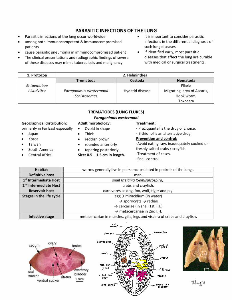
TREMATODES (LUNG FLUKES) Paragonimus westermani
Geographical distribution: primarily in Far East especially
Japan
Korea
Taiwan
South America
Central Africa.
Adult morphology:
Ovoid in shape
Thick
reddish brown
rounded anteriorly
tapering posteriorly. Size: 0.5 – 1.5 cm in length.
Treatment: - Praziquantel is the drug of choice. - Bithionol is an alternative drug. Prevention and control: -Avoid eating raw, inadequately cooked or freshly salted crabs / crayfish. -Treatment of cases. -Snail control.
Habitat worms generally live in pairs encapsulated in pockets of the lungs.
Definitive host man.
1st Intermediate Host snail Melania (Semisulcospira).
2nd Intermediate Host crabs and crayfish.
Reservoir host carnivores as dog, fox, wolf, tiger and pig.
Stages in the life cycle egg→ miracidium (in water) → sporocysts → rediae
→ cercariae (in snail 1st I.H.) → metacercariae in 2nd I.H.
Infective stage metacercariae in muscles, gills, legs and viscera of crabs and crayfish.

Thug’s
METHOD OF INFECTION 1. The defenitive host infection occurs by eating raw/
insufficiently cooked infected crabs or crayfish. 2. Metacercariae excyst in the small intestine pass through
the intestinal wall 3. grow for about one week into young flukes 4. penetrate the diaphragm and pleural cavity and come to
rest in the lung, forming cystic cavities then get mature. 5. The life cycle is completed in 6-8 months. 6. The eggs escape from the pulmonary pockets through the
bronchioles and are coughed out with sputum, or swallowed and pass immature with feces.
7. Eggs require from 15 days to several weeks in water to complete embryonation then hatch and miracidia escape.
8. Miracidium enters the snail first I.H. then
develops into sporocysts rediae cercariae in 3-5 months.
9. The released cercariae penetrate the crustaceans second I.H then develop into metacercariae, which require 6-8 weeks to become infective.
DIAGNOSIS 1-Clinical signs & diet history in endemic area.
Chest pain
Chronic cough
Brownish sputum may show eggs
Pleural effusion
Life threatening haemoptysis if eroding into adjacent bronchi
2-Characteristic immature eggs in sputum, feces or in aspirated pleural effusion. 3-Plain x-ray chest and computerized tomography show nodular shadows & cavities (Radiographic features of paragonimiasis include patchy air space consolidation, cystic changes and ring shadows. Pleural effusions and pneumothoraces may occur.) 4-Immunodiagnostic tests as ELISA to detect early, chronic and extra-pulmonary infection. 5- Eosinophilia.
Blood Flukes Schistosoma
Stage of migration: By schistosomula In the lung:
verminous pneumonitis
minute haemorrhage
cough
haemoptysis.
The body of cercaria enters the skin or mucous membrane leaving the tail where it is transformed into schistosomulum. It is carried after 2 days by the blood
Rt side of heart
lung Lt side of the heart systemic circulation
intestinal capillary bed
via mesentric-portal vessels
intrahepatic branches of the portal vein where it matures in 7 weeks.
Thug’s
CESTODES (HYDATID DISEASE)
Mode of infection in man Ingestion of eggs of Echinococus granulosus through: • Hand to mouth from fur of dogs
while playing with them. • Contaminated food or drink with
faeces of infected dogs
CLINICAL MANIFESTATIONS • Hydatid Cysts may cause symptoms by compression of adjacent
structures, and lung cysts may present with chest pain, cough, haemoptysis or pneumothorax.
• Symptoms may also occur if antigenic material is released from the cyst, causing a hypersensitivity reaction with fever, wheeze and urticaria and, rarely, anaphylaxis.
• Cysts may become secondarily infected causing empyema or lung abscess formation.
NEMATODES
FILARIA Wuchereria bancrofti and Brugia malayi
MIGRATING LARVAE OF Ascaris, Hook worm, Toxocara
• found in endemic regions of south east Asia, India, China and Africa.
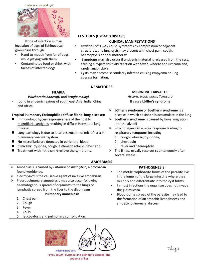
Tropical Pulmonary Eosinophilia (diffuse filarial lung disease): Immunologic hyper-responsiveness of the host to
microfilarial antigens resulting in diffuse interstitial lung disease.
Lung pathology is due to local destruction of microfilaria in pulmonary vascular system.
No microfilaria are detected in peripheral blood. Clinically: dyspnea, cough, asthmatic attacks, fever and Treatment with hetrazan →relieve the symptoms.
It cause Löffler's syndrome Löffler's syndrome or Loeffler's syndrome is a
disease in which eosinophils accumulate in the lung Loeffler's syndrome is caused by larval migration
into the alveoli which triggers an allergic response leading to
respiratory symptoms including 1. cough, wheeze, dyspnoea, 2. chest pain 3. fever and haemoptysis.
The illness usually resolves spontaneously after several weeks.
AMOEBIASIS
Amoebiasis is caused by Entamoeba histolytica, a protozoan found worldwide.
E histolytica is the causative agent of invasive amoebiasis Pleuropulmonary amoebiasis may also occur following
haematogenous spread of organisms to the lungs or lymphatic spread from the liver to the diaphragm
Pulmonary amoebiasis 1. Chest pain 2. Cough 3. Fever 4. Chills 5. leucocytosis and pulmonary consolidation
PATHOGENESIS • The motile trophozoite forms of the parasite live
in the lumen of the large intestine where they multiply and differentiate into the cyst forms.
• In most infections the organism does not invade the gut mucosa.
• Blood-borne spread of the parasite may lead to the formation of an amoebic liver abscess and amoebic pulmonary abscess.

Thug’s
Development of Schistosoma inside the body of infected human Development of Hydatid Cyst inside human body
Larval migration of Ascaris Larval migration of Ancylostoma duodenale
Conclusion:
• Direct identification of the causative organisms may be achieved definitively through
microscopic examination of stool or respiratory tract samples, or indirectly via serological
testing.
• If identified early, most parasitic lung diseases are curable with medical treatment.