parkinsonism and impaired axonal transport in a mouse model of
TRANSCRIPT

Parkinsonism and impaired axonal transportin a mouse model of frontotemporal dementiaLars M. Ittner*†‡, Thomas Fath†§, Yazi D. Ke*†, Mian Bi*, Janet van Eersel*, Kong M. Li¶, Peter Gunning§,and Jurgen Gotz*‡
*Alzheimer’s and Parkinson’s Disease Laboratory, Brain and Mind Research Institute, University of Sydney, Camperdown, NSW 2050, Australia;§Oncology Research Unit, Children’s Hospital at Westmead, Westmead, NSW 2145, Australia; and ¶LC Laboratory, Discipline of Pharmacology,University of Sydney, Sydney, NSW 2006, Australia
Communicated by Etienne-Emile Baulieu, College de France, Le Kremlin-Bicetre, France, August 18, 2008 (received for review April 8, 2008)
Frontotemporal dementia (FTD) is characterized by cognitive andbehavioral changes and, in a significant subset of patients, Par-kinsonism. Histopathologically, FTD frequently presents with tau-containing lesions, which in familial cases result from mutations inthe MAPT gene encoding tau. Here we present a novel transgenicmouse strain (K3) that expresses human tau carrying the FTDmutation K369I. K3 mice develop a progressive histopathology thatis reminiscent of that in human FTD with the K369I mutation. Inaddition, K3 mice show early-onset memory impairment and amy-otrophy in the absence of overt neurodegeneration. Different fromour previously generated tau transgenic strains, the K3 miceexpress the transgene in the substantia nigra (SN) and show anearly-onset motor phenotype that reproduces Parkinsonism withtremor, bradykinesia, abnormal gait, and postural instability. In-terestingly, motor performance of young, but not old, K3 miceimproves upon L-dopa treatment, which bears similarities to Par-kinsonism in FTD. The early-onset symptoms in the K3 mice aremechanistically related to selectively impaired anterograde axonaltransport of distinct cargos, which precedes the loss of dopami-nergic SN neurons that occurs in aged mice. The impaired axonaltransport in SN neurons affects, among others, vesicles containingthe dopamine-synthesizing enzyme tyrosine hydroxylase. Distinctmodes of transport are also impaired in sciatic nerves, which mayexplain amyotrophy. Together, the K3 mice are a unique model ofFTD-associated Parkinsonism, with pathomechanistic implicationsfor the human pathologic process.
tau � Alzheimer � transgenic � NFT � memory
How neuronal dysfunction and cell loss is brought about inneurodegenerative diseases such as Alzheimer disease
(AD) and frontotemporal dementia (FTD) is only partiallyunderstood. In familial cases of FTD with tau aggregation (i.e.,FTDP-17), mutations were identified in the MAPT encoding themicrotubule (MT)-associated protein tau (1), and in FTD caseswithout tau aggregation, they were identified in PGRN encodingprogranulin (2, 3). Of the 42 known MAPT mutations, severalhave been expressed in transgenic mice. The mice reproduceselective aspects of the disease which is, in part, determined bythe choice of promoter and tau isoform, inclusion of FTDP-17mutations, the integration site, and copy number of the trans-gene (4).
In FTD and AD, tau becomes increasingly hyperphosphory-lated, i.e., more phosphorylated at physiological sites and, inaddition, de novo at pathological sites (5). Hyperphosphorylationdetaches tau from MTs, and makes it prone to form filamentousinclusions, including neurofibrillary tangles (NFTs) in AD andFTD, and Pick bodies in Pick disease (PiD) (6–9). However, itis only partly understood how aggregated tau interferes withcellular functions.
Here we report a novel transgenic mouse strain that expressesK369I mutant human tau in neurons (K3 mice). This mutationhas been identified in a patient with a PiD neuropathology (10).Different from previously generated tau transgenic strains, K3
mice express the transgene in the SN, in addition to other brainareas. The mice develop memory impairment and an early-onsetmotor phenotype reminiscent of Parkinsonism. Sciatic nerveligations and an analysis of SN neurons assisted in identifyingimpaired axonal transport of distinct cargos as pathomechanism.
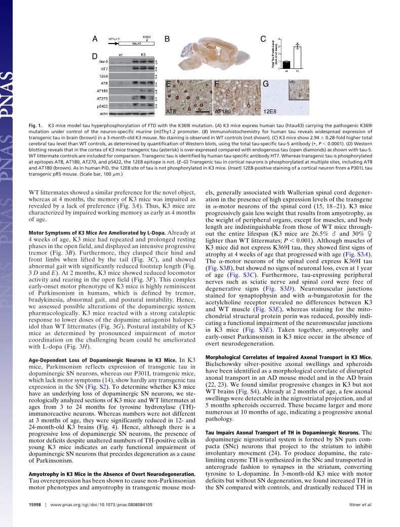
ResultsHyperphosphorylation and Deposition of Tau in K3 Mice. K3 trans-genic mice express K369I mutant human tau driven by theneuron-specific mThy1.2 promoter [Fig. 1A and supportinginformation (SI) Text] . K369I tau is expressed in several brainareas including cortex, hippocampus, and basal ganglia (Fig.1B). In K3 forebrains, total tau protein levels are 2.9-foldhigher than in WT controls (Fig. 1 C and D). Tau is phos-phorylated at multiple sites including AT8, AT180, AT270, andpS422 (Fig. 1 D–F). However, reminiscent of human PiD, tauis not phosphorylated at the 12E8 (S262/S356) phospho-epitope, even in old (�12 months) K3 mice (Fig. 1 D and G).In frontal cortex of K3 mice, ovoid intraneuronal tau aggre-gates are predominant, resembling Pick bodies. Like in PiD,they are identified by Bielschowsky, but not Gallyas, silverimpregnation (Fig. 2A). Their numbers increase with age (Fig.2B). Antibodies pS422 and AT100 reactive with pathologicallyphosphorylated tau (11, 12) stained them as well, and thenumbers of tau-positive inclusions correlate with these ofBielschowsky-positive deposits (Fig. 2 A and C). Thiazin redstaining for fibrillar tau (13) co-localized with AT8 immuno-staining of inclusions (Fig. 2D). A Western blot analysis ofsarkosyl extractions (Fig. 2E) revealed high levels of insolubletau in K3 mice, P301L transgenic pR5 mice (14), and WTtau-transgenic ALZ17 mice (15). It was much more phosphor-ylated at the PHF1 epitope in K3 than in P301L transgenic pR5mice (14), whereas again, the 12E8 epitope was not phosphor-ylated (Fig. 2E). As in the human K369I mutation carrier (10),tau in K3 mice was ubiquitinated (Fig. S1). Together, thisshows that, histopathologically, the K3 mice model FTD withthe K369I mutation.
Memory Deficits in K3 Mice. To test memory functions of K3 micewe used the novel object recognition task (16, 17). Here, the timespent exploring two objects on the first test day is equal, whereason the second test day mice with normal memory will spendmore time exploring a novel object. At 2 months of age, K3 and
Author contributions: L.M.I., T.F., Y.D.K., and J.G. designed research; L.M.I., T.F., Y.D.K.,M.B., J.v.E., K.M.L., and J.G. performed research; L.M.I., T.F., Y.D.K., M.B., J.v.E., and J.G.analyzed data; and L.M.I., T.F., Y.D.K., P.G., and J.G. wrote the paper.
The authors declare no conflict of interest.
†L.I.M., T.F., and Y.D.K. contributed equally to this work.
‡To whom correspondence may be addressed. E-mail: [email protected] [email protected].
This article contains supporting information online at www.pnas.org/cgi/content/full/0808084105/DCSupplemental.
© 2008 by The National Academy of Sciences of the USA
www.pnas.org�cgi�doi�10.1073�pnas.0808084105 PNAS � October 14, 2008 � vol. 105 � no. 41 � 15997–16002
NEU
ROSC
IEN
CE
WT littermates showed a similar preference for the novel object,whereas at 4 months, the memory of K3 mice was impaired asrevealed by a lack of preference (Fig. 3A). Thus, K3 mice arecharacterized by impaired working memory as early as 4 monthsof age.
Motor Symptoms of K3 Mice Are Ameliorated by L-Dopa. Already at4 weeks of age, K3 mice had repeated and prolonged restingphases in the open field, and displayed an intensive progressivetremor (Fig. 3B). Furthermore, they clasped their hind andfront limbs when lifted by the tail (Fig. 3C), and showedabnormal gait with significantly reduced footstep length (Fig.3 D and E). At 2 months, K3 mice showed reduced locomotoractivity and rearing in the open field (Fig. 3F). This complexearly-onset motor phenotype of K3 mice is highly reminiscentof Parkinsonism in humans, which is defined by tremor,bradykinesia, abnormal gait, and postural instability. Hence,we assessed possible alterations of the dopaminergic systempharmacologically. K3 mice reacted with a strong catalepticresponse to lower doses of the dopamine antagonist haloper-idol than WT littermates (Fig. 3G). Postural instability of K3mice as determined by pronounced impairment of motorcoordination on the challenging beam could be amelioratedwith L-dopa (Fig. 3H).
Age-Dependent Loss of Dopaminergic Neurons in K3 Mice. In K3mice, Parkinsonism reflects expression of transgenic tau indopaminergic SN neurons, whereas our P301L transgenic mice,which lack motor symptoms (14), show hardly any transgenic tauexpression in the SN (Fig. S2). To determine whether K3 micehave an underlying loss of dopaminergic SN neurons, we ste-reologically analyzed sections of K3 mice and WT littermates atages from 3 to 24 months for tyrosine hydroxylase (TH)-immunoreactive neurons. Whereas numbers were not differentat 3 months of age, they were significantly reduced in 12- and24-month-old K3 brains (Fig. 4). Hence, although there is aprogressive loss of dopaminergic SN neurons, the presence ofmotor deficits despite unaltered numbers of TH-positive cells inyoung K3 mice indicates an early functional impairment ofdopaminergic SN neurons that precedes degeneration as a causeof Parkinsonism.
Amyotrophy in K3 Mice in the Absence of Overt Neurodegeneration.Tau overexpression has been shown to cause non-Parkinsonianmotor phenotypes and amyotrophy in transgenic mouse mod-
els, generally associated with Wallerian spinal cord degener-ation in the presence of high expression levels of the transgenein �-motor neurons of the spinal cord (15, 18–21). K3 miceprogressively gain less weight that results from amyotrophy, asthe weight of peripheral organs, except for muscles, and bodylength are indistinguishable from those of WT mice through-out the entire lifespan (K3 mice are 26.5% � and 30% �lighter than WT littermates; P � 0.001). Although muscles ofK3 mice did not express K369I tau, they showed first signs ofatrophy at 4 weeks of age that progressed with age (Fig. S3A).The �-motor neurons of the spinal cord express K369I tau(Fig. S3B), but showed no signs of neuronal loss, even at 1 yearof age (Fig. S3C). Furthermore, tau-expressing peripheralnerves such as sciatic nerve and spinal cord were free ofdegenerative signs (Fig. S3D). Neuromuscular junctionsstained for synaptophysin and with �-bungarotoxin for theacetylcholine receptor revealed no differences between K3and WT muscle (Fig. S3E), whereas staining for the mito-chondrial structural protein porin was reduced, possibly indi-cating a functional impairment of the neuromuscular junctionsin K3 mice (Fig. S3E). Taken together, amyotrophy andearly-onset Parkinsonism in K3 mice occur in the absence ofovert neurodegeneration.
Morphological Correlates of Impaired Axonal Transport in K3 Mice.Bielschowsky silver-positive axonal swellings and spheroidshave been identified as a morphological correlate of disruptedaxonal transport in an AD mouse model and in the AD brain(22, 23). We found similar progressive changes in K3 but notWT brains (Fig. S4). Already at 2 months of age, a few axonalswellings were detectable in the nigrostriatal projection, and at5 months spheroids occurred. These became larger and morenumerous at 10 months of age, indicating a progressive axonalpathology.
Tau Impairs Axonal Transport of TH in Dopaminergic Neurons. Thedopaminergic nigrostriatal system is formed by SN pars com-pacta (SNc) neurons that project to the striatum to inhibitinvoluntary movement (24). To produce dopamine, the rate-limiting enzyme TH is synthesized in the SNc and transported inanterograde fashion to synapses in the striatum, convertingtyrosine to L-dopamine. In 3-month-old K3 mice with motordeficits but without SN degeneration, we found increased TH inthe SN compared with controls, and drastically reduced TH in
A B C
D
E F G
Fig. 1. K3 mice model tau hyperphosphorylation of FTD with the K369I mutation. (A) K3 mice express human tau (htau43) carrying the pathogenic K369Imutation under control of the neuron-specific murine (m)Thy1.2 promoter. (B) Immunohistochemistry for human tau reveals widespread expression oftransgenic tau in brain (brown) in a 3-month-old K3 mouse. No staining is observed in WT controls (not shown). (C) K3 mice show 2.94 � 0.28-fold higher totalcerebral tau level than WT controls, as determined by quantification of Western blots, using the total tau-specific tau-5 antibody (*, P � 0.0001). (D) Westernblotting reveals that in the cortex of K3 mice transgenic tau (asterisk) is over-expressed compared with endogenous tau (open diamonds) as shown with tau-5.WT littermate controls are included for comparison. Transgenic tau is identified by human tau-specific antibody HT7. Whereas transgenic tau is phosphorylatedat epitopes AT8, AT180, AT270, and pS422, the 12E8 epitope is not. (E–G) Transgenic tau in cortical neurons is phosphorylated at multiple sites, including AT8and AT180 (brown). As in human PiD, the 12E8 site of tau is not phosphorylated in K3 mice. (Inset) 12E8-positive staining of a cortical neuron from a P301L tautransgenic pR5 mouse. (Scale bar, 100 �m.)
15998 � www.pnas.org�cgi�doi�10.1073�pnas.0808084105 Ittner et al.

striatal synapses compared with controls (Fig. 5 A and B).Numbers of TH-positive synaptic boutons of SNc neurons werenot reduced, suggesting that less TH is transported along thenigrostriatal projection. Reduced availability of TH can causereduced dopamine levels. We found that, already at 6 weeks ofage, K3 mice had a significant (P � 0.05) 15.3 � 3.9% (mean �SD) decrease of basal dopamine levels compared with WTcontrols.
Tau has previously been reported to interfere with axonaltransport in cell culture, resulting in an altered distribution oforganelles (19, 25). Hence, we assessed the distribution of TH indopaminergic SN neurons of K3 mice stained for TH (Fig. 5C).Compared with WT controls, SN neurons of K3 mice thatexpress transgenic tau showed a reduced TH intensity in growthcones, along with enrichment in cell bodies, supporting thenotion of reduced TH transport (Fig. 5 D–F). Taken together,young K3 mice have less TH in striatal synapses and thus lowerbasal dopamine levels.
Selective Axonal Transport Impairment in K3 Mice. To further ad-dress transport defects in the dopaminergic system of K3 mice,
we compared levels of different cargos transported between SNand striatum by Western blotting (Fig. 5 G and H). In thestriatum of K3 mice, the markers of amyloid � precursor protein(APP)-containing vesicles, APP, PS1, and Gap43 (26), the APPfamily member amyloid � precursor-like protein 2 (APLP2), the
Fig. 2. K3 mice show a progressive histopathology reminiscent of FTD withthe K369I mutation. (A) The intraneuronal ovoid inclusions are Bielschowskysilver-positive (black; arrow), and Gallyas silver-negative (Inset, Gallyas-positive human NFT). The round Pick body-like lesions are also reactive withantibodies pS422 and AT100 for pathologically phosphorylated tau. Theinclusions are 12E8-negative (not shown). No staining is found in controls (notshown). (B) Numbers of Bielschowsky-positive cells in K3 mice increase withage (*, P � 0.01 vs. previous age-group). (C) Numbers of Bielschowsky- andpS422-positive cells correlate in K3 brains (P � 0.001). (D) Staining with the AT8antibody (green) and Thiazin red (TR) for fibrillar deposits co-localized (yel-low) in intraneuronal lesions in K3 brains. (E) Comparison of sarkosyl-insolubletau in brains of WT, WT tau-expressing ALZ17, P301L mutant tau-expressingpR5, and K3 mice. Western blotting reveals insoluble tau in ALZ17, pR5, andK3 mice. Insoluble tau is phosphorylated at the 12E8 epitope only in pR5 mice,whereas phosphorylation at the PHF-1 epitope is most abundant in K3 mice (*,P � 0.05).
Fig. 3. K3 mice present with early-onset memory deficits and Parkinsonism-like motor deficits that can be partially ameliorated with L-dopa. (A) At 2months of age, K3 mice (white bars) explore novel objects with the samepreference as WT littermates (black bars). At 4 months of age, their memoryis impaired as indicated by an equal exploration of both the known and thenovel object (*, P � 0.001). (B) At 4 weeks of age, K3 mice (white dots) presentwith a slight tremor that becomes progressively more intense, until at 8 weeksof age all K3 mice suffer from severe tremor. No tremor is observed in WTlittermates (black dots). Scoring of tremor is described in Methods. (C) Alreadyat 6 weeks of age, K3 mice, but not WT littermates, clasp their hind limbs whenlifted by the tail. (D and E) Abnormal gait shown by coloring of paws. Barsindicate foot step length that is 38.3% shortened in K3 mice versus WTlittermates (*, P � 0.0001). (F) Already at 2 months of age, the locomotoractivity of K3 mice (white bar) is reduced as they travel less than WT littermates(black bar) in the open field arena. This difference persists throughout age (*,P � 0.05). (G) Experimental induction of catalepsy with the dopamine antag-onist haloperidol (administered i.p.) in the bar test reveals a four-times-increased sensitivity of the dopaminergic system in K3 mice versus WT litter-mates (*, P � 0.001). (H) The challenging beam task shows a pronouncedpostural instability of K3 mice compared with WT littermates, as indicated byrepeated slipping when crossing a narrow beam (**, P � 0.0001). Single dosesof L-dopa ameliorate the balance deficit of 3-month-old K3 mice (gray bar; *,P � 0.05). The L-dopa responsiveness is lost when K3 mice reach 6 months ofage.
Ittner et al. PNAS � October 14, 2008 � vol. 105 � no. 41 � 15999
NEU
ROSC
IEN
CE

motor proteins kinesin heavy chain and kinesin light chain, andthe scaffold protein Jip1 were markedly reduced. Mitochondrialrespiratory chain protein complexes NADH-ubiquinol oxi-doreductase (Co-I) and ATP synthase (Co-V) were also re-duced. No changes were found for synaptophysin and synapto-tagmin, markers of different transport cargos (27, 28), APLP1,and the motor protein kinesin-III. As levels of these markerswere not reduced in the K3 SN, this suggests selectively impairedaxonal transport in the nigrostriatal system.
To directly address axonal transport in vivo, we ligated thesciatic nerve in K3 mice and WT littermates, taking advantageof transgene expression in spinal cord �-motor neurons (Figs.S3B and S5 A and B). Ligation causes accumulation of antero-gradely transported cargos proximal and retrogradely trans-ported cargos distal to the ligation site (26, 29). In ligated WT
nerves, APP, Gap43, kinesin heavy chain (Kif5B), kinesin lightchain (KLC), and Jip1 accumulated proximal to the ligationsite, different from K3 mice in which accumulation of APP,Gap43, and Jip1 was significantly reduced. Co-V accumulatedproximally and distally of the ligation in WT nerves, repre-senting bidirectional transport of mitochondria. In ligated K3nerves, however, Co-V accumulated only in the distal part,suggesting impaired anterograde and unaffected retrogradetransport of mitochondria in K3 mice. A tau dependency ofthis impairment is further supported by immunohistochemistryas only K369I tau-expressing axons show reduced numbers ofmitochondria (Fig. S5C). Retrograde transport was unaf-fected, as assessed also by striatal injections of FluoroGold, aretrogradely transported dye (Fig. S6). Accumulation ofmarker proteins for other transport cargos and motor proteinsat the ligation sites in WT and K3 nerves was comparable.These included the slow transport marker tubulin (29), syn-aptic vesicle precursor proteins synaptophysin and synapto-tagmin, motor protein kinesin-III (27), and calcitonin gene-related peptide (30). Together, this shows that tau specificallyimpairs the anterograde axonal transport of APP- and TH-containing vesicles and mitochondria, and leaves the transportof other cargos unaffected. Hence, early-onset amyotrophyand Parkinsonism in the K3 mice are both likely caused byimpaired axonal transport.
DiscussionPossibly ref lecting a unique expression pattern of the K369Itau transgene that included the SN, we obtained a novel mousestrain, K3, that not only models histopathological character-istics of FTD with the K369I tau mutation, but also memoryimpairment, amyotrophy, and Parkinsonism. We identifiedcargos such as TH-containing vesicles and mitochondria whose
Fig. 4. Progressive loss of dopaminergic SN neurons in K3 mice. (A) Immu-nohistochemistry of dopaminergic TH (brown)-containing neurons revealsthat, not at 3, but at 24 months of age, significantly fewer cells are TH-positivein K3 than in WT SN. (Scale bar, 50 �m.) (B) At 3 months of age, numbers ofTH-positive cells on serial sagittal sections are equal in WT and K3 SN; however,at 12 and 24 months of age, numbers of TH-positive cells are significantlyreduced in K3 mice (*, P � 0.01; **, P � 0.0001).
Fig. 5. TH transport is impaired in the nigrostriatal projection of K3 mice and in primary SN neuronal cultures established from them. (A) Staining for TH (red)on sagittal sections of 3-month-old K3 and WT mice reveals that TH intensity (I) is increased in the SN pars compacta (broken lines), and reduced in synapticboutons of the striatum (CPu). Numbers of TH-positive synaptic boutons are unaltered in K3 mice (Right). (B) Quantification of immunohistochemical and Westernblot (WB) data reveals that TH accumulates in K3 SN neurons (*, P � 0.0005). Consequently, TH is decreased in the K3 CPu (*, P � 0.0001), indicating that TH isnot delivered properly to the axonal terminals of SN neurons in the K3 striatum. (C) Tau-expressing primary cultured K3 SN neurons (arrowhead) show a markedlydecreased TH staining of axonal growth cones (Inset; open arrow) compared with WT neurons (arrow; Inset). This occurs in the absence of overt morphologicalalterations. (Scale bar, 50 �m.) (D) K3 SN neurons have 1.82-fold higher total tau levels than WT neurons (*, P � 0.01). (E) Quantification of fluorescence intensitiesshows increased TH in the soma of primary SN neurons of K3 mice compared with WT neurons (*, P � 0.05). (F) In contrast, TH fluorescence intensity is markedlydecreased in growth cones of K3 compared with WT neurons (*, P � 0.0001). (G) SN and striatal (CPu) extracts from WT and K3 mice analyzed in parallel showa marked reduction of selective proteins in the CPu of K3 mice. Changes are found exclusively for motor proteins and markers of distinct vesicles and mitochondria(see Results). For comparison, protein levels are not reduced in the SN. (H) Quantification of protein levels in the CPu of K3 mice shown as expression (in fold)compared with WT (dashed line; *, P � 0.05; **, P � 0.01).
16000 � www.pnas.org�cgi�doi�10.1073�pnas.0808084105 Ittner et al.

axonal transport was selectively impaired. Hence, the K3 miceare particularly suited in which to study the role of tau inimpaired axonal transport, which has been implicated as acentral pathomechanism in AD and related disorders (31).
The K369I mutation was initially identified in a human patientwith a neuropathologic process indistinguishable from sporadicPiD (10). PiD is a subtype of FTD characterized by rapid diseaseprogression with frontotemporal and limbic neuronal loss (9). Incases with generalized PiD, additional brain areas may beaffected, such as the SN (32). The definition of PiD, however, hasundergone several changes over time (33). While Pick body-likeinclusions have been frequently seen in carriers of distinctMAPT mutations (10, 34, 35), the very existence of an autoso-mal-dominant PiD resulting from MAPT mutations has beenquestioned (36). PiD is generally characterized by a frontalsyndrome and the presence of Pick bodies made mainly of 3R tau(37, 38). Pick bodies are also 12E8- and AT100-negative (39–41).For comparison, K3 mice develop ovoid tau inclusions that are12E8-negative but AT100-positive. In this respect, they modelthe human K369I histopathology, as there the inclusions are also12E8-negative but AT100-positive (10). The K3 mice do, how-ever, model other aspects of sporadic PiD, such as ubiquitinationand the presence of Bielschowsky- but not Gallyas-positive tauaggregates.
The Pick body-like inclusions, Parkinsonism, and amnesia thatcharacterize K3 mice are part of the human PiD syndrome,although in the K369I case, no Parkinsonism has been reported,possibly reflecting the absence of a tau pathologic process in theSN (data not shown). Similarly, amyotrophy is absent in thehuman case. However, as expression of the K369I transgene inthe sciatic nerve also caused an impaired transport of distinctcargos associated with a pathologic process, this may reveal acentral pathomechanism.
AD and FTD both present with a progressive decline ofmemory function leading to dementia, although in FTD it isoften preceded by behavioral changes and motor symptomssuch as Parkinsonism (9). The K3 mice present with early-onset memory impairment at an age of 4 months, beforeBielschowsky-positive tau deposits are detectable. This is inline with the observation that memory deficits precede his-topathological changes in other tau-expressing strains (21, 42).Pathological tau thus interferes with distinct cellular mecha-nisms causing neuronal dysfunction before tau is sequesteredinto deposits. Consequently, recovery of memory function hasbeen observed in mice when expression of transgenic tau wassuppressed, despite a persistent accumulation of NFTs, sug-gesting that tau mediates neuronal dysfunction independent ofNFT formation (43). We speculate that the memory impair-ment in the K3 mice may also be caused by impaired axonaltransport, in agreement with previous findings in AD brain andAPP transgenic mice (31).
K3 mice develop a complex motor phenotype and amyotrophyas early as 4 weeks of age, which, again, is before tau aggregates.Early-onset tremor, bradykinesia, muscle rigidity, and posturalinstability establish K3 mice as a unique mouse model forParkinsonism in FTD. Interestingly, as in the treatment ofParkinsonism in patients with FTD, the symptoms of K3 miceare partially reversible with L-dopa at an early, but not late, stageof disease.
Increasing evidence suggests that neuronal dysfunction result-ing from failure of axonal transport is an important pathomecha-nism in neurodegeneration, including AD (31, 44). The func-tional impairment of K3 mice is accompanied by progressivemorphological changes including axonal swellings and spheroidsthat are histopathological correlates of disrupted axonal trans-port (22, 23), preceding loss of SN neurons. In vivo experiments
revealed that tau can directly inhibit axonal transport (18).Although mechanistically not fully understood, it has beenproposed that increases in tau, as found in AD and FTD,decrease binding of motor proteins to MT tracks (45, 46). In K3mice, we found impaired TH transport in the nigrostriatalsystem. This suggests that the motor deficits are, at least in part,a result of early-onset neuronal dysfunction caused by impairedaxonal distribution of TH in the nigrostriatal system, and thus,reduced basal dopamine levels. Similar to K3 mice, viral expres-sion of both WT or P301L tau in the SN of rats caused reducedTH immunoreactivity in the striatum, decreased dopaminelevels, and formation of axonal spheroids that preceded neuronalloss by several months (23). Therefore, it is not unlikely that, inthe rat model, neuronal dysfunction is also a result of tau-impaired axonal transport, as in K3 mice. Reduced striatalsynaptic supply of transported cargos other than TH is likely tocontribute to the K3 phenotype, as we have shown that transportof additional cargos such as APP-containing vesicles or mito-chondria is also affected. Finally, sciatic nerve ligations revealeda similarly impaired axonal transport in a second, unrelatedsystem. Neuronal dysfunction resulting from impaired antero-grade axonal transport is therefore likely to cause amyotrophy inthe K3 mice, in the absence of overt neurodegeneration.
In conclusion, K3 mice are a unique model for aspects of PiD,but mainly for FTD-associated Parkinsonism and memory im-pairment. Future therapeutic strategies that target transportimpairment in tauopathies may be beneficial in the treatment ofL-dopa-resistant Parkinsonism in FTD.
Materials and MethodsMice. A human K369I mutant tau cDNA, lacking exon 3 and containing fourMT-binding domains, was cloned together with a Kozak consensus sequenceinto a murine Thy1.2 expression vector as described (14). Transgenic mice wereproduced by pronuclear injection of B6D2F1�B6D2F1 oocytes (47). Founderswere identified by PCR using primers 5�-GGGTGTCTCCAATGCCTGCTTCT-TCAG-3� and 5�-AAGTCACCCAGCAGGGAGGTGCTCAG-3�. Strains were estab-lished from founder mice, and the K3 strain was backcrossed 8 times onto aC57BL/6 background. pR5 mice express P301L mutant tau (14), whereas ALZ17mice express WT human tau (15). Animal experiments were approved by theAnimal Ethics Committee of the University of Sydney.
Cell Culture and Immunocytochemistry. For nigrostriatal primary cultures, SN andstriatum were prepared separately from neonatal mice. The tissue was dissectedin Hanks’ balanced salt solution and incubated with 2.5 mg/ml trypsin in thepresence of 1 mg/ml DNaseI (Sigma) for 20 min at 37°C. Subsequently, it wastriturated in plating medium containing DMEM/10% FBS (Gibco) using fire-polished glass Pasteur pipettes. SN cells (2 � 105) were plated overnight at 37°ConpolyD-lysine (0.1mg/ml; Sigma)-coated12-mmglass coverslipsmounted in thecenter of a 12-well culture plate well. Subsequently, 1 � 104 striatal cells wereplated in a ring around the coverslips to support SN cell growth. After 2 h, theplating medium was replaced by 1 ml of Neurobasal medium containing B27supplement and Glutamax (Gibco). For immunofluorescence staining, cells werefixed with 4% paraformaldehyde in 80-mM Pipes, 1 mM MgCl2, and 1 mM EGTA(pH 6.8) after 7 days. Cells were permeabilized with 0.1% Triton in PBS solutionand stained with antibodies for TH and tau.
Statistics. Statistical analysis was performed with Prizm 4 software for Win-dows (GraphPad) using Student’s t test. All values are given as mean � SE.
ACKNOWLEDGMENTS. We thank Matthias Minderer, Daniel Schuppli, DeniseNergenau, Fabien Delerue, and Drs. Manuela Neumann, Malcolm France, andPhil Robinson for support and helpful comments; and Mamdouh Nessiem foranimal care. We thank Drs. Peter Seubert, Peter Davies, and Walter Born forantibodies and Dr. Akihiko Takashima for thiazin red. This research wassupported by grants from the University of Sydney, The Medical Foundation(University of Sydney), the NHMRC, the Judith Jane Mason & Harold StannettWilliams Memorial Foundation, the ARC, and the New South Wales Govern-ment through the Ministry for Science and Medical Research (BioFirst Grant)(J.G.); and the ARC, the University of Sydney, and the Deutsche Forschungs-gesellschaft (L.M.I.). J.G. is a Medical Foundation Fellow.
Ittner et al. PNAS � October 14, 2008 � vol. 105 � no. 41 � 16001
NEU
ROSC
IEN
CE
1. Ballatore C, Lee VM, Trojanowski JQ (2007) Tau-mediated neurodegeneration inAlzheimer’s disease and related disorders. Nat Rev Neurosci 8:663–672.
2. Baker M, et al. (2006) Mutations in progranulin cause tau-negative frontotemporaldementia linked to chromosome 17. Nature 442:916–919.
3. Cruts M, et al. (2006) Null mutations in progranulin cause ubiquitin-positive fronto-temporal dementia linked to chromosome 17q21. Nature 442:920–924.
4. Gotz J, Ittner LM (2008) Animal models of Alzheimer’s disease and frontotemporaldementia. Nat Rev Neurosci 9:532–544.
5. Alonso AC, Grundke-Iqbal I, Iqbal K (1996) Alzheimer’s disease hyperphosphorylatedtau sequesters normal tau into tangles of filaments and disassembles microtubules. NatMed 2:783–787.
6. Goedert M, Wischik CM, Crowther RA, Walker JE, Klug A (1988) Cloning and sequenc-ing of the cDNA encoding a core protein of the paired helical filament of Alzheimerdisease: identification as the microtubule-associated protein tau. Proc Natl Acad SciUSA 85:4051–4055.
7. Feany MB, Dickson DW (1995) Widespread cytoskeletal pathology characterizes cor-ticobasal degeneration. Am J Pathol 146:1388–1396.
8. Constantinidis J, Richard J, Tissot R (1974) Pick’s disease. Histological and clinicalcorrelations. Eur Neurol 11:208–217.
9. Lee VM, Goedert M, Trojanowski JQ (2001) Neurodegenerative tauopathies. Annu RevNeurosci 24:1121–1159.
10. Neumann M, et al. (2001) Pick’s disease associated with the novel Tau gene mutationK369I. Ann Neurol 50:503–513.
11. Bussiere T, et al. (1999) Phosphorylated serine422 on tau proteins is a pathologicalepitope found in several diseases with neurofibrillary degeneration. Acta Neuropathol97:221–230.
12. Gotz J, Chen F, van Dorpe J, Nitsch RM (2001) Formation of neurofibrillary tangles inP301l tau transgenic mice induced by Abeta 42 fibrils. Science 293:1491–1495.
13. Uchihara T, et al. (2001) Different conformation of neuronal tau deposits distinguishedby double immunofluorescence with AT8 and thiazin red combined with Gallyasmethod. Acta Neuropathol 102:462–466.
14. Gotz J, Chen F, Barmettler R, Nitsch RM (2001) Tau filament formation in transgenicmice expressing P301L tau. J Biol Chem 276:529–534.
15. Probst A, et al. (2000) Axonopathy and amyotrophy in mice transgenic for humanfour-repeat tau protein. Acta Neuropathol (Berl) 99:469–481.
16. Ennaceur A, Delacour J (1988) A new one-trial test for neurobiological studies ofmemory in rats. 1: Behavioral data. Behav Brain Res 31:47–59.
17. Bevins RA, Besheer J (2006) Object recognition in rats and mice: A one-trial non-matching-to-sample learning task to study ‘recognition memory’. Nat Protocols1:1306–1311.
18. Ishihara T, et al. (1999) Age-dependent emergence and progression of a tauopathy intransgenic mice overexpressing the shortest human tau isoform. Neuron 24:751–762.
19. Spittaels K, et al. (1999) Prominent axonopathy in the brain and spinal cord oftransgenic mice overexpressing four-repeat human tau protein. Am J Pathol 155:2153–2165.
20. Lewis J, et al. (2000) Neurofibrillary tangles, amyotrophy and progressive motordisturbance in mice expressing mutant (P301L) tau protein. Nat Genet 25:402–405.
21. Leroy K, et al. (2007) Early axonopathy preceding neurofibrillary tangles in mutant tautransgenic mice. Am J Pathol 171:976–992.
22. Stokin GB, et al. (2005) Axonopathy and transport deficits early in the pathogenesis ofAlzheimer’s disease. Science 307:1282–1288.
23. Klein RL, Dayton RD, Lin WL, Dickson DW (2005) Tau gene transfer, but not alpha-synuclein, induces both progressive dopamine neuron degeneration and rotationalbehavior in the rat. Neurobiol Dis 20:64–73.
24. Waxman SG (2003) Clinical Neuroanatomy (McGraw-Hill, New York).25. Stamer K, Vogel R, Thies E, Mandelkow E, Mandelkow EM (2002) Tau blocks traffic of
organelles, neurofilaments, and APP vesicles in neurons and enhances oxidative stress.J Cell Biol 156:1051–1063.
26. Kamal A, Stokin GB, Yang Z, Xia CH, Goldstein LS (2000) Axonal transport of amyloidprecursor protein is mediated by direct binding to the kinesin light chain subunit ofkinesin-I. Neuron 28:449–459.
27. Okada Y, Yamazaki H, Sekine-Aizawa Y, Hirokawa N (1995) The neuron-specific kinesinsuperfamily protein KIF1A is a unique monomeric motor for anterograde axonaltransport of synaptic vesicle precursors. Cell 81:769–780.
28. Otsuka AJ, et al. (1991) The C. elegans unc-104 gene encodes a putative kinesin heavychain-like protein. Neuron 6:113–122.
29. Kamal A, Almenar-Queralt A, LeBlanc JF, Roberts EA, Goldstein LS (2001) Kinesin-mediated axonal transport of a membrane compartment containing beta-secretaseand presenilin-1 requires APP. Nature 414:643–648.
30. Verkade P, Verkleij AJ, Annaert WG, Gispen WH, Oestreicher AB (1996) Ultrastructurallocalization of B-50/growth-associated protein-43 to anterogradely transported syn-aptophysin-positive and calcitonin gene-related peptide-negative vesicles in the re-generating rat sciatic nerve. Neuroscience 71:489–505.
31. Stokin GB, Goldstein LS (2006) Axonal transport and Alzheimer’s disease. Annu RevBiochem 75:607–627.
32. Wang LN, Zhu MW, Feng YQ, Wang JH (2006) Pick’s disease with Pick bodies combinedwith progressive supranuclear palsy without tuft-shaped astrocytes: A clinical, neuro-radiologic and pathological study of an autopsied case. Neuropathology 26:222–230.
33. Cairns NJ, et al. (2007) Neuropathologic diagnostic and nosologic criteria for fronto-temporal lobar degeneration: Consensus of the Consortium for Frontotemporal LobarDegeneration. Acta Neuropathol 114:5–22.
34. Pickering-Brown S, et al. (2000) Pick’s disease is associated with mutations in the taugene. Ann Neurol 48:859–867.
35. Rizzini C, et al. (2000) Tau gene mutation K257T causes a tauopathy similar to Pick’sdisease. J Neuropathol Exp Neurol 59:990–1001.
36. Bergeron C, Morris HR, Rossor M (2003). Pick’s disease. Neurodegeneration: Themolecular pathology of dementia and movement disorders, ed Dickson DW (ISNNeuropth Press, Basel), pp 124–131.
37. de Silva R, et al. (2006) An immunohistochemical study of cases of sporadic andinherited frontotemporal lobar degeneration using 3R- and 4R-specific tau monoclo-nal antibodies. Acta Neuropathol 111:329–340.
38. Zhukareva V, et al. (2002) Sporadic Pick’s disease: A tauopathy characterized by aspectrum of pathological tau isoforms in gray and white matter. Ann Neurol 51:730–739.
39. Shiarli AM, et al. (2006) Comparison of extent of tau pathology in patients withfrontotemporal dementia with Parkinsonism linked to chromosome 17 (FTDP-17),frontotemporal lobar degeneration with Pick bodies and early onset Alzheimer’sdisease. Neuropathol Appl Neurobiol 32:374–387.
40. Mailliot C, et al. (1998) Phosphorylation of specific sets of tau isoforms reflects differentneurofibrillary degeneration processes. FEBS Lett 433:201–204.
41. Probst A, Tolnay M, Langui D, Goedert M, Spillantini MG (1996) Pick’s disease: Hyper-phosphorylated tau protein segregates to the somatoaxonal compartment. ActaNeuropathol 92:588–596.
42. Gotz J, et al. (2007) A decade of tau transgenic animal models and beyond. Brain Pathol17:91–103.
43. Santacruz K, et al. (2005) Tau suppression in a neurodegenerative mouse modelimproves memory function. Science 309:476–481.
44. Roy S, Zhang B, Lee VM, Trojanowski JQ (2005) Axonal transport defects: A commontheme in neurodegenerative diseases. Acta Neuropathol 109:5–13.
45. Ebneth A, et al. (1998) Overexpression of tau protein inhibits kinesin-dependenttrafficking of vesicles, mitochondria, and endoplasmic reticulum: implications forAlzheimer’s disease. J Cell Biol 143:777–794.
46. Dixit R, Ross JL, Goldman YE, Holzbaur EL (2008) Differential regulation of dynein andkinesin motor proteins by tau. Science 319:1086–1089.
47. Ittner LM, Gotz J (2007) Pronuclear injection for the production of transgenic mice. NatProtocols 2:1206–1215.
16002 � www.pnas.org�cgi�doi�10.1073�pnas.0808084105 Ittner et al.