crecimiento axonal
TRANSCRIPT
8/6/2019 Crecimiento Axonal
http://slidepdf.com/reader/full/crecimiento-axonal 1/18
Extracellular regulators of axonal growth
in the adult central nervous system
Betty P. Liu, William B. J. Cafferty, Stephane O. Budel
and Stephen M. Strittmatter*
Department of Neurology, Yale University School of Medicine, PO Box 208018,
333 Cedar Street, New Haven, CT 06520, USA
Robust axonal growth is required during development to establish neuronal connectivity. However,stable fibre patterns are necessary to maintain adult mammalian central nervous system (CNS)function. After adult CNS injury, factors that maintain axonal stability limit the recovery of function.Extracellular molecules play an important role in preserving the stability of the adult CNS axons andin restricting recovery from pathological damage. Adult axonal growth inhibitors include a group of
proteins on the oligodendrocyte, Nogo-A, myelin-associated glycoprotein, oligodendrocyte-myelinglycoprotein and ephrin-B3, which interact with axonal receptors, such as NgR1 and EphA4.
Extracellular proteoglycans containing chondroitin sulphates also inhibit axonal sprouting in theadult CNS, particularly at the sites of astroglial scar formation. Therapeutic perturbations of theseextracellular axonal growth inhibitors and their receptors or signalling mechanisms provide a degreeof axonal sprouting and regeneration in the adult CNS. After CNS injury, such interventions supporta partial return of neurological function.
Keywords: spinal cord injury; axon regeneration; myelin; Nogo; Nogo receptor;chondroitin sulphate proteoglycans
1. INTRODUCTION
During development, growth–inhibitory interactions
mediated by membrane-bound and diffusible cues
regulate the guidance of axons towards their targetsand contribute to synapse selection and formation. The
last decade and a half has witnessed an explosion of
molecular knowledge regarding these events. It is now
clear that extracellular semaphorins, ephrins, netrins,
slits and repulsive guidance molecules interact with their
axonal receptors, plexins, neuropilins, Eph kinases,
deleted in colorectal carcinoma, Unc5s, robos and
neogenin, to generate the precision of axonal guidance
required for the formation of a functional nervous system
(Tessier-Lavigne & Goodman 1996; Yu & Bargmann
2001; Monnier et al . 2002; Matsunaga et al . 2004;
Rajagopalan et al . 2004; figure 1). Growth–inhibitory
mechanisms are at least as crucial as growth-stimulating
effects during the axonal guidance period. Once axonal
target zones are identified, synaptic connections are
refined in juveniles during a period of experience-
dependent plasticity that relies heavily on N -methyl-
D-aspartate (NMDA) receptors and neurotrophins
(Fox & Wong 2005; Hensch2005; Taha& Stryker 2005).
After the establishment and refinement of central
nervous system (CNS) tracts during embryonic and
early postnatal periods, the mammalian CNS main-
tains a relatively stable pattern of connectivity.
The growth and rearrangement of adult fibres over
distances greater than 1 mm is rare (Holtmaat et al .
2005). While gross axonal stability is necessary to
maintain higher order CNS function, this fixed ‘wiring’
of neurons limits the ability of the adult CNS to recover
from traumatic, ischaemic and inflammatory injury.It is of fundamental biological interest and crucial
therapeutic implication to define the molecular basis
for disparities in the stability of neuronal connections as
a function of developmental epoch and anatomical
region. For injured adult CNS neurons, axonal growth
is limited by both the lack of cell-autonomous intrinsic
growth promoters and the presence of extrinsic
environmental inhibitors. In contrast, damage to the
peripheral nervous system (PNS) results in successful
axonal regeneration, which relies on both the induction
of a neuronal growth response and the exposure to a
permissive environment. Those cell-autonomous
factors, which promote peripheral axonal regeneration,
but are lacking from injured adult central neurons, have
been reviewed elsewhere (Skene 1989; Bomze et al .
2001). This paper focuses on the environmental
inhibitors located in CNS myelin and astroglial scar,
which restrict the plasticity and regeneration of adult
CNS axons.
The non-permissive nature of the mature CNS was
first appreciated by the observations made by Cajal in
1927. Subsequently, Aguayo & Richardson demon-
strated that providing damaged CNS axons with a
permissive substrate allows them to regenerate over
long distances (David & Aguayo 1981; Richardsonet al . 1982). The permissive environment used in these
studies was a peripheral nerve graft devoid of CNS glia,
thereby implicating these cells as pivotal in mediating
the inhibition of axon regeneration in the adult CNS.
Phil. Trans. R. Soc. B (2006) 361, 1593–1610
doi:10.1098/rstb.2006.1891
Published online 31 July 2006
One contribution of 13 to a Theme Issue ‘The regenerating brain’.
*Author for correspondence ([email protected]).
1593 q 2006 The Royal Society
8/6/2019 Crecimiento Axonal
http://slidepdf.com/reader/full/crecimiento-axonal 2/18
Subsequent experiments revealed that CNS myelinhomogenates were capable of collapsing growth conesand blocking neurite outgrowth in vitro (Savio &Schwab 1989; Bandtlow et al . 1990). Furthermore,axons regenerating in vivo appeared to becomedystrophic and cease elongation when entering areasof astrogliosis (Davies et al . 1999). These investigationsled to the hypothesis that axon growth inhibitorsassociated with adult CNS myelin and glial scar existand are responsible for restricting CNS axon regen-eration. Several such inhibitors have been identified inthe last several years. These include chondroitinsulphate proteoglycans (CSPGs) and the myelin-associated molecules, Nogo-A, myelin-associatedglycoprotein (MAG), oligodendrocyte-myelin glyco-protein (OMgp) and ephrin-B3.
2. MYELIN-DERIVED INHIBITORS
(a) Nogo
Nogo’s inhibitory activity was characterized by Schwaband colleagues over 17 years ago through sizefractionation of adult CNS myelin by SDS-PAGE(Caroni & Schwab 1988). Membrane-associated
proteins of 35 and 250 kDa, termed NI-35/250,inhibited neurite outgrowth. Following the publicationof six partial peptide sequences derived from aproteolytic digest of the bovine homologue of ratNI250 (Spillmann et al . 1998), three groups indepen-dently identified a cDNA encoding NI-250, termedNogo-A (Chen et al . 2000; GrandPre et al . 2000;Prinjha et al . 2000).
Alternative transcription of the Nogo gene results inthe three different variants: Nogo-A (1162 aminoacids), Nogo-B (373 amino acids) and Nogo-C (199amino acids). They have a common carboxyl terminusof 188 amino acids that is homologous to members of
the reticulon gene family (Chen et al . 2000; GrandPreet al . 2000; Prinjha et al . 2000). Reticulons constitute afamily of endoplasmic reticulum-associated proteinswith largely uncharacterized functions. Nogo-Aexpression is high in CNS oligodendrocytes and
minimal in peripheral myelinating Schwann cells,
consistent with a role in mediating axonal growthinhibition specifically in the CNS.
All the three Nogo isoforms contain two hydro-phobic domains that render these macromolecules
integral membrane proteins. The loop between the twotransmembrane domains, termed Nogo-66, can bedetected in part at the extracellular surface and hasaxon growth–inhibitory effects (GrandPre et al . 2000;
Prinjha et al . 2000). Nogo-66 binds to a neuronalreceptor termed the Nogo-66 receptor that transducesits inhibitory activity (discussed in detail below). Theamino-terminal domain of Nogo-A contains a uniquesequence (D20) that inhibits both axon outgrowth and
fibroblast spreading in vitro, independently of theNogo-66 receptor (Fournier et al . 2001; Oertle et al .2003). The monoclonal antibody IN-1 neutralizes theamino-terminal domain of Nogo-A (Fiedler et al .
2002). The neuronal receptor for this amino-terminaldomain of Nogo-A is not yet known.
Two main topologies have been proposed forNogo-A, one with amino-Nogo localized intracellularlyand the other with amino-Nogo facing extracellularly(figure 2). Full characterization of these topologies iscritical for understanding the Nogo-A access to intact
and regenerating axons. Antibodies directed againstNogo-66 and amino-Nogo stain the surface of differentiated oligodendrocytes in culture, indicatingthat both Nogo-66 and amino-Nogo can be extracellu-
lar (Oertle et al . 2003). Additional evidence for atopology oriented with both domains in the extracellu-lar space derives from the observation that an amino-Nogo-A-24, can bind to the receptor for Nogo-66 (Huet al . 2005). The combination of Nogo-66 with thisamino-Nogo-A-24 domain creates a substantiallyhigher affinity for the NgR1, with a dissociationconstant in the subnanomolar range. It is possible
that Nogo-A can assume different topologies indifferent cell types and that its topology might beregulated in response to cellular signals such as contactwith axons or in response to axon damage (Hu &Strittmatter 2004).
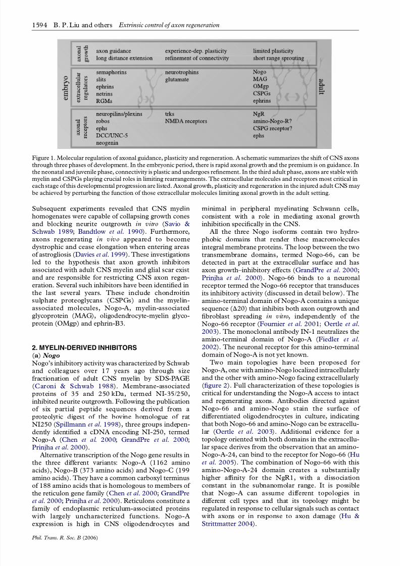
Figure 1. Molecular regulation of axonal guidance, plasticity and regeneration. A schematic summarizes the shift of CNS axons
through three phases of development. In the embryonic period, there is rapid axonal growth and the premium is on guidance. In
the neonatal and juvenile phase, connectivity is plastic and undergoes refinement. In the third adult phase, axons are stable with
myelin and CSPGs playing crucial roles in limiting rearrangements. The extracellular molecules and receptors most critical in
each stage of this developmental progression are listed. Axonal growth, plasticity and regeneration in the injured adult CNS may
be achieved by perturbing the function of those extracellular molecules limiting axonal growth in the adult setting.
1594 B. P. Liu and others Extrinsic control of axon regeneration
Phil. Trans. R. Soc. B (2006)
8/6/2019 Crecimiento Axonal
http://slidepdf.com/reader/full/crecimiento-axonal 3/18
While Nogo-A is strongly expressed by oligoden-drocytes, Nogo isoforms are also present in manyneurons (GrandPre et al . 2000; Hasegawa et al . 2004).Recently, Hunt et al . (2002) showed that Nogo-A islocalized at branch points, varicosities and synapses of
cultured neurons. For the most part, Nogo isoformsand other reticulons are localized to the endoplasmicreticulum of neurons and are likely to be involved inintracellular membrane trafficking. Several lines of evidence suggest that they may regulate Rab- andSNARE-dependent vesicle trafficking in and out of the endoplasmic reticulum (Steiner et al . 2004; Genget al . 2005).
( b) Myelin-associated glycoprotein
MAG, also known as Siglec-4, was first isolated as amajor constituent of CNS myelin in the early 1970s
(Quarles et al . 1972, 1973). MAG is expressed by bothCNS and PNS myelinating glial cells and has beenshown to participate in the formation and maintenanceof the myelin sheath (Schachner & Bartsch 2000).MAG, an immunoglobulin superfamily protein with
a single transmembrane domain, is found on the
surface of myelinating cells and is localized in perinodal
regions (figure 2). There are two MAG isoforms,
S-MAG (short form) and L-MAG (long form), which
differ in their cytoplasmic domain by 44 amino acids
(Tropak et al . 1988). A number of studies indicate thatL-MAG can function as a signalling molecule through
its longer cytoplasmic domain. Antibody ligation of
MAG leads to Fyn kinase activation (Umemori et al .
1994); together, Fyn and MAG play a role in
oligodendrocyte maturation (Biffiger et al . 2000).
Mice lacking MAG exhibit deficits in axoglial
apposition and a degree of slow axonal degeneration.
MAG may also participate in calcium-regulated
signalling events in myelinating glia by interacting
with calcium-binding protein, S100b and calmodulin
(Kursula et al . 2000).
MAG’s capability for inhibiting axonal outgrowth
was identified using different myelin protein fraction-
ation and assay methods (McKerracher et al . 1994;
Mukhopadhyay et al . 1994) than those for Nogo-A.
MAG is capable of inhibiting axonal growth of diverse
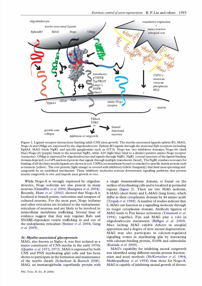
Figure 2. Ligand–receptor interactions limiting adult CNS axon growth. The myelin-associated ligands ephrin-B3, MAG,
Nogo-A and OMgp are expressed by the oligodendrocyte. Ephrin-B3 signals through the neuronal Eph receptors including
EphA4. MAG binds NgR1 and specific gangliosides such as GT1b. Nogo has two inhibitory domains; Nogo-66 (darkblue)/Nogo-24 (purple) binds to the neuronal NgR1, while D20 (light blue) bind to a distinct putative amino-Nogo receptor
(turquoise). OMgp is expressed by oligodendrocytes and signals through NgR1. NgR1 (crystal structure of the ligand-binding
domain depicted) is a GPI-anchored protein that signals through multiple transducers (listed). The NgR1 residues necessary for
binding of all the three myelin ligands are shown in red. CSPGs are membrane bound or attached to specific matrix protein such
as tenascin (yellow). The core protein (light orange) is covered with inhibitory GAGs (burgundy) that limit axon sprouting and
outgrowth by an undefined mechanism. These inhibitory molecules activate downstream signalling pathways that prevent
neurite outgrowth in vitro and impede axon growth in vivo.
Extrinsic control of axon regeneration B. P. Liu and others 1595
Phil. Trans. R. Soc. B (2006)
8/6/2019 Crecimiento Axonal
http://slidepdf.com/reader/full/crecimiento-axonal 4/18
adult neuron types: retinal, superior cervical ganglion,spinal and hippocampal neurons. However, it promotesneurite outgrowth from neonatal dorsal root ganglionneurons (Mukhopadhyay et al . 1994), suggesting thatMAG may provide a permissive substrate for axonoutgrowth during early development, but restrictaberrant growth in adulthood. Since MAG is expressedin both the peripheral and central myelin fractions, itsexpression does not correlate precisely with axonregenerative permissiveness, suggesting that it mightbe less relevant than Nogo-A. However, MAGexpression in Schwann cells might be compensatedfor by more rapid clearance of MAG in the peripherythan in the CNS during Wallerian degeneration.Furthermore, MAG expression is upregulated in
response to cortical axotomy, indicating that itmay limit axonal regeneration after CNS injury(Mingorance et al . 2005).
MAG, like Nogo-66, can exert its axon-inhibitoryactivity by binding to the Nogo-66 receptor (Domeniconiet al . 2002; Liu et al . 2002). MAG also interacts withNogo receptor 2 (NgR2; Venkatesh et al . 2005). As asiglec protein, MAG has affinity for ganglioside GT1b,and in some model systems this glycolipid serves as aprincipal cell surface-binding site for MAG (Vinsonet al . 2001). The role of these gangliosides in mediatingaxonal growth inhibition by MAG is not clear (Tanget al . 1997).
(c) Oligodendrocyte-myelin glycoprotein
By focusing selectively on the glycosylphosphatidylino-sitol (GPI)-linked proteins of CNS myelin as inhibitorsof axonal growth, He and colleagues (Wang et al .
2002b) described the activity of a third myelin protein,termed OMgp. OMgp had been identified by Mickol &Stefansson in 1988 as a peanut agglutinin-bindingprotein derived from CNS white matter (Vourc’h &Andres 2004). It is a 120 kDa glycoprotein with fivetandem leucine-rich repeats that is anchored to theouter leaflet of the plasma membrane by GPI linkagesite (figure 2). Although OMgp was originally purifiedfrom CNS myelin, immunohistochemical studies haveshown that OMgp is also highly expressed on neuronsin the hypothalamus, brainstem, hippocampus andcerebellum (Habib e t al . 1998). Recent studiesdemonstrate that OMgp is localized to perinodal
regions and plays a role in determining the spacing of nodes of Ranvier (Huang et al . 2005). The Nogo-66
receptor has been shown to provide a high-affinitybinding site for OMgp, a site that is competent toinitiate signal transduction to inhibit axonal growth(Wang et al . 2002b).
3. RECEPTOR MECHANISMS FOR MYELININHIBITORS
(a) NgR1
A receptor for the Nogo-66 fragment of Nogo-A,termed Nogo-66 receptor 1 (NgR1; figure 2), was
identified through an expression-cloning strategy withNogo-66 alkaline phosphatase fusion protein as aligand (Fournier et al . 2001). NgR1 binds Nogo-66with nanomolar affinity. As described above, the fusionamino-Nogo-A-24 to Nogo-66 yields a subnanomolar
affinity for NgR1 (Hu et al . 2005). This receptor islocalized to the axolemma and found in many classes of
CNS neurons (Fournier et al . 2001; Wang et al . 2002c;Hasegawa et al . 2004). Transfection of NgR1 confers aNogo-66 response in otherwise non-responsive
neurons, demonstrating that NgR1 functions as areceptor for Nogo-66. The human homologue of mouse NgR1 shares 89% amino acid identity (Fournieret al . 2001). NgR2 and NgR3 were later identified andfound to possess 55% identity in the leucine rich repeat(LRR) domains to NgR1 (Barton et al . 2003; He et al .2003; Lauren et al . 2003; Pignot et al . 2003). NgR2 andNgR3 do not bind to Nogo-66 (Barton et al . 2003).
Surprisingly, two other myelin-associated inhibitors,MAG and OMgp, also bind to NgR1 and can signalthrough this receptor despite their lack of sequencesimilarity to Nogo-A (Domeniconi et al . 2002; Liu et al .2002; Wang et al . 2002b). In the recent work by Gigerand colleagues, it has been reported that MAG canbind to both NgR1 and NgR2, but not NgR3
(Venkatesh et al . 2005). We have since confirmedthat NgR2 has preferential affinity for clustered MAG(B. P. Liu & S. M. Strittmatter 2005, unpublished
studies) as compared to AP-MAG (Barton et al . 2003).The necessity of NgR1 for responses to myelin
ligands has been explored in several in vitro experi-ments. An anti-NgR1 monoclonal antibody, 7E11,blocks Nogo, MAG and OMgp binding to NgR1 andeffectively promotes neurite outgrowth from neuronscultured on CNS myelin substrates (Li et al . 2004b;Schimmele & Pluckthun 2005). Suppression of NgR1expression by siRNA alleviates Nogo-66 and myelininhibition (Ahmed et al . 2005). Truncated NgR1
protein in a soluble or membrane-bound formfunctions as a dominant negative to block myelinligand responses (Fournier et al . 2002; Wang et al .2002a). To explore the necessity of NgR1 genetically,NgR1 K/K mice were generated (Kim et al . 2004;Zheng et al . 2005). Mice lacking NgR1 are viable but
display mild hypoactivity and motor impairment. Of note, Nogo-A levels are increased in these mice,strongly supporting the notion that Nogo/NgR1 forma ligand/receptor pair in vivo. Dorsal root ganglia(DRG) neurons lacking NgR1 do not bind Nogo-66,and their growth cones are not collapsed by Nogo-66 ormyelin (Kim etal . 2004). Despite the potential of NgR2
for binding some forms of MAG (Venkatesh et al .2005), NgR1K/K DRG neurons are not inhibited byMAG (Kim et al . 2004). Studies on a second line of NgR1 K/K mice confirmed the upregulation of Nogo-A, but yielded neurons that were inhibited bymyelin in vitro (Zheng et al . 2005). Differences in myelinpreparation or presentation may account for thesedisparities between in vitro assays (e.g. soluble nativeprotein in growth cone collapse assays versus dried and
partially denatured protein in outgrowth assays). At thispoint, we conclude that NgR1 is partially responsible formediating myelin inhibition of axonal growth.
Owing to the observation that NgR1 can bind
multiple myelin-associated inhibitors, an understand-ing of NgR1 interactions could lead to the design of specific receptor antagonists. NgR1 contains eightLRRs flanked by an amino terminal LRR domain(LRRNT) and a carboxyl terminal cysteine-rich LRR
1596 B. P. Liu and others Extrinsic control of axon regeneration
Phil. Trans. R. Soc. B (2006)
8/6/2019 Crecimiento Axonal
http://slidepdf.com/reader/full/crecimiento-axonal 5/18
(LRRCT). A unique domain consisting of 100 aminoacid residues that link the LRRCT to the GPI anchor isthe least conserved among the three NgR familymembers (Fournier et al . 2002; Wang et al . 2002a;figure 2). We analysed the function of various NgR1domains in outgrowth inhibition. The leucine-richrepeat domain is necessary and sufficient for Nogo-66binding and NgR1 multimerization heterologous cells(Fournier et al . 2002). Viral-mediated expression of mutated NgR1 in primary neurons demonstrates thatthe NgR1 C-terminal domain is required for inhibitorysignalling but not for ligand binding. The NgR1 GPIdomain is not essential for signalling, but appears tofacilitate Nogo-A responses. A similar deletion analysisdemonstrates that the binding of MAG and OMgp to
cell-surface NgR1 also requires the entire LRR regionof NgR1, but not other portions of the protein(Fournier et al . 2002; Barton et al . 2003).
The crystal structure of NgR1 has been determined(Barton et al . 2003; He et al . 2003) and most closelyresembles that of platelet glycoprotein Iba (Huizingaet al . 2002). The LRR segments are aligned in a parallelfashion to form a beta pleated sheet, creating a banana-shaped structure with a concave and convex surfacecapable of protein–protein interactions (Barton et al .2003; He et al . 2003). Once the structure of thereceptor in complex with different ligands has beensolved, then the molecular basis for multiple bindingsites can be fully appreciated.
( b) Signal transduction
NgRs do not possess intracellular signalling domainsand therefore require co-receptors to transduce inhibi-
tory activity. The low-affinity neurotrophic factorreceptor, p75NTR, has been reported to transducegrowth inhibitory signals through a membranecomplex involving NgR1 and another transmembraneprotein, LINGO-1 (Wang et al . 2002a; Wong et al .2002; Mi et al . 2004). Furthermore, TAJ/TROY, anorphan tumour necrosis factor receptor family memberexpressed in many adult neurons, can serve as analternative co-receptor for NgR1, substituting for
p75NTR (Park et al . 2005; Shao et al . 2005). Recently,epidermal growth factor receptor (EGFR) activationhas also been linked to NgR1–ligand binding (Koprivicaet al . 2005). Transactivation of EGFR after NgR1
engagement was shown to be necessary for axonalgrowth inhibition, but the molecular basis for thislinkage has not been defined.
RhoA stimulation is critical for myelin-derivedinhibitory signalling downstream from NgR1 acti-vation. RhoA activation in neurons inhibits neuriteformation by stimulating Rho-associated kinase(ROCK) and actinomyosin contractility. This, inturn, leads to neurite retraction and growth conecollapse. Blocking RhoA activity with either dominant-negative RhoA or C3 exoenzyme, or inhibiting ROCK with Y27632, alleviates myelin substrate inhibitionin vitro ( Jin & Strittmatter 1997; Lehmann et al . 1999;
Vinson et al . 2001; Dergham et al . 2002; Fournier et al .2003; Schweigreiter et al . 2004). Measurement of in vitro RhoA activity in neurons also confirmed RhoAactivation following exposure to myelin components(Winton et al . 2002; Fournier et al . 2003; Schweigreiter
et al . 2004). In one study, modulation of Rho-GDIactivity by p75NTR coupling to NgR1 was linked toRho activation (Yamashita & Tohyama 2003). Further-more, several in vivo spinal cord injury (SCI) studies inwhich RhoA signalling was inhibited showed improvedaxon regeneration and functional recovery (see below).
Protein kinase C (PKC) activation has beenassociated with NgR1-based signalling (Sivasankaranet al . 2004). As for the EGFR activation, the molecularsteps linking PKC activation to NgR1 engagement arenot delineated. Another signalling mechanism reporteddownstream of NgR1 activation is regulated intra-membranous proteolysis of p75NTR (Domeniconiet al . 2005). Sequential extracellular metalloproteinaseand presenilin-dependent g-secretase-dependent clea-
vage of a small fraction of p75NTR has been implicatedin myelin/NgR1 signalling. Whether signalling via Taj isassociated with protease activation has not been studied.
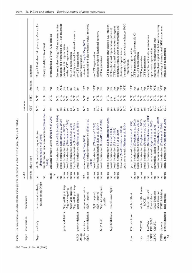
4. MYELIN-TARGETED INTERVENTIONS FOR
AXONAL REGENERATION IN VIVO
(a) Nogo
The identification of Nogo-A as a CNS myelininhibitor of axonal growth in vitro has led to the designof Nogo-targeted intervention studies in vivo (table 1).Rodent models of SCI allow anatomical and functionalassessment of pharmacological and genetic approachesto inhibit the action of Nogo. Schwab and colleaguespioneered the delivery of anti-Nogo strategiesin vivo bytransplanting hybridoma cells modified to secrete theIN-1 monoclonal antibody directed against the amino-terminal of Nogo-A. IN-1-treated animals that under-
went a dorsal over-hemisection injury exhibitedsignificant axonal regeneration and functional recovery(Schnell & Schwab 1990). Further studies from thisgroup illustrated the efficacy of IN-1 and other anti-Nogo preparations in other models of experimentalSCI (Raineteau et al . 1999; Brosamle et al . 2000; Buffoet al . 2000; Merkler et al . 2001; Raineteau et al . 2001;Bareyre et al . 2002, 2004; Fiedler et al . 2002) corticallesions (Wenk et al . 1999; Emerick et al . 2003) and
ischaemic lesions (Papadopoulos et al . 2002) aftermiddle cerebral artery occlusion (MCAO).
Genetic studies have also shed light on the role of Nogo in limiting CNS regeneration. To assess the
sufficiency of Nogo for limiting axonal regeneration,transgenic mice expressing Nogo-A or Nogo-C inperipheral Schwann cells were generated. Axon regen-eration is delayed after sciatic nerve crush in mice withperipheral Nogo-A or C expression, demonstrating thatNogo can partially override the permissiveness of thePNS environment (Pot et al . 2002; Kim et al . 2003a).
While there may be additional myelin-derivedinhibitors, in vitro experiments with cells or myelinfrom Nogo-A null mice have demonstrated thatNogo-A plays a detectably significant role in myelinblockade of axonal outgrowth. CNS myelin preparedfrom Nogo-A/B knockout mice in our laboratory, and
in two other laboratories, exhibits reduced inhibition of neurite outgrowth (Kim et al . 2003b; Simonen et al .2003; Zheng et al . 2003). Nogo-A and Nogo-A/Bmutant mice have normal brain histology and myelindistribution. There are no obvious defects in axons
Extrinsic control of axon regeneration B. P. Liu and others 1597
Phil. Trans. R. Soc. B (2006)
8/6/2019 Crecimiento Axonal
http://slidepdf.com/reader/full/crecimiento-axonal 6/18
8/6/2019 Crecimiento Axonal
http://slidepdf.com/reader/full/crecimiento-axonal 7/18
or oligodendrocytes, demonstrating that Nogo isdispensable (or compensated for) in the developmentof the mouse CNS. In vivo SCI studies with differentNogo-A/B mouse strains have yielded different resultswith regard to the importance of this one molecule inlimiting corticospinal regeneration after SCI (Kim et al .2003b; Simonen et al . 2003; Zheng et al . 2003). AfterSCI, corticospinal axons of one Nogo-A/BK/K strainsprout rostral to a dorsal hemisection and some fibresregenerate into distal cord segments in about 50% of lesioned mice (Kim et al . 2003b). This growth isassociated with recovery of locomotor function.Complimentary studies utilizing the same experimentalparadigm yielded either moderate (Simonen et al .2003) or a complete lack of corticospinal tract (CST)
regeneration (Zheng et a l . 2003). However, theexchange of mouse strains between the laboratoriesinvolved in these studies has made it clear that suchCST growth is only seen with certain Nogo mutantalleles regardless of surgical technique ( J. K. Lee,
J. E. Kim & S. M. Strittmatter, unpublished studies).Moreover, strain background and age modulate theCST regeneration phenotype. The determinants of Nogo-A K/K phenotypic variation and penetranceremain a subject of investigation (see Woolf (2003)for comment).
( b) Myelin-associated glycoprotein
The analysis of mice lacking MAG has provided insightinto its role in the maintenance of myelin architectureas well as its role in limiting plasticity in the adult CNS(table 1). CNS myelin formation is delayed in MAGknockout mice and the cytoplasmic collars of mature
CNS myelin are frequently missing or reduced (Li et al .1994; Montag et al . 1994). Redundant myelination iscommon in the CNS of the adult MAG knockouts(Bartsch et al . 1995) and the mice display oligoden-drogliopathy (Lassmann et al . 1997). Older mutantmice exhibit perturbations in axon integrity (Fruttigeret al . 1995; Weiss et al . 2000). MAG-null mice exhibitlate-onset progressive PNS axonal atrophy and anincreased rate of Wallerian degeneration (Bjartmaret al . 1999). Mice deficient in only L-MAG displaymost of the CNS abnormalities exhibited by the MAGnull mice, but their PNS axon and myelin integrityappeared normal (Fujita et al . 1998). Thus, S-MAG is
sufficient to maintain PNS myelin, and L-MAG may beselectively necessary for signalling within oligodendro-cytes and for the maintenance of CNS myelin.
The role of MAG in limiting in vivo CNS axonregeneration was explored by dorsal hemisection(Bartsch et al . 1995). MAGK/Kmice failed to exhibitaxonal regeneration after SCI. However, in Wld
s
,MAGK/K mice there is a moderate increase inperipheral axonal regeneration (Schafer et al . 1996).The Wld
s mice exhibit slow Wallerian degeneration sothat peripheral nerve myelin (and MAG) persists formuch longer distal to a nerve injury. The absence of adefinitive phenotype may indicate that other ligands
that bind NgR1, such as Nogo or OMgp, maycompensate for the absence of MAG. In order toexplore this issue further, experiments are underway inour laboratory to assess the regenerative phenotype of mice triple mutant for Nogo-A, MAG and OMgp.
(c) NgR1
The Nogo66 receptor 1 binds three myelin-associated
ligands, Nogo, MAG and OMgp. The role of NgR1 in
limiting recovery from CNS trauma has been studied in
several experiments using various strategies in animal
SCI models (table 1). A peptide antagonist of NgR1,
NEP1-40, is a subfragment of Nogo-66 and competes
for binding of Nogo-66 to NgR1 (GrandPreetal
. 2002).The peptide blocks Nogo, but not MAG/OMgp, action
in vitro. This peptide has shown significant efficacy in
promoting regeneration and recovery after experi-
mental SCI (GrandPre et al . 2002). When infused
locally at the time of a lesion (GrandPre et al . 2002) or
subcutaneously after a one week delay (Li & Strittmat-
ter 2003), the NgR1 antagonist promotes corticospinal
and raphespinal axon sprouting. Fibre growth is
correlated with a recovery in locomotor function.
A recombinant protein consisting of the ecto-domain
of the NgR1 (27–310) inhibits Nogo-66, MAG and
OMgp binding to immobilized NgR1 in vitro and
promotes neurite outgrowth on myelin. Transgenicexpression or intrathecal delivery of the NgR1 (27–310)
in vivo significantly improves histological and functional
recovery after dorsal hemisection injury of the thoracic
spinal cord (Li etal . 2004a). Ramer and colleagues have
demonstrated that soluble NgR1-Fc infusion promotes
post-injury sprouting of intact serotonergic and tyrosine
hydroxylase-positive fibres in the spinal cord
(MacDermid et al . 2004). In addition, expression of a
truncated, dominant negative NgR1 in retinal ganglion
cells improves optic nerve regeneration in vivo, particu-
larly when combined with manipulations that enhance
the intrinsic growth state of these neurons (Fischer etal .
2004). The NgR1-ecto protein also promotes cortico-fugal sprouting and functional recovery when infused
i.c.v. after a hemispheral stroke created by MCAO (Lee
etal . 2004). Thus, a range of studies has demonstrated a
role for NgR1 and/or its ligands in limiting axonal
growth in the injured adult rodent CNS.
Two genetic studies assessed the necessity of NgR1
in limiting axonal regeneration after SCI (Kim et al .
2004; Zheng et al . 2005). In both studies, the CST did
not grow after a dorsal hemisection of the spinal cord.
Of note, the upregulation of Nogo-A and the increased
NgR1-independent actions of amino-Nogo may
explain the lack of CST regeneration here as compared
to the NgR1-ecto and NEP1-40 experiments.A complete SCI assessment of NgR1 K/K mice in
our laboratory revealed that the mice recover better
locomotor function significantly when compared to
wild-type littermates (Kim et al . 2004). Mice which are
lacking NgR1 exhibit regeneration of rubrospinal tract
axons and significant sprouting of dorsal raphe fibres
after either dorsal hemisection or complete spinal
transection (Kim et al . 2004). The study from Zheng
et al . (2005) did not assess animals functionally and
concentrated solely on CST anatomy. The recovery of
NgR1K/K mice from focal ischaemic lesions in the
cerebral cortex has also been examined (Leeet al
.2004). The absence of NgR1 allows greater corticofu-
gal fibre sprouting from the non-ischaemic cortex and
greater behavioural recovery. In summary, NgR1 has a
partial role in limiting axonal growth after CNS trauma.
Extrinsic control of axon regeneration B. P. Liu and others 1599
Phil. Trans. R. Soc. B (2006)
8/6/2019 Crecimiento Axonal
http://slidepdf.com/reader/full/crecimiento-axonal 8/18
5. ASTROCYTE-DERIVED INHIBITORS
(a) Chondroitin sulphate proteoglycans
Aside from oligodendroctyes, other CNS glia such asastrocytes and oligodendroctye precursors (OPs) havebeen the focus of many regeneration studies due totheir key role in the establishment of the glial scar. Theglial scar is a dynamic entity that forms at the site of aCNS lesion almost immediately after damage (Berryet al . 1983; Fitch & Silver 1997). Hypertrophicastrocytic processes enmesh the lesion site and beginto express and deposit an inhibitory extracellularmatrix (ECM) consisting primarily of CSPGs, (Levine1994; Davies et al . 1997; 1999, 2004; Fitch et al . 1999;Asher et al . 2000; Bradbury et al . 2002; Jones et al .2002; Tang et al . 2003). This tissue reaction ultimatelyresults in the formation a dense complicated structurethat is exquisitely inhibitory to regenerating axons(Davies et al . 1997, 1999; Bradbury et al . 2002).
The ECM of CNS scar tissue consists principallyof collagens, glycoproteins (such as tenascin-C,
tenascin-R), CSPGs and heparan sulphate proteogly-cans. These components can interact with cell adhesionmolecules and integrins. CSPGs present in the ECM
have been shown to be inhibitory to axonal growth bothin vitro (Snow et al . 1990; McKeon et al . 1991; Dou &Levine 1994; Friedlander et al . 1994; Smith-Thomaset al . 1994, 1995; McKeon et al . 1995; Fidler et al .1999, 2002; Niederost et al . 1999; Ughrin et al . 2003)and in vivo (Davies et al . 1997, 1999; Moon et al . 2001;Bradbury et al . 2002). However, conflicting reportshave emerged to suggest that CSPGs may also promoteneurite outgrowth in vitro (Streit et al . 1993; Faissneret al . 1994) and in vivo (Oakley & Tosney 1991;
Yaginumaet al
. 1991; Joneset al
. 2003), perhapsreflecting the diversity and versatility of this class of proteins. It now appears clear that the chondroitin
sulphate moieties contribute a substantial fraction of the axon-inhibitory function of CSPGs, although thecore proteins may contribute additional specificity and
alternative mechanisms of action.CSPGs comprise a range of complex molecules that
have been shown to have crucial roles in cell adhesion,cell migration, pathfinding and barrier formation(Margolis & Margolis 1997; Bandtlow & Zimmermann2000). CSPGs are characterized as having a centralcore protein to which long unbranched glucosamino-
glycan (GAG) side chains are covalently attached.Modification by epimerases and sulphotransferasesresult in structural heterogeneity of bound GAGs,and dictate their function. The large aggregatingproteoglycans, also known as the hyalectans orlecticans, include aggrecan (Paulsson et al . 1987;
Krueger et al . 1992; Domowicz et al . 1995; Li et al .1996), versican (Krusius et al . 1987; Dours-Zimmer-mann & Zimmermann 1994), the CNS-specific
proteoglycan, neurocan (Rauch etal . 1992; Friedlanderet al . 1994; Grumet et al . 1994; Oohira et al . 1994) andbrevican (Yamada et al . 1994, 1997). They all share anN-terminal globular domain followed by a hyaluronic-
binding region, a C-terminal EGF motif, lectin andcomplement regulatory protein domains and severalregions upon which CS side chains are attached. TheCSPG NG2 shares no sequence homology with thelecticans and is a transmembrane proteoglycan
(Levine & Card 1987; Stallcup & Beasley 1987).Phosphacan/DSD-1 is the extracellular CSPG domainof a receptor-type protein tyrosine phosphatase(Grumet et al . 1993, 1994; Maeda et al . 1994; Maurelet al . 1994).
( b) Neurocan
The prototypic lectican, neurocan, is produced as a275 kDa secreted protein that can be proteolyticallyprocessedinto two smaller fragments (Rauch etal . 1992;Matsui et al . 1994). Both the C-terminal (neurocan-C)and N-terminal fragment (neurocan-130) aresulphated. They are deposited by astrocytes andoligodendrocyte progenitors (OPs) exclusively in theCNS, and are concentrated in white matter. Neurocanhas been shown to be inhibitory to neurite outgrowthin vitro (Asher et al . 2000). In keeping with its role as akey inhibitor of axonal regeneration, neurocanexpression is elevated at the site of mechanical (Asheret al . 2000; Tang et al . 2003; Davies et al . 2004) orischaemic (Deguchi etal .2005) injury to the adult CNS.Neurocan is known to interact with NCAM, Ng-CAM/L1, TAG-1 and tenascin (Friedlander etal . 1994; Milevet al . 1994, 1996, 1998) and to inhibit axon growth byNg-CAM/L1. Neurocan has also been implicated inpathfinding during development as it is transientlyexpressed in the optic chiasm during retino-tectalmapping and in the dorsal midline of the spinal cord
(Sango et al . 2003; Leung et al . 2004; Popp et al . 2004).However, mice which are null mutant for neurocanappear to develop normally, with only mild LTP deficits(Zhou etal . 2001), illustrating that neurocan may have aredundant or subtle role during development.
(c) Versican
Versican is a large proteoglycan, which has the same Nand C terminal motifs as the other members of thelectican family, but also contains unique a and b
domains. The mRNA is alternatively spliced toresult in three isoforms of versican, full length V0,which possess both a and b domains, V1, which lacksthe a domain, and V2, which lacks the b domain(Dours-Zimmermann & Zimmermann 1994; Milevet al . 1998; Schmalfeldt et al . 1998). Recently, versicanwas shown to be expressed in a subset of interneuronsin cytoarchitectonically distinct laminae of the embryo-
nic tectum (Yamagata & Sanes 2005). It was theorizedthat graded versican expression in the tectum mightrestrict retinal axons from inappropriate pathways.Versican expression has also been implicated in neuralcrest cell migration (Perissinotto et al . 2000). The roleof versican in restricting axonal projections and cellmigration during development may parallel a role inrestricting plasticity and regeneration after injury to theadult CNS. Expression of versican is elevated withintwo weeks after SCI and remains chronically elevated inthe lesion perimeter ( Jones et al . 2003; Tang etal . 2003).
(d) Phosphacan
Phosphacan is another CSPG found throughout theadult CNS (Snow et al . 1990; Maurel et al . 1994; Tanget al . 2003). It is expressed in secreted and membrane-bound forms (RPTP-b). Phosphacan has been shownto block the growth-promoting effects of N-CAM,
1600 B. P. Liu and others Extrinsic control of axon regeneration
Phil. Trans. R. Soc. B (2006)
8/6/2019 Crecimiento Axonal
http://slidepdf.com/reader/full/crecimiento-axonal 9/18
TAG-1 and tenascin. Phosphacan is highly expressedby radial glial cells and consequently has been shown tobe crucial in the oriented movement of post-mitoticcells during cortical development. Phosphacanexpression after SCI initially declines within the firstfew weeks post-injury and peaks after one month, incontrast to neurocan expression, which is elevatedwithin days of the injury (Tang et al . 2003). Conse-quently, protracted phosphacan expression maycontribute to chronic inhibition of axonal regeneration.
(e) NG2
NG2 is a 300 kDa proteoglycan, an integral membraneprotein cloned in 1991 by Stallcup and colleagues(Nishiyama et al . 1991) characterized by a short 25residue transmembrane domain, a cytoplasmic domainof 76 amino acids and a large extracellular domain(2225 residues), with a single chondroitin sulphate-binding site at serine 999 (Stallcup & Dahlin-Huppe2001). NG2 was initially identified as a cell surface
antigen of neural tumour-derived cell lines that hadproperties intermediate between neurons and glial cells(therefore nerve glia (NG)). Later investigationsrevealed that NG2 was expressed by a subset of smallstellate cells within the CNS parenchyma and thoughtto represent a group of progenitors. Subsequent studiesinspired by the identification of O2A progenitors byRaff et al . (1983, 1998) confirmed the identity of NG2expressing cells as OPs (Levine & Card 1987; Stallcup &Beasley 1987).
NG2 was shownto be inhibitoryto neurite outgrowthin vitro independent of the integrity of its chondroitinsulphate side chain as pre-treatment of NG2-rich
substrata with chondroitinase ABC (ChABC) failed toalleviate inhibition. However, treatment of thesecultures with polyclonal antibodies raised against NG2was able to partially reverse inhibition (Dou & Levine1994). Similarly, membrane preparations from culturedOPs were inhibitory to neurite outgrowth fromcerebellar explants (Chen et al . 2002) in a mannerreversed in the presence of NG2 polyclonal antibodies.A more thorough investigation of three separate
domains within the extracellular portion of themembrane-bound form of NG2 has implicated inde-pendent inhibitory regions which may have implicationson the function of this CSPG in a membrane bound or
secreted form (Ughrin et al . 2003).NG2 is upregulated at the lesion margins after brainlesion (Levine 1994), spinal cord hemisection ( Joneset al . 2002) and spinal transection (de Castro et al .2005), areas correlated with axon regeneration failureand consistent with the role for NG2 in limiting axonalregeneration. Indeed, preliminary reports infusingmonoclonal antibodies against NG2 appeared toenhance primary afferent regeneration after experi-mental SCI (Tang et al . 2003). However, mice nullmutant for NG2 failed to show enhanced axonalregeneration after complete transection (de Castroet al . 2005). Moreover, wild-type mice illustrated a
more robust axonal regeneration. These results impli-cate a detrimental effect of the loss of NG2. Therefore,in the absence of a specific receptor, the action of NG2may be mediated by interactions with other ECMmolecules (Burg et al . 1996; Dou & Levine 1997).
6. PROTEOGLYCAN-TARGETED INTERVENTION
FOR AXONAL REGENERATION
Early in vitro reports identified CSPGs as the potentinhibitory species expressed by astrocytes (Snow et al .1990; McKeon et al . 1991; Dou & Levine 1994;
Smith-Thomas et al . 1994). Subsequent investigationsrevealed that cultured retinal ganglion cells werecapable of enhanced growth on CSPG-rich substrate,if the substrate was pre-treated with the bacterialenzyme ChABC (McKeon et al . 1995). ChABCliberates CS GAGs from CSPG core proteins andthus ultimately identified the GAG moiety of CSPGs ascrucial in mediating axonal growth arrest in vitro. Morerecently, ChABC has been used with great effect in vivo
to enhance regeneration of damaged CNS pathways.ChABC was capable of enhancing regeneration of microlesioned nigrostriatal axons back to their targets(Moon et al . 2001). Intrathecal delivery of ChABCrestored functional corticospinal connectivity afterbilateral dorsal column lesion in the rat, with
concomitant restoration of sensory and propriospinalfunction (Bradbury et al . 2002). Furthermore, Clarke’snucleus neurons regenerate into a peripheral nerve
graft (Yick et al . 2000) and past the lesion site afterdorsal hemisection (Yick et al . 2003). These studieshave spearheaded the use of ChABC in a number of combinatorial studies that have used a multi-dimensional stratagem to overcome regeneration fail-ure. ChABC has been successfully combined withSchwann cell seeded guidance channels (Chau et al .2004), Schwann cell and olfactory ensheathing cellbridges (Fouad et al . 2005) and neural stem/progenitorcells with resultant enhanced axonal regeneration. An
alternative strategy to the use of ChABC in reducingthe effects of GAG moiety-associated inhibition was theuse of a DNA enzyme that targets the mRNA of xylosyltransferase-1, an enzyme critical for the additionof GAG side chains to core proteins. Administration of this enzyme reduced the expression of GAGs on CSPG
core proteins on astroctyes in vitro and in areasundergoing reactive gliosis after SCI in vivo (Grimpe &Silver 2004).
Another approach to targeting CSPG-associatedinhibition has focused on modulating the ability of the damaged CNS glial cells to proliferate and produceinhibitory ECM (Rhodes et al . 2003). The molecular
mechanisms that cause upregulation of CSPG afterinjury have been partially described (Asher et al . 2000).Intraspinal delivery of the small CSPG, decorin, afterspinal stab injury has been shown to negate theupregulation of neurocan, phosphacan and NG2,suppress the formation of the glial limitans of the scarand enhance axon regeneration while reducingexpression of the cytokine transforming growth factor(TGF) beta (Davies et al . 2004). Inflammation is
known to exacerbate the production of CSPGs (Asheret al . 2000) and therefore anti-inflammatory therapyprovides another mechanism whereby extrinsic inhibi-tory factors can be controlled.
The diverse structure and function of CSPGs hasmade them complicated targets for intervention, and todate no general chondroitin sulphate receptors have beenidentifiedthat mediate their function.Activationof RhoAis a necessary step in mediating CSPG-associated
Extrinsic control of axon regeneration B. P. Liu and others 1601
Phil. Trans. R. Soc. B (2006)
8/6/2019 Crecimiento Axonal
http://slidepdf.com/reader/full/crecimiento-axonal 10/18
inhibition in vitro. Several laboratories have reporteddisinhibition of neurite outgrowth on CSPG-richsubstrates in the presence of ROCK inhibitors (Borisoff et al . 2003; Monnier et al . 2003). Therefore, blockadeof the Rho/ROCK axis holds the potential to blocksignalling by myelin, CSPG and ephrins. In vivo studiestargeting RhoA with C3 exoenzyme have yielded littleevidence of regeneration (Fournier et al . 2003) exceptin studies with a modified cell permanent version of theprotein (Winton et al . 2002). ROCK inhibitors haveyielded a degree of axonal growth and functionalimprovement in several studies, but a mixture of beneficial and deleterious effects may ultimately limittheir usefulness (Borisoff et al . 2003; Fournier et al .2003; Park et al . 2005). One study has implicated PKC
activation as critical in transducing CSPG-mediatedRhoA activation and hence growth cone collapse(Sivasankaran et al . 2004). Intrathecal delivery of thePKC inhibitor Go6976 resulted in modest regen-eration of ascending dorsal column afferents afterdorsal hemisection (Sivasankaran et al . 2004). Morerecently, EGF receptor activation (Koprivica et al .2005) has emerged as another intermediate in transdu-cing the action of both CSPG and myelin inhibitoryfunction. These investigators showed that inhibitionof EGF kinase function was capable of ablatingthe neurite outgrowth–inhibitory effects of CSPGs.Furthermore, local delivery of EGF receptor inhibitorsenhanced axon growth after optic nerve crush.
7. EPHRINS: DEVELOPMENT, MYELIN AND
SCAR TISSUE
The ephrins and their cognate Eph receptors playmajor roles in axonal pathfinding and target recog-nition during CNS development (Klein 2004). Recentstudies of Eph/ephrins expression after CNS injuryhave elucidated a range of roles for this receptor–ligandfamily after CNS trauma. Several of these proteinsappear to be expressed by reactive astrocytes.Bidirectional signalling between ephrin-B2-expressingastrocytes and EphB2-expressing meningeal fibroblasts
may regulate the interaction between these two celltypes during CNS scar formation (Bundesen et al .2003). Since scar tissue is clearly inhibitory for axonaloutgrowth, this ephrin/Eph interaction may seconda-
rily alter axonal growth after trauma. There is alsoevidence that EphA4 is upregulated in reactiveastrocytes after SCI. In mice lacking EphA4, astro-gliosis is reduced and inhibition of axonal growth bothin vivo and in vitro is reduced. Furthermore, functionalrecovery from spinal hemisection is enhanced inEphA4K/K mice (Goldshmit et al . 2004).
Ephrins have also been implicated in myelin-dependent inhibition of axonal growth. Ephrin-B3 isselectively expressed on mature myelinating oligoden-drocytes (Benson et al . 2005). Furthermore, myelinderived from ephrin-B3 knockout mice is less inhibi-tory than wild-type myelin for neurite outgrowth.
During development, ephrin-B3 is known to functionas a midline glial repellent for descending EphA4receptor-expressing corticospinal tract axons (Kullanderet al . 2001). Thus, at least for EphA4-expressing adultaxonal tracts, ephrin-B3 expression in oligodendrocytes
probably contributes to limited adult CNS axonalgrowth. In vitro studies with tissue and cells from genetargeted lines suggest that both ephrin-B3 and NgR1ligands play detectable roles in CNS myelin inhibitionof adult axon growth (Benson et al . 2005).
8. SEMAPHORINS AND OTHER AXON GUIDANCE
MOLECULES: DEVELOPMENT, MYELINAND SCAR TISSUE
Like the ephrins, the semaphorins have prominentaxon repulsive functions during early neuronaldevelopment (Nakamura et al . 2000; Fujisawa 2004).These functions are mediated largely through plexin-class receptors. A number of soluble semaphorinligands use neuropilins as high-affinity binding co-receptors together with plexins (He & Tessier-Lavigne1997; Kolodkin et al . 1997; Winberg et al . 1998;Takahashi et al . 1999; Tamagnone et al . 1999). Ourunderstanding of the role of semaphorins and theirreceptors in limiting adult axonal growth is incomplete.
Expression of inhibitory sema3 proteins by meningealfibroblasts at sites of CNS scar has been documented,but their functional role is unclear (De Winter et al .2002). Oligodendrocytes, especially those near injurysites, have been to shown to express a transmembranesema4D, which is capable of inhibiting axonal growthfrom several neuronal types (Moreau-Fauvarque et al .2003). In optic nerve, oligodendrocytes also expressanother axon-inhibitory transmembrane semaphorin,sema5A (Oster et al . 2003).
Netrins are secreted proteins with both axonalattractive and repulsive roles during development(Tessier-Lavigne & Goodman 1996). In the adult,
netrin-1 expression has been documented in matureoligodendrocytes (Manitt et a l . 2001). Since theinhibitory netrin receptors, Unc5H1-3, are expressedin adult spinal cord, this protein may also have anaxonal growth-limiting action after CNS injury (Manittet al . 2004).
9. ROLE OF MYELIN/CSPG INHIBITORS
IN NORMAL PHYSIOLOGY
In vivo injury studies demonstrate a pathological role formyelin/NgR1 and CSPGs in limiting axonal growth inthe adult CNS after injury. An obvious issue is the
physiological function of these molecular pathways.Since local sprouting and plasticity from uninjuredfibres contribute to the positive effects of NgR1 andCSPG perturbations in the recovery from injury, it isimportant to consider whether these targets mightparticipate in physiological, experience-dependentplasticity. The visual system provides an ideal in vivo
model in which this question can be addressed (McGeeet al . 2005). It is well established that the visual systemexhibits a striking ability to rearrange in response tosensory input during a defined window in the latejuvenile phase of development (Hensch 2005; Hensch &Fagiolini 2005; Taha & Stryker 2005; Yang et al . 2005).
Monocular deprivation (MD) during this time periodleads to reduce responsiveness to the deprived eyewithout any damage to the nervous system itself. Thereduced visual acuity in the deprived eye has a clinicalcorrelation in amblyopia. After the critical period for
1602 B. P. Liu and others Extrinsic control of axon regeneration
Phil. Trans. R. Soc. B (2006)
8/6/2019 Crecimiento Axonal
http://slidepdf.com/reader/full/crecimiento-axonal 11/18
ocular dominance plasticity (P19-32 in mice), MDproduces only minimal ‘sub-threshold’ changes incortical function (Hensch 2005) that can be detectedby visually evoked potentials.
In mice lacking NgR1, the visual cortex developsnormally, exhibiting electrical response properties andcortical maps indistinguishable from those in wild-typemice (McGee et al . 2005). Closing one eye for severaldays shifts the responses to the open eye to the samedegree in both NgR1 K/K and wild-type mice duringthe critical period. However, at P45 or P120, well afterthe closure of the critical period, the NgR1K/K visualcortex continues to exhibit full ocular dominanceplasticity to 4 day MD while the wild-type cortexexhibits no plasticity of single unit recordings to such
deprivation. Thus, NgR1 plays a role in the normalclosure of the critical period. At the anatomical andcellular level, the basis for continued ocular dominanceplasticity in the NgR1K/K mouse visual cortex is notyet defined.
CSPGs have also been implicated in limitingplasticity and closing of the critical period (Pizzorussoet al . 2002). These ECM components are expressedmost highly in condensed lattice-like structures calledperineuronal nets (PNNs) surrounding parvalbumin-positive GABAergic neurons, and reach maximumexpression as the critical period closes. Removal of GAG side chains from CSPGs by enzymatic digestionwith chondroitinase ABC partially restores oculardominance plasticity to older animals after closure of the critical period (Pizzorusso et al . 2002). Twodifferent mechanisms for CSPG-mediated plasticityhave been proposed. In one, CSPG may dampenstructural changes through their inhibitory actions onneurite outgrowth. In the other, the negatively chargedECM milieu created by these molecules maybuffer requisite ionic and transmitter concentrations(Pizzorusso et al . 2002; Hensch & Fagiolini 2005).Structures akin to PNNs have been located in thedorsal horn of the spinal cord termed periaxonal netswhere they appear to serve a similar function to theircounterparts in the cortex. Primary afferent terminalswithin spinal grey matter are decorated with CSPGs.Intraspinal delivery of ChABC degrades these intrinsi-cally expressed CSPGs and encourages primaryafferent re-organization after deafferentation of lesionsto the spinal cord ( W. B. J. Cafferty & S. McMahon2005, unpublished observations).
10. SUMMARY
Historically, the failure of axonal regeneration withinthe damaged CNS has been ascribed to two limitingfactors, an intrinsically reduced growth capacity of mature CNS neurons and the presence of inhibitorymolecules produced by non-neuronal cells unique tothe CNS: oligodendrocytes and astrocytes. CNS axonshave been shown to grow considerable distances in vivo
if presented with a permissive environment lacking
oligodendrocytes and astrocytes. The most instructivedemonstration of this capacity came from seminalexperiments illustrating the ability of damaged CNSaxons to regenerate into tissue bridges consisting of transplanted peripheral nerve (David & Aguayo 1981;
Richardson et al . 1982). Thus, while adult CNSneurons may have a diminished growth capacity(Kalil & Reh 1982; Schreyer & Jones 1983; Schreyer &Skene 1993; Goldberg & Barres 1998), they retain
adequate intrinsic growth potential to extend if theenvironment is acceptable.
Much research over the last decade has focused on
identifying the factors produced by CNS glial cells thatarrest axonal sprouting and regeneration. Earlierstudies from Schwab and colleagues identifieddamaged CNS myelin as a substrate inhibitory toinjured axons (Schwab & Thoenen 1985; Schwab &Caroni 1988; Savio & Schwab 1989). Later studiesidentified Nogo, MAG and OMgp as factors present inCNS myelin thatmediate its inhibitory nature(McGee &Strittmatter 2003). All the three of these proteins caninteract with an axonal NgR1 protein. Perturbation of these molecules or downstream signalling eventspermits a degree of CNS axonal growth and repair.
Derived primarily from the reactive astrocytes,CSPGs also play a crucial inhibitory role in the adultCNS (Silver & Miller 2004; Carulli et al . 2005). In vivo
evidence indicates that CSPG digestion can promoteCNS axonal growth and functional recovery (Bradburyet al . 2002). A number of protein factors important for
embryonic axonal guidance may also contribute tolimiting axonal growth in the adult. While this arearequires further investigation, the data implicatingmembers of the ephrin family are the most advanced(Goldshmit et al . 2004; Benson et al . 2005).
Those myelin proteins and CSPGs that play aprominent role in limiting axonal growth after CNSinjury also play a crucial role during late juvenile
development to consolidate connectivity optimized byexperience-dependent plasticity in the nervous system.Thus, the evidence for improved functional recoveryafter adult CNS injury may be ascribed to the ability of myelin or CSPG perturbations to reverse the last stagesof brain development and to reinstate the plasticity of the juvenile brain. In this sense, further investigationsof the penultimate stages of brain development may bemost useful for developing therapeutic interventionsfor adult CNS injuries.
This work was supported in part by grants from the NationalInstitutes of Health, the Christopher Reeve ParalysisFoundation and the Falk Medical Research Trust to S.M.S.
REFERENCESAhmed, Z., Dent, R. G., Suggate, E. L., Barrett, L. B.,
Seabright, R. J., Berry, M. & Logan, A. 2005 Disinhibition
of neurotrophin-induced dorsal root ganglion cell neurite
outgrowth on CNS myelin by siRNA-mediated knock-
down of NgR, p75NTR and Rho-A. Mol. Cell. Neurosci.
28, 509–523. (doi:10.1016/j.mcn.2004.11.002)
Asher, R. A. et al . 2000 Neurocan is upregulated in injured
brain and in cytokine-treated astrocytes. J. Neurosci. 20,
2427–2438.
Bandtlow, C. E. & Zimmermann, D. R. 2000 Proteoglycans
in the developing brain: new conceptual insights for oldproteins. Physiol. Rev. 80, 1267–1290.
Bandtlow, C., Zachleder, T. & Schwab, M. E. 1990
Oligodendrocytes arrest neurite growth by contact inhi-
bition. J. Neurosci. 10, 3837–3848.
Extrinsic control of axon regeneration B. P. Liu and others 1603
Phil. Trans. R. Soc. B (2006)

8/6/2019 Crecimiento Axonal
http://slidepdf.com/reader/full/crecimiento-axonal 12/18
Bareyre, F. M., Haudenschild, B. & Schwab, M. E. 2002
Long-lasting sprouting and gene expression changes
induced by the monoclonal antibody IN-1 in the adult
spinal cord. J. Neurosci. 22, 7097–7110.
Bareyre, F. M., Kerschensteiner, M., Raineteau, O.,
Mettenleiter, T. C., Weinmann, O. & Schwab, M. E.
2004 The injured spinal cord spontaneously forms a new
intraspinal circuit in adult rats. Nat. Neurosci. 7, 269–277.
(doi:10.1038/nn1195)Barton, W. A., Liu, B. P., Tzvetkova, D., Jeffrey, P. D., Fournier,
A. E., Sah, D., Cate,R., Strittmatter, S. M. & Nikolov,D. B.
2003 Structure and axon outgrowth inhibitor binding of the
Nogo-66 receptor and related proteins. EMBO J. 22,
3291–3302. (doi:10.1093/emboj/cdg325)
Bartsch, U. et al . 1995 Lack of evidence that myelin-
associated glycoprotein is a major inhibitor of axonal
regeneration in the CNS. Neuron 15, 1375–1381. (doi:10.
1016/0896-6273(95)90015-2)
Benson, M. D., Romero, M. I., Lush, M. E., Lu, Q. R.,
Henkemeyer, M. & Parada, L. F. 2005 Ephrin-B3 is a
myelin-based inhibitor of neurite outgrowth. Proc. Natl
Acad. Sci. USA 102, 10 694–10 699. (doi:10.1073/pnas.
0504021102)Berry, M., Maxwell, W. L., Logan, A., Mathewson, A.,
McConnell, P., Ashhurst, D. E. & Thomas, G. H. 1983
Deposition of scar tissue in the central nervous system.
Acta Neurochir. 32(Suppl. (Wien)), 31–53.
Biffiger, K., Bartsch, S., Montag, D., Aguzzi, A., Schachner,
M. & Bartsch, U. 2000 Severe hypomyelination of the
murine CNS in the absence of myelin-associated glyco-
protein and fyn tyrosine kinase. J. Neurosci. 20,
7430–7437.
Bjartmar, C., Yin, X. & Trapp, B. D. 1999 Axonal pathology
in myelin disorders. J. Neurocytol. 28, 383–395. (doi:10.
1023/A:1007010205037)
Bomze, H. M., Bulsara, K. R., Iskandar, B. J., Caroni, P. &
Skene, J. H. 2001 Spinal axon regeneration evoked by
replacing two growth cone proteins in adult neurons. Nat.
Neurosci. 4, 38–43. (doi:10.1038/82881)
Borisoff, J. F., Chan, C. C., Hiebert, G. W., Oschipok, L.,
Robertson, G. S., Zamboni, R., Steeves, J. D. & Tetzlaff,
W. 2003Suppression of Rho-kinase activity promotes axonal
growthon inhibitory CNSsubstrates. Mol. Cell.Neurosci.22,
405–416. (doi:10.1016/S1044-7431(02)00032-5)
Bradbury, E. J., Moon, L. D., Popat, R. J., King, V. R.,
Bennett, G. S., Patel, P. N., Fawcett, J. W. & McMahon,
S. B. 2002 Chondroitinase ABC promotes functional
recovery after spinal cord injury. Nature 416, 636–640.
(doi:10.1038/416636a)
Brosamle, C., Huber, A. B., Fiedler, M., Skerra, A. &Schwab, M. E. 2000 Regeneration of lesioned corticosp-
inal tract fibers in the adult rat induced by a recombinant,
humanized IN-1 antibody fragment. J. Neurosci. 20,
8061–8068.
Buffo, A., Zagrebelsky, M., Huber, A. B., Skerra, A., Schwab,
M. E., Strata, P. & Rossi, F. 2000 Application of
neutralizing antibodies against NI-35/250 myelin-associ-
ated neurite growth inhibitory proteins to the adult rat
cerebellum induces sprouting of uninjured Purkinje cell
axons. J. Neurosci. 20, 2275–2286.
Bundesen, L. Q., Scheel, T. A.,Bregman, B.S. & Kromer, L. F.
2003 Ephrin-B2 and EphB2 regulation of astrocyte–
meningeal fibroblast interactions in response to spinal cord
lesions in adult rats. J. Neurosci. 23, 7789–7800.Burg, M. A., Tillet, E., Timpl, R. & Stallcup, W. B. 1996
Binding of the NG2 proteoglycan to type VI collagen and
other extracellular matrix molecules. J. Biol. Chem. 271,
26 110–26 116. (doi:10.1074/jbc.271.42.26110)
Caroni, P. & Schwab, M. E. 1988 Two membrane protein
fractions from rat central myelin with inhibitory properties
for neurite growth and fibroblast spreading. J. Cell Biol.
106, 1281–1288. (doi:10.1083/jcb.106.4.1281)
Carulli, D., Laabs, T., Geller, H. M. & Fawcett, J. W. 2005
Chondroitin sulfate proteoglycans in neural development
and regeneration. Curr. Opin. Neurobiol. 15, 116–120.
(doi:10.1016/j.conb.2005.03.018)
Chau, C. H., Shum, D. K., Li, H., Pei, J., Lui, Y. Y., Wirthlin,L., Chan, Y. S. & Xu, X. M. 2004 Chondroitinase ABC
enhances axonal regrowth through Schwann cell-seeded
guidance channels after spinal cord injury. FASEB J. 18,
194–196.
Chen, M. S., Huber, A. B., van der Haar, M. E., Frank, M.,
Schnell, L., Spillmann, A. A., Christ, F. & Schwab, M. E.
2000 Nogo-A is a myelin-associated neurite outgrowth
inhibitor and an antigen for monoclonal antibody IN-1.
Nature 403, 434–439. (doi:10.1038/35000219)
Chen, Z. J., Ughrin, Y. & Levine, J. M. 2002 Inhibition of
axon growth by oligodendrocyte precursor cells. Mol. Cell.
Neurosci. 20, 125–139. (doi:10.1006/mcne.2002.1102)
David, S. & Aguayo, A. J. 1981 Axonal elongation into
peripheral nervous system ‘bridges’ after central nervous
system injury in adult rats. Science 214, 931–933.
Davies, S. J., Fitch, M. T., Memberg, S. P., Hall, A. K.,
Raisman, G. & Silver, J. 1997 Regeneration of adult axons
in white matter tracts of the central nervous system.
Nature 390, 680–683.
Davies, S. J., Goucher, D. R., Doller, C. & Silver, J. 1999
Robust regeneration of adult sensory axons in degenerat-
ing white matter of the adult rat spinal cord. J. Neurosci.
19, 5810–5822.
Davies, J. E., Tang, X., Denning, J. W., Archibald, S. J. &
Davies, S. J. 2004 Decorin suppresses neurocan, brevican,
phosphacan and NG2 expression and promotes axon
growth across adult rat spinal cord injuries. Eur.
J. Neurosci. 19, 1226–1242. (doi:10.1111/j.1460-9568.
2004.03184.x)
de Castro Jr, R., Tajrishi, R., Claros, J. & Stallcup, W. B. 2005
Differential responses of spinal axons to transection:
influence of the NG2 proteoglycan. Exp. Neurol. 192,
299–309. (doi:10.1016/j.expneurol.2004.11.027)
De Winter, F., Holtmaat, A. J. & Verhaagen, J. 2002
Neuropilin and class 3 semaphorins in nervous system
regeneration. Adv. Exp. Med. Biol. 515, 115–139.
Deguchi, K. et al . 2005 Expression of neurocan after transient
middle cerebral artery occlusion in adult rat brain. Brain
Res. 1037, 194–199. (doi:10.1016/j.brainres.2004.12.
016)
Dergham, P., Ellezam, B., Essagian, C., Avedissian, H.,
Lubell, W. D. & McKerracher, L. 2002 Rho signaling
pathway targeted to promote spinal cord repair. J. Neurosci. 22, 6570–6577.
Domeniconi, M. et al . 2002 Myelin-associated glycoprotein
interacts with the Nogo66 receptor to inhibit neurite
outgrowth. Neuron 35, 283–290. (doi:10.1016/S0896-
6273(02)00770-5)
Domeniconi, M., Zampieri, N., Spencer, T., Hilaire, M.,
Mellado, W., Chao, M. V. & Filbin, M. T. 2005 MAG
induces regulated intramembrane proteolysis of the p75
neurotrophin receptor to inhibit neurite outgrowth.
Neuron 46, 849–855. (doi:10.1016/j.neuron.2005.05.029)
Domowicz, M., Li, H., Hennig, A., Henry, J., Vertel, B. M. &
Schwartz, N. B. 1995 The biochemically and immunolo-
gically distinct CSPG of notochord is a product of the
aggrecan gene. Dev. Biol. 171, 655–664. (doi:10.1006/dbio.1995.1312)
Dou, C. L. & Levine, J. M. 1994 Inhibition of neurite growth
by the NG2 chondroitin sulfate proteoglycan. J. Neurosci.
14, 7616–7628.
1604 B. P. Liu and others Extrinsic control of axon regeneration
Phil. Trans. R. Soc. B (2006)
8/6/2019 Crecimiento Axonal
http://slidepdf.com/reader/full/crecimiento-axonal 13/18
Dou, C. L. & Levine, J. M. 1997 Identification of a neuronal
cell surface receptor for a growth inhibitory chondroitin
sulfate proteoglycan (NG2). J. Neurochem. 68, 1021–1030.
Dours-Zimmermann, M. T. & Zimmermann, D. R. 1994 A
novel glycosaminoglycan attachment domain identified in
two alternative splice variants of human versican. J. Biol.
Chem. 269, 32 992–32 998.
Emerick, A. J., Neafsey, E. J., Schwab, M. E. & Kartje, G. L.
2003 Functional reorganization of the motor cortex inadult rats after cortical lesion and treatment with
monoclonal antibody IN-1. J. Neurosci. 23, 4826–4830.
Faissner, A., Clement, A., Lochter, A., Streit, A., Mandl, C.
& Schachner, M. 1994 Isolation of a neural chondroitin
sulfate proteoglycan with neurite outgrowth promoting
properties. J. Cell Biol. 126, 783–799. (doi:10.1083/jcb.
126.3.783)
Fidler, P. S. etal . 1999 Comparing astrocytic cell lines that are
inhibitory or permissive for axon growth: the major axon-
inhibitory proteoglycan is NG2. J. Neurosci. 19,
8778–8788.
Fiedler, M., Horn, C., Bandtlow, C., Schwab, M. E. &
Skerra, A. 2002 An engineered IN-1 F(ab) fragment with
improved affinity for the Nogo-A axonal growth inhibitor
permits immunochemical detection and shows enhanced
neutralizing activity. Protein Eng. 15, 931–941. (doi:10.
1093/protein/15.11.931)
Fischer, D., He, Z. & Benowitz, L. I. 2004 Counteracting the
Nogo receptor enhances optic nerve regeneration if retinal
ganglion cells are in an active growth state. J. Neurosci. 24,
1646–1651. (doi:10.1523/JNEUROSCI.5119-03.2004)
Fitch, M. T. & Silver, J. 1997 Glial cell extracellular matrix:
boundaries for axon growth in development and regen-
eration. Cell Tissue Res. 290, 379–384. (doi:10.1007/
s004410050944)
Fitch, M. T., Doller, C., Combs, C. K., Landreth, G. E. &
Silver, J. 1999 Cellular and molecular mechanisms of glial
scarring and progressive cavitation: in vivo and in vitro
analysis of inflammation-induced secondary injury after
CNS trauma. J. Neurosci. 19, 8182–8198.
Fouad, K., Klusman, I. & Schwab, M. E. 2004 Regenerating
corticospinal fibers in the Marmoset (Callitrix jacchus)
after spinal cord lesion and treatment with the anti-Nogo-
A antibody IN-1. Eur. J. Neurosci. 20, 2479–2482. (doi:10.
1111/j.1460-9568.2004.03716.x)
Fouad, K., Schnell, L., Bunge, M. B., Schwab, M. E.,
Liebscher, T. & Pearse, D. D. 2005 Combining Schwann
cell bridges and olfactory-ensheathing glia grafts with
chondroitinase promotes locomotor recovery after
complete transection of the spinal cord. J. Neurosci. 25,
1169–1178. (doi:10.1523/JNEUROSCI.3562-04.2005)
Fournier, A. E., GrandPre, T. & Strittmatter, S. M. 2001
Identification of a receptor mediating Nogo-66 inhibitionof axonal regeneration. Nature 409, 341–346. (doi:10.
1038/35053072)
Fournier, A. E., Gould, G. C., Liu, B. P. & Strittmatter, S. M.
2002 Truncated soluble Nogo receptor binds Nogo-66
and blocks inhibition of axon growth by myelin.
J. Neurosci. 22, 8876–8883.
Fournier, A. E., Takizawa, B. T. & Strittmatter, S. M. 2003
Rho kinase inhibition enhances axonal regeneration in the
injured CNS. J. Neurosci. 23, 1416–1423.
Fox, K. & Wong, R. O. 2005 A comparison of experience-
dependent plasticity in the visual and somatosensory
systems. Neuron 48, 465–477. (doi:10.1016/j.neuron.
2005.10.013)
Friedlander, D. R., Milev, P., Karthikeyan, L., Margolis,R. K., Margolis, R. U. & Grumet, M. 1994 The neuronal
chondroitin sulfate proteoglycan neurocan binds to the
neural cell adhesion molecules Ng-CAM/L1/NILE and
N-CAM, and inhibits neuronal adhesion and neurite
outgrowth. J. Cell Biol. 125, 669–680. (doi:10.1083/jcb.
125.3.669)
Fruttiger, M., Montag, D., Schachner, M. & Martini, R.
1995 Crucial role for the myelin-associated glycoprotein
in the maintenance of axon-myelin integrity. Eur.
J. Neurosci. 7, 511–515. (doi:10.1111/j.1460-9568.1995.
tb00347.x)
Fujisawa, H. 2004 Discovery of semaphorin receptors,
neuropilin and plexin, and their functions in neuraldevelopment. J. Neurobiol. 59, 24–33. (doi:10.1002/neu.
10337)
Fujita, N., Kemper, A., Dupree, J., Nakayasu, H., Bartsch,
U., Schachner, M., Maeda, N., Suzuki, K. & Popko, B.
1998 The cytoplasmic domain of the large myelin-
associated glycoprotein isoform is needed for proper
CNS but not peripheral nervous system myelination.
J. Neurosci. 18, 1970–1978.
Geng, J., Shin, M. E., Gilbert, P. M., Collins, R. N. & Burd,
C. G. 2005 Saccharomyces cerevisiae Rab-GDI displace-
ment factor ortholog Yip3p forms distinct complexes with
theYpt1 RabGTPase andthe reticulon Rtn1p. EukaryotCell
4, 1166–1174. (doi:10.1128/EC.4.7.1166-1174.2005)
Goldberg, J. L. & Barres, B. A. 1998 Neuronal regeneration:
extending axons from bench to brain. Curr. Biol. 8,
R310–R312. (doi:10.1016/S0960-9822(98)70195-2)
Goldshmit, Y., Galea, M. P., Wise, G., Bartlett, P. F. & Turnley,
A. M. 2004 Axonal regeneration andlack of astrocytic gliosis
in EphA4-deficient mice. J. Neurosci. 24, 10 064–10 073.
(doi:10.1523/JNEUROSCI.2981-04.2004)
GrandPre, T., Li, S. & Strittmatter, S. M. 2002 Nogo-66
receptor antagonist peptide promotes axonal regeneration.
Nature 417, 547–551.
GrandPre, T., Nakamura, F., Vartanian, T. & Strittmatter,
S. M. 2000 Identification of the Nogo inhibitor of axon
regeneration as a Reticulon protein. Nature 403, 439–444.
(doi:10.1038/35000226)
Grimpe, B. & Silver, J. 2004 A novel DNA enzyme reduces
glycosaminoglycan chains in the glial scar and allows
microtransplanted dorsal root ganglia axons to regenerate
beyond lesions in the spinal cord. J. Neurosci. 24,
1393–1397. (doi:10.1523/JNEUROSCI.4986-03.2004)
Grumet, M., Flaccus, A. & Margolis, R. U. 1993 Functional
characterization of chondroitin sulfate proteoglycans of
brain: interactions with neurons and neural cell adhesion
molecules. J. Cell Biol. 120, 815–824. (doi:10.1083/jcb.
120.3.815)
Grumet, M., Milev, P., Sakurai, T., Karthikeyan, L.,
Bourdon, M., Margolis, R. K. & Margolis, R. U. 1994
Interactions with tenascin and differential effects on cell
adhesion of neurocan and phosphacan, two major
chondroitin sulfate proteoglycans of nervous tissue.
J. Biol. Chem. 269, 12 142–12 146.Habib, A. A., Marton, L. S., Allwardt, B., Gulcher, J. R.,
Mikol, D. D., Hognason, T., Chattopadhyay, N. &
Stefansson, K. 1998 Expression of the oligodendrocyte-
myelin glycoprotein by neurons in the mouse central
nervous system. J. Neurochem. 70, 1704–1711.
Hasegawa,Y., Fujitani, M., Hata,K., Tohyama, M., Yamagishi,
S. & Yamashita, T. 2004 Promotion of axon regeneration by
myelin-associated glycoproteinand Nogo through divergent
signals downstream of Gi/G. J. Neurosci. 24, 6826–6832.
(doi:10.1523/JNEUROSCI.1856-04.2004)
He, Z. & Tessier-Lavigne, M. 1997 Neuropilin is a receptor
for the axonal chemorepellent Semaphorin III. Cell 90,
739–751. (doi:10.1016/S0092-8674(00)80534-6)
He, X. L., Bazan, J. F., McDermott, G., Park, J. B., Wang, K.,Tessier-Lavigne, M., He, Z. & Garcia, K. C. 2003
Structure of the Nogo receptor ectodomain: a recognition
module implicated in myelin inhibition. Neuron 38,
177–185. (doi:10.1016/S0896-6273(03)00232-0)
Extrinsic control of axon regeneration B. P. Liu and others 1605
Phil. Trans. R. Soc. B (2006)
8/6/2019 Crecimiento Axonal
http://slidepdf.com/reader/full/crecimiento-axonal 14/18
Hensch, T. K. 2005 Critical period mechanisms in develop-
ing visual cortex. Curr. Top. Dev. Biol. 69, 215–237.
Hensch, T. K. & Fagiolini, M. 2005 Excitatory-inhibitory
balance and critical period plasticity in developing visual
cortex. Prog. Brain Res. 147, 115–124.
Holtmaat, A. J., Trachtenberg, J. T., Wilbrecht, L., Shepherd,
G. M., Zhang, X., Knott, G. W. & Svoboda, K. 2005
Transient and persistent dendritic spines in the neocortex
in vivo. Neuron 45, 279–291. (doi:10.1016/j.neuron.2005.01.003)
Hu, F. & Strittmatter, S. M. 2004 Regulating axon
growth within the postnatal central nervous system.
Semin. Perinatol. 28, 371–378. (doi:10.1053/j.semperi.
2004.10.001)
Hu, F., Liu, B. P., Budel, S., Liao, J., Chin, J., Fournier, A. &
Strittmatter, S. M. 2005 Nogo-A interacts with the Nogo-
66 receptor through multiple sites to create an isoform-
selective subnanomolar agonist. J. Neurosci. 25,
5298–5304. (doi:10.1523/JNEUROSCI.5235-04.2005)
Huang, J. K. et al . 2005 Glial membranes at the Node
of Ranvier prevent neurite outgrowth. Science 310,
1813–1817.
Huizinga, E. G., Tsuji, S., Romijn, R. A., Schiphorst, M. E.,de Groot, P. G., Sixma, J. J. & Gros, P. 2002 Structures of
glycoprotein Ibalpha and its complex with von Willebrand
factor A1 domain. Science 297, 1176–1179. (doi:10.1126/
science.107355)
Hunt, D., Mason, M. R., Campbell, G., Coffin, R. &
Anderson, P. N. 2002 Nogo receptor mRNA expression
in intact and regenerating CNS neurons. Mol. Cell.
Neurosci. 20, 537–552. (doi:10.1006/mcne.2002.1153)
Ji, B., Li, M., Budel, S., Pepinsky, R. B., Walus, L., Engber,
T. M., Strittmatter, S. M. & Relton, J. K. 2005 Effect of
combined treatment with methylprednisolone and soluble
Nogo-66 receptor after rat spinal cord injury. Eur.
J. Neurosci. 22, 587–594. (doi:10.1111/j.1460-9568.
2005.04241.x)
Jin, Z. & Strittmatter, S. M. 1997 Rac1 mediates collapsin-1-
induced growth cone collapse. J. Neurosci. 17, 6256–6263.
Jones, L. L., Yamaguchi, Y., Stallcup, W. B. & Tuszynski,
M. H. 2002 NG2 is a major chondroitin sulfate
proteoglycan produced after spinal cord injury and is
expressed by macrophages and oligodendrocyte progeni-
tors. J. Neurosci. 22, 2792–2803.
Jones, L. L., Sajed, D. & Tuszynski, M. H. 2003 Axonal
regeneration through regions of chondroitin sulfate
proteoglycan deposition after spinal cord injury: a balance
of permissiveness and inhibition. J. Neurosci. 23,
9276–9288.
Kalil, K. & Reh, T. 1982 A light and electron microscopicstudy of regrowing pyramidal tract fibers. J. Comp. Neurol.
211, 265–275. (doi:10.1002/cne.902110305)
Kim, J. E., Bonilla, I. E., Qiu, D. & Strittmatter, S. M. 2003 a
Nogo-C is sufficient to delay nerve regeneration. Mol. Cell.
Neurosci. 23, 451–459. (doi:10.1016/S1044-7431(03)
00076-9)
Kim, J. E., Li, S., GrandPre, T., Qiu, D. & Strittmatter, S. M.
2003b Axon regeneration in young adult mice lacking
Nogo-A/B. Neuron 38, 187–199. (doi:10.1016/S0896-
6273(03)00147-8)
Kim, J. E., Liu, B. P., Park, J. H. & Strittmatter, S. M. 2004
Nogo-66 receptor prevents raphespinal and rubrospinal
axon regeneration and limits functional recovery from
spinal cord injury. Neuron 44, 439–451. (doi:10.1016/j.neuron.2004.10.015)
Klein, R. 2004 Eph/ephrin signaling in morphogenesis,
neural development and plasticity. Curr. Opin. Cell Biol.
16, 580–589. (doi:10.1016/j.ceb.2004.07.002)
Kolodkin, A. L., Levengood, D. V., Rowe, E. G., Tai, Y. T.,
Giger, R. J. & Ginty, D. D. 1997 Neuropilin is a
semaphorin III receptor. Cell 90, 753–762. (doi:10.1016/
S0092-8674(00)80535-8 )
Koprivica, V. et al . 2005 EGFR activation mediates inhibition
of axon regeneration by myelin and chondroitin sulfate
proteoglycans. Science 310, 106–110. (doi:10.1126/
science.1115462)
Krueger Jr, R. C., Hennig,A. K. & Schwartz, N. B. 1992 Twoimmunologically and developmentally distinct chondroi-
tin sulfate proteolglycans in embryonic chick brain. J. Biol.
Chem. 267, 12 149–12 161.
Krusius, T., Gehlsen, K. R. & Ruoslahti, E. 1987 A fibroblast
chondroitin sulfate proteoglycan core protein contains
lectin-like and growth factor-like sequences. J. Biol. Chem.
262, 13 120–13 125.
Kullander, K., Mather, N. K., Diella, F., Dottori, M., Boyd,
A. W. & Klein, R. 2001 Kinase-dependent and kinase-
independent functions of EphA4 receptors in major axon
tract formation in vivo. Neuron 29, 73–84. (doi:10.1016/
S0896-6273(01)00181-7 )
Kursula, P., Lehto, V. P. & Heape, A. M. 2000 S100beta
inhibits the phosphorylation of the L-MAG cytoplasmicdomain by PKA. Brain Res. Mol. Brain Res. 76, 407–410.
(doi:10.1016/S0169-328X(00)00018-8)
Lassmann, H., Bartsch, U., Montag, D. & Schachner, M.
1997 Dying-back oligodendrogliopathy: a late sequel of
myelin-associated glycoprotein deficiency. Glia 19,
104–110. (doi:10.1002/(SICI)1098-1136(199702)19:
2!104::AID-GLIA2O3.0.CO;2-0)
Lauren, J., Airaksinen, M. S., Saarma, M. & Timmusk, T.
2003 Two novel mammalian Nogo receptor homologs
differentially expressed in the central and peripheral
nervous systems. Mol. Cell. Neurosci. 24, 581–594.
(doi:10.1016/S1044-7431(03)00199-4)
Lee, J. K., Kim, J. E., Sivula, M. & Strittmatter, S. M. 2004
Nogo receptor antagonism promotes stroke recovery byenhancing axonal plasticity. J. Neurosci. 24, 6209–6217.
(doi:10.1523/JNEUROSCI.1643-04.2004)
Lehmann, M., Fournier, A., Selles-Navarro, I., Dergham, P.,
Sebok, A., Leclerc, N., Tigyi, G. & McKerracher, L. 1999
Inactivation of Rho signaling pathway promotes CNS
axon regeneration. J. Neurosci. 19, 7537–7547.
Leung, K. M., Margolis, R. U. & Chan, S. O. 2004
Expression of phosphacan and neurocan during early
development of mouse retinofugal pathway. Brain Res.
Dev. Brain Res. 152, 1–10. (doi:10.1016/j.devbrainres.
2004.05.010)
Levine, J. M. 1994 Increased expression of the NG2
chondroitin-sulfate proteoglycan after brain injury.
J. Neurosci. 14, 4716–4730.Levine, J. M. & Card, J. P. 1987 Light and electron
microscopic localization of a cell surface antigen (NG2)
in the rat cerebellum: association with smooth proto-
plasmic astrocytes. J. Neurosci. 7, 2711–2720.
Li, S. & Strittmatter, S. M. 2003 Delayed systemic Nogo-66
receptor antagonist promotes recovery from spinal cord
injury. J. Neurosci. 23, 4219–4227.
Li, C., Tropak, M. B., Gerlai, R., Clapoff, S., Abramow-
Newerly, W., Trapp, B., Peterson, A. & Roder, J. 1994
Myelination in the absence of myelin-associated glyco-
protein. Nature 369, 747–750. (doi:10.1038/369747a0)
Li, H., Domowicz, M., Hennig, A. & Schwartz, N. B. 1996
S103L reactive chondroitin sulfate proteoglycan (aggrecan)
mRNA expressed in developing chick brain and cartilage isencoded by a single gene. Brain Res. Mol. Brain Res. 36,
309–321. (doi:10.1016/0169-328X(95)00269-X)
Li, S. et al . 2004a Blockade of Nogo-66, myelin-associated
glycoprotein, and oligodendrocyte myelin glycoprotein by
1606 B. P. Liu and others Extrinsic control of axon regeneration
Phil. Trans. R. Soc. B (2006)
8/6/2019 Crecimiento Axonal
http://slidepdf.com/reader/full/crecimiento-axonal 15/18
soluble Nogo-66 receptor promotes axonal sprouting and
recovery after spinal injury. J. Neurosci. 24, 10 511–10 520.
(doi:10.1523/JNEUROSCI.2828-04.2004)
Li, W. et al . 2004b A neutralizing anti-Nogo66 receptor
monoclonal antibody reverses inhibition of neurite out-
growth by central nervous system myelin. J. Biol. Chem.
279, 43 780–43 788. (doi:10.1074/jbc.M401803200)
Li, S., Kim, J. E., Budel, S., Hampton, T. G. & Strittmatter,
S. M. 2005 Transgenic inhibition of Nogo-66 receptorfunction allows axonal sprouting and improved loco-
motion after spinal injury. Mol. Cell. Neurosci. 29, 26–39.
Liu, B. P., Fournier, A., GrandPre, T. & Strittmatter, S. M.
2002 Myelin-associated glycoprotein as a functional
ligand for the Nogo-66 receptor.[comment]. Science 297,
1190–1193. (doi:10.1126/science.1073031)
MacDermid, V. E., McPhail, L. T., Tsang, B., Rosenthal, A.,
Davies, A. & Ramer, M. S. 2004 A soluble Nogo receptor
differentially affects plasticity of spinally projecting axons.
Eur. J. Neurosci. 20, 2567–2579. (doi:10.1111/j.1460-
9568.2004.03715.x)
Maeda, N., Hamanaka, H., Shintani, T., Nishiwaki, T. &
Noda, M. 1994 Multiple receptor-like protein tyrosine
phosphatases in the form of chondroitin sulfate proteogly-can. FEBS Lett. 354, 67–70. (doi:10.1016/0014-5793(94)
01093-5)
Manitt, C., Colicos, M. A., Thompson, K. M., Rousselle, E.,
Peterson, A. C. & Kennedy, T. E. 2001 Widespread
expression of netrin-1 by neurons and oligodendrocytes in
the adult mammalian spinal cord. J. Neurosci. 21,
3911–3922.
Manitt, C., Thompson, K. M. & Kennedy, T. E. 2004
Developmental shift in expression of netrin receptors in
the rat spinal cord: predominance of UNC-5 homologues
in adulthood. J. Neurosci. Res 77, 690–700. (doi:10.1002/
jnr.20199)
Margolis, R. U. & Margolis, R. K. 1997 Chondroitin sulfate
proteoglycans as mediators of axon growth and pathfind-
ing. Cell Tissue Res. 290, 343–348. (doi:10.1007/
s004410050939)
Matsui, F., Watanabe, E. & Oohira, A. 1994 Immunological
identification of two proteoglycan fragments derived from
neurocan, a brain-specific chondroitin sulfate proteogly-
can. Neurochem. Int. 25, 425–431. (doi:10.1016/0197-
0186(94)90018-3)
Matsunaga, E., Tauszig-Delamasure, S., Monnier, P. P.,
Mueller, B. K., Strittmatter, S. M., Mehlen, P. &
Chedotal, A. 2004 RGM and its receptor neogenin
regulate neuronal survival. Nat. Cell Biol. 6, 749–755.
Maurel, P., Rauch, U., Flad, M., Margolis, R. K. & Margolis,
R. U. 1994 Phosphacan, a chondroitin sulfate proteogly-can of brain that interacts with neurons and neural cell-
adhesion molecules, is an extracellular variant of a
receptor-type protein tyrosine phosphatase. Proc. Natl
Acad. Sci. USA 91, 2512–2516. (doi:10.1073/pnas.91.7.
2512)
McGee, A. W. & Strittmatter, S. M. 2003 The Nogo-66
receptor: focusing myelin inhibition of axon regeneration.
Trends Neurosci. 26, 193–198. (doi:10.1016/S0166-2236
(03)00062-6)
McGee, A. W., Yang, Y., Fischer, Q. S., Daw, N. W. &
Strittmatter, S. M. 2005 Experience-driven plasticity of
visual cortex limited by myelin and Nogo receptor. Science
309, 2222–2226. (doi:10.1126/science.1114362)
McKeon, R. J., Schreiber, R. C., Rudge, J. S. & Silver, J. 1991Reduction of neurite outgrowth in a model of glial scarring
following CNS injury is correlated with the expression of
inhibitory molecules on reactive astrocytes. J. Neurosci. 11,
3398–3411.
McKeon, R. J., Hoke, A. & Silver, J. 1995 Injury-induced
proteoglycans inhibit the potential for laminin-mediated
axon growth on astrocytic scars. Exp. Neurol. 136, 32–43.
(doi:10.1006/exnr.1995.1081)
McKerracher, L., David, S., Jackson, D. L., Kottis, V., Dunn,
R. J. & Braun, P. E. 1994 Identification of myelin-
associated glycoprotein as a major myelin-derived
inhibitor of neurite growth. Neuron 13, 805–811.
(doi:10.1016/0896-6273(94)90247-X)Merkler, D., Metz, G. A., Raineteau, O., Dietz, V., Schwab,
M. E. & Fouad, K. 2001 Locomotor recovery in spinal
cord-injured rats treated with an antibody neutralizing the
myelin-associated neurite growth inhibitor Nogo-A.
J. Neurosci. 21, 3665–3673.
Mi, S. et al . 2004 LINGO-1 is a component of the Nogo-66
receptor/p75 signaling complex. Nat. Neurosci. 7,
221–228. (doi:10.1038/nn1188)
Milev, P., Friedlander, D. R., Sakurai, T., Karthikeyan, L.,
Flad, M., Margolis, R. K., Grumet, M. & Margolis, R. U.
1994 Interactions of the chondroitin sulfate proteoglycan
phosphacan, the extracellular domain of a receptor-type
protein tyrosine phosphatase, with neurons, glia, and
neural cell adhesion molecules. J. Cell Biol. 127,
1703–1715. (doi:10.1083/jcb.127.6.1703)
Milev, P., Maurel, P., Haring, M., Margolis, R. K. &
Margolis, R. U. 1996 TAG-1/axonin-1 is a high-affinity
ligand of neurocan, phosphacan/protein-tyrosine phos-
phatase-zeta/beta, and N-CAM. J. Biol. Chem. 271,
15 716–15 723. (doi:10.1074/jbc.271.26.15716)
Milev, P., Maurel, P., Chiba, A., Mevissen, M., Popp, S.,
Yamaguchi, Y., Margolis, R. K. & Margolis, R. U. 1998
Differential regulation of expression of hyaluronan-bind-
ing proteoglycans in developing brain: aggrecan, versican,
neurocan, and brevican. Biochem. Biophys. Res. Commun.
247, 207–212. (doi:10.1006/bbrc.1998.8759)
Mingorance, A., Fontana, X., Soriano, E. & Del Rio, J. A.
2005 Overexpression of myelin-associated glycoprotein
after axotomy of the perforant pathway. Mol. Cell.
Neurosci. 29, 471–483. (doi:10.1016/j.mcn.2005.03.016)
Monnier, P. P. et al . 2002 RGM is a repulsive guidance
molecule for retinal axons. Nature 419, 392–395. (doi:10.
1038/nature01041)
Monnier, P. P., Sierra, A., Schwab, J. M., Henke-Fahle, S. &
Mueller, B. K. 2003 The Rho/ROCK pathway mediates
neurite growth–inhibitory activity associated with the
chondroitin sulfate proteoglycans of the CNS glial scar.
Mol. Cell. Neurosci. 22, 319–330. (doi:10.1016/S1044-
7431(02)00035-0)
Montag, D. et al . 1994 Mice deficient for the myelin-
associated glycoprotein show subtle abnormalities in
myelin. Neuron 13, 229–246. (doi:10.1016/0896-6273
(94)90472-3)Moon, L. D., Asher, R. A., Rhodes, K. E. & Fawcett, J. W.
2001 Regeneration of CNS axons back to their target
following treatment of adult rat brain with chondroitinase
ABC. Nat. Neurosci. 4, 465–466.
Moreau-Fauvarque, C. et al . 2003 The transmembrane
semaphorin Sema4D/CD100, an inhibitor of axonal
growth, is expressed on oligodendrocytes and upregulated
after CNS lesion. J. Neurosci. 23, 9229–9239.
Mukhopadhyay, G., Doherty, P., Walsh, F. S., Crocker, P. R.
& Filbin, M. T. 1994 A novel role for myelin-associated
glycoprotein as an inhibitor of axonal regeneration. Neuron
13, 757–767. (doi:10.1016/0896-6273(94)90042-6)
Nakamura, F., Kalb, R. G. & Strittmatter, S. M. 2000
Molecular basis of semaphorin-mediated axon guidance. J. Neurobiol. 44, 219–229. (doi:10.1002/1097-4695
(200008)44:2!219::AID-NEU11O3.0.CO;2-W )
Niederost, B. P., Zimmermann, D. R., Schwab, M. E. &
Bandtlow, C. E. 1999 Bovine CNS myelin contains
Extrinsic control of axon regeneration B. P. Liu and others 1607
Phil. Trans. R. Soc. B (2006)
8/6/2019 Crecimiento Axonal
http://slidepdf.com/reader/full/crecimiento-axonal 16/18
neurite growth–inhibitory activity associated with chon-
droitin sulfate proteoglycans. J. Neurosci. 19, 8979–8989.
Nishiyama, A., Dahlin, K. J., Prince, J. T., Johnstone, S. R. &
Stallcup, W. B. 1991 The primary structure of NG2, a
novel membrane-spanning proteoglycan. J. Cell Biol. 114,
359–371. (doi:10.1083/jcb.114.2.359)
Oakley, R. A. & Tosney, K. W. 1991 Peanut agglutinin and
chondroitin-6-sulfate are molecular markers for tissues
that act as barriers to axon advance in the avian embryo.Dev. Biol. 147, 187–206. (doi:10.1016/S0012-1606(05)
80017-X)
Oertle, T. et al . 2003 Nogo-A inhibits neurite outgrowth and
cell spreading with three discrete regions. J. Neurosci. 23,
5393–5406.
Oohira, A., Matsui, F., Watanabe, E., Kushima, Y. & Maeda,
N. 1994 Developmentally regulated expression of a brain
specific species of chondroitin sulfate proteoglycan,
neurocan, identified with a monoclonal antibody IG2 in
the rat cerebrum. Neuroscience 60, 145–157. (doi:10.1016/
0306-4522(94)90210-0 )
Oster, S. F., Bodeker, M. O., He, F. & Sretavan, D. W. 2003
Invariant Sema5A inhibition serves an ensheathing
function during optic nerve development. Development
130, 775–784. (doi:10.1242/dev.00299 )
Papadopoulos, C. M., Tsai, S. Y., Alsbiei, T., O’Brien, T. E.,
Schwab, M. E. & Kartje, G. L. 2002 Functional recovery
and neuroanatomical plasticity following middle cerebral
artery occlusion and IN-1 antibody treatment in the adult
rat. Ann. Neurol. 51, 433–441. (doi:10.1002/ana.10144)
Park, J. B., Yiu, G., Kaneko, S., Wang, J., Chang, J., He,
X. L., Garcia, K. C. & He, Z. 2005 A TNF receptor family
member, TROY, is a coreceptor with Nogo receptor in
mediating the inhibitory activity of myelin inhibitors.
Neuron 45, 345–351. (doi:10.1016/j.neuron.2004.12.040)
Paulsson, M., Morgelin, M., Wiedemann, H., Beardmore-
Gray, M., Dunham, D., Hardingham, T., Heinegard, D.,
Timpl, R. & Engel, J. 1987 Extended and globular protein
domains in cartilage proteoglycans. Biochem. J. 245,
763–772.
Perissinotto, D. et al . 2000 Avian neural crest cell migration is
diversely regulated by the two major hyaluronan-binding
proteoglycans PG-M/versican and aggrecan. Development
127, 2823–2842.
Pignot, V. et al . 2003 Characterization of two novel proteins,
NgRH1 and NgRH2, structurally and biochemically
homologous to the Nogo-66 receptor. J. Neurochem. 85,
717–728.
Pizzorusso, T., Medini, P., Berardi, N., Chierzi, S., Fawcett,
J. W. & Maffei, L. 2002 Reactivation of ocular dominance
plasticity in the adult visual cortex. Science 298,
1248–1251. (doi:10.1126/science.1072699)
Popp, S., Maurel, P., Andersen, J. S. & Margolis, R. U. 2004Developmental changes of aggrecan, versican and neuro-
can in the retina and optic nerve. Exp. Eye Res. 79,
351–356. (doi:10.1016/j.exer.2004.05.016)
Pot, C. et al . 2002 Nogo-A expressed in Schwann cells
impairs axonal regeneration after peripheral nerve injury.
J. Cell Biol. 159, 29–35. (doi:10.1083/jcb.200206068)
Prinjha, R., Moore, S. E., Vinson, M., Blake, S., Morrow, R.,
Christie, G., Michalovich, D., Simmons, D. L. & Walsh,
F. S. 2000 Inhibitor of neurite outgrowth in humans.
Nature 403, 383–384. (doi:10.1038/35000287)
Quarles, R. H., Everly, J. L. & Brady, R. O. 1972
Demonstration of a glycoprotein which is associated with
a purified myelin fraction from rat brain. Biochem. Biophys.
Res. Commun. 47, 491–497. (doi:10.1016/0006-291X(72)90741-3)
Quarles, R. H., Everly, J. L. & Brady, R. O. 1973 Myelin-
associated glycoprotein: a developmental change. Brain
Res. 58, 506–509. (doi:10.1016/0006-8993(73)90022-X)
Raff, M. C., Miller, R. H. & Noble, M. 1983 A glial
progenitor cell that develops in vitro into an astrocyte or an
oligodendrocyte depending on culture medium. Nature
303, 390–396. (doi:10.1038/303390a0)
Raff, M. C., Durand, B. & Gao, F. B. 1998 Cell number
control and timing in animal development: the oligoden-
drocyte cell lineage. Int. J. Dev. Biol. 42, 263–267.
Raineteau, O., Z’Graggen, W. J., Thallmair, M. & Schwab,
M. E. 1999 Sprouting and regeneration after pyramidot-omy and blockade of the myelin-associated neurite growth
inhibitors NI 35/250 in adult rats. Eur. J. Neurosci. 11,
1486–1490. (doi:10.1046/j.1460-9568.1999.00560.x)
Raineteau, O., Fouad, K., Noth, P., Thallmair, M. & Schwab,
M. E. 2001 Functional switch between motor tracts in the
presence of the mAb IN-1 in the adult rat. Proc. Natl Acad.
Sci. USA 98, 6929–6934. (doi:10.1073/pnas.111165498)
Rajagopalan, S., Deitinghoff, L., Davis, D., Conrad, S.,
Skutella, T., Chedotal, A., Mueller, B. K. & Strittmatter,
S. M. 2004 Neogenin mediates the action of repulsive
guidance molecule. Nat. Cell Biol. 6, 756–762.
Rauch, U., Karthikeyan, L., Maurel, P., Margolis, R. U. &
Margolis, R. K. 1992 Cloning and primary structure of
neurocan, a developmentally regulated, aggregating chon-droitin sulfate proteoglycan of brain. J. Biol. Chem. 267,
19 536–19 547.
Rhodes, K. E., Moon, L. D. & Fawcett, J. W. 2003 Inhibiting
cell proliferation during formation of the glial scar: effects
on axon regeneration in the CNS. Neuroscience 120,
41–56. (doi:10.1016/S0306-4522(03)00285-9)
Richardson, P. M., McGuinness, U. M. & Aguayo, A. J. 1982
Peripheral nerve autografts to the rat spinal cord: studies
with axonal tracing methods. Brain Res. 237, 147–162.
(doi:10.1016/0006-8993(82)90563-7)
Sango, K., Oohira, A., Ajiki, K., Tokashiki, A., Horie, M. &
Kawano, H. 2003 Phosphacan and neurocan are repulsive
substrata for adhesion and neurite extension of adult rat
dorsal root ganglion neurons in vitro. Exp. Neurol. 182,1–11. (doi:10.1016/S0014-4886(03)00090-6)
Savio, T. & Schwab, M. E. 1989 Rat CNS white matter, but
not gray matter, is nonpermissive for neuronal cell
adhesion and fiber outgrowth. J. Neurosci. 9, 1126–1133.
Schachner, M. & Bartsch, U. 2000 Multiple functions of
the myelin-associated glycoprotein MAG (siglec-4a) in
formation and maintenance of myelin. Glia 29, 154–165.
(doi:10.1002/(SICI)1098-1136(20000115)29:2!154::
AID-GLIA9O3.0.CO;2-3)
Schafer, M., Fruttiger, M., Montag, D., Schachner, M. &
Martini, R. 1996 Disruption of the gene for the myelin-
associated glycoprotein improves axonal regrowth along
myelin in C57BL/Wlds mice. Neuron 16, 1107–1113.
(doi:10.1016/S0896-6273(00)80137-3)Schimmele, B. & Pluckthun, A. 2005 Identification of a
functional epitope of the Nogo receptor by a combinator-
ial approach using ribosome display. J. Mol. Biol. 352,
229–241. (doi:10.1016/j.jmb.2005.06.073)
Schmalfeldt, M., Dours-Zimmermann, M. T., Winterhalter,
K. H. & Zimmermann, D. R. 1998 Versican V2 is a major
extracellular matrix component of the mature bovine
brain. J. Biol. Chem. 273, 15 758–15 764. (doi:10.1074/
jbc.273.25.15758)
Schnell, L. & Schwab, M. E. 1990 Axonal regeneration in the
rat spinal cord produced by an antibody against myelin-
associated neurite growth inhibitors. Nature 343,
269–272. (doi:10.1038/343269a0)
Schreyer,D. J. & Jones,E. G. 1983 Growingcorticospinalaxonsby-pass lesions of neonatal rat spinal cord. Neuroscience 9,
31–40. (doi:10.1016/0306-4522(83)90044-1)
Schreyer, D. J. & Skene, J. H. 1993 Injury-associated
induction of GAP-43 expression displays axon branch
1608 B. P. Liu and others Extrinsic control of axon regeneration
Phil. Trans. R. Soc. B (2006)
8/6/2019 Crecimiento Axonal
http://slidepdf.com/reader/full/crecimiento-axonal 17/18
specificity in rat dorsal root ganglion neurons. J. Neurobiol.
24, 959–970. (doi:10.1002/neu.480240709)
Schwab, M. E. & Thoenen, H. 1985 Dissociated neurons
regenerate into sciatic but not optic nerve explants in
culture irrespective of neurotrophic factors. J. Neurosci. 5,
2415–2423.
Schwab, M. E. & Caroni, P. 1988 Oligodendrocytes and CNS
myelin are nonpermissive substrates for neurite growth
and fibroblast spreading in vitro. J. Neurosci. 8,2381–2393.
Schweigreiter, R. et al . 2004 Versican V2 and the central
inhibitory domain of Nogo-A inhibit neurite growth via
p75NTR/NgR-independent pathways that converge at
RhoA. Mol. Cell. Neurosci. 27, 163–174. (doi:10.1016/
j.mcn.2004.06.004)
Seymour, A. B. et al . 2005 Delayed treatment with
monoclonal antibody IN-1 1 week after stroke results in
recovery of function and corticorubral plasticity in adult
rats. J. Cereb. Blood Flow Metab. 25, 1366–1375. (doi:10.
1038/sj.jcbfm.9600134)
Shao, Z. et al . 2005 TAJ/TROY, an orphan TNF receptor
family member, binds Nogo-66 receptor 1 and regulates
axonal regeneration. Neuron 45, 353–359. (doi:10.1016/
j.neuron.2004.12.050)
Silver, J. & Miller, J. H. 2004 Regeneration beyond the glial
scar. Nat. Rev. Neurosci. 5, 146–156. (doi:10.1038/
nrn1326)
Simonen, M. et al . 2003 Systemic deletion of the myelin-
associated outgrowth inhibitor Nogo-A improves regen-
erative and plastic responses after spinal cord injury.
Neuron 38, 201–211. (doi:10.1016/S0896-6273(03)
00226-5)
Sivasankaran, R., Pei, J., Wang, K. C., Zhang, Y. P., Shields,
C. B., Xu, X. M. & He, Z. 2004 PKC mediates inhibitory
effects of myelin and chondroitin sulfate proteoglycans on
axonal regeneration. Nat. Neurosci. 7, 261–268. (doi:10.
1038/nn1193)
Skene, J. H. 1989 Axonal growth-associated proteins. Annu.
Rev. Neurosci. 12, 127–156. (doi:10.1146/annurev.ne.12.
030189.001015)
Smith-Thomas, L. C., Fok-Seang, J., Stevens, J., Du, J. S.,
Muir, E., Faissner, A., Geller, H. M., Rogers, J. H. &
Fawcett, J. W. 1994 An inhibitor of neurite outgrowth
produced by astrocytes. J. Cell Sci. 107, 1687–1695.
Smith-Thomas, L. C., Stevens, J., Fok-Seang, J., Faissner, A.,
Rogers, J. H. & Fawcett, J. W. 1995 Increased axon
regeneration in astrocytes grown in the presence of
proteoglycan synthesis inhibitors. J. Cell Sci. 108,
1307–1315.
Snow, D. M., Lemmon, V., Carrino, D. A., Caplan, A. I. &
Silver, J. 1990 Sulfated proteoglycans in astroglial barriers
inhibit neurite outgrowth in vitro. Exp. Neurol. 109,111–130. (doi:10.1016/S0014-4886(05)80013-5)
Spillmann, A. A., Bandtlow, C. E., Lottspeich, F., Keller, F.
& Schwab, M. E. 1998 Identification and characterization
of a bovine neurite growth inhibitor (bNI-220). J. Biol.
Chem. 273, 19 283–19 293. (doi:10.1074/jbc.273.30.
19283)
Stallcup, W. B. & Beasley, L. 1987 Bipotential glial precursor
cells of the optic nerve express the NG2 proteoglycan.
J. Neurosci. 7, 2737–2744.
Stallcup, W. B. & Dahlin-Huppe, K. 2001 Chondroitin
sulfate and cytoplasmic domain-dependent membrane
targeting of the NG2 proteoglycan promotes retraction
fiber formation and cell polarization. J. Cell Sci. 114,
2315–2325.Steiner, P., Kulangara,K., Sarria, J. C., Glauser, L.,Regazzi, R.
& Hirling, H. 2004 Reticulon 1-C/neuroendocrine-specific
protein-C interacts with SNARE proteins. J. Neurochem.89,
569–580. (doi:10.1111/j.1471-4159.2004.02345.x)
Streit, A., Nolte, C., Rasony, T. & Schachner, M. 1993
Interaction of astrochondrin with extracellular matrix
components and its involvement in astrocyte process
formation and cerebellar granule cell migration. J. Cell
Biol. 120, 799–814. (doi:10.1083/jcb.120.3.799)
Taha, S. A. & Stryker, M. P. 2005 Molecular substrates of
plasticity in the developing visual cortex. Prog. Brain Res.
147, 103–114.
Takahashi, T., Fournier, A., Nakamura, F., Wang, L. H.,Murakami, Y., Kalb, R. G., Fujisawa, H. & Strittmatter,
S. M. 1999 Plexin-neuropilin-1 complexes form
functional semaphorin-3A receptors. Cell 99, 59–69.
(doi:10.1016/S0092-8674(00)80062-8)
Tamagnone, L. et al . 1999 Plexins are a large family of
receptors for transmembrane, secreted, and GPI-
anchored semaphorins in vertebrates. Cell 99, 71–80.
(doi:10.1016/S0092-8674(00)80063-X)
Tang, S., Woodhall, R. W., Shen, Y. J., deBellard, M. E.,
Saffell, J. L., Doherty, P., Walsh, F. S. & Filbin, M. T. 1997
Soluble myelin-associated glycoprotein (MAG) found
in vivo inhibits axonal regeneration. Mol. Cell. Neurosci.
9, 333–346. (doi:10.1006/mcne.1997.0633)
Tang, X., Davies, J. E. & Davies, S. J. 2003 Changes in
distribution, cell associations, and protein expression
levels of NG2, neurocan, phosphacan, brevican, versican
V2, and tenascin-C during acute to chronic maturation of
spinal cord scar tissue. J. Neurosci. Res 71, 427–444.
(doi:10.1002/jnr.10523)
Teng, F. Y. & Tang, B. L. 2005 Why do Nogo/Nogo-66
receptor gene knockouts result in inferior regeneration
compared to treatment with neutralizing agents?
J. Neurochem. 94, 865–874. (doi:10.1111/j.1471-4159.
2005.03238.x)
Tessier-Lavigne, M. & Goodman, C. S. 1996 The molecular
biology of axon guidance. Science 274, 1123–1133.
(doi:10.1126/science.274.5290.1123)
Tropak, M. B., Johnson, P. W., Dunn, R. J. & Roder, J. C.
1988 Differential splicing of MAG transcripts during CNS
and PNS development. Brain Res. 464, 143–155.
Ughrin, Y. M., Chen, Z. J. & Levine, J. M. 2003 Multiple
regions of the NG2 proteoglycan inhibit neurite growth
and induce growth cone collapse. J. Neurosci. 23, 175–186.
Umemori, H., Sato, S., Yagi, T., Aizawa, S. & Yamamoto, T.
1994 Initial events of myelination involve Fyn tyrosine
kinase signalling. Nature 367, 572–576. (doi:10.1038/
367572a0)
Venkatesh, K., Chivatakarn, O., Lee, H., Joshi, P. S., Kantor,
D. B., Newman, B. A., Mage, R., Rader, C. & Giger, R. J.
2005 The Nogo-66 receptor homolog NgR2 is a sialic
acid-dependent receptor selective for myelin-associated
glycoprotein. J. Neurosci. 25, 808–822. (doi:10.1523/
JNEUROSCI.4464-04.2005)Vinson, M., Strijbos, P. J., Rowles, A., Facci, L., Moore, S. E.,
Simmons, D. L. & Walsh, F. S. 2001 Myelin-associated
glycoproteininteracts with ganglioside GT1b. A mechanism
for neurite outgrowth inhibition. J. Biol. Chem. 276,
20 280–20 285. (doi:10.1074/jbc.M100345200)
Vourc’h, P. & Andres, C. 2004 Oligodendrocyte myelin
glycoprotein (OMgp): evolution, structure and function.
Brain Res. Brain Res. Rev. 45, 115–124. (doi:10.1016/
j.brainresrev.2004.01.003 )
Wang, K. C., Kim, J. A., Sivasankaran, R., Segal, R. & He, Z.
2002a P75 interacts with the Nogo receptor as a
co-receptor for Nogo, MAG and OMgp. Nature 420,
74–78. (doi:10.1038/nature01176)
Wang, K. C., Koprivica, V., Kim, J. A., Sivasankaran, R.,Guo, Y., Neve, R. L. & He, Z. 2002b Oligodendrocyte-
myelin glycoprotein is a Nogo receptor ligand that inhibits
neurite outgrowth. Nature 417, 941–944. (doi:10.1038/
nature00867)
Extrinsic control of axon regeneration B. P. Liu and others 1609
Phil. Trans. R. Soc. B (2006)
8/6/2019 Crecimiento Axonal
http://slidepdf.com/reader/full/crecimiento-axonal 18/18
Wang, X., Chun, S. J., Treloar, H., Vartanian, T., Greer,
C. A. & Strittmatter, S. M. 2002c Localization of Nogo-A
and Nogo-66 receptor proteins at sites of axon-myelin and
synaptic contact. J. Neurosci. 22, 5505–5515.
Weiss, M. D., Hammer, J. & Quarles, R. H. 2000
Oligodendrocytes in aging mice lacking myelin-associated
glycoprotein are dystrophic but not apoptotic. J. Neurosci.
Res 62, 772–780. (doi:10.1002/1097-4547(20001215)62:
6!
772::AID-JNR3O
3.0.CO;2-X)Wenk, C. A., Thallmair, M., Kartje, G. L. & Schwab, M. E.
1999 Increased corticofugal plasticity after unilateral
cortical lesions combined with neutralization of the IN-1
antigen in adult rats. J. Comp. Neurol. 410, 143–157.
(doi:10.1002/(SICI)1096-9861(19990719)410:1!143::
AID-CNE12O3.0.CO;2-)
Winberg, M. L., Noordermeer, J. N., Tamagnone, L.,
Comoglio, P. M., Spriggs, M. K., Tessier-Lavigne, M. &
Goodman, C. S. 1998 Plexin A is a neuronal semaphorin
receptor that controls axon guidance. Cell 95, 903–916.
(doi:10.1016/S0092-8674(00)81715-8)
Winton, M. J., Dubreuil, C. I., Lasko, D., Leclerc, N. &
McKerracher, L. 2002 Characterization of new cell
permeable C3-like proteins that inactivate Rho and
stimulate neurite outgrowth on inhibitory substrates.
J. Biol. Chem. 277, 32 820–32 829. (doi:10.1074/jbc.
M201195200)
Wong, S. T., Henley, J. R., Kanning, K. C., Huang, K. H.,
Bothwell, M. & Poo, M. M. 2002 A p75(NTR) and Nogo
receptor complex mediates repulsive signaling by myelin-
associated glycoprotein. Nat. Neurosci. 5, 1302–1308.
(doi:10.1038/nn975)
Woolf, C. J. 2003 No Nogo: now where to go? Neuron 38,
153–156. (doi:10.1016/S0896-6273(03)00233-2)
Yaginuma, H., Homma, S., Kunzi, R. & Oppenheim, R. W.
1991 Pathfinding by growth cones of commissural
interneurons in the chick embryo spinal cord: a light and
electron microscopic study. J. Comp. Neurol. 304, 78–102.
(doi:10.1002/cne.903040107)Yamada, H., Watanabe, K., Shimonaka, M. & Yamaguchi, Y.
1994 Molecular cloning of brevican, a novel brain
proteoglycan of the aggrecan/versican family. J. Biol.
Chem. 269, 10 119–10 126.
Yamada, H., Fredette, B., Shitara, K., Hagihara, K., Miura,
R., Ranscht, B., Stallcup, W. B. & Yamaguchi, Y. 1997
The brain chondroitin sulfate proteoglycan brevican
associates with astrocytes ensheathing cerebellar glomeruli
and inhibits neurite outgrowth from granule neurons.
J. Neurosci. 17, 7784–7795.
Yamagata, M. & Sanes, J. R. 2005 Versican in the developing
brain: lamina-specific expression in interneuronal subsets
and role in presynaptic maturation. J. Neurosci. 25,8457–8467. (doi:10.1523/JNEUROSCI.1976-05.2005)
Yamashita, T. & Tohyama, M. 2003 The p75 receptor acts as
a displacement factor that releases Rho from Rho-GDI.
Nat. Neurosci. 6, 461–467.
Yang, Y., Fischer, Q. S., Zhang, Y., Baumgartel, K., Mansuy,
I. M. & Daw, N. W. 2005 Reversibleblockade of experience-
dependent plasticity by calcineurin in mouse visual cortex.
Nat. Neurosci. 8, 791–796. (doi:10.1038/nn1464)
Yick, L. W., Wu, W., So, K. F., Yip, H. K. & Shum, D. K.
2000 Chondroitinase ABC promotes axonal regeneration
of Clarke’s neurons after spinal cord injury. Neuroreport 11,
1063–1067.
Yick, L. W., Cheung, P. T., So, K. F. & Wu, W. 2003 Axonal
regeneration of Clarke’s neurons beyond the spinal cord
injury scar after treatment with chondroitinase ABC.
Exp. Neurol. 182, 160–168. (doi:10.1016/S0014-4886(02)
00052-3)
Yu, T. W. & Bargmann, C. I. 2001 Dynamic regulation of
axon guidance. Nat. Neurosci. 4(Suppl.), 1169–1176.
(doi:10.1038/nn748)
Zheng, B., Ho, C., Li, S., Keirstead, H., Steward, O. &
Tessier-Lavigne, M. 2003 Lack of enhanced spinal
regeneration in Nogo-deficient mice. Neuron 38,
213–224. (doi:10.1016/S0896-6273(03)00225-3)
Zheng, B., Atwal, J., Ho, C., Case, L., He, X. L., Garcia,
K. C., Steward, O. & Tessier-Lavigne, M. 2005 Genetic
deletion of the Nogo receptor does not reduce neurite
inhibition in vitro or promote corticospinal tract regen-
eration in vivo. Proc. Natl Acad. Sci. USA 102, 1205–1210.(doi:10.1073/pnas.0409026102)
Zhou, X. H. et al . 2001 Neurocan is dispensable for brain
development. Mol. Cell. Biol. 21, 5970–5978. (doi:10.
1128/MCB.21.17.5970-5978.2001 )
1610 B. P. Liu and others Extrinsic control of axon regeneration