overexpression of diacylglycerol kinase enhances g q-coupled g protein-coupled receptor...
TRANSCRIPT
1521-0111/85/5/800–810$25.00 http://dx.doi.org/10.1124/mol.113.091280MOLECULAR PHARMACOLOGY Mol Pharmacol 85:800–810, May 2014Copyright ª 2014 by The American Society for Pharmacology and Experimental Therapeutics
Overexpression of Diacylglycerol Kinase h EnhancesGaq-Coupled G Protein–Coupled Receptor Signaling s
Joseph E. Rittiner, Victoria E. Brings, and Mark J. ZylkaDepartment of Cell Biology and Physiology, University of North Carolina Neuroscience Center, University of North Carolina,Chapel Hill, North Carolina
Received December 11, 2013; accepted March 7, 2014
ABSTRACTMultiple genome-wide association studies have linked diacylgly-cerol kinase h (DGKh) to bipolar disorder (BPD). Moreover, DGKhexpression is increased in tissue from patients with BPD. Howincreased levels of this lipid kinase might affect cellular functionsis currently unclear. Here, we overexpressed mouse DGKhin human embryonic kidney 293 cells to examine substratespecificity and signaling downstream of endogenous Gprotein–coupled receptors (GPCRs). We found that DGKh canphosphorylate diacylglycerol (DAG) with different acyl side chains(8:0, 12:0, 18:1). In addition, overexpression of DGKh enhancedcalcium mobilization after stimulating muscarinic receptors withcarbachol and after stimulating purinergic receptors with ATP. Thiseffect required DGKh catalytic activity, as assessed using a kinase-dead (G389D) mutant and multiple truncation constructs. DGKh
was localized throughout the cytosol and did not translocate to theplasma membrane after stimulation with carbachol. Since proteinkinase C (PKC) can be activated by DAG and promotes receptordesensitization, we also examined functional interactions betweenPKC and DGKh. We found that acute activation of PKC withphorbol 12-myristate 13-acetate shortened carbachol-evokedcalcium responses and occluded the effect of overexpressedDGKh. Moreover, inhibition of PKC activity with bisindolylmaleimideI (BIM I) produced the same enhancing effect on carbachol-evokedcalciummobilization as overexpressed DGKh, and overexpressionof DGKh produced no additional effect on calcium mobiliza-tion in the presence of BIM I. Taken together, our data suggestthat DGKh enhances GPCR signaling by reducing PKCactivation.
IntroductionDiacylglycerol kinases (DGKs) are a large family of enzymes
that catalyze the phosphorylation of the membrane lipiddiacylglycerol (DAG) to phosphatidic acid (van Blitterswijkand Houssa, 2000; Sakane et al., 2007). DAG and phosphatidicacid are important second messengers and regulate diverseproteins and pathways, including protein kinase C (PKC)(Mellor and Parker, 1998), ion channels (Lucas et al., 2003),endocannabinoid production (Gregg et al., 2012), and phospho-inositide synthesis (Jenkins et al., 1994). DGKs are thus wellpositioned to regulate diverse intracellular signaling pathways(Merida et al., 2008).In recent years, several studies have identified genetic
associations betweenDGKH and bipolar disorder (BPD) (Baumet al., 2008; Squassina et al., 2009; Takata et al., 2011; Weberet al., 2011; Yosifova et al., 2011; Zeng et al., 2011). DGKH is
the gene that encodes diacylglycerol kinase h (DGKh). Inaddition, Moya and colleagues found that DGKh mRNA wasexpressed at higher levels in postmortem tissue samples frompatients with BPD than unaffected controls (Moya et al., 2010).DGKh is a type 2 DGK with two known splice variants (Klaucket al., 1996;Murakami et al., 2003) andwas recently implicatedin lung cancer (Nakano et al., 2014). However, how alterationsin DGKh levels might affect cellular functions or contribute toBPD pathogenesis is currently unknown.Dysregulation of G protein–coupled receptor (GPCR) activity
is involved in the pathology of many psychiatric disorders,including BPD (Catapano and Manji, 2007). Indeed, tissuesfrom BPD patients exhibit changes in GPCR (Pantazopouloset al., 2004) and G protein subunit expression (Young et al.,1993; Rao et al., 2009), enhanced receptor–G protein coupling(Friedman andWang, 1996), and decreased expression of GPCRkinase 3 (GRK3) (Rao et al., 2009). Furthermore, therapeuticconcentrations of lithium and valproate, common treatments ofBPD, inhibit G protein activation after GPCR stimulation in cellmembranes (Avissar et al., 1988) and platelets from bipolarpatients (Hahn et al., 2005).Given that DGKh is expressed at higher levels in BPD
patients and has the potential to affect GPCR signaling, we
This work was supported by the National Institutes of Health NationalInstitute of Neurological Disorders and Stroke [Grants R01-NS081127 andR01-NS067688].
dx.doi.org/10.1124/mol.113.091280.s This article has supplemental material available at molpharm.
aspetjournals.org.
ABBREVIATIONS: AUC, area under the curve; BIM I, bisindolylmaleimide I; BPD, bipolar disorder; BSA, bovine serum albumin; Co-IP,coimmunoprecipitation; DAG, diacylglycerol; DGK, diacylglycerol kinase; DTT, DL-dithiothreitol; EGFR, epidermal growth factor receptor; GFP,green fluorescent protein; Gö6976, 5,6,7,13-tetrahydro-13-methyl-5-oxo-12H-indolo[2,3-a]pyrrolo[3,4-c]carbazole-12-propanenitrile; GPCR,G protein–coupled receptor; GRK, G protein–coupled receptor kinase; HEK, human embryonic kidney; PA, phosphatidic acid; PBS, phosphate-buffered saline; PCR, polymerase chain reaction; PKC, protein kinase C; PMA, phorbol-12-myristate-13-acetate; RFP, red fluorescent protein;TBST, Tris-buffered saline/Tween 20; WT, wild-type.
800
at University of S
outh Dakota on June 5, 2014
molpharm
.aspetjournals.orgD
ownloaded from
http://molpharm.aspetjournals.org/content/suppl/2014/03/07/mol.113.091280.DC1.html Supplemental Material can be found at:
sought to determine if overexpression of DGKh affected GPCRsignaling in human embryonic kidney (HEK) 293 cells, a modelcell line with well characterized GPCR signaling cascades (Luoet al., 2008). Here, we found that overexpression of DGKhdramatically increased the duration of calcium responses afterstimulating endogenous Gaq-coupled GPCRs. The effect ofDGKh overexpression was dependent on DGKh catalyticactivity and was blocked by inhibition of PKC. Taken together,our data suggest that DGKh enhances GPCR signaling byattenuating PKC activity, possibly by attenuating PKC-dependent receptor desensitization.
Materials and MethodsCarbamoylcholine chloride (carbachol), D-sorbitol, n-butanol,
EDTA, phenylmethanesulfonyl fluoride, sodium fluoride, DL-dithiothreitol(DTT), sodium deoxycholate, ATP, HEPES sodium salt, glycerol,sodium pyrophosphate, Dulbecco’s phosphate-buffered saline (PBS),fatty acid–free bovine serum albumin (BSA), poly-D-lysine, bromophe-nol blue, and phorbol 12-myristate 13-acetate (PMA) were purchasedfrom Sigma-Aldrich (St. Louis, MO). Concentrated hydrochloric acid,Tris hydrochloride (Tris-HCl), sodium chloride (NaCl), magnesiumchloride hexahydrate (MgCl2), SDS, Triton X-100, Tween 20, andD-glucose were purchased from Fisher Scientific (Waltham, MA).Bisindolylmaleimide I (BIM I) was purchased from Cayman Chemical(Ann Arbor, MI). Gö6976 (5,6,7,13-tetrahydro-13-methyl-5-oxo-12H-indolo[2,3-a]pyrrolo[3,4-c]carbazole-12-propanenitrile) was purchasedfrom Tocris Bioscience (Bristol, UK). BisNonidet P40 substitute (NP40)was purchased from USB Corporation (Cleveland, OH). g-32P–labeledATP (6000 Ci/mmol, 150 mCi/ml) was purchased from PerkinElmer(Waltham, MA).
Molecular Biology. A full-length clone of mouse DGKh isoform 1was generated by polymerase chain reaction (PCR) amplificationusing cDNA from C57BL/6 mouse neurons as a template (bases1–3471 from GenBank accession #NM_001081336.1) (Murakamiet al., 2003). The initial clone was found to be unstable due to highGC content at the 59 end of the DGKh coding sequence. To remedy thisproblem, the first 69 bases of the DGKh coding sequence weremodified to decrease GC content while preserving the wild-type (WT)DGKh amino acid sequence (native sequence: ATG GCC GGG GCCGGC AGC CAG CAC CAC CCT CAG GGC GTC GCG GGA GGA GCGGTC GCT GGG GCC AGC GCG; modified sequence: ATG GCA GGAGCA GGA AGT CAG CAT CAT CCT CAG GGA GTT GCA GGA GGAGCA GTT GCA GGA GCA ACT GCA). The resulting construct wasstable and was used to generate all subsequent constructs. DGKh
truncation constructs were generated by PCR amplification. TheG389D point mutant was generated by traditional PCR-basedmutagenesis. Full-length DGKh and all DGKh constructs wereinserted into the multiple cloning site of pcDNA 3.1(1) downstreamof monomeric red fluorescent protein (RFP) lacking a stop codon tocreate fusion constructs with N-terminal RFP tags. A DGKh-646Dfusion construct with an N-terminal Venus tag was generated in thesame fashion. All constructs contained a Kozak consensus sequenceand were sequence verified.
Cell Culture. HEK293 cells were grown in Dulbecco’s modifiedEagle’s medium (Gibco, Grand Island, NY) containing 10% fetalbovine serum, 100 U/ml penicillin, and 100 mg/ml streptomycin at37°C and 5%CO2. Cells were plated on polylysine-coated glass-bottomdishes (MatTek, Ashland, MA) for calcium imaging, polylysine-coatedglass coverslips (Brain Research Laboratories, Waban, MA) forimmunostaining, and polylysine-coated six-well plates (Corning,Corning, NY) for all other experiments. Twenty-four hours afterplating, cells were transfected with Lipofectamine/Plus (Invitrogen,Carlsbad, CA) in serum-free Dulbecco’s modified Eagle’s mediumaccording to the manufacturer’s instructions. Each plate/well wastransfected with 500 ng of each DNA construct, and the total amount
of DNA per transfection was normalized to 1 mg with empty vector.After 4 hours, the transfection medium was replaced with freshgrowth medium. All experiments were performed 24 hours aftertransfection.
Calcium Imaging. Calcium imaging was performed as describedpreviously (Rittiner et al., 2012). Briefly, HEK293 cells expressingvarious DGKh constructs and controls were washed twice in Hanks’balanced salt solution assay buffer (Invitrogen; 140 mg/l CaCl2, 100mg/l MgCl2-6H2O, 100 mg/l MgSO4-7H2O, 400 mg/l KCl, 60 mg/lKH2PO4, 350 mg/l NaHCO3, 8 g/l NaCl, 48 mg/l Na2HPO4, 1 g/lD-glucose, supplemented with 2.4 g/l HEPES, 2 g/l D-glucose, and 0.1%fatty acid–free BSA, pH 7.3) and loaded with 2 mM Fura-2 AM(Invitrogen) in 0.02% Pluronic F-127 (Invitrogen) in assay buffer for1 hour at room temperature. For experiments lacking extracellularcalcium, identical assay buffer lacking CaCl2 was used throughout theprocedure. Cells were then washed three times in assay buffer,incubated at room temperature for 30 minutes, and imaged on anEclipse Ti microscope (Nikon, Tokyo, Japan) at room temperature. ADG-4 light source (Sutter, Novato, CA), CFI Plan Fluor 20� objective(Nikon), Clara DR-328G-C01-SIL CCD camera (Andor, Belfast, UK)and NIS Elements imaging software (Nikon) were used to imagecalcium responses. All experiments used 500-millisecond excitation at340 nm and 250-millisecond excitation at 380 nm. Fura-2 emissionwas measured at 510 nm.
Fresh assay buffer was added prior to each experiment. After 40seconds of baseline imaging, the assay buffer was aspirated andagonist solution was added. All solutions were aspirated and pipettedmanually. Aspiration or addition of buffer alone did not cause calciumresponses (data not shown, see Fig. 6B, PMA-only trace for equivalentexperiment). For PKC inhibition experiments, inhibitor was added inassay buffer 30 minutes prior to imaging and was present in all washand agonist solutions. Only cells that expressed visible RFPfluorescence, had low baseline Fura-2 ratios (,0.6), and respondedto agonist stimulation (Fura-2 ratio . 0.8 at any time after agonistaddition) were analyzed. For experiments using cells transfected withRFP-DGKh-D645 and Venus-DGKh-646D, only cells expressing bothvisible RFP fluorescence and visible Venus fluorescence wereanalyzed. Real-time response profiles were generated as describedpreviously (Rittiner et al., 2012). Area under curve (AUC) and peakresponse values were calculated relative to baseline Fura-2 ratios ona cell-by-cell basis and averaged over all cells in each condition. Tocontrol for day-to-day variation in calcium responses, control cellsexpressing RFP alone were tested as part of each experiment. AUCand peak response values were then normalized such that RFP alonewas set at a value of 1. To create scatter plots comparing expressionand AUC values, RFP-DGKh expression levels were calculated foreach cell by quantifying RFP fluorescence.
Confocal Microscopy. HEK293 cells expressing RFP-DGKh
were washed twice in Hanks’ balanced salt solution assay buffer andimaged using a CSU-10 spinning disc confocal scanner (Yokogawa,Tokyo, Japan) mounted on a TE2000 microscope (Nikon). An Argon/Krypton laser light source, Orca-ER camera (Hamamatsu Photonics,Hamamatsu, Japan), and SimplePCI imaging software (Hamamatsu)were used to image RFP-tagged DGKh. After approximately 1 minuteof baseline imaging, assay buffer was replaced with buffer containing10 mM carbachol or 500 mM sorbitol, and imaging continued for anadditional 15 minutes. Solutions were removed and added by manualpipetting.
Immunostaining. HEK293 cells expressing RFP were incubatedin PBS alone or 300 nM PMA in PBS for 10 minutes at 37°C, thenfixed in ice-cold 4% paraformaldehyde (Fisher Scientific) for 15minutes. After incubating in 0.5% Triton X-100 in PBS for 15minutes,coverslips were blocked for 30 minutes in blocking solution—0.1%Triton X-100 and 5% normal goat serum (Invitrogen) in PBS—thenincubated with rabbit anti-PKCd primary antibody (Santa CruzBiotechnology, Santa Cruz, CA) in blocking solution overnight at 4°C.Cells were then washed with 10% goat serum in PBS for 30 minutesand incubated in Alexa Fluor 488–conjugated goat anti-rabbit
DGKh Regulates GPCR Signaling 801
secondary antibody (Invitrogen) and DRAQ5 nuclear dye (CellSignaling Technology, Beverly, MA) in blocking solution for 1 hourat room temperature. Cells were washed three times (5 minutes/wash) with PBS between each step. Coverslips were mounted ontomicroscope slides (Fisher Scientific) with Fluoro-Gel (ElectronMicroscopy Sciences, Hatfield, PA) and stored at 4°C. Slides wereimaged at room temperature the following day on an LSM 510microscope (Zeiss, Oberkochen, Germany), using multiline Argon andHelium-Neon laser light sources, a 63� objective, and ZEN imagingsoftware (Zeiss).
In Vitro Kinase Assay. The DGK in vitro kinase assay wasadapted from previous studies (Kanoh et al., 1992; Ohanian andHeagerty, 1994). HEK293 cells expressing RFP-tagged DGKh (WT ortruncation constructs) or RFP alone were washed twice with ice-coldPBS and scraped into ice-cold lysis buffer—50mMTris-HCl pH 7.5, 150mM NaCl, 1 mM EDTA, 1 mM phenylmethanesulfonyl fluoride, 1�Complete Mini protease inhibitor cocktail (Roche, Basel, Switzerland),1� Phosphatase Inhibitor Cocktail 2 (Sigma-Aldrich)—to preserve anypotential phosphorylation events on protein. Cells were disrupted bybrief sonication on ice, and debris was collected by centrifugation at10,000g for 10 minutes at 4°C. Clarified lysates were kept on ice.
The final reaction mixture contained 50 mM Tris-HCl pH 7.4,100 mM NaCl, 20 mM NaF, 10 mM MgCl2, 1 mM DTT, 1 mM EDTA,1 mM sodium deoxycholate, 0.5 mM DAG (8:0, 12:0, or 18:1), 1 mM[g-32P]ATP (50mCi/rxn), and lysate; reactionvolumewas50ml. A calculatedamount of 1,2-dioctanoyl-glycerol (8:0 DAG), 1,2-dilauroyl-glycerol (12:0 DAG), or 1,2-dioleoyl-glycerol (18:1 DAG) in chloroform (Avanti PolarLipids, Alabaster, AL) was deposited in a glass tube under a stream ofdry nitrogen gas. The DAG was then resuspended in an appropriateamount of 5� kinase buffer (250 mM Tris-HCl pH 7.4, 500 mM NaCl,100 mM NaF, 50 mM MgCl2, 5 mM DTT, 5 mM EDTA) and 10 mMsodium deoxycholate by brief bath sonication. 15 ml of the DAGsuspension was added to a tube containing 20 ml of water, followed by5 ml of 10 mM [g-32P]ATP. The reaction was initiated by adding 10 mlof lysate, incubated for 3 minutes at 30°C, and then stopped by adding25 ml of 12 N HCl followed by 750 ml of water saturated with n-butanol.Lipids were extracted from the reaction mixture by adding 500 ml ofn-butanol, mixing thoroughly, and separating by centrifugation at1000g for 5minutes. The organic phase (450ml) waswashedwith 500mlof n-butanol–saturated water and separated by centrifugation, withcare taken not to disturb the aqueous phase. Next, 350ml of this organicphase was washed again in the same fashion. Finally, 250 ml of thewashed organic phase was transferred to a scintillation vial containing2 ml of ScintiSafe Econo 2 scintillation fluid (Fisher Scientific), mixedgently, and counted on a Wallac Rackbeta 1209 liquid scintillationcounter (LKB Instruments, Mount Waverley, VIC, Australia).
For experiments comparing different DGKh truncation constructs,each lysate sample was analyzed by anti-RFP western blotting asdescribed below, and the band intensity was quantified using ImageJimage analysis software (NIH, Bethesda, MD). The DGKh activitydata were corrected for differences in expression, and normalized suchthat RFP alone was set at a value of 0, and wild-type DGKhwas set ata value of 1.
Western Blotting. A 15-ml portion of each lysate used in the in vitrokinase assay was mixed with an appropriate amount of 4� Laemmlisample buffer—125 mM Tris-HCl, 40% (v/v) glycerol, 2% (v/v) SDS, 10%(v/v) b-mercaptoethanol (Bio-Rad, Hercules, CA), 0.02% (w/v) bromophe-nol blue—and separated by SDS-PAGE on a 4% to 15% gradient gel (Bio-Rad) at 100 V for 1 hour. Proteins were transferred to a polyvinylidinefluoride membrane (Bio-Rad) at 100 V for 70 minutes on ice and blockedwith 5% nonfat milk (Bio-Rad) in Tris-buffered saline/Tween 20 (TBST;100 mM Tris-HCl pH 7.5, 165 mM NaCl, 0.1% Tween 20) for 1 hour atroom temperature. Blots were then incubated with rabbit anti-RFPprimary antibody (Invitrogen) in 5% BSA (Sigma-Aldrich) in TBSTovernight at 4°C. The following day, blots were washed three times(5 minutes/wash) with TBST and incubated with IRDye800-conjugateddonkey anti-rabbit secondary antibody (LI-COR, Lincoln, NE) in 5%nonfat milk/TBST for 1.5 hours at room temperature. Blots were then
washed three times (5 minutes/wash) with TBST and imaged on anOdyssey CLx infrared imaging system (LI-COR).
Coimmunoprecipitation. HEK293 cells expressing taggedDGKh truncation constructs or fluorescent tags alone were washedtwice with ice-cold PBS and scraped into ice-cold glycerol coimmuno-precipitation (Co-IP) buffer containing 50 mM HEPES pH 7.4, 250mMNaCl, 2 mMEDTA, 10% (v/v) glycerol, 0.5% (v/v), BisNonidet P40substitute, 1� protease inhibitor cocktail, and 1� phosphataseinhibitor cocktail. Cellular debris was removed by centrifugation at10,000g for 10 minutes at 4°C, and input aliquots of the clarifiedlysate were set aside. The remaining lysate was then incubated withchicken anti–green fluorescent protein (GFP) primary antibody (AvesLaboratories, Tigard, OR) or normal chicken IgY control (Santa CruzBiotechnology) for 3 hours at 4°C with gentle mixing. Agarose beadscoupled to goat anti-chicken IgY (Aves Laboratories) were then addedto each sample and incubated for 1 hour at 4°C. The beads werewashed four times (5 minutes/wash) with ice-cold glycerol Co-IPbuffer, and bound proteins were eluted from the beads by incubationwith 4� Laemmli sample buffer for 15 minutes at 37°C. Input and IPfractions were then separated by SDS-PAGE, transferred to poly-vinylidine fluoride membrane, and blotted as described above, usingrabbit anti-RFP primary antibody. After imaging, blots were reprobedwith rabbit anti-GFP primary antibody (Invitrogen) to determine thespecificity of the precipitation. Samples containing RFP alone wereheated for 5 minutes at 94°C before SDS-PAGE; samples containingRFP-DGKh constructs were not heated, because we found that DGKh
protein and DGKh truncation constructs were undetectable onWestern blots when heated at 94°C.
ResultsMouse DGKh PhosphorylatesMultiple DAG Substrates.
First, we set out to evaluate whether mouse DGKh isoform 1was catalytically active (Fig. 1A shows domain structure ofisoform 1, henceforth referred to as DGKh or DGKh1). Toaccomplish this, we transfected HEK293 cells with RFP-taggedDGKh or RFP only (control) expression constructs. One daylater, we prepared cell lysates, incubated these lysates withpurified DAG substrates and [g-32P]ATP, then monitored32P-labeled phosphatidic acid (32P-PA) formation (Fig. 1B).This in vitro assay was previously used to monitor DGKactivity (Kanoh et al., 1992; Ohanian and Heagerty, 1994). Wefound that kinase reactions containing RFP-DGKh producedsignificantly more 32P-PA than reactions containing RFP alone(Fig. 1C) when 500 mM dioctanoyl (8:0), dilauroyl (12:0), ordioleoyl (18:1) glycerol was used as substrate. Given thatmouseDGKh could phosphorylate multiple DAG substrates, and that18:1 DAG was previously used to characterize human DGKhand other DGK isoforms (Murakami et al., 2003), we elected touse 18:1 DAG for subsequent experiments. Furthermore, wefound that 500 mM18:1 DAGwas a saturating concentration ofsubstrate (Fig. 1D), justifying our use of this DAG concentra-tion for subsequent in vitro kinase reactions.DGKh Increases Intracellular Calcium Responses
after GPCR Stimulation. Since DGKh is expressed athigher levels in patients with bipolar disorder (Moya et al.,2010), we next sought to determine if overexpression of DGKh
affects endogenous GPCR signaling. We focused our researchon HEK293 cells because they endogenously express the M3
muscarinic acetylcholine receptor, a Gaq-coupled GPCR thatmobilizes intracellular calcium following stimulation withcarbachol (Luo et al., 2008). Carbachol causes calcium releaseby specifically activating the M3, but not the M1, receptor inHEK293 cells (Luo et al., 2008). Moreover, M3 receptor
802 Rittiner et al.
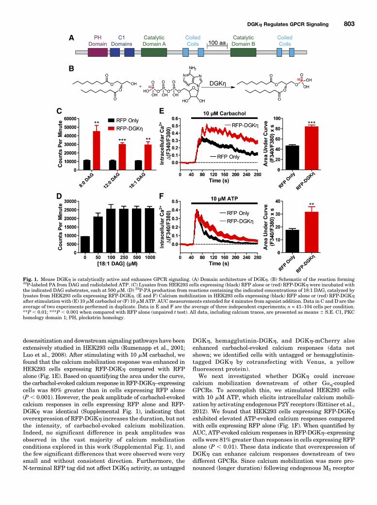
desensitization and downstream signaling pathways have beenextensively studied in HEK293 cells (Rumenapp et al., 2001;Luo et al., 2008). After stimulating with 10 mM carbachol, wefound that the calcium mobilization response was enhanced inHEK293 cells expressing RFP-DGKh compared with RFPalone (Fig. 1E). Based on quantifying the area under the curve,the carbachol-evoked calcium response in RFP-DGKh–expressingcells was 80% greater than in cells expressing RFP alone(P , 0.001). However, the peak amplitude of carbachol-evokedcalcium responses in cells expressing RFP alone and RFP-DGKh was identical (Supplemental Fig. 1), indicating thatoverexpression of RFP-DGKh increases the duration, but notthe intensity, of carbachol-evoked calcium mobilization.Indeed, no significant difference in peak amplitudes wasobserved in the vast majority of calcium mobilizationconditions explored in this work (Supplemental Fig. 1), andthe few significant differences that were observed were verysmall and without consistent direction. Furthermore, theN-terminal RFP tag did not affect DGKh activity, as untagged
DGKh, hemagglutinin-DGKh, and DGKh-mCherry alsoenhanced carbachol-evoked calcium responses (data notshown; we identified cells with untagged or hemagglutinin-tagged DGKh by cotransfecting with Venus, a yellowfluorescent protein).We next investigated whether DGKh could increase
calcium mobilization downstream of other Gaq-coupledGPCRs. To accomplish this, we stimulated HEK293 cellswith 10 mM ATP, which elicits intracellular calcium mobili-zation by activating endogenous P2Y receptors (Rittiner et al.,2012). We found that HEK293 cells expressing RFP-DGKh
exhibited elevated ATP-evoked calcium responses comparedwith cells expressing RFP alone (Fig. 1F). When quantified byAUC, ATP-evoked calcium responses in RFP-DGKh–expressingcells were 81% greater than responses in cells expressing RFPalone (P , 0.01). These data indicate that overexpression ofDGKh can enhance calcium responses downstream of twodifferent GPCRs. Since calcium mobilization was more pro-nounced (longer duration) following endogenous M3 receptor
Fig. 1. Mouse DGKh is catalytically active and enhances GPCR signaling. (A) Domain architecture of DGKh. (B) Schematic of the reaction forming32P-labeled PA from DAG and radiolabeled ATP. (C) Lysates from HEK293 cells expressing (black) RFP alone or (red) RFP-DGKh were incubated withthe indicated DAG substrates, each at 500 mM. (D) 32P-PA production from reactions containing the indicated concentrations of 18:1 DAG, catalyzed bylysates from HEK293 cells expressing RFP-DGKh. (E and F) Calcium mobilization in HEK293 cells expressing (black) RFP alone or (red) RFP-DGKhafter stimulation with (E) 10 mM carbachol or (F) 10 mMATP. AUCmeasurements extended for 4 minutes from agonist addition. Data in C and D are theaverage of two experiments performed in duplicate. Data in E and F are the average of three independent experiments; n = 41–104 cells per condition.**P , 0.01; ***P , 0.001 when compared with RFP alone (unpaired t test). All data, including calcium traces, are presented as means 6 S.E. C1, PKChomology domain 1; PH, pleckstrin homology.
DGKh Regulates GPCR Signaling 803
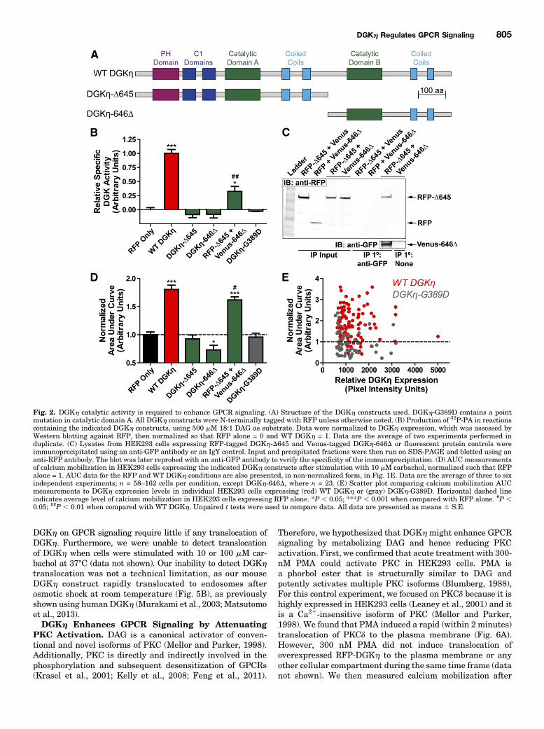
activation, we focused the remainder of our experiments onDGKh modulation of M3 receptor activation.DGKh Catalytic Activity Is Required to Enhance
Carbachol-Evoked Calcium Mobilization. DGKh, like alltype 2 DGKs, possesses a split catalytic domain (Sakane et al.,2007). To determine if DGKh catalytic activity was required toenhance carbachol-evoked calcium mobilization, we generatedtwo DGKh deletion constructs, DGKh-D645 and DGKh-646D,each containing one-half of the catalytic domain (Fig. 2A). Wealso generated DGKh-G389D, containing a point mutation thatis predicted to abolish ATP binding based on homology to otherDGK proteins (Yamada et al., 2003). We found that DGKh-D645, DGKh-646D, and DGKh-G389D were expressed (Sup-plemental Fig. 2) but were catalytically inactive, as theygenerated no more 32P-PA than lysates containing RFP alone(Fig. 2B). In contrast, lysates from cells coexpressing DGKh-D645 and DGKh-646D displayed specific activity (catalyticactivity normalized for expression; Supplemental Fig. 2)approximately 32% of that of wild-type RFP-DGKh (Fig. 2B),suggesting the two halves directly interact. Indeed, we foundthat RFP-DGKh-D645 coimmunoprecipitated with Venus-DGKh-646D (Fig. 2C). This interaction was specific to DGKh-D645 and DGKh-646D and did not involve the fluorescent tagsor nonspecific binding to the immunoprecipitation beads, asevidenced by controls showing no interaction when eitherconstruct was replaced with a fluorescent protein alone orwhen the anti-GFP antibody was replaced with an IgY control(Fig. 2C).Additionally, none of these catalytically dead constructs
(DGKh-D645, DGKh-646D, or DGKh-G389D) enhanced cal-cium mobilization after carbachol stimulation (Fig. 2D). Cellsexpressing DGKh-646D alone appeared unhealthy and dis-played a slight reduction in calcium mobilization. In contrast,coexpression of each half of DGKh (RFP-tagged DGKh-D645and Venus-tagged DGKh-646D) increased calcium mobiliza-tion by 62% (based on AUC measurement, Fig. 2D). Thesedata indicate that DGKh catalytic activity is required toenhance carbachol-evoked calcium mobilization.We next analyzed calcium mobilization AUC data on a cell-
by-cell basis, to determine if the expression level of DGKhcorrelated with how effectively DGKh enhanced calciummobilization responses. Notably, there was no relationshipbetween the level of DGKh expression and the extent ofcalcium mobilization in cells expressing WT DGKh or kinase-dead DGKh-G389D (Fig. 2E). There was also no significantdifference in the expression level of WT DGKh and DGKh-G389D across individual cells. Together, these data suggestthat the amount of DGKh required to maximally enhancecarbachol-evoked calcium mobilization is very small.We also tested the catalytic activity of several DGKh
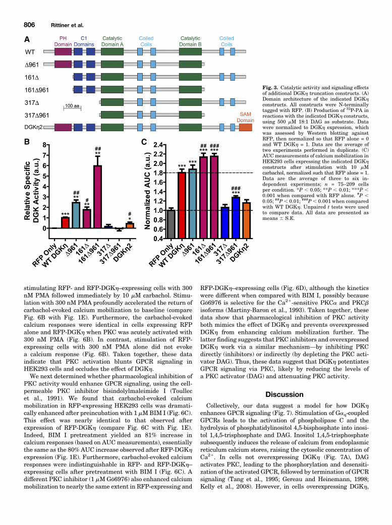
truncation constructs lacking other structural domains (Fig.3A), again normalized to their expression in HEK293 cells(Supplemental Fig. 2). We found that the pleckstrin homologydomain andC-terminal tail negatively regulatedDGKh catalyticactivity, as measured using the in vitro kinase assay (Fig. 3B).Truncation of either domain increased DGKh specific activity inan additive fashion, and the construct lacking both domains(161D961) displayed a specific activity 6-fold higher than WTDGKh (Fig. 3B). The C1 domains were required for catalyticactivity in this assay, as both constructs lacking C1 domainswere inactive (Fig. 3B). Furthermore, the presence of catalyticactivity among the DGKh truncation constructs correlated with
the ability to enhance carbachol-evoked calcium mobilization(Fig. 3, B and C). However, this correlation was not quantitative.The DGKh truncation constructs with greatly increased cata-lytic activity (compared with WT DGKh) enhanced calciummobilization to an equal or only marginally increased extent.These data suggest that beyond a certain point, increased DGKhcatalytic activity does not result in an increased effect uponcalcium mobilization. Lastly, we tested the long splice isoformDGKh2, which contains a C-terminal sterile a motif domain(Fig. 3A), in both the in vitro kinase and calcium mobilizationassays. We found that DGKh2 had decreased catalytic activitycompared with WT DGKh1 (Fig. 3B) as has been reportedpreviously (Murakami et al., 2003), and produced only a negli-gible (P 5 0.057 compared with RFP alone) enhancement ofcarbachol-evoked calcium mobilization (Fig. 3C). As with WTDGKh, we observed no correlation between DGKh expressionand calcium mobilization for any of the DGKh truncationconstructs on a cell-by-cell basis (Supplemental Fig. 3).DGKh Does Not Affect Endoplasmic Reticulum
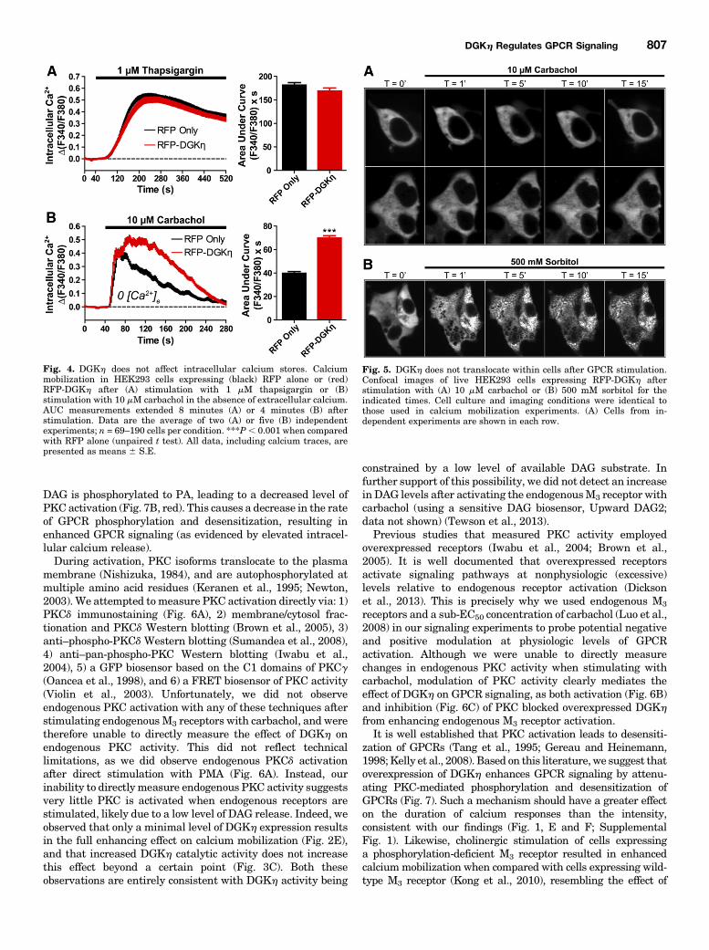
Calcium Loading. Next, to determine if DGKh enhancedcalcium responses by affecting the loading of intracellularcalcium stores, and not GPCR signaling per se, we treatedRFP-DGKh–expressing cells and RFP-expressing controls with1 mM thapsigargin in the presence of extracellular calcium.Thapsigargin is a noncompetitive sarco/endoplasmic reticulumcalcium ATPase inhibitor that causes the release of intracel-lular calcium store contents into the cytosol (Jackson et al.,1988). Calcium release was not significantly different betweencells expressing RFP alone and RFP-DGKh (Fig. 4A), in-dicating that DGKh has no effect on the loading of intracellularcalcium stores.When intracellular calcium stores become depleted,
calcium-sensing proteins trigger the opening of calciumchannels in the plasma membrane, allowing the entry ofextracellular calcium, a process known as store-operatedcalcium entry (Venkatachalam et al., 2002). To determineif DGKh enhanced calcium mobilization by affecting thisprocess, we stimulated cells with carbachol in the absence ofextracellular calcium (to abolish calcium entry through anychannels or transporters). In the absence of extracellularcalcium, carbachol-evoked calcium mobilization was reduced,particularly in the later stages of the response (compare Fig. 4Bwith Fig. 1E). However, overexpression of DGKh still enhancedthe calcium response by 77% (based on AUC measurements)when compared with RFP alone. This effect was nearly identicalto the 80%AUC increase in the presence of extracellular calcium(Fig. 1E). Thus, these data indicate that DGKh enhances thecarbachol-evoked calcium response via a mechanism that is notsolely dependent upon the modulation of calcium entry.DGKhDoes Not Translocate after GPCR Stimulation.
HumanDGKh localizes to the cytosol under baseline conditionsand translocates to endosomes after osmotic shock (Murakamiet al., 2003; Matsutomo et al., 2013). However, whether DGKhtranslocates to the plasmamembrane or intracellular compart-ments following GPCR stimulation is unknown. Using live cellconfocal imaging, we found that mouse DGKh is localizedthroughout the cytoplasm in unstimulated cells and did nottranslocate to the plasma membrane or to any intracellularcompartment after stimulating with 10 mM carbachol at roomtemperature (Fig. 5A). This experiment was conducted underconditions that were identical to those used in the carbachol-evoked calciummobilization assay, suggesting that the effects of
804 Rittiner et al.
DGKh on GPCR signaling require little if any translocation ofDGKh. Furthermore, we were unable to detect translocationof DGKh when cells were stimulated with 10 or 100 mM car-bachol at 37°C (data not shown). Our inability to detect DGKhtranslocation was not a technical limitation, as our mouseDGKh construct rapidly translocated to endosomes afterosmotic shock at room temperature (Fig. 5B), as previouslyshown using humanDGKh (Murakami et al., 2003; Matsutomoet al., 2013).DGKh Enhances GPCR Signaling by Attenuating
PKC Activation. DAG is a canonical activator of conven-tional and novel isoforms of PKC (Mellor and Parker, 1998).Additionally, PKC is directly and indirectly involved in thephosphorylation and subsequent desensitization of GPCRs(Krasel et al., 2001; Kelly et al., 2008; Feng et al., 2011).
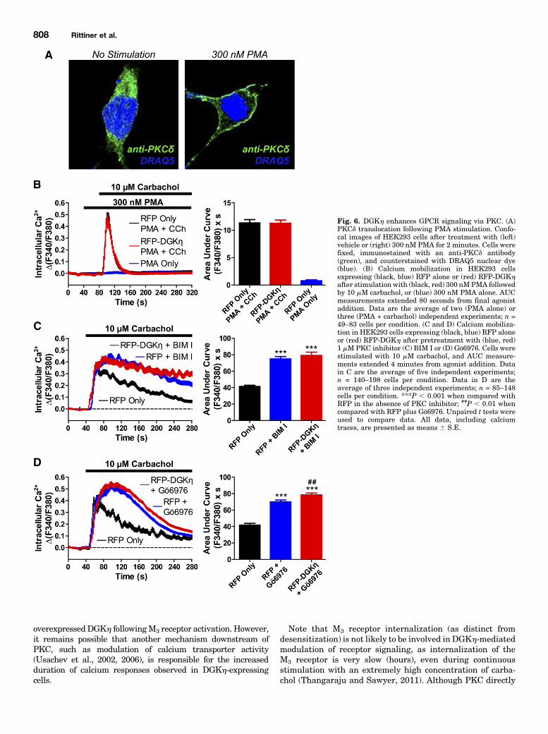
Therefore, we hypothesized that DGKhmight enhance GPCRsignaling by metabolizing DAG and hence reducing PKCactivation. First, we confirmed that acute treatment with 300-nM PMA could activate PKC in HEK293 cells. PMA isa phorbol ester that is structurally similar to DAG andpotently activates multiple PKC isoforms (Blumberg, 1988),For this control experiment, we focused on PKCd because it ishighly expressed in HEK293 cells (Leaney et al., 2001) and itis a Ca21-insensitive isoform of PKC (Mellor and Parker,1998). We found that PMA induced a rapid (within 2 minutes)translocation of PKCd to the plasma membrane (Fig. 6A).However, 300 nM PMA did not induce translocation ofoverexpressed RFP-DGKh to the plasma membrane or anyother cellular compartment during the same time frame (datanot shown). We then measured calcium mobilization after
Fig. 2. DGKh catalytic activity is required to enhance GPCR signaling. (A) Structure of the DGKh constructs used. DGKh-G389D contains a pointmutation in catalytic domain A. All DGKh constructs were N-terminally tagged with RFP unless otherwise noted. (B) Production of 32P-PA in reactionscontaining the indicated DGKh constructs, using 500 mM 18:1 DAG as substrate. Data were normalized to DGKh expression, which was assessed byWestern blotting against RFP, then normalized so that RFP alone = 0 and WT DGKh = 1. Data are the average of two experiments performed induplicate. (C) Lysates from HEK293 cells expressing RFP-tagged DGKh-D645 and Venus-tagged DGKh-646D or fluorescent protein controls wereimmunoprecipitated using an anti-GFP antibody or an IgY control. Input and precipitated fractions were then run on SDS-PAGE and blotted using ananti-RFP antibody. The blot was later reprobed with an anti-GFP antibody to verify the specificity of the immunoprecipitation. (D) AUC measurementsof calcium mobilization in HEK293 cells expressing the indicated DGKh constructs after stimulation with 10 mM carbachol, normalized such that RFPalone = 1. AUC data for the RFP and WT DGKh conditions are also presented, in non-normalized form, in Fig. 1E. Data are the average of three to sixindependent experiments; n = 58–162 cells per condition, except DGKh-646D, where n = 23. (E) Scatter plot comparing calcium mobilization AUCmeasurements to DGKh expression levels in individual HEK293 cells expressing (red) WT DGKh or (gray) DGKh-G389D. Horizontal dashed lineindicates average level of calcium mobilization in HEK293 cells expressing RFP alone. *P , 0.05; ***P , 0.001 when compared with RFP alone. #P ,0.05; ##P , 0.01 when compared with WT DGKh. Unpaired t tests were used to compare data. All data are presented as means 6 S.E.
DGKh Regulates GPCR Signaling 805
stimulating RFP- and RFP-DGKh–expressing cells with 300nM PMA followed immediately by 10 mM carbachol. Stimu-lation with 300 nM PMA profoundly accelerated the return ofcarbachol-evoked calcium mobilization to baseline (compareFig. 6B with Fig. 1E). Furthermore, the carbachol-evokedcalcium responses were identical in cells expressing RFPalone and RFP-DGKh when PKC was acutely activated with300 nM PMA (Fig. 6B). In contrast, stimulation of RFP-expressing cells with 300 nM PMA alone did not evokea calcium response (Fig. 6B). Taken together, these dataindicate that PKC activation blunts GPCR signaling inHEK293 cells and occludes the effect of DGKh.We next determined whether pharmacological inhibition of
PKC activity would enhance GPCR signaling, using the cell-permeable PKC inhibitor bisindolylmaleimide I (Toullecet al., 1991). We found that carbachol-evoked calciummobilization in RFP-expressing HEK293 cells was dramati-cally enhanced after preincubation with 1 mMBIM I (Fig. 6C).This effect was nearly identical to that observed afterexpression of RFP-DGKh (compare Fig. 6C with Fig. 1E).Indeed, BIM I pretreatment yielded an 81% increase incalcium responses (based on AUCmeasurements), essentiallythe same as the 80% AUC increase observed after RFP-DGKh
expression (Fig. 1E). Furthermore, carbachol-evoked calciumresponses were indistinguishable in RFP- and RFP-DGKh–expressing cells after pretreatment with BIM I (Fig. 6C). Adifferent PKC inhibitor (1 mMGö6976) also enhanced calciummobilization to nearly the same extent in RFP-expressing and
RFP-DGKh–expressing cells (Fig. 6D), although the kineticswere different when compared with BIM I, possibly becauseGö6976 is selective for the Ca21-sensitive PKCa and PKCbisoforms (Martiny-Baron et al., 1993). Taken together, thesedata show that pharmacological inhibition of PKC activityboth mimics the effect of DGKh and prevents overexpressedDGKh from enhancing calcium mobilization further. Thelatter finding suggests that PKC inhibitors and overexpressedDGKh work via a similar mechanism—by inhibiting PKCdirectly (inhibitors) or indirectly (by depleting the PKC acti-vator DAG). Thus, these data suggest that DGKh potentiatesGPCR signaling via PKC, likely by reducing the levels ofa PKC activator (DAG) and attenuating PKC activity.
DiscussionCollectively, our data suggest a model for how DGKh
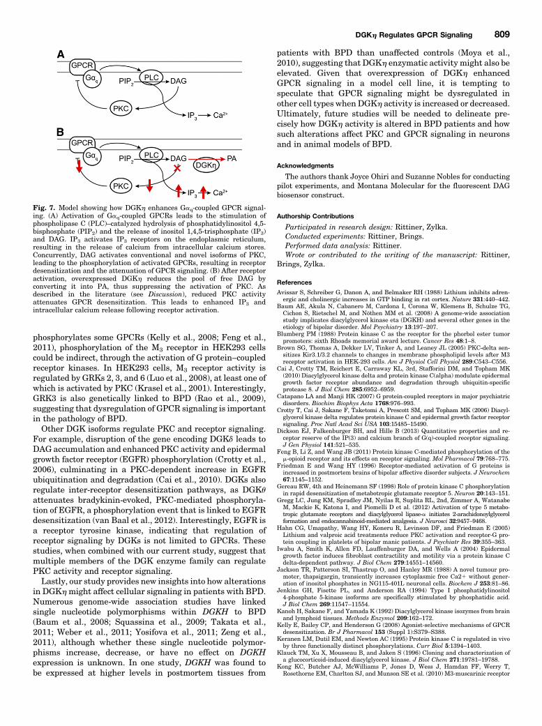
enhances GPCR signaling (Fig. 7). Stimulation of Gaq-coupledGPCRs leads to the activation of phospholipase C and thehydrolysis of phosphatidylinositol 4,5-bisphosphate into inosi-tol 1,4,5-trisphosphate and DAG. Inositol 1,4,5-trisphosphatesubsequently induces the release of calcium from endoplasmicreticulum calcium stores, raising the cytosolic concentration ofCa21. In cells not overexpressing DGKh (Fig. 7A), DAGactivates PKC, leading to the phosphorylation and desensiti-zation of the activated GPCR, followed by termination of GPCRsignaling (Tang et al., 1995; Gereau and Heinemann, 1998;Kelly et al., 2008). However, in cells overexpressing DGKh,
Fig. 3. Catalytic activity and signaling effectsof additional DGKh truncation constructs. (A)Domain architecture of the indicated DGKhconstructs. All constructs were N-terminallytagged with RFP. (B) Production of 32P-PA inreactions with the indicated DGKh constructs,using 500 mM 18:1 DAG as substrate. Datawere normalized to DGKh expression, whichwas assessed by Western blotting againstRFP, then normalized so that RFP alone = 0and WT DGKh = 1. Data are the average oftwo experiments performed in duplicate. (C)AUCmeasurements of calcium mobilization inHEK293 cells expressing the indicated DGKhconstructs after stimulation with 10 mMcarbachol, normalized such that RFP alone = 1.Data are the average of three to six in-dependent experiments; n = 75–209 cellsper condition. *P , 0.05; **P , 0.01; ***P ,0.001 when compared with RFP alone. #P ,0.05; ##P, 0.01; ###P, 0.001 when comparedwith WT DGKh. Unpaired t tests were usedto compare data. All data are presented asmeans 6 S.E.
806 Rittiner et al.
DAG is phosphorylated to PA, leading to a decreased level ofPKC activation (Fig. 7B, red). This causes a decrease in the rateof GPCR phosphorylation and desensitization, resulting inenhanced GPCR signaling (as evidenced by elevated intracel-lular calcium release).During activation, PKC isoforms translocate to the plasma
membrane (Nishizuka, 1984), and are autophosphorylated atmultiple amino acid residues (Keranen et al., 1995; Newton,2003). We attempted tomeasure PKC activation directly via: 1)PKCd immunostaining (Fig. 6A), 2) membrane/cytosol frac-tionation and PKCd Western blotting (Brown et al., 2005), 3)anti–phospho-PKCd Western blotting (Sumandea et al., 2008),4) anti–pan-phospho-PKC Western blotting (Iwabu et al.,2004), 5) a GFP biosensor based on the C1 domains of PKCg(Oancea et al., 1998), and 6) a FRET biosensor of PKC activity(Violin et al., 2003). Unfortunately, we did not observeendogenous PKC activation with any of these techniques afterstimulating endogenous M3 receptors with carbachol, and weretherefore unable to directly measure the effect of DGKh onendogenous PKC activity. This did not reflect technicallimitations, as we did observe endogenous PKCd activationafter direct stimulation with PMA (Fig. 6A). Instead, ourinability to directly measure endogenous PKC activity suggestsvery little PKC is activated when endogenous receptors arestimulated, likely due to a low level of DAG release. Indeed, weobserved that only a minimal level of DGKh expression resultsin the full enhancing effect on calcium mobilization (Fig. 2E),and that increased DGKh catalytic activity does not increasethis effect beyond a certain point (Fig. 3C). Both theseobservations are entirely consistent with DGKh activity being
constrained by a low level of available DAG substrate. Infurther support of this possibility, we did not detect an increaseinDAG levels after activating the endogenousM3 receptor withcarbachol (using a sensitive DAG biosensor, Upward DAG2;data not shown) (Tewson et al., 2013).Previous studies that measured PKC activity employed
overexpressed receptors (Iwabu et al., 2004; Brown et al.,2005). It is well documented that overexpressed receptorsactivate signaling pathways at nonphysiologic (excessive)levels relative to endogenous receptor activation (Dicksonet al., 2013). This is precisely why we used endogenous M3
receptors and a sub-EC50 concentration of carbachol (Luo et al.,2008) in our signaling experiments to probe potential negativeand positive modulation at physiologic levels of GPCRactivation. Although we were unable to directly measurechanges in endogenous PKC activity when stimulating withcarbachol, modulation of PKC activity clearly mediates theeffect of DGKh on GPCR signaling, as both activation (Fig. 6B)and inhibition (Fig. 6C) of PKC blocked overexpressed DGKhfrom enhancing endogenous M3 receptor activation.It is well established that PKC activation leads to desensiti-
zation of GPCRs (Tang et al., 1995; Gereau and Heinemann,1998; Kelly et al., 2008). Based on this literature, we suggest thatoverexpression of DGKh enhances GPCR signaling by attenu-ating PKC-mediated phosphorylation and desensitization ofGPCRs (Fig. 7). Such a mechanism should have a greater effecton the duration of calcium responses than the intensity,consistent with our findings (Fig. 1, E and F; SupplementalFig. 1). Likewise, cholinergic stimulation of cells expressinga phosphorylation-deficient M3 receptor resulted in enhancedcalciummobilization when compared with cells expressing wild-type M3 receptor (Kong et al., 2010), resembling the effect of
Fig. 4. DGKh does not affect intracellular calcium stores. Calciummobilization in HEK293 cells expressing (black) RFP alone or (red)RFP-DGKh after (A) stimulation with 1 mM thapsigargin or (B)stimulation with 10 mM carbachol in the absence of extracellular calcium.AUC measurements extended 8 minutes (A) or 4 minutes (B) afterstimulation. Data are the average of two (A) or five (B) independentexperiments; n = 69–190 cells per condition. ***P, 0.001 when comparedwith RFP alone (unpaired t test). All data, including calcium traces, arepresented as means 6 S.E.
Fig. 5. DGKh does not translocate within cells after GPCR stimulation.Confocal images of live HEK293 cells expressing RFP-DGKh afterstimulation with (A) 10 mM carbachol or (B) 500 mM sorbitol for theindicated times. Cell culture and imaging conditions were identical tothose used in calcium mobilization experiments. (A) Cells from in-dependent experiments are shown in each row.
DGKh Regulates GPCR Signaling 807
overexpressedDGKh followingM3 receptor activation. However,it remains possible that another mechanism downstream ofPKC, such as modulation of calcium transporter activity(Usachev et al., 2002, 2006), is responsible for the increasedduration of calcium responses observed in DGKh-expressingcells.
Note that M3 receptor internalization (as distinct fromdesensitization) is not likely to be involved in DGKh-mediatedmodulation of receptor signaling, as internalization of theM3 receptor is very slow (hours), even during continuousstimulation with an extremely high concentration of carba-chol (Thangaraju and Sawyer, 2011). Although PKC directly
Fig. 6. DGKh enhances GPCR signaling via PKC. (A)PKCd translocation following PMA stimulation. Confo-cal images of HEK293 cells after treatment with (left)vehicle or (right) 300 nM PMA for 2 minutes. Cells werefixed, immunostained with an anti-PKCd antibody(green), and counterstained with DRAQ5 nuclear dye(blue). (B) Calcium mobilization in HEK293 cellsexpressing (black, blue) RFP alone or (red) RFP-DGKhafter stimulation with (black, red) 300 nMPMA followedby 10 mM carbachol, or (blue) 300 nM PMA alone. AUCmeasurements extended 80 seconds from final agonistaddition. Data are the average of two (PMA alone) orthree (PMA + carbachol) independent experiments; n =49–83 cells per condition. (C and D) Calcium mobiliza-tion in HEK293 cells expressing (black, blue) RFP aloneor (red) RFP-DGKh after pretreatment with (blue, red)1 mMPKC inhibitor (C) BIM I or (D) Gö6976. Cells werestimulated with 10 mM carbachol, and AUC measure-ments extended 4 minutes from agonist addition. Datain C are the average of five independent experiments;n = 140–198 cells per condition. Data in D are theaverage of three independent experiments; n = 85–148cells per condition. ***P , 0.001 when compared withRFP in the absence of PKC inhibitor; ##P , 0.01 whencompared with RFP plus Gö6976. Unpaired t tests wereused to compare data. All data, including calciumtraces, are presented as means 6 S.E.
808 Rittiner et al.
phosphorylates some GPCRs (Kelly et al., 2008; Feng et al.,2011), phosphorylation of the M3 receptor in HEK293 cellscould be indirect, through the activation of G protein–coupledreceptor kinases. In HEK293 cells, M3 receptor activity isregulated by GRKs 2, 3, and 6 (Luo et al., 2008), at least one ofwhich is activated by PKC (Krasel et al., 2001). Interestingly,GRK3 is also genetically linked to BPD (Rao et al., 2009),suggesting that dysregulation of GPCR signaling is importantin the pathology of BPD.Other DGK isoforms regulate PKC and receptor signaling.
For example, disruption of the gene encoding DGKd leads toDAG accumulation and enhanced PKC activity and epidermalgrowth factor receptor (EGFR) phosphorylation (Crotty et al.,2006), culminating in a PKC-dependent increase in EGFRubiquitination and degradation (Cai et al., 2010). DGKs alsoregulate inter-receptor desensitization pathways, as DGKu
attenuates bradykinin-evoked, PKC-mediated phosphoryla-tion of EGFR, a phosphorylation event that is linked to EGFRdesensitization (van Baal et al., 2012). Interestingly, EGFR isa receptor tyrosine kinase, indicating that regulation ofreceptor signaling by DGKs is not limited to GPCRs. Thesestudies, when combined with our current study, suggest thatmultiple members of the DGK enzyme family can regulatePKC activity and receptor signaling.Lastly, our study provides new insights into how alterations
in DGKhmight affect cellular signaling in patients with BPD.Numerous genome-wide association studies have linkedsingle nucleotide polymorphisms within DGKH to BPD(Baum et al., 2008; Squassina et al., 2009; Takata et al.,2011; Weber et al., 2011; Yosifova et al., 2011; Zeng et al.,2011), although whether these single nucleotide polymor-phisms increase, decrease, or have no effect on DGKHexpression is unknown. In one study, DGKH was found tobe expressed at higher levels in postmortem tissues from
patients with BPD than unaffected controls (Moya et al.,2010), suggesting that DGKh enzymatic activity might also beelevated. Given that overexpression of DGKh enhancedGPCR signaling in a model cell line, it is tempting tospeculate that GPCR signaling might be dysregulated inother cell types when DGKh activity is increased or decreased.Ultimately, future studies will be needed to delineate pre-cisely how DGKh activity is altered in BPD patients and howsuch alterations affect PKC and GPCR signaling in neuronsand in animal models of BPD.
Acknowledgments
The authors thank Joyce Ohiri and Suzanne Nobles for conductingpilot experiments, and Montana Molecular for the fluorescent DAGbiosensor construct.
Authorship Contributions
Participated in research design: Rittiner, Zylka.Conducted experiments: Rittiner, Brings.Performed data analysis: Rittiner.Wrote or contributed to the writing of the manuscript: Rittiner,
Brings, Zylka.
References
Avissar S, Schreiber G, Danon A, and Belmaker RH (1988) Lithium inhibits adren-ergic and cholinergic increases in GTP binding in rat cortex. Nature 331:440–442.
Baum AE, Akula N, Cabanero M, Cardona I, Corona W, Klemens B, Schulze TG,Cichon S, Rietschel M, and Nöthen MM et al. (2008) A genome-wide associationstudy implicates diacylglycerol kinase eta (DGKH) and several other genes in theetiology of bipolar disorder. Mol Psychiatry 13:197–207.
Blumberg PM (1988) Protein kinase C as the receptor for the phorbol ester tumorpromoters: sixth Rhoads memorial award lecture. Cancer Res 48:1–8.
Brown SG, Thomas A, Dekker LV, Tinker A, and Leaney JL (2005) PKC-delta sen-sitizes Kir3.1/3.2 channels to changes in membrane phospholipid levels after M3receptor activation in HEK-293 cells. Am J Physiol Cell Physiol 289:C543–C556.
Cai J, Crotty TM, Reichert E, Carraway KL, 3rd, Stafforini DM, and Topham MK(2010) Diacylglycerol kinase delta and protein kinase C(alpha) modulate epidermalgrowth factor receptor abundance and degradation through ubiquitin-specificprotease 8. J Biol Chem 285:6952–6959.
Catapano LA and Manji HK (2007) G protein-coupled receptors in major psychiatricdisorders. Biochim Biophys Acta 1768:976–993.
Crotty T, Cai J, Sakane F, Taketomi A, Prescott SM, and Topham MK (2006) Diacyl-glycerol kinase delta regulates protein kinase C and epidermal growth factor receptorsignaling. Proc Natl Acad Sci USA 103:15485–15490.
Dickson EJ, Falkenburger BH, and Hille B (2013) Quantitative properties and re-ceptor reserve of the IP(3) and calcium branch of G(q)-coupled receptor signaling.J Gen Physiol 141:521–535.
Feng B, Li Z, and Wang JB (2011) Protein kinase C-mediated phosphorylation of them-opioid receptor and its effects on receptor signaling. Mol Pharmacol 79:768–775.
Friedman E and Wang HY (1996) Receptor-mediated activation of G proteins isincreased in postmortem brains of bipolar affective disorder subjects. J Neurochem67:1145–1152.
Gereau RW, 4th and Heinemann SF (1998) Role of protein kinase C phosphorylationin rapid desensitization of metabotropic glutamate receptor 5. Neuron 20:143–151.
Gregg LC, Jung KM, Spradley JM, Nyilas R, Suplita RL, 2nd, Zimmer A, WatanabeM, Mackie K, Katona I, and Piomelli D et al. (2012) Activation of type 5 metabo-tropic glutamate receptors and diacylglycerol lipase-a initiates 2-arachidonoylglycerolformation and endocannabinoid-mediated analgesia. J Neurosci 32:9457–9468.
Hahn CG, Umapathy, Wang HY, Koneru R, Levinson DF, and Friedman E (2005)Lithium and valproic acid treatments reduce PKC activation and receptor-G pro-tein coupling in platelets of bipolar manic patients. J Psychiatr Res 39:355–363.
Iwabu A, Smith K, Allen FD, Lauffenburger DA, and Wells A (2004) Epidermalgrowth factor induces fibroblast contractility and motility via a protein kinase Cdelta-dependent pathway. J Biol Chem 279:14551–14560.
Jackson TR, Patterson SI, Thastrup O, and Hanley MR (1988) A novel tumour pro-moter, thapsigargin, transiently increases cytoplasmic free Ca21 without gener-ation of inositol phosphates in NG115-401L neuronal cells. Biochem J 253:81–86.
Jenkins GH, Fisette PL, and Anderson RA (1994) Type I phosphatidylinositol4-phosphate 5-kinase isoforms are specifically stimulated by phosphatidic acid.J Biol Chem 269:11547–11554.
Kanoh H, Sakane F, and Yamada K (1992) Diacylglycerol kinase isozymes from brainand lymphoid tissues. Methods Enzymol 209:162–172.
Kelly E, Bailey CP, and Henderson G (2008) Agonist-selective mechanisms of GPCRdesensitization. Br J Pharmacol 153 (Suppl 1):S379–S388.
Keranen LM, Dutil EM, and Newton AC (1995) Protein kinase C is regulated in vivoby three functionally distinct phosphorylations. Curr Biol 5:1394–1403.
Klauck TM, Xu X, Mousseau B, and Jaken S (1996) Cloning and characterization ofa glucocorticoid-induced diacylglycerol kinase. J Biol Chem 271:19781–19788.
Kong KC, Butcher AJ, McWilliams P, Jones D, Wess J, Hamdan FF, Werry T,Rosethorne EM, Charlton SJ, and Munson SE et al. (2010) M3-muscarinic receptor
Fig. 7. Model showing how DGKh enhances Gaq-coupled GPCR signal-ing. (A) Activation of Gaq-coupled GPCRs leads to the stimulation ofphospholipase C (PLC)–catalyzed hydrolysis of phosphatidylinositol 4,5-bisphosphate (PIP2) and the release of inositol 1,4,5-trisphosphate (IP3)and DAG. IP3 activates IP3 receptors on the endoplasmic reticulum,resulting in the release of calcium from intracellular calcium stores.Concurrently, DAG activates conventional and novel isoforms of PKC,leading to the phosphorylation of activated GPCRs, resulting in receptordesensitization and the attenuation of GPCR signaling. (B) After receptoractivation, overexpressed DGKh reduces the pool of free DAG byconverting it into PA, thus suppressing the activation of PKC. Asdescribed in the literature (see Discussion), reduced PKC activityattenuates GPCR desensitization. This leads to enhanced IP3 andintracellular calcium release following receptor activation.
DGKh Regulates GPCR Signaling 809
promotes insulin release via receptor phosphorylation/arrestin-dependent activa-tion of protein kinase D1. Proc Natl Acad Sci USA 107:21181–21186.
Krasel C, Dammeier S, Winstel R, Brockmann J, Mischak H, and Lohse MJ (2001)Phosphorylation of GRK2 by protein kinase C abolishes its inhibition by calmod-ulin. J Biol Chem 276:1911–1915.
Leaney JL, Dekker LV, and Tinker A (2001) Regulation of a G protein-gated inwardlyrectifying K1 channel by a Ca(21)-independent protein kinase C. J Physiol 534:367–379.
Lucas P, Ukhanov K, Leinders-Zufall T, and Zufall F (2003) A diacylglycerol-gatedcation channel in vomeronasal neuron dendrites is impaired in TRPC2 mutantmice: mechanism of pheromone transduction. Neuron 40:551–561.
Luo J, Busillo JM, and Benovic JL (2008) M3 muscarinic acetylcholine receptor-mediated signaling is regulated by distinct mechanisms. Mol Pharmacol 74:338–347.
Martiny-Baron G, Kazanietz MG, Mischak H, Blumberg PM, Kochs G, Hug H,Marmé D, and Schächtele C (1993) Selective inhibition of protein kinase C iso-zymes by the indolocarbazole Gö 6976. J Biol Chem 268:9194–9197.
Matsutomo D, Isozaki T, Sakai H, and Sakane F (2013) Osmotic shock-dependentredistribution of diacylglycerol kinase h1 to non-ionic detergent-resistant mem-brane via pleckstrin homology and C1 domains. J Biochem 153:179–190.
Mellor H and Parker PJ (1998) The extended protein kinase C superfamily. BiochemJ 332:281–292.
Mérida I, Avila-Flores A, and Merino E (2008) Diacylglycerol kinases: at the hub ofcell signalling. Biochem J 409:1–18.
Moya PR, Murphy DL, McMahon FJ, and Wendland JR (2010) Increased gene ex-pression of diacylglycerol kinase h in bipolar disorder. Int J Neuropsychopharmacol13:1127–1128.
Murakami T, Sakane F, Imai S, Houkin K, and Kanoh H (2003) Identification andcharacterization of two splice variants of human diacylglycerol kinase eta. J BiolChem 278:34364–34372.
Nakano T, Iravani A, Kim M, Hozumi Y, Lohse M, Reichert E, Crotty TM, StafforiniDM, and Topham MK (2014) Diacylglycerol kinase h modulates oncogenic prop-erties of lung cancer cells. Clin Transl Oncol 16:29–35.
Newton AC (2003) Regulation of the ABC kinases by phosphorylation: protein kinaseC as a paradigm. Biochem J 370:361–371.
Nishizuka Y (1984) The role of protein kinase C in cell surface signal transductionand tumour promotion. Nature 308:693–698.
Oancea E, Teruel MN, Quest AF, and Meyer T (1998) Green fluorescent protein(GFP)-tagged cysteine-rich domains from protein kinase C as fluorescent indicatorsfor diacylglycerol signaling in living cells. J Cell Biol 140:485–498.
Ohanian J and Heagerty AM (1994) Membrane-associated diacylglycerol kinase ac-tivity is increased by noradrenaline, but not by angiotensin II, in arterial smoothmuscle. Biochem J 300:51–56.
Pantazopoulos H, Stone D, Walsh J, and Benes FM (2004) Differences in the cellulardistribution of D1 receptor mRNA in the hippocampus of bipolars and schizo-phrenics. Synapse 54:147–155.
Rao JS, Rapoport SI, and Kim HW (2009) Decreased GRK3 but not GRK2 expressionin frontal cortex from bipolar disorder patients. Int J Neuropsychopharmacol 12:851–860.
Rittiner JE, Korboukh I, Hull-Ryde EA, Jin J, Janzen WP, Frye SV, and Zylka MJ(2012) AMP is an adenosine A1 receptor agonist. J Biol Chem 287:5301–5309.
Rümenapp U, Asmus M, Schablowski H, Woznicki M, Han L, Jakobs KH, Fahimi-Vahid M, Michalek C, Wieland T, and Schmidt M (2001) The M3 muscarinic ace-tylcholine receptor expressed in HEK-293 cells signals to phospholipase D via G12but not Gq-type G proteins: regulators of G proteins as tools to dissect pertussistoxin-resistant G proteins in receptor-effector coupling. J Biol Chem 276:2474–2479.
Sakane F, Imai S, Kai M, Yasuda S, and Kanoh H (2007) Diacylglycerol kinases: whyso many of them? Biochim Biophys Acta 1771:793–806.
Squassina A, Manchia M, Congiu D, Severino G, Chillotti C, Ardau R, Piccardi M,and Zompo MD (2009) The diacylglycerol kinase eta gene and bipolar disorder:a replication study in a Sardinian sample. Mol Psychiatry 14:350–351.
Sumandea MP, Rybin VO, Hinken AC, Wang C, Kobayashi T, Harleton E, Sievert G,Balke CW, Feinmark SJ, and Solaro RJ et al. (2008) Tyrosine phosphorylationmodifies protein kinase C delta-dependent phosphorylation of cardiac troponin I.J Biol Chem 283:22680–22689.
Takata A, Kawasaki H, Iwayama Y, Yamada K, Gotoh L, Mitsuyasu H, Miura T,Kato T, Yoshikawa T, and Kanba S (2011) Nominal association between a poly-morphism in DGKH and bipolar disorder detected in a meta-analysis of East Asiancase-control samples. Psychiatry Clin Neurosci 65:280–285.
Tang H, Shirai H, and Inagami T (1995) Inhibition of protein kinase C prevents rapiddesensitization of type 1B angiotensin II receptor. Circ Res 77:239–248.
Tewson PH, Quinn AM, and Hughes TE (2013) A multiplexed fluorescent assay forindependent second-messenger systems: decoding GPCR activation in living cells.J Biomol Screen 18:797–806.
Thangaraju A and Sawyer GW (2011) Comparison of the kinetics and extent ofmuscarinic M1-M5 receptor internalization, recycling and downregulation in Chi-nese hamster ovary cells. Eur J Pharmacol 650:534–543.
Toullec D, Pianetti P, Coste H, Bellevergue P, Grand-Perret T, Ajakane M, Baudet V,Boissin P, Boursier E, and Loriolle F et al. (1991) The bisindolylmaleimide GF109203X is a potent and selective inhibitor of protein kinase C. J Biol Chem 266:15771–15781.
Usachev YM, DeMarco SJ, Campbell C, Strehler EE, and Thayer SA (2002) Brady-kinin and ATP accelerate Ca(21) efflux from rat sensory neurons via protein ki-nase C and the plasma membrane Ca(21) pump isoform 4. Neuron 33:113–122.
Usachev YM, Marsh AJ, Johanns TM, Lemke MM, and Thayer SA (2006) Activationof protein kinase C in sensory neurons accelerates Ca21 uptake into the endo-plasmic reticulum. J Neurosci 26:311–318.
van Baal J, de Widt J, Divecha N, and van Blitterswijk WJ (2012) Diacylglycerolkinase u counteracts protein kinase C-mediated inactivation of the EGF receptor.Int J Biochem Cell Biol 44:1791–1799.
van Blitterswijk WJ and Houssa B (2000) Properties and functions of diacylglycerolkinases. Cell Signal 12:595–605.
Venkatachalam K, van Rossum DB, Patterson RL, Ma HT, and Gill DL (2002) Thecellular and molecular basis of store-operated calcium entry. Nat Cell Biol 4:E263–E272.
Violin JD, Zhang J, Tsien RY, and Newton AC (2003) A genetically encoded fluo-rescent reporter reveals oscillatory phosphorylation by protein kinase C. J Cell Biol161:899–909.
Weber H, Kittel-Schneider S, Gessner A, Domschke K, NeunerM, Jacob CP, ButtenschonHN, Boreatti-Hümmer A, Volkert J, and Herterich S et al. (2011) Cross-disorderanalysis of bipolar risk genes: further evidence of DGKH as a risk gene for bipolardisorder, but also unipolar depression and adult ADHD.Neuropsychopharmacology 36:2076–2085.
Yamada K, Sakane F, Imai Si, Tsushima S, Murakami T, and Kanoh H (2003)Regulatory role of diacylglycerol kinase gamma in macrophage differentiation ofleukemia cells. Biochem Biophys Res Commun 305:101–107.
Yosifova A, Mushiroda T, Kubo M, Takahashi A, Kamatani Y, Kamatani N, StoianovD, Vazharova R, Karachanak S, and Zaharieva I et al. (2011) Genome-wide asso-ciation study on bipolar disorder in the Bulgarian population. Genes Brain Behav10:789–797.
Young LT, Li PP, Kish SJ, Siu KP, Kamble A, Hornykiewicz O, and Warsh JJ (1993)Cerebral cortex Gs alpha protein levels and forskolin-stimulated cyclic AMP for-mation are increased in bipolar affective disorder. J Neurochem 61:890–898.
Zeng Z, Wang T, Li T, Li Y, Chen P, Zhao Q, Liu J, Li J, Feng G, and He L et al. (2011)Common SNPs and haplotypes in DGKH are associated with bipolar disorder andschizophrenia in the Chinese Han population. Mol Psychiatry 16:473–475.
Address correspondence to: Mark J. Zylka, Department of Cell Biology &Physiology, 5109D NRB, CB #7545, The University of North Carolina atChapel Hill, Chapel Hill, NC 27599-7545. E-mail: [email protected]
810 Rittiner et al.