outcome of primary root canal treatment: systematic … · ng y-l, mann v, rahbaran s, lewsey j,...
TRANSCRIPT

REVIEW
Outcome of primary root canal treatment:systematic review of the literature – Part 1.Effects of study characteristics on probabilityof success
Y.-L. Ng1, V. Mann2, S. Rahbaran1, J. Lewsey3 & K. Gulabivala11Unit of Endodontology, UCL Eastman Dental Institute, University College London; 2Department of Medical Statistics, LondonSchool of Tropical Medicine and Hygiene; and 3Clinical effectiveness Unit, The Royal College of Surgeons of England, London, UK
Abstract
Ng Y-L, Mann V, Rahbaran S, Lewsey J, Gulabivala K.
Outcome of primary root canal treatment: systematic review of
the literature – Part 1. Effects of study characteristics on
probability of success. International Endodontic Journal,
40, 921–939, 2007.
Aims The aims of this study were (i) to conduct a
comprehensive systematic review of the literature on
the outcome of primary (initial or first time) root canal
treatment; (ii) to investigate the influence of some
study characteristics on the estimated pooled success
rates.
Methodology Longitudinal clinical studies investi-
gating outcome of primary root canal treatment,
published up to the end of 2002, were identified
electronically (MEDLINE and Cochrane database
1966–2002 December, week 4). Four journals (Inter-
national Endodontic Journal, Journal of Endodontics,
Oral Surgery Oral Medicine Oral Pathology Endodontics
Radiology and Dental Traumatology & Endodontics),
bibliographies of all relevant papers and review articles
were hand-searched. Three reviewers (Y-LN, SR and
KG) independently assessed, selected the studies based
on specified inclusion criteria, and extracted the data
onto a pre-designed proforma. The study inclusion
criteria were: longitudinal clinical studies investigating
root canal treatment outcome; only primary root canal
treatment carried out on the teeth studied; sample size
given; at least 6-month postoperative review; success
based on clinical and/or radiographic criteria (strict,
absence of apical radiolucency; loose, reduction in size
of radiolucency); overall success rate given or could be
calculated from the raw data. The findings by individual
study were summarized and the pooled success rates by
each potential influencing factor were calculated for this
part of the study.
Results Of the 119 articles identified, 63 studies
published from 1922 to 2002, fulfilling the inclusion
criteria were selected for the review: six were random-
ized trials, seven were cohort studies and 48 were
retrospective studies. The reported mean success rates
ranged from 31% to 96% based on strict criteria or
from 60% to 100% based on loose criteria, with
substantial heterogeneity in the estimates of pooled
success rates. Apart from the radiographic criteria of
success, none of the other study characteristics could
explain this heterogeneity. Twenty-four factors (patient
and operative) had been investigated in various com-
binations in the studies reviewed. The influence of
preoperative pulpal and periapical status of the teeth on
treatment outcome were most frequently explored, but
the influence of treatment technique was poorly
investigated.
Conclusions The estimated weighted pooled success
rates of treatments completed at least 1 year prior to
review, ranged between 68% and 85% when strict
criteria were used. The reported success rates had not
improved over the last four (or five) decades. The
quality of evidence for treatment factors affecting
primary root canal treatment outcome is sub-optimal;
there was substantial variation in the study–designs. It
Correspondence: Dr Y.-L. Ng, Unit of Endodontology, UCLEastman Dental Institute, UCL, 256 Grays Inn Road, LondonWC1X 8LD, UK (Tel.: 020 7915 1233; fax: 020 7915 2371;e-mail: [email protected]).
doi:10.1111/j.1365-2591.2007.01322.x
ª 2007 International Endodontic Journal International Endodontic Journal, 40, 921–939, 2007 921

would be desirable to standardize aspects of study–
design, data recording and presentation format of
outcome data in the much needed future outcome
studies.
Keywords: outcome, root canal treatment, success,
systematic review.
Received 27 March 2007; accepted 6 July 2007
Introduction
There has been a surge of interest in formulating
clinical guidelines for optimal treatment of diseases
based on properly conceived and executed research.
The gold standard for informing clinical practice is
putatively the randomized controlled trial (RCT); how-
ever, neither medical nor dental practice has been
generally well supported by such evidence. Sackett
et al. (1996) now famous definition, ‘the conscientious,
explicit and judicious use of current best evidence in
making decisions about the care of individual patients’,
not only embraces the notion of grades of evidence but
also recognizes that optimal levels of evidence may not
be available for all situations. There is therefore a need
to synthesize an objective over-view based on available
evidence. Depending upon the quality and quantity of
the data, systematic reviews can be of several different
kinds: traditional reviews; meta-analysis leading to an
estimate of effect size; best evidence synthesis; and the
hypothetico-deductive approach, in which the effort is
directed at evaluating the evidence for and against a
given theory, in an attempt to solve the problem of why
contradictory results appear, rather than simply aver-
aging often incompatible data (Eysenck 1994).
For the outcome of endodontic treatment, there are
eight published systematic reviews, which have used
different approaches in synthesis of information from
the literature. Basmadjian-Charles et al. (2002) and
Paik et al. (2004) used a systematic approach for
literature search but a traditional approach for evalu-
ating the variables impacting on the success and failure
of the root canal retreatment. Two reviews (Hepworth
& Friedman 1997, Peterson & Gutmann 2001) calcu-
lated the weighted-average success rates by each factor
under investigation. Neiderman & Theodosopoulou
(2003) estimated the number needed to treat when
comparing two types of treatments. Three reviews
(Lewsey et al. 2001, Kojima et al. 2004, Sathorn et al.
2005) estimated the size of effect of individual factors
which included presence of preoperative pulpal and
periapical status, apical extent of root filling & number
of treatment visits, using meta-analysis. Except for
Lewsey et al. (2001), none have investigated the
influence of study characteristics such as radiographic
criteria for determination of treatment outcome and
year of publication, on the data heterogeneity.
In the absence of sufficient gold standard level data,
there is a need to synthesize ‘sub-standard’ data but
there is a lack of formal guidelines to achieve this. In
the absence of such guidelines, the authors proposed
the use of a process of ‘triangulation’ of different
analytical approaches as a sensible strategy. The
purpose of this systematic review and synthesis was
to: (i) identify the probable dominant factors influenc-
ing outcome; (ii) help prioritize the questions that need
to be addressed; (iii) inform the design and data
collection protocol for future RCTs. The outcome of
this analysis will be presented in two parts.
The aim of the first part of this paper is to present
the estimated pooled success rates of primary root
canal treatment by aspects of study characteristics:
decade of publication, study-specific criteria for suc-
cess, unit of outcome measure, duration after treat-
ment, geographical location of study and qualification
of the operator.
Materials and methods
Literature search
Longitudinal clinical studies investigating the out-
come of primary root canal treatment that were
published up to the end of 2002 were identified
electronically (MEDLINE database 1966–2002 Decem-
ber, week 4) using six keywords (root canal treat-
ment, root canal therapy, endodontic treatment,
endodontics, treatment outcome and success) and
eight strategies (1 AND 5, 1 AND 6, 2 AND 5, 2 AND
6, 3 AND 5, 3 AND 6, 4 AND 5 and 4 AND 6). A
Cochrane Library search was also conducted. PubMed
was independently searched using the ‘related articles’
feature. Four journals (International Endodontic Jour-
nal, Journal of Endodontics, Oral Surgery Oral Med-
icine Oral Pathology Endodontics Radiology and
Dental Traumatology & Endodontics) and bibliogra-
phies of all relevant papers and review articles were
hand-searched. Unpublished studies were identified by
Outcome of primary root canal treatment – Part 1 Ng et al.
International Endodontic Journal, 40, 921–939, 2007 ª 2007 International Endodontic Journal922

searching abstracts and conference proceedings. Per-
sonal contacts were also used to identify ongoing or
unpublished studies. Full articles were obtained for all
relevant titles identified through either electronic or
other search methods.
Study selection, quality assessment and dataextraction
Three reviewers (Y-LN, SR and KG) independently
assessed and selected the studies based on the following
inclusion criteria:
1. Clinical study on primary root canal treatment.
2. Stratified analysis of primary root canal treatment
available, if root canal re-treatment cases had been
included.
3. Sample size given.
4. At least 6-month postoperative review.
5. Success based on clinical and/or radiographic
(strict, absence of apical radiolucency; loose, reduction
in size of radiolucency) criteria.
6. Overall success rate given or could be calculated
from the raw data.
7. Presentations in English, German, Chinese and
Japanese languages were accepted.
Disagreements on study inclusion were resolved by
discussion. The reasons for study rejection at this or
subsequent stages were recorded.
Data were extracted by all three reviewers indepen-
dently using custom-designed data collection forms.
The data collection form was piloted on several papers
and modified for optimal utility before final use. The
data extracted could be classified into six groups;
success rates, study characteristics, demographic data
of patients, pre-, intra- and postoperative factors. Any
disagreement was discussed and data were excluded if
agreement could not be reached.
Estimation of pooled success rates
stata version 9.2 statistical software (StataCorp, College
Station, TX, USA) was used to perform all statistical
analyses. Un-weighted pooled success rate by each factor
was calculated by dividing the total number of successful
unitswith the total number of units within the respective
category (according to Hepworth & Friedman 1997). In
addition, the weighted pooled success rates were esti-
mated using random effects meta-analysis with DerSi-
monian and Laird’s methods (DerSimonian & Laird
1986). Statistical heterogeneity amongst the studies was
assessed by Cochran’s (Q) test (Cochran 1954).
Meta-regression models (Thompson & Higgins 2002)
were used to explore the potential sources of statistical
heterogeneity and to assess the effect of factors on
estimating the pooled success rate. Factors related with
study characteristics considered in the meta-regression
analyses as covariates (and their sub-categories) were:
decade of publication, study specific criteria (radio-
graphic, combined radiographical & clinical) for suc-
cess, unit of outcome measure (tooth and root),
duration after treatment when assessing success (‘at
least 4 years’ or ‘<4 years’), geographical location of
the study (North American, Scandinavian and other
countries), qualification of the operator (undergraduate
students, postgraduate students, general dental practi-
tioners (GDP), specialist or mixed group). If either the
estimated proportion of total variation because of
heterogeneity across studies (I2) or the estimated
between-study variance (s2) from the meta-regression
model without covariate in the model was reduced
substantially (>10%) when a covariate was included
into the model, the respective covariate was considered
to be a potential source of heterogeneity.
Results
A total of 119 papers were identified in the initial
search, 51 articles were excluded for the reasons given
in Table 1. Some papers presented different parts of the
same study, so their data were combined for analyses in
this review: (i) Heling & Tamshe (1970, 1971); (ii)
Barbakow et al. (1980a,b, 1981); (iii) Morse et al.
(1983a,b,c); (iv) Ørstavik et al. (1987) & Eriksen et al.
(1988). Conversely, Kerekes (1978) presented two
separate data sets in their paper, and were therefore
considered as two separate studies in this review. As a
result, 63 studies fulfilling the inclusion criteria were
selected for this review. The year of publication of the
selected studies ranged from 1922 to 2002 with the
highest number of studies published in the 1980s
(n ! 16) (Table 2).
Each reviewer had entered 174 data points per
selected study and the initial agreements amongst the
three reviewers were moderate (j ! 0.57–0.61). As
per protocol, following discussion about any disagree-
ments, there was 100% concurrence on used data.
Methodological characteristics of included studies
Of the 63 studies included in this review, six were RCTs
(Table 2). Others were cohort studies (n ! 8) or
retrospective observational studies (n ! 49). Although
Ng et al. Outcome of primary root canal treatment – Part 1
ª 2007 International Endodontic Journal International Endodontic Journal, 40, 921–939, 2007 923

Table 1 Reasons for exclusion of the 51 articles
Article Inclusion criteria (1–6) not fulfilled or other reasons for exclusion
Grove (1921) 1Clinical study for primary root canal treatment
Hinman (1921) 3Sample size given6Overall success rate given or could be calculated
Grove (1923) 1Clinical study for primary root canal treatment
Coolidge (1926) 6Overall success rate given or could be calculated
Rhein et al. (1926) 4At least 6-month postoperative review
Puterbaugh (1926) 3Sample size given6Overall success rate given or could be calculated
Hall (1928) 1Clinical study for primary root canal treatment
Appleton (1932) 4At least 6-month postoperative review
Buchbinder (1936) 2Stratified analysis available
Macphee (1936) 6Overall success rate given or could be calculated
Strindberg (1956) 2Stratified analysis of one RCT available
Frostell (1963) 2Stratified analysis of one RCT available
Nichols (1963) 1Clinical study for primary root canal treatment
Grossman et al. (1964) 2Stratified analysis of one RCT available
Engstrom et al. (1964) 2Stratified analysis of one RCT available
Ingle et al. (1965) 2Stratified analysis of one RCT available
Curson (1966) 1Clinical study for primary root canal treatment
Oliet & Sorin (1969) 5Success based on clinical and/or radiographic criteria
Storms (1969) 2Stratified analysis of one RCT available
Ratliff (1973) 5Success based on clinical and/or radiographic criteria
Cvek et al. (1976) 2Stratified analysis of one RCT available
Adenubi (1978) Same data set as Adenubi & Rule (1976)
Taintor et al. (1978) 1Clinical study for primary root canal treatment
Vernieks & Messer (1978) 2Stratified analysis of one RCT available
Kerekes & Tronstad (1979) 2Stratified analysis of one RCT available
Markitziu & Heling (1981) 1Clinical study for primary root canal treatment
Hession (1981) 2Stratified analysis of one RCT available
Thoden van Velzen et al. (1981) Same data set as Kerekes & Tronstad (1979)
Ashkenaz (1984) 5Success based on clinical and/or radiographic criteria
Seto et al. (1985) 5Success based on clinical and/or radiographic criteria
Ørstavik et al. (1986) 6Overall success rate given or could be calculated
Teo et al. (1986) 2Stratified analysis of one RCT available
Kullendorff et al. (1988) 1Clinical study for primary root canal treatment
Molven & Halse (1988) 2Stratified analysis of one RCT available
Same data set as Halse & Molven (1987)
Augsburger & Peters (1990) 4At least 6-month postoperative review5Success based on clinical and/or radiographic criteria
Stabholz (1990) 1Clinical study for primary root canal treatment
Wong et al. (1992) 5Success based on clinical and/or radiographic criteria
Ørstavik & Horsted-Bindslev (1993) 6Overall success rate given or could be calculated
Gutknecht et al. (1996) 4At least 6-month postoperative review
Friedman (1997) 1Clinical study for primary root canal treatment
Ricucci & Langeland (1997) 1Clinical study for primary root canal treatment
Weine & Buchanan (1997) 1Clinical study for primary root canal treatment
Shi et al. (1997) 5Success based on clinical and/or radiographic criteria
Weiger et al. (1998) 1Clinical study for primary root canal treatment
Caplan & White (2001) 5Success based on clinical and/or radiographic criteria
Oliver & Abbott (2001) 1Clinical study for primary root canal treatment
Waltimo et al. (2001) 6Overall success rate given or could be calculated
Lazarski et al. (2001) 5Success based on clinical and/or radiographic criteria
Lynch et al. (2002) 5Success based on clinical and/or radiographic criteria
Caplan et al. (2002) 5Success based on clinical and/or radiographic criteria
Murakami et al. (2002) 4At least 6-month postoperative review
Outcome of primary root canal treatment – Part 1 Ng et al.
International Endodontic Journal, 40, 921–939, 2007 ª 2007 International Endodontic Journal924

nine studies (Grahnen & Hansen 1961, Storms 1969,
Selden 1974, Heling & Kischinovsky 1979, Pekruhn
1986, Sjogren et al. 1990, Friedman et al. 1995,
Chugal et al. 2001, Hoskinson et al. 2002) had included
previously root-filled teeth in their sample, they had
provided stratified analysis for primary treatment.
The recall rates (percentage of patients attending for
follow-up after treatment) were reported by 39 studies
and ranged from 11% to 100% with a median of
52.7%. Either root (27 studies) or tooth (36 studies)
was used as the unit of outcome measure. The sample
sizes ranged from 22 to 2921 teeth or 38 to 2921
roots; some studies only included single-rooted teeth,
hence the number of teeth and roots were the same.
The treatment outcome was determined by radio-
graphic examination alone (27 studies) or in combi-
nation with clinical findings (36 studies) (Table 2).
Different radiographic criteria of success have been
used and these were divided into: ‘strict’ (complete
resolution of peri-apical lesion at recall) or ‘loose’
(reduction in size of existing peri-apical lesion at recall).
For the radiographic assessment of the outcome of
treatment, only 19 studies (Table 2) employed at least
two observers to carry out the assessment. Observer(s)
were calibrated prior to evaluation of radiographs in
eight studies and intra- or inter-observer reliability tests
were carried out in nine studies (Table 2).
Different studies have evaluated the influence of a
range of different clinical prognostic factors on outcome
but the combinations of factors reported vary (Table 3).
The statistical methods used for analysing the associa-
tion between potential influencing factors and treatment
outcome were the chi-square test (31 studies), relative
incidence distribution (two studies), logistical regression
models (three studies), anova (two studies), survival
analysis (one study) and logistic regressionmodels using
generalized estimating equations (one study) (Table 2).
Twenty-three studies did not analyse the data statisti-
cally or did not present such information.
Success rates by study characteristics
Outcome measure used
The reported success rates of root canal treatment
ranged from 31% to 96% based on strict criteria and
from 60% to 100% based on loose criteria. The
weighted pooled success rates from studies using ‘strict’
criteria (data available from 40 studies) were about
10% lower than those from studies using ‘loose’ criteria
(data available from 38 studies) regardless of exami-
nation method used (Table 4). Some studies (n ! 14)
presented the success rates stratified by both strict and
loose criteria.
After combining the data from the two examination
methods, the pooled success rates estimated by meta-
analyses were 74.7% (95% CI: 69.8–79.5%) from 40
studies using strict radiographic criteria and 85.2%
(95% CI: 82.2–88.3%) from 36 studies using loose
radiographic criteria. The estimated success rates by
individual studies as well as the weighted pooled
success rates by the two radiographic criteria are
presented as Forrest plots in Figs 1 and 2. Meta-
regression analyses showed the reported success rates
based on strict radiographic criteria were 10.5%
(4.4–16.7%, P ! 0.001) lower than the success rates
based on loose radiographic criteria. The radiographic
criteria were also found to be responsible for part of the
statistical heterogeneity, therefore the estimated suc-
cess rates by individual factors were calculated sepa-
rately for data based on the use of strict or loose
criteria.
Duration after treatment completion
Most studies did not standardize the duration after
treatment completion when the outcomes were
reviewed, which ranged from 6 months to 30 years.
Only 15 studies (Table 2) followed-up all the cases for
at least 4 years. Attempts to pool data on success rates
by different follow-up durations are confounded by the
relatively small study numbers in some groups and
may have produced distorted results. When strict
criteria were used, the pooled success rates increased
with longer follow-ups; the substantial increases were
between 6 and 12 months and between 24 and
36 months after treatment (Table 4). However, there
was no obvious trend in success rate by duration after
treatment when loose criteria were used.
Year of publication
The pooled success rates based on ‘loose’ outcome
criteria for each decade since the 1920s appeared to be
similar with the highest pooled success rate at 88.2%
during the 1980s (Table 4). However, the pooled
success rates based on ‘strict’ outcome criteria for
studies published during 1960s (79.7%) and 1970s
(79.0%) were the highest. More importantly, the
expected trend of progressively increasing success rates
over the last century was clearly not in evidence.
Geographical location of study
About one-third of the studies were carried out in the
USA or Canada (24 studies) and the rest were carried
Ng et al. Outcome of primary root canal treatment – Part 1
ª 2007 International Endodontic Journal International Endodontic Journal, 40, 921–939, 2007 925

Table 2 Study characteristics
Author (year)
Geographic
location of
study
aStudy–
design
Recall
rate
(%)
‡4 year
follow-up
after
treatment
bUnit of
measure
Sample
size
cAssessment
of success
dRadiographic
criteria
of success
‡2radiographic
observers Calibration
Reliability
test
eStatistical
analysis
Blayney (1922) USA R 28 T 104 C&R L
Auerbach (1938) USA R 22 T 211 C&R L
Buchbinder (1941) USA R – Ro 245 Ra S
Morse & Yates (1941) USA R – T 265 Ra L
Castagnola & Orlay (1952) Scandinavia R 68 T 1000 C&R S
Grahnen & Hansen (1961) Scandinavia R 44 4 Ro 1277 C&R S 4
Seltzer et al. (1963) USA C – Ro 2921 Ra L
Zeldow & Ingle (1963) USA C – T 42 C&R L
Bender et al. (1964) USA R 30 Ro 706 Ra L 4
Engstrom & Lundberg (1965) Scandinavia R 74 Ro 181 Ra S
Harty et al. (1970) UK R 60 Ro 1139 C&R S v2
Heling & Tamshe (1970, 1971) Israel R 27 T 213 C&R S
Cvek (1972) Scandinavia R – Ro 55 Ra S ANOVA
Selden (1974) USA R 11 T 556 Ra L v2
Werts (1975) USA R 23 4 T 47 C&R S
Adenubi & Rule (1976) UK R – Ro 870 C&R S v2
Heling & Shapira (1978) Israel R 17 T 118 C&R S
Jokinen et al. (1978) Scandinavia R 45 Ro 2459 C&R S 4 v2
Kerekes (1978) Scandinavia R – Ro 379 Ra S 4 v2
Kerekes (1978) Scandinavia R – Ro 188 Ra S 4 v2
Soltanoff (1978) USA R – T 266 Ra L
Heling & Kischinovsky (1979) Switzerland R 13 Ro 202 C&R L v2
Barbakow et al. (1980a,b, 1981) South Africa R 60 T 335 C&R L v2
Cvek et al. (1982) Scandinavia R 83 4 Ro 45 Ra S v2
Nelson (1982) UK R – T 299 C&R L v2
Boggia (1983) UK R – T 52 Ra S
Klevant & Eggink (1983) Holland R 76 T 319 Ra S v2
Morse et al. (1983a,b,c) USA R – Ro 458 C&R L v2
Oliet (1983) USA C – T 338 C&R L v2
Swartz et al. (1983) USA R – Ro 1770 C&R L v2
Pekruhn (1986) Saudi Arabia R 81 T 925 C&R S v2
Bystrom et al. (1987) USA C 56 Ro 79 Ra S 4 v2
Halse & Molven (1987) Scandinavia R 63 4 Ro 551 Ra S v2
Matsumoto et al. (1987) Japan R 38 T 85 C&R L v2
Ørstavik et al. (1987) &
Eriksen et al. (1988)
Scandinavia RCT 36 4 Ro 289 Ra L 4 4 RIDIT
Safavi et al. (1987) USA R – T 464 C&R S 4 4 4 v2
Outco
meofprim
ary
rootca
naltre
atm
ent–Part
1Ngetal.
Intern
ational
Endodontic
Journal,
40,921–9
39,2007
ª2007Intern
ational
Endodontic
Journal
926

Akerblom & Hasselgren (1988) Scandinavia R 73 Ro 64 C&R S 4
Shah (1988) India C 70 T 65 C&R L
Sjogren et al. (1990) Scandinavia R 46 4 Ro 573 Ra S 4 4 4 LR
Murphy et al. (1991) USA R – T 89 Ra S
Cvek (1992) Scandinavia R 76 4 Ro 610 Ra S v2
Reid et al. (1992) Australia RCT 44 4 Ro 74 C&R S 4 v2
Jurcak et al. (1993) USA R 58 T 102 C&R L 4
Smith et al. (1993) UK R 54 4 T 821 C&R L v2
Peak (1994) UK R – T 136 C&R S 4 v2
Friedman et al. (1995) Canada C 78 T 250 C&R S v2
Calisken & Sen (1996) Turkey R – T 172 C&R S
Østavik (1996) Scandinavia C 81 4 Ro 599 Ra L 4 4 RIDIT
Peretz et al. (1997) Israel R – T 28 C&R S v2
Sjogren et al. (1997) Scandinavia C 96 4 Ro 53 Ra S 4 4
Lilly et al. (1998) USA R – T 22 C&R S 4
Trope et al. (1999) USA RCT – T 102 Ra S 4 4 4 v2
Ricucci et al. (2000) Italy R – T 110 Ra S 4 v2
Weiger et al. (2000) Germany RCT 92 T 67 C&R S 4 LR
Chugal et al. (2001) USA R 75 4 R 322 Ra S 4 LR
Deutsch et al. (2001) USA R 42 T 153 C&R L v2
Heling et al. (2001) Israel R – T 319 Ra L ANOVA
Peak et al. (2001) UK R – 4 T 406 C&R L 4
Pettiette et al. (2001) USA RCT 66 T 40 Ra L v2
Benenati & Khajotia (2002) USA R 29 T 894 Ra S v2
Cheung (2002) Hong Kong R 28 4 T 282 C&R S Survival
Hoskinson et al. (2002) UK R 42 4 Ro 413 C&R S 4 4 4 GEE
Peters & Wesselink (2002) Holland RCT 100 Ro 38 C&R S 4 4 4 v2
aR, retrospective study; C, prospective cohort study; RCT, randomized controlled trial.bT, teeth; Ro, root (unit of measure was recorded as ‘root’ for those studies which has only included single rooted teeth in their sample).cC&R, combined clinical and radiographic examination; Ra, radiographic examination only.dS, strict criteria; L, loose criteria.eLR, single level logistic regression; GEE, generalized estimating equations; v2, chi-squared test; RIDIT, relative incidence distribution; ANOVA, analysis of variance; Survival, survivalanalysis.
Ngetal.
Outco
meofprim
ary
rootca
naltre
atm
ent–Part
1
ª2007Intern
ational
Endodontic
Journal
Intern
ational
Endodontic
Journal,
40,921–9
39,2007
927
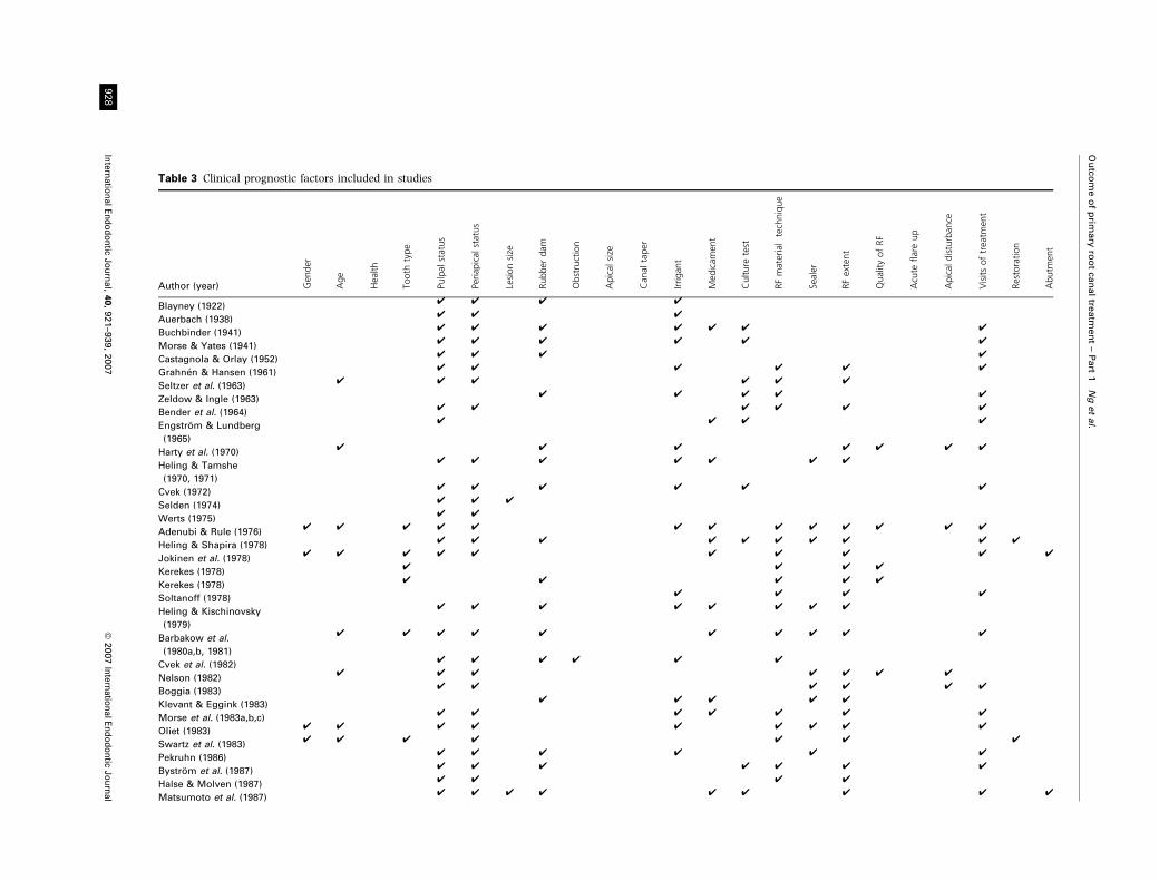
Table 3 Clinical prognostic factors included in studies
Author (year) Gen
der
Age
Health
Tooth
type
Pulpal
status
Periap
ical
status
Lesionsize
Rubber
dam
Obstruction
Apical
size
Can
altaper
Irrigan
t
Med
icam
ent
Culture
test
RFmaterialtechnique
Sealer
RFextent
QualityofRF
Acute
flareup
Apical
disturban
ce
Visitsoftreatm
ent
Restoration
Abutm
ent
Blayney (1922)4 4 4 4
Auerbach (1938)4 4 4
Buchbinder (1941)4 4 4 4 4 4 4
Morse & Yates (1941)4 4 4 4 4 4
Castagnola & Orlay (1952)4 4 4 4
Grahnen & Hansen (1961)4 4 4 4 4 4
Seltzer et al. (1963)4 4 4 4 4 4
Zeldow & Ingle (1963)4 4 4 4 4
Bender et al. (1964)4 4 4 4 4 4
Engstrom & Lundberg
(1965)
4 4 4 4
Harty et al. (1970)4 4 4 4 4 4 4
Heling & Tamshe
(1970, 1971)
4 4 4 4 4 4 4
Cvek (1972)4 4 4 4 4 4
Selden (1974)4 4 4
Werts (1975)4 4
Adenubi & Rule (1976)4 4 4 4 4 4 4 4 4 4 4 4 4
Heling & Shapira (1978)4 4 4 4 4 4 4 4 4 4
Jokinen et al. (1978)4 4 4 4 4 4 4 4 4 4
Kerekes (1978)4 4 4 4
Kerekes (1978)4 4 4 4 4
Soltanoff (1978)4 4 4 4
Heling & Kischinovsky
(1979)
4 4 4 4 4 4 4 4
Barbakow et al.
(1980a,b, 1981)
4 4 4 4 4 4 4 4 4 4
Cvek et al. (1982)4 4 4 4 4 4
Nelson (1982)4 4 4 4 4 4 4
Boggia (1983)4 4 4 4 4 4
Klevant & Eggink (1983)4 4 4 4 4
Morse et al. (1983a,b,c)4 4 4 4 4 4 4
Oliet (1983)4 4 4 4 4 4 4 4 4
Swartz et al. (1983)4 4 4 4 4 4 4
Pekruhn (1986)4 4 4 4 4 4
Bystrom et al. (1987)4 4 4 4 4 4 4
Halse & Molven (1987)4 4 4 4
Matsumoto et al. (1987)4 4 4 4 4 4 4 4 4
Outco
meofprim
ary
rootca
naltre
atm
ent–Part
1Ngetal.
Intern
ational
Endodontic
Journal,
40,921–9
39,2007
ª2007Intern
ational
Endodontic
Journal
928

Ørstavik et al. (1987) &
Eriksen et al. (1988)
4 4 4
Safavi et al. (1987)4 4 4 4 4 4 4 4
Akerblom &
Hasselgren (1988)
4 4 4 4 4 4 4 4 4
Shah (1988)4 4 4 4 4 4
Sjogren et al. (1990)4 4 4 4 4 4 4 4 4 4 4
Murphy et al. (1991)4 4
Cvek (1992)4 4 4 4 4
Reid et al. (1992)4 4 4 4 4
Jurcak et al. (1993)4 4 4
Smith et al. (1993)4 4 4 4 4 4 4 4 4 4
Peak (1994)4 4 4 4 4
Friedman et al. (1995)4 4 4 4 4 4
Calisken & Sen (1996)4 4 4 4 4 4 4 4 4 4 4
Østavik (1996)4 4 4 4
Peretz et al. (1997)4 4 4
Sjogren et al. (1997)4 4 4 4 4 4 4
Lilly et al. (1998)4 4 4 4 4 4
Trope et al. (1999)4 4 4 4 4 4 4
Ricucci et al. (2000)4 4 4 4
Weiger et al. (2000)4 4 4 4 4 4 4 4
Chugal et al. (2001)4
Deutsch et al. (2001)4 4 4 4 4 4
Heling et al. (2001)4 4 4 4 4 4 4
Peak et al. (2001)4 4 4 4 4
Pettiette et al. (2001)4 4
Benenati & Khajotia (2002)4 4 4 4
Cheung (2002)4 4 4 4 4 4 4 4 4 4
Hoskinson et al. (2002)4 4 4 4 4 4 4 4 4 4 4 4 4 4
Peters & Wesselink (2002)4 4 4 4 4 4 4 4 4 4 4
Total 8 13 4 13 51 49 6 33 2 1 2 32 20 14 38 25 29 7 0 5 35 8 2
RF, root filling.
Ngetal.
Outco
meofprim
ary
rootca
naltre
atm
ent–Part
1
ª2007Intern
ational
Endodontic
Journal
Intern
ational
Endodontic
Journal,
40,921–9
39,2007
929

Table 4 Estimated success rates by study characteristics
Factor/categories
aNo.
studies
identified
Strict radiographic criteria Loose radiographic criteria
No.
studies
No.
units
Estimated pooled success ratesNo.
studies
No.
units
Estimated pooled success rates
cUn-weighted (%) dWeighted (%) cUn-weighted (%) dWeighted (%)
Outcome measure used
Radiographic 27 17 4745 74.4 74.1 (66.9–81.3) 14 (13)b 7177 83.4 84.1 (79.0–89.3)
Clinical + radiographic 36 23 10799 72.8 75.0 (68.4–81.7) 24 (23)b 10430 82.0 85.8 (81.8–89.9)
Duration after treatment (months)
6 4 2 1120 17.3 29.6 (14.2–73.3) 2 4633 89.1 89.1 (78.8–99.5)
12 9 5 2080 68.6 67.7 (39.0–96.4) 4 798 76.7 69.5 (52.3–86.6)
24 6 3 2328 75.8 67.3 (43.8–90.9) 3 1103 82.7 82.5 (75.2–89.8)
36 5 2 941 80.4 80.6 (78.0–83.1) 3 254 69.3 66.6 (36.1–95.9)
48 6 5 2931 84.5 83.8 (79.3–88.3) 2 301 93.5 61.7 (25.0–96.0)
>48 9 8 1162 86.8 85.4 (80.3–90.6) 1 821 84.3 84.3 (81.8–86.8)
Year of publication
Before 1960 5 2 1245 68.2 68.2 (65.6–70.8) 4 1580 82.2 84.2 (72.2–96.1)
1960s 5 2 1458 81.3 79.7 (74.0–85.4) 3 3669 80.4 80.4 (79.2–81.7)
1970s 12 9 5468 69.8 79.0 (66.7–91.3) 6 (5)b 4400 77.0 84.0 (71.3–96.6)
1980s 16 9 2834 77.8 74.8 (62.5–87.0) 10 (9)b 3770 89.7 88.2 (85.0–91.4)
1990s 14 10 2007 83.9 76.9 (69.7–84.1) 8 2271 83.3 85.5 (80.2–90.9)
2000s 11 8 2532 65.2 68.0 (60.5–75.4) 7 1917 85.2 85.1 (78.0–92.3)
Geographic location of study
USA or Canada 24 9 2412 67.5 74.1 (64.9–83.2) 20 (19)b 9393 86.5 88.1 (84.9–91.2)
Scandinavian country 15 12 6435 71.5 80.5 (71.1–89.8) 3 (2)b 3347 70.4 70.3 (61.3–79.2)
Other country 24 19 6697 76.9 71.2 (64.4–78.1) 15 4867 83.4 84.5 (80.4–88.5)
Qualification of operators
Undergraduate students 21 14 8306 68.4 74.8 (67.0–82.7) 11 (10)b 7808 79.9 83.3 (75.8–90.9)
GDP 7 6 1353 64.4 65.7 (56.3–75.1) 5 1228 85.5 86.2 (82.9–89.5)
Postgraduate students 4 4 1336 82.9 77.2 (64.5–89.8) 2 959 93.1 93.1 (91.5–94.7)
Specialist 23 11 3288 87.6 84.8 (80.1–89.4) 17 (16)b 6368 84.7 87.6 (83.9–91.3)
GDP, general dental practitioners.aTotal number of studies identified for the respective study characteristics is equal to or smaller than the summation of number of studies under strict and loose criteria as some studiesreported success rates based on both criteria.bNumber in bracket indicating the number of studies included in the meta-analysis after those studies with 100% rates by the respective factor under investigation have been excluded.cUn-weighted pooled success rates were estimated based on the Hepworth & Friedman (1997)’s approach.dWeighted pooled success rates were estimated using random effects meta-analysis (where there was only one study, its reported success rate and confident intervals were presented).
Outco
meofprim
ary
rootca
naltre
atm
ent–Part
1Ngetal.
Intern
ational
Endodontic
Journal,
40,921–9
39,2007
ª2007Intern
ational
Endodontic
Journal
930

out in Scandinavian (15 studies, Sweden/Norway) or
other countries (24 studies) including: UK (eight
studies), Israel (four studies), Holland (two studies),
Switzerland (one study), Australia (one study), Ger-
many (one study), Hong Kong (one study), India (one
study), Italy (one study), Japan (one study), Saudi
Figure 1 Probability of success based on strict radiographic criteria.
Figure 2 Probability of success based on loose radiographic criteria.
Ng et al. Outcome of primary root canal treatment – Part 1
ª 2007 International Endodontic Journal International Endodontic Journal, 40, 921–939, 2007 931

Arabia (one study), South Africa (one study) and
Turkey (one study) (Table 2). The studies performed in
the North American countries reported the treatment
outcome data more frequently based on loose radio-
graphic criteria than on strict criteria. In contrast, most
of the outcome data from the Scandinavian countries
were based on strict rather than loose criteria. Based on
the loose criteria, the pooled estimate of success rate of
treatment carried out in Scandinavian countries
(70.3%) was much lower than for those in North
American (88.1%) or other (84.5%) countries; how-
ever, the pooled estimate for the Scandinavian coun-
tries, only consisted of two studies. In stark contrast,
the pooled estimate of success rate from outcome data
based on strict criteria from the Scandinavian countries
(80.5%) was the highest (Table 4).
Qualification of operators (undergraduate, postgraduate,
GDP and specialist)
Only two studies compared the outcome of root canal
treatment by qualification of operators. Ingle et al.
(1965) (a study excluded from this review; Table 1),
found no significant difference in success rates of
treatment carried out by undergraduates or private
practitioners, in agreement with Cheung (2002) who
reported the qualification and experience of operator
had no influence on treatment outcome.
The majority of the reviewed studies classified
operator qualification as: undergraduate students (21
studies), GDP (seven studies), postgraduate students
(four studies) or specialists (23 studies). In five studies,
treatment was carried out by a mixed group of
operators and three studies did not provide this
information. From the results, treatment carried out
by postgraduate students and specialists had the
highest weighted pooled estimate of success, regardless
of strict or loose criteria (Table 4).
Source of heterogeneity
As the radiographic criteria for success have already
been shown to have a significant effect on the pooled
success rates, further meta-regression analyses were
therefore carried out, separately on success rates based
on strict or loose criteria, to explore which of the other
study characteristics were potentially responsible for
the statistical heterogeneity. None had significant
effects on the success rates reported by the studies or
could account for the heterogeneity (Table 5) in
estimating the pooled success rate of primary root
canal treatment.
Discussion
Most of the selected studies were prospective cohort or
retrospective studies, therefore the levels of evidence
provided by them are grade B (levels 2 or 3) based on
the criteria given by the Oxford centre for evidence-
based medicine (Phillips et al. 1998 http://www.cebm.
net/index.aspx?0=1025). There were only six random-
ized trials investigating different aspects of root canal
treatment procedures on outcomes, including
the effects of sealers (AH26 [DeTrey AG, Zurich,
Switzerland]; Procosol [Star Dental, Conshohocken,
PA, USA], Kloropercha) (Ørstavik et al. 1987); root
filling materials (Hydron! [Hydron Technologies,
Pompano Beach, FL, USA], gutta-percha with AH26!
sealer) (Reid et al. 1992); single-visit versus multiple-
visit root canal treatment (Trope et al. 1999, Weiger
et al. 2000, Peters & Wesselink 2002); use of Ca(OH)2dressing versus no medicament in multiple-visit treat-
ment (Trope et al. 1999); and the use of stainless steel
versus nickel–titanium hand files (Pettiette et al. 2001).
Table 5 Results of meta-regression analysis to account for the
source of heterogeneity
Covariate included
Strict Loose
I2 s2 I2 s2
No. covariate
included
0.985 0.0247 0.973 0.0085
Year of publication
(before 1970s, 1970–1989,
1990–2002)
0.983 0.0265 0.971 0.0098
Geographical
location of study
(USA, Scandinavian
or other countries)
0.984 0.0244 0.952 0.0069
Unit of measure
(root or tooth)
0.984 0.0253 0.961 0.0081
Qualification of operator
(specialist, postgraduate,
undergraduate, GDP or
mixed group)
0.979 0.0209 0.974 0.0073
Criteria for success
(radiographic vs. combined
radiographic & clinical)
0.986 0.0254 0.974 0.0088
Duration after treatment
(at least 4 years or shorter)
0.985 0.0228 0.973 0.0085
Recall rate 0.986 0.0218 0.975 0.0117
GDP, general dental practitioners; I2, proportion of total varia-tion due to heterogeneity across studies; s2, estimate ofbetween-study variance (if the I2 and s2 values were reducedby 10% after including a covariate in the regression model ascompared with the values estimated without any covariatesentered, the respective covariate was considered to be apotential source of heterogeneity).
Outcome of primary root canal treatment – Part 1 Ng et al.
International Endodontic Journal, 40, 921–939, 2007 ª 2007 International Endodontic Journal932

The Cochrane Oral Health group’s current guidelines
for a systematic review states that ‘the scope of the
review is to include all RCTs, where RCTs are inappro-
priate, rather than unavailable, other levels of evidence
may be considered’ (http://www.ohg.cochrane.org/
forms/writing_review.pdf September 2006). However,
the authors decided that the numerous observational
studies, whilst not having the feature of randomization
or control groups, represented useful and useable data
that could not be deemed inferior by any other criteria.
Instead of using exclusion rules to control the heter-
ogeneity of design, this systematic review followed the
recommendation by Stroup et al. (2000). Broad inclu-
sion criteria for studies were used and analyses were
performed to investigate the effect of study character-
istics on the estimated pooled success rates. Despite
using broad inclusion criteria, several well designed
and executed studies such as those by Strindberg
(1956), Ørstavik and Horsted-Bindslev (1993); Ørsta-
vik et al. (1986) and others (Table 1) had to be
excluded.
The goal was to explore the available data and
partition it to reveal the effect of study characteristics,
general patient factors, individual pre-, intra-, and
postoperative factors on treatment outcome, whilst
triangulating the outcomes of different approaches of
exploration.
Preliminary data collection was carried out by
two authors (Y-LN and KG) to explore its diversity.
Amongst the studies reviewed, there were substantial
variations in study characteristics such as sample
selection, definition of successful cases, duration after
treatment, type and strategy of data collection as
well as data analyses. Some of the potential clinical
prognostic factors (tooth type, age grouping, size
of apical preparation, definition of apical disturbance,
apical extent of root filling, quality of root filling
and quality of coronal restoration) were sub-classified
differently between studies. Therefore, a strategy
for pooling the data by recalculation of the avail-
able figures was derived. Based on this strategy, a
data collection form was designed, tested, refined
and adopted. Despite this, disagreement amongst
the reviewers existed (j ! 0.57–0.61). The disagree-
ment could be traced to a lack of clarity in the
presentation of methodology and results. For
some studies, data had to be extracted from the
discussion section where it was sometimes first
introduced. When there was a disagreement, an
agreement was negotiated between the examiners by
presenting the case for each view. In the majority of
cases, the source of errors were easily identified and
corrected.
The un-weighted pooled success rate by each factor
was calculated based on the approach used by Hep-
worth & Friedman (1997). However, this approach
does not take into consideration, the within and
between study variations as opposed to the study–
design-specific weighted pooled success rates estimated
using random effects meta-analysis. The discrepancies
in the success rates estimated using the two approaches
are well demonstrated in Table 4. Therefore, the results
based on the un-weighted pooled success rates were not
considered in the following discussions. The estimation
of pooled success rates for some sub-group analyses
within some study characteristics were based on small
data sets, restricting their value.
The significant difference in success rates judged by
strict or loose radiographic criteria has already been
iterated but in addition, the data based on strict criteria
revealed a clear trend for differences in the pooled
success rates from studies adopting different follow-up
durations; for example, 6 month follow-up compared
with longer duration. It should be mandatory to state
the criteria for success as part of the methodology of
future clinical root canal treatment outcome studies,
preferably stratified by both loose and strict radio-
graphic criteria. The European Society of Endodontol-
ogy’s (2006) suggest a clinical and radiographic
follow-up after at least 1 year with annual recall for
up to 4 years before a case is judged a failure. The
American Association of Endodontists suggests clinical
and radiographic evaluation for a 4- to 5-year period,
with the additional proviso of determining the func-
tionality of the treated tooth (http://www.aae.org/
dentalpro/guidelines.htm). The origin of this is proba-
bly based on the work of Strindberg (1956). From a
research perspective and based on this review, the cases
should be reviewed for a minimum of 1 year and
preferably for at least 3 years, after completion of
treatment. It would be preferable to standardize the
duration after treatment for all the patients or at least
to include the duration as a covariate into the statistical
model to account for any variations in the success rate
because of the different follow-up times. The best choice
of statistical analysis would be to analyse time to
healing (success) with survival analysis techniques,
however, it would require regular follow-up of all
patients. The reality is that the longer the duration of
follow-up after treatment, the greater the drop-out rate
at recall. Therefore, a balance has to be struck between
these competing ideals in both the medical and dental
Ng et al. Outcome of primary root canal treatment – Part 1
ª 2007 International Endodontic Journal International Endodontic Journal, 40, 921–939, 2007 933

fields, although the use of financial incentives may help
improve recall rates (Wang et al. 2004).
This review also highlighted two important method-
ological shortcomings in published root canal treat-
ment outcome studies. The variability in radiographic
assessment because of subjectivity in radiograph read-
ing is well recognized (Goldman et al. 1972), yet the
good practice of employing at least two pre-calibrated
observers with intra- & inter-observer agreement tests,
was not adopted by most of the studies (n ! 56). In
addition, the statistical methods used for analysing the
association between potential influencing factors and
treatment outcome, did not take account of the effects
of potential confounders.
The overall success rates were not affected by ‘year of
publication’ or ‘geographic location of study’. In the
former category, the measure of relevance should really
be the ‘year in which treatment was carried out’ but
few studies provide this information. Nevertheless, the
absence of obvious improvement in success rates by the
year of publication suggests that the advances in
technology and materials used for root canal treatment
do not appear to have influenced treatment outcome
significantly. Such a suggestion is strongly refuted by
endodontists on the grounds that the apparent lack of
improvement in success rates is a function of more
adventurous case selection fuelled by confidence in
better skills and outcomes. The validity of this propo-
sition is explored further in the second part. For the
present, it is argued that the lack of improvement in
success rates could be attributed to the fact that, whilst
technology has improved instruments and materials to
achieve a set of goals, the principles underpinning
those goals have not changed over the duration
covered by this review (Hall 1928). This brings to the
fore, the classic debate about the relative value of
biological versus the technical principles in dentistry
(Noyes 1922, Naidorf 1972). Noyes (1922) lamented
that dentists were not trained to think in biological
concepts but to act in mechanical procedures; whilst
Naidorf (1972) applauding the technical excellence
achieved by the pre-occupation of dentists with this
element, deplored the lack of biological awareness of
the basic pathology of the problem or the biological
consequences of the treatment. The clinical academics
in this discipline would probably sustain the validity of
these assertions, even today. It is interesting to note
that the success rates of studies from the North
American countries, where the use of contemporary
technology is probably most widely recommended fared
no better than those from other countries. Further-
more, the adoption of strict radiographic criteria and
microbiological awareness in their approach appeared
to bring better results in studies performed in the
Scandinavian countries. This speculation is important
because it centres around the debates that raged in the
1960s and 1970s about the value of the microbial
culture test in informing the progress of treatment, a
practice, long as abandoned as unnecessary (Engstrom
et al. 1964, Mikkelsen & Theilade 1969, Oliet & Sorin
1969, Morse 1971, Sims 1973, Frank et al. 1978,
Molander et al. 1996a,b). This ultimately led to the
adoption of single-visit treatment by many endodontists
on the basis of the cost-benefit analysis (Spangberg
2001) an issue that will be explored further in the
second part. The historical importance of this biological
versus technical debate is important to appreciate,
because it fundamentally changed the way root canal
treatment was conceived and practiced; from a micro-
bially aware post-focal infection era, to one dominated
by a technological awareness but relative microbiolog-
ical ignorance. The problem of geographical location
also merits close inspection, as sometimes, a single
study may report pooled data from multi-centre eval-
uations (Friedman et al. 1995).
Although the educational and experience back-
ground of the operators had no significant influence
on their respective success rates in individual studies,
the estimated pooled success rates for endodontists or
postgraduates were higher than for other dentist
groups in this review. The important influence of the
background of operators on the technical outcome of
endodontic procedures has been demonstrated in lab-
oratory studies (Gulabivala et al. 2000, Van Zyl et al.
2005) but there is a lack of appropriate tools or
methodology to objectively quantify operator skills. The
role of such refined technical skills must surely be
balanced against the overall understanding of the
problem and the motivation and integrity with which
the procedure is performed.
Conclusion
The estimated weighted pooled success rates of treat-
ments completed at least 1 year previously, ranged
between 68% and 85% when strict criteria were used.
The reported success rates have failed to improve over
the last four or five decades. The quality of evidence for
treatment factors affecting primary root canal treat-
ment outcome is sub-optimal; there was substantial
variation in the study–designs. It would be desirable to
standardize aspects of study–design, data recording and
Outcome of primary root canal treatment – Part 1 Ng et al.
International Endodontic Journal, 40, 921–939, 2007 ª 2007 International Endodontic Journal934

presentation format of outcome data in the much
needed future outcome studies. The second part of this
paper will present the results of meta-analyses and
meta-regression to investigate the effect of individual
clinical factors on the success rates of primary root
canal treatment.
Acknowledgement
The authors acknowledge the mentorship provided by
Professor Mark Gilthorpe of the University of Leeds
(previously based at the Eastman Dental Institute)
during the early stages of this project.
References
Adenubi JO (1978) The criteria for success in endodontics.
Nigerian Medical Journal 8, 404–10.
Adenubi JO, Rule DC (1976) Success rate for root fillings in
young patients. British Dental Journal 141, 237–41.
Akerblom A, Hasselgren G (1988) The prognosis for endodon-
tic treatment of obliterated root canals. Journal of Endodontics
14, 565–7.
Appleton JLT (1932) A note on the clinical value of bacteri-
ological controlling the treatment of periapical infection.
Dental Cosmos IXXiv, 798–800.
Ashkenaz PJ (1984) One-visit endodontics. Dental Clinics North
America 28, 853–63.
Auerbach MB (1938) Clinical approach to the problem of pulp
canal therapy. Journal of American Dental Association 25,
939–42.
Augsburger RA, Peters DD (1990) Radiographic evaluation of
extruded obturation materials. Journal of Endodontics 16,
492–7.
Barbakow FH, Cleaton-Jones P, Friedman D (1980a) An
evaluation of 566 cases of root canal therapy in general
dental practice. 1. Diagnostic criteria and treatment details.
Journal of Endodontics 6, 456–60.
Barbakow FH, Cleaton-Jones P, Friedman D (1980b) An
evaluation of 566 cases of root canal therapy in general
dental practice. 2. Postoperative observations. Journal of
Endodontics 6, 485–9.
Barbakow FH, Cleaton-Jones PE, Friedman D (1981) Endo-
dontic treatment of teeth with periapical radiolucent areas
in a general dental practice. Oral Surgery, Oral Medicine, Oral
Patholology 51, 552–9.
Basmadjian-Charles CL, Farge P, Bourgeois DM, Lebrun T
(2002) Factors influencing the long-term results of end-
odontic treatment: a review of the literature. International
Dental Journal 52, 81–6.
Bender IB, Seltzer S, Turkenkopf S (1964) To culture or not to
culture? Oral Surgery, Oral Medicine, Oral Patholology 18,
527–40.
Benenati FW, Khajotia SS (2002) A radiographic recall
evaluation of 894 endodontic cases treated in a dental
school setting. Journal of Endodontics 28, 391–5.
Blayney JR (1922) The clinical results of pulp treatment.
Journal of National Dental Association 16, 198–208.
Boggia R (1983) A single-visit treatment of septic root canals
using periapically extruded endomethasone. British Dental
Journal 155, 300–5.
Buchbinder M (1936) A statistical study of root-canal therapy.
The Dental Cosmos 78, 20–6.
Buchbinder M (1941) A statistical comparison of cultured and
non-cultured root canal cases. Journal of Dental Research 2,
93–6.
Bystrom A, Happonen RP, Sjogren U, Sundqvist G (1987)
Healing of periapical lesion of pulpless teeth after endodontic
treatment with controlled asepsis. Endodontics & Dental
Traumatology 3, 58–63.
Calisken MK, Sen BH (1996) Endodontic treatment of teeth
with apical periodontitis using calcium hydroxide: a long-
term study. Endodontics & Dental Traumatology 12, 215–21.
Caplan DJ, White BA (2001) Clinical factors related to
noncompletion of root canal therapy. Journal of Public
Health in Dentistry 61, 6–13.
Caplan DJ, Kolker J, Rivera EM, Walton RE (2002) Relation-
ship between number of proximal contacts and survival of
root canal treated teeth. International Endodontic Journal 35,
193–9.
Castagnola L, Orlay HG (1952) Treatment of gangrene of
the pulp by the Walkhoff method. British Dental Journal 19,
93–100.
Cheung GS (2002) Survival of first-time nonsurgical root
canal treatment performed in a dental teaching hospital.
Oral Surgery, Oral Medicine, Oral Pathology, Oral Radiology
and Endodontics 93, 596–604.
Chugal NM, Clive JM, Spangberg LSW (2001) A prognostic
model for assessment of the outcome of endodontic treat-
ment: effect of biologic and diagnostic variables. Oral
Surgery, Oral Medicine, Oral Pathology, Radiology and Endo-
dontics 91, 342–52.
Cochran WG (1954) The combination of estimates from
different experiments. Biometrics 10, 101–29.
Coolidge ED (1926) A discussion of clinical results of root-
canal treatment and filling. The Dental Cosmo Ixix, 1280–8.
Curson I (1966) Endodontic techniques – prognosis in
endodontics. British Dental Journal 20, 568–70.
Cvek M (1972) Treatment of non-vital permanent incisors
with calcium hydroxide. Odontologisk Revy 23, 27–44.
Cvek M (1992) Prognosis of luxated non-vital maxillary
incisors treated with calcium hydroxide and filled with
gutta-percha. A retrospective clinical study. Endodontics &
Dental Traumatology 8, 45–55.
Cvek M, Hollender L, Nord C-E (1976) Treatment of non-vital
permanent incisors with calcium hydroxide. Odontologisk
Revy 27, 93–108.
Ng et al. Outcome of primary root canal treatment – Part 1
ª 2007 International Endodontic Journal International Endodontic Journal, 40, 921–939, 2007 935

Cvek M, Granath L, Lundberg M (1982) Failures and healing
in endodontically treated non-vital anterior teeth with
posttraumatically reduced pulpal lumen. Acta Odontologica
Scandinavica 40, 223–8.
DerSimonian R, Laird N (1986) Meta-analysis in clinical trials.
Control Clinical Trials 7, 177–88.
Deutsch AS, Musikant BL, Cohen BI, Kase D (2001) A study of
one visit treatment using EZ-fill root canal sealer. Endodontic
Practice 4, 29–36.
Engstrom B, Lundberg M (1965) The correlation between
positive culture and the prognosis of root canal therapy after
pulpectomy. Odontologisk Revy 16, 193–204.
Engstrom B, Segerstad LHA, Ramstrom G, Frostell G (1964)
Correlation of positive cultures with the prognosis for root
canal treatment. Odontologisk Revy 15, 257–70.
Eriksen HM, Ørstavik D, Kerekes K (1988) Healing of apical
periodontitis after endodontic treatment using three differ-
ent root canal sealers. Endodontics & Dental Traumatology 4,
114–7.
European Society of Endodontology (2006) Quality guidelines
for endodontic treatment: consensus report of the European
Society of Endodontology. International Endodontic Journal
39, 921–30.
Eysenck HJ (1994) Meta-analysis and its problems. British
Medical Journal 309, 789–92.
Frank AL, Abou Rass M, Glick DH (1978) Changing trends in
endodontics. Journal of American Dental Association 96, 202–
10.
Friedman S (1997) Success and failure of initial endodontic
therapy. Ontario Dentist 74, 35–8.
Friedman S, Lost C, Zarrabian M, Trope M (1995) Evaluation
of success and failure after endodontic therapy using glass
ionomer cement sealer. Journal of Endodontics 21, 384–90.
Frostell G (1963) Clinical significance of the root canal culture.
Transactions of Third International Conference on Endodontics,
112–22.
Goldman M, Pearson AH, Darzenta N (1972) Endodontic
success – who’s reading the radiograph?. Oral Surgery, Oral
Medicine, Oral Pathology 33, 432–7.
Grahnen H, Hansen L (1961) The prognosis of pulp and root
canal therapy. Odontologisk Revy 12, 146–65.
Grossman LI, Shepard LI, Person LA (1964) Roentgenologic
and clinical evaluation of endodontically treated teeth. Oral
Surgery 17, 368–74.
Grove CJ (1921) Nature’s method of making perfect root
fillings following pulp removal, with a brief consideration of
the development of secondary cementum. The Dental Cosmo
11, 698–981.
Grove CJ (1923) A method of pulp removal to prevent
periapical infection. Journal of American Dental Association
10, 93–108.
Gulabivala K, Abdo S, Sherriff M, Regan JD (2000) The
influence of interfacial forces and duration of filing on root
canal shaping. Endodontics & Dental Traumatology 16, 166–
74.
Gutknecht N, Kaiser F, Hassan A, Lampert F (1996) Long-
term clinical evaluation of endodontically treated teeth by
Nd:YAG lasers. Journal of Clinical Laser Medicine and Surgery
14, 7–11.
Hall EM (1928) Pulpless teeth. The Dental Cosmo Ixx, 145–
50.
Halse A, Molven O (1987) Overextended gutta-percha and
Kloroperka N-Ø root canal fillings. Radiographic findings
after 10–17 years. Acta Odontologica Scandinavica 45, 171–
7.
Harty FJ, Parkins BJ, Wengraf AM (1970) Success rate in root
canal therapy. A retrospective study on conventional cases.
British Dental Journal 128, 65–70.
Heling B, Kischinovsky D (1979) Factors affecting successful
endodontic therapy. Journal of British Endodontic Society 12,
83–9.
Heling B, Shapira J (1978) Roentgenologic and clinical
evaluation of endodontically treated teeth, with or without
negative culture. Quintessence International 11, 79–84.
Heling B, Tamshe A (1970) Evaluation of the success of
endodontically treated teeth. Oral Surgery, Oral Medicine,
Oral Pathology 30, 533–6.
Heling B, Tamshe A (1971) Statistical evaluation of the
success of endodontically treated teeth. New York Journal of
Dentistry 41, 69–82.
Heling I, Bialla-Shenkman S, Turetzky A, Horwitz J, Sela J
(2001) The outcome of teeth with periapical periodontitis
treated with nonsurgical endodontic treatment: a comput-
erized morphometric study. Quintessence International 32,
397–400.
Hepworth MJ, Friedman S (1997) Treatment outcome of
surgical and non-surgical management of endodontic fail-
ures. Journal of Canadian Dental Association 63, 364–71.
Hession RW (1981) Long-term evaluation of endodontic
treatment: anatomy, instrumentation, obturation – the
endodontic practice triad. International Endodontic Journal
14, 179–84.
Hinman TP (1921) The interpretation of X-ray pictures of
apical granulations, giving differential diagnosis of cases
favourable and cases unfavourable for treatment and
root-canal filling. Journal of National Dental Association 8,
83–7.
Hoskinson SE, Ng YL, Hoskinson AE, Moles DR, Gulabivala K
(2002) A retrospective comparison of outcome of root canal
treatment using two different protocols. Oral Surgery, Oral
Medicine, Oral Patholology, Oral Radiolology and Endodontics
93, 705–15.
Ingle JL, Beveridge EE, Glick DH, Weichman JA (1965) Modern
endodontic therapy. In: Ingle JL, Bakland LK, eds. Endodon-
tics, 4th edn. Baltimore, MA: Williams and Wilkins 1994,
pp. 27–53.
Jokinen MA, Kotilainen R, Poikkeus P, Poikkeus R, Sarkki L
(1978) Clinical and radiographic study of pulpectomy and
root canal therapy. Scandinavian Journal of Dental Research
86, 366–73.
Outcome of primary root canal treatment – Part 1 Ng et al.
International Endodontic Journal, 40, 921–939, 2007 ª 2007 International Endodontic Journal936

Jurcak JJ, Bellizzi R, Loushine RJ (1993) Successful single-visit
endodontics during operation desert shield. Journal of
Endodontics 19, 412–3.
Kerekes K (1978) Radiographic assessment of an endodontic
treatment method. Journal of Endodontics 4, 210–3.
Kerekes K, Tronstad L (1979) Long-term results of endodontic
treatment performed with a standardized technique. Journal
of Endodontics 5, 83–90.
Klevant FJH, Eggink CO (1983) The effect of canal preparation
on periapical disease. International Endodontic Journal 16,
68–75.
Kojima K, Inamoto K, Nagamatsu K et al. (2004) Success rate
of endodontic treatment of teeth with vital and nonvital
pulps. A meta-analysis. Oral Surgery, Oral Medicine, Oral
Pathology, Oral Radiology and Endodontics 97, 95–9.
Kullendorff B, Grondahl K, Rohlin M, Henrikson CO (1988)
Subtraction radiography for the diagnosis of periapical bone
lesions. Endodontics and Dental Traumatology 4, 253–9.
Lazarski MP, Walker WA III, Flores CM, Schindler WG,
Hargreaves KM (2001) Epidemiological evaluation of the
outcomes of non-surgical root canal treatment in a large
cohort of insured dental patients. Journal of Endodontics 27,
791–6.
Lewsey JD, Gilthorpe MS, Gulabivala K (2001) An introduction
to meta-analysis within the framework of multilevel model-
ling using the probability of success of root canal treatment
as an illustration. Community Dental Health 18, 131–7.
Lilly JP, Cox D, Arcuri M, Krell KV (1998) An evaluation of
root canal treatment in patients who have received irradi-
ation to the mandible and maxilla. Oral Surgery, Oral
Medicine, Oral Pathology, Oral Radiology and Endodontics 86,
224–6.
Lynch CD, Burke FM, NıRiordain R, Hannigan A (2002)
Preoperative periapical status and survival of root filled
teeth: a pilot investigation. International Endodontic Journal
35, 492 (Abstract).
Macphee GG (1936) The problem of the pulpless tooth. British
Dental Journal 58, 119–25.
Markitziu A, Heling I (1981) Endodontic treatment of patients
who have undergone irradiation of the head and neck. A
longitudinal follow-up of eleven endodontically treated
teeth. Oral Surgery, Oral Medicine, Oral Pathology 52, 294–8.
Matsumoto T, Nagai T, Ida Km Ito M et al. (1987) Factors
affecting successful prognosis of root canal treatment.
Journal of Endodontics 13, 239–42.
Mikkelsen L, Theilade E (1969) False negative cultures from
root canals. Inhibition of bacterial growth by some drugs
used in root canal therapy. Acta Odontologica Scandinavica
27, 387–96.
Molander A, Reit C, Dahlen G (1996a) Microbiological root
canal sampling; diffusion of a technology. International
Endodontic Journal 29, 163–7.
Molander A, Reit C, Dahlen G (1996b) Reasons for dentists’
acceptance or rejection of microbiological root canal sam-
pling. International Endodontic Journal 29, 168–72.
Molven O, Halse A (1988) Success rates for gutta-percha and
Kloropercha N-Ø root fillings made by undergraduate
students: radiographic findings after 10–17 years. Interna-
tional Endodontic Journal 21, 243–50.
Morse DR (1971) The endodontic culture technique: an
impractical and unnecessary procedure. Dental clinics of
North America 15, 793–806.
Morse FW, Yates MF (1941) Follow up studies of root-filled
teeth in relation to bacteriologic findings. Journal of American
Dental Association 28, 956–71.
Morse DR, Esposito JV, Pike C, Furst ML (1983a) A radio-
graphic evaluation of the periapical status of teeth treated
by the gutta-percha–eucapercha endodontic method: a one-
year follow-up study of 458 root canals. Part I. Oral Surgery,
Oral Medicine, Oral Pathology 55, 607–10.
Morse DR, Esposito JV, Pike C, Furst ML (1983b) A radio-
graphic evaluation of the periapical status of teeth treated
by the gutta-percha-eucapercha endodontic method: a one-
year follow-up study of 458 root canals. Part II. Oral
Surgery, Oral Medicine, Oral Pathology 56, 89–96.
Morse DR, Esposito JV, Pike C, Furst ML (1983c) A radio-
graphic evaluation of the periapical status of teeth treated
by the gutta-percha-eucapercha endodontic method: a one-
year follow-up study of 458 root canals. Part III. Oral
Surgery, Oral Medicine, Oral Pathology 56, 190–7.
Murakami M, Inoue S, Inoue N (2002) Clinical evaluation of
audiometric control root canal treatment: a retrospective
case study. Quintessence International 33, 465–74.
Murphy WK, Kaugars GE, Collett WK, Dodds RN (1991)
Healing of periapical radiolucencies after nonsurgical end-
odontic therapy. Oral Surgery, Oral Medicine, Oral Pathology
71, 620–4.
Naidorf IJ (1972) Inflammation and infection of pulp and
periapical tissues. Oral Surgery, Oral Medicine, Oral Pathology
34, 486–97.
Neiderman R, Theodosopoulou JN (2003) A systematic review
of in vivo retrograde obturation materials. International
Endodontic Journal 36, 577–85.
Nelson IA (1982) Endodontics in general practice – a retro-
spective study. International Endodontic Journal 15, 168–72.
Nichols E (1963) Assessment of the periapical status of
pulpless teeth. British Dental Journal 114, 453–9.
Noyes FE (1922) (In Blayney JR). The clinical results of pulp
treatment. Journal of National Dental Association16, 198–208.
Oliet S (1983) Single-visit endodontics: a clinical study. Journal
Endodontics 9, 147–52.
Oliet S, Sorin SM (1969) Evaluation of clinical results based
upon culturing root canals. Journal of British Endodontic
Society 3, 3–6.
Oliver CM, Abbott PV (2001) Correlation between clinical
success and apical dye penetration. International Endodontics
Journal 34, 637–44.
Ørstavik D, Horsted-Bindslev P (1993) A comparison of
endodontic treatment results at two dental schools. Interna-
tional Endodontic Journal 26, 348–54.
Ng et al. Outcome of primary root canal treatment – Part 1
ª 2007 International Endodontic Journal International Endodontic Journal, 40, 921–939, 2007 937

Ørstavik D, Kerekes K, Eriksen HM (1986) The periapical
index: a scoring system for radiographic assessment of
apical periodontitis. Endodontics & Dental Traumatology 2,
20–34.
Ørstavik D, Kerekes K, Eriksen HM (1987) Clinical perfor-
mance of three endodontic sealers. Endodontics & Dental
Traumatology 3, 178–86.
Østavik D (1996) Time-course and risk analyses of the
development and healing of chronic apical periodontitis in
man. International Endodontic Journal 29, 150–5.
Paik S, Sechrist C, Torabinejad M (2004) Levels of evidence for
the outcome of endodontic retreatment. Journal of Endodon-
tics 30, 745–50.
Peak JD (1994) The success of endodontic treatment in
general dental practice: a retrospective clinical and radio-
graphic study. Primary Dental Care 1, 9–13.
Peak JD, Hayes SJ, Bryant ST, Dummer PM (2001) The
outcome of root canal treatment. A retrospective study
within the armed forces (Royal Air Force). British Dental
Journal 190, 140–4.
Pekruhn RB (1986) The incidence of failure following single-
visit endodontic therapy. Journal of Endodontics 12, 68–72.
Peretz B, Yakir O, Fuks AB (1997) Follow up after root canal
treatment of young permanent molars. Journal of Clinical
Pediatric Dentistry 21, 237–40.
Peters LB, Wesselink PR (2002) Periapical healing of end-
odontically treated teeth in one and two visits obturated in
the presence or absence of detectable microorganisms.
International Endodontic Journal 35, 660–7.
Peterson J, Gutmann JL (2001) The outcome of endodontic
re-surgery: a systematic review. International Endodontic
Journal 34, 169–75.
Pettiette MT, Delano O, Trope M (2001) Evaluation of success
rate of endodontic treatment performed by students with
stainless-steel K-files and nickel–titanium hand files. Journal
of Endodontics 27, 124–7.
Phillips B, Ball C, Sackett D et al. (1998) Oxford Centre for
Evidence-Based Medicine Levels of Evidence in Level of Evi-
dence and Grades of Recommendation. Oxford centre for
evidence-based medicine http://www.cebm.net/levels_of_
evidence.asp#levels.
Puterbaugh PG (1926) Pulp canal therapeutics. Journal of
American Dental Association Oct, 1384–91.
Ratliff DP (1973) The success rate of root canal filling in
permanent incisors in children. University of Newcastle Upon
Tyne Medical Gazette 67, 42–5.
Reid RJ, Abbott PV, McNamara JR, Heithersay GS (1992) A
five-year study of hydron root canal fillings. International
Endodontic Journal 25, 213–20.
Rhein ML, Krasnow F, Gies WJ (1926) A prolonged study of
the electrolytic treatment of dental focal infection – a
preliminary report. The Dental Cosmo Ixviii, 971–81.
Ricucci D, Langeland K (1997) Incomplete calcium hydroxide
removal from the root canal: a case report. International
Endodontic Journal 30, 418–21.
Ricucci D, Grondahl K, Bergenholtz G (2000) Periapical status
of root-filled teeth exposed to the oral environment by loss of
restoration or caries. Oral Surgery, Oral Medicine, Oral
Pathology, Oral Radiology and Endodontics 90, 354–9.
Sackett DL, Rosenberg WM, Gray JA, Haynes RB, Richardson
WS (1996) Evidence based medicine: what it is and what it
isn’t. British Medical Journal 13, 71–2.
Safavi KE, Dowden WE, Langeland K (1987) Influence of
delayed coronal permanent restoration on endodontic
prognosis. Endodontics & Dental Traumatology 3, 187–91.
Sathorn C, Parashos P, Messer HH (2005) Effectiveness of
single- versus multiple-visit endodontic treatment of teeth
with apical periodontitis: a systematic review and meta-
analysis. International Endodontic Journal 38, 347–55.
Selden HS (1974) Pulpoperiapical disease: diagnosis and
healing. A clinical endodontic study. Oral Surgery, Oral
Medicine, Oral Pathology 27, 271–83.
Seltzer S, Bender IB, Turkenkopf S (1963) Factors affecting
successful repair after root canal therapy. Journal of Amer-
ican Dental Association 57, 651–62.
Seto BG, Beumer J, Kagawa T, Klokkevold P, Wolinsky L
(1985) Analysis of endodontic therapy in patients irradiated
for head and neck cancer. Oral Surgery, Oral Medicine, Oral
Pathology 60, 540–5.
Shah N (1988) Non-surgical management of periapical
lesions: a prospective study. Oral Surgery, Oral Medicine,
Oral Pathology 66, 365–71.
Shi Y, Wang J, Cao C (1997) Clinical studies on pulpitis and
periapical periodontitis caused by traumatic occlusion.
Chinese Journal of Stomatology 32, 23–5.
Sims W (1973) Some comments on the microbiological
examination of root canals. Journal of Dentistry 2, 2–6.
Sjogren U, Hagglund B, Sundqvist G, Wing K (1990) Factors
affecting the long-term results of endodontic treatment.
Journal of Endodontics 16, 498–504.
Sjogren U, Figdor D, Persson S, Sundqvist G (1997)
Influence of infection at the time of root filling on the
outcome of endodontic treatment of teeth with apical
periodontitis. International Endodontic Journal 30, 297–
306.
Smith CS, Setchell DJ, Harty FJ (1993) Factors influencing the
success of conventional root canal therapy – a five-year
retrospective study. International Endodontic Journal 26,
321–33.
Soltanoff W (1978) A comparative study of the single-visit and
the multiple-visit endodontic procedure. Journal of Endodon-
tics 4, 278–81.
Spangberg LS (2001) Evidence-based endodontics: the one-
visit treatment idea. Oral Surgery, Oral Medicine, Oral
Pathology, Oral Radiology and Endodontics 91, 617–8.
Stabholz A (1990) Success rate in endodontics. Alpha Omegan
83, 20–4.
Storms JL (1969) Factors that influence the success of
endodontic treatment. Journal of Canadian Dental Association
35, 83–97.
Outcome of primary root canal treatment – Part 1 Ng et al.
International Endodontic Journal, 40, 921–939, 2007 ª 2007 International Endodontic Journal938

Strindberg LZ (1956) The dependence of the results of pulp
therapy on certain factors – an analytical study based on
radiographic and clinical follow-up examinations. Acta
Odontologica Scandinavica 14, 1–175.
Stroup DF, Berlin JA, Morton SC et al. (2000) Meta-analysis of
observational studies in epidemiology: a proposal for
reporting. Meta-analysis Of Observational Studies in Epide-
miology (MOOSE) group. Journal of American Medical Asso-
ciation 19, 2008–12.
Swartz DB, Skidmore AE, Griffin JA (1983) Twenty years of
endodontic success and failure. Journal of Endodontics 9,
198–202.
Taintor JF, Biesterfeld RC, Bonness BW (1978) Evaluating
clinical endodontic success. Dental Practice Dec, 36–41.
Teo CS, Chan NC, Lim SS (1986) Success rate in endodontic
therapy – a retrospective study. Part I. Dental Journal of
Malaysia 9, 7–10.
Thoden van Velzen SK, Duivenvoorden HJ, Schuurs AH
(1981) Probabilities of success and failure in endodontic
treatment: a Bayesian approach. Oral Surgery, Oral Medicine,
Oral Pathology 52, 85–90.
Thompson SG, Higgins JP (2002) How should meta-regression
analyses be undertaken and interpreted? Statistics in Med-
icine 15, 1559–73.
Trope M, Delano O, Ørstavik D (1999) Endodontic treatment of
teeth with apical periodontitis: single vs multivisit treat-
ment. Journal of Endodontics 25, 345–50.
Van Zyl S, Gulabivala K, Ng Y-L (2005) The effect of
customization of master gutta-percha on the apical control
of root canal fillings. International Endodontic Journal 38,
658–66.
Vernieks AA, Messer LB (1978) Calcium hydroxide induced
healing of periapical lesions: a study of 78 non-vital teeth.
Journal of British Endodontic Society 11, 61–8.
Waltimo TM, Boiesen J, Eriksen HM, Ørstavik D (2001)
Clinical performance of 3 endodontic sealers. Oral Surgery,
Oral Medicine, Oral Pathology, Oral Radiology and Endodontics
92, 89–92.
Wang N, Knight K, Dao T, Friedman S (2004) Treatment
outcome in endodontics – The Toronto Study. Phases I and
II: apical surgery. Journal of Endodontics 30, 751–61.
Weiger R, Axmann-Krcmar D, Lost C (1998) Prognosis of
conventional root canal treatment reconsidered. Endodontics
& Dental Traumatology 14, 1–9.
Weiger R, Rosendahl R, Lost C (2000) Influence of calcium
hydroxide intracanal dressings on the prognosis of teeth
with endodontically induced periapical lesions. International
Endodontic Journal 33, 219–26.
Weine FS, Buchanan SL (1997) Controversies in clinical
endodontics: Part 2. Single-appointment vsmultiple-appoint-
ment treatment. The Compendium of Continuing Education in
Dentistry 18, 140–4, 146, 148 passim; quiz 156.
Werts R (1975) Endodontic treatment: a five-year follow-up.
Dental Practice Jan, 29–30.
Wong M, Shelley JJ, Bodey T, Hall R (1992) Delayed root canal
therapy: an analysis of treatment over time. Journal of
Endodontics 18, 387–90.
Zeldow BJ, Ingle JI (1963) Correlation of the positive culture to
the prognosis of endodontically treated teeth: a clinical
study. Journal of American Dental Association 66, 23–7.
Ng et al. Outcome of primary root canal treatment – Part 1
ª 2007 International Endodontic Journal International Endodontic Journal, 40, 921–939, 2007 939