orthodontic topics for fifth years - college of dentsitry ... · web viewthis involves surgical...
TRANSCRIPT
ORTHODONTIC TOPICS FOR FIFTH YEARS
DIAGNOSTIC AIDS 1
TREATMENT PLANNING 5
EXTRACTION IN ORTHODONTICS 11
CROWDING 17
SPACING 19
OPEN BITE 21
ANTERIOR DEEP BITE 25
CROSSBITES 26
CANINE 31
ADULT ORTHODONTICS 35

Orthodontics for Fifth Years (2016) Prof. Akram Faisal Alhuwaizi
DIAGNOSTIC AIDS
Diagnostic aids include the use of:1. Study models2. Orthopantomography3. Cephalometrics4. Other views: hand wrist and periapical radiographs5. Photography6. 3D imaging
INTRAORAL RADIOGRAPHSThe intraoral radiographs are the easiest to take for most orthodontic patients. The most
frequently used views include:A. Intraoral periapical radiographsB. Bitewing radiographsC. Occlusal radiographs
A. Intraoral Periapical RadiographsThey are ideal for the detection of anomalies related to changes in tooth structure and /
or the lamina dura and/ or the periapical region. They are ideal for localized views in relatively small areas of interest because of the excellent clarity that they allow.
They are recommended for:1. Adult cases with periodontal disease2. Evaluation of the dental health of the deciduous and/or permanent teeth periapically3. Detection of pathologic conditions in the early stage especially dental caries4. Assessment of traumatized teeth after an injury (especially root fractures)5. Calculation of the total space analysis6. Detection of root resorption, before during and after treatment.
B. Bitewing RadiographsBitewing radiographs are used primarily to record the coronal portion of the upper and
the lower posterior dentition. They are used for:• the detection of proximal caries• the study of interdental bone height• the detection of secondary caries under restorations• the detection of overhanging margins of proximal restorations.
C. lntraoral Occlusal RadiographsThey are useful in localization of impacted teeth or for the study of the labio-lingual
position of the root apices in the anterior segments of the upper and the lower dentition.
HAND WRIST RADIOGRAPHSThe hand-wrist region is made up of numerous small bones. These bones show a
predictable and scheduled pattern of appearance, ossification and union from birth to maturity. Hence, this region is one of the most suited to assess skeletal age.
1

Orthodontics for Fifth Years (2016) Prof. Akram Faisal Alhuwaizi
SKELETAL AGESkeletal/radiological/anatomical age is considered to be the most reliable age for
assessment of growth for orthodontic purposes. It is closely related to the growth of an individual. The stages of growth can be accurately determined using methods based on the skeletal maturation indicators and these can be used by the orthodontist to decide the type of treatment and determine the prognosis of a particular case.
Anatomical regions suitable for skeletal maturational assessment should have ideally:• Region should be small to restrict radiation exposure and expense• Region should be easily accessible• Should have many ossification centers which ossify at separate times and which can be
standardized (e.g. cervical vertebrae or hand wrist and fingers)
FACIAL PHOTOGRAPHSFacial photographs are the easiest to store, occupy the least amount of space and
provide immense information to the clinician as well as the patient. Photographs can be: Extraoral photographs lntraoral photographs
Extraoral PhotographsExtraoraI photographs are considered essential records and should be taken before
starting treatment and after completion of treatment. The information provided by these photographs is valuable to the orthodontist and patient.
Uses of extraoral photographs1. Evaluation of craniofacial relationships and proportions before and after treatment2. Assessment of soft tissue profile3. Detection and recording muscle imbalances4. Detecting and recording facial asymmetry5. Identifying patients
The requirements for extraoral photographs include:1. High quality and standardized facial photographs taken in natural head position2. Patients head oriented accurately in all three planes of space and in FH plane3. Background free of distractions4. Quality lightening revealing facial contours with no shadows in the background5. Ears exposed for purpose of orientation6. Eyes open and looking straight ahead with glasses removed
The views taken regularly for patients include:1. Frontal facial with lips relaxed2. Facial profile with lips relaxed3. Three-quarter view, smiling4. Frontal facial smiling.
For facial deformity cases or cases likely to undergo orthognathic correction, further views are required:1. Frontal facial in maximum intercuspal position, lips sealed2. Left and right, facial profile in maximum intercuspation, lips sealed
2

Orthodontics for Fifth Years (2016) Prof. Akram Faisal Alhuwaizi
3. Left and right, facial profile, lips relaxed4. Left and right three-quarter view, smiling5. Bird view from above the patient (by standing behind and above the patient)
Intraoral PhotographsThese views are used in:
1. recording the structure and color of enamel2. patient motivation3. assessing and recording health or disease of the teeth and soft tissue structures4. monitoring of treatment progress5. studying of relationships before, immediately following and several years after
treatment, to improve treatment planning
The requirements for intraoral photographs include:1. High quality and standardized intraoral color prints2. Photographs should be oriented accurately in all three planes of the space3. Free of distractions - retractors, labels etc.4. Quality lightening revealing anatomical contours and free of shadows5. Tongue should be retracted posteriorly6. Free of saliva and/or bubbles7. Clean dentition
The views include:1. One frontal photograph in maximum intercuspation2. Two lateral views-right and left3. Two occlusal views-upper and lower
3D IMAGINGExcept for a few structures of interest which lie in the midsagittal plane, it is difficult to
make accurate measurements using cephalograms. Conventional facial photos too lose depth information by projecting images of structures at different heights upon a single plane. Moreover, the dental cast (a three dimensional representation of oral tissues) must be integrated into facial images.
Conventional computed tomography (CT) imaging involves the use of rotating X-ray equipment, combined with a digital computer, to obtain images of the body. Using CT imaging, cross-sectional images of body organs and tissues can be produced.
Their use in orthodontic treatment has been limited due to the following reasons:1. The dose of ionizing radiation has been high2. Economically costly3. Slices of relatively thick4. Distortions are produced if CT scans are done with orthodontic appliances in place
Cone beam computed tomography (CBCT)CBCT is a faster, more compact version of traditional CT with a lower dose of radiation.
Through the use of a cone-shaped X-ray beam, the size of the scanner, radiation dosage and time needed for scanning are all dramatically reduced.
The 3D views produced may be useful in certain orthodontic cases:
3

Orthodontics for Fifth Years (2016) Prof. Akram Faisal Alhuwaizi
1. Accurate location of impacted teeth and a more accurate assessment of any associated pathology, particularly resorption of adjacent teeth.
2. Assessment of alveolar bone coverage3. Assessment of alveolar bone height and volume (which may be relevant in potential
implant cases)4. Study the placement of microimplants (used to provide anchorage)5. TMJ or airway analysis
CBCT allows the acquisition of detailed 3D images of the face in high resolution. Using this virtual 3D information, computer-aided surgery (CAS) software allows surgical planning and simulation which offers a number of possibilities:1. A more detailed vision of the anatomy of the patient in three dimensions.2. The data from CBCT can be combined with the data captured from 3D facial camera
systems. This allows the clinicians to see the relationship of the soft tissues with the underlying hard tissues. Virtual surgery can then be undertaken on this 3D model and the effect on the overlying soft tissues assessed.
3. Virtual surgery will allow the surgeon to calculate the most appropriate and safest osteotomy lines in advance of the operation.
4. This virtual setup can be used to manufacture positioning splints and construct customized fixation plates.
Although the radiation dose is considerably smaller than conventional CT scanning, the dose is still higher than for the conventional radiographs. Therefore, CBCT should therefore only be used when conventional radiography has failed to give, or is very unlikely to give, the necessary diagnostic information.
Other 3D imaging techniques are also being developed for use in orthodontics, such as optical laser scanning and stereo-photogrammetry.
Digital Study ModelsComputerized software are commercially available which are capable of scanning study
models and storing the scanned data as 3D images. The scanned data is calibrated to the actual size of the study models. Hence, certain model analyses can be done directly on the computer.
The biggest advantage of this system is to allow storage of valuable information contained in these 3D records, which otherwise may be lost due to the plaster study model breaking.
4

Orthodontics for Fifth Years (2016) Prof. Akram Faisal Alhuwaizi
TREATMENT PLANNING
In order to formulate an appropriate treatment plan the clinician needs to be competent in history taking, examination of the patient and collection of appropriate records. The clinician also needs to have an understanding of growth and development, facial and dental aesthetics, occlusion, the aetiology of malocclusion, different orthodontic appliances and mechanics, the physiology of tooth movement, the risks and benefits of treatment, retention and relapse.
GENERAL OBJECTIVES OF ORTHODONTIC TREATMENT:When planning orthodontic treatment the following areas need to be considered:
• Aesthetics• Oral health• Function• Stability
Ideally, orthodontic treatment should:1- ensure a good aesthetic result, both facially and dentally;2- it should not compromise dental health;3- it should promote good function; and4- it should produce as stable a result as possible.
Orthodontic treatment should never compromise dental health or function, but occasionally, it may not be possible to produce a treatment plan that creates ideal aesthetics and the most stable result. In these cases a compromise may need to be reached and this must be discussed with the patient as part of the informed consent process.
FORMING AN ORTHODONTIC PROBLEM LISTBy following a logical process, the clinician can draw up a problem list that will help to
provide the information needed to form the treatment plan.The history, examination and collection of appropriate records are required to identify
the problems in any case. This list of problems helps to formulate a diagnosis. Problems can be divided into pathological problems and developmental problems. Pathological problems are problems related to disease, such as caries and periodontal disease, and need to be addressed before any orthodontic treatment is undertaken. Developmental problems are those factors related to the malocclusion and make up the orthodontic problem list. In order to make this problem list more understandable, it can be classified into four sections:
1) The patient’s concerns, expectations and motivationA patient will only be satisfied if those aspects of their malocclusion which trouble them
are addressed. An appropriate history should reveal which features they are unhappy with and importantly, the result they are hoping for, or expect, at the end of treatment. Where possible the clinician should formulate a plan that addresses the patient’s area of complaint. However, occasionally the patient’s perception of their problem or expectations may be unrealistic. The role of the orthodontist is then to counsel the patient carefully to
5

Orthodontics for Fifth Years (2016) Prof. Akram Faisal Alhuwaizi
explain what can or cannot be achieved. If the patient’s expectations are unrealistic, then treatment should not be undertaken.
Undergoing orthodontics requires a great deal of active participation and co-operation from the patient. No matter how skilful the orthodontist, treatment will not succeed unless the patient is sufficiently motivated to co-operate with all aspects of their orthodontic care. If the patient is not sufficiently motivated, then treatment should not be undertaken.
2) Facial and smile aestheticsStraight teeth do not necessarily create a good smile and appropriate facial aesthetics.
The position of the teeth within the face, and the effects of tooth movements on the overlying soft tissues of the lips, need to be considered. This is a complex area for a number of reasons.
The area of facial aesthetics is affected by personal and cultural factors and also by fashions and trends. There has been a recent trend towards more protrusive profiles, with proclination of both the upper and lower dentitions to produce more lip support. Advocates suggest that this treatment approach leads to increased lip protrusion and can produce a more youthful appearance, but it does not come without potential risks. Firstly, proclination of incisors may move the teeth into areas of increased instability, with a tendency for the lips and cheeks to push the teeth back and cause relapse. In addition, excess expansion and proclination may lead to teeth perforating the buccal plate, causing bony dehiscences and possibly compromising future periodontal health.
The effect of tooth movement on the overlying soft tissues is unpredictable. It is untrue to suggest that extracting teeth and retroclining the upper incisors will automatically compromise the facial aesthetics. However, care must be taken in cases where excessive retroclination of the upper labial segment is being considered, to avoid flattening of the facial profile. This would be particularly contraindicated in patients with an increased nasio-labial angle, large nose and retrognathic mandible.
Many aspects of facial or smile aesthetics cannot be influenced by orthodontics alone, and in some cases cannot be influenced at all. This needs to be discussed with the patient, and if appropriate, surgical and restorative options may need to be considered.
3) Alignment and symmetry in each archAssessing the amount of space required in each arch to treat the case (space analysis)
includes:• the amount of crowding or spacing• the inclination of incisors• any tooth size discrepancies• the shape and symmetry of the arch
4) Skeletal and dental relationships in all three dimensionsAssessing the patient in all three dimensions (transverse, anteroposterior and vertical)
aims to describe the occlusion, distinguishing between the dental and skeletal factors contributing to the malocclusion in each plane. Generally, it is easier to correct malocclusions that are due to dental problems alone than ones with underlying skeletal problems.
AIMS OF ORTHODONTIC TREATMENTThe next stage is to work through the orthodontic problem list deciding which problems
will be addressed and which will be accepted. This will result in a list, which contains the
6

Orthodontics for Fifth Years (2016) Prof. Akram Faisal Alhuwaizi
aims of treatment. Once the aims have been decided, possible solutions can be considered, which will lead to the formulation of the final definitive treatment plan.
There is often more than one treatment plan possible for each patient. The clinician must discuss the realistic options available to the patient, explaining the risks and benefits of each approach, including the effects of no treatment at all. This forms the basis of informed consent.
SKELETAL PROBLEMS AND TREATMENT PLANNINGThere are three options for treating malocclusions with underlying skeletal problems:
1- Orthodontic camouflage2- Growth modification3- Combined orthodontic and surgical approach
1- Orthodontic camouflageTreatment with orthodontic camouflage means that the skeletal discrepancy is
accepted, but the teeth are moved into a Class I relationship. The smaller the skeletal contribution to the malocclusion, the more likely that orthodontic camouflage will be possible. It is easier to camouflage anteroposterior skeletal problems than vertical problems, which in turn are easier to camouflage than transverse problems.
2- Growth modification (dentofacial orthopaedics)This is only possible in growing patients. By use of orthodontic appliances, minor
changes can be made to the skeletal pattern. Most growth modification is used to correct anteroposterior discrepancies as it is harder to make changes in the vertical dimension and even more difficult to alter transverse skeletal discrepancies.
Any growth modification that does occur is usually minimal. In most cases, growth modification is used for treatment of Class II malocclusions using headgear or functional appliances. It can also be used for the early treatment of patients with a Class III skeletal pattern with a retrognathic maxilla, using a protraction facemask.
3- Combined orthodontic and orthognathic surgical treatmentThis involves surgical correction of the jaw discrepancy in combination with
orthodontics, to position the dentition to produce optimum dental and facial aesthetics. This is undertaken on patients who are fully grown. This may be indicated for patients with severe skeletal or very severe dento-alveolar problems, who are beyond the scope of orthodontics alone. It is also sometimes indicated if the patient is too old for growth modification, and orthodontic camouflage would produce a compromised facial result.
BASIC PRINCIPLES IN ORTHODONTIC TREATMENT PLANNINGThese basic principles can be used in conjunction with space analysis to formulate a
treatment plan.
Oral healthThe first part of any orthodontic treatment plan is to establish and maintain good oral
health. While definitive restorations, such as crowns and bridges may be placed after alignment of the teeth, all active disease must be fully treated before beginning any orthodontic treatment.
7

Orthodontics for Fifth Years (2016) Prof. Akram Faisal Alhuwaizi
The lower archTraditionally treatment planning is based around the lower labial segment. The rest of
the occlusion can be planned around it. In most cases it is advisable to maintain the current position of the lower labial segment. This is because the lower labial segment is positioned in an area of relative stability between the tongue lingually, and the lips and cheeks labially and buccally. Any excessive movement of the lower labial segment would increase the risk of relapse.
Lower incisors may be proclined in the following cases:• Cases presenting with very mild lower incisor crowding• Treatment of deep overbites, particularly in Class II division 2 cases• Patients who had a digit-sucking habit (where the lower incisors have been retroclined)• To prevent unfavourable profile changes in reduction of large overjets when surgery is
not indicated or declinedThe lower incisors can also be retroclined to camouflage a Class III malocclusion, or in
the treatment of bimaxillary dental proclination.
The upper archOnce the lower arch has been planned, the upper arch position can be planned in order
to obtain a Class I incisor relationship. The key to achieving a Class I incisor relationship is to get the canines into a Class I relationship. This gives the clinician an idea of how much space will be required and how far the upper canine will need to be moved. This will give an indication of the type of movement, appliance design, and anchorage requirement.
Buccal segmentsAlthough the aim is usually to obtain a Class I canine relationship, it is not necessary to
always have a Class I molar relationship. If teeth are extracted in the upper arch, but not in the lower, the molars will be in a Class II relationship. Conversely, if teeth are extracted in the lower arch but not in the upper, the molars will be in a Class III relationship. Whether extractions are needed or not will depend upon the space requirement in each arch.
Typically, more extractions are needed in the upper arch in Class II cases, to allow retraction of the upper labial segment to camouflage the underlying skeletal pattern. However, in Class III cases treated orthodontically extractions are more likely in the lower arch to allow retroclination of the lower labial segment.
AnchorageAnchorage planning is about resisting unwanted tooth movement. Whenever teeth are
moved there is always an equal and opposite reaction. This means that when teeth are moved there is often a side-effect of unwanted tooth movement of other teeth in the arch. When planning a case it is therefore important to decide how to limit the movement of teeth that do not need to move.
Treatment mechanicsOnce the aims of treatment are clear, the final result could be achieved using many
different types of appliances and treatment mechanics. The choice of treatment mechanics is determined by the clinician’s expertise and experience with different techniques. The clinician should utilize mechanics that produce the desired result in the most efficient and predictable way, while avoiding any risks or undesirable side-effects and minimizing the compliance required from the patient.
8

Orthodontics for Fifth Years (2016) Prof. Akram Faisal Alhuwaizi
RetentionAt the end of orthodontic treatment almost every case needs to be retained to prevent
relapse back towards the original malocclusion. It is vital that retention must be considered, planned for and discussed at the beginning of treatment. Wearing retainers requires commitment from the patient and they should be made aware of the need for these retainers before treatment begins.
SPACE ANALYSISSpace analysis is a process that allows an estimation of the space required in each arch
to fulfil the treatment aims. It helps to determine whether the treatment aims are feasible, and assists with the planning of treatment mechanics and anchorage control.
Space planning is carried out in two phases:1- to determine the space required including creating space for any planned prostheses2- calculates the amount of space that will be created during treatment.
Before undertaking a space analysis, the aims of the treatment should be determined as this will affect the amount of space required or created.
Calculating the space requirementsSpace is required to correct the following:
1- CrowdingThe amount of crowding present can be calculated by measuring the mesiodistal widths
of any misaligned teeth in relation to the available space in the arch. This process is repeated for all the misaligned teeth in the arch to give the total extent of crowding.
2- Incisor anteroposterior changeIt is often necessary to alter the anteroposterior position of the upper incisors,
particularly when reducing an overjet. The aim is to create an overjet of 2mm at the end of treatment.• If incisors are retracted, this requires space; every millimetre of incisor retraction requires
2mm of space in the dental arch.• If incisors are proclined then space is created; for every millimetre of incisor proclination
2mm of space are created in the arch.For example, if a patient presented with an overjet of 6mm and the incisors needed to
be retracted to create a normal overjet of 2mm. So to reduce the overjet by 4mm would require 8mm of space.
3- Levelling occlusal curvesWhere there is no occlusal stop the lower
incisors may over-erupt resulting in an occlusal curve which runs from the molars to the incisors (Curve of Spee). Levelling an increased curve of Spee requires 1 to 2mm of space depending on the depth of the curve, which is measured from the premolar cusps to a flat plane joining the distal cusps of first permanent molars and incisors.
4- Arch contraction and expansion:Arch contraction requires space while arch expansion creates space.
9

Orthodontics for Fifth Years (2016) Prof. Akram Faisal Alhuwaizi
5- Correction of upper incisor angulation (mesiodistal tip) and inclination (torque):The space requirements to correct incisor angulation and inclination are usually
minimal.INFORMED CONSENT
It is advisable to obtain a written consent for the treatment. A copy should be given to the patient with clear details of the:• aims of the treatment• risks and benefits• types of appliances to be used• details of any teeth to be extracted• commitment required• likely duration of treatment• any financial implications• long-term retention requirements
Treatment alternatives, which must always include no treatment as an option, must be clearly explained, with the risks and benefits of each approach carefully discussed.
Patients who are 16 years or older can give consent for themselves. Patients who are younger than this, but fully understand the process, can give consent. However, it is preferable to have full parental support for the treatment if possible. If the opposite occurs, the parent wants the treatment, but the child does not, then it is best not to proceed. Orthodontic treatment requires a great deal of compliance, and unless the patient is totally committed, it is best to delay until such time as they are.
It is useful to give the patient a summary of exactly what is expected from them. This involves not only information about good oral hygiene, appropriate diet and regular attendance, but also any specific requirements relevant to their case, such as headgear wear, turning expansion screws and elastic wear. A fully prepared and committed patient is more likely to result in more successful orthodontic treatment.
TYPES OF ORTHODONTIC TREATMENT1- Preventive Orthodontics
It is action taken to preserve normal occlusion at a specific time. It requires the ability to understand dentofacial development and growth. It involves:• the elimination of bad oral habits• the correction of general contributory causes, such as incorrect posture and malnutrition• the maintenance of tooth form by proper restoration of individual teeth• timely removal of retained deciduous teeth• use of space maintainers after premature loss of deciduous teeth
2- Interceptive OrthodonticsIt recognizes and eliminates potential irregularities and malpositions in the developing
dentofacial complex to permit future normal development. Interceptive measures include caries control, anatomical dental restorations, space maintenance, transitory oral habit correction, genetic and congenital anomalies, and supervising the exfoliation of deciduous teeth. Preventive and interceptive measures overlap. However, interception always recognizes the existence of a malocclusion or malformation whereas prevention is aimed at preventing the malocclusion or malformation from occurring.
10

Orthodontics for Fifth Years (2016) Prof. Akram Faisal Alhuwaizi
3- Corrective OrthodonticsIt, like interceptive orthodontics, recognizes the existence of a malocclusion and the
need for employing certain technical procedures to reduce or eliminate the problem and the attendant sequelae. The procedures employed in correction may be mechanical, functional or surgical in nature.
11

Orthodontics for Fifth Years (2016) Prof. Akram Faisal Alhuwaizi
EXTRACTION IN ORTHODONTICS
The indications of extraction of teeth as a part of orthodontic treatment include:
1- Arch Length-Tooth Material Discrepancy (Crowding)If the dentition is too large to fit in the dental arch without irregularity, it may be
necessary to reduce the dentition size by the extraction of teeth. It is not normally acceptable to increase the dental arch size, because the increased dental arch dimension would not be tolerated by the oral musculature.
Guidelines for extraction in class I crowding/protrusion:• Less than 4 mm arch length discrepancy - extraction rarely indicated.• 5-9 mm arch length discrepancy- non-extraction or extraction possible; depends on the
details of the therapy• 10 mm or more arch length discrepancy - extraction almost always required
2- Correction of Sagittal (anteroposterior) Interarch RelationshipAbnormal sagittal malrelationship such as Class II /Ill malocclusion may require
extraction to achieve a normal interarch relationship.In Class I malocclusion, it is preferable to extract in both the arches because it is not
advisable to discourage the development of only one arch more than the other.In most Class II cases (with abnormal upper proclination, normal alignment of the lower
teeth and where A point is abnormally forward relative to the B point), it is advisable to extract teeth only in the upper arch and to retract the upper incisors and canines. However, when the lower arch is crowded or molars are not in full cusp Class II molar relationship, it might be preferable to extract in both the arches.
Class III cases are usually treated by extracting teeth only in the lower arch.
SELECTION OF TEETH TO BE EXTRACTED:Extraction for orthodontic reasons will be governed by:
1- Condition of the teeth: Fractured, hypoplastic, grossly carious teeth, root canal treated teeth and teeth with large restorations are preferred for extraction over healthy teeth. The main consideration is the long-term prognosis for the tooth rather than the appearance.
2- Position of the crowding: Crowding in one part of the arch is more readily corrected if extractions are done in that part rather than a remote area of the arch. However, incisor crowding is usually relieved by premolar extraction as it gives a more pleasing appearance and occlusal balance than with incisor extraction. The first premolar, positioned in the center of each quadrant, is usually near the area of crowding whether in the anterior or buccal segment. Hence, it is also the tooth most frequently extracted along with orthodontic treatment.
3- Position of the teeth: Grossly malpositioned teeth which are difficult to align may often be the teeth of choice for extraction. The position of the apex of the tooth must be considered as it is more difficult to move the apex than the crown.
12

Orthodontics for Fifth Years (2016) Prof. Akram Faisal Alhuwaizi
TYPES OF EXTRACTION PROCEDURESBalancing Extractions
Balancing extractions may be defined as the removal of a tooth on the opposite side of the same arch (although not necessarily the same) in order to preserve symmetry.
1f a tooth is removed from one side of the dental arch which is crowded, or which has complete contact of teeth all around, there is a tendency for the remaining teeth to move towards the extraction space. This is in the form of forward movement of teeth behind the space, or movement of anterior teeth across the center of the arch, resulting in asymmetry. It is usual to balance extractions in order to prevent such asymmetry.
Compensating ExtractionsRemoval of the equivalent tooth in the opposing arch to maintain buccal occlusion. In
some Class I crowding cases, it is necessary to extract in both arches to maintain lateral symmetry. Compensating extractions preserve interarch relationship by allowing the posterior teeth to drift forward together.
Enforced ExtractionsThese extractions are carried out because they are necessary as in the case of grossly
decayed teeth, poor periodontal status, fractured tooth, impacted tooth, etc.
EXTRACTION OF INCISORSUpper Incisors
The incisors, especially the upper central incisors, are rarely extracted as a part of orthodontic therapy. Indications for upper incisor extraction:1- Unfavorably impacted upper incisors.2- Grossly carious incisor that cannot be restored.3- Trauma/irreparable damage to incisors by fracture.4- Buccally or lingually blocked out lateral incisor with good contact between central incisor
and canines.5- If a lateral incisor is crowded in linguo-occlusion with its apex palatally displaced and if
the canine is erupting in a forward position and is upright or distally inclined, lateral incisor extraction is indicated.
Lower IncisorsDisadvantages of mandibular incisor extraction:1- Remaining anterior teeth may imbricate2- Although crowding may be relieved in the short term, forward movement of buccal teeth
leaves incisor contacts and positions less than ideal3- Lower intercanine width (ICW) decreases resulting in a secondary reduction in the
upper lCW with crowding in the upper labial segment4- Retroclination of lower incisors5- Deep bite6- It is not possible to fit four upper incisors around three lower incisors, either an increase
in overjet or upper incisor crowding have to be accepted.
Indications for mandibular incisor extraction:1- When one incisor is completely excluded from the arch and there are satisfactory
approximal contacts between other incisors.2- Poor prognosis as in case of trauma, caries, bone loss, etc.
13

Orthodontics for Fifth Years (2016) Prof. Akram Faisal Alhuwaizi
3- Severely malpositioned incisor.4- Lower canines are severely inclined distally and lower incisors are fanned-it is very
difficult to correct this condition by extractions further back in the arch. The most upright incisor is selected for extraction so that other teeth can be tipped into correct position.
5- In mild Class III incisor relation with an acceptable upper arch and lower incisor crowding, a lower incisor may be extracted to achieve normal overjet, overbite and to relieve crowding.
6- Tooth size discrepancy with Bolton's mandibular anterior excess of 4 mm.
Contraindications for mandibular incisor extraction:1- Deep bite cases with horizontal growth pattern.2- All cases which require upper first premolar extraction while canines are in a Class I
relationship.3- Bimaxillary crowding cases with no tooth size discrepancy in the incisor area.4- Cases having anterior discrepancy due to either small lower incisors or large upper
incisors.
EXTRACTION OF CANINESThe permanent canines are important teeth and are not frequently extracted as a part of
orthodontic treatment. Their extraction causes flattening of the face, altered facial balance and change in facial expression.
When the lower canine is crowded, it is sometimes tempting to extract this tooth. However, this is avoided because the approximal contact between the lateral incisor and first premolar is rarely satisfactory.
Indications:Canine may be extracted in one of the following instances:
• Mandibular canine may be extracted when it is likely to be very difficult to align, e.g. when it is excluded from the arch and the apex is severely malpositioned or when it is unfavorably impacted.
• Upper canines develop far away from their final location and have a long path of eruption from their development site to their final position in the oral cavity. Therefore, they are commonly impacted or ectopic and their alignment is difficult, even impossible. Extraction may be required in such cases.
• When upper canine is completely excluded from the arch and approximal contact between lateral incisor and first premolar is good, extraction of the canine may be considered.
EXTRACTION OF FIRST PREMOLARSlt is the tooth most commonly extracted as part of orthodontic therapy especially for the
relief of crowding because:• It is positioned near the center of each quadrant of the arch and is therefore near the site
of crowding, i.e. the space gained by their extraction can be utilized for correction both in the anterior and posterior region.
• First premolar extraction is the least likely to upset molar occlusion and is the best alternative to maintain vertical dimension.
• The contact between the canine and second premolar is satisfactory.• First premolar extraction leaves behind a posterior segment that offers adequate
anchorage for retraction of the 6 anterior teeth.
14

Orthodontics for Fifth Years (2016) Prof. Akram Faisal Alhuwaizi
Indications1. To relieve moderate to severe anterior crowding in both arches. In lower arch crowding,
where canines are mesially inclined, spontaneous improvement in incisor alignment will follow.
2. Correction of moderate to severe anterior proclination as in Class lI div 1 or Class I bimaxillary protrusion.
3. In high anchorage cases, it is preferred over second premolars.4. As a part of serial extraction.
Timing of ExtractionThe first premolars should not be extracted until all premolars, permanent incisors and
canines have erupted sufficiently for brackets to be placed on them, as mesial migration is greatly increased by extraction. The only exception to this rule is when second premolars cannot erupt because they are impacted.
The first premolars should not be extracted more than three weeks before starting active treatment to avoid mesial migration of posterior teeth and therefore leaving insufficient space for retraction.
EXTRACTION OF SECOND PREMOLARSIndications:1. Second premolar extraction is preferred in mild anterior crowding cases as space
closure and vertical control is easier after anterior alignment. The presence of first premolar anterior to extraction site strengthens the anterior anchorage, thereby facilitating closure from behind.
2. In open bite cases second premolar is preferred for extraction as it encourages deepening of the bite.
3. When second premolar is completely excluded from the arch following forwards drift of first molar after early loss of deciduous second molar.
4. Second premolar extraction is preferred when one wishes to maintain soft tissue profile and esthetics.
5. Unfavorably impacted second premolars.6. Grossly carious or periodontally compromised second premolar.
EXTRACTION OF FIRST MOLARSThe first permanent molar is considered as the cornerstone of the dentition. Extraction
of first molars is avoided because:1- It does not give adequate space to relieve anterior crowding.2- Deepening of bite3- Poor approximal contact between second premolar and second molar4- Second premolar and second molar may tip into extraction space5- Mastication is affected
However, if fixed appliances are used skillfully most problems caused by enforced first molar extractions can be overcome, but treatment lasts somewhat longer than with first premolar extraction.
15

Orthodontics for Fifth Years (2016) Prof. Akram Faisal Alhuwaizi
Indications1. Minimum space requirement for correction of anterior crowding or mild proclination2. Grossly decayed/periodontally compromised molar with poor prognosis3. Impacted molar (rarely seen).
Time for ExtractionWhen crowding is absent or confined to the premolar segment and no space is needed
for anterior alignment then first molar is removed before second molar erupts to allow it to move forward during eruption and take up the first molar position.
Lower first molar needs to be removed earlier than upper first molar because second molar moves forward less readily in the lower jaw.
When space is required for alignment of anteriors, it is preferable to wait for second molar eruption before first molar extraction to avoid space closure by forward movement of second molar.
EXTRACTION OF SECOND MOLARSSecond molars are positioned at the end of the dental arch and therefore is away from
the site of crowding. Its extraction does not help in relieving the crowding.
Mandibular Second Molar:Extraction may be indicated in the following cases:
1. To relieve impaction of second premolars: When forward drift of the first permanent molar following premature loss of the second deciduous molar causes insufficient space for second premolar eruption, second molar extraction allows distal movement of the first permanent molar. This provides enough space for premolar eruption.
2. To relieve impaction of mandibular third molar: Extraction of lower second molar can result in reasonable third molar position if:a. the third molar is upright or its long axis is not tilted mesially more than 30° to the
long axis of second molar.b. the second molar is extracted only after calcification of third molar crown or just after
root formation of third molar has started, usually between 12 and 14 years.3. Severely carious, ectopically erupted or severely rotated second molar.4. Open bite cases, extraction may help in correcting anterior open bite.
Upper Second MolarsIndications:1. In mildly crowded cases, where less than 3-4 mm space is required for the labial
segments, good results can be obtained after retraction of the buccaI segments.2. To make space for crowded second premolar by distalization of first molar.3. When second molar is impacted against first molar.
Advantages of second molar extraction• Facilitates treatment using removable appliances• Eruption of third molar is faster• Prevention of dished-in appearance of the face• Few residual spaces at the end of treatment• Good mandibular arch form• Less chances of relapse
16

Orthodontics for Fifth Years (2016) Prof. Akram Faisal Alhuwaizi
• Increases overbite hence, in openbite cases
Disadvantages of second molar extraction• Too much tooth substance is removed in mild crowding cases.• Extraction site away from area of crowding.• It is not certain that third molars will erupt even after second molar extraction.• Final third molar position may be unacceptable.• Increases overbite.
Criteria for upper second molar extraction and replacement by third molar:• The age of the patient should be past the average eruption time of second molars• Size, shape and root area of third molar should be sufficient to serve in place of second
molar• Upper tuberosity should be insufficient to accommodate all 3 molars• If second molar is in buccal occlusion and third molar is positioned in the tuberosity• Upper third molar in favorable angulation for eruption• Second molar severely carious with questionable prognosis.
EXTRACTION OF THIRD MOLARSExtraction of third molars during orthodontic treatment does not yield space for
decrowding or reduction of proclination.
Indications:1. Impacted third molar: third molars are commonly impacted and unless other teeth are
missing or have been extracted, there is rarely room to accommodate them in the arch. Third molar extraction is frequently carried out to relieve impaction. The conventional timing of extraction of a third molar is when two-thirds of its root is formed.Extraction of third molar should not be delayed because:• More difficult to remove when roots are completed.• Danger of root dilacerations which may make removal more difficult.• Pericoronitis can develop and cause bone loss and pocket formation may occur distal
to second molar.2. Erupting mandibular third molars in an attempt to prevent or minimize late lower anterior
crowding.3. Malformed third molars, which interfere with normal occlusion, should be extracted.
17

Orthodontics for Fifth Years (2016) Prof. Akram Faisal Alhuwaizi
CROWDING
Crowding affects approximately 60% of Caucasians. Both jaw size and tooth size are mainly genetically determined and appear to be reducing; however, environmental factors, for example premature deciduous tooth loss, can increase crowding. The introduction of a less abrasive diet, so that less interproximal tooth wear occurs during the lifetime of an individual may also increase crowding.
CLASSIFICATION OF CROWDING:Considering the amount of space deficiency, crowding is divided into:
Mild crowding (0-4mm) Moderate crowding (5-8mm) Severe crowding (>8mm)
Considering its etiology, crowding is divided into: Primary crowding (hereditary): crowding is determined genetically and is caused by
disproportionately sized teeth and jaws. The malalignment of the anterior teeth is characteristic of this type of crowding.
Secondary crowding: it is an acquired anomaly caused by mesial drift of the posterior teeth after premature loss of deciduous teeth in the lateral segments.
Tertiary crowding: occurs between the ages of 18 and 20 primarily of the lower anterior teeth. It may be attributed to: mesial migration of the posterior teeth owing to forces from the erupting third
molars. The third molar has a weak association with late lower incisor crowding. Furthermore, this crowding can still occur in patients with congenitally absent third molars. Therefore, prophylactic removal of lower third molars to prevent lower labial segment crowding cannot be justified.
uprightening of the lower incisors as a result of forward growth of the mandible when maxillary growth has slowed
soft tissue pressures being stronger from the lips and cheeks than from the tongue reduction in lower intercanine width: In most individuals intercanine width increases
up to around 12 to 13 years of age, and this is followed by a very gradual diminution throughout adult life. The rate of decrease is most noticeable during the mid to late teens.
CREATING SPACEThe amount of space that will be created during treatment can also be assessed. The
aim is to balance the space required with the space created. Space can be created by one or more of the following:
1- DerotationDerotating anterior teeth needs space while derotating posterior teeth creates space.
2- UprighteningUprightening tilted teeth creates space.
18

Orthodontics for Fifth Years (2016) Prof. Akram Faisal Alhuwaizi
3- Distal movement of molarsDistal movement of molars in the upper arch can be achieved with headgear. Extra-oral
traction using headgear will usually produce up to 2–3 mm per side. It is used:• when there is a mild space requirement where extractions may produce too much space• in addition to extractions when there is a very high space requirement.
Temporary bone anchorage devices (TADS) offer an alternative to headgear. Appliances attached to these anchorage devices can be used to distalize upper molars.
Distal movement of the lower first molar is very difficult and in reality the best that can be achieved is uprighting of this tooth.
4- ExpansionSpace can be created by expanding the upper arch laterally; approximately 0.5 mm is
created for every 1 mm of posterior arch expansion. Expansion should ideally only be undertaken when there is a crossbite. Expansion without a crossbite may increase the risk of instability and the risk of perforation of the buccal plate.
Expansion of the lower arch may be indicated if a lingual crossbite of the lower premolars and/or molars exists. Any significant expansion in the lower arch, particularly the lower intercanine width, is unstable.
5- Proclination of incisorsSpace can be created by proclining incisors, but this will be dictated by the aims of the
treatment. Each millimetre of incisor advancement creates approximately 2mm of space within the dental arch.
6- Enamel strippingEnamel interproximal reduction or ’stripping‘ is the removal of a small amount of enamel
on the mesial and distal aspect of teeth. In addition to creating space, the process has been advocated for improving the shape and contact points of teeth, and possibly enhancing stability at the end of treatment.
On the anterior teeth approximately 0.5mm can be removed on each tooth (0.25mm mesial and distal) without compromising the health of the teeth. Enamel can be carefully removed with an abrasive strip, then treated topically with fluoride.
A high speed air-turbine handpiece can be used to remove enamel from the posterior teeth. However, both teeth and periodontium can be damaged unless care is taken.
It is important that teeth are reasonably aligned before enamel reduction begins.
7- ExtractionsBefore planning extractions of any permanent teeth, it is essential to ensure that all
remaining teeth are present and developing appropriately.
19

Orthodontics for Fifth Years (2016) Prof. Akram Faisal Alhuwaizi
SPACING
Generalized spacing is not common and is due to either hypodontia or small teeth in well-developed arches. Orthodontic management of generalized spacing is frequently difficult as there is usually a tendency for the spaces to re-open unless permanently retained. In milder cases it may be wiser to encourage the patient to accept the spacing, or if the teeth are narrower than average, acid-etch composite additions or porcelain veneers can be used to widen them and thus improve aesthetics. In severe cases of hypodontia a combined orthodontic–restorative approach to localize space for the provision of prostheses, or implants, may be required.
Localized spacing may be due to hypodontia; or loss of a tooth as a result of trauma or extraction. This problem is most noticeable if an upper incisor is missing as the symmetry of the smile is affected, a feature which is usually noticed more by the lay public than other aspects of a malocclusion.
HypodontiaHypodontia is defined as the congenital absence of one or more teeth. The prevalence
in a Caucasian population (excluding the third molars) being between 3.5 to 6.5%. One or more third molar is missing in approximately 25–35% of the population.
The next most commonly missing teeth are the second premolars (3%) followed by the upper lateral incisors (2%).
MANAGEMENT OF MISSING UPPER INCISORSUpper central incisors are rarely congenitally absent. They can be lost as a result of
trauma, or occasionally their extraction may be indicated because of dilaceration. Upper lateral incisors are congenitally absent in approximately 2% of a Caucasian population, but can also be lost following trauma. Both can occur unilaterally or bilaterally.
Whatever the reason for their absence, there are two treatment options:1- closure of the space (and camouflage the adjacent teeth)2- opening of the space and placement of a fixed or removable prosthesis
The choice for a particular patient will depend upon a number of factors:1- Skeletal relationship: space closure by incisor retraction may be preferable in Class II
div 1 as it will aid overjet reduction, but unfavorable in Class III malocclusion.2- Presence of crowding or spacing.3- Colour and form of adjacent teeth: if the permanent canines are much darker than the
incisors and/or pointed in shape, modification to make them resemble lateral incisors will be difficult; also, if a lateral incisor is to be brought forward to replace a missing single upper central incisor, an aesthetically pleasing result will only be possible if the lateral is fairly large and has a broad gingival circumference.
4- The inclination of adjacent teeth, as this will influence whether it is easier to open or close the space.
5- The buccal segment occlusion, if the buccal segment relationship is Class I space opening is preferable.
6- The patient’s wishes and ability to co-operate with complex treatment: some patients have definite ideas about whether they are willing to proceed with appliance treatment, and whether they wish to have the space closed or opened for a prosthetic replacement.
20

Orthodontics for Fifth Years (2016) Prof. Akram Faisal Alhuwaizi
7- Long-term maintenance/ replacement of a prosthesis.SPACE CLOSURE
It is carried out by:1- Molar protraction: early extraction of any deciduous teeth allows forward movement of
the first permanent molars, but fixed appliances are required to complete alignment and correct the axial inclinations. Temporary anchorage screws may be helpful where large spaces need to be closed.
2- Incisor retraction where there is an increased overjet3- Conservative closure of the space: If any masking procedures (for example
contouring a canine incisally, palatally, and interproximally to resemble a lateral incisor) or acid-etch composite additions are required, these should be carried out prior to the placement of appliances to facilitate final tooth alignment (although definitive restorations e.g. crowns or veneers, are best delayed until treatment is completed).
Placement of a bonded retainer post-treatment is advisable in the majority of cases.
SPACE OPENINGIn cases with congenitally absent upper lateral incisors early extraction of the deciduous
predecessor may be indicated to encourage the permanent canine to erupt mesially achieving a greater volume of alveolar bone. Later the canine is retracted during active space opening.
Definitive treatment when the permanent dentition is established will require fixed appliances to open the space. Whenever space is opened prior to bridgework, it is important to retain with a partial denture for at least 3 to 6 months.
Implants are commonly use but are relatively expensive and require root parallism. Moreover, in growing patients orthodontically re-positioned roots may show some relapse.
AutotransplantationThis is the surgical repositioning of a tooth into a surgically created socket within the
same patient. It is successful to transplant open apex premolars from crowded arches into the sockets of uvulsed central incisors.
MEDIAN DIASTEMAAs median diastemas tend to reduce or close with the eruption of the canines,
management can be subdivided as follows.• Before eruption of the permanent canines intervention is only necessary if the diastema
is greater than 3 mm and there is a lack of space for the lateral incisors to erupt. Care is required not to cause resorption of the incisor roots against the unerupted canines.
• After eruption of the permanent canines space closure is usually straightforward. Fixed appliances are required to achieve uprighting of the incisors after space closure. Prolonged retention is usually necessary as diastemas exhibit a great tendency to re-open, particularly if there is a familial tendency, the upper arch is spaced or the initial diastema was greater than 2 mm. Alternatively, if the central incisors are narrow a restorative solution, for example veneers, can be considered.If it is thought that the fraenum is a contributory factor, then fraenectomy should be
considered. Opinions differ as to whether this should be done before treatment; during space closure; or following completion of closure of the diastema. There is currently no strong evidence upon which to base the timing of this procedure.
21

Orthodontics for Fifth Years (2016) Prof. Akram Faisal Alhuwaizi
OPEN BITE
• Anterior open bite: there is no vertical overlap of the incisors when the buccal segment teeth are in occlusion.
• Posterior open bite: when the teeth are in occlusion there is a space between the posterior teeth.
• Incomplete overbite: the lower incisors do not occlude with the opposing upper incisors or the palatal mucosa when the buccal segment teeth are in occlusion. The overbite may be decreased or increased.
AETIOLOGY OF ANTERIOR OPEN BITEThe aetiology of anterior open bite includes both inherited and environmental factors.
These factors include skeletal pattern, soft tissues, habits, and localized failure of development. In many cases the aetiology is multifactorial, and the presenting malocclusion is similar.
Prevalence differs between racial groups: Caucasians: 2–4% Afro-Caribbean: 5–10%
1. Skeletal patternIndividuals with a tendency to vertical rather than horizontal facial growth exhibit
increased vertical skeletal proportions. Where the lower face height is increased there will be an increased inter-occlusal distance between the maxilla and mandible. Although the labial segment teeth appear to be able to compensate for this to a limited extent by further eruption, where the inter-occlusal distance exceeds this compensatory ability an anterior open bite will result. If the vertical, downwards, and backwards pattern of growth continues, the anterior open bite will become more marked. In this group of patients the anterior open bite is usually symmetrical and in the more severe cases may extend distally around the arch so that only the posterior molars are in contact when the patient is in maximal interdigitation. The vertical development of the labial segments results in typically extended alveolar processes when viewed on a lateral cephalometric radiograph.
2. Soft tissue patternIn order to be able to swallow it is necessary to create an anterior oral seal. In younger
children the lips are often incompetent and some will achieve anterior seal by positioning the tongue forward between the anterior teeth during swallowing. Individuals with increased vertical skeletal proportions have an increased likelihood of incompetent lips and may continue to achieve an anterior oral seal in this manner even when the soft tissues have matured. This type of swallowing pattern is also seen in patients with an anterior open bite due to a digit-sucking habit. In these situations the behaviour of the tongue is adaptive.
An endogenous or primary tongue thrust is rare, but it is difficult to distinguish it from an adaptive tongue thrust as the occlusal features are similar. However, it has been suggested that an endogenous tongue thrust is associated with sigmatism (lisping), and in some cases both the upper and lower incisors are proclined by the action of the tongue.
3. HabitsThe effects of a habit depend upon its duration and intensity. If a persistent digit-sucking
habit continues into the mixed and permanent dentitions, this can result in an anterior open bite due to restriction of development of the incisors by the finger or thumb.
22

Orthodontics for Fifth Years (2016) Prof. Akram Faisal Alhuwaizi
Characteristically, the anterior open bite produced is asymmetrical and it is often associated with a posterior crossbite. Constriction of the upper arch is believed to be caused by cheek pressure and a low tongue position.
After a sucking habit stops the open bite tends to resolve, although this may take several months. During this period the tongue may come forward during swallowing to achieve an anterior seal. Where the habit has continued until growth is complete the open bite may persist.
4. Localized failure of developmentThis is seen in patients with a cleft of the lip and palate, although rarely it may occur for
no apparent reason.
5. Mouth breathingIt has been suggested that the open-mouth posture adopted by individuals who
habitually mouth breathe, either due to nasal obstruction or habit, results in overdevelopment of the buccal segment teeth. This leads to an increase in the height of the lower third of the face and consequently a greater incidence of anterior open bite.
MANAGEMENT OF ANTERIOR OPEN BITEManagement of an anterior open bite due purely to a digit-sucking habit can be
straightforward, but where the skeletal pattern, growth, and/or soft tissue environment are unfavourable, correction without resort to orthognathic surgery may not be possible.
In the mixed dentition, a digit-sucking habit that has resulted in an anterior open bite should be gently discouraged. If a child is keen to stop, a removable appliance can be fitted to act as a reminder. After fitting, the acrylic behind the upper incisors should be trimmed to allow any spontaneous alignment. Once the permanent dentition is established, more active steps can be taken.
A period of observation may be helpful in the management of children with an anterior open bite which is not associated with a digit-sucking habit. In some cases an anterior open bite may reduce spontaneously, possibly because of maturation of the soft tissues and improved lip competence, or favourable growth. Skeletal open bites with increased vertical proportions are often associated with a downward and backward rotation of the mandible with growth. Obviously, if growth is unfavourable, it is better to know this before planning treatment.
There is no evidence to show that correction of anterior open bite improves lisping/speech problems.
APPROACHES TO THE MANAGEMENT OF ANTERIOR OPEN BITEThere are three possible approaches to management.
1- Acceptance of the anterior open bite:In this case treatment is aimed at relief of any crowding and alignment of the arches.
This approach can be considered in the following situations (particularly if the open bite does not present a problem to the patient):• mild cases• lips are markedly incompetent and/or an endogenous tongue thrust is suspected• severe malocclusions where the patient refuses surgery
23

Orthodontics for Fifth Years (2016) Prof. Akram Faisal Alhuwaizi
2- Orthodontic correction of the anterior open bite:If growth and the soft tissue environment are favourable, an orthodontic solution to the
anterior open bite can be considered. Extrusion of the incisors to close an anterior open bite is inadvisable, as the condition will relapse once the appliances are removed. Rather, treatment should aim to intrude the molars, or at least control their vertical development.
Methods of intruding the molars:• High-pull headgear• Fixed appliance mechanics• Buccal capping on a removable/functional appliance• Repelling magnets• Temporary anchorage devices (TADs)
In the milder malocclusions the use of high-pull headgear during conventional treatment may suffice. In marked anterior open bite associated with a Class II skeletal pattern, a removable appliance or a functional appliance incorporating buccal blocks and high-pull headgear can be used to try to restrain vertical maxillary growth.
In order to achieve true growth modification it is necessary to apply an intrusive force to the maxilla for at least 14–16 hours per day during the pubertal growth spurt, and preferably continuing until the growth rate has slowed. This is only achievable with excellent patient co-operation and favourable growth.
A greater degree of molar intrusion can be achieved utilizing bone anchorage either with screws or plates. Both palatal and buccal implants are used to decrease the risk of tipping the molars buccally.
When incisors are proclined, retraction and alignment of the incisors can result in reduction of an open bite.
SurgeryThis option can be considered once growth has slowed to adult levels for severe
problems with a skeletal aetiology and/or where dental compensation will not give an aesthetic or stable result. In some patients an anterior open bite is associated with a ‘gummy’ smile which can be difficult to reduce by orthodontics alone necessitating a surgical approach.
Contra-indications:The management of patients with increased vertical skeletal proportions requires careful
planning to prevent worsening the case. The following should be avoided:1- Cervical-pull headgear as it causes molar extrusion2- Upper arch expansion: When the upper arch is expanded the upper molars are tilted
buccally which results in the palatal cusps being tipped downwards. If arch expansion is required, this is best achieved using a fixed appliance so that buccal root torque can be used to limit downward tipping of the palatal cusps.
3- Class II or Class III intermaxillary traction as this may extrude the molars.
RETENTION AFTER ANTERIOR OPEN BITE CORRECTIONRelapse into anterior open bite can occur by any combination of depression of the
incisors and elongation of the molars:
24

Orthodontics for Fifth Years (2016) Prof. Akram Faisal Alhuwaizi
• Digit sucking can produce intrusive forces on the incisors, while at the same time leading to an altered posture of the jaw that allows posterior teeth to erupt. If the habit continues after orthodontic treatment, relapse is guaranteed.
• Tongue thrust swallowing has little role in relapse into open bite.• In patients who do not place some object between the front teeth, return of open bite is
almost always the result of elongation of the posterior teeth, particularly the upper molars, without any evidence of intrusion of incisors. Controlling eruption of the upper molars therefore is the key to retention in open bite patients.
An appliance with posterior bite plane that creates several millimeters of jaw separation (an open bite activator or bionator) stretches the patient's soft tissues to provide a force opposing eruption of posterior teeth.
Excessive vertical growth and eruption of the posterior teeth often continue until late in the teens or early twenties, so retention also must continue till then.
POSTERIOR OPEN BITEPosterior open bite occurs more rarely than anterior open bite and the aetiology is less
well understood. Possible causes include:1- increased vertical skeletal proportions, although it is more commonly associated with an
anterior open bite which also extends posteriorly.2- in association with early extraction of first permanent molars, possibly occurring as a
result of lateral tongue spread.3- with eruption disturbances, primary failure of eruption is a genetic condition which
affects molars. They may erupt slower than the vertical development becoming relatively submerged or may fail to erupt at all. Although these teeth are not ankylosed they do not respond normally to orthodontic force and indeed usually become ankylosed if traction is applied. Extraction is the only treatment alternative.
4- in association with unilateral condylar hyperplasia (rare) which also results in facial asymmetry. If this problem is suspected, a bone scan will indicate excessive cell division in the condylar head region. A condylectomy alone or in combination with surgery to correct the resultant deformity may be required.
25

Orthodontics for Fifth Years (2016) Prof. Akram Faisal Alhuwaizi
ANTERIOR DEEP BITE
Significant deep bites affect approximately 20%of mixed dentition patients. The problem may result from reduced lower face height, lack of eruption of posterior teeth, or overeruption of the anterior teeth. The possible treatments are quite different.
Short Face/Deep BiteSome children exhibit a skeletal vertical deficiency (short face), almost always with an
anterior deep bite and some degree of mandibular deficiency and often with a Class II division 2 malocclusion; often accompanied by everted and prominent lips.
Children with vertical deficiency can be identified at an early age. They tend to have a low mandibular plane angle (skeletal deep bite) and a long mandibular ramus. Growth is expressed in an anterior direction, with a tendency toward upward and forward rotation of the mandible. The goal in correcting these problems is to increase eruption of posterior teeth and influence the mandible to rotate downward without decreasing chin prominence too much.
In Class II malocclusion patients, cervical headgear can be used to extrude upper molars. An anterior bite plane can be added to open the bite so that both upper and lower teeth can erupt.
Functional appliances (usually with mandibular advancement) that inhibits eruption of upper posterior teeth and allows free eruption of the lower posterior teeth can be used. Since Class II correction, is easier if the lower molar erupts more than the upper, functional appliance are preferred to headgear.
In adults, lack of mandibular growth makes molar extrusion unstable, therefore deep bite is usually treated by intrusion of the anterior teeth.
Retention After Deep Bite CorrectionThis is done by using a removable upper retainer with an anterior bite plane, that
contacts the lower incisors, but does not separate the posterior teeth. Because vertical growth continues into the late teens, this retainer can be worn, only at night, for several years after fixed appliance orthodontics is completed.
26

Orthodontics for Fifth Years (2016) Prof. Akram Faisal Alhuwaizi
CROSSBITES
• Crossbite: a discrepancy in the buccolingual relationship of the upper and lower teeth. The transverse relationship of the arches is described in terms of the position of the lower teeth relative to the upper teeth.
• Buccal crossbite: the buccal cusps of the lower teeth occlude buccal to the buccal cusps of the upper teeth.
• Lingual crossbite: the buccal cusps of the lower teeth occlude lingual to the lingual cusps of the upper teeth. This is also known as a scissors bite.
• Displacement: on closing from the rest position the mandible encounters a premature contact(s) and is displaced to the left or the right, and/or anteriorly, into maximum interdigitation.
AETIOLOGY:A variety of factors acting either singly or in combination can lead to the development of
a crossbite.
1- Local factors:The most common local cause is crowding where one or two teeth are displaced from
the arch. For example, a crossbite of an upper lateral incisor often arises owing to lack of space between the upper central incisor and the deciduous canine, which forces the lateral incisor to erupt palatally and in linguo-occlusion with the opposing teeth. Posteriorly, early loss of a second deciduous molar in a crowded mouth may result in forward movement of the upper first permanent molar, forcing the second premolar to erupt palatally. Also, retention of a primary tooth can deflect the eruption of the permanent successor leading to a crossbite.
2- Skeletal factors:Generally, the greater the number of teeth in crossbite, the greater is the skeletal
component of the aetiology. A crossbite of the buccal segments may be due purely to a mismatch in the relative width of the arches, or to an anteroposterior discrepancy, which results in a wider part of one arch occluding with a narrower part of the opposing jaw.
For this reason, buccal crossbites of an entire buccal segment are most commonly associated with Class III malocclusions, and lingual crossbites are associated with Class II malocclusions. Anterior crossbites are associated with Class III skeletal patterns. Crossbites can also be associated with true skeletal asymmetry and/or asymmetric mandibular growth.
3- Soft tissues:A posterior crossbite is often associated with a digit-sucking habit, as the position of the
tongue is lowered and a negative pressure is generated intra-orally.
4- Rarer causes:These include cleft lip and palate, where growth in the width of the upper arch is
restrained by the scar tissue of the cleft repair. Trauma to, or pathology of, the temporomandibular joints can lead to restriction of growth of the mandible on one side, leading to asymmetry.
27
Orthodontics for Fifth Years (2016) Prof. Akram Faisal Alhuwaizi
TYPES OF CROSSBITE
Anterior crossbiteAn anterior crossbite is present when one or more of the upper incisors is in linguo-
occlusion (i.e. in reverse overjet) relative to the lower arch. It may be associated with forward mandibular shift.
Posterior crossbitesCrossbites of the premolar and molar region involving one or two teeth or an entire
buccal segment can be subdivided as follows:
1- Unilateral buccal crossbite with displacement:This type of crossbite can affect only one or two teeth per quadrant, or the whole of the
buccal segment. When a single tooth is affected, the problem usually arises because of the displacement of one tooth from the arch leading to a deflecting contact on closure into the crossbite.
When the whole of the buccal segment is involved, the underlying aetiology is usually that the upper arch is of a similar width to the lower arch (i.e. it is too narrow) with the result that on closure from the rest position the buccal segment teeth meet cusp to cusp. In order to achieve a more comfortable and efficient intercuspation, the patient displaces their mandible to the left or right.
It is often difficult to detect this displacement on closure as the patient soon learns to close straight into the position of maximal interdigitation. This type of crossbite may be associated with a midline shift in the lower arch in the direction of the mandibular displacement.
2- Unilateral buccal crossbite with no displacement:This category of crossbite is less common. It can arise as a result of deflection of two
(or more) opposing teeth during eruption, but the greater the number of teeth in a segment that are involved, the greater the likelihood that there is an underlying skeletal asymmetry.
28
Crossbite
Anteriorwith displacement
no displacement
Posterior
BuccalUnilateral
with displacement
no displacement
Bilateral
LingualUnilateral
Bilateral (Scissors bite)

Orthodontics for Fifth Years (2016) Prof. Akram Faisal Alhuwaizi
3- Bilateral buccal crossbite:Bilateral crossbites are more likely to be associated with a skeletal discrepancy, either
in the anteroposterior (Class III malocclusion) or transverse dimension, or in both.
4- Unilateral lingual crossbite:This type of crossbite is most commonly due to displacement of an individual tooth as a
result of crowding or retention of the deciduous predecessor.
5- Bilateral lingual crossbite (scissors bite):This crossbite is typically associated with an underlying skeletal discrepancy, often a
Class II malocclusion with the upper arch further forward relative to the lower so that the lower buccal teeth occlude with a wider segment of the upper arch.
MANAGEMENTRationale for treatment
Displacing contacts may predispose towards temporomandibular joint dysfunction syndrome in a susceptible individual. Unilateral crossbites associated with mandibular displacement may result in asymmetric mandibular growth. Therefore, a crossbite associated with a displacement is a functional indication for orthodontic treatment.
However, treatment for a bilateral crossbite without displacement should be approached with caution, as partial relapse may result in a unilateral crossbite with displacement. In addition, a bilateral crossbite is probably as efficient for chewing as the normal buccolingual relationship of the teeth. However, the same cannot be said of a lingual crossbite where the cusps of affected teeth do not meet together at all.
Anterior crossbites, as well as being frequently associated with displacement, can lead to movement of a lower incisor labially through the labial supporting tissues, resulting in gingival recession. In this case early treatment is advisable.
Treatment of anterior crossbiteThe following factors should be considered:
• What type of movement is required? If bodily or apical movement is required then fixed appliances are indicated; however, if in the mixed dentition tipping movements is enough, a removable appliance can be used.
• How much overbite is expected at the end of treatment? For treatment to be successful there must be some overbite present to retain the corrected incisor position. However, when planning treatment it should be remembered that proclination of an upper incisor will result in a reduction of overbite compared with the pre-treatment position.
• Is there space available within the arch to accommodate the tooth/ teeth to be moved? If not, are extractions required and if so which teeth?
• Is reciprocal movement of the opposing tooth/teeth required?
In the mixed dentition, provided that there is sufficient overbite and tilting movements will be enough, treatment can often be accomplished more readily with a removable appliance. The appliance should incorporate good anterior retention to counteract the displacing effect of the active element (where two or more teeth are to be proclined, a screw appliance may solve this problem) and posterior bite plain just thick enough to free the occlusion with the opposing arch. Otherwise, it may be advisable to wait until the permanent dentition is established and comprehensive fixed appliance treatment can be carried out.
29

Orthodontics for Fifth Years (2016) Prof. Akram Faisal Alhuwaizi
Treatment of posterior crossbiteIt is important to consider the aetiology of this feature before starting treatment. For
example, is the crossbite due to displacement of one tooth from the arch, in which case correction will involve aligning this tooth, or is reciprocal movement of two or more opposing teeth required? Also, if there is a skeletal component, will it be possible to compensate for this by tooth movement? The inclination of the affected teeth should also be evaluated. Upper arch expansion is more likely to be stable if the teeth to be moved were initially tilted palatally.
Even when fixed appliances are used, expansion of the upper buccal segment teeth will result in some tipping down of the palatal cusps. This has the effect of hinging the mandible downwards leading to an increase in lower face height, which may be undesirable in patients who already have an increased lower facial height and/or reduced overbite. If expansion is indicated in these patients, fixed appliances are required to apply buccal root torque to the buccal segment teeth in order to try and resist this tendency, perhaps with high-pull headgear as well.
Removal of premature contacts in the deciduous dentition is effective in preventing posterior crossbites continuing into mixed/permanent dentition. Furthermore, when grinding alone is not effective an upper removable appliance can be used to expand the upper arch to reduce the risk of the crossbite persisting.
Transverse problems are best treated in the pre-pubertal growth spurt.As expansion will create additional space, it may be advisable to delay a decision
regarding extractions until after the expansion phase has been completed.Where a crossbite is due to skeletal asymmetry then a thorough assessment is required
to determine the aetiology and contribution of both the maxilla and mandible to the presenting features. Correction will require a combined approach involving orthognathic surgery once growth has slowed to adult levels.
Unilateral buccal crossbiteWhere this problem has arisen owing to the displacement of one tooth from the arch, for
example an upper premolar tooth which has been crowded palatally, treatment will involve movement of the displaced tooth into the line of the arch, relieving crowding where and if necessary. If the displacement is marked, consideration can be given to extracting the displaced tooth itself. If correction of a crossbite requires movement of the opposing teeth in opposite directions, this can be achieved by the use of cross elastics attached to bands or bonded brackets on the teeth involved.
A unilateral crossbite involving all the teeth in the buccal segment is usually associated with a displacement, and treatment is directed towards expanding the upper arch so that it fits around the lower arch at the end of treatment. If the upper buccal teeth are tilted palatally, this can be accomplished with an upper removable appliance incorporating a midline screw and buccal capping. More commonly a quadhelix appliance can be used, particularly if comprehensive fixed appliance treatment is indicated. As a degree of relapse can be anticipated, some slight overexpansion of the upper arch is advisable, but stability is aided by good cuspal interdigitation. It is important to avoid severe over-expansion as a lingual crossbite or fenestration of the buccal periodontal support may result.
Bilateral buccal crossbiteUnless the upper buccal segment teeth are tilted palatally to a significant degree,
bilateral buccal crossbites are often accepted. Rapid maxillary expansion can be used to try and expand the maxillary basal bone, but even with this technique a degree of relapse
30
Orthodontics for Fifth Years (2016) Prof. Akram Faisal Alhuwaizi
in the buccopalatal tooth position occurs following treatment, with the risk of development of a unilateral crossbite with displacement. Surgically assisted RME can also be considered.
Bilateral buccal crossbites are common in patients with a repaired cleft of the palate. Expansion of the upper arch by stretching of the scar tissue is often indicated in these cases and is readily achieved using a quadhelix appliance.
Lingual crossbiteIf a single tooth is affected, this is often the result of displacement due to crowding. If
extraction of the displaced tooth itself is not indicated to relieve crowding, then fixed appliances can be used to move the affected upper tooth palatally. More severe cases with a greater skeletal element usually need a combination of buccal movement of the affected lower teeth and palatal movement of the upper teeth with fixed appliances.
The quadhelix applianceThe quadhelix is a very efficient fixed slow expansion appliance. The quadhelix
appliance can also be adjusted to give more expansion anteriorly or posteriorly as required and can also be used to derotate rotated molars. When active expansion is complete it can be made passive to aid retention of the expansion.
A quadhelix is fabricated from 1 mm stainless steel wire and attached to the teeth by bands cemented to a molar tooth on each side. The usual activation is about half a tooth width each side.
31

Orthodontics for Fifth Years (2016) Prof. Akram Faisal Alhuwaizi
CANINE
The development of the upper canine starts around 4 to 5 months of age, high in the maxilla. Crown calcification is complete around 6 to 7 years of age. The permanent canine then migrates forwards and downwards to lie buccal and mesial to the apex of the deciduous canine before erupting down the distal aspect of the root of the upper lateral incisor. Pressure from the unerupted canine on the root of the lateral incisor, leads to flaring of the incisor crowns (ugly duckling stage), which resolves as the canine erupts. In normal development the upper canines should be palpable in the labial sulcus by age 11 years.
In a Caucasian population, the prevalence of common canine problems are:• congenital absence of upper canines (0.3%) and lower canines (0.1%)• impaction of upper canines (1-2%) and lower canines (0.35%)• resorption of upper incisors due to impacted canine (0.7% of 10-13 year olds)• transposition of upper canines (0.33%)
AETIOLOGY OF UPPER CANINE DISPLACEMENTCanine displacement is generally classified into buccal or palatal displacement. More
rarely, canines can be found lying horizontally above the apices of the teeth of the upper arch or displaced high adjacent to the nose.
Possible causative factors of canine displacement include:1. Displacement of the crypt. This is the probable aetiology behind severe
displacements.2. Long path of eruption.3. Short-rooted or absent upper lateral incisor. Patients with absent or short-rooted
lateral incisors are more likely to have palatally displaced canines due to the lack of guidance during eruption. Therefore, it is important to follow up canine eruption in patients with missing or peg-shaped lateral incisors.
4. Crowding. As the upper canine is the last tooth anterior to the molar to erupt, it usually has insufficient space to erupt in crowded arches. In normal development the canine lies buccal to the arch and in the presence of crowding will be deflected buccally. On the other hand, most of palatal displacements have sufficient space for eruption.
5. Retention of the deciduous canine. This usually results in mild displacement of the permanent tooth buccally. However, if the permanent canine itself is displaced, normal resorption of the deciduous canine will not occur. In this situation the retained deciduous tooth is an indicator, rather than the cause, of displacement.
6. Genetic factors. The evidence to support inheritance of palatal displacement of the upper canine includes:a) greater prevalence in Caucasiansb) affects females more commonly than malesc) familial occurrenced) 8% of the cases are bilaterale) occurs in association with other dental anomalies
32

Orthodontics for Fifth Years (2016) Prof. Akram Faisal Alhuwaizi
INTERCEPTION OF DISPLACED CANINESBecause management of ectopic canines is difficult and early detection of an abnormal
eruption path gives the opportunity for interceptive measures, it is essential to routinely palpate for unerupted canines when examining any child aged 10 years and older.
Canines, which are palpable in the normal developmental position, which is buccal and slightly distal to the upper lateral incisor root, have a good prognosis for eruption. Clinically, if a definite hollow and/or asymmetry is found on palpation, OPG examination may show asymmetry in the position and development of the canines.
If a palatally displaced canine is detected in the mixed dentition, extraction of the deciduous canine may improve the unerupted canine position.
ASSESSING UPPER CANINE POSITIONThe position of an unerupted canine should initially be assessed clinically, followed by
radiographic examination if displacement is suspected.
Clinically: by palpation (in the buccal sulcus and palatally) and by the inclination of the lateral incisor.
Radiographically: The radiographic assessment of a displaced canine should include:• locate the position of canine crown and root apex relative to adjacent teeth and the arch• the prognosis of adjacent teeth and deciduous canine, if present• the presence of resorption, particularly of the adjacent central and/or lateral incisors
The views commonly used for assessing ectopic canines include the following:• Orthopantomogram (OPG). This film gives a good overall assessment of the
development of the dentition and canine position. However, it shows the canine to be further away from the midline and more vertical than reality, i.e. more favourably positioned for alignment. This view should be supplemented with an intra-oral view.
• Periapical. This view is useful for assessing the prognosis of a retained deciduous canine and for detecting resorption.
• Upper anterior occlusal. Taken at 70-75° to use vertical parallax with OPG radiograph.• Lateral cephalometric. For accurate localization this view should be combined with an
anteroposterior view.• Cone beam computerized tomography (CBCT). Due to the increased radiographic dose,
CBCT is restricted to those ectopic canines where accurate localization is not possible with conventional views and/or root resorption of adjacent teeth is suspected.
MANAGEMENT OF BUCCAL DISPLACEMENTBuccal displacement is usually associated with crowding, and therefore relief of
crowding prior to eruption of the canine will usually effect some spontaneous improvement. Buccal displacements are more likely to erupt than palatal displacements because of the thinner buccal mucosa and bone.
More rarely a buccally displaced canine tooth does not erupt or its eruption is so delayed that treatment for other aspects of the malocclusion is compromised. In these situations exposure of the impacted tooth may be indicated. To ensure an adequate width of attached gingiva either an apically repositioned or, preferably, a replaced flap should be used. In the latter case, in order to be able to apply traction to align the canine, an attachment can be bonded to the tooth at the time of surgery, and a gold chain or a stainless steel ligature can be attached to the bond and used to apply traction.
33
Orthodontics for Fifth Years (2016) Prof. Akram Faisal Alhuwaizi
In severely crowded cases where the upper lateral incisor and first premolar are in contact and no additional space exists to accommodate the wider canine tooth, extraction of the canine itself may be indicated. In some patients the canine is so severely displaced that a good result is unlikely, necessitating removal of the canine tooth and the use of fixed appliances to close any residual spacing.
MANAGEMENT OF PALATAL DISPLACEMENTThe treatment options available are as follows:
1- Surgical removal of canineThis option can be considered under the following conditions:
• The retained deciduous canine has an acceptable appearance and the patient is happy with the aesthetics and/or reluctant to do more complicated treatment. However, the primary canine will be lost eventually and a prosthetic replacement required.
• The upper arch is very crowded and the upper first premolar is adjacent to the upper lateral incisor with acceptable aesthetics.
• The canine is severely displaced. Any residual spacing can be closed orthodontically or by prosthetic replacement.
2- Surgical exposure and orthodontic alignmentIndications are as follows:
• well-motivated patient• well-cared-for dentition• favourable canine position• space available (or can be created)
Whether orthodontic alignment is feasible or not depends upon the three-dimensional position of the unerupted canine:• Height: highly positioned canines have poorer prognosis and more restricted access for
surgical exposure.• Anteroposterior position: the nearer the canine crown is to the midline, the more difficult
alignment will be.• Position of the apex: the further away the canine apex is from normal, the poorer the
prognosis for successful alignment.• Inclination: more horizontal canines need for greater traction.
If these factors are favourable, the usual sequence of treatment is as follows:1) Make space available (permanent extractions should be delayed until after the canine
has been exposed and traction successfully started).2) Arrange exposure and allow the tooth to erupt for 2 to 3 months.3) With deeply buried canines, an attachment plus a gold chain can be bonded to the tooth
at the time of exposure or 2 days after pack removal.4) Start traction using either a removable or fixed appliance. To complete alignment a fixed
appliance is necessary, as movement of the root apex buccally is required to complete positioning of the canine into a functional relationship with the lower arch.
34

Orthodontics for Fifth Years (2016) Prof. Akram Faisal Alhuwaizi
RESORPTIONUnerupted and impacted canines can cause resorption of adjacent lateral incisor roots
and may sometimes progress to cause resorption of the central incisor. CBCT has shown that ⅔ of upper lateral incisors associated with ectopic canines showed signs of resorption. This sequelae is more common in females than males.
Swift intervention is essential, as resorption often proceeds at a rapid rate. Extraction of the canine may be necessary to halt the resorption. However, if the resorption is severe it may be wiser to extract the affected incisor(s), thus allowing the canine to erupt.
TRANSPLANTATIONTransplantation should be carried out when the canine root is ⅔ to ¾ of its final length;
unfortunately, by the time most ectopic canines are diagnosed root development is almost complete.
If transplantation is to be attempted, it must be possible to remove the canine intact and there must be space available to accommodate the canine within the arch and occlusion. The transplanted canine should be positioned out of occlusion and splinted with a sectional archwire for 6 weeks.
The main causes of failure of transplanted canines are:1- Replacement resorption occurs when the root surface is damaged during the surgical
procedure.2- Ankylosis is promoted by rigid splinting of the transplanted tooth, which encourages
healing by bony rather than fibrous union.3- Inflammatory resorption follows death of the pulpal tissues, and therefore the vitality of
the transplanted tooth must be carefully monitored.
TRANSPOSITIONTransposition is the interchange in the position of two teeth. This anomaly is rare but
almost always affects the canine tooth. It affects males and females equally and is more common in the maxilla. It usually involves:1- the upper canine and first premolar2- the upper canine and lateral incisor3- the lower canine and lateral incisor
Management depends upon:1- whether the transposition is complete (i.e. the apices of the affected teeth are
transposed) or partial2- the malocclusion3- the presence or absence of crowding.
Possible treatment options include:1- acceptance (particularly if transposition is complete)2- extraction of the most displaced tooth if the arch is crowded3- orthodontic alignment
35
Orthodontics for Fifth Years (2016) Prof. Akram Faisal Alhuwaizi
ADULT ORTHODONTICS
The demand for adult orthodontics is increasing. There are really two distinct groups of adults that request orthodontic treatment:• The first group is looking for comprehensive treatment, having, for whatever reason,
missed out on orthodontics as a child. With dental awareness growing, an increasing demand for improved dental aesthetics and better social acceptance of orthodontic appliances, more adults are willing to wear appliances.
• The second group of adults requires orthodontic treatment to facilitate restorative and/or periodontal care.
SPECIFIC PROBLEMS IN ADULT ORTHODONTIC TREATMENTIn many ways the approach to treatment in adult patients follows the same process as
that for children. There are however some problems that are specific to adult patients:
1- Lack of growthAlthough growth continues at a very slow rate throughout adulthood, the majority of
growth changes have occurred by the end of puberty. This means that there is no scope for growth modification, so skeletal discrepancies can only be treated with either orthodontic camouflage, or combined orthodontics and orthognathic surgery.
It can also be more difficult to reduce overbites without the benefit of growth. Where possible, overbite reduction should be achieved by intrusion of the incisors, rather than the extruding the molars (provided this does not compromise the smile aesthetics). This is because extrusion of posterior teeth is more prone to relapse in adults.
2- Periodontal diseaseAdult patients are more likely to be suffering, or have suffered, from periodontal
disease. A reduced periodontium is not a contraindication to orthodontic treatment, but it is vital that any active periodontal disease is treated and stabilized before orthodontic treatment can begin.
3- Missing or heavily restored teethTooth loss may lead to drifting and/or tilting of adjacent teeth and overeruption of
opposing teeth into the space. In addition, atrophy of the alveolar bone can occur, leading to a narrowing or ‘necking’ in the site of the missing tooth or teeth. This can make tooth movement into these areas more difficult.
Heavily restored teeth are more common in adults and may complicate the orthodontic treatment. The choice of extractions may be determined by the prognosis of the restored teeth, and bonding to certain restorative materials is more difficult than bonding directly to enamel.
The patient needs to be warned that the restoration may be damaged when removing the fixed appliance. For this reason, if possible, it is best to leave any definitive restorations until after the orthodontic treatment.
4- Physiological factors affecting tooth movementThere is a reduced tissue blood supply and decreased cell turnover in adults, which can
mean that initial tooth movement is slower in adults, and may be more painful. Lighter initial forces are therefore advisable.
36

Orthodontics for Fifth Years (2016) Prof. Akram Faisal Alhuwaizi
5- Adult motivation and attitude towards treatmentAdults have the potential to be excellent, well-motivated patients. Physiological factors
might suggest that adult treatment should take longer than it does in children; however the increased co-operation may compensate for slower initial tooth movement.
Adults tend to be more conscious of the appearance of the appliance, so there has been a drive towards more aesthetic orthodontic appliances. Although distal movement of the upper molars with headgear is technically feasible, adults are more reluctant to wear extra-oral appliances. Alternative sources of anchorage are therefore more commonly used in adult patients, such as implant-based anchorage.
ORTHODONTICS AND PERIODONTAL DISEASEPeriodontal disease is more common in adults, and is therefore an important factor that
must be considered in all adult orthodontic patients. Periodontal attachment loss is not a contraindication to orthodontic treatment, but active periodontal disease must be treated and stabilized before treatment begins. The presence of plaque is the most important factor in the initiation, progression and recurrence of periodontal disease. Teeth with reduced periodontal support can be safely moved provided there is adequate plaque control.
Loss of periodontal support can lead to pathological tooth migration of a single tooth or a group of teeth e.g. labial migration and spacing of the incisors. The teeth lie in an area of balance between the tongue lingually and the lips and cheeks buccally. The forces from the tongue are higher than those exerted by the lips and cheeks, but a normal healthy periodontium resists these proclining forces from the tongue. If however periodontal attachment is lost as a result of disease, then the teeth will be proclined forwards. In addition, if posterior teeth are lost then this lack of posterior support produces more pressures on the labial segment, leading to further proclination of the incisors.
Once the periodontal disease has been fully stabilized, and the patient is able to maintain a good standard of oral hygiene, treatment can begin. Lighter forces are required, due to the reduced periodontal support, and ideally bonds rather than bands should be used on the molars to aid oral hygiene.
Due to the reduced alveolar bone support the centre of resistance of the tooth moves apically giving a greater tendency for teeth to tip excessively, so this must be carefully controlled with appropriate treatment mechanics.
At the end of treatment, with reduced periodontal attachment there will always be a tendency for the forces of the tongue to procline the incisors. These cases require permanent retention, often in the form of bonded retainers, and the patient must be taught how to maintain excellent oral hygiene around these retainers.
ORTHODONTIC TREATMENT AS AN ADJUNCT TO RESTORATIVE WORKOrthodontic treatment in these cases does not necessarily require comprehensive
correction aiming for an ideal occlusion. The aims of adjunctive orthodontic treatment are to:• Facilitate restorative work by appropriate positioning of teeth
37

Orthodontics for Fifth Years (2016) Prof. Akram Faisal Alhuwaizi
• Improve the periodontal health by reducing areas that harbour plaque, and making it simpler for the patient to maintain good oral hygiene
• Position the teeth so that occlusal forces are transmitted along the long axis of the tooth, and tooth wear is more evenly distributed throughout the arch
The following are examples of problems that benefit from a joint approach between the orthodontist and the restorative dentist:• Uprighting of abutment teeth: following tooth loss adjacent teeth may drift into the space.
Uprighting these abutment teeth can facilitate the placement of replacement prosthetic teeth.
• Redistribution or closure of spaces: following tooth loss it may be possible to close the remaining space, or move a proposed abutment tooth into the middle of an edentulous span, in order to aid construction of a more robust prosthesis. If implants are required then the roots may need to be repositioned to permit surgical placement.
• Intrusion of over-erupted teeth: one of the side effects of tooth loss is over-eruption of the opposing tooth. This can interfere with restoration of the space, so the over-erupted tooth can be intruded using orthodontics.
• Extrusion of fractured teeth: sometimes it is necessary to extrude a fractured tooth, to bring the fracture line supragingivally to allow placement of a crown or restoration. There is a limit to this, as excess extrusion will reduce the amount of root supported by bone.
AESTHETIC ORTHODONTIC APPLIANCESAlthough aesthetic orthodontic appliances are not restricted to adult patients, the drive
for less visible appliances has come from adults. This demand has led to the development of a number of orthodontic appliances with improved aesthetics.
A- Aesthetic orthodontic brackets and wiresAesthetic orthodontic brackets are made of clear or tooth coloured material. Although
not invisible, they can significantly reduce the appearance of fixed appliances. They can either be made of ceramic materials or polycarbonate (plastic) brackets. At the present time ceramic brackets are preferred. Despite their aesthetic advantages, ceramic brackets do have some potential disadvantages:1- Frictional resistance. Ceramic brackets offer more friction to sliding of the archwire, than
standard metal brackets, which may increase the treatment time.2- Bracket breakage, particularly of the tie-wings, is more common with ceramic brackets.3- Iatrogenic enamel damage. Ceramic brackets are harder than enamel, so if these
brackets are in occlusal contact with the opposing teeth there is a significant risk of enamel wear. Hence, these brackets should be avoided in the lower arch if there is a possibility of occlusal contact.
4- Debonding. Removing metal brackets at the end of treatment is not usually a problem, as they are relatively pliable and the base can be easily distorted. Ceramic brackets are more rigid and the sudden force used to debond brackets can shatter the bracket, or on occasion, may cause enamel fractures.
Both stainless steel and nickel titanium wires are available coated with white epoxy or Teflon, but the coating can become discoloured and wear off during clinical use.
38

Orthodontics for Fifth Years (2016) Prof. Akram Faisal Alhuwaizi
Rhodium coating reduces the reflectivity of the wire giving it a matt white or frosted appearance, which although not tooth coloured is more aesthetic than the normal metallic wire appearance.
Non-metallic aesthetic wires have recently been marketed as perfectly clear flexible fibre glass wires.
The elastomeric modules that holds the wire in the place have a good appearance initially, but discolour with time, usually due to food colouring in the diet. Therefore, self-ligating aesthetic brackets have recently been developed to overcome this problem, as they require no elastomeric modules.
B- Clear orthodontic aligners: the Invisalign ® conceptThe use of clear plastic appliances was first described using plastic retainer materials.
Mildly irregular cases were treated by producing a series of patient casts with the teeth cut off and progressively repositioned until the teeth were in the correct position. A series of clear plastic tooth positioners, or aligners, were fabricated over these casts. The patient would then wear this series of clear plastic appliances to move the teeth. This technique was demanding and labour intensive, until the process was computerized by Align Technology in the late 1990s and the Invisalign ® concept was created. There are now several companies offering variations of this clear aligner treatment.
Accurate impressions (usually using poly-vinyl siloxane) are taken to allow the construction of precision casts which can be scanned to produce a virtual 3-D model. This 3-D model can then be manipulated by the orthodontist and the malocclusion ‘virtually’ corrected using software.
A series of clear plastic aligners are produced that gradually correct the malocclusion towards the clinician’s goals. Each aligner is worn for 2 weeks, and is only removed for eating, drinking, brushing and flossing. Each aligner will move the teeth approximately 0.25 mm.
The potential advantages of Invisalign® are:1- Excellent aesthetics2- Ease of use and comfort for patient3- Ease of care and oral hygiene
Potential disadvantages are:1- Limited control over root movement2- Limited intermaxillary correction (limited anteroposterior changes) without the use of
elastics between the aligners3- Cost
The limited control over root position means that movements such as root paralleling, correction of severe rotations, tooth uprighting and tooth extrusion, are more difficult. This makes space closure more challenging, so in general Invisalign ® is better at treating simple to moderate non-extraction alignments, rather than corrections requiring extractions.
The technology is constantly developing to try and improve the control over tooth movements. This includes the addition of customized composite attachments to the teeth, designed to offer more control over tooth movements, power ridges built into the plastic anteriorly to improve torque control, and customized attachments for intermaxillary elastics.
39
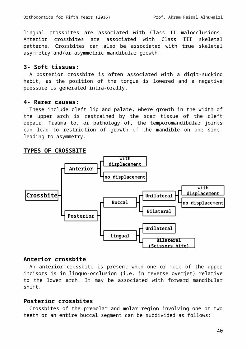
Orthodontics for Fifth Years (2016) Prof. Akram Faisal Alhuwaizi
At the present time, Invisalign ® is most effective at treating milder malocclusions presenting with malalignment, but it may be used successfully in combination with other techniques to treat more complex cases. The other treatment techniques may include restorative work, such as veneers, and even a short phase of fixed appliances. In more complex cases clear aligner treatment may not replace the use of fixed appliances, but it may reduce the amount of time the patient needs to wear less aesthetic labial fixed appliances.
C- Lingual orthodonticsLingual appliances in many ways offer the ultimate in aesthetic appliances, as the whole
system is bonded to the lingual aspect of the teeth. After much attention in the early 1980s their popularity fell, partly due to the introduction of ceramic brackets, but also due to a number of problems with the appliance. Recent technological improvements and an increased demand for ‘invisible’ appliances have led to a recent increase in interest in lingual orthodontics.
Advantages:1- Aesthetics2- No risk to the labial enamel through decalcification3- Position of the teeth can be seen more accurately as it is not obscured by the appliance4- Some lingual brackets create a bite-plane effect on the upper incisors and canines,
making these types of brackets useful for treating deep overbites
Disadvantages:1- Speech alteration2- Discomfort to the patient’s tongue3- Masticatory difficulties4- More technically demanding for the operator, which increases the chair-time and
therefore the cost of this approach5- Operator proficiency in indirect bonding is required and rebonding failed brackets can
be difficult6- More difficult to clean7- Initial alignment can be more challenging in more crowded cases due to reduced
interbracket span8- Increased bracket loss
The majority of tongue discomfort is related to the mandibular arch, so patients may choose to have a lingual appliance in the upper arch, where aesthetics is more crucial and a labial appliance in the lower.
Lingual orthodontics can range from simple alignment of the upper labial segment using round wires, to comprehensive treatment using appliances made using state-of-the-art computer-assisted design/manufacture (CAD/CAM) technology.
CAD/CAM has allowed the production of fully customized appliances, with individualized production of brackets and wires. One of the challenges of aligning teeth from the lingual aspect is the unique morphology of the lingual aspect of teeth, and the range of bucco-lingual thickness of teeth. Customization of appliances overcomes these problems, improving the fit of the appliances, increasing the finishing control, as well as reducing speech problems and tongue irritation. Also if the customized brackets debond during treatment they can be rebonded directly, as the bracket base-to-tooth fi t is so good that incorrect positioning is unlikely.
40