original article dopamine receptor expression on primary
TRANSCRIPT
Int J Clin Exp Med 2018;11(3):1765-1771www.ijcem.com /ISSN:1940-5901/IJCEM0061586
Original ArticleDopamine receptor expression on primary osteoblasts and bone marrow mesenchymal stem cells of rats
Pek-U Cheong1, Ting Ma1, Yan Zheng1, Xi-Yuan Ge2, Yu Zhang1, Ye Lin1
1Department of Oral Implantology, Peking University, School of Stomatology, Beijing 100081, P.R. China; 2Central Laboratory, Peking University School and Hospital of Stomatology, Beijing 100081, P.R. China
Received April 15, 2017; Accepted January 5, 2018; Epub March 15, 2018; Published March 30, 2018
Abstract: Bone metabolism and the underlying mechanisms are of great interest, particularly in the context of clinical bone tissue repair and tissue engineering. Neurotransmitters play an important role in bone metabolism. Dopamine is an important neurotransmitter with several critical functions in the central nervous system, and is likely involved in bone metabolism. However, the expression of dopamine receptors in the bone marrow stem cells and osteoblasts has not been extensively studied to date. In this study, we determined whether two types of dopa-mine receptors, dopamine receptor 1 (D1R) and dopamine receptor 2 (D2R), were expressed on osteoblasts and bone marrow mesenchymal stem cells (BMMSCs) in rats, by using polymerase chain reaction, western blot, and immunofluorescence. Immunohistochemistry was used to confirm the expression of D1R and D2R in the mandible of the rats. The results showed that D1R and D2R are expressed on rat osteoblasts and BMMSCs. Both receptors were distributed in the dental pulp, periodontal tissue, and bone tissue. Our results suggested that dopamine could affect the biological behavior of osteoblasts and BMMSCs by directly binding to its receptors. D1R and D2R could therefore directly modulate bone metabolism.
Keywords: Dopamine, dopamine receptor, osteoblast, bone marrow mesenchymal stem cell, mandible, rats
Introduction
Leptin is a polypeptide hormone that exerts gonadal functions by binding to specific re- ceptors in the hypothalamus. The discovery of leptin-mediated bone mass modulation via the sympathetic nervous system suggested that neurotransmitters play a role in bone formation [1, 2]. Dopamine, a catecholamine neurotrans-mitter widely expressed in the central nervous system (CNS) and some peripheral areas, regu-lates cognitive and behavioral functions in the CNS, including voluntary movement, memo-ry, attention, learning, and reward [3]. Several effects of dopamine and its receptors in the modulation of bone modeling have been report-ed. Long-term treatment with D2R antagonist induced bone loss in female rats [4]. The ab- sence of dopamine transporter caused lower bone mass in mice [5]. In clinical trials, patients with Schizophrenia undergoing treatment with D2R antagonist showed lower bone mineral density [6]. On the other hand, the risk of osteo-
porosis was higher in patients with Parkinson’s disease. These were related to dopamine and its receptors [7, 8]. Based on these data, we hypothesized that dopamine could be involved in mediating bone metabolism.
Dopamine exerts its physiological actions by activating specific seven-transmembrane G- protein-coupled dopamine receptors. Five such receptors, D1R, D2R, D3R, D4R, and D5R, have been discovered. These receptors have been divided into the D1-like and D2-like subfamilies, based on pharmacology and the ability to mod-ulate cyclic adenosine monophosphate (cAMP) concentration. D1R and D5R are included in the D1-like family and tend to increase the con-centration of cAMP. The D2-like family, which includes D2R, D3R, and D4R, inhibits the pro-duction of cAMP by inhibiting adenylate cycla- se [3, 9]. Dopamine receptors are widely ex- pressed in the CNS and peripheral tissues [9], such as the renal system [10] and coronary ves-sels [11].
Rat osteoblast and MSC dopamine receptor expression
1766 Int J Clin Exp Med 2018;11(3):1765-1771
Hanami et al detected all five dopamine recep-tors in human osteoclasts. Further, the number of osteoclasts decreased after culture with D2-like receptor agonist but not with D1-like receptor agonist [12]. Lee et al reported that mouse MC3T3-E1 osteoblastic cells expressed all the dopamine receptors, based on RT-PCR and western blot data [13]. However, previous studies showed that only D1R, D3R, and D5R were expressed in MC3T3-E1 cells [14]. Thus, the expression pattern of dopamine receptors in rats remains unclear. In this study, we aimed to determine whether rat primary osteoblasts and BMMSCs expressed D1R and D2R, by using polymerase chain reaction (PCR), west-ern blot, and immunofluorescence. We also studied the distribution of dopamine receptors in the rat mandible by immunohistochemistry. The results could improve our understanding of the mechanisms by which dopamine modu-lates bone metabolism.
Materials and mthods
Cell culture
Primary Sprague-Dawley (SD) rat calvarial os- teoblasts were obtained and cultured as report-ed previously [15]. Briefly, the fibrous layers of the periosteum from the parietal and frontal bones were isolated and cleaned with 0.1M phosphate-buffered saline (1×PBS). The isolat-ed material was digested by treating with 0.2% collagenase I (Sigma, USA) for 10 min twice and 3 times for 15 min at 37°C. The reaction mix-tures were centrifuged for 6 min at 1500 rpm. Cells were resuspended in Dulbecco’s mini-mum essential medium (DMEM, Hyclone, USA) supplemented with 10% fetal bovine serum (Gibco, USA) and 1% penicillin/streptomycin, and cultured at 37°C in a 5% CO2 humidified atmosphere.
Rat BMMSCs (Cyagen Bioscience Inc., USA) were cultured in α-Minimal Essential Media (α-MEM, Gibco, USA) supplemented with 10% fetal bovine serum (Gibco, USA) and 1% peni- cillin/streptomycin, and cultured at 37°C in a 5% CO2 humidified atmosphere. Culture medi-um was replaced every 2-3 days.
Polymerase chain reaction (PCR)
RNA was isolated from osteoblasts and BM- MSCs by using Trizol reagent (Invitrogen, USA) according to the manufacturer’s instructions. The isolated RNA was reverse-transcribed to cDNA by using the PrimeScript RT Reagent Kit (TaKaRa Bio Inc. Japan). Glyceraldehyde 3-phosphate dehydrogenase (GAPDH) was us- ed as a normalizing control. PCR amplification was performed using the following cycling settings: 29 cycles of 94°C for 40 s, 55°C for 45 s, and 72°C for 40 s, and finally 70°C for 5 min. After amplification, electrophoresis was conducted in 1% agarose gel stained with Gel- RedTM Nucleic Acid Gel Stain (Biotium, Inc., CA, USA), and the bands were visualized and ph- otographed using the Vilber Lourmat Fusion FX5 system (Vilber Lourmat, Marne-la-Vallée, France). The primers used are listed in Table 1.
Western blot
We used PC12 as the positive control and beta actin as a loading control. The processing of cells, including PC12, osteoblasts, and BM- MSCs, was conducted as reported previously [16]. Cells lysis was performed in radio immu-noprecipitation assay buffer (50 mmol/L Tris-HCL: pH 8.0, 5 mmol/L EDTA, 150 mmol/L NaCl, 1% Triton X-100, 1 mmol/L phenylmethyl-sulfonyl fluoride and phosphatase & protease inhibitor cocktail). The mixture was centrifuged at 14,000 rpm, and the cell lysate (superna-tant) was collected and loaded onto 12% Nu- PAGE SDS-PAGE Gel (Invitrogen, Carlsbad, CA, USA). The resolved bands were transferred to PVDF membranes, blocked in 5% skimm- ed milk for 30 min, and incubated overnight at 4°C with primary antibodies against D1R and D2R (ratio 1:200; Abcam, Cambridge, MA, USA). The membrane was washed and incu- bated with horseradish peroxidase-conjugated goat anti-rabbit IgG secondary antibody (Mi- lipore, Billerica, MA, USA) at 37°C for 1 h. The bands were detected using chemiluminesce-
Table 1. Primers used for PCRGene Sequence (5’-3’)D1R* 5’-CAGTCCATGCCAAGAATTGCC-3’
5’-AATCGATGCAGAATGGCTGGG-3’D2R 5’-CACCACGGCCTACATAGCAA-3’
5’-GGCGTGCCCATTCTTCTCT-3’GAPDH 5’-ATGCTCTACCCACGGCAAG-3’
5’-GGAAGATCCTGATCCCTTTC-3’*D1R gene reference was obtained from Zhou et al [29].
Rat osteoblast and MSC dopamine receptor expression
1767 Int J Clin Exp Med 2018;11(3):1765-1771
nce reagent and visualized by the EasySee western blot kit (Transgen, China).
Immunofluorescence
Immunofluorescence studies were performed according to previously described protocols
mounted on glass slides. The sections were then subjected to immunohistochemistry.
Immunohistochemistry
Slide-mounted tissue sections were heated for 30 min at 58°C, deparaffinized in xylene, and
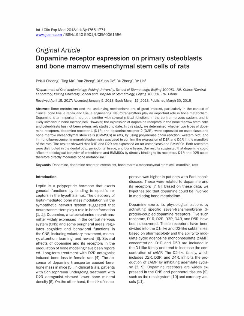
Figure 1. Representative immu-nofluorescence images showing the expression of D1R (A), D2R (B), and negative control (C) on osteoblasts. Antibodies are rep-resented in green and nuclei in blue. Scale bar = 150 μm.
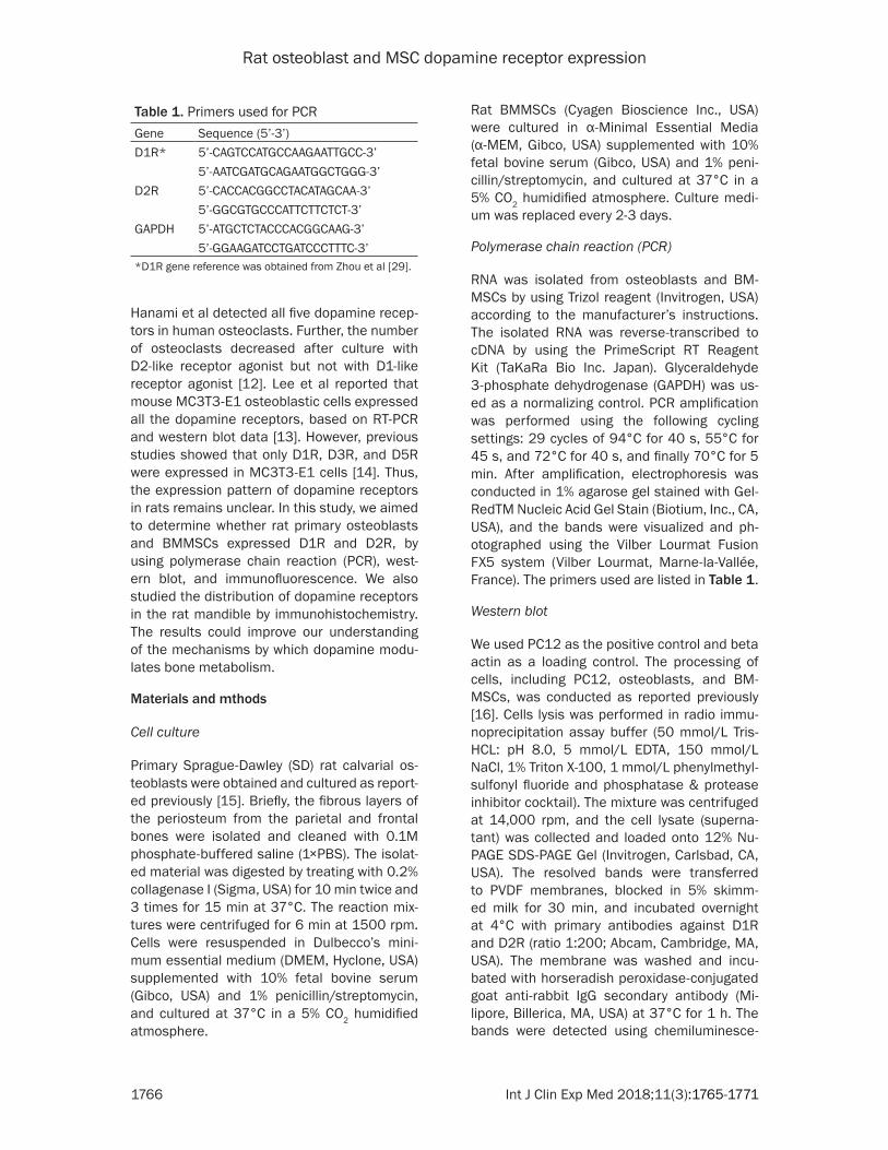
[12]. The osteoblasts and BM- MSCs were soaked in parafor-maldehyde for 20 min. The ce- lls were incubated with 20% goat serum and then immu-nostained overnight at 4°C with antibodies against D1R and D2R (1:500; Abcam, Ca- mbridge, MA, USA) after seri-ally washing in PBS. Cells in- cubated in PBS were used as negative control. Subsequen- tly, the cells were incubated with horseradish peroxidase-conjugated goat anti-rabbit IgG antibodies (Milipore, Bill- erica, MA, USA). The fluores-cence signal was observed and photographed with a fluo-rescent microscope (NIKON, ECLIPSE E600).
Tissue preparation
After the Sprague-Dawley (SD) rats were sacrificed by cervi-cal dislocation, the mandibles were removed and fixed in 4% paraformaldehyde. After 1 mo- nth, the tissues were immers- ed in a 10% EDTA solution for demineralization, until the de- ntal hard tissue could be pu- nctured with a fine needle wi- thout obvious resistance. Af- ter rinsing under running wa- ter, the tissue samples were dehydrated in an ethanol gra-dient series (50%-60%-80%-90%-95), and sequentially im- mersed in 100% ethanol for 3 h twice, normal butanol for 20 h, and trichloromethane for 5 h. Finally, the tissue was subjected to wax immersion for 24 h twice to obtain a wax-embedded block. The bl- ock was cut into 5-μm sagit- tal serial sections parallel to the long axis of the teeth and
Figure 2. Representative immu-nofluorescence images showing the expression of D1R (A), D2R (B), and negative control (C) on BMMSCs. Antibodies are repre-sented in green and nuclei in blue. Scale bar = 150 μm.
Rat osteoblast and MSC dopamine receptor expression
1768 Int J Clin Exp Med 2018;11(3):1765-1771
rehydrated in an ethanol gradient series. After blocking endogenous peroxidase with 3% H2O2 for 10 min at room temperature, the sections were incubated in 1 mg/ml trypsin at 37°C for 10 min for antigen retrieval. 10% Goat Serum was used for blocking at room temperature, and the sections were incubated with rabbit anti-dopamine receptor D1R polyclonal anti-body (1:400; Abcam, Cambridge, MA, USA) or rabbit anti-dopamine receptor D2R polyclonal antibody (1:400; Abcam, Cambridge, MA, USA) at 4°C overnight in a humid atmosphere. One of the sections was incubated in PBS as a nega-tive control. After washing in PBS, the sections were incubated in the presence of peroxidase-conjugated goat anti-rabbit IgG (H+L; 1:400; Zhongshan Golden Bridge Biotechnology, Bei- jing, China) for 1 h to visualize DR subtypes. Images were captured using a digital micro-scopic system (Olympus BX51/DP72, Tokyo, Japan).
Results
Immunofluorescence analysis of dopamine receptors
We used immunofluorescence to determine whether D1R and D2R were expressed in os- teoblasts and BMMSCs. As shown in Figures 1 and 2 significant D1R and D2R expression was observed in both cell types. Higher D1R expression was observed in BMMSCs, while similar levels of D1R and D2R were observed in osteoblasts.
D1R and D2R gene expression in osteoblasts and BMMSCs
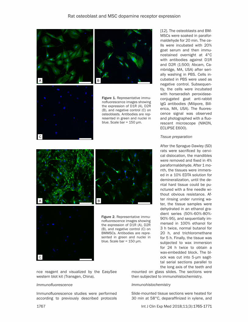
D1R and D2R gene expression in osteoblasts and BMMSCs was confirmed by PCR. As shown in Figure 3, gel electrophoresis of the D1R and D2R PCR products from BMMSCs and osteo-blasts yielded identical DNA bands.
Western blot analysis of dopamine receptor expression in osteoblasts and BMMSCs
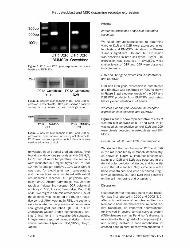
Figures 4 and 5 show representative results of western blot analysis of D1R and D2R. PC12 was used as the positive control. D1R and D2R were clearly detected in osteoblasts and BM- MSCs.
Distribution of D1R and D2R in rat mandible
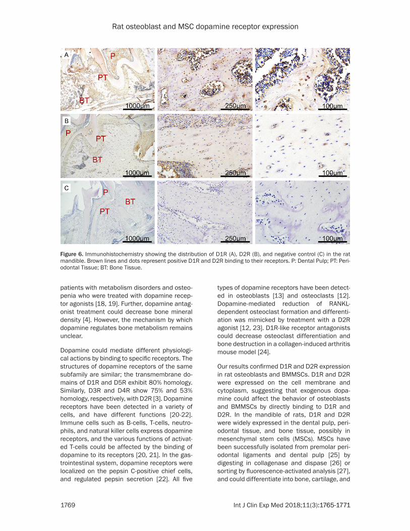
We studied the distribution of D1R and D2R in the rat mandible by immunohistochemistry. As shown in Figure 6, immunohistochemical staining of D1R and D2R was observed in the dental pulp, periodontal tissue, and bone tis-sue in the rat mandible. Only some cells in the bone were stained, and were distributed irregu-larly. Additionally, D1R and D2R were observed on the cell membrane and cytoplasm.
Discussion
Neurotransmitter-mediated bone mass regula-tion was first reported in 2000 and 2002 [1, 2], after which evidence of neurotransmitter invo- lvement in bone metabolism accumulated rap-idly. Dopamine, an important neurotransmit- ter involved in severe central nervous system (CNS) diseases such as Parkinson’s disease, is associated with a high risk of osteoporosis [17], and is likely involved in bone metabolism. In- creased bone mineral density was observed in
Figure 3. D1R and D2R gene expression in osteo-blasts and BMMSCs.
Figure 4. Western blot analysis of D1R and D2R ex-pression in osteoblasts. PC12 was used as a positive control. Beta actin was used as a loading control.
Figure 5. Western blot analysis of D1R and D2R ex-pression in bone marrow mesenchymal stem cells. PC12 was used as a positive control. Beta actin was used as a loading control.
Rat osteoblast and MSC dopamine receptor expression
1769 Int J Clin Exp Med 2018;11(3):1765-1771
patients with metabolism disorders and osteo-penia who were treated with dopamine recep-tor agonists [18, 19]. Further, dopamine antag-onist treatment could decrease bone mineral density [4]. However, the mechanism by which dopamine regulates bone metabolism remains unclear.
Dopamine could mediate different physiologi-cal actions by binding to specific receptors. The structures of dopamine receptors of the same subfamily are similar; the transmembrane do- mains of D1R and D5R exhibit 80% homology. Similarly, D3R and D4R show 75% and 53% homology, respectively, with D2R [3]. Dopamine receptors have been detected in a variety of cells, and have different functions [20-22]. Immune cells such as B-cells, T-cells, neutro-phils, and natural killer cells express dopamine receptors, and the various functions of activat-ed T-cells could be affected by the binding of dopamine to its receptors [20, 21]. In the gas-trointestinal system, dopamine receptors were localized on the pepsin C-positive chief cells, and regulated pepsin secretion [22]. All five
types of dopamine receptors have been detect-ed in osteoblasts [13] and osteoclasts [12]. Dopamine-mediated reduction of RANKL-dependent osteoclast formation and differenti-ation was mimicked by treatment with a D2R agonist [12, 23]. D1R-like receptor antagonists could decrease osteoclast differentiation and bone destruction in a collagen-induced arthritis mouse model [24].
Our results confirmed D1R and D2R expression in rat osteoblasts and BMMSCs. D1R and D2R were expressed on the cell membrane and cytoplasm, suggesting that exogenous dopa-mine could affect the behavior of osteoblasts and BMMSCs by directly binding to D1R and D2R. In the mandible of rats, D1R and D2R were widely expressed in the dental pulp, peri-odontal tissue, and bone tissue, possibly in mesenchymal stem cells (MSCs). MSCs have been successfully isolated from premolar peri-odontal ligaments and dental pulp [25] by digesting in collagenase and dispase [26] or sorting by fluorescence-activated analysis [27], and could differentiate into bone, cartilage, and
Figure 6. Immunohistochemistry showing the distribution of D1R (A), D2R (B), and negative control (C) in the rat mandible. Brown lines and dots represent positive D1R and D2R binding to their receptors. P: Dental Pulp; PT: Peri-odontal Tissue; BT: Bone Tissue.
Rat osteoblast and MSC dopamine receptor expression
1770 Int J Clin Exp Med 2018;11(3):1765-1771
adipose cells under specific conditions [26]. The possibility of other cell types expre- ssing D1R and D2R in dental pulp and peri-odontal tissue, however, could not be excluded, and requires further study. In our study, the expression of D2R appeared lower than that of D1R, although published data suggests that D2R is pivotal in bone remodeling [23]. Based on our results, we assumed that D1R and D2R are expressed throughout bone remodeling, and may be involved in the regulation of osteo-blastic induction of BMMSCs.
Our results on D1R and D2R expression seem to be in broad agreement with clinical and experimental evidence of dopamine receptor modulation of bone metabolism [4, 12, 13, 18, 23, 24, 28]. Expression of the remaining members of the dopamine receptor subfami-lies, with homologous transmembrane domains and similar functions [3], in osteoblasts, BM- MSCs, and the rat mandible could not be ex- cluded in our study. Furthermore, the functio- nal effects of different receptors and the un- derlying molecular mechanisms require further study.
In conclusion, we found that D1R and D2R were expressed on osteoblasts and BMM- SCs, and widely distributed in the rat mandible dental pulp, periodontal tissue, and bone tis-sue. Dopamine could affect the biological be- havior of osteoblasts and BMMSCs by directly binding to these receptors.
Acknowledgements
We thank the Central Laboratory of Peking University School and Hospital of Stomatology for providing the experimental facilities and technical support. This work was supported by the Science Foundation of Peking University School and Hospital of Stomatology (No. PKU- SS20150106) and National Key R&D Program of China (2016YFC1102705).
Disclosure of conflict of interest
None.
Address correspondence to: Yu Zhang and Ye Lin, Department of Oral Implantology, Peking University, School of Stomatology, 22 South Avenue, Zhong- guancun, Haidian District, Beijing 100081, P.R. China. Tel: (8610) 62179977-5344; Fax: (8610)
62173402; E-mail: [email protected] (YZ); [email protected] (YL)
References
[1] Ducy P, Amling M, Takeda S, Priemel M, Schil-ling AF, Beil FT, Shen JH, Vinson C, Rueger JM and Karsenty G. Leptin inhibits bone formation through a hypothalamic relay: a central control of bone mass. Cell 2000; 100: 197-207.
[2] Takeda S, Elefteriou F, Levasseur R, Liu XY, Zhao LP, Parker KL, Armstrong D, Ducy P and Karsenty G. Leptin regulates bone forma-tion via the sympathetic nervous system. Cell 2002; 111: 305-317.
[3] Beaulieu JM and Gainetdinov RR. The physiol-ogy, signaling, and pharmacology of dopamine receptors. Pharmacol Rev 2011; 63: 182-217.
[4] Kunimatsu T, Kimura J, Funabashi H, Inoue T and Seki T. The antipsychotics haloperidol and chlorpromazine increase bone metabo-lism and induce osteopenia in female rats. Regul Toxicol Pharm 2010; 58: 360-368.
[5] Bliziotes M, Gunness M, Eshleman A and Wi-ren K. The role of dopamine and serotonin in regulating bone mass and strength: studies on dopamine and serotonin transporter null mice. J Musculoskelet Neuronal Interact 2002; 2: 291-295.
[6] Meaney AM and O’Keane V. Reduced bone mineral density in patients with schizophrenia receiving prolactin raising anti-psychotic me- dication. J Psychopharmacol 2003; 17: 455-458.
[7] Malochet-Guinamand S, Durif F and Thomas T. Parkinson’s disease: a risk factor for osteopo-rosis. Joint Bone Spine 2015; 82: 406-410.
[8] Kartaginer J, Ataya K, Mercado A and Abbasi A. Osteoporosis associated with neuroleptic trea- tment. A case report. J Reprod Med 1990; 35: 198-202.
[9] Emilien G, Maloteaux JM, Geurts M, Hoogen-berg K and Cragg S. Dopamine receptors-phys-iological understanding to therapeutic inter-vention potential. Pharmacol Therapeut 1999; 84: 133-156.
[10] Cuevas S, Villar VA, Jose PA and Armando I. Renal dopamine receptors, oxidative stress, and hypertension. Int J Mol Sci 2013; 14: 17553-17572.
[11] Tonnarini G, Parlapiano C, Cavallotti D, Tego A, Curione M, Giancaspro G, Vincentelli GM, Le-one S and Cavallotti C. Dopamine receptor subtypes in the human coronary vessels of healthy subjects. J Recept Sig Transd 2011; 31: 33-38.
[12] Hanami K, Nakano K, Saito K, Okada Y, Yama-oka K, Kubo S, Kondo M and Tanaka Y. Dopa-mine D2-like receptor signaling suppresses
Rat osteoblast and MSC dopamine receptor expression
1771 Int J Clin Exp Med 2018;11(3):1765-1771
human osteoclastogenesis. Bone 2013; 56: 1-8.
[13] Lee DJ, Tseng HC, Wong SW, Wang ZY, Deng M and Ko CC. Dopaminergic effects on in vitro os-teogenesis. Bone Res 2015; 3: 15020.
[14] Ko CC, Wang ZY, Tseng H, Lee DJ and Guez C. Design, synthesis, and evaluation of polydopa-mine-laced gelatinous hydroxyapatite nanoco- mposites for orthopedic applications. Ceram Trans 2014; 247: 135-148.
[15] Rath B, Nam J, Knobloch TJ, Lannutti JJ and Agarwal S. Compressive forces induce osteo-genic gene expression in calvarial osteoblasts. J Biomech 2008; 41: 1095-1103.
[16] Kumar JR, Rajkumar R, Farooq U, Lee LC, Tan FCK and Dawe GS. Evidence of D-2 receptor expression in the nucleus incertus of the rat. Physiol Behav 2015; 151: 525-534.
[17] Zhao Y, Shen L and Ji HF. Osteoporosis risk and bone mineral density levels in patients with Parkinson’s disease: a meta-analysis. Bone 2013; 52: 498-505.
[18] Meaney AM and O’Keane V. Reduced bone mineral density in patients with schizophrenia receiving prolactin raising anti-psychotic me- dication. J Psychopharmacol 2003; 17: 455-458.
[19] Takeno A, Yamamoto M, Okazaki K, Yamaguchi T and Toshitsugu S. Successful improvement of metabolic disorders, including osteopenia, by a dopamine agonist in a male patient with macro-prolactinoma. Am J Case Rep 2016; 17: 160-164.
[20] McKenna F, McLaughlin PJ, Lewis BJ, Sibbring GC, Cummerson JA, Bowen-Jones D and Moots RJ. Dopamine receptor expression on human T- and B-lymphocytes, monocytes, neutrophils, eosinophils and NK cells: a flow cytometric study. J Neuroimmunol 2002; 132: 34-40.
[21] Levite M, Marino F and Cosentino M. Dopa-mine, T cells and multiple sclerosis (MS). J Neural Transm 2017; 124: 525-542.
[22] Wang Q, Ji T, Zheng LF, Feng XY, Wang ZY, Lian H, Song J, Li XF, Zhang Y and Zhu JX. Cellular localization of dopamine receptors in the gas-tric mucosa of rats. Biochem Bioph Res Co 2012; 417: 197-203.
[23] Yang HL, Xu YZ, Zhu M, Gu Y, Zhang W, Shao HG, Wang YJ, Ping ZC, Hu XY, Wang LL and Geng DC. Inhibition of titanium-particle-in-duced inflammatory osteolysis after local ad-ministration of dopamine and suppression of osteoclastogenesis via D2-like receptor signal-ing pathway. Biomaterials 2016; 80: 1-10.
[24] Nakashioya H, Nakano K, Watanabe N, Miyas-aka N, Matsushita S and Kohsaka H. Thera-peutic effect of D1-like dopamine receptor an-tagonist on collagen-induced arthritis of mice. Mod Rheumatol 2011; 21: 260-266.
[25] Kadar K, Kiraly M, Porcsalmy B, Molnar B, Racz GZ, Blazsek J, Kallo K, Szabo EL, Gera I, Gerber G and Varga G. Differentiation potential of stem cells from human dental origin-promise for tissue engineering. J Physiol Pharmacol 2009; 60: 167-175.
[26] Kim SS, Kwon DW, Im I, Kim YD, Hwang DS, Holliday LS, Donatelli RE, Son WS and Jun ES. Differentiation and characteristics of undiffer-entiated mesenchymal stem cells originating from adult premolar periodontal ligaments. Korean J Orthod 2012; 42: 307-317.
[27] Alvarez R, Lee HL, Wang CY and Hong C. Char-acterization of the osteogenic potential of mes-enchymal stem cells from human periodontal ligament based on cell surface markers. Int J Oral Sci 2015; 7: 213-219.
[28] Di Somma C, Colao A, Di Sarno A, Klain M, Landi ML, Facciolli G, Pivonello R, Panza N, Salvatore M and Lombardi G. Bone marker and bone density responses to dopamine ago-nist therapy in hyperprolactinemic males. J Clin Endocr Metab 1998; 83: 807-813.
[29] Zhou QY, Grandy DK, Thambi L, Kushner JA, Vantol HHM, Cone R, Pribnow D, Salon J, Bun-zow JR and Civelli O. Cloning and expression of human and rat D1 dopamine-receptors. Na-ture 1990; 347: 76-80.