the dopamine d4 receptor, the ultimate disordered protein.pdf
DESCRIPTION
ATRANSCRIPT

The dopamine D4 receptor, the ultimate disordered protein
Amina S. WoodsStructural Biology Unit, Cellular Neurobiology Branch, NIDA IRP, NIH, Baltimore MD, USA
AbstractThe human D4 dopamine receptor is a synaptic neurotransmitter receptor responsible for neuronalsignaling in the mesolimbic system of the brain, an area of the brain that regulates emotion andcomplex behavior. Its structure makes it a very unusual and interesting G protein-coupled receptor(GPCR) as it has several polymorphic variants of its gene in the region encoding the third intracellularloop (IL3). This region contains from two to seven or more similar 48 base pair repeats. These repeatscause this protein to have a very high disorder index and this, in turn, makes it very interactive withother proteins. Among GPCRs in general, the unusually proline-rich IL3 is unique to the D4 receptor(D4R). We believe that, as in the D2R, this region of the receptor plays a role in it’s interaction withother receptors.
KeywordsDopamine D4 Receptor; Disordered proteins; AKAP; Heteromers; Adenosine A2A Receptor
IntroductionThe neurotransmitter dopamine interacts with two dopamine receptors’ type, the D1 and D2.The D1 has one subtype (D5), while the D2 has two subtypes the D3 and D4. However, D1and D2 have the largest expression in the brain. The D2, D3 and D4 receptors can inhibitadenylate cyclase, while D1 and D5 activate it (1–3). The D4 receptor (D4R) has structural,functional, and pharmacological characteristics closely related to the dopamine D2 receptor.According to Van Tol, both genomic structure (i.e. intron-exon organization) and primarysequence show very high similarity to the D2 receptor (4–7). We have previously demonstratedthat the D2 receptor (D2R; P14416) has an arginine (Arg) richepitope 217RRRRKRVNpTKRpS228 at the amino-terminus of the third intracellular loop(IL3) and that this epitope is involved in heteromer formation with an adenosine A2A receptor’s(A2A R) [P29274] epitope located in its carboxy terminus and containing a phosphorylatedserine residue [Ser374] from a casein kinase (CK) consensus site (Figure 1A and Table 1) (8–12). Downstream from that Arg-rich area are Thr225 and Ser228 which have consensus sitesamenable to phosphorylation with protein kinase A/protein kinase C (PKA/PKC). However,in all D4R subtypes the Arg-rich domain is located in the carboxy terminus of IL3 while thephosphorylatable residues, two Thr, are located in a PKA/PKC consensus sites, onedownstream and another upstream, flank the Arg-rich epitope sequence,QpTRRRRRAKIpT. (Figure 1B).
Address for Correspondence: Amina S. Woods, Ph.D. NIDA IRP, NIH Structural Biology Unit Cellular Neurobiology Branch 333 CassellDrive Baltimore, MD 21224, USA. Tel: 443-740-2747. Fax: 443-740- 2144. [email protected] of interestThis research was supported by the Intramural Research Program of the National Institute on Drug Abuse, NIH. The authors thank theOffice of National Drug Control Policy (ONDCP) for instrumentation funding, without which this and other projects could not have beenaccomplished.
NIH Public AccessAuthor ManuscriptJ Recept Signal Transduct Res. Author manuscript; available in PMC 2010 October 13.
Published in final edited form as:J Recept Signal Transduct Res. 2010 October ; 30(5): 331–336. doi:10.3109/10799893.2010.513842.
NIH
-PA Author Manuscript
NIH
-PA Author Manuscript
NIH
-PA Author Manuscript

The D4 receptors presynaptic localization is in glutamatergic terminals while the postsynapticlocalization is in the dendrites of the GABAergic efferent neurons. The (A2A R) is mostlylocalized in the striatum in glutamatergic terminals and in GABAergic efferent neurons, thestriatopallidal neurons. Their co-localization suggests the possibility of heteromer formationbetween A2A R and D4 R (11,13,14). The dopamine D4 has several subtypes that differ in thenumber of polymorphic repeat sequence of 2–10 repeated units of 16 amino acids that occurin the IL3. D4 receptor genes encoding receptors with two (D4.2), four (D4.4) [Q8NGM5-1],or seven (D4.7) [P21917] repeat units are the most abundant (4–7). The D4 sequence is alsounusually rich in its content of the amino acid proline. Src homology 3 binding domains (SH3)domains are recognized by proline-rich proteins.
Disordered proteinsMany proteins or segments of proteins lack three-dimensional structure. These unstructuredregions are called ‘disordered regions’. According to Dyson and Wright’s excellent review(15), these proteins are most often then not highly conserved between species in bothcomposition and sequence. This is the case for the A2AR, D2R, D3R, and D4R (16) and, contraryto the traditional view that functional proteins always have a stable three-dimensional structure(some do not), nevertheless disordered regions are often functional. They are rich in disorderpromoting amino acids (A, R, G, Q, S, P, E, and K) and have a low content of order-promotingamino acids (W, C, F, I, Y, V, L, and N) (16). Thus, they are characterized by an overall highnet charge and low hydrophobicity, which supports our findings that epitopes involved in theinitiation of receptor heteromerization do it through salt bridge formation driven by anelectrostatic interaction not a hydrophobic one (9–12). Usually disordered segments of aprotein fold on binding to their biological targets, which gives them the conformation that adisorder protein lacks. The disorder to order transitions, which take place during bindinginteractions of the disordered regions, makes the interaction highly reversible and at the sametime highly specific. D2, D3, D4, and A2A receptors also qualify for the title of ‘hubproteins’ (15–18), which are defined as proteins with greater than five interaction partners.Bioinformatics analysis of the A2AR and D2 R using various programs (16–18) clearly showsthat the IL3 of the D2R and the carboxyl terminus of the A2AR have both a high disorder index.This explains their propensity to interact with each other and with other receptors (17–22). Italso emphasizes the importance of the electrostatic interaction between the D2R, D3R, andD4R IL3′ and the A2AR carboxyl terminus epitope and other receptors similar epitope (12), asindeed the first and necessary step in heteromer formation.
Heteromer formationTo test for the possibility of heteromer formation between the D4R Arg-rich epitope and theadenosine A2A receptor epitope SAQEpSQGNT that interacts with the Arg-rich regions ofD2R, D3R, and D4 R, synthetic peptides representing the phosphorylated and non-phosphorylated D4 epitope [QpTRRRRRAKIpT and QTRRRRRAKIT] and the A2ARepitope were made and an equimolar solution of all three peptides was analyzed by MALDImass spectrometry in positive ion mode. As in the case of the D2R-A2A interaction, the D4Rand A2AR interaction seen in the MALDI mass spectrum in Figure 2, shows formation of non-covalent complexes (NCXs) between the epitopes of the D4R and A2AR (relative abundance6 and 25% respectively). The normalized relative abundance of the MH+ of the NCXs ofSAQEpSQGNT with the epitopes of the D4 R QpTRRRRRAKIpT andQTRRRRRAKIT was 11.0% and 25.0%, respectively. Normalization was obtained bydividing the relative abundance of the molecular ion [MH+] of the NCXs by the relativeabundance of the MH+ of the corresponding phosphorylated and non-phosphorylated epitopesof the D4 receptor. We previously demonstrated that heteromerization of adenosine A2A withdopamine D2 receptors and glutamate N-methyl-d-aspartate (NMDA) receptors (NR1-1
Woods Page 2
J Recept Signal Transduct Res. Author manuscript; available in PMC 2010 October 13.
NIH
-PA Author Manuscript
NIH
-PA Author Manuscript
NIH
-PA Author Manuscript

subunit) with dopamine D1 receptors depends on epitope–epitope electrostatic interactions(9,10) and that this is a general mechanism for receptor heteromerization (12). We foundstriking similarities between the epitopes involved in the A2A-D2 and NMDA-D1 receptorheteromerization. Similarities were seen in an (1) Arg-rich epitope, localized in the N-terminalportion of the IL3 of the D2 receptor, and the C-terminus of the NR1-1 subunit of the NMDAreceptor (2); on the opposing epitopes of the C-termini of both A2A and D1 receptors, a serine(Ser) residue susceptible of phosphorylation by CK1 (pSer374 and pSer397 respectively) (3);a PKA consensus site adjacent to the Arg-rich epitope of the D2 receptor and the NR1-1 subunitof the NMDA receptor. The same parameters are found with the D4R-A2A interaction.
The role of prolineAmino acid composition and modifications determine the structure and function of a protein.When perusing the amino acid sequence of the D4R subtypes, one immediately notices theoverwhelming presence of the amino acid residue proline which is the only cyclic amino acid.Its cyclic structure strongly influences the secondary structures of proteins, as its amino groupis a secondary amine, so formation of a peptide bond yields an amide in which there is no N-H hydrogen bond donor function. Due to its rigidity, proline is frequently located in turns orbends, which are often on the surface of a protein (23). Thus the kinking of protein strandsgenerated by proline results in destabilized α-helix formation. This is clearly seen in Figure 3where a model of the D4-4 Rpts-IL3 has more α-helices than the D4-7 Rpts-IL3. Proline-richsegments found in the IL3 of the D4 receptor in all species allow it to serve as a docking sitefor signaling proteins that become targets for phospholipase methylation [phospholipidmethylation (PLM)]-based modulation.
The frequency of proline residues in most proteins is 5%. However, as seen in Table 2 it isaround 12 and 15% in D4-4 Rpts and D4-7 Rpts, respectively and 27 and 30% in their IL3,while it is somewhat elevated in D2R and D3 R IL3 (8 and 9% respectively; Table 2). This isan unusually high proline content for mammalian proteins, as high proline concentration isusually seen in thermophiles’ proteins. In humans and other primates, the D4 receptor possessesanywhere from 2 to 11 additional proline-rich repeat segments (Figure 3), and a higher numberof repeats (i.e. seven) brings an increased risk of attention-deficit hyperactivity disorder(ADHD) (5).
A-kinase anchoring proteinsA-kinase anchoring proteins (AKAPs) are scaffold proteins which coordinate the subcellularlocalization of second-messenger-regulated enzymes such as: PKA or PKC and proteinphosphatases. They also have domains that interact with G protein-coupled receptors (GPCRs).AKAPs facilitate and localize dopamine-initiated signal cascades, leading to activation ofprotein kinases, as well as the turning-off of such signals. Second messengers regulate synapticplasticity by influencing the balance between kinase and phosphatase activity. AKAP interactswith PKA and a calcium-dependent phosphatase PP2B. AKAPs (24) are signal organizingmolecules that compartmentalize kinases and phosphatases that are regulated by secondmessengers. D4R IL3 contains several SH3 in its polymorphic repeat sequence. SH3 domainsare found in numerous proteins involved with signal transduction pathways, and form β-barrellike structures that bind proline-rich sequences. Therefore the presence of multiple repeatsgreatly increases the # of prolines and subsequently the # of SH3 recognition sites. Thus,proteins such as AKAP, which contain SH3 domains recognized by the proline-rich D4 IL3,can tether the kinases PKA and PKC, and phosphatases making phosphorylation/dephosphorylation of the D4,7 repeats more likely than phosphorylation of the D4,4 repeatsvariants.
Woods Page 3
J Recept Signal Transduct Res. Author manuscript; available in PMC 2010 October 13.
NIH
-PA Author Manuscript
NIH
-PA Author Manuscript
NIH
-PA Author Manuscript

The SH3 domains β-barrel shape consisting of five β-strands is arranged as two tightly packedanti-parallel β sheets (25,26). The SH3-type fold is found in both eukaryotes and prokaryotes.SH3 domains are usually found in disordered proteins that have the propensity to interact withother proteins thus mediating the assembly of certain protein complexes, by binding to proline-rich motifs in certain proteins. SH3 domains are mostly found in intracellular proteins domainwhich is the case for the D4R IL3.
SH3-binding epitopes usually have the consensus sequence:
-X -P -p -X -P-
1 2 3 4 5
Residues 1 and 4 are usually aliphatic, 2 and 5 are always proline, while 3 is also sometimesa proline. The protein AKAP contains SH3 domains recognized by the proline-rich D4 IL3.
In conclusion, what might drive the function of the D4R is the unusual predominance ofprolines in its sequence, leading to anchoring of several AKAPs on its IL3. These AKAPsanchor kinases and phosphatases in the proximity of the basic epitope located at the carboxyterminus of IL3, thereby locating all the needed enzymes for phosphorylation anddephosphorylation in the proximity of the important epitope. When phosphorylated,[QpTRRRRRAKIpT] is less likely to interact with phosphorylated epitopes of the A2AR(11%) and when not phosphorylated [QTRRRRRAKIT] the interaction likelihood doublesto 25%. The human D4 dopamine receptor is a synaptic neurotransmitter receptor responsiblefor neuronal signaling in the mesolimbic system of the brain, a part of the brain that regulatesemotion and complex behavior. Phosphorylation/dephosphorylation has always beenconsidered to be an ON/OFF switch. The D4R structure makes it a very efficient switch andis a particularly interesting GPCR because the large number of polymorphic variants of thereceptor gene in humans. This results in variations that affect the efficiency of this receptor asa switch. It seems that the more repeats the more efficient it probably becomes. The gene variesin the nucleotide sequence of the region encoding IL3, which contains from two to seven ormore similar 48 base pair repeats. (These nucleotide repeats code for more than 18 differentsequences.) Among GPCR, the proline-rich IL3 is unique to the D4R. We believe that as inthe D2R, the role of this region of the receptor is to interact with other receptors (12,27).
In addition, proline-rich segments found in the IL3 of the D4 receptor in all species allow it toserve as a docking site for signaling proteins that become targets for PLM-based modulation.In humans and other primates, the D4R proline-rich segments (Figure 4), and a higher numberof repeats (i.e. seven) shows maximum disorder (28) 90 and 97%, respectively for the D4R-4rpts and D4R-7Rpts (Table 3) and is accompanied by an increased risk of ADHD (5).
The D4 R SH3-binding sites are within a region involved in the control of receptorinternalization. Through heteromerization, receptors establish new functional interactions, andacquire new functional properties that differ from those of either receptor unit when not partof a receptor heteromer unit.
We would like to emphasize that SH3 are protein domains found in many proteins involvedwith signal transduction pathways. SH3 domains form β-barrel like structures that bind proline-rich sequences. Hence the presence of multiple repeats greatly increases the # of prolines andtherefore the # of SH3 recognition sites. This fact results in proteins such as AKAP, whichcontain SH3 domains, to be recognized by the proline-rich D4 IL3. This can, in turn, tether thekinases PKA and PKC and phosphatases thus making phosphorylation/dephosphorylation ofthe D4,7 repeats more likely than phosphorylation of the D4,4 repeats or other variantscontaining less prolines. One cannot emphasize enough the role of AKAPs as scaffold proteinswhich coordinates the subcellular localization of second-messenger-regulated enzymes such
Woods Page 4
J Recept Signal Transduct Res. Author manuscript; available in PMC 2010 October 13.
NIH
-PA Author Manuscript
NIH
-PA Author Manuscript
NIH
-PA Author Manuscript

as: PKA or PKC and protein phosphatases. AKAPs facilitate and localize dopamine-initiatedsignal cascades, leading to activation of protein kinases, as well as the turning-off of suchsignals. Second messengers regulate synaptic plasticity by influencing the balance betweenkinase and phosphatase activity. AKAP interacts with PKA and a calcium-dependentphosphatase PP2B.
References1. Missale C, Nash SR, Robinson SW, Jaber M, Caron MG. Dopamine receptors: from structure to
function. Physiol Rev 1998;78:189–225. [PubMed: 9457173]2. Aizman O, Brismar H, Uhlén P, Zettergren E, Levey AI, Forssberg H, Greengard P, Aperia A.
Anatomical and physiological evidence for D1 and D2 dopamine receptor co-localization in neostriatalneurons. Nat Neurosci 2000;3:226–230. [PubMed: 10700253]
3. Surmeier DJ, Song WJ, Yan Z. Coordinated expression of dopamine receptors in neostriatal mediumspiny neurons. J Neurosci 1996;16:6579–6591. [PubMed: 8815934]
4. Van Tol HH, Wu CM, Guan HC, Ohara K, Bunzow JR, Civelli O, Kennedy J, Seeman P, Niznik HB,Jovanovic V. Multiple dopamine D4 receptor variants in the human population. Nature 1992;358:149–152. [PubMed: 1319557]
5. Oak JN, Oldenhof J, Van Tol HH. The dopamine D(4) receptor: one decade of research. Eur J Pharmacol2000;405:303–327. [PubMed: 11033337]
6. Livak KJ, Rogers J, Lichter JB. Variability of dopamine D4 receptor (DRD4) gene sequence withinand among nonhuman primate species. Proc Natl Acad Sci USA 1995;92:427–431. [PubMed:7831304]
7. Oldenhof J, Vickery R, Anafi M, Oak J, Ray A, Schoots O, Pawson T, von Zastrow M, Van Tol HH.SH3 binding domains in the dopamine D4 receptor. Biochemistry 1998;37:15726–15736. [PubMed:9843378]
8. Ciruela F, Burgueño J, Casadó V, Canals M, Marcellino D, Goldberg SR, Bader M, Fuxe K, AgnatiLF, Lluis C, Franco R, Ferré S, Woods AS. Combining mass spectrometry and pulldown techniquesfor the study of receptor heteromerization. Direct epitope-epitope electrostatic interactions betweenadenosine A2A and dopamine D2 receptors. Anal Chem 2004;76:5354–5363. [PubMed: 15362892]
9. Woods AS, Ciruela F, Fuxe K, Agnati LF, Lluis C, Franco R, Ferré S. Role of electrostatic interactionin receptor-receptor heteromerization. J Mol Neurosci 2005;26:125–132. [PubMed: 16012186]
10. Woods AS, Ferré S. Amazing stability of the arginine-phosphate electrostatic interaction. J ProteomeRes 2005;4:1397–1402. [PubMed: 16083292]
11. Fuxe K, Marcellino D, Guidolin D, Woods AS, Agnati LF. Heterodimers and receptor mosaics ofdifferent types of g-protein- coupled receptors. Physiology 2008;23:322–332. [PubMed: 19074740]
12. Woods AS, et al. Electrostatic interactions as key determinants of the quaternary structure of receptorheteromers. JBC. 2010 [Epub ahead of print].
13. Tanganelli S, Sandager Nielsen K, Ferraro L, Antonelli T, Kehr J, Franco R, Ferré S, Agnati LF, FuxeK, Scheel-Krüger J. Striatal plasticity at the network level. Focus on adenosine A2A and D2interactions in models of Parkinson’s Disease. Parkinsonism Relat Disord 2004;10:273–280.[PubMed: 15196505]
14. Fuxe K, Canals M, Torvinen M, Marcellino D, Terasmaa A, Genedani S, Leo G, Guidolin D, Diaz-Cabiale Z, Rivera A, Lundstrom L, Langel U, Narvaez J, Tanganelli S, Lluis C, Ferré S, Woods A,Franco R, Agnati LF. Intramembrane receptor-receptor interactions: a novel principle in molecularmedicine. J Neural Transm 2007;114:49–75. [PubMed: 17066251]
15. Dyson HJ, Wright PE. Intrinsically unstructured proteins and their functions. Nat Rev Mol Cell Biol2005;6:197–208. [PubMed: 15738986]
16. Agnati LF, Fuxe K, Woods A, Genedani S, Guidolin D. Theoretical considerations on the topologicalorganization of receptor mosaics. Curr Protein Pept Sci 2009;10:559–569. [PubMed: 19751189]
17. Romero P, Obradovic Z, Li X, Garner EC, Brown CJ, Dunker AK. Sequence complexity of disorderedprotein. Proteins 2001;42:38–48. [PubMed: 11093259]
18. Dunker AK, Brown CJ, Obradovic Z. Identification and functions of usefully disordered proteins.Adv Protein Chem 2002;62:25–49. [PubMed: 12418100]
Woods Page 5
J Recept Signal Transduct Res. Author manuscript; available in PMC 2010 October 13.
NIH
-PA Author Manuscript
NIH
-PA Author Manuscript
NIH
-PA Author Manuscript
19. Romero P, Obradovic Z, Dunker AK. Sequence data analysis for long disordered regions predictionin the calcineurin family. Genome Inform 1997;8:110–124.
20. Linding R, Jensen F, Diella P, Bork T, Gibson J, Russell RB. Protein disorder prediction: implicationsfor structural proteomics. Structure 2003;11:1453–1459. [PubMed: 14604535]
21. Vullo A, Bortolami O, Pollastri G, Tosatto SCE. Spritz: a server for the prediction of intrinsicallydisordered regions in protein sequences using kernel machines. Nucleic Acids Res 2006;34:W164–W168. [PubMed: 16844983]
22. Shimizu K, Muraoka Y, Hirose S, Tomii K, Noguchi T. Predicting mostly disordered proteins byusing structure-unknown protein data. BMC Bioinformatics 2007;8:78. [PubMed: 17338828]
23. Creighton, TE. Proteins Structure and Molecular properties. 2. New-York: W.H. Freeman andCompany; 1993. Chapter 3: amino acid residues; p. 6-20.
24. Malbon CC, Tao J, Hsien-yu Wang HY. AKAPs (A-kinase anchoring proteins) and molecules thatcompose their G-protein-coupled receptor signalling complexes. Biochem J 2004;379:1–9.[PubMed: 14715081]
25. Pawson T, Schlessingert J. SH2 and SH3 domains. Curr Biol 1993;3:434–42. [PubMed: 15335710]26. Mayer BJ. SH3 domains: complexity in moderation. J Cell Sci 2001;114:1253–63. [PubMed:
11256992]27. Ferré S, Baler R, Bouvier M, Caron MG, Devi LA, Durroux T, Fuxe K, George SR, Javitch JA, Lohse
MJ, Mackie K, Milligan G, Pfleger KD, Pin JP, Volkow ND, Waldhoer M, Woods AS, Franco R.Building a new conceptual framework for receptor heteromers. Nat Chem Biol 2009;5:131–134.[PubMed: 19219011]
28. Radivojac P, Obradovic Z, Brown CJ, Bunker AK. Prediction of boundaries between intrinsicallyordered and disordered protein regions. Pac Symp Biocomput 2003;8:216–227. [PubMed: 12603030]
Woods Page 6
J Recept Signal Transduct Res. Author manuscript; available in PMC 2010 October 13.
NIH
-PA Author Manuscript
NIH
-PA Author Manuscript
NIH
-PA Author Manuscript

Figure 1.(A) Location of Arg-rich epitope. (B) AKAP anchoring kinases and phosphatases.
Woods Page 7
J Recept Signal Transduct Res. Author manuscript; available in PMC 2010 October 13.
NIH
-PA Author Manuscript
NIH
-PA Author Manuscript
NIH
-PA Author Manuscript

Figure 2.Mass spectrum showing the non-covalent complex formed between the phosphorylated andnon-phosphorylated the epitopes of the D4R and A2AR.
Woods Page 8
J Recept Signal Transduct Res. Author manuscript; available in PMC 2010 October 13.
NIH
-PA Author Manuscript
NIH
-PA Author Manuscript
NIH
-PA Author Manuscript

Figure 3.T model of the D4,4 repeats (A) third intracellular loop (IL3) has more α-helices than the D4,7repeats-IL3 (B).
Woods Page 9
J Recept Signal Transduct Res. Author manuscript; available in PMC 2010 October 13.
NIH
-PA Author Manuscript
NIH
-PA Author Manuscript
NIH
-PA Author Manuscript
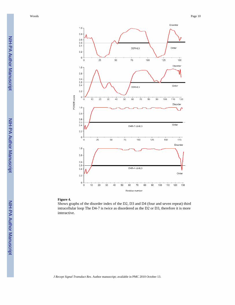
Figure 4.Shows graphs of the disorder index of the D2, D3 and D4 (four and seven repeat) thirdintracellular loop The D4-7 is twice as disordered as the D2 or D3, therefore it is moreinteractive.
Woods Page 10
J Recept Signal Transduct Res. Author manuscript; available in PMC 2010 October 13.
NIH
-PA Author Manuscript
NIH
-PA Author Manuscript
NIH
-PA Author Manuscript

NIH
-PA Author Manuscript
NIH
-PA Author Manuscript
NIH
-PA Author Manuscript
Woods Page 11
Table 1
Dopamine D2, D3 and D4 conserved basic epitope.
IL3 # aa Receptor Arg-rich epitope sequence
211–373 163 Dopamine D2 VLRRRRKRVNpTKRpSSR
210–329 120 Dopamine D3 KQRRRKRILpTRQNSQC
214–346 133 Dopamine D4 QpTRRRRRAKIpTGRERK
IL3, third intracellular loop.
J Recept Signal Transduct Res. Author manuscript; available in PMC 2010 October 13.

NIH
-PA Author Manuscript
NIH
-PA Author Manuscript
NIH
-PA Author Manuscript
Woods Page 12
Table 2
Percentage of prolines in the D2, D3 and D4 receptors and in their third intracellular loop.
Receptor # aa Res # Of prolines % Of total aa Res
D4-7 Rpts 467 69 14.8%
D4-7 Rpts-IL3 181 54 29.3%
D4-4 Rpts 419 50 11.9%
D4-4 Rpts-IL3 133 35 26.5%
D2 443 27 6.1%
D2-IL3 163 15 9.2%
D3 400 20 5.0%
D3-IL3 120 10 8.3%
IL3, third intracellular loop.
J Recept Signal Transduct Res. Author manuscript; available in PMC 2010 October 13.

NIH
-PA Author Manuscript
NIH
-PA Author Manuscript
NIH
-PA Author Manuscript
Woods Page 13
Table 3
% Disorder in IL3 of dopamine receptor.
Receptor % Disorder
D4R-7 rpts 97
D4R-4 rpts 90
D2R 26
D3R 22
D2, D3, D4 receptor; IL3, third intracellular loop.
J Recept Signal Transduct Res. Author manuscript; available in PMC 2010 October 13.