original article - ipsonline.in · department of prosthodontics, faculty of dental medicine,...
TRANSCRIPT

282 © 2017 The Journal of Indian Prosthodontic Society | Published by Wolters Kluwer - Medknow
The potential of mangosteen (Garcinia mangostana) peel extract, combined with demineralized freeze‑dried bovine bone xenograft, to reduce ridge resorption and alveolar bone regeneration in preserving the tooth extraction socket
Utari Kresnoadi, Maretaningtias Dwi Ariani, Eha Djulaeha, Nike HendrijantiniDepartment of Prosthodontics, Faculty of Dental Medicine, Airlangga University, Surabaya, Indonesia
Original Article
Background: Following the extraction of a tooth, bone resorption can cause significant problems for a subsequent denture implant and restorative dentistry. Thus, the tooth extraction socket needs to be maintained to reduce the chance of any alveolar ridge bone resorption.Objective: The objective of this study is to determine whether the administration of mangosteen peel extracts (MPEs), combined with demineralized freeze-dried bovine bone xenograft (DFBBX) materials for tooth extraction socket preservation, could potentially reduce inflammation by decreased the expression of nuclear factor κβ (NfKb) and receptor activator of nuclear factor-κβ ligand (RANKL), to inhibit alveolar bone resorption, and increased of bone morphogenetic protein-2 (BMP2) expressions to accelerate alveolar bone regeneration.Materials and Methods: This study consists of several stages. First, a dosage of MPE combined with graft materials was applied to a preserved tooth extraction socket of a Cavia cobaya. Second, the C. cobaya was examined using immune histochemical expression of NfKb, RANKL, BMP2, as well as histology of osteoblasts and osteoclasts. The research was statistically analyzed, using an analysis of variance test and Tukey honest significant difference test.Results: The results of this research were that it was determined that MPEs combined with graft materials on a preserved tooth extraction socket can reduce NfKb, RANK, and osteoclasts also increase of BMP2 and osteoblast.Conclusion: The induction of MPEs and DFBBX is effective in reducing inflammation, lowering osteoclasts, decreasing alveolar bone resorption, and also increasing BMP2 expression and alveolar bone regeneration.
Keywords: Bone morphogenetic protein-2, graft materials, mangosteen peel extracts, nuclear factor κβ, preserved tooth extraction socket, receptor activator of nuclear factor-κβ ligand expressions
Abstract
Address for correspondence: Prof. Utari Kresnoadi, Department of Prosthodontics, Faculty of Dentistry, Airlangga University, Jalan Mayjend, Prof. Dr. Moestopo No. 47 Surabaya 60132, East Java, Indonesia. E-mail: [email protected]: 07th March, 2017, Accepted: 19th May, 2017
Access this article onlineQuick Response Code:
Website:
www.j-ips.org
DOI:
10.4103/jips.jips_64_17
How to cite this article: Kresnoadi U, Ariani MD, Djulaeha E, Hendrijantini N. The potential of mangosteen (Garcinia mangostana) peel extract, combined with demineralized freeze-dried bovine bone xenograft, to reduce ridge resorption and alveolar bone regeneration in preserving the tooth extraction socket. J Indian Prosthodont Soc 2017;17:282-8.
This is an open access article distributed under the terms of the Creative Commons Attribution-NonCommercial-ShareAlike 3.0 License, which allows others to remix, tweak, and build upon the work non-commercially, as long as the author is credited and the new creations are licensed under the identical terms.
For reprints contact: [email protected]
[Downloaded free from http://www.j-ips.org on Saturday, February 24, 2018, IP: 183.82.145.117]

Kresnoadi, et al.: The potential of mangosteen peel extract to reduce ridge resorption and alveolar bone regeneration
The Journal of Indian Prosthodontic Society | Volume 17 | Issue 3 | July-September 2017 283
INTRODUCTION
The posttooth‑extraction bone resorption process can result in significant problems for denture implants and restorative dentistry. To deter the denture from falling apart, one of the most important factors in denture construction is its retention factor, which is heavily affected by other anatomical factors, such as a high scaling ridge as a cantilever for the denture implant. To gain a high positional ridge, the ridge resorption, usually caused by trauma after a tooth extraction, needs to be reduced. Inflammation will trigger osteoclast growth and also cause alveolar bone resorption. Original tooth extraction socket preservation in patients is required to reduce the likelihood of occurrence of alveolar bone resorption. Conservation of the original tooth extraction socket in patients, along with an optimal technique of dental implant therapy is needed. The same principal can be applied to the edentulous area, to increase the aesthetic point on both fixed and removable dentures.[1]
Inflammation can raise the number of osteoclast cells, pro‑inflammation cytokines (tumor necrosis factor (TNF)‑a and interleukin 1 β (IL‑1 β), and also receptor activator of nuclear factor‑κβ ligand (RANKL) and RANK (receptor activator of nuclear factor‑κβ).[2,3] The RANKL process is a signal of an osteoclast, which occurs because the TNF or TNF receptor family plays an important role in controlling the death of cells and also the proliferation of cells. Family molecule RANK and RANKL receptors are key regulators of bone remodeling and are essential for osteoclast activation and development.[4] When osteoclast cell growth rises, alveolar bone resorption will take place.
Hayden, et al.[5] have stated that nuclear factor κβ (NfKb) is one aspect of the cause of inflammation. The pro‑inflammation cytokine production path indicates a specific dependency on NfKb, just as TNF‑a activates NfKb; therefore, NfKb is a fundamental part of propagation and cytokine response. According to Bellanti,[6] the level of inflammation can be categorized as acute, sub‑acute, or chronic. Acute inflammation takes place from the 1st day until the third; subsequently, there will be chronic inflammation, based on the history of illness. In the fields of Medicine and Dentistry, the use of grafts to mend or augment a bone defect is a common practice,[7‑12] but it was not stable enough to produce the expected result. In the bone growth process, bone morphogenetic protein‑2 (BMP2) triggers the process of osteoblast genesis, stimulating either an osteoblastic‑specific factor‑2 or a core binding factor‑1, affecting the produced osteoblast; the collagen 1 and noncollagen product osteocalcin will increase, and another alveolar bone growth will appear.[13]
Demineralized freeze‑dried bovine bone xenograft (DFDBBX) is a product made by Batan Research Tissue Bank and offered in powder form; it has been relieved of its immunogenic attribute by gamma (γ) ray radiation.
There are various uses for mangosteen peel in the medical field, one of them being as an anti‑inflammatory; it may also function as an antioxidant, and is also capable of activating wound‑healing hormones.[14]
Pedraza‑Chaverri, et al.,[14] Palakawong, et al.,[15] Khairunisa,[16] Yatman,[17] stated in their report that xanthones were detected inside the mangosteen peel and that these contain dibenzopyran, an anti‑inflammatory substance. Dibenzofuran is a close relative to the flavonoids and another anti‑inflammatory which works by inhibiting the production of the cyclooxygenase‑2 (COX‑2) enzyme, the main cause of inflammation. Various illnesses are caused by free radicals; in protecting the body from free radicals, the antioxidant substance stabilizes them. Mangosteen peels contain compounds that are useful as antioxidants and are also pharmacologically active: flavonoid, xantone, and tannin.[18]
Based on the above description, the question arises as to whether the administration of a combination of mangosteen peel extract (MPE) and DFDBBX on tooth extraction sockets can decrease the generation of NfKb, RANKL as an indicator of inflammation, reducing osteoclasts, lowering the chance of ridge resorption and also increasing BMP2 expression and alveolar bone regeneration. This research is an effort at tissue engineering, with the objective of preserving tooth extraction sockets by combining MPE and DFDBBX to fill in the extraction socket to reduce the inflammation caused by the extraction, which can cause ridge resorption and increase osteoblast cells, resulting in alveolar bone regeneration.
MATERIALS AND METHODS
Research procedure and animal modelEvery procedure performed in this research meets the ethical requirements as stated by the Dentistry Ethics Commission of Airlangga University as stipulated in their decree No 1/KKEPK.FKG/III/2015.
The materials used are mangosteen (Garcia mangostana) peel extract, sterile distilled water (aquades), DFDBBX, absolute alcohol, 70% alcohol, monoclonal antibody anti‑NfKb p 56 (Santa Cruz), monoclonal antibody RANKL (Santa Cruz), monoclonal antibody BMP2 (Santa Cruz), immune staining kit reagent (Novocastra‑Leica), and a reagent
[Downloaded free from http://www.j-ips.org on Saturday, February 24, 2018, IP: 183.82.145.117]

Kresnoadi, et al.: The potential of mangosteen peel extract to reduce ridge resorption and alveolar bone regeneration
284 The Journal of Indian Prosthodontic Society | Volume 17 | Issue 3 | July-September 2017
for hematoxylin and eosin (HE) coloring. Indirect immunohistochemically examination can be conducted using primary antibodies as well as secondary antibodies. The experimental animals used were healthy and active male Cavia cobaya (Guinea pigs), weighing around 300–350 g, aged 3–3.5 months.
The MPE was obtained using a maceration technique; after that the most appropriate dosage of the mixture of MPE and xenograft + polyethylene glycol (PEG) was calculated, followed by a Histopathologic Examination of Osteoblasts and Osteoclasts by HE, along with a staining and immunohistochemistry examination using monoclonal antibody NfKb p 56, RANKL, and BMP2.
Research procedureFifty‑six C. cobayas were divided into eight groups, each group containing seven animals. The groups were then separated into 7‑day groups and 30‑day groups, each group received four treatments. The tooth extracted was the bottom right incisor. Afterward, the C. cobaya were taken from their treatment facility and anesthetized intravenously with 0.2 CC/300 g ketamine BB.[19] The bottom left incisor extraction was conducted with specialized (needle holder) pliers. The extraction socket was then filled with PEG, DFDBBX + PEG, MPE + PEG, and a combination of MPE + DFDBBX + PEG. Each mixture was allowed as much as 0.1 CC, corresponding with the volume of the tooth extraction socket, which was then sewn up with Braun Aesculap brand polyamide monofilament sewing thread, DS 12 3/8c, 12 mm, 6/10 met, 0.7 sterile.[20] The groups was sorted as follows. Groups 1 and 2: Tooth extraction performed, followed by filling in the socket with PEG only. Groups 3 and 4: Tooth extraction performed followed by filling in the socket using DFDBBX + PEG. Groups 5 and 6: Tooth extraction was performed, followed by filling in the socket with MPE and PEG. Groups 7 and 8: Tooth extraction was performed followed by filling in the socket with a combination of MPE + DFDBBX + PEG. An examination after 7 and 30 days for each group were conducted.
Histopathologic specimen preparationAfter 7 days and 30 days had passed, the C. cobayas were euthanized and their jaws were removed with a surgical incision. Paraffin block was prepared. The paraffin block was then sliced, using a rotary microtome with a thickness of approximately 4 µ, after which it was laid out on a glass slide. Deparaffinization was conducted by dissolving the specimen in xylol for 2 × 3 min. The remainder of the xylol was washed out with absolute alcohol, 99%, 95%, 90%, 80%, 70%, for 2 × 1 min each, the calculation for NfkB,
RANKL, and BMP2 expressions, and also Osteoblast and Osteoclast cells, was carried out under a light microscope each slide was examined with 1000 times magnification and a maximum of 10 fields of view. The calculation results were noted on a worksheet with a mean value per field of view.
The calculation results were then recorded and tabulated to be statistically analyzed by the Kolmogorov–Smirnov method and a homogeneity test. An An analysis of variance (ANOVA) was conducted to discern the differences among the groups. A multiple comparison by Tuckey honest significant difference (HSD) was then carried out, to discover the group with the best result.
RESULTS
The preliminary results for the best dosage, number of calculated mean and standard deviations (SDs) of osteoblasts and osteoclasts after 30 days of treatment, showed that the dose of 2% had the highest osteoblast result at 22.825. The lowest osteoblast count occurred in the control group that was only filled with PEG, i.e., 5.5714, while the highest osteoclast count was observed from the dose control with a result of 22.428; meanwhile, the dose of 2% obtained the lowest yield at 7.
The osteoblast and osteoclast calculation results with 2% dosage and with treatment for 7 and 30 days are presented in Figure 1.
The block diagram [Figure 1] displays the difference between the control groups, the DFDBBX + PEG, the MPE + PEG, and the MPE + DFDBBX + PEG group. The group that was treated with MPE + DFDBBX + PEG had the highest osteoblast number, at 12.50 ± SD 1.049.
Figure 1: Mean of osteoblast and osteoclast, axis X: Account of osteoclast and osteoblast. Axis Y: treatment filled socket with PEG (control), DFDBBX + PEG (Graft), MPE + PEG and combination of MPE + DFDBBX + PEG at 7 and 30 days. PEG: Polyethylene glycol, DFDBBX: Demineralized freeze dried bovine bone xenograft, MPE: Mangosteen peel extract
[Downloaded free from http://www.j-ips.org on Saturday, February 24, 2018, IP: 183.82.145.117]
Kresnoadi, et al.: The potential of mangosteen peel extract to reduce ridge resorption and alveolar bone regeneration
The Journal of Indian Prosthodontic Society | Volume 17 | Issue 3 | July-September 2017 285
Meanwhile, the lowest osteoblast count was in the control group. Against this, for osteoclasts, the lowest number was the precise group with MPE + DFDBBX + PEG with a value of 8.50 ± SD 1.761, while the highest osteoclast number was in the control group (or PEG‑only‑group) with a value of 19.33 SD ± 3.
A microscopic appearance of osteoblasts and osteoclasts can be seen in Figure 2. From the normality test, we obtain P = 0.000 < 0.05, the result showing that there are significant differences between the groups. On the Levene test, a homogeneity test which was carried out before the ANOVA test obtained P = 0.167 > 0.05, which means that we can continue with the ANOVA test.
The ANOVA statistical test on the 7 and 30 days treatment shows a significant difference of the osteoblasts and osteoclasts on the control group, the DFDBBX + PEG group, the MPE + PEG group, and the MPE + DFDBBX + PEG combination group, characterized by a significance value of 0.00 < 0.05. There are significant differences between Osteoblast cells in the control group and in the MPE group, and also in the MPE and DFDBBX combination group with P = 0.001 < 0.05. Meanwhile, the results of the ANOVA statistical test on osteoclast cells for the 7 and 30 days treatment are as follows: results show significant differences of the osteoclast cells between control group, DFDBBX + OEG group, MPE Group, and MPE + DFDBBX + PEG combination group, indicated by P = 0.00 < 0.05. This was then followed by statistical Tukey HSD test for osteoclast cells, with the results indicating significant differences between osteoclast cells in the control group, and the MPE group, and also the MPE + DFDBBX + PEG combination group
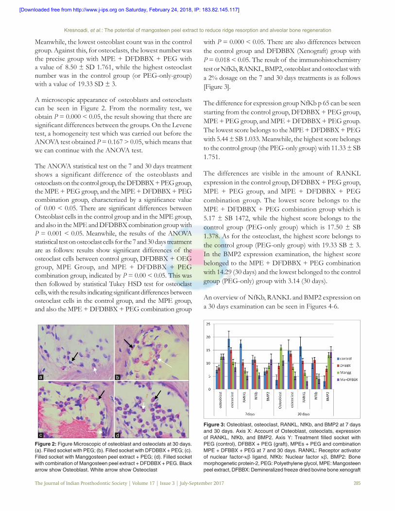
with P = 0.000 < 0.05. There are also differences between the control group and DFDBBX (Xenograft) group with P = 0.018 < 0.05. The result of the immunohistochemistry test or NfKb, RANKL, BMP2, osteoblast and osteoclast with a 2% dosage on the 7 and 30 days treatments is as follows [Figure 3].
The difference for expression group NfKb p 65 can be seen starting from the control group, DFDBBX + PEG group, MPE + PEG group, and MPE + DFDBBX + PEG group. The lowest score belongs to the MPE + DFDBBX + PEG with 5.44 ± SB 1.033. Meanwhile, the highest score belongs to the control group (the PEG‑only group) with 11.33 ± SB 1.751.
The differences are visible in the amount of RANKL expression in the control group, DFDBBX + PEG group, MPE + PEG group, and MPE + DFDBBX + PEG combination group. The lowest score belongs to the MPE + DFDBBX + PEG combination group which is 5.17 ± SB 1472, while the highest score belongs to the control group (PEG‑only group) which is 17.50 ± SB 1.378. As for the osteoclast, the highest score belongs to the control group (PEG‑only group) with 19.33 SB ± 3. In the BMP2 expression examination, the highest score belonged to the MPE + DFDBBX + PEG combination with 14.29 (30 days) and the lowest belonged to the control group (PEG‑only) group with 3.14 (30 days).
An overview of NfKb, RANKL and BMP2 expression on a 30 days examination can be seen in Figures 4‑6.
Figure 3: Osteoblast, osteoclast, RANKL, NfKb, and BMP2 at 7 days and 30 days. Axis X: Account of Osteoblast, osteoclats, expression of RANKL, NfKb, and BMP2. Axis Y: Treatment filled socket with PEG (control), DFBBX + PEG (graft), MPEs + PEG and combination MPE + DFBBX + PEG at 7 and 30 days. RANKL: Receptor activator of nuclear factor‑κβ ligand, NfKb: Nuclear factor κβ, BMP2: Bone morphogenetic protein‑2, PEG: Polyethylene glycol, MPE: Mangosteen peel extract, DFBBX: Demineralized freeze dried bovine bone xenograft
Figure 2: Figure Microscopic of osteoblast and osteoclats at 30 days. (a). Filled socket with PEG; (b). Filled socket with DFDBBX + PEG; (c). Filled socket with Manggosteen peel extract + PEG; (d). Filled socket with combination of Mangosteen peel extract + DFDBBX + PEG. Black arrow show Osteoblast. White arrow show Osteoclast
dc
ba
[Downloaded free from http://www.j-ips.org on Saturday, February 24, 2018, IP: 183.82.145.117]

Kresnoadi, et al.: The potential of mangosteen peel extract to reduce ridge resorption and alveolar bone regeneration
286 The Journal of Indian Prosthodontic Society | Volume 17 | Issue 3 | July-September 2017
ANOVA statistical calculations on NfKb expression on the 7 and 30 days treatments show significant differences of the NfKb results on the control group, DFBBX + PEG group, MPE + PEG group, and MPE + DFBBX + PEG combination group, as indicated by significant values of P = 0.00 < 0.05 on 7 and 30 days treatments.
This was then followed by a Tukey HSD Statistical test on the NfKb expression results, which showed significant differences of the NfKb expression for the 7 and 30 days treatments between the control group and MPE group, with a value of P = 0.000 < 0.05. Furthermore, between the control group and MPE + DFBBX + PEG combination group, with a value of P = 0.000 < 0.05.
ANOVA statistical calculations of RANKL expression on the 7 and 30 days treatments show significant differences on the RANKL expression results on the control group, DFDBBX + PEG group, MPE + PEG group, and MPE + DFDBBX + PEG combination group, as indicated by a significant value of P = 0.00 < 0.05. This was then followed by a Tukey HSD Statistical test on RANKL expression results, which showed significant differences of the RANKL expression for the 7 and 30 days treatments between the control group and MPE group, with a value of P = 0.000 < 0.05. Furthermore, the control group and MPE + DFDBBX + PEG combination group with a value of P = 0.002 < 0.05. There is also a significant difference between control groups and DFDBBX + PEG groups, with a value of P = 0.002 < 0.05.
A decline in the number of osteoclast cells for the amount of NfKb and RANKL expression occurred in
every group on the 7 days treatment, from the control group, the DFBBX group, the MPE group, and the MPE + DFBBX + PEG group. The lowest decline was observed on the MPE group + DFBBX + PEG group.
DISCUSSION
Trauma resulting from the extraction of teeth can cause inflammation; according to Yenary et al.,[21] when trauma occurs, the immunocompetent macrophage cells and mast cells will increase the TNF‑a (pro‑inflammatory cytokines). As part of the immune response is controlled by NfKb which contains cytosol and is bound to Ikappa B (IkB), this will lead to IkB phosphorylation, which then causes a proteasome degradation and release of NfKb from the translocation into the nucleus. Even though the inflammation from extraction trauma has subsided, and the extraction wound is closed, the mastication process in the oral cavity would cause mechanical stimuli to be directly accepted by residual ridge through the neurogenic receptor, which will activate the TNF‑a and IL‑1. This would mean that even when the extraction wound has closed, the alveolar bone resorption will continue to occur.[20]
There has been a distinction in this study on the block diagram of NfKb expression for the control group, DFBBX + PEG group, MPE + PEG group, and the MPE + DFBBX + PEG group. This study shows that stuffing/filling the tooth extraction socket with a combination of MPE + DFBBX + PEG was effective in reducing the NfKb and RANKL expression significantly; this is in accordance with a study by Chen et al.,[22] which stated that Garciana mangostana Linn can be used as an anti‑inflammatory. The types of Xanthone known to be the
Figure 5: Microscopic figure of NfKb expression at 30 days. (a) Filled with PEG; (b) filled with DFDBBX + PEG; (c) filled with MPE + PEG; (d) filled with combination of MPE + DFDBBX + PEG. Black arrow shows NfKb expression. NfKb: Nuclear factor κβ, DFDBBX: Demineralized freeze dried bovine bone xenograft, MPE: Mangosteen peel extract, PEG: Poly etylene glycol
dc
ba
Figure 4: Microscopic figure of expression of RANKL at 30 days. (a) Filled with PEG; (b) filled with DFDBBX + PEG; (c) filled with MPE + PEG; (d) filled with combination of MPE + DFDBBX + PEG. Black arrow show RANKL expression. RANKL: Receptor activator of nuclear factor‑κβ ligand, DFDBBX: Demineralized freeze dried bovine bone xenograft, MPE: Mangosteen peel extract, PEG: Poly etylene glycol
dc
ba
[Downloaded free from http://www.j-ips.org on Saturday, February 24, 2018, IP: 183.82.145.117]

Kresnoadi, et al.: The potential of mangosteen peel extract to reduce ridge resorption and alveolar bone regeneration
The Journal of Indian Prosthodontic Society | Volume 17 | Issue 3 | July-September 2017 287
most beneficial are the a‑mangostin and γ‑mangostin. Both of these are quite capable of inhibiting the production of nitric oxide and PGE2 from liposacharida cell stimulation RAW 2647. In the human body, Xanthone serves as an antioxidant, antiproliferative, anti‑inflammatory, and anti‑microbial. Xanthone is a strong antioxidant which is extremely important in the balancing of pro‑oxidants in the body, also known as “free radicals” in our natural surroundings.[16] According to Nakatani et al.,[23] γ‑mangostin possesses the ability to inhibit the production of the enzyme COX, the cause of inflammation, and directly retards the activity of IkB kinase enzyme, while also preventing COX‑2 gene transcription (NfKb target genes), as well as suppressing the production of PGE2 in the inflammatory process.
This s tudy i s in accordance wi th a s tudy by Jinsart, et al.[24] which states that in the mangosteen peel there are xanthones, which contain an anti‑inflammatory: The a and γ mangostin, which is the main bioactive compound.
In this study, the block diagram of the RANKL expression result shows the differences among each group, from the control group, DFDBBX + PEG group, MPE + PEG group, and MPE + DFBBX + PEG combination group. It shows that the combination of MPE + DFDBBX + PEG produces the lowest RANKL expression; it indicates that there is a significant decrease of inflammation because of the preservation of the tooth extraction socket by filling it with MPE + DFBBX + PEG.
This is in accordance with another study from Porth and Matfin[25] which states that inflammation will cause macrophages to induce pro‑inflammatory synthetic cytokines IL‑1 and TNF‑a, which play a role in the release of phospholipids from the fibroblast cell membrane, cell mast, neutrophil, macrophage, and lymphocytes; hence, the phospholipase A2 enzyme may start working as arachidonic acid metabolism initiates and produces the COX‑2 enzyme. An increase in the level of COX‑2 plays a role in the synthesis of prostaglandins, especially PGE‑2 since it can cause an increase in vasodilatation and can increase endothelial permeability, thereby facilitating the infiltration of inflammatory cells.[26] RANKL is a key mediator of the osteoclast development process The RANKL binding to the RANK receptor on the preosteoblast surface will activate the jun terminal kinase and NfKb, which leads to osteoclast development.[26] RANKL also plays an important role in osteoimmunology. When COX‑2 is inhibited and the pro‑inflammatory cytokines (IL‑1, TNF‑a) and also PGE‑2 decline, osteoclast development, either directly or through RANKL, will be hampered, so a differentiation and fusion of osteoclast precursors into osteoclasts will not occur. Aside from that, inhibition may also occur in the bounding process of the RANKL to RANK on the surface of preosteoblasts which will hamper the activation of NfKb; where the NfKb leads to osteoclast development, decreasing NfKb will reduce the osteoclast cell numbers.[27] Socket preservation procedure aims to reduce the loss of bone following tooth extraction, to maintain the alveolar bone volume. This is carried out by placing a bone graft into the extraction socket immediately after the tooth is extracted.[28] There has been an increase in the number of BMP2 expressions, the highest number being with the insertion of MPE + DFDBBX + PEG combination; the BMP2 is an alveolar bone growth factor, and it shows in the increasing number of osteoblast cells. In accordance with a study by Pagni, et al.,[29] BMP is a growth factor; it can induce differentiation of stem cells into cells that will develop the bone, in a process called osteoinduction. A study once introduced a method to preserve the socket after a tooth extraction process by utilizing the BMP2 absorbed in a sponge collagen. This was done by Howell, et al. in 1997, which shows that this BMP2 grafting material is safe; the result of the preservation on the patient shows an increase of alveolar bone material.
The result of this study shown in the block diagram makes it clear that the lowest osteoclast count is observed in the MPE + DFDBBX + PEG group, while the highest osteoclast count belongs to the control group. From this study, we can conclude that utilizing the combinations of MPE with DFDBBX and PEG as filling/stuffing proved
Figure 6: Microscopic figure of BMP2 expression at 30 days. (a) Filled socket with PEG; (b) filled socket with DFDBBX + PEG; (c) Filled socket with mangosteen peel extract + PEG; (d) filled with combination of mangosteen peel extract + DFDBBX + PEG. Black arrow show BMP2 expression. BMP2: Bone morphogenetic protein‑2, DFDBBX: Demineralized freeze dried bovine bone xenograft, PEG: Poly etylene glycol
dc
ba
[Downloaded free from http://www.j-ips.org on Saturday, February 24, 2018, IP: 183.82.145.117]

Kresnoadi, et al.: The potential of mangosteen peel extract to reduce ridge resorption and alveolar bone regeneration
288 The Journal of Indian Prosthodontic Society | Volume 17 | Issue 3 | July-September 2017
effective in decreasing NfKb and RANKL expression, which then led to decrease in inflammation, reducing osteoclast cell growth and ridge resorption. This research also provides an insight into the process of homeostasis in bones, that when the osteoclast cells responsible for ridge resorption decrease[30] then osteoblast cells as bone‑forming elements will rise.
CONCLUSION
The conclusion of this research is the induction of combination of MPEs and DFDBBX is effective in reducing inflammation, lowering osteoclasts, decreasing alveolar bone resorption, and also increasing BMP2 expression and alveolar bone regeneration.
AcknowledgmentsWe would like to thank the Rector of Airlangga University who provided us with the leading research grant for universities No. 519/UN3/2015, dated March 26, 2015, from the Ministry of Research, Technology and Higher Education, through the Institute of Research and Innovation at Airlangga University.
Financial support and sponsorshipThis study was supported by Airlangga University.
Conflicts of interestThere are no conflicts of interest.
REFERENCES
1. Darby I, Chen S, De Poi R. Ridge preservation: What is it and when should it be considered. Aust Dent J 2008;53:11‑21.
2. Lundy FT, Linden GJ. Neuropeptides and neurogenic mechanisms in oral and periodontal inflammation. Crit Rev Oral Biol Med 2004;15:82‑98.
3. Kulka M, Sheen CH, Tancowny BP, Grammer LC, Schleimer RP. Neuropeptide active human mast cell degranulation and chemokin production. Immunology 2008;123:398‑410.
4. Soepribadi I. Regeneration and wound healing in dental medicine. 1st ed., Vol. 77‑88. Jakarta: Sagung Seto; 2013. p. 101‑2.
5. Hayden MS, West AP, Ghosh S. NF‑kappaB and the immune response. Oncogene 2006;25:6758‑80.
6. Bellanti JA. Imunologi III, Indonesia translation. Jogjakarta: Gajah Mada University Press; 1993. p. 223‑33.
7. Lieberman JR, Friedlaender GE. Bone regeneration and repair. 1st ed. Totowa, New Jersey: Humana Press; 2005. p. 22‑32.
8. Boix D, Weiss P, Gauthier O, Guicheux J, Bouler JM, Pilet P, et al. Injectable bone substitute to preserve alveolar ridge resorption after tooth extraction: A study in dog. J Mater Sci Mater Med 2006;17:1145‑52.
9. Steigmann M. A bovine‑bone mineral block for the treatment of severe ridge deficiencies in the anterior region: A clinical case report. Int J Oral Maxillofac Implants 2008;23:123‑8.
10. Barone A, Aldini NN, Fini M, Giardino R, Calvo Guirado JL, Covani U. Xenograft versus extraction alone for ridge preservation after tooth removal: A clinical and histomorphometric study. J Periodontol
2008;79:1370‑7.11. Mardas N, Chadha V, Donos N. Alveolar ridge preservation with guided
bone regeneration and a synthetic bone substitute or a bovine‑derived xenograft: A randomized, controlled clinical trial. Clin Oral Implants Res 2010;21:688‑98.
12. Pelegrine AA, da Costa CE, Correa ME, Marques JF Jr. Clinical and histomorphometric evaluation of extraction sockets treated with an autologous bone marrow graft. Clin Oral Implants Res 2010;21:535‑42.
13. Khudhany LS. Determination of Mandibular Bone Density Index Post‑Menopausal Women by Taking into Account the Occurrence of Osteoporosis. Disertation Post Graduate Indonesia University; 2003. p. 34, 52.
14. Pedraza‑Chaverri J, Cárdenas‑Rodríguez N, Orozco‑Ibarra M, Pérez‑Rojas JM. Medicinal properties of mangosteen (Garcinia mangostana). Food Chem Toxicol 2008;46:3227‑39.
15. Palakawong C, Shopanodora P, Pisuchpen S, Phongpaichit S. Antioxidant and antimicrobial activities of crude extract from mangosteen (Garcinia mangostana L) part and some essential oils. Int Food Res J 2010;17:583‑9.
16. Khairunisa ND. Comparison of MPE (Garcinia mangostana Linn) with 2% in Inhibiting Ketokonazola Growth of Pityrosporum ovale on Dandruff. Article Research Papers Sciences, Graduate Medicine Programe, Diponegoro University, Semarang, Indonesia; 2011. p. 45.
17. Pedraza‑Chaverri J, Cárdenas‑Rodríguez N, Orozco‑Ibarra M, Pérez‑Rojas JM. Medicinal properties of mangosteen (Garcinia mangostana). Food and Chemical Toxicology 2008;46:3227‑39.
18. Dungir SG, Katja DG, Kamu VS. Anti oxidant activity and fenolic extract from Manggosteen peel extract (Garcinia mangostana L). J Mipa Unsrat Online 2012;1:11‑5.
19. Kusumawati D. Friends with experimental animal. 1st ed. Jogjakarta, Indonesia: Gajah Mada University Press; 2004. p. 67.
20. Utari K. Tall Like Receptor 2 as a Signaling Pathway Osteogenesis Alveol Bone Induced by Combine of Aloe vera dan Graft, Disertation, PhD Programe in Medicine Science, Medicine Faculty, Airlangga University; 2012. p. 45‑60.
21. Yenary MA, Liu J, Zheng Z, Vertex ZS, Lee JE, Giffard RG. Antiapoptotic and anti inflamatory mechanisme of Heat Shock Protein (Hsp) protection. Ann N Y Acad Sci 2005;1053:74‑83.
22. Chen LG, Yang LL, Wang CC. Anti‑inflammatory activity of mangostins from Garcinia mangostana. Food Chem Toxicol 2008;46:688‑93.
23. Nakatani K, Yamakuni T, Kondo N, Arakawa T, Oosawa K, Shimura S, et al. gamma‑Mangostin inhibits inhibitor‑kappaB kinase activity and decreases lipopolysaccharide‑induced cyclooxygenase‑2 gene expression in C6 rat glioma cells. Mol Pharmacol 2004;66:667‑74.
24. Jinsart W, Ternai B, Buddhasukh D, Polya GM. Inhibition of wheat embryo calcium‑dependent protein kinase and other kinases by mangostin and gamma‑mangostin. Phytochemistry 1992;31:3711‑3.
25. Porth MC, Matfin G. Pathophysiology Concept of Altered Health Science. 8th ed. New York: Mosby; 2009.
26. Carranza F, Henry H, Michael GN. Clinical Periodontology. 10th ed. New York: WB Saunders; 2006. p. 30‑1.
27. Hakozaki A, Yoda M, Tohmonda T, Furukawa M, Hikata T, Uchikawa S, et al. Receptor activator of NF‑kappaB (RANK) ligand induces ectodomain shedding of RANK in murine RAW264.7 macrophages. J Immunol 2010;184:2442‑8.
28. Kotsakis G, Markou N, Chrepa V, Krompa V, Kotsakis A. Alveolar ridge preservation utilizing the socket plug technique. Int J Oral Implantol Clin Res 2012;3:24‑30.
29. Pagni G, Pellegrini G, Giannobile WV, Rasperini G. Postextraction alveolar ridge preservation: Biological basis and treatments. Int J Dent 2012;2012:151030.
30. Roodman GD. Regulation of osteoclast differentiation. Ann N Y Acad Sci 2006;1068:100‑9.
[Downloaded free from http://www.j-ips.org on Saturday, February 24, 2018, IP: 183.82.145.117]