original article /artÍculo original -...
TRANSCRIPT
The Biologist(Lima)
ISSN Versión Impresa 1816-0719 ISSN Versión en linea 1994-9073 ISSN Versión CD ROM 1994-9081
ORIGINAL ARTICLE /ARTÍCULO ORIGINAL
REDISCOVERING THE FIRST MONOGRAPH ON PLANT ANATOMY - ANATOME PLANTARUM (1675) BY MARCELLO MALPIGHI
REDESCUBRIENDO LA PRIMERA MONOGRAFÍA SOBRE LA ANATOMÍA DE LAS PLANTAS- ANATOME PLANTARUM (1675) DE MARCELLO MALPIGHI
1Marius-Nicusor Grigore1Faculty of Biology, Alexandru Ioan Cuza University Iasi, Carol 20 A, Iasi, Romania
The Biologist (Lima), 14(2), jul-dec: 155-170.
155
ABSTRACT
Keywords: illustrations – Malpighi – microscope – plant anatomy
Malpighi's Anatome Plantarum (1675) arises as a natural step in the progress of plant biology and especially of plant morphology and anatomy. The book is well written and the language used is generally accurate, except for the limitations imposed by the level of knowledge at the time of Malpighi. Malpighi treated the plant as a system and recognized that its organs work in a synergic manner. Many of the terms used today in modern plant morphology and anatomy were already used by Malpighi. He introduced many relevant figures to support the information provided, some of which were derived from microscopic observations. Overall, despite the progress made since then, his work should be regarded as a modern monograph in plant anatomy.
RESUMEN
Palabras clave: ilustraciones – microscopio – Malpighi – anatomía de plantas
Anatome Plantarum de Malpighi (1675) surge como un paso natural en el progreso de la biología de las plantas y en especial de la morfología y la anatomía vegetal. El libro está bien escrito y el lenguaje utilizado es generalmente exacto, salvo las limitaciones impuestas por el nivel del conocimiento en la época de Malpighi. Malpighi trata la planta como un sistema y reconoce que sus órganos trabajan de una manera sinérgica. Muchos de los términos utilizados hoy día en la morfología y anatomía vegetal moderna fueron ya empleados por Malpighi. El autor también introdujo en su obra muchas figuras relevantes para apoyar la información aportada, algunas derivadas de observaciones microscópicas. A pesar de los avances registrados desde entonces su obra debe ser considerada, en general, como una monografía moderna de anatomía de las plantas.
156
new methodology by the field of religion and relationship with God. Since then, mechanistic methods for the exploration of nature penetrate all areas, including natural sciences, still dominated until then, by the writings of Theophrastus and Aristotle.
In this way, within 20-30 years, which experienced an extra-sensitive reality - the effervescence of baroque illustrated by paintings as religious ecstasy - a real, concrete, tangible world subtly occurs. This is the new world of illustrations derived from microscopic observations. In this sense, the modern human eye could hardly associate St. Teresa's image, as Italian artist Bernini figured her in religious ecstasy (sculpture St. Teresa's Ecstasy, from Beata Ludovica Alberoni, located in San Francesco a Ripa, Rome, Italy, and coincidentally finished in the same year of 1675 – see Salvat 1981, p. 33 ) with drawings from microscopic observations made by Antonie van Leeuwenhoek (1632-1723) on microorganisms, and by Marcello Malpighi (1628-1694) and Nehemiah Grew (1641-1712), on plants (“not available”. “Personal comment”).
The discovery of the principle of microscopy, although still debated from historical point of view, is attributed to brothers Hans and Zacharias Jansen around the year of 1590 (Ivănescu & Toma 2006). Subsequently, many "amateurs" were striving to improve and refine microscopes, so in the 60s and 70s of the seventeenth century, optical microscopy has become widely used for scientific research (naturally understood for that time), especially in Italy, the Netherlands and England.
As a result, Hooke discovers in 1665 the plant cell, while van Leeuwenhoek did it for some groups of microorganisms. Thus, only in few decades, an abyssal perceptive dichotomy is mentally produced; people started to move from the saints' mystic ecstasy horribly dying in the hands of sad angels, to a real universe
The seventeenth century, so intensely debated and commented, is the century that makes it unable to nominate with a single phrase, as historians are used to do, referring to a certain historical period. The seventeenth century is being considered so rich in events, thus offering practically everything that can cover the distance between the sensory and spiritual; it is being regarded as an accurate panorama just by its contradictions (Adriani 1982).
However, the strictly historical hierarchy often does not perfectly fits with cultural progress, with occurrence and flowering or collapsing among current or artistic movements. Yet, when making such hierarchy, it has merely an operational purpose, derived from the need to integrate achievements from various fields on the time scale. Martin (1982), in his exceptional work about the Baroque, was of the opinion that the seventeenth century recalls the two faces of Janus: a period of extraordinary progress in science and philosophy and radical changes in the economy and in the development of the modern state. However, this century is characterized by further theological controversy, an intense concern for personal religious experience and providential spirit, inherited from early Christianity (Martin 1982).
Sometimes, the entirely century is superimposed over the Baroque era (Oprescu 1985, Semenzato 1981), but Chaunu (1986) refers to the period 1680-1770 (1780) as a dense reality, difficult to be delineated; this époque is yet undeniable: European Enlightenment also called and the 'Age of Reason', triggered and maintained by the philosophy of Bacon, Descartes, Locke, and Spinoza. Amazing border between last decades of the seventeenth and the first of eighteenth century's cover dilated and dense temporal dimensions. Descartes separated its
INTRODUCTION
The Biologist (Lima). Vol. 14, Nº2, jul-dec 2016 Grigore
157
Rediscovering the first monograph on plant anatomy
completely unknown until then. This micro-universe is invisible to the naked eye; it is invisible, but miraculously and simply exists. Mysterious and curious ”animalicules” do really exist; they fearful perhaps swarming everywhere, move and live. However, the scrutinizer eye, curiosity and rational move beyond the barriers of prejudices, fears and inability to understand (“not available.” “Personal comment”).
There are relatively abundant published resources about Malpighi's life, historical impact of his scientific contribution, especially in the field of animal and human anatomy, physiology, disease and medicine (Arber 1942, Jay 1999, Piccolino 1999, Pearce 2007, Meli 2011, Reverón 2011, West 2013) and, of course, the large information included in the five-volume work by Adelmann (1966), dealing with Marcello Malpighi and evolution of embryology. Surprisingly, data about Malpighi's contribution to plant anatomy are relatively few and scattered, and especially a work focused strictly on the analysis of his book (1675) seems to lack. Neither Adelmann's book is not so generous with information dealing with the content of Malpighi's book; in a chapter referring on "Intensified researches on plants: the Anatome Plantarum takes form", Adelmann states that most of material is 'beyond the competence of the present writer'.
The objective of this research was to (re) discover a less known valuable historical resource for plant sciences in general and for plant morphology and anatomy, in special, while for other Malpighi's contributions to biology and medicine fields there are relatively abundant researches.
We downloaded Malpighi's work as electronic version (Biodiversity Heritage Library - not in
MATERIAL AND METHODS
copyright) and then worked on it as printed material. Several clarifications regarding the terms introduced by Malpighi can be useful. First of all, the term cortex, - icis occurring throughout the Malpighi's work is being used and thus must be primarily understood with the sense of 'bark', as explained by Evert (2006), in the tradition founded by Esau's plant anatomy terminology: 'a nontechnical term applied to all tissues outside the vascular cambium or the xylem; in older trees may be divided into dead outer bark and living inner bark, which consists of secondary phloem'. Indeed, many examples provided by Malpighi refer to woody species. The Latin common sense also covers this definition, with reference rather to rhytidome. In this direction, the current English anatomical term 'cortex' and Romanian 'scoar ă' do not fit with Malpighi's cortex; in modern plant anatomy, the cortex is the tissue region located between epidermis and central cylinder in roots and stem. Another example is that of foliola used by Malpighi to designate the small leaves from plants' buds; no doubt, it is a diminutive form of folia (leaf) and thus it explains the logical derivation. However, Romanian anatomical language also has this term - 'foliolă', but it refers on a leaflet from compound leaf; therefore, it has been inherited from Latin, but has changed his meaning. A logical connection still exists, since the leaflet (foliolă) is indeed, a small leaf as a part of a compound leaf.
Regarding Latin and Greek dictionaries that have been consulted and used for our work, in the reference list only those explicitly cited in the text are being listed. For Malpighi's mentioned species, corresponding common English names were identified using Martin's (1969) 'The concise British flora in colour.' For general English morphological and anatomical terms, several works have been consulted (Harris & Harris 2001, Evert 2006, Grigore et al. 2014) selected among a plethora of monographs.
The Biologist (Lima). Vol. 14, Nº2, jul-dec 2016
158
Grigore
We underline that Malpighi's terms used in this book are mentioned during our work, either in their original form (Latin) or translated into English. In both cases, we used Italic characters, both within text and especially in the explanation of the figures. At the same time, we tried, whenever possible, to avoid excessive equivalence of terms used by Malpighi with terms belonging to modern plant morphology and anatomy. This was the case especially with illustrations and correspondent explanations offered by Italian author. We wanted thus to maintain the originality of a language used in an anatomical work from seventeenth-century.
Finally, we should emphasize that at this moment, there is no English or other language
translation of Malpighi's valuable work on plant anatomy.
Dissecting the first part of Anatome Plantarum by Marcello Malpighi (1675). Facts and insights.
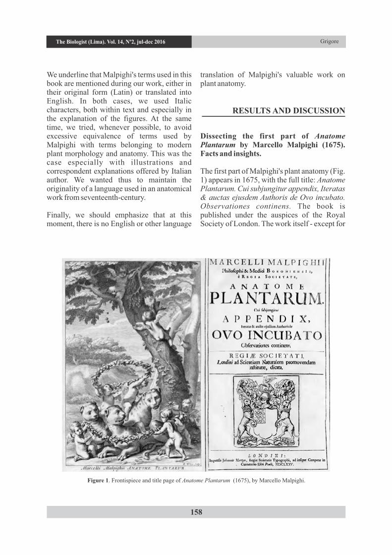
The first part of Malpighi's plant anatomy (Fig. 1) appears in 1675, with the full title: Anatome Plantarum. Cui subjungitur appendix, Iteratas & auctas ejusdem Authoris de Ovo incubato. Observationes continens. The book is published under the auspices of the Royal Society of London. The work itself - except for
RESULTS AND DISCUSSION
Figure 1. Frontispiece and title page of Anatome Plantarum (1675), by Marcello Malpighi.
The Biologist (Lima). Vol. 14, Nº2, jul-dec 2016
159
cutting up, the art of cutting up', being hence used in surgery.
The Latin people were using two different words for „to cut / cutting" and for "to dissect / dissection"; in this context, the term anatome will have been used only to designate a dissection (cutting the body of an organism, especially animal, but later also a plant organism), in order to identify and visualize the internal structures. Nevertheless, since in the case of animal's dissections (where the primarily Greek anatome applies), their organs can be observed with the naked eye (thus, without any magnifier device), probably Malpighi has used the term anatome rather in the sense of morphology (morpho-anatomy), i.e. not only what can be observed using a microscope, but also details that can be detected by the naked eye (or magnifying glass).
Malpighi's book is structured as follow. It starts with Anatomes Plantarum Idea (actually, an Introduction, in the current meaning), where the author explains the reasons which led him to write this book. According to Malpighi, incidentally, covered by a spirit heating (exaltation) of the age, penetrated (tempted) by anatomy field, and realizing the importance of plants as animate organisms, he took the responsibility for the first study of its kind (prima studia iter mihi aperirem). At the same time, he introduces a series of specialized terms - they appear within his text with Italic letters. First of all, one may notice the tree trunk (truncos), then the bark (cortex).
Of great significance is the fact that he recognizes and treats the plant as a whole, as a system that can be decomposed into individual parts. At the exterior (exterior) of the plant the cuticle (cuticula) is located, with utriculae (utriculis) or regularly disposed horizontal sacules (seu sacculis horizontali ordine locatis) (disposed – a very important observation, from anatomical point of view);
Annex about observations of the phenomenon of incubation of chicken egg - has 82 pages, is written in Latin, and includes at the end of the paper, 54 plates in black & white with 336 figures.
However, it should be noted that, from the total number of these figures, only a slight amount (about 14 figures, either as complete plates or as isolated images within a plate) contain f igures resul t ing from microscopic observations in the basic sense of the word. Most of them are in reality morphological (stricto sensu) representations of organs / parts of organs of plants.
This observation is very important, if connected with the book's title and the current meaning of the term "anatomy". Of course, in the beginning, the term had a broader sense and dealt, as we shall describe, not only with microscopic observations. The etymology of the word "anatomy" is derived the ancient Greek ( – νατομή / anatomy – to dissect, to cutreferring especially on animal body) (Liddel &Scott, 1883); however, it seems that the term does not appear in this form in either of ancient Greek texts known nowadays. Most likely, it was taken and Latinized thus becoming 'anatomy' during the Middle Age. However, our survey reveals that this word is mentioned only in one recent consulted resource (Diccionario Ilustrado Latino-Español, Español-Latino 1997) from more than 12 Latin dictionaries we dealt with. Surprisingly, anatomie, -mia mica, ae, - in Spanish dictionary is considered an ecclesiastical term, although his meaning is that of 'anatomy, dissection'. Neither the massif Oxford Latin Dictionary (1968, 2126 pages) has mention about or , as the latter anatomia anatomeappears in the work of Malpighi. However, satisfactory contributions in order to clarify this term are made by White & Riddle (1872), in their Latin dictionary. The Greek origin is recognized and it appears in the form anatomiaor , - , also , meaning 'a anatomica ae anatomice
Rediscovering the first monograph on plant anatomyThe Biologist (Lima). Vol. 14, Nº2, jul-dec 2016
160
these are being formed annually (...) and confer rigidity and at maturity degrade and fall. Sometimes, after the fall of these, epidermis can be seen (interestingly, this term does not occur with Italics). Then, Malpighi reminds of a network of woody fibers (ligno fibrosis retibus) and, finally, he probably refers on phloem (libro ?), most likely with a purely anatomical meaning, as we know it today, yet perhaps not necessarily with the same sense. The word liber, -bri in Latin dictionaries has very different meanings, most of them not related with the phloem (or bast). However, deeper research enables to discriminate his sense in various resources: 'the inner bark of a tree, rind, bast' (Oxford Latin Dictionary 1968). A special observation is made in relation to a particular type of structures (lactiferum), which occupies the middle portion of the bark from a species of Ficus; likely, there are laticiferous tissues, which may
explain the etymology of the modern term. Under the bark, woody portion (lignea portio) is located, which is described in quite great detail; the term alburnus is used (alburna), located between bark and wood. Inside the stem, the pith (medulla) is localized.
Subsequently, a number of terms, rather of morphology field are introduced: caudex, buds (gemmae). Regarding the last term, Malpighi clearly understood the correlation between their opening and leaves' occurrence; he also recognizes that some species may have underground buds. However, he describes the buds in a metaphorical but rather confusing manner; 'thus, (buds) are like delicate defended (protected) children who grow on the branches until, as the opening of the uterus, produce eggs (quasi ab aperto utero, ovo producuntur) ". Then, he uses the terms: leaves, flowers, seeds.
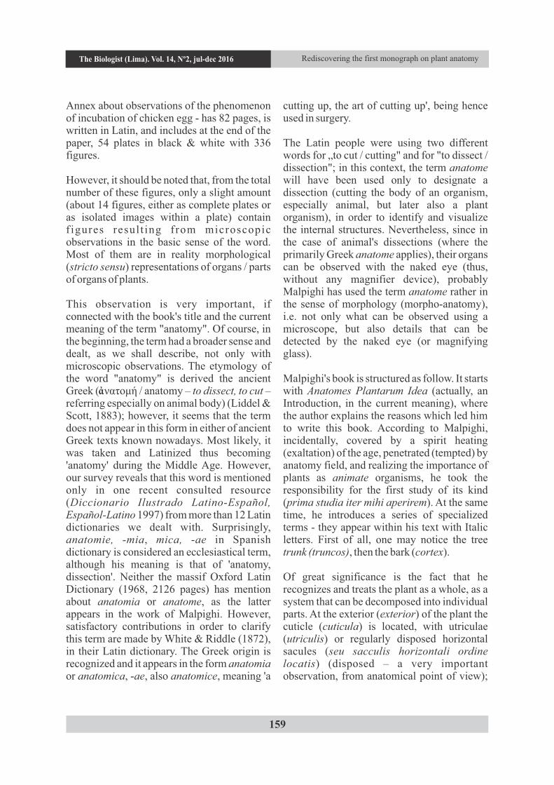
Figure 2. Longitudinal section (?) through the stem of Portulaca (succulent cortex; A – fibrous epidermal network; B – utriculae spaces, filled with transparent juice).
GrigoreThe Biologist (Lima). Vol. 14, Nº2, jul-dec 2016
161
The book is then followed by a Praefatio, where attention should be paid on the fact that the author motivates the language used in his work: a moderate (accessible) language, located under the compulsion to be progressive without sterile meditations or driven by the temptation of a rigid, academic language.
The work is then divided into several major chapters: (About ..):
1. Bark (de cortice). At a first glance, it may seem surprising that a special chapter is dedicated to cortex. However, coincidentally or not, it is the only title of the chapters, which appears in capital letters. From author's explanations, we can assume that he speaks about it as a universal structural element, common to all plants. Thus, Nature has covered the plants at periphery (peripheriam, very important observation) with "(...) Cortex
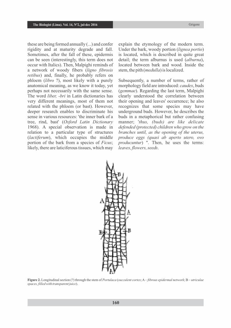
(dicitur) inderdum viscus appellatur". Therefore, Malpighi placed the cortex at the plant' periphery, but not necessarily to its direct exterior, i.e. in contact with the external environment. In addition, the term viscus means entrails, organs, or whatever lies under the skin (Nădejde & Nădejde-Gesticone 1930). The term epidermis appears, moreover, within Malpighi's work. In this part of the book, the expression 'cross section' has been noticed. Reference to figures from the end of the book is being made in the text of the book, as a modern element; the number of a mentioned plate is given outside the text body, on the edge of page, as in the older works specific to late Middle Ages or Baroque. We exemplify by two examples; one drawing from a species of Portulaca (Fig. 2) and another of chicory (Fig. 3); we keep the explanations as offered by Malpighi (original translation provided by terms written in Italics).
Figure 3. Cross section through the stem of chicory (A – utriculae, located under a thin cuticle; B – all the cortex occupied by woody/lignified substance; C – woody/lignified fistulae or fibers, disposed in bundles; D – laticiferous vessels).
Rediscovering the first monograph on plant anatomyThe Biologist (Lima). Vol. 14, Nº2, jul-dec 2016
At first glance, the language used by Malpighi seems quite accurate; however, it is the first book (monograph) of plant anatomy in the history of botany. Referring on Portulaca - a genus with many succulent species - Malpighi noticed very well that the cells are turgid (turgor means swollen, full of .., with attention to Romanian and English etymology of several botanical terms). In addition, the term utricule probably refers in fact to cells or any other regular, well-defined forms; ligneam in adjectival form, may be in fact the correspondent for lignified, as we understand it today. Likely, modern anatomy taken from the Latin ligno (which appears to Malpighi) both terms of wood (xylem, 'lemn' in Romanian) and a derivative adjective – lignified (lignum, - i, wood).
2. Parts of the stem (de partibus caulem vel caudicem componentibus). Interestingly, Malpighi uses in this case two different terms, apparently synonymous: caudex and caulis, as they distinguish in modern morphology. Nevertheless, Latin language seems to distinguish between these two term: caudex, - icis refers to a 'trunk or stem of a tree', while caulis, - is seems to refer on a stem of a non-woody plant (Oxford Latin Dictionary 1968, Stăureanu 1932, Nădejde & Nădejde-Gesticone 1930). In this chapter, the term 'culm' also appears, referring to wheat, but it is written with normal letters, which may suggest that it was already a known, popular word and not a new introduced term. Surprisingly, Nădejde and Nădejde-Gesticone (1930) consider that the terms 'culmus' and 'calamus'
162
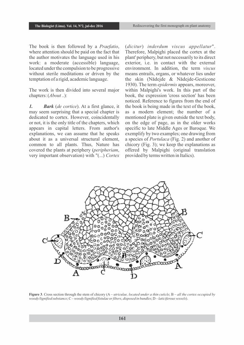
Figure 4. Cross section through a two-years old branch of horse-chestnut (A - six arranged fibrous arrays – bark; B, C – an old pair of layers extending and producing a new extension, D, where woody bundles, E, occur; F – pith, with several different appendices, G, H, I).
GrigoreThe Biologist (Lima). Vol. 14, Nº2, jul-dec 2016
are two forms of the same word, despite in the rigorous and modern anatomical language they are described as two words with different meaning. In the same direction it should be noted that Malpighi uses the term truncus when referring to the stem (trunk) of trees.
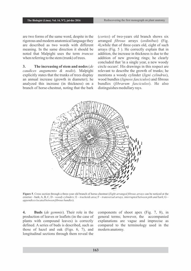
3. The increasing of stem and nodes (de caudices augumento & nodis). Malpighi explicitly states that the trunks of trees display an annual increase (growth in diameter); he analyzed this increase (in thickness) on a branch of horse-chestnut, noting that the bark
(cortex) of two-years old branch shows six arranged fibrous arrays (ordinibus) (Fig. 4),while that of three-years old, eight of such arrays (Fig. 5 ). He correctly explain that in addition, the increase in thickness is due to the addition of new growing rings; he clearly concluded that 'in a single year, a new woody circle occurs'. His drawings in this respect are relevant to describe the growth of trunks; he mentions a woody cylinder (ligni cylindrus), wood bundles (ligneos fasciculos) and fibrous bundles (fibrarum fasciculos). He also distinguishes medullary rays.
Figure 5. Cross section through a three-year old branch of horse-chestnut (Eight arranged fibrous arrays can be noticed at the exterior – bark; A, B, C, D – woody cylinders; E – tracheids area; F – transversal arrays, interrupted between pith and bark; G – appendices located between fibrous bundles).
4. Buds (de gemmis). Their role in the production of leaves or leaflets (in the case of plants with compound leaves) is correctly defined. A series of buds is described, such as those of hazel and oak (Figs. 6, 7), and longitudinal sections through them reveal the
components of shoot apex (Fig. 7, 8), in general terms; however, the accompanied explanations are vague and imprecise as compared to the terminology used in the modern anatomy.
Rediscovering the first monograph on plant anatomy
163
The Biologist (Lima). Vol. 14, Nº2, jul-dec 2016
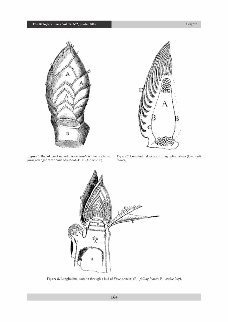
Figure 6. Bud of hazel and oak (A – multiple scales-like leaves form, arranged at the basis of a shoot - B; C – foliar scar).
Figure 7. Longitudinal section through a bud of oak (D – small leaves).
Figure 8. Longitudinal section through a bud of Ficus species (E – falling leaves; F – stable leaf).
Grigore
164
The Biologist (Lima). Vol. 14, Nº2, jul-dec 2016
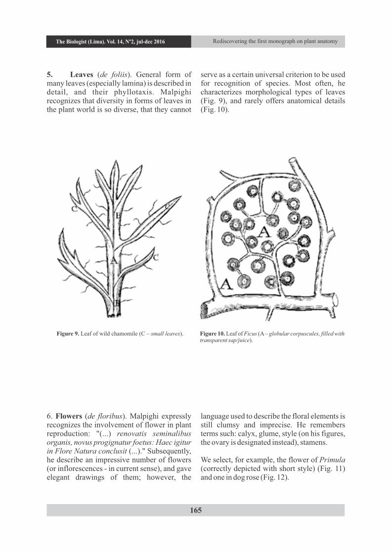
5. Leaves (de foliis). General form of many leaves (especially lamina) is described in detail, and their phyllotaxis. Malpighi recognizes that diversity in forms of leaves in the plant world is so diverse, that they cannot
serve as a certain universal criterion to be used for recognition of species. Most often, he characterizes morphological types of leaves (Fig. 9), and rarely offers anatomical details (Fig. 10).
Figure 9. Leaf of wild chamomile (C – small leaves). Figure 10. Leaf of Ficus (A – globular corpuscules, filled with transparent sap/juice).
6. Flowers (de floribus). Malpighi expressly recognizes the involvement of flower in plant reproduction: "(...) renovatis seminalibus organis, novus progignatur foetus: Haec igitur in Flore Natura conclusit (...)." Subsequently, he describe an impressive number of flowers (or inflorescences - in current sense), and gave elegant drawings of them; however, the
language used to describe the floral elements is still clumsy and imprecise. He remembers terms such: calyx, glume, style (on his figures, the ovary is designated instead), stamens.
We select, for example, the flower of Primula (correctly depicted with short style) (Fig. 11) and one in dog rose (Fig. 12).
Rediscovering the first monograph on plant anatomy
165
The Biologist (Lima). Vol. 14, Nº2, jul-dec 2016

7. Seeds (seed formation, seminum generatione) covers, in fact, a more extended and complex issue; from the book's text and illustrations provided by Malpighi (indeed, detailed but inaccurate explained), we can deduce that he deals with fertilization, embryo formation and other components of the seed as well as early stages of seedling' emergence. Incidentally, he mentions again the foetus (in utero), which may reinforce the assumption that the term foetus, refers even in a universal way, to the result of the fusion of sexual gametes, thus, ultimately, to the embryo. In addition, the term seedling explicitly occurs, whose origin is clearly recognized as derived from the seed (seminalis plantula).
8. Fruit formation (increasing, growing) and its forms (de uterorum augumento & ipsorum succedente forma). He not uses the
term fruit, but uterus, which is still quite relevant, because Malpighi probably uses with the sense of the ovary, which correctly explains its involvement in the fruit formation. However, he describes the shape and structures of many fruits from various species (Figs. 13, 14, 15), and in a quite accurate manner, except, again, here and there, the imprecise language. Term 'pericarp' also occurs. In the case of mentioned drupes, the endocarp is described as osseum nucleum, an obvious observation about its sclerified structure. He correctly states that fruits content the seed or the seeds; in the case of silicle from shepherd's purse, he illustrates the seeds correctly (Fig. 13). The poricidal capsule of poppy is accurately described (Fig. 15), with all its details: the fenestrated operculum, the pores (openings), from where small seeds will be released.
Figure 11. Flower of Primula species (E – style). Figure 12. Flower of dog rose (B – uterus; C – style; D – tubes; E – fistules).
Grigore
166
The Biologist (Lima). Vol. 14, Nº2, jul-dec 2016
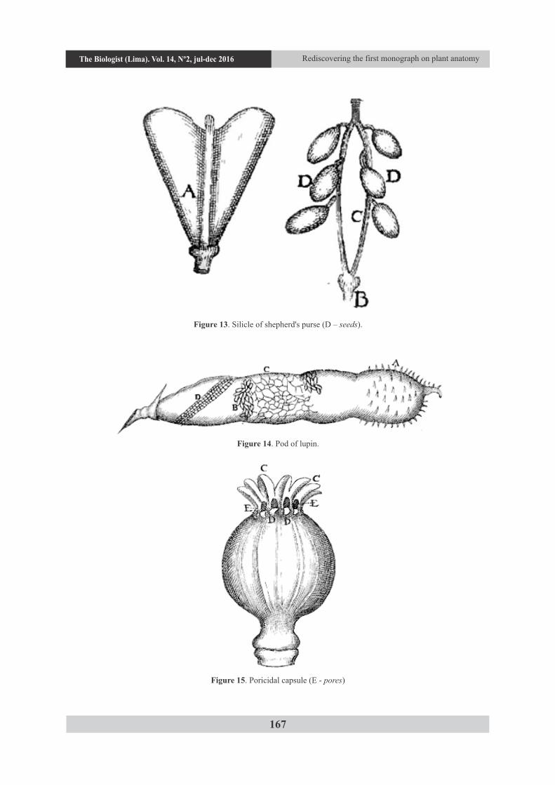
Figure 13. Silicle of shepherd's purse (D – seeds).
Figure 14. Pod of lupin.
Figure 15. Poricidal capsule (E - pores)
Rediscovering the first monograph on plant anatomy
167
The Biologist (Lima). Vol. 14, Nº2, jul-dec 2016
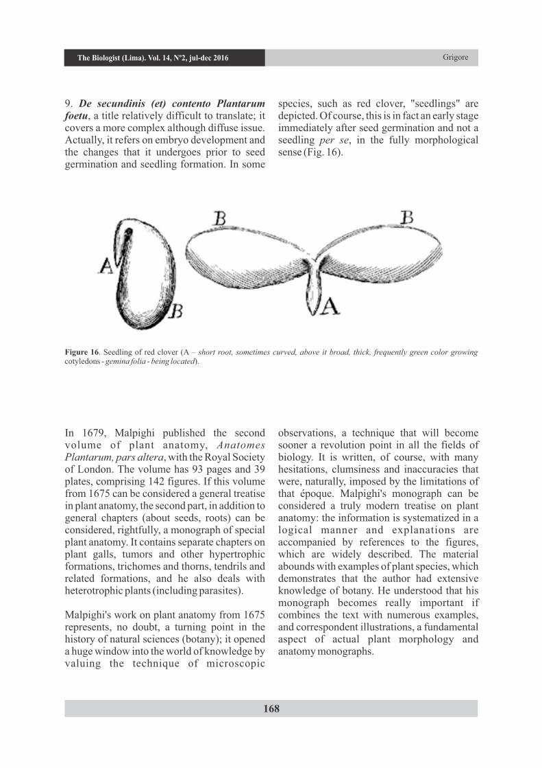
9. De secundinis (et) contento Plantarum foetu, a title relatively difficult to translate; it covers a more complex although diffuse issue. Actually, it refers on embryo development and the changes that it undergoes prior to seed germination and seedling formation. In some
species, such as red clover, "seedlings" are depicted. Of course, this is in fact an early stage immediately after seed germination and not a seedling per se, in the fully morphological sense (Fig. 16).
Figure 16. Seedling of red clover (A – short root, sometimes curved, above it broad, thick, frequently green color growing cotyledons - gemina folia - being located).
In 1679, Malpighi published the second volume of plant anatomy, Anatomes Plantarum, pars altera, with the Royal Society of London. The volume has 93 pages and 39 plates, comprising 142 figures. If this volume from 1675 can be considered a general treatise in plant anatomy, the second part, in addition to general chapters (about seeds, roots) can be considered, rightfully, a monograph of special plant anatomy. It contains separate chapters on plant galls, tumors and other hypertrophic formations, trichomes and thorns, tendrils and related formations, and he also deals with heterotrophic plants (including parasites).
Malpighi's work on plant anatomy from 1675 represents, no doubt, a turning point in the history of natural sciences (botany); it opened a huge window into the world of knowledge by valuing the technique of microscopic
observations, a technique that will become sooner a revolution point in all the fields of biology. It is written, of course, with many hesitations, clumsiness and inaccuracies that were, naturally, imposed by the limitations of that époque. Malpighi's monograph can be considered a truly modern treatise on plant anatomy: the information is systematized in a logical manner and explanations are accompanied by references to the figures, which are widely described. The material abounds with examples of plant species, which demonstrates that the author had extensive knowledge of botany. He understood that his monograph becomes really important if combines the text with numerous examples, and correspondent illustrations, a fundamental aspect of actual plant morphology and anatomy monographs.
Grigore
168
The Biologist (Lima). Vol. 14, Nº2, jul-dec 2016
169
My special thanks go to John B. West, from Department of Medicine, University of California San Diego, La Jolla, California, who offered me valuable information inside Adelmann's book (1966), a resource I wasn't able to consult by myself.
ACKNOWLEDGEMENTS
BIBLIOGRAPHIC REFERENCES
Adelmann, H.B. 1966. Marcello Malpighi and the Evolution of Embryology (5 vols). Ithaca, NY: Cornell University Press. 2475 pp.
Adriani, G. 1982. Pictura germană în secolul al XVII-lea. Ed. Meridiane, Bucure ti. 210 pp.
Arber, A. 1942. Nehemiah Grew (1641-1712) and Marcello Malpighi (1628-1694): An essay in comparison. Isis, 34 (1): 7-16.
Chaunu, P. 1986. Civiliza ia Europei în secolul luminilor, vol. I. Ed. Meridiane, Bucure ti. 467 pp.s
Diccionario Vox Ilustrado Latino-Español, Español-Latino, 1997. Bibliograf, S.A., Barcelona, 715 pp.
Evert, R. F. 2006. Esau's Plant Anatomy (third ed.). John Wiley and Sons, New York. 601 pp.
Grigore, M. N., Ivănescu L. & Toma C. 2014. Halophytes. An integrative anatomical study. Springer, Cham, Heidelberg, New York, Dordrecht, London. 548 pp.
Harris, J. G. & Harris M. W. 2001. Plant iden t i f i ca t ion terminology. An illustrated glossary, second ed. Spring Lake Publishing, Spring Lake, Utah. 206 pp.
Ivănescu L. & Toma C. 2006. De la descoperirea celulei (1665, 1667) la teoria celulară (1838, 1839). Studii i sComunicări. Muzeul de tiin e ale .Naturii ”Ion Borcea” Bacău, 21: 13-19.
Jay, V. 1999. Marcello Malpighi. Archives of
Pathology & Laboratory Medicine, 123: 874.
Liddell, H. G. & Scott R. 1883. Greek-English th
Lexicon, 7 ed. New York, Harper & Brothers, Franklin-Square. 1776 pp.
Malpighi, M. 1675. Anatome Plantarum. Regiae Societati, Londini ad Scientiam Naturalem promovendam institutae, dicata. 82 pp.
Martin, J. R. 1982. Barocul. Ed. Meridiane, Bucure ti. 293 pp.
Martin, W. K. 1969. The concise British flora in colour. Ebury Press and Michael Joseph. 254 pp.
Meli, D. B. 2011. Mechanism, experiment, disease: Marcello Malpighi and S e v e n t e e n t h - c e n t u r y a n a t o m y . Baltimore, MD, Johns Hopkins University Press. 456 pp.
Nădejde I. & Nădejde-Gesticone A. 1930. Dic ionar Latin-Român complet pentru .licee, seminarii i universită i. Ed. tNa ională Mecu SA, Ia i. 704 pp.t .
Oprescu, G. 1985. Manual de istoria artei. Barocul. Ed. Meridiane, Bucure ti. 240 pp.
Oxford Latin Dictionary, 1968. Oxford at the Clarendon Press, 2126 pp.
Pearce, J. M. S. 2007. Malpighi and the Discovery of Capillaries. European Neurology, 58: 253-255.
Piccolino, M. 1999. Marcello Malpighi and the difficult birth of modern life sciences. Endeavour, 23: 175-179.
Reverón, R. R. 2011. Marcello Malpighi (1628-1694), founder of microanatomy. International Journal of Morphology, 29: 399-402.
Salvat, J. (ed.). 1981. Historia del Arte, vol. 7. Salvat Editores, S.A., Barcelona, 299 pp.
Semenzato, C. 1981. Arte, part 2. Culturama. Gran Enciclopedia temático-visual, vol. 7. Ediciones Danae, s.a., Barcelona, Spain. 536 pp.
Stăureanu, M. 1932. Dic ionar Latin-Român. tEdit. “Scrisul Românesc”, Craiova, 578 pp.
Rediscovering the first monograph on plant anatomyThe Biologist (Lima). Vol. 14, Nº2, jul-dec 2016
170
English Dictionary, vol. I, 4th ed. London, Longmans, Green, and Co. 1034 pp.
West, J. B. 2013. Marcello Malpighi and the discovery of the pulmonary capillaries and alveoli. American Journal of Physiology - Lung Cellular and Molecular Physiology, 304: L383-L390.
White, J. T. & Riddle, J. E. 1872. A Latin-Received January 14, 2016.
Accepted February 23, 2016.
GrigoreThe Biologist (Lima). Vol. 14, Nº2, jul-dec 2016