original article cigarette smoke impairs cytokine ... · and bcg containment in alveolar...
TRANSCRIPT

ORIGINAL ARTICLE
Cigarette smoke impairs cytokine responsesand BCG containment in alveolar macrophagesRichard N van Zyl-Smit,1 Anke Binder,1 Richard Meldau,1 Patricia L Semple,1
Alicia Evans,2 Peter Smith,2 Eric D Bateman,1 Keertan Dheda1,3,4
▸ Additional material ispublished online only. To viewplease visit the journal online(http://dx.doi.org/10.1136/thoraxjnl-2013-204229).1Lung Infection and ImmunityUnit, Division of Pulmonology& UCT Lung Institute,Department of Medicine,University of Cape Town,South Africa2Division of Pharmacology,Department of Medicine,University of Cape Town,South Africa3Institute of Infectious Diseasesand Molecular Medicine,University of Cape Town,South Africa4Department of Infection,University College LondonMedical School, UK
Correspondence toProfessor Keertan Dheda,Groote Schuur Hospital,H47 Old Main Building,Observatory, 7925,South Africa;[email protected]
Received 24 July 2013Revised 7 November 2013Accepted 10 November 2013
To cite: van Zyl-Smit RN,Binder A, Meldau R, et al.Thorax Published OnlineFirst: [please include DayMonth Year] doi:10.1136/thoraxjnl-2013-204229
ABSTRACTBackground There is a strong epidemiological linkbetween smoking and tuberculosis (TB), but theassociation is confounded by socioeconomic and otherfactors. A direct relationship between cigarette smokeand poor treatment-related outcomes in patients withTB is therefore questionable. We investigated whetherconstituents of tobacco smoke impair mycobacterial hostimmune responses in vitro.Methodology Preparation of a cigarette smoke extract(CSE) from Marlboro Red cigarettes was standardisedand reproducibility verified by mass spectroscopy.Macrophages were derived from peripheral bloodmonocytes (MDM) and alveolar macrophages frombronchoalveolar lavage fluid from healthy non-smokingvolunteers. Mycobacterial uptake (flow cytometricdetection of fluorescence using green fluorescentprotein-labelled BCG), cytokine responses (ELISA) andmycobacterial containment (colony forming units) wasevaluated in both macrophage populations with andwithout co-culture with CSE, nicotine and a nicotinereceptor blocker.Results Cigarette smoke failed to impair the uptake ofmycobacteria by monocyte-derived or alveolarmacrophages. CSE (vs no CSE) reduced the mean (SD)BCG-driven macrophage (MDM) interferon γ (IFN-γ),tumour necrosis factor α (TNF-α) and interleukin 10(IL-10) responses by 56.4 (18.6)%, 67.0 (33.4)% and77.7 (27.7)%, respectively (p<0.001). Nicotine aloneimpaired IL-10 and TNF-α production by 48.8 (37)%and 49 (50)%, respectively (p<0.05) through an α-7nicotine receptor-independent mechanism. In 5-daycultures, CSE impaired mycobacterial (BCG) containmentin both monocyte-derived and alveolar macrophages.Conclusions Cigarette smoke attenuates effectorcytokine responses and impairs mycobacterialcontainment within infected human macrophagesderived from the peripheral blood and alveolarcompartments, thus supporting the hypothesis thatcigarette smoke subverts mycobacteria-related immunity.
INTRODUCTIONThere are an estimated 1.3 billion smokers world-wide.1 Tobacco smoking is the most important pre-ventable cause of death, with an estimated 8% ofall adult deaths per year (5 million people) attribut-able to tobacco smoking. More than 80% of thesedeaths occur in the developing world.1 One-thirdof the world’s population is thought to be latentlyinfected with Mycobacterium tuberculosis and anadditional 8.8 million new cases are diagnosedwith active tuberculosis (TB) each year.2 Several
modifiable factors including malnutrition, over-crowding, poverty and HIV co-infection are asso-ciated with susceptibility to and spread of activeTB.3 In recent years, smoking has been confirmedas another risk factor for TB. Three comprehensiveindependent systematic reviews and meta-analysessupport this association.4–6 Compared with non-smokers, smokers have almost twice the risk of TBinfection and of progression from latent to activedisease. Smokers are also almost twice as likely todie from active TB.4–6 Based on these data, an esti-mated 15.8% of TB cases worldwide are probablyattributable to tobacco smoking, higher than thatattributable to HIV infection (∼11%), alcoholabuse (∼8%) and diabetes (∼7%).7
In the light of these facts, implementation ofsmoking cessation strategies has been proposed asan important component of TB control pro-grammes in tandem with addressing other modifi-able factors such as overcrowding, poverty andalcohol abuse.8 However, in poorly resourcedregions, smoking cessation programmes will incuradditional cost and place extra demands uponalready overburdened clinic staff. It is thereforerightly questioned what priority smoking cessationshould receive, particularly because the epidemio-logical association is weakened by confoundingfactors such as overcrowding, poverty and alcohol
Key messages
What is the key question?Is the epidemiological association of smoking andheightened risk (approximately double) oftuberculosis (TB) disease and death merely aconfounder of socioeconomic or other factors or isit supported by biologically plausible mechanisticdata?
What is the bottom line?An animal study has demonstrated higherM tuberculosis colony growth in mice exposed tocigarette smoke but substantial human data arelacking.
Why read on?This study provides novel human datademonstrating several biologically plausiblemechanisms by which tobacco smokes subvertshuman immunity and possibly increases the riskfor TB disease.
van Zyl-Smit RN, et al. Thorax 2013;0:1–8. doi:10.1136/thoraxjnl-2013-204229 1
Tuberculosis Thorax Online First, published on November 28, 2013 as 10.1136/thoraxjnl-2013-204229
Copyright Article author (or their employer) 2013. Produced by BMJ Publishing Group Ltd (& BTS) under licence.
on 13 February 2019 by guest. P
rotected by copyright.http://thorax.bm
j.com/
Thorax: first published as 10.1136/thoraxjnl-2013-204229 on 28 N
ovember 2013. D
ownloaded from

usage, and there is limited experimental data to support the bio-logical plausibility for the association between smoking andTB.9 10 In animal models, mice exposed to cigarette smoke for14 weeks have a significantly higher mycobacterial burden inthe lungs and spleen 30 days after challenge with aerosolisedM tuberculosis.9 In non-TB models, cigarette smoke has beendemonstrated to impair phagocytic function (Staphylococcus,Listeria, Candida) and cytokine responses (lipopolysaccharide,Legionella).11–15 There are no human data on the impact of cig-arette smoke extract (CSE) on mycobacterial containment. Wehypothesised that constituents of tobacco smoke may attenuateeffector cytokine responses and mycobacterial containment inhuman alveolar and monocyte-derived macrophages (MDM).
METHODSParticipants and obtaining macrophagesHealthy HIV-negative non-smoking participants were recruitedto provide venous blood samples and/or undergo bronchoscopyand bronchoalveolar lavage. Detailed methods used are pro-vided in the online supplement.
MDM were prepared from peripheral blood mononuclearcells obtained by density sedimentation throughFicoll-Hypaque. The peripheral blood mononuclear cells wereseeded at a concentration of 1×10⁶/mL into 24-well plates andallowed to adhere for 6 days. Alveolar macrophages wereobtained by bronchoscopy with low-pressure suction using a300 mL sterile saline lavage. Non-adherent cells were removedat 4 h and appropriate cell concentrations were prepared foreach of the experiments performed.
Macrophage infection with BCGMycobacterium bovis Bacillus Calmette-Guérin (BCG) expres-sing green fluorescent protein (BCG-GFP) was used in all infec-tion experiments. MDM were infected with BCG-GFP at amultiplicity of infection of 2:1 and for alveolar macrophages at2.5:1. MDM were washed with warm phosphate-buffered salineafter 18 h to remove non-ingested mycobacteria.
Preparation of CSE and nicotineA standardised cigarette smoking device was constructed basedon the apparatus used in the studies reported by Freed and cow-orkers (see online supplement). Reproducibility of the extractwas assessed using an ABSciex 3200 Qtrap mass spectrometerconnected to an Aglient 1200 Series high-performance liquidchromatography. For each experiment, fresh extract was usedand added to cultures within 15 min of preparation.
Determination of mycobacterial uptakeFlow cytometric analysis was performed to determine thenumber of macrophages containing intracellular BCG-GFP.Immediately before acquisition of the cells, 10 μL7-aminoactinomycin D (7AAD; eBiosciences) was added inorder to establish cell viability. Once acquired, the cells wereanalysed on a FACsCalibur using Cell Quest software.
Cytokine assaysCytokine concentrations were determined using commerciallyavailable ELISA kits and performed according to the manufac-turers’ instructions. Triplicates of each experimental condition wereprepared and pooled whenever sufficient cells were available. Toexplore the hypothesis that nicotine can modulate tumour necrosisfactor α (TNF-α) production, experiments were conducted usingan α7 receptor blocker, α-bungarotoxin (Sigma Aldrich).Macrophages were pre-incubated with α-bungarotoxin-FITC
(1.5 μg/mL) for 15 min prior to the addition of nicotine and infec-tion with BCG.
Mycobacterial containment endpointsTo determine the capacity of human macrophages to containmycobacterial infection, adherent macrophages were infectedwith BCG and then cultured for 5 days in the presence of 10%CSE. On days 1, 2, 3 and 5, intracellular colony forming units(CFUs) were determined.
Statistical methodsStatistical comparisons were made with the appropriate para-metric (t test) and non-parametric tests (Mann–Whitney U test)and, where applicable, paired tests (paired t test or Wilcoxonmatched pairs signed rank test). For data involving more thantwo categories, an analysis of variance (ANOVA) was used(one-way ANOVA or repeated measures ANOVA as appropri-ate). To correct for multiple comparisons the Tukey test wasused. A p value of 0.05 was considered significant. Statisticalanalysis was performed using GraphPad Prism (V.5.00,GraphPad Software, San Diego, California, USA, http://www.graphpad.com) and OpenEpi (V.2.3.1. http://www.OpenEpi.com, updated 19 September 2010).
RESULTSCSE and toxicityThe CSE was prepared 26 times over a 9-month experimentalperiod. The mean (SD) concentration of nicotine in the extractwas 6.4 (2.6) μg/mL. Exposure of adherent MDM to concentra-tions of CSE >10% for 24 h showed a dose-dependent reduc-tion in cell viability (see figure E4 in online supplement).Furthermore, at concentrations of ≥20%, spontaneous celldetachment increased (see figure E5 in online supplement).Experiments were conducted using 10% CSE. For uptake andcytokine experiments, CSE was added to the macrophage cul-tures immediately prior to infection.
Mycobacterial uptakeMDM viability was not affected by BCG infection after 18 h orby co-exposure to either nicotine or cigarette smoke. The mean(SD) percentage viability of uninfected unexposed MDM (deter-mined by 7AAD staining) was 63.3 (5.5)%. Exposure to 10%CSE or 1 μg/mL nicotine did not affect viability (CSE: 67.7(4.05)%, nicotine: 64.1 (3.8)%; p=0.59). After BCG infection,MDM viability (67.3 (5.8)%) was not significantly differentfrom uninfected macrophage viability (exposed or unexposed)nor from that of infected/CSE-exposed macrophages (68.1(2.7)%) or infected/nicotine-exposed macrophages (60.9(9.7)%; p=0.63).
Significant variability in BCG uptake was seen between indivi-duals at 4 h and 18 h. At 4 h BCG uptake was low, but signifi-cantly higher in unexposed MDM than in those exposed to10% CSE (4.5 (2.6)% vs 3.2 (1.6)%; p=0.03). At 18 h,however, no significant difference in uptake was demonstratedbetween unexposed MDM and those exposed to 10% CSE(24.9 (6.9)% vs 21.3 (6.8)%; p=0.09, figure 1).
Alveolar macrophages exhibited a higher uptake of BCG at18 h (42.6 (17.4)%), but this was not significantly differentfrom alveolar macrophages exposed to 10% CSE (44.3(16.1)%) or 1 μg/mL nicotine (35.9 (16.8)%); p=0.28, figure1). Viability of alveolar macrophages was not affected by infec-tion or co-exposure to 10% CSE or nicotine (see figures E6 andE7 in online supplement).
2 van Zyl-Smit RN, et al. Thorax 2013;0:1–8. doi:10.1136/thoraxjnl-2013-204229
Tuberculosis
on 13 February 2019 by guest. P
rotected by copyright.http://thorax.bm
j.com/
Thorax: first published as 10.1136/thoraxjnl-2013-204229 on 28 N
ovember 2013. D
ownloaded from

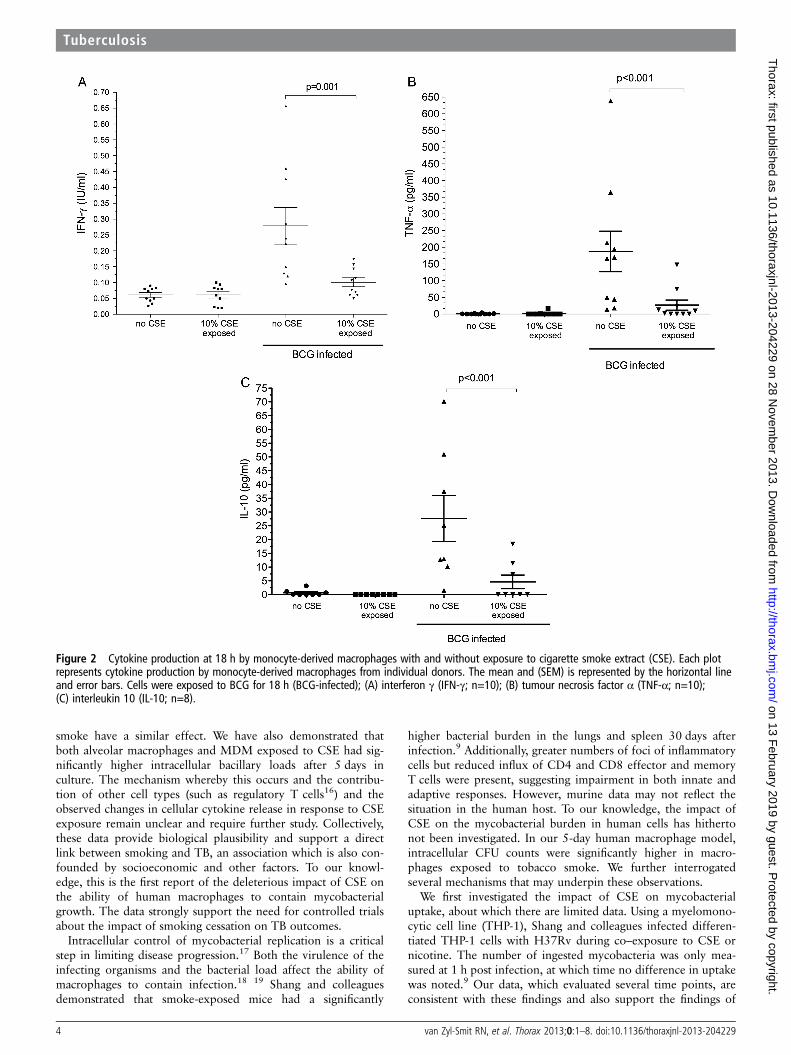
Cytokine productionIFN-γ production by MDM was measured at 4 h and 18 h afterinfection. At 4 h the mean (SD) IFN-γ production was minimal(0.05 (0.01) IU/mL) and was not affected by co-exposure to10% CSE (0.04 (0.18) IU/mL; p=0.43). At 18 h, IFN-γ produc-tion by unstimulated MDM as well as those exposed to 10%CSE remained negligible (0.06 (0.03) IU/mL and 0.06(0.03) IU/mL, respectively; p=1.0). Following BCG infection, asignificant increase in mean (SD) IFN-γ was detected (0.28(0.18) IU/mL). However, macrophages co-exposed to 10% CSEduring infection demonstrated significantly less IFN-γ produc-tion (0.10 (0.04) IU/mL; p=0.001, figure 2). Production ofIFN-γ was confirmed by intracellular staining for IFN-γ and bydemonstrating upregulation of IFN-γ mRNA transcription (seefigures E8 and E9 and table E2 in online supplement). Due tohigh basal cytokine production by alveolar macrophages in the24 h after lavage, cytokine data for alveolar macrophages werenot interpretable (data not shown).
TNF-α production was measured at 18 h after infection withBCG. Similarly to IFN-γ, negligible amounts of TNF-α weresecreted by unstimulated and CSE-exposed macrophages (mean(SD) 0.73 (1.8) pg/mL and 1.7 (5.3) pg/mL, respectively;p=0.3). Following infection, TNF-α production increased sig-nificantly (137.5 (111.7) pg/mL) but was significantly reducedafter co-exposure to 10% CSE (21.63 (45.97) pg/mL; p<0.001vs control, figure 3).
Interleukin 10 was measured at 18 h after infection with andwithout 10% CSE exposure. The results were similar to thosefor TNF-α and IFN-γ production. Unstimulated andCSE-exposed macrophages produced little or no measurableIL-10 (0.65 (1.65) pg/mL and 0.0 (0.0) pg/mL, respectively).Following infection, IL-10 production was 27.68 (22.5) pg/mLand was significantly reduced by co-exposure to CSE duringinfection (4.64 (6.75) pg/mL; p<0.001, figure 3).
Compared with the maximal cytokine production of unex-posed MDMs, 10% CSE reduced mean (SD) BCG-drivenIFN-γ, TNF-α and IL-10 production by 56.4 (18.6)%, 67.0(33.4)% and 77.7 (27.7)%, respectively. Alveolar macrophage
cytokine production and response to CSE or nicotine exposurewas not interpretable as basal cytokine production was highwhen tested 24 h after lavage (data not shown).
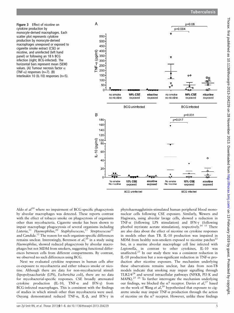
Cytokine response to nicotine exposureExposure of MDM to nicotine (1 μg mL) alone (as opposed towhole smoke extract) resulted in significantly reducedBCG-driven IL-10 production (40.1 (60.3) pg/mL vs 11.3(16.4) pg/mL; p=0.03). TNF-α production was similarlyreduced but did not reach statistical significance (181.6(186.1) pg/mL vs 78.7 (52.5) pg/mL; p=0.08, figure 3). The cal-culated mean (SD) reduction in cytokine production in responseto nicotine exposure was 48.8 (37)% for IL-10 and 49 (50)%for TNF-α. Blocking of the nicotine α7 receptor withα-bungarotoxin did not restore TNF-α production by cellsexposed to CSE or nicotine (figure 4).
Mycobacterial containmentOver a 5-day period, BCG-infected MDM and alveolar macro-phages exposed to 10% CSE showed higher CFU counts notaccounted for by adherent cell numbers, as the latter did notchange (figure 5). At each time point, prior to cell lysis toperform the CFU count, control and CSE-containing wells wereinspected under an inverted microscope. No difference in thenumber and integrity of adherent macrophages could beidentified.
DISCUSSIONOur studies of the effect of CSE on the responses of humanMDM and alveolar macrophages to mycobacterial infection(BCG) demonstrate that, while the uptake of mycobacteriaremained unaffected, the production of key cytokines in theimmune response to TB infection (ie, TNF-α, IFN-γ and IL-10)was significantly reduced by exposure to CSE. Furthermore,nicotine alone similarly impaired both IL-10 and TNF-α pro-duction, suggesting that it too contributes to this effect. Sincethe effect of smoke that does not contain nicotine was notexamined, it is not clear whether other components of cigarette
Figure 1 BCG-GFP uptake byalveolar and monocyte-derivedmacrophages after 18 h. Uptake(percentage of cells positive for GFPby flow cytometry) is depicted forsubjects (n=7) following 18 h ofinfection. Uptake by macrophages withand without exposure to 10% cigarettesmoke extract (CSE) from individualdonors is depicted by joined lines. Thehorizontal line represents the medianvalue for all individuals.
van Zyl-Smit RN, et al. Thorax 2013;0:1–8. doi:10.1136/thoraxjnl-2013-204229 3
Tuberculosis
on 13 February 2019 by guest. P
rotected by copyright.http://thorax.bm
j.com/
Thorax: first published as 10.1136/thoraxjnl-2013-204229 on 28 N
ovember 2013. D
ownloaded from
smoke have a similar effect. We have also demonstrated thatboth alveolar macrophages and MDM exposed to CSE had sig-nificantly higher intracellular bacillary loads after 5 days inculture. The mechanism whereby this occurs and the contribu-tion of other cell types (such as regulatory T cells16) and theobserved changes in cellular cytokine release in response to CSEexposure remain unclear and require further study. Collectively,these data provide biological plausibility and support a directlink between smoking and TB, an association which is also con-founded by socioeconomic and other factors. To our knowl-edge, this is the first report of the deleterious impact of CSE onthe ability of human macrophages to contain mycobacterialgrowth. The data strongly support the need for controlled trialsabout the impact of smoking cessation on TB outcomes.
Intracellular control of mycobacterial replication is a criticalstep in limiting disease progression.17 Both the virulence of theinfecting organisms and the bacterial load affect the ability ofmacrophages to contain infection.18 19 Shang and colleaguesdemonstrated that smoke-exposed mice had a significantly
higher bacterial burden in the lungs and spleen 30 days afterinfection.9 Additionally, greater numbers of foci of inflammatorycells but reduced influx of CD4 and CD8 effector and memoryT cells were present, suggesting impairment in both innate andadaptive responses. However, murine data may not reflect thesituation in the human host. To our knowledge, the impact ofCSE on the mycobacterial burden in human cells has hithertonot been investigated. In our 5-day human macrophage model,intracellular CFU counts were significantly higher in macro-phages exposed to tobacco smoke. We further interrogatedseveral mechanisms that may underpin these observations.
We first investigated the impact of CSE on mycobacterialuptake, about which there are limited data. Using a myelomono-cytic cell line (THP-1), Shang and colleagues infected differen-tiated THP-1 cells with H37Rv during co–exposure to CSE ornicotine. The number of ingested mycobacteria was only mea-sured at 1 h post infection, at which time no difference in uptakewas noted.9 Our data, which evaluated several time points, areconsistent with these findings and also support the findings of
Figure 2 Cytokine production at 18 h by monocyte-derived macrophages with and without exposure to cigarette smoke extract (CSE). Each plotrepresents cytokine production by monocyte-derived macrophages from individual donors. The mean and (SEM) is represented by the horizontal lineand error bars. Cells were exposed to BCG for 18 h (BCG-infected); (A) interferon γ (IFN-γ; n=10); (B) tumour necrosis factor α (TNF-α; n=10);(C) interleukin 10 (IL-10; n=8).
4 van Zyl-Smit RN, et al. Thorax 2013;0:1–8. doi:10.1136/thoraxjnl-2013-204229
Tuberculosis
on 13 February 2019 by guest. P
rotected by copyright.http://thorax.bm
j.com/
Thorax: first published as 10.1136/thoraxjnl-2013-204229 on 28 N
ovember 2013. D
ownloaded from
Aldo et al10 where no impairment of BCG-specific phagocytosisby alveolar macrophages was detected. These reports contrastwith the effect of tobacco smoke on phagocytosis of organismsother than mycobacteria. Cigarette smoke has been shown toimpair macrophage phagocytosis of several organisms includingListeria,11 Haemophilus,20 Staphylococcus,13 Streptococcus21
and Candida.12 The reason for such organism-specific differencesremains unclear. Interestingly, Berenson et al,20 in a study usingHaemophilus, showed reduced phagocytosis by alveolar macro-phages but not MDM from smokers, suggesting functional differ-ences between cells from different compartments. By contrast,we observed no such differences using BCG.
Next we evaluated cytokine responses in human cells afterco-exposure to mycobacteria and either tobacco smoke or nico-tine. Although there are data for non-mycobacterial stimuli(lipopolysaccharide (LPS), Escherichia coli), there are no datafor mycobacterial-specific responses. CSE broadly attenuatedcytokine production (IL-10, TNF-α and IFN-γ) fromBCG-infected macrophages. This is consistent with the findingsof studies in which stimuli other than mycobacteria were used.Ouyang demonstrated reduced TNF-α, IL-β, and IFN-γ in
phytohaemagglutinin-stimulated human peripheral blood mono-nuclear cells following CSE exposure. Similarly, Wewers andHagiwara, using alveolar lavage cells, showed a reduction inTNF-α (following LPS stimulation) and IFN-γ (followingphorbol myristate acetate stimulation), respectively.14 15 Thereare also data about the effect of nicotine on cytokine responsesin models other than TB. IL-10 production was impaired inMDM from healthy non-smokers exposed to nicotine patches22
but, in a murine alveolar macrophage cell line infected withLegionella, in contrast to other cytokines, IL-10 wasunaffected.23 In our study there was a consistent reduction inIL-10 production but a non-significant reduction in TNF-α pro-duction after nicotine exposure. The mechanism underlyingthese observations remains unclear, but data from non-TBmodels indicate that smoking may impair signalling throughTLR2/424 and several intracellular pathways (NFKB, PI3 K andMAPK).25 26 To further interrogate the mechanism underlyingour findings, we blocked the α7 receptor. Davies et al,27 basedon the work of Wang et al,28 hypothesised that exposure to cig-arette smoke may impair TNF-α production through the actionof nicotine on the α7 receptor. However, unlike these findings
Figure 3 Effect of nicotine oncytokine production bymonocyte-derived macrophages. Eachscatter plot represents cytokineproduction by monocyte-derivedmacrophages unexposed or exposed tocigarette smoke extract (CSE) ornicotine, and uninfected (left handpanel) or following an 18 h BCGinfection (right; BCG-infected). Thehorizontal bars represent mean (SEM)values. (A) Tumour necrosis factor α(TNF-α) responses (n=7). (B)Interleukin 10 (IL-10) responses (n=5).
van Zyl-Smit RN, et al. Thorax 2013;0:1–8. doi:10.1136/thoraxjnl-2013-204229 5
Tuberculosis
on 13 February 2019 by guest. P
rotected by copyright.http://thorax.bm
j.com/
Thorax: first published as 10.1136/thoraxjnl-2013-204229 on 28 N
ovember 2013. D
ownloaded from
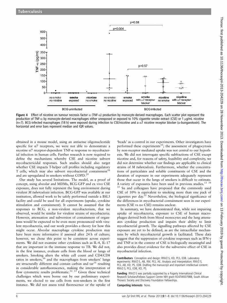
obtained in a mouse model, using an antisense oligonucleotidespecific for α7 receptors, we were not able to demonstrate anicotine α7 receptor-dependent TNF-α response to mycobacter-ial infection in human cells. Further research is now required todefine the mechanisms whereby CSE and nicotine subvertmycobactericidal responses. Such studies should also targetwhether CSE impacts T-helper cell profiles including regulatoryT cells, which may also subvert mycobacterial containment16
and are upregulated in smokers without COPD.29
Our study has several limitations. The model, as a proof ofconcept, using alveolar and MDMs, BCG-GFP and ex vivo CSEexposure, does not fully represent the lung environment duringvirulent M tuberculosis infection. BCG-GFP was available at ourinstitution, allowed work to be safely performed outside a BSL3facility and could be used for all experiments (uptake, cytokinestimulation and containment). It cannot be assumed that theresponses to BCG, a non-virulent mycobacterium, that weobserved, would be similar for virulent strains of mycobacteria.However, attenuation and subversion of containment of organ-isms would be expected to be even more pronounced with viru-lent mycobacteria, and our work provides a theory for how thismight occur. Alveolar macrophage cytokine production mayhave been more informative if assessed after 24 h of culture;however, we chose this point to be consistent across experi-ments. We did not examine other cytokines such as IL-4, IL-17that are important in the immune response to TB. We did not,in the first instance, evaluate cells from the blood or lungs ofsmokers. Smoking alters the white cell count and CD4/CD8ratios in smokers,30 and the macrophages from smokers’ lungsare structurally different and contain carbon and tar31 resultingin considerable autofluorescence, making the interpretation offlow cytometric results problematic.32 33 Given these technicalchallenges which were borne out by our preliminary experi-ments, we elected to use cells from non-smokers in the firstinstance. We did not assess total fluorescence or the uptake of
‘beads’ as a control in our experiments. Other investigators haveperformed these experiments10; the assessment of phagocytosisby non-receptor mediated uptake was not central to our hypoth-esis. We did not interrogate specific subfractions of CSE exceptnicotine and, for reasons of safety, feasibility and complexity, wedid not determine whether our findings are applicable to clinicalstrains of M tuberculosis. Furthermore, whether the concentra-tions of particulates and soluble constituents of CSE and theduration of exposure in our experiments adequately representthose that occur in the lungs of smokers is difficult to estimate.A variety of exposures have been used in previous studies.13 34
35 Su and colleagues have proposed that the commonly usedCSE of 10% is equivalent to smoking more than one pack ofcigarettes per day.35 Nevertheless, the biological significance ofthe differences in mycobacterial containment seen in our experi-ments (CSE vs no CSE) remains unclear.
In summary, we have demonstrated that, while not impairinguptake of mycobacteria, exposure to CSE of human macro-phages derived both from blood monocytes and the lung attenu-ates cytokine production and impairs their ability to limitmycobacterial growth. The signalling pathways affected by CSEexposure are yet to be defined, as are the intracellular mechan-isms by which mycobacterial growth is facilitated. These datasuggest that the suppression of cytokine responses such as IFN-γand TNF-α in the context of CSE is biologically meaningful andalso provides direct evidence for the subversive effect of CSE inmycobacterial infection.
Contributors Conception and design: RNVZ-S, KD, PLS, EDB. Laboratoryexperiments: RNVZ-S, AB, RM, PLS, AE. Analysis and interpretation: RNVZ-S,PLS, AB, KD, PS, EDB. Drafting the manuscript and important intellectual content:RNVZ-S, PLS, EDB, KD, PS.
Funding RNVZ-S was partially supported by a Fogarty International ClinicalResearch Scholars/Fellows Support Centre NIH grant R24TW007988, South AfricanThoracic Society and Discovery Foundation Fellowships.
Competing interests None.
Figure 4 Effect of nicotine on tumour necrosis factor α (TNF-α) production by monocyte-derived macrophages. Each scatter plot represent theproduction of TNF-α by monocyte-derived macrophages either unexposed or exposed to 10% cigarette smoke extract (CSE) or 1 μg/mL nicotine(n=7). BCG-infected macrophages (18 h) were exposed during infection to CSE/nicotine and a α7 nicotine receptor blocker (α-bungarotoxin). Thehorizontal and error bars represent median and IQR values.
6 van Zyl-Smit RN, et al. Thorax 2013;0:1–8. doi:10.1136/thoraxjnl-2013-204229
Tuberculosis
on 13 February 2019 by guest. P
rotected by copyright.http://thorax.bm
j.com/
Thorax: first published as 10.1136/thoraxjnl-2013-204229 on 28 N
ovember 2013. D
ownloaded from

Ethics approval The University of Cape Town Research Ethics Committee grantedapproval for this study.
Patient consent Written informed consent was obtained from all participants priorto enrolment in the study.
Provenance and peer review Not commissioned; externally peer reviewed.
REFERENCES1 World Health Organization. WHO report on the global tobacco epidemic, 2008. The
MPOWER package. Geneva: World Health Organization, 2008.2 World Health Organization. Global tuberculosis control – surveillance, planning,
financing. Report No. WHO/HTM/TB/2009.411. Geneva: World Health Organization,2009.
3 van Zyl Smit RN, Pai M, Yew WW, et al. Global lung health: the colliding epidemicsof tuberculosis, tobacco smoking, HIV and COPD. Eur Respir J 2010;35:27–33.
4 Lin HH, Ezzati M, Murray M. Tobacco smoke, indoor air pollution and tuberculosis:a systematic review and meta-analysis. PLoSMed 2007;4:e20.
5 Bates MN, Khalakdina A, Pai M, et al. Risk of tuberculosis from exposure totobacco smoke: a systematic review and meta-analysis. Arch Intern Med2007;167:335–42.
6 Slama K, Chiang CY, Enarson DA, et al. Tobacco and tuberculosis: a qualitativesystematic review and meta-analysis. Int J Tuberc Lung Dis 2007;11:1049–61.
7 Lonnroth K, Castro KG, Chakaya JM, et al. Tuberculosis control and elimination2010–50: cure, care, and social development. Lancet 2010;375:1814–29.
8 Basu S, Stuckler D, Bitton A, et al. Projected effects of tobacco smoking on worldwidetuberculosis control: mathematical modelling analysis. BMJ 2011;343:d5506.
9 Shang S, Ordway D, Henao-Tamayo M, et al. Cigarette smoke increasessusceptibility to tuberculosis–evidence from in vivo and in vitro models. J Infect Dis2011;203:1240–8.
10 Ando M, Sugimoto M, Nishi R, et al. Surface morphology and function of humanpulmonary alveolar macrophages from smokers and non-smokers. Thorax1984;39:850–6.
11 King TE Jr, Savici D, Campbell PA. Phagocytosis and killing of Listeriamonocytogenes by alveolar macrophages: smokers versus nonsmokers. J Infect Dis1988;158:1309–16.
12 Ortega E, Barriga C, Rodriguez AB. Decline in the phagocytic function of alveolarmacrophages from mice exposed to cigarette smoke. Comp Immunol MicrobiolInfect Dis 1994;17:77–84.
13 Green GM. Mechanisms of tobacco smoke toxicity on pulmonary macrophage cells.Eur J Respir Dis Suppl 1985;139:82–5.
Figure 5 Serial BCG colony counts over 5 days in (A) monocyte-derived macrophages and (B) alveolar macrophages. Monocyte-derivedmacrophages and alveolar macrophages infected with BCG were cultured for 5 days after the addition of 10% cigarette smoke extract (CSE) on day0 (post infection). The solid line represents unexposed macrophages and the dashed line represents CSE-exposed macrophages. Each day representsthe time point (post infection) when macrophages were lysed and organism load derived by counting the number of colony forming units (CFU) onsolid media. Monocyte-derived macrophages, n=9 (day 1–3) and n=5 (day 5); alveolar macrophages, n=5 (day 1–4) and n=4 (day 5).
van Zyl-Smit RN, et al. Thorax 2013;0:1–8. doi:10.1136/thoraxjnl-2013-204229 7
Tuberculosis
on 13 February 2019 by guest. P
rotected by copyright.http://thorax.bm
j.com/
Thorax: first published as 10.1136/thoraxjnl-2013-204229 on 28 N
ovember 2013. D
ownloaded from

14 Wewers MD, Diaz PT, Wewers ME, et al. Cigarette smoking in HIV infection inducesa suppressive inflammatory environment in the lung. Am J RespirCrit Care Med1998;158(5 Pt 1):1543–9.
15 Hagiwara E, Takahashi KI, Okubo T, et al. Cigarette smoking depletes cellsspontaneously secreting Th(1) cytokines in the human airway. Cytokine2001;14:121–6.
16 Semple PL, Binder AB, Davids M, et al. Regulatory T-cells attenuate mycobacterialstasis in alveolar and blood-derived macrophages from patients with TB. Am JRespir Crit Care Med 2013;187:1249–58.
17 Schwander S, Dheda K. Human lung immunity against mycobacterium tuberculosis:insights into pathogenesis and protection. Am J Respir Crit Care Med2011;183:696–707.
18 Keane J, Remold HG, Kornfeld H. Virulent Mycobacterium tuberculosisstrains evade apoptosis of infected alveolar macrophages. J Immunol2000;164:2016–20.
19 Lee J, Hartman M, Kornfeld H. Macrophage apoptosis in tuberculosis. Yonsei Med J2009;50:1–11.
20 Berenson CS, Garlipp MA, Grove LJ, et al. Impaired phagocytosis of nontypeableHaemophilus influenzae by human alveolar macrophages in chronic obstructivepulmonary disease. J Infect Dis 2006194:1375–84.
21 Phipps JC, Aronoff DM, Curtis JL, et al. Cigarette smoke exposure impairspulmonary bacterial clearance and alveolar macrophage complement-mediatedphagocytosis of Streptococcus pneumoniae. Infect Immun 2010;78:1214–20.
22 Madretsma S, Wolters LM, van Dijk JP, et al. In-vivo effect of nicotine on cytokineproduction by human non-adherent mononuclear cells. Eur J Gastroenterol Hepatol1996;8:1017–20.
23 Matsunaga K, Klein TW, Friedman H, et al. Involvement of nicotinic acetylcholinereceptors in suppression of antimicrobial activity and cytokine responses of alveolarmacrophages to Legionella pneumophila infection by nicotine. J Immunol2001;167:6518–24.
24 Chen H, Cowan MJ, Hasday JD, et al. Tobacco smoking inhibits expression ofproinflammatory cytokines and activation of IL-1R-associated kinase, p38, and
NF-kappaB in alveolar macrophages stimulated with TLR2 and TLR4 agonists.J Immunol 2007;179:6097–106.
25 Hope JC, Thom ML, McCormick PA, et al. Interaction of antigen presenting cellswith mycobacteria. Vet Immunol Immunopathol 2004;100:187–95.
26 Tobian AA, Potter NS, Ramachandra L, et al. Alternate class I MHC antigenprocessing is inhibited by Toll-like receptor signaling pathogen-associated molecularpatterns: mycobacterium tuberculosis 19-kDa lipoprotein, CpG DNA, andlipopolysaccharide. J Immunol 2003;171:1413–22.
27 Davies PD, Yew WW, Ganguly D, et al. Smoking and tuberculosis: theepidemiological association and immunopathogenesis. Trans R Soc Trop Med Hyg2006;100:291–8.
28 Wang H, Yu M, Ochani M, et al. Nicotinic acetylcholine receptor alpha7 subunit isan essential regulator of inflammation. Nature 2003;421:384–8.
29 Barcelo B, Pons J, Ferrer JM, et al. Phenotypic characterisation of T-lymphocytes inCOPD: abnormal CD4+CD25+ regulatory T-lymphocyte response to tobaccosmoking. Eur Respir J 2008;31:555–62.
30 Ginns LC, Goldenheim PD, Miller LG, et al. T-lymphocyte subsets in smoking andlung cancer: analysis of monoclonal antibodies and flow cytometry. Am Rev RespirDis 1982;126:265–9.
31 Brody AR, Craighead JE. Cytoplasmic inclusions in pulmonary macrophages ofcigarette smokers. Lab Invest 1975;32:125–32.
32 Skold CM, Hed J, Eklund A. Smoking cessation rapidly reduces cell recovery inbronchoalveolar lavage fluid, while alveolar macrophage fluorescence remains high.Chest 1992;101:989–95.
33 Umino T, Skold CM, Pirruccello SJ, et al. Two-colour flow-cytometric analysis ofpulmonary alveolar macrophages from smokers. Eur Respir J 1999;13:894–9.
34 Roth MD, Whittaker K, Salehi K, et al. Mechanisms for impaired effector function inalveolar macrophages from marijuana and cocaine smokers. J Neuroimmunol2004;147:82–6.
35 Su Y, Han W, Giraldo C, et al. Effect of cigarette smoke extract on nitric oxidesynthase in pulmonary artery endothelial cells. Am J Respir Cell Mol Biol1998;19:819–25.
8 van Zyl-Smit RN, et al. Thorax 2013;0:1–8. doi:10.1136/thoraxjnl-2013-204229
Tuberculosis
on 13 February 2019 by guest. P
rotected by copyright.http://thorax.bm
j.com/
Thorax: first published as 10.1136/thoraxjnl-2013-204229 on 28 N
ovember 2013. D
ownloaded from