asxl1 impairs osteoclast formation by epigenetic
TRANSCRIPT
Washington University School of MedicineDigital Commons@Becker
Open Access Publications
2018
ASXL1 impairs osteoclast formation by epigeneticregulation of NFATc1Nidhi RohatgiWashington University School of Medicine in St. Louis
Wei ZouWashington University School of Medicine in St. Louis
Patrick L. CollinsWashington University School of Medicine in St. Louis
Jonathan R. BrestoffWashington University School of Medicine in St. Louis
Timothy H. ChenWashington University School of Medicine in St. Louis
See next page for additional authors
Follow this and additional works at: https://digitalcommons.wustl.edu/open_access_pubs
This Open Access Publication is brought to you for free and open access by Digital Commons@Becker. It has been accepted for inclusion in OpenAccess Publications by an authorized administrator of Digital Commons@Becker. For more information, please contact [email protected].
Recommended CitationRohatgi, Nidhi; Zou, Wei; Collins, Patrick L.; Brestoff, Jonathan R.; Chen, Timothy H.; Abu-Amer, Yousef; and Teitelbaum, Steven L.,,"ASXL1 impairs osteoclast formation by epigenetic regulation of NFATc1." Blood Advances.2,19. 2467-2477. (2018).https://digitalcommons.wustl.edu/open_access_pubs/7242

AuthorsNidhi Rohatgi, Wei Zou, Patrick L. Collins, Jonathan R. Brestoff, Timothy H. Chen, Yousef Abu-Amer, andSteven L. Teitelbaum
This open access publication is available at Digital Commons@Becker: https://digitalcommons.wustl.edu/open_access_pubs/7242
REGULAR ARTICLE
ASXL1 impairs osteoclast formation by epigenetic regulation of NFATc1
Nidhi Rohatgi,1 Wei Zou,1 Patrick L. Collins,1 Jonathan R. Brestoff,1 Timothy H. Chen,2 Yousef Abu-Amer,2 and Steven L. Teitelbaum1,3
1Department of Pathology and Immunology, 2Department of Orthopedic Surgery, and 3Division of Bone and Mineral Diseases, Department of Medicine, Washington UniversitySchool of Medicine, St. Louis, MO
Key Points
• ASXL1 deletion inmyeloid lineage cellspromotes osteoclastdifferentiation resultingin low bone mass.
• ASXL1 modulatesH3K27 methylation ofosteoclastogenic genepromoters, includingNFATc1.
Additional sex comb-like 1 (ASXL1) mutations are commonly associated with myeloid
malignancies and are markers of aggressive disease. The fact that ASXL1 is necessary for
myeloid differentiation raises the possibility it also regulates osteoclasts. We find deletion
of ASXL1 in myeloid cells results in bone loss with increased abundance of osteoclasts.
Because ASXL1 is an enhancer of trithorax and polycomb (ETP) protein, we asked if it
modulates osteoclast differentiation by maintaining balance between positive and
negative epigenetic regulators. In fact, loss of ASXL1 induces concordant loss of inhibitory
H3K27me3 with gain of H3K4me3 at key osteoclast differentiation genes, including nuclear
factor for activated T cells 1 (NFATc1) and itgb3. In the setting of ASXL1 deficiency, increased
NFATc1 binds to the Blimp1 (Prdm1) promoter thereby enhancing expression of this
pro-osteoclastogenic gene. The global reduction of K27 trimethylation in ASXL1-deficient
osteoclasts is also attended by a 40-fold increase in expression of the histone demethylase
Jumonji domain‐containing 3 (Jmjd3). Jmjd3 knockdown in ASXL1-deficient osteoclast
precursors increases H3K27me3 on theNFATc1 promoter and impairs osteoclast formation.
Thus, in addition to promoting myeloid malignancies, ASXL1 controls epigenetic reprog-
ramming of osteoclasts to regulate bone resorption and mass.
Introduction
Osteoclast is the bone-resorbing, myeloid lineage polykaryon. In keeping with its myeloid ontogeny,molecules which control myelopoiesis also regulate osteoclasts.
Osteoclast formation requires activation of RANK ligand (RANKL)/RANK signaling pathways includingNF-kB and MAPKs such as extracellular signal-regulated kinase and p38.1 These immediate signalsresult in synthesis of c-Fos, which collaborates with the key osteoclastogenic transcription factor,NFATc1, to autoactivate the latter’s promoter thereby inducing osteoclastogenesis.2 Although RANKLinduces epigenetic changes that contribute to osteoclast formation, the mechanisms underlying thisepigenetic remodeling in osteoclastogenesis still remain unclear.3
Additional sex comb-like (ASXL) genes encode enhancer of trithorax and polycomb (ETP) proteins,which, by facilitating histone methylation or demethylation, repress or stimulate gene transcription in acell-specific context.4,5 Transcriptional repression by ASXL proteins is mediated by recruiting polycombreceptor complex 2 (PRC2) to promoters, thereby increasing H3K27 methylation. Trimethylation of K27results in gene inactivation, a process reversed by recruitment of a histone demethylase that removesH3K27me3 methyl groups.6 These events also exist during osteoclastogenesis.7
An ASXL family gene, ASXL2, positively effects osteoclast formation as its deletion diminishes thenumber of bone resorptive cells, resulting in osteopetrosis.8 Another member of the ASXL family, namelyASXL1, is associated with and dictates prognosis of a number of myeloid malignancies such asmastocytosis with associated myelodysplasia, which predisposes to osteoclast-dependent osteoporosis.9,10
Submitted 5 March 2018; accepted 18 August 2018. DOI 10.1182/bloodadvances.2018018309.
The full-text version of this article contains a data supplement.
© 2018 by The American Society of Hematology
9 OCTOBER 2018 x VOLUME 2, NUMBER 19 2467
Like ASXL2, ASXL1 also recognizes PPARg, but whereas ASXL2promotes adipogenesis, the process is arrested by ASXL1.11
Because ASXL1 suppresses the nuclear receptor (PPARg), whoseactivity was presumed to promote physiological osteoclast forma-tion, we postulated that specific deletion of ASXL1 in myeloid lineagewould induce the bone resorptive cell and diminish bone mass.12
Although mice lacking ASXL1, exclusively in myeloid lineage cells,have low bone mass, their robust osteoclast formation is independentof canonical RANKL- and PPARg-activated signals, including c-Fosexpression. Like its induction of hematopoietic malignancies,however, the osteoclastogenic properties of ASXL1 deficiencyinvolve demethylation of a repressive histone mark, H3K27me3.Consequently, the genes most affected by absence of ASXL1are involved in osteoclast differentiation. A subset of these genesgain K4 trimethylation, a mark of active chromatin, and wereenriched in NFAT-like motifs. One such gene is prdm1 (Blimp1),the promoter of which exhibits increased binding to NFATc1 inknockout (KO) cells. The reciprocal increase in H3K4me3 onosteoclastic genes in ASXL1-deficient myeloid lineage cells mayalso be because of activation of the histone demethylase Jumonjidomain‐containing 3 (Jmjd3), which specifically removes K27 methylgroups from promoters. Thus, ASXL1 regulates osteoclast epigenomeand its inactivation promotes bone loss.
Methods
Animals
All animals were housed in the animal care unit ofWashington UniversitySchool ofMedicine, where theyweremaintained according to guidelinesof the Association for Assessment and Accreditation of LaboratoryAnimal Care. All animal experimentation was approved by the AnimalStudies Committee of Washington University School of Medicine.
Generation of ASXL1-deficient mice
Asxl1tm1a(EUCOMM)Wtsi were purchased from European Con-ditional Mouse Consortium. For deletion of the LacZ-neomycin-resistance cassette and for the generation of mice with LoxP-flankedASXL1 allele (ASXL1f/1), those transmitting 100% were bred toFLPo (JAX mice 012930, C57BL/6 mice), which have transgenicexpression of an enhanced form of the recombinase FLP driven bythe GT(ROSA)26Sor promoter. ASXL1f/1 mice were intercrossedto generate ASXL1f/f mice. ASXL1fl/fl mice generated weresubsequently crossed with LysM-Cre mice (Jackson Laboratory). Tofully delete ASXL1 using LysM-Cre, ASXL1fl/fl, LysMCre1/2 micewere crossed again to obtain animals bearing 2 copies of theLysM-Cre allele (ASXL1fl/fl LysMCre1/1). LysMCre1/1 andASXL1fl/fl littermate without Cre mice served as control.
Macrophage isolation and osteoclast culture
All in vitro experiments were performed at least 3 times. Primarybone marrow macrophages (BMMs) were prepared as described13
with slight modification. Marrow was extracted from femora andtibiae of 6- to 8-week-old mice with a minimum essential medium(a-MEM) and cultured in a-MEM containing 10% inactivated fetalbovine serum, 100 IU/mL penicillin, and 100 mg/mL streptomycin(a-10 medium) with 1:10 of mMCSF producing cell line, CMG14-12 condition media on petri-plastic dishes. Cells were incubatedat 37°C in 6% CO2 for 3 days and then washed with phosphate-buffered saline (PBS) and lifted with 13 trypsin/EDTA in PBS. Atotal of 1.23 104 BMMs were cultured in 500 mL a-MEM containing
10% heat-inactivated fetal bovine serum with glutathione-Stransferase–RANKL and 30 ng/mL of mouse recombinantmacrophage colony-stimulating factor (M-CSF) in 48-well tissueculture plates, some containing sterile bovine bone slices. Cellswere fixed and stained for tartrate-resistant acid phosphatase(TRAP) activity after 5 days in culture, using a commercial kit (Sigma387-A; Sigma-Aldrich, St. Louis, MO). The images were capturedusing Nikon Eclipse E400 (Melville, NY) upright microscope.
Histone extraction, histone western blot
Histones were extracted by standard acid extraction protocol. Briefly,cells were lysed in triton extraction buffer (PBS containing 0.5%triton3 100 [vol/vol]) on ice for 10minutes. The cells were centrifugedat 2000 rpm for 10 minutes followed by another wash with half thevolume of triton extraction buffer. Pellet was resuspended in 0.2N HCl.Histones were acid extracted overnight at 4°C. The samples werecentrifuged and supernatant removed. Protein was determined usingBradford assay.
Lentivirus infection
293-T cells were transfected with shJMJD3 together with a packagingplasmid (pHR0 8.2 d R) and the envelope (pCMV-VSV-G) plasmid.After 48 hours, medium containing lentiviruses was collected andfiltered. Macrophages obtained from 8- to 12-week-old male micewere infected with virus for 24 hours in the presence of 1:10 CMGand 10 mg/mL protamine (Sigma-Aldrich). Cells were selectedin the presence of CMG and 1 mg/mL puromycin (Calbiochem) for3 days before use as osteoclast precursors.
Chromatin immunoprecipitation (ChIP) assay
For immunoprecipitation, 60 mL of magnetic protein A beads wereused. The beads were washed thrice with PBS containing 0.02%tween 20. After the final wash, beads were resuspended with theantibody overnight at 4°C. The next day, 1 to 2million cells were platedin tissue culture plates (6 RANKL). Formaldehyde was added directlyto cell culture media for 10 minutes at room temperature such thatthe final concentration was 1%. Cross-linking was quenched by theaddition of glycine to a final concentration of 0.125 M. Cells werewashed 3 times with PBS, scraped off the plates in a small amountof PBS, and centrifuged, and the pellet was washed with PBScontaining protease inhibitors (Complete, Roche). Cells were thenresuspended in 1 mL PBS, and protease inhibitors centrifugedat 5000 rpm for 5 minutes at 4°C. The pellet was resuspended incold sonication buffer (50 mM tris(hydroxymethyl)aminomethane[Tris]–HCl pH 8, 10 mM EDTA, 0.1% sodium dodecyl sulfate [SDS],0.5% sodium deoxycholate and protease inhibitors) for 25 to30 minutes on ice. Cells were aliquoted at 1 3 106 cells per mLconcentration and chromatin sheared using Bioruptor (6 cycles,10 minutes at high speed per cycle). The samples were centrifugedto pellet the cellular debris. Five percent of the cells were collected in aseparate tube to be used as input control. The supernatant wasdivided equally between immunoprecipitation samples to includeisotype control. Sheared DNA was incubated with the bead-antibody slurry. The next day, DNA-protein complexes were washedin low salt buffer (SDS 0.1%, Triton X100 1%, EDTA 2 mM, Tris-HCl pH 8.0 20 mM, NaCl 150 mM) followed by high salt buffer(SDS 0.1%, Triton X100 1%, EDTA 2 mM, Tris-HCl pH 8.0 20 mM,NaCl 500 mM), LiCl buffer (LiCl 0.25 M, nonidet P-40 1%,deoxycholate 1%, EDTA 1 mM, Tris-HCl pH 8.0, 10 mM), and Tris-EDTA buffer. DNA was eluted by adding 250 mL of elution buffer
2468 ROHATGI et al 9 OCTOBER 2018 x VOLUME 2, NUMBER 19
(SDS 1%, NaHCO3 0.1 M). Samples and inputs were de-cross-linked and cleaned for quantitative polymerase chain reaction (qPCR)analysis.
Chromatin immunoprecipitation and DNA sequencing
(ChIP-seq) and analysis
ChIP or input DNA was used for indexed library preparation (Ilumina)and then subjected to 50-bp single-end sequencing per manufacturer’sprotocol on Illumina HiSeq3000. Sequenced libraries were aligned tothe reference genome (mm10) using NovaAlign base settings. Peakcalling was performed using MACS2.0 for H3K4me3 ChIP-seq14 andHOMER for H3K27me3 ChIP-seq,15 using paired inputs as peakcalling controls. For heat maps and quantification of reads over peaks,genes, or promoter regions, reads per kilobase per million wereextracted using Deeptools.16 For log ratio (M) and average mean (A)(MA) plot analysis, reads were then further quantile normalized usingthe R package preprocessCore, prior to direct comparison. Directvisualization of ChIP-seq tracks was accomplished using the Universityof California, Santa Cruz Genome Browser.17
Statistics
Statistical significance was determined using multiple comparisonin a 1‐way or 2-way analysis of variance (ANOVA) test, or with unpairednonparametric Student t test when only 2 groups were present, usingGraphPad Prism v7 built‐in statistical analysis (GraphPad SoftwareInc., La Jolla, CA). P , .05 was considered significant. Allquantitative reverse transcription–PCR and qPCR data wereexpressed as mean from at least 3 independent biological experi-ments with at least 3 technical replicates.
Results
ASXL1 deletion in myeloid lineage cells promotes
osteoclastogenesis resulting in low bone mass
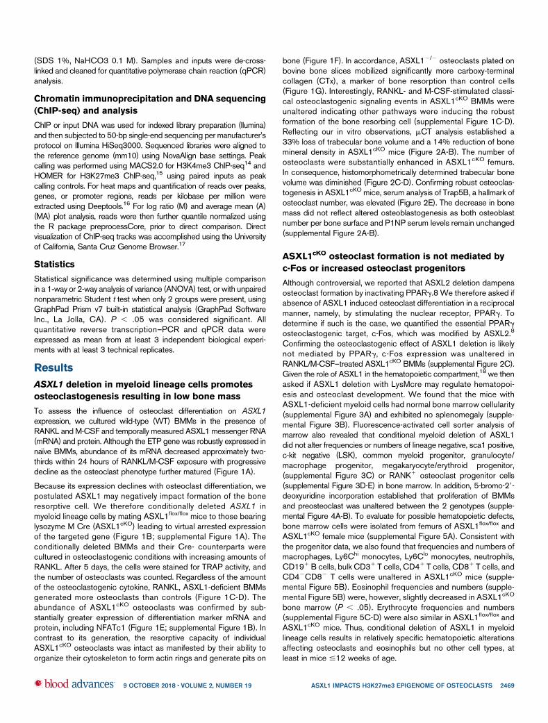
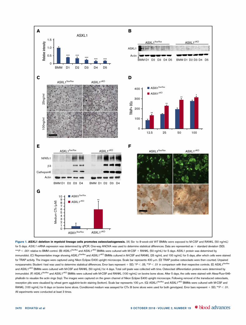
To assess the influence of osteoclast differentiation on ASXL1expression, we cultured wild-type (WT) BMMs in the presence ofRANKL and M-CSF and temporally measured ASXL1 messenger RNA(mRNA) and protein. Although the ETP gene was robustly expressed innaıve BMMs, abundance of its mRNA decreased approximately two-thirds within 24 hours of RANKL/M-CSF exposure with progressivedecline as the osteoclast phenotype further matured (Figure 1A).
Because its expression declines with osteoclast differentiation, wepostulated ASXL1 may negatively impact formation of the boneresorptive cell. We therefore conditionally deleted ASXL1 inmyeloid lineage cells by mating ASXL1flox/flox mice to those bearinglysozyme M Cre (ASXL1cKO) leading to virtual arrested expressionof the targeted gene (Figure 1B; supplemental Figure 1A). Theconditionally deleted BMMs and their Cre- counterparts werecultured in osteoclastogenic conditions with increasing amounts ofRANKL. After 5 days, the cells were stained for TRAP activity, andthe number of osteoclasts was counted. Regardless of the amountof the osteoclastogenic cytokine, RANKL, ASXL1-deficient BMMsgenerated more osteoclasts than controls (Figure 1C-D). Theabundance of ASXL1cKO osteoclasts was confirmed by sub-stantially greater expression of differentiation marker mRNA andprotein, including NFATc1 (Figure 1E; supplemental Figure 1B). Incontrast to its generation, the resorptive capacity of individualASXL1cKO osteoclasts was intact as manifested by their ability toorganize their cytoskeleton to form actin rings and generate pits on
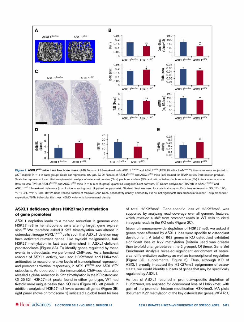
bone (Figure 1F). In accordance, ASXL12/2 osteoclasts plated onbovine bone slices mobilized significantly more carboxy-terminalcollagen (CTx), a marker of bone resorption than control cells(Figure 1G). Interestingly, RANKL- and M-CSF-stimulated classi-cal osteoclastogenic signaling events in ASXL1cKO BMMs wereunaltered indicating other pathways were inducing the robustformation of the bone resorbing cell (supplemental Figure 1C-D).Reflecting our in vitro observations, mCT analysis established a33% loss of trabecular bone volume and a 14% reduction of bonemineral density in ASXL1cKO mice (Figure 2A-B). The number ofosteoclasts were substantially enhanced in ASXL1cKO femurs.In consequence, histomorphometrically determined trabecular bonevolume was diminished (Figure 2C-D). Confirming robust osteoclas-togenesis in ASXL1cKO mice, serum analysis of Trap5B, a hallmark ofosteoclast number, was elevated (Figure 2E). The decrease in bonemass did not reflect altered osteoblastogenesis as both osteoblastnumber per bone surface and P1NP serum levels remain unchanged(supplemental Figure 2A-B).
ASXL1cKO osteoclast formation is not mediated by
c-Fos or increased osteoclast progenitors
Although controversial, we reported that ASXL2 deletion dampensosteoclast formation by inactivating PPARg.8 We therefore asked ifabsence of ASXL1 induced osteoclast differentiation in a reciprocalmanner, namely, by stimulating the nuclear receptor, PPARg. Todetermine if such is the case, we quantified the essential PPARgosteoclastogenic target, c-Fos, which was modified by ASXL2.8
Confirming the osteoclastogenic effect of ASXL1 deletion is likelynot mediated by PPARg, c-Fos expression was unaltered inRANKL/M-CSF–treated ASXL1cKO BMMs (supplemental Figure 2C).Given the role of ASXL1 in the hematopoietic compartment,18 we thenasked if ASXL1 deletion with LysMcre may regulate hematopoi-esis and osteoclast development. We found that the mice withASXL1-deficient myeloid cells had normal bone marrow cellularity(supplemental Figure 3A) and exhibited no splenomegaly (supple-mental Figure 3B). Fluorescence-activated cell sorter analysis ofmarrow also revealed that conditional myeloid deletion of ASXL1did not alter frequencies or numbers of lineage negative, sca1 positive,c-kit negative (LSK), common myeloid progenitor, granulocyte/macrophage progenitor, megakaryocyte/erythroid progenitor,(supplemental Figure 3C) or RANK1 osteoclast progenitor cells(supplemental Figure 3D-E) in bone marrow. In addition, 5-bromo-29-deoxyuridine incorporation established that proliferation of BMMsand preosteoclast was unaltered between the 2 genotypes (supple-mental Figure 4A-B). To evaluate for possible hematopoietic defects,bone marrow cells were isolated from femurs of ASXL1flox/flox andASXL1cKO female mice (supplemental Figure 5A). Consistent withthe progenitor data, we also found that frequencies and numbers ofmacrophages, Ly6Chi monocytes, Ly6Clo monocytes, neutrophils,CD191 B cells, bulk CD31 T cells, CD41 T cells, CD81 T cells, andCD42CD82 T cells were unaltered in ASXL1cKO mice (supple-mental Figure 5B). Eosinophil frequencies and numbers (supple-mental Figure 5B) were, however, slightly decreased in ASXL1cKO
bone marrow (P , .05). Erythrocyte frequencies and numbers(supplemental Figure 5C-D) were also similar in ASXL1flox/flox andASXL1cKO mice. Thus, conditional deletion of ASXL1 in myeloidlineage cells results in relatively specific hematopoietic alterationsaffecting osteoclasts and eosinophils but no other cell types, atleast in mice #12 weeks of age.
9 OCTOBER 2018 x VOLUME 2, NUMBER 19 ASXL1 IMPACTS H3K27me3 EPIGENOME OF OSTEOCLASTS 2469
0123456
Med
ium
CTx
(M
)
789
10G
ASXL1cKO
ASXI1flox/flox
ASXL1cKO
**
ASXI1flox/flox
0
0.5
1.0
1.5Re
lative
inte
nsity
BMM D1 D2
ASXL1A
D3
*********
*** ***
D4 D5
B
BMMActin
ASXL1
D1 D2 D3 D4 D5 BMM D1 D2 D3 D4 D5
ASXL1cKOASXL1flox/flox
C
25ng
/ml
100n
g/m
l
ASXL1cKOASXL1flox/floxD
0
100
200
300
400
TRAP
+ OC
s12.5
**
25
**
50
**
100
*ASXI1cKO
ASXI1flox/flox
E
BMM
Actin
CathepsinK
NFATc1
3
D1 D2 D3 D4 BMM D1 D2 D3 D4
ASXL1cKOASXL1flox/floxF
ASXL1cKOASXL1flox/flox
Figure 1. ASXL1 deletion in myeloid lineage cells promotes osteoclastogenesis. (A) Six- to 8-week-old WT BMMs were exposed to M-CSF and RANKL (50 ng/mL)
for 5 days. ASXL1 mRNA expression was determined by qPCR. One-way ANOVA was used to determine statistical differences. Data are represented as 1 standard deviation (SD).
***P , .001 relative to BMM control. (B) ASXL1flox/flox and ASXL1cKO BMMs were cultured with M-CSF 1 RANKL (50 ng/mL) for 5 days. ASXL1 protein was determined by
immunoblot. (C) Representative image showing ASXL1flox/flox and ASXL1cKO BMMs cultured in M-CSF and RANKL (25 ng/mL and 100 ng/mL) for 5 days, after which cells were stained
for TRAP activity. The images were captured using Nikon Eclipse E400 upright microscope. Scale bar represents 400 mm. (D) TRAP positive osteoclasts were then counted. Unpaired
nonparametric Student t test was used to determine statistical differences. Error bars represent 1 SD; *P , .05, **P , .01 in comparison with their respective controls. (E) ASXL1flox/flox
and ASXL1cKO BMMs were cultured with M-CSF and RANKL (50 ng/mL) for 4 days. Total cell lysate was collected with time. Osteoclast differentiation proteins were determined by
immunoblot. (F) ASXL1flox/flox and ASXL1cKO BMMs were cultured with M-CSF and RANKL (100 ng/mL) on bovine bone slices. After 5 days, the cells were stained with Alexa-Fluor-546-
phallodin to visualize the actin rings (top). The images were captured on the green channel of Nikon Eclipse E400 upright microscope. Following removal of the transduced osteoclasts,
resorption pits were visualized by wheat germ agglutinin-lectin staining (bottom). Scale bar represents 100 mm. (G) ASXL1flox/flox and ASXL1cKO BMMs were cultured with M-CSF and
RANKL (100 ng/mL) for 6 days on bovine bone slices. Conditioned medium was assayed for CTx (6 bone slices were used for both genotypes). Error bars represent 1 SD; **P , .01.
All experiments were conducted at least 3 times.
2470 ROHATGI et al 9 OCTOBER 2018 x VOLUME 2, NUMBER 19
ASXL1 deficiency alters H3K27me3 methylation
of gene promoters
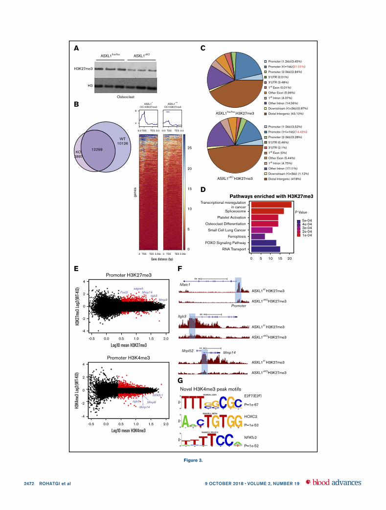
ASXL1 depletion leads to a marked reduction in genome-wideH3K27me3 in hematopoietic cells altering target gene expres-sion.18 We therefore asked if K27 trimethylation was altered inosteoclast lineage ASXL1cKO cells such that ASXL1 deletion mayhave activated relevant genes. Like myeloid malignancies, bulkH3K27 methylation in fact was diminished in ASXL1-deficientpreosteoclasts (Figure 3A). To identify genes regulated by theseevents in osteoclasts, we performed ChIP-seq. As a functionalreadout of ASXL1 activity, we used H3K27me3 and H3K4me3antibodies to measure relative levels of transcriptional repressionand promoter activation, respectively, in ASXL1flox/flox and ASXL1cKO
osteoclasts. As observed in the immunoblot, ChIP-seq data alsorevealed a global reduction in K27 trimethylation in the KO osteoclast.Of 25 321 H3K27me3 peaks found in either genotype, WT hadfivefold more unique peaks than KO cells (Figure 3B, left panel). Inaddition, analysis of H3K27me3 levels across all genes (Figure 3B,right panel shows chromosome 1) indicated a global trend for loss
of total H3K27me3. Gene-specific loss of H3K27me3 wassupported by analyzing read coverage over all genomic features,which revealed a shift from promoter reads in WT cells to distalintragenic reads in the KO cells (Figure 3C).
Given chromosome-wide depletion of H3K27me3, we asked ifgenes most affected by ASXL1 loss were specific to osteoclastdevelopment. A total of 663 genes in KO osteoclast exhibitedsignificant loss of K27 methylation (criteria used was greaterthan twofold change between the 2 groups). Of these, Gene SetEnrichment Analysis revealed significant enrichment of osteo-clast differentiation pathway as well as transcriptional regulation(Figure 3D; supplemental Figure 6). Thus, although KO ofASXL1 broadly impacted the H3K27me3 epigenome of osteo-clasts, we could identify subsets of genes that may be specificallyregulated by ASXL1.
As loss of ASXL1 resulted in promoter-specific depletion ofH3K27me3, we analyzed for concordant loss of H3K27me3 withgain of the promoter histone modification H3K4me3. MA plotsdocument K27 methylation of the key osteoclastic genes, NFATc1,
ASXL1flox/flox ASXL1cKO
A
ASXL1flox/flox ASXL1cKO
C
0.25
***0.2
0.15
BV/T
V
0.10.05
0ASXL1flox/flox ASXL1cKO
**
Conn
-Den
s(1
/mm
^3)
250200150100
500
ASXL1flox/flox ASXL1cKO
*
Tb-S
p (m
m)
0.250.2
0.150.1
0.050
ASXL1flox/flox ASXL1cKO
**
TbTh
(mm
)
0.060.050.040.030.020.01
0ASXL1flox/flox ASXL1cKO
TbN
(1/m
m)
8ns6
4
2
0ASXL1flox/flox ASXL1cKO
**
vBM
D(m
g HA
/ccm
) 250200150100
500
ASXL1flox/flox ASXL1cKO
B
***
8
OcN/
BS(N
/mm
) 6
4
2
0ASXL1flox/flox ASXL1cKO
**
25201510
50
BV/T
V
ASXL1flox/flox ASXL1cKO
D
**0.35
0.30.25
0.20.15
0.10.05
0TR
AP5B
(U/m
l)ASXL1flox/flox ASXL1cKO
E
Figure 2. ASXL1cKO
mice have low bone mass. (A-B) Femurs of 13-week-old male ASXL1 flox/flox and ASXL1cKO (ASXL1flox/flox LysMcre/cre) littermates were subjected to
mCT analysis (n 5 5 in each group). Scale bar represents 100 mm. (C-D) Femurs of ASXL1flox/flox and ASXL1cKO mice (left) stained for TRAP activity (red reaction product).
Scale bar represents 1 mm. Histomorphometric analysis of osteoclast number (OcN) per bone surface (BS) and ratio of trabecular bone volume (BV) to total marrow space
(total volume [TV]) of ASXL1flox/flox and ASXL1cKO mice (n 5 5 in each group) quantified using BioQuant software. (E) Serum analysis for TRAP5B in ASXL1flox/flox and
ASXL1cKO 12-week-old male mice (n 5 7 mice in each group). Unpaired nonparametric Student t test was used for statistical analysis. Error bars represent 1 SD; *P , .05,
**P , .01, ***P , .001. BV/TV, bone volume fraction of marrow; Conn-Dens, connectivity density, normed by TV; ns, not significant; TbN, trabecular number; TbSp, trabecular
separation; TbTh, trabecular thickness; vBMD, volumetric bone mineral density.
9 OCTOBER 2018 x VOLUME 2, NUMBER 19 ASXL1 IMPACTS H3K27me3 EPIGENOME OF OSTEOCLASTS 2471
Osteoclast
ASXL1flox/flox
H3K27me3
H3
ASXL1cKO
A
ASXL1f/f
OC-H3K27me3
8
4
-3.0 TSS TES 3.0
ASXL1cKO
OC-H3K27me3
-3.0 TSS TES 3.0
-3 TSS TES 3.0kb -3 TSS
Gene distance (bp)
gene
s
TES 3.0kb
25
20
15
10
5
0
12298
WT10126
KO2897
B
4
2
0
H3K2
7me3
Log
2(W
T-KO
)
Log10 mean H3K27me3
-2
-4
-0.5 0.0 0.5
FosI2adgre5
Mmp14
Mmp9itgb3
Promoter H3K27me3
1.0 1.5 2.0
4
2
0
H3K4
me3
Log
2(W
T-KO
)
-2
-4
Mmp9
Mmp14
itgb3
NFATc1
Promoter H3K4me3
Log10 mean H3K4me3-0.5 0.0 0.5 1.0 1.5 2.0
E
ASXL1cKOH3K27me3
ASXL1f/f H3K27me3
ASXL1cKOH3K27me3
ASXL1f/f H3K27me3
ASXL1cKOH3K27me3
ASXL1f/f H3K27me3
Nfatc1
Itgb3
Mrpl52 Mmp14
Promoter
F
Novel H3K4me3 peak motifsE2F7(E2F)
P=1e-67
P=1e-52
P=1e-52
NFATc2
HOXC2
G
ASXL1flox/flox H3K27me3
Promoter (1.2kb)(3.45%)
Promoter (<=1kb)(21.01%)
Promoter (2-3kb)(2.84%)
5’UTR (0.51%)
3’UTR (3.48%)
1st Exon (0.01%)
Other Exon (5.99%)
1st Intron (4.07%)
Other Intron (14.56%)
Downstream (<=3kb)(0.97%)
Distal Intergenic (43.12%)
ASXL1cKO H3K27me3
Promoter (1-2kb)(3.52%)
Promoter (1<=1kb)(14.42%)
Promoter (2-3kb)(3.28%)
5’UTR (0.46%)
3’UTR (2.1%)
1st Exon (0%)
Other Exon (5.44%)
1st Intron (4.75%)
Other Intron (17.11%)
Downstream (<=3kb) (1.12%)
Distal Intergenic (47.8%)
C
Transcriptional misregulationin cancer
Spliceosome
Platelet Activation
Osteoclast Differentiation
Small Cell Lung Cancer
Ferroptosis
FOXO Signaling Pathway
RNA Transport
0 5 10 15 20
P Value
5e-044e-043e-042e-041e-04
Pathways enriched with H3K27me3D
Figure 3.
2472 ROHATGI et al 9 OCTOBER 2018 x VOLUME 2, NUMBER 19
itgb3, Mmp9, and Mmp14 to have increased K27 methylation in theASXL1flox/flox in comparison with KO (Figure 3E, top). On the otherhand, alterations of K4 methylation, an indicator of activechromatin, were much less dramatic on some of these genepromoters (Figure 3E, bottom). Importantly, K27 methylationon promoters upstream of TSS of specific genes such asNFATc1, itgb3, and Mmp14 visualized using University ofCalifornia, Santa Cruz Genome Browser was decreased (Figure3F). These observations raised the possibility that depletion ofASXL1 de-represses transcription factors that drive the forma-tion of new H3K4me3 positive promoters. To identify thesepotential regulatory pathways, we performed a motif enrichmentanalysis of novel H3K4me3 peaks specific to the ASXL1cKO cells.Utilizing de novo motif prediction, and a highly stringent statistical cutoff (P, 1e-50), we identified enrichment of NFAT-like motifs in the KO
promoters (P value 51e-52) (Figure 3G; supplemental Tables 1and 2). ASXL1cKO osteoclast lineage cells exhibit specific loss ofH3K27me3 with gain of H3K4me3 at the NFATc1 promoter, alongwith genome-wide enrichment of NFAT motifs in the novel ASXL1-KO promoters.
Increased pro-osteoclastogenic transcription factors
in ASXL1-deficient osteoclasts
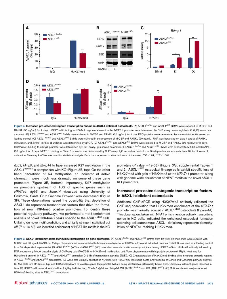
Additional ChIP-qPCR using H3K27me3 antibody validated theChIP-seq observation that H3K27me3 enrichment of the NFATc1promoter was markedly reduced in ASXL1cKO osteoclasts (Figure 4A).This observation, taken with NFAT enrichment on actively transcribinggenes in KO cells, indicated the enhanced osteoclast formationattending cell-autonomous ASXL1 deficiency represents demethy-lation of NFATc1-residing H3K27me3.
A50 ASXL1flox/flox
ASXL1cKO
***
NFA
Tc1
prom
oter
(5%
of i
nput
)
40
30
20
10
0IgG H3K27me3
B
EZH2
Suz12
EED
Actin
RANKL - + - +
ASXL1flox/flox ASXl1cKOC
Relat
ive in
tens
ity
ASXL1flox/flox
ASXL1cKO
0BMM D1
Blimp1
**
**
D2
5
10
15
20
25
D
0IgG H3K27me3
0.1
0.2
0.3
0.4
0.5
0.6
Blim
p1 p
rom
oter
(5%
of i
nput
)
ASXL1flox/flox
ASXL1cKO
E
**
0IgG NFATc1
2
4
6
8
Blim
p1 p
rom
oter
(5%
of i
nput
)
ASXL1flox/flox
ASXL1cKO
Figure 4. Increased pro-osteoclastogenic transcription factors in ASXL1-deficient osteoclasts. (A) ASXL1flox/flox and ASXL1cKO BMMs were exposed to M-CSF and
RANKL (50 ng/mL) for 2 days. H3K27me3 binding to NFATc1 response element in the NFATc1 promoter was determined by ChIP assay. Immunoglobulin G (IgG) served as
a control. (B) ASXL1flox/flox and ASXL1cKO BMMs were cultured in M-CSF and RANKL (50 ng/mL) for 1 day. PRC proteins were determined by immunoblot. Actin served as
loading control. (C) ASXL1flox/flox and ASXL1cKO BMMs were cultured in the presence of M-CSF and RANKL (50 ng/mL). RNA was harvested on days 1 and 2 of RANKL
stimulation, and Blimp1 mRNA abundance was determined by qPCR. (D) ASXL1flox/flox and ASXL1cKO BMMs were exposed to M-CSF and RANKL (50 ng/mL) for 2 days.
H3K27me3 binding to Blimp1 promoter was determined by ChIP assay. IgG served as control. (E) ASXL1flox/flox and ASXL1cKO BMMs were exposed to M-CSF and RANKL
(50 ng/mL) for 2 days. NFATc1 binding to Blimp1 promoter was determined by ChIP assay. IgG served as control. n 5 3 independent experiments from 10- to 12-week-old
male mice. Two-way ANOVA was used for statistical analysis. Error bars represent 1 standard error of the mean; **P , .01, ***P , .001.
Figure 3. ASXL1 deficiency alters H3K27me3 methylation on gene promoters. (A) ASXL1flox/flox and ASXL1cKO BMMs from 12-week-old male mice were cultured with
M-CSF and 50 ng/mL RANKL for 2 days. Representative immunoblot of bulk histone methylation for H3K27me3 on acid extracted histones. Total H3 was used as a loading control
(n 5 3 independent experiments). (B) ASXL1flox/flox (WT) and ASXL1cKO (KO) osteoclast were chromatin immunoprecipitated using H3K27me3 or H3K4me3 antibody followed by
DNA sequencing. Model based analysis of ChIP-seq data (MACS2) for H3K27me3 methylation. Left: Venn diagram made with http://jolars.co/eulerr/. Right: Heat map for
H3K27me3 on chr1 in ASXL1flox/flox and ASXL1cKO osteoclast (13 kb of transcription start site [TSS]). (C) Characterization of H3K27me3 binding sites in various genomic regions
in ASXL1flox/flox and ASXL1cKO osteoclasts. (D) Gene sets uniquely enriched in KO mice with H3K27me3 loss using Kyoto Encyclopedia of Genes and Genomes pathway analysis.
(E) MA plots for H3K27me3 (up) and H3K4me3 (down) to visualize genes (data points) that are being identified as differentially bound (red). Osteoclast relevant genes identified in
blue. (F) H3K27me3 peaks at individual loci (highlighted blue bar), NFATc1, itgb3, and Mmp14; WT (ASXL1flox/flox) and KO (ASXL1cKO). (G) Motif enrichment analysis of novel
H3K4me3 binding sites in ASXL1cKO osteoclasts.
9 OCTOBER 2018 x VOLUME 2, NUMBER 19 ASXL1 IMPACTS H3K27me3 EPIGENOME OF OSTEOCLASTS 2473
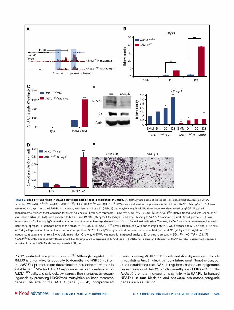
ASXL1 regulates transcription by interacting with PRC2, whichincludes EZH2, Suz12, and EED.19 Thus, ASXL1 deficiency mayalter the PRC2 complex proteins, thereby attenuating H3K27me3methylation. For example, robust osteoclastogenesis attendingASXL1 deficiency may represent an abundance of EZH2, whichdelivers methylated H3K27 to the promoter of Irf8, thereby silencingits osteoclast-inhibiting properties.20 To explore this possibility, wemeasured PRC2 proteins in ASXL1cKOmacrophages and osteoclasticcells. Despite decreased H3K27me3 methylation in ASXL1cKO
osteoclasts, expression of PRC2 core members in mutant BMMswas unchanged, even as they underwent osteoclast differenti-ation (Figure 4B). Blimp1 (encoded by Prdm121), an establishedpositive regulator of osteoclastogenesis, and a transcriptionalrepressor of antiosteoclastogenic genes, increased in ASXL1-KOosteoclasts (Figure 4C). In contrast toNFATc1, however, H3K27me3on Blimp1 promoter was not significantly different between the controland KO osteoclasts (Figure 4D), indicating absence of ASXL1likely did not exert its Blimp1 inductive effect directly by regulatinghistone methylation. Consistent with previous reports that NFATc1regulates Blimp1 expression,17 ASXL1-deletion markedly en-hanced NFATc1 occupancy on the Blimp1 promoter (Figure 4E).
Loss of H3K27me3 in ASXL1-deficient osteoclasts is
mediated by JMJD3
The loss of K27 methylation in ASXL1cKO osteoclasts could likelybe because of removal of methyl groups from the lysine residueson histone H3, by a demethylase specific to K27 trimethylation,22
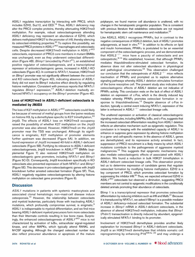
Jmjd3. The effects of ASXL1 loss on H3K27me3 occupancyraised the possibility of whether K27 methylation on the Jmjd3promoter was also altered. However, H3K27me3 on the Jmjd3promoter near the TSS was unchanged. Although its signifi-cance is enigmatic, K27 methylation of promoter elementsfurther upstream was decreased (Figure 5A). In addition, wefound expression of Jmjd3 increased 40-fold in ASXL1-deficientosteoclasts (Figure 5B). Fortifying its relevance to ASXL1-deficientosteoclastogenesis, Jmjd3 knockdown in ASXL1cKO BMMs (sup-plemental Figure 7) also restored H3K27me3 methylation onosteoclastogenic gene promoters, including NFATc1 and Blimp1(Figure 5C-D). Consequently, Jmjd3 knockdown specifically in KOosteoclasts also prevented expression of both NFATc1 and Blimp1(Figure 5E). This decrease in pro-osteoclastogenic genes with Jmjd3knockdown further arrested osteoclast formation (Figure 5F). Thus,ASXL1 negatively regulates osteoclastogenesis by altering histonemethylation on osteoclast gene promoters (Figure 6).
Discussion
ASXL1 mutations in patients with systemic mastocytosis andassociated clonal hematologic non–mast-cell disease induceosteoporosis.10 However, the relationship between osteoclastsand myeloid leukemias, particularly those with inactivating ASXL1mutations, which profoundly compromise survival, is enigmatic.9
ASXL1 is indispensable to myeloid differentiation, and we find micebearing ASXL1-deficient myeloid precursors form more osteoclaststhan their littermate controls resulting in low bone mass. Surpris-ingly, the enhanced osteoclastogenesis of ASXL1cKO mice is notcharacterized by activation of IkBa, extracellular signal-regulatedkinase, and other MAPKs, which typically attend RANKL andM-CSF signaling. Although the changed osteoclast number mayalso reflect precursor abundance or apoptosis of the mature
polykaryon, we found marrow cell abundance is unaltered, with nochanges in the hematopoietic progenitor population. This is consistentwith previous literature that establishes osteoclasts are not requiredfor hematopoietic stem cell maintenance and mobilization.23
Like ASXL2, ASXL1 recognizes PPARg, but in contrast to thenegative consequences of ASXL2 deletion, lack of ASXL1 promotesadipogenesis, at least in vitro.8,11 In addition to its effects on lipidand insulin homeostasis, PPARg is postulated to be an essentialcomponent of the osteoclastogenic process, and we demonstratedthat ASXL22/2 mice have a paucity of osteoclasts and areosteopetrotic.8,12 We established, however, that although PPARgmediates thiazolidinedione-stimulated osteoclast formation, itsabsence does not alter osteoclast abundance in physiologicaland pathological circumstances.24 This observation challengedour conclusion that the osteopetrosis of ASXL22/2 mice reflectsinactivation of PPARg and prompted us to explore alternativesignaling pathways whereby ASXL1 deletion stimulates formationof the bone resorptive cell. The present study also indicates thatosteoclastogenic effects of ASXL1 deletion are not indicative ofPPARg activity. This conclusion rests on the lack of effect of ASXL1deletion on expression of c-Fos, which is the directly induced keymediator whereby PPARg promotes osteoclast formation in re-sponse to thiazolidinediones.12 Despite absence of c-Fos in-duction, typically a central event inducing NFATc1, expression of thelatter is enhanced in ASXL12/2 osteoclast lineage cells.
The unaltered expression or activation of classical osteoclastogenicsignaling molecules, including MAPKs, IkBa, and c-Fos, suggests thatthe increased osteoclast differentiation resulting from ASXL1 deletionreflects an alternative mechanism such as epigenetic regulation. Thisconclusion is in keeping with the established capacity of ASXL1 toenhance or suppress gene expression by altering histone methylationin a gene- and cell-specific manner. In the absence of ASXL1, genesare demethylated, and its repressive activity arrested. In fact,suppression of PRC2 recruitment is a likely means by which ASXL1mutations contribute to the pathogenesis of aggressive myeloidmalignancies.18 This suggests that epigenetic mechanisms mayalso mediate osteoclastogenic effects of associated with ASXL1deletion. We found a reduction in bulk H3K27 trimethylation inASXL1-deficent osteoclast lineage cells. This observation promp-ted us to determine expression of candidate genes that regulateosteoclast formation by modifying histone methylation. EZH2 is akey component of PRC2, which promotes osteoclast formation bysuppressing the inhibitor Irf8.20 Thus, we expected enhanced EZH2 inASXL1cKO osteoclasts but observed a diminution, suggesting PRC2members are not central to epigenetic modifications in the conditionallydeleted animals promoting their abundance of osteoclasts.
Blimp 1 is a transcriptional repressor that promotes osteoclastdifferentiation by reducing inhibitors such as Irf8 andMafb.21 Becauseit is transduced by NFATc1, we asked if Blimp1 is a possible mediatorof ASXL1 deficiency–induced osteoclast formation. The substantialincrease in Blimp1 mRNA in ASXL1-deficient osteoclasts, in theabsence of altered H3K27me3 methylation, indicates that Blimp1(Prdm1) transactivation is directly induced by abundant, epigenet-ically stimulated NFATc1 binding to its promoter.
Involvement of H3K27me3 demethylase provides another likelyexplanation for increased Blimp1 in ASXL1-deficient osteoclasts.Jmjd3 is an H3K27me3 demethylase that inhibits somatic cellreprogramming by enhancing P16INK4a 25 and may undergo a
2474 ROHATGI et al 9 OCTOBER 2018 x VOLUME 2, NUMBER 19
PRC2-mediated epigenetic switch.26 Although regulation ofJMJD3 is enigmatic, its capacity to demethylate H3K27me3 onthe NFATc1 promoter and thus stimulate osteoclast formation isestablished.7 We find Jmjd3 expression markedly enhanced inASXL1cKO cells, and its knockdown arrests their increased osteoclas-togenesis by promoting H3K27me3 methylation on bone resorptivegenes. The size of the ASXL1 gene (;6 kb) compromised
overexpressing ASXL1 in KO cells and directly assessing its rolein regulating Jmjd3, which will be a future goal. Nonetheless, ourstudy establishes that ASXL1 regulates osteoclast epigenomevia expression of Jmjd3, which demethylates H3K27m3 on theNFATc1 promoter increasing its sensitivity to RANKL. EnhancedNFATc1 in turn binds to and activates pro-osteoclastogenicgenes such as Blimp1.
10 kb
kdm6b(Jmjd3)
Promoter Upstream Element
ASXL1f/f H3K27me3
ASXL1cKO H3K27me3
A
ASXL1flox/flox
ASXL1cKO
50Jmjd3
40
30
Relat
ive in
tens
ity
20
10
0BMM D1
**
***
D2
B
ASXL1cKO Scr
ASXL1cKO ShJmjd3
400
300
200
100
0IgG H3K27me3
NFA
Tc1
prom
oter
(5%
of i
nput
)
***
C
3.53.02.52.0
Relat
ive in
tens
ity
1.51.00.5
0BMM D1
ASXL1cKO-Scr ASXL1cKO-Sh JMJD3
D2 D3 BMM
Blimp1
D1 D2 D3
*
**
**
Scr
NFATc1
3
Actin
shJmjd3
E
ASXL1cKO Scr
ASXL1cKO ShJmjd3
1.0
0.8
0.6
0.4
0.2
0IgG H3K27me3
Blim
p1 p
rom
oter
(5%
of i
nput
)
***
D
SCR RNA ShJmjd3
F
Figure 5. Loss of H3K27me3 in ASXL1-deficient osteoclasts is mediated by Jmjd3. (A) H3K27me3 peaks at individual loci (highlighted blue bar) on Jmjd3
promoter; WT (ASXL1flox/flox) and KO (ASXL1cKO). (B) ASXL1flox/flox and ASXL1cKO BMMs were cultured in the presence of M-CSF and RANKL (25 ng/mL). RNA was
harvested on days 1 and 2 of RANKL stimulation, and histone H3 Lys 27 (H3K27) demethylase Jmjd3 mRNA abundance was determined by qPCR. Unpaired
nonparametric Student t test was used for statistical analysis. Error bars represent 1 SD; **P , .01, ***P , .001. (C-D) ASXL1cKO BMMs, transduced with scr or Jmjd3
short hairpin RNA (shRNA), were exposed to M-CSF and RANKL (25 ng/mL) for 2 days. H3K27me3 binding to NFATc1 promoter (C) and Blimp1 promoter (D) was
determined by ChIP assay. IgG served as control. n 5 2 independent experiments from 10- to 12-week-old male mice. Two-way ANOVA was used for statistical analysis.
Error bars represent 1 standard error of the mean; ***P , .001. (E) ASXL1cKO BMMs, transduced with scr or Jmjd3 shRNA, were exposed to M-CSF and 1 RANKL
for 3 days. Expression of osteoclast differentiation proteins NFATc1 and b3 integrin was determined by immunoblot (left) and Blimp1 by qPCR (right). n 5 3
independent experiments from 8-week-old male mice. One-way ANOVA was used for statistical analysis. Error bars represent 1 SD; *P , .05, **P , .01. (F)
ASXL1cKO BMMs, transduced with scr or shRNA for Jmjd3, were exposed to M-CSF and 1 RANKL for 5 days and stained for TRAP activity. Images were captured
on Nikon Eclipse E400. Scale bar represents 400 mm.
9 OCTOBER 2018 x VOLUME 2, NUMBER 19 ASXL1 IMPACTS H3K27me3 EPIGENOME OF OSTEOCLASTS 2475
Although the relationship between myeloid linage cells andosteoclasts is established, these findings show that mutationspromoting hematopoietic malignancies may also directly modulateosteoclast formation. Thus, the osteoclastogenesis of ASXL1deficiency does not involve classical RANKL- or M-CSF-stimulatedmolecules such as MAPKs and c-Fos but activation of NFATC1 byreversal of suppressive histone methylation. Epigenetic modifica-tion of myeloid lineage genes is therefore central to skeletalhomeostasis and may participate in malignancy associated boneloss.
Acknowledgments
The authors thank Genome Technology Access Center atWashington University for performing ChIP-seq and providingtechnical assistance.
This work was supported by grants from the National Instituteof Arthritis and Musculoskeletal and Skin Diseases, National Insti-tutes of Health (R37 AR046523 [S.L.T.], R01 AR054326 [Y.A.-A.],
R01 AR072623 [Y.A.-A.], and P30 AR057235 [S.L.T. and Y.A.-A.]);the National Institute of Diabetes and Digestive and Kidney Diseases,National Institutes of Health (grant R01 DK111389) (S.L.T.); andShriners Hospitals for Children (grant 85400-STL) (S.L.T.).
Authorship
Contribution: N.R. designed and performed experiments and wrotethe manuscript; W.Z. and J.R.B. designed and performed experi-ments; P.L.C. performed experiments and edited the manuscript;T.H.C. performed experiments; Y.A.-A. designed experiments; andS.L.T. designed experiments and wrote the manuscript.
Conflict-of-interest disclosure: The authors declare no compet-ing financial interests.
Correspondence: Steven L. Teitelbaum, Department of Pathol-ogy and Immunology, Washington University School of Medicine,Campus Box 8118, 660 South Euclid Ave, St. Louis, MO 63110;e-mail: [email protected].
References
1. Novack DV, Teitelbaum SL. The osteoclast: friend or foe? Annu Rev Pathol. 2008;3(1):457-484.
2. Asagiri M, Sato K, Usami T, et al. Autoamplification of NFATc1 expression determines its essential role in bone homeostasis. J Exp Med. 2005;202(9):1261-1269.
3. Nishikawa K, Iwamoto Y, Kobayashi Y, et al. DNA methyltransferase 3a regulates osteoclast differentiation by coupling to an S-adenosylmethionine-producing metabolic pathway. Nat Med. 2015;21(3):281-287.
4. Baskind HA, Na L, Ma Q, Patel MP, Geenen DL, Wang QT. Functional conservation of Asxl2, a murine homolog for the Drosophila enhancer of trithoraxand polycomb group gene Asx. PLoS One. 2009;4(3):e4750.
5. Gildea JJ, Lopez R, Shearn A. A screen for new trithorax group genes identified little imaginal discs, the Drosophila melanogaster homologue of humanretinoblastoma binding protein 2. Genetics. 2000;156(2):645-663.
ASXL1
WT
PRC2
Off
Blimp1 gene expression off
Osteoclastogenesis
SUZ12
me3 me3 me3
EZH2 EED
On
Increased Osteoclastogenesis
me3
ASXL1cKO
ASXL1
JMJD3
NFATc1
SUZ12
EZH2 EED
ASXSXSXSSSXXXSXXL1
Figure 6. Schematic representation for role of ASXL1 in regulating osteoclastogenesis. ASXL1 binds to PRC2 proteins to methylate promoters of key osteoclast
differentiation genes such as NFATc1 and regulate their expression (left). Absence of ASXL1 prevents PRC2-mediated histone methylation (right). Jmjd3, specific demethylase
for H3K27me3 in turn removes K27 methyl groups from promoters such as NFATc1, resulting in increased osteoclastogenesis.
2476 ROHATGI et al 9 OCTOBER 2018 x VOLUME 2, NUMBER 19
6. Margueron R, Reinberg D. The Polycomb complex PRC2 and its mark in life. Nature. 2011;469(7330):343-349.
7. Yasui T, Hirose J, Tsutsumi S, Nakamura K, Aburatani H, Tanaka S. Epigenetic regulation of osteoclast differentiation: possible involvement of Jmjd3in the histone demethylation of Nfatc1. J Bone Miner Res. 2011;26(11):2665-2671.
8. Izawa T, Rohatgi N, Fukunaga T, et al. ASXL2 regulates glucose, lipid, and skeletal homeostasis. Cell Reports. 2015;11(10):1625-1637.
9. Seitz S, Barvencik F, Koehne T, et al. Increased osteoblast and osteoclast indices in individuals with systemic mastocytosis.Osteoporos Int. 2013;24(8):2325-2334.
10. Damaj G, Joris M, Chandesris O, et al. ASXL1 but not TET2 mutations adversely impact overall survival of patients suffering systemic mastocytosiswith associated clonal hematologic non-mast-cell diseases. PLoS One. 2014;9(1):e85362.
11. Park UH, Yoon SK, Park T, Kim EJ, Um SJ. Additional sex comb-like (ASXL) proteins 1 and 2 play opposite roles in adipogenesis via reciprocal regulationof peroxisome proliferator-activated receptor gamma. J Biol Chem. 2011;286(2):1354-1363.
12. Wan Y, Chong LW, Evans RM. PPAR-gamma regulates osteoclastogenesis in mice. Nat Med. 2007;13(12):1496-1503.
13. Lam J, Takeshita S, Barker JE, Kanagawa O, Ross FP, Teitelbaum SL. TNF-alpha induces osteoclastogenesis by direct stimulation of macrophagesexposed to permissive levels of RANK ligand. J Clin Invest. 2000;106(12):1481-1488.
14. Zhang Y, Liu T, Meyer CA, et al. Model-based analysis of ChIP-Seq (MACS). Genome Biol. 2008;9:R137.
15. Heinz S, Benner C, Spann N, et al. Simple combinations of lineage-determining transcription factors prime cis-regulatory elements required formacrophage and B cell identities. Mol Cell. 2010;38(4):576-589.
16. Ramırez F, Ryan DP, Gruning B, et al. deepTools2: a next generation web server for deep-sequencing data analysis. Nucleic Acids Res. 2016;44(W1):W160-W165.
17. Karolchik D, Baertsch R, Diekhans M, et al; University of California Santa Cruz. The UCSCGenome Browser Database. Nucleic Acids Res. 2003;31(1):51-54.
18. Abdel-Wahab O, Adli M, LaFave LM, et al. ASXL1 mutations promote myeloid transformation through loss of PRC2-mediated gene repression.Cancer Cell. 2012;22(2):180-193.
19. Abdel-Wahab O, Dey A. The ASXL-BAP1 axis: new factors in myelopoiesis, cancer and epigenetics. Leukemia. 2013;27(1):10-15.
20. Fang C, Qiao Y, Mun SH, et al. Cutting edge: EZH2 promotes osteoclastogenesis by epigenetic silencing of the negative regulator IRF8. J Immunol.2016;196(11):4452-4456.
21. Nishikawa K, Nakashima T, Hayashi M, et al. Blimp1-mediated repression of negative regulators is required for osteoclast differentiation. Proc Natl AcadSci USA. 2010;107(7):3117-3122.
22. Hong S, Cho YW, Yu LR, Yu H, Veenstra TD, Ge K. Identification of JmjC domain-containing UTX and JMJD3 as histone H3 lysine 27 demethylases.Proc Natl Acad Sci USA. 2007;104(47):18439-18444.
23. Miyamoto T. Role of osteoclasts in regulating hematopoietic stem and progenitor cells. World J Orthop. 2013;4(4):198-206.
24. ZouW, Rohatgi N, Chen TH, Schilling J, Abu-Amer Y, Teitelbaum SL. PPAR-g regulates pharmacological but not physiological or pathological osteoclastformation. Nat Med. 2016;22(11):1203-1205.
25. Zhao W, Li Q, Ayers S, et al. Jmjd3 inhibits reprogramming by upregulating expression of INK4a/Arf and targeting PHF20 for ubiquitination. Cell. 2013;152(5):1037-1050.
26. Shi X, Zhang Z, Zhan X, et al. An epigenetic switch induced by Shh signalling regulates gene activation during development and medulloblastoma growth.Nat Commun. 2014;5:5425.
9 OCTOBER 2018 x VOLUME 2, NUMBER 19 ASXL1 IMPACTS H3K27me3 EPIGENOME OF OSTEOCLASTS 2477