octoplus qplex - dyeagnostics · 2020-02-03 · octoplus qplex is equipped with a fine tuned set of...
TRANSCRIPT

1
OCTOPLUSQPLEXFLUORESCENCE IMAGER
for fast & powerful 2D gel image acquisition

2 3
Made in Germany
Octoplus QPLEX Fluorescence ImagerThe new Octoplus QPLEX fluorescence imager sets a novel standard for fluorescence 2D gel imaging. It combines the sensitivity of laser-based systems with the rapid image acquisition of CCD cameras. The Octoplus QPLEX system introduces the latest improvements in fluorescence excitation and emission detection technologies and is developed for the specific 4-color multiplexing (quadruplexing) fluorescence imaging of Refraction-2DTM QPLEX dyes.In addition, Octoplus QPLEX performs fluorescent and chemiluminescent Western blot analysis to the highest standards available. The robust setup of the device is designed for daily use and does not require any maintainance.

4 5
“Refraction-2D™ multiplex fluorescence 2D gel analysis helps us to identify novel protein biomarkers for cancer diagnostics.”
Prof. Dr. Dr. J. Habermann and Dr. Timo Gemoll. University of Lübeck
“We very much appreciate the rapid 2D gel image acquisition by a highly specific and sensitive
CCD camera based system.“
Prof. Dr. Barbara Sitek, Medical Proteom Center Bochum
Ultra-sensitive 4-color fluorescence detectionHigh resolution 2D gel imagingRapid image acquisition
Octoplus QPLEX Fluorescence Imager
Quadroplexfluorescence
2D gel imaging

6 7
Refraction-2D™ QPLEX analysis of three different Arabidopsis thaliana ecotypes.
Quadruplex fluorescence 2D gel imaging
Octoplus QPLEX is especially designed for Refraction-2D™ and Saturn-2D™ multiplex fluorescence 2D gel analysis. The powerful combination of carefully developed system components for fluorescence light excitation and detection create a perfect system for sensitive image acquisition.The image acquisition of a Refraction-2D™ QPLEX gel size 24 x 20 cm (images for G-Dye100, G-Dye200 G-Dye300 and G-Dye400) is performed within minutes. A complete series of gels (e.g. six Refraction-2D™ QPLEX gels) can be imaged in less than one hour.
G-Dye100 + G-Dye200 + G-Dye300 + G-Dye400
= 5 min image acquisition
Octoplus QPLEX Blue HP LED, G100BP filterBit depth: 16 bitGel size: 24 x 20 cm, pH 3-10Sample: 50 µg total protein from E. coli
Octoplus QPLEX Green HP LED, G200BP filterBit depth: 16 bitGel size: 24 x 20 cm, pH 3-10Sample: 50 µg total protein from E. coli
Octoplus QPLEX Red LED, G300BP filter
Bit depth: 16 bit
Gel size: 24 x 20 cm, pH 3-10
Sample: 50 µg total protein from E. coli
Octoplus QPLEX Infra Red LED, G400BP filter
Bit depth: 16 bit
Gel size: 24 x 20 cm, pH 3-10
Sample: 50 µg total protein from E. coli
G-Dye100 image
Fluorescent label: G-Dye100
G-Dye300 image G-Dye400 image
G-Dye200 image
Fluorescent label: G-Dye400Fluorescent label: G-Dye300
Fluorescent label: G-Dye200

8 9
100 200 300 400 M
Sensitivity of fluorescence 2D gel imaging
The latest developments in high power LED technology (light emitting diode) combined with a high quantum efficiency CCD sensor (charge coupled device) over a spectrum of 470 to 770 nm enables the Octoplus QPLEX system to compete with laser based fluorescence imaging devices (Fig. 1+2).
Fig 1. Green HP LED; G200BP filter; bit depth: 16 bit; gel size: 24 x 20 cm, pH 4-7;Sample: 50 µg total protein from E. coli pre-labeled with G-Dye100 (minimal labeling);Detailed view of the 2D gel shown below.
Fig 2. 532 nm green SHG laser; LPG (575LP) filter; bit depth: 16 bit; gel size: 24 x 20 cm, pH 4-7; Sample: 50 µg total protein from E. coli pre-labeled with G-Dye100 (minimal labeling);Detailed view of the 2D gel shown below.
Image acquired by Octoplus QPLEX Scan acquired by Typhoon FLA 9000
Octoplus QPLEX
Typhoon FLA 9000
Acknowlegement: The 2D gel was provided by courtesy of Prof. Dr. Dr. J. Habermannand Dr. T. Gemoll, University of Lübeck, Germany
Specificity of detection of fluorescence light
Multiplex fluorescence imaging requires highly specific fluorescence light excitation and emission. Octoplus QPLEX is equipped with a fine tuned set of 4 highly specific excitation and emission band pass filters.
To test for detection specificity 4 x 5 µg of E. coli total protein was pre-labeled with G-Dye100, G-Dye200, G-Dye300 and G-Dye400 and separated by 1D SDS-PAGE (lanes 1-4, lane 5: QPLEX pre-labeled protein marker, Fig. 3). To analyze filter specificity (cross talk), the specific lane was detected by using the corresponding excitation (HP LED respectively laser) and emission filter set (Figures below).
G-Dye100 imageBlue HP LEDG100BP filter
G-Dye200 imageGreen HP LED
G200BP filter
G-Dye300 imageRed HP LED
G300BP filter
G-Dye400 imageIR HP LEDG400BP filter
G-Dye100 scanBlue LD laserLPB (510LP) filter
G-Dye200 scanGreen SHG laser LPG (575LP) filter
G-Dye300 scanRed LD laser LPR (665LP) filter
not available
100 200 300 400 M
100 200 300 400 M
100 200 300 400 M
100 200 300 400 M
100 200 300 400 M
100 200 300 400 M
100 200 300 400 M
Fig. 3. G-Dye100 - 400 overlay Image acquired by Octoplus QPLEX
Octoplus QPLEX
Typhoon FLA 9000

10 11
PTM (Phosphorylation) 3
4
1
2 PTM (Glycosylation)
Is 0 h 12 h 24 h
Is 0 h 12 h 24 h
12 h upregulation (2.3 fold)
24 h downregulation (1.8 fold)
Refraction-2D™ QPLEX 2D gel analysis of Arabidopsis thaliana
(Col.) 0 h, 72 h and 144 h upon treatment with Bion® (salicylic acid
analogon, stimulates pathogen defense). Imaging by Octoplus QPLEX.
2
3
4
1
World´s firstquadruplex 2D gelanalysis
• Octoplus QPLEX fluorescence imaging
• Refraction-2D™ QPLEX protein labeling

12 13
Refraction-2D™ QPLEX4-color 2D gel protein labeling
Comparing up to 4 different samples within the same analysis: Refraction-2DTM QPLEX introduces the world´s first 4-color coding for 2D gels. The G-Dyes (G-Dye100, G-Dye200, G-Dye300 and G-Dye400) combine the most powerful fluorescence properties with a superior photo stability and easy, accurate spot picking.
Kit content
• G-Dye100 – high performance fluorescence dye
• G-Dye200 – high performance fluorescence dye
• G-Dye300 – high performance fluorescence dye
• G-Dye400 – high performance fluorescence dye
• G-Dye labeling stop solution
• G-Dye solvent
• G-Dye low retention tips
• G-Dye low retention micro centrifuge tubes
• Extra G-Dye100 for easy spot picking (included in 12G kits)
FluorescenceChemiluminescenceColorimetric
1D gel imaging
Simultaneous fluorescence imaging of 4 different dyes
Sensitive & quantitative chemiluminescence
Bright colorimetric imaging

14 15
Fluo
resc
ence
inte
nsity
(A
U)
1D Fluorescence Imaging
The Octoplus QPLEX HP LED module emittes light with 4 different wavelenghts to excite the diverse fluorophores. Highly specific excitation and emission filters avoid any crosstalk issues. Figure 4 shows 4 differently fluorescent labeled proteins separated by SDS-PAGE and imaged by Octoplus QPLEX. Complex samples of less than 1 µg of protein can be analyzed with high accuracy (Fig. 5).
Fig. 4. Serial dilution of T-Dye labeled proteins: BSA + T-Green 210, Casein + T-Rex 330, Lactoglobulin + G-Dye100, RNase A + T-Red 410. Proteins were separated by SDS-PAGE and imaged by Octoplus QPLEX.
Imaging times: T-Green 210: 35 sec, T-Rex 330: 5 sec, G-Dye100: 10 sec, T-Red 410: 30 sec.
Fig. 5. E. coli total protein extract pre-labeled with T-Green 210. Separation by SDS-PAGE. Octoplus QPLEX imaging time: 20 sec. To ensure protein quantificaytion all signals are below saturation (max. grey value < 65,536 px).
1.000 0.500 0.250 0.125 0.062 0.031 0.016Protein load (μg)
5.000 1.000 0.500 0.250 0.125 0.062Protein load (μg)
Fig. 6. Sample multiplexing of 2 μg T-Red 310 and T-Green 210 labeled protein derived from domestic pig (Sus scrofa domesticus) comparing two different extraction protocols. Evaluation by 1D software.
Multiplex fluorescence Western blot
Multiplex fluorescence Western blot analysis allows the specific detection of different proteins within one sample. The specific and rapid imaging (< 1 sec) is performed by Octoplus QPLEX.
Fig. 7. Multiplex fluorescence Western blot. Protein derived from human serum was extracted and pre-labeled with T-Green 210. 2 x 10 µg of protein (lane 1 & 2) was separated by SDS-PAGE and then transfered onto a low fluorescent membrane. The blot was subjected to a lactoglobulin antibody and then to a T-Red 310 conjugated secondary antibody. The proteins were detected by Octoplus QPLEX imaging. Exposure time: 0.3 sec.
Serum Lactoglobulin Overlay
Sample multiplexing
16 17
0
20
40
60
80
100
Amount of protein (µg)
Sign
al in
tens
ity (
AU
)
R² = 0.996
0.0 0.10 0.30 0.40 0.50 0.60 0.70 0.80 0.90 1.00.20
4
5
6
7
8
9
lg s
igna
l int
ensi
ty (
AU
)
0.4 4 40 4000.04
Amount of G-Dye200 (pmol)
R² = 0.998
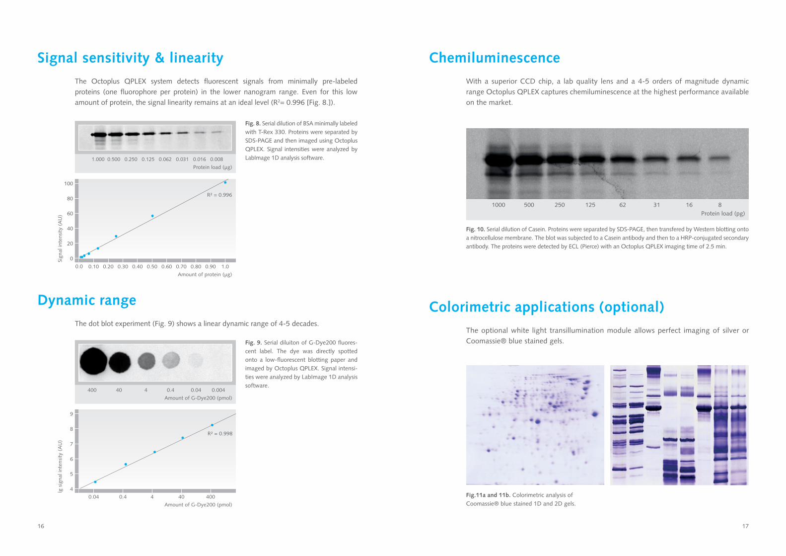
Signal sensitivity & linearity
The Octoplus QPLEX system detects fluorescent signals from minimally pre-labeled proteins (one fluorophore per protein) in the lower nanogram range. Even for this low amount of protein, the signal linearity remains at an ideal level (R2= 0.996 [Fig. 8.]).
Fig. 8. Serial dilution of BSA minimally labeled with T-Rex 330. Proteins were separated by SDS-PAGE and then imaged using Octoplus QPLEX. Signal intensities were analyzed by LabImage 1D analysis software.1.000 0.500 0.250 0.125 0.062 0.031 0.016 0.008
Protein load (μg)
Fig. 9. Serial diluiton of G-Dye200 fluores-cent label. The dye was directly spotted onto a low-fluorescent blotting paper and imaged by Octoplus QPLEX. Signal intensi-ties were analyzed by LabImage 1D analysis software.
400 40 4 0.4 0.04 0.004
Amount of G-Dye200 (pmol)
Dynamic range
The dot blot experiment (Fig. 9) shows a linear dynamic range of 4-5 decades.
Chemiluminescence
With a superior CCD chip, a lab quality lens and a 4-5 orders of magnitude dynamic range Octoplus QPLEX captures chemiluminescence at the highest performance available on the market.
Colorimetric applications (optional)
The optional white light transillumination module allows perfect imaging of silver or Coomassie® blue stained gels.
Fig. 10. Serial dilution of Casein. Proteins were separated by SDS-PAGE, then transfered by Western blotting onto a nitrocellulose membrane. The blot was subjected to a Casein antibody and then to a HRP-conjugated secondary antibody. The proteins were detected by ECL (Pierce) with an Octoplus QPLEX imaging time of 2.5 min.
Fig.11a and 11b. Colorimetric analysis of Coomassie® blue stained 1D and 2D gels.
1000 500 250 125 62 31 16 8Protein load (pg)

18 19
Instrument specifications
CCD Camera Kodak KAF-3200 CCD full frame chip with microlens technology16-bit (65,536 grey values)
Cooling 4-stage Peltier cooling (∆T -60 K)
Chip resolution 3.2 MP(2,184 w x 1,472 h pixel)
Pixel size 6.8 x 6.8 µmfull well capacity 55,000 e-
Quantum efficiency 475 nm ≈ 65% 525 nm ≈ 75%575 nm ≈ 85%665 nm ≈ 85%760 nm ≈ 65%
Dynamic range 4-5 orders of magnitude
Lens Schneider-Kreuznach (F 0.95 / 25 mm)
Focusing Manual remote operation
Binning modes 1 x 1, 2 x 2, ... , 10 x 10
Fluorescence unit RGB-IR fluorescence high performance (HP) LED unit including specific filter and diffusor lenses
Filter G100BP (blue)G200BP (green)G300BP (red)G400BP (infra red)WL (white light)upgradable to 4 additional filters
Max. sample size 30 x 22 cm
Sample tray 10 stage sample tray mounted on dual-action dampers
Interface USB 2.0 / Ethernet
Operating system Windows 7
Operating temperature
Up to 30°C
Size (W x H x D) 41 cm x 90 cm x 40 cm
Weight Approx. 45 kg
Image capture software
The easy to use image capture soft-ware performs different capture modes for optimal image acquisition. The raw image data are saved and a copy is tak-en for further analysis by 1D and 2D software. This data separation allows you to always revert back to your origi-nal images. 1D and 2D gel image anal-ysis software is available separately.
19
Octoplus QPLEX inside
High sensitivity 16-bit chip with dark pixel for
low noise
Highly specific QPLEX filter set (blue,
green, red, infra red)
Special optical lens for large imaging area and
high image quality
HP LED unit with specific filters and diffusor lenses for
homogenous illumination
10 stage sample tray size 30 x 22 cm on
dual-action dampers
Robust housing for daily routine

20
Ordering information
PR130 Octoplus QPLEX Fluorescence Imager • Quadruplex HP LED module (specific blue, red, green & infra red fluorescence detection) • Quadruplex emission filter set, additional WL filter • Chemiluminescence • Image capture software • Control PC
PR132 White-light transmission module PR137 Display 24“ PR136 1D analysis software LabImage PR134 2D analysis software Delta2D (fix/ floating/ consumable)
Related products
PR03 Low fluorescent glass cassettes, size 8 x 10 cm PR04 Low fluorescent glass cassettes, size 22 x 27.5 cm
Related consumables Please refer to our protein labeling kit eBrochure available on our website: PDF NH DyeAGNOSTICS Protein Labeling Kits eBrochure 2012-2013 (engl.)
Contact
NH DyeAGNOSTICS GmbH Weinbergweg 23 D-06120 Halle Germany
Fon: +49 (0) 345-2799 6413 Fax: +49 (0) 345-2799 6412 E-Mail: [email protected] [email protected] www.dyeagnostics.com