oct (optic coherence tomography) 1)noninvasive 2) non-contact imaging 3)millimeter penetration
DESCRIPTION
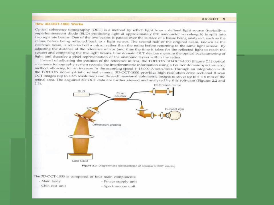
OCT (Optic Coherence Tomography) 1)Noninvasive 2) non-contact imaging 3)Millimeter penetration Aproximately 2-3 mm in tissue with micrometer scale (axial and transverse resolution) not only the retina and optic nerve but also anterior segment of the eye. - PowerPoint PPT PresentationTRANSCRIPT


OCT(Optic Coherence Tomography)
1)Noninvasive
2) non-contact imaging
3)Millimeter penetration
Aproximately 2-3 mm in tissue with micrometer scale (axial and transverse resolution) not only the retina and optic nerve but also anterior segment of the eye


4) Ist demonstrated in 1991 (20 years ago)
5) Today we have the conventional OCT
with an axial resolution of < 10µm and 3Dimentional (3D) OCT with a much
higher resolution of 5µm
OCT principle
1) Is similar to ultrasound
2) Uses echoes to locate structures within the body

Light speed and sound
The speed of light being almost a million times faster than sound allows measurement of structures with a resolution of < 10 microns compared to 100 microns scale for ultrasound.
OCT
Can clearly image the cornea, sclera, iris and lens in the anterior segment. using infrared light having a wave length of 13.1 nm
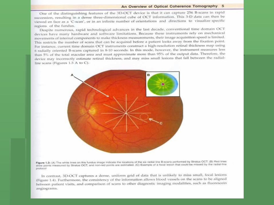
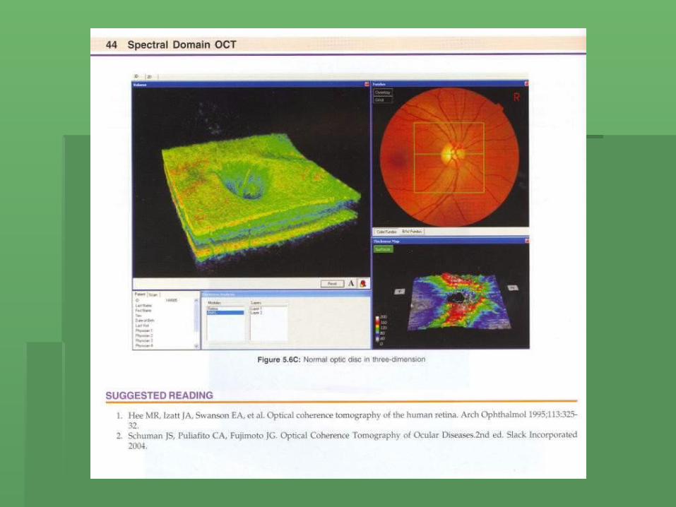
Radial line OCT compared to 3D- OCT
In contrast to radial – line ,3D- OCT captures a dense , uniform grid of data that is unlikely to miss small , focal lesions.
Role of OCT imaging
OCT is useful in the diagnosis and staging of diseases which help in their management , assessing the response to treatment and in monitoring the progress of the disease.

Resolution power
Current commercial OCT scanners offer a resolution of 8-10 micron that is at least 10 time better than ultrasound

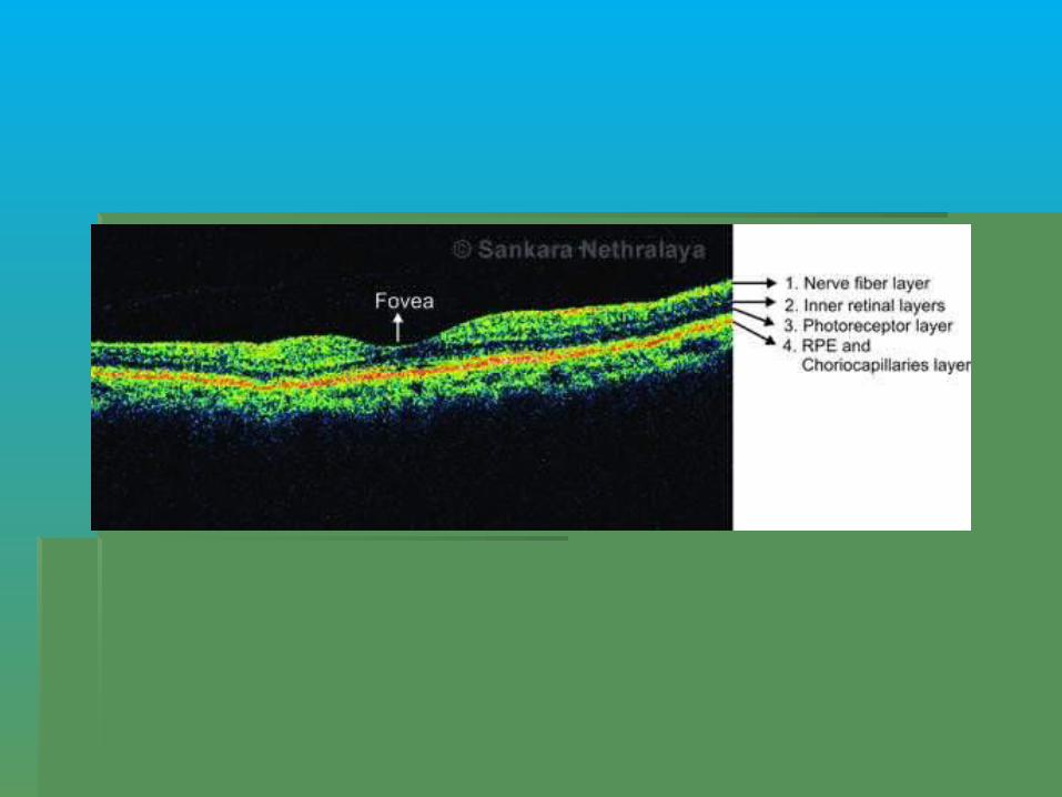
OCT Colors
Tissues with higher reflectivity such as RPE appear in brighter colors ( red-white)– and less dense structures , such as vitreous and intraretinal fluid , appear in darker colors.
(blue- black )

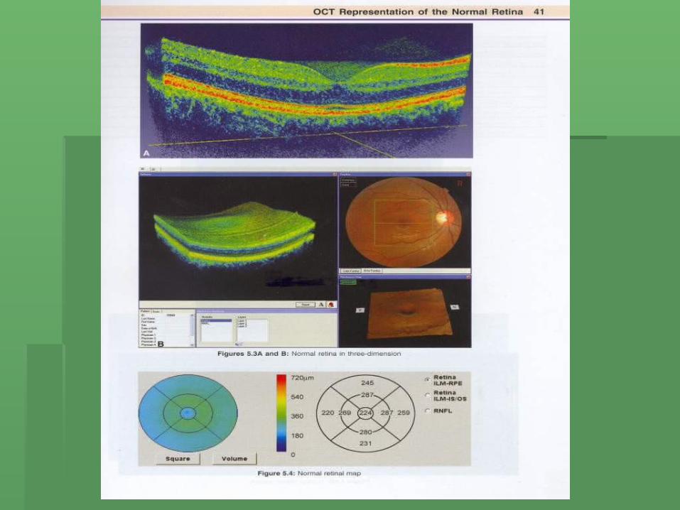
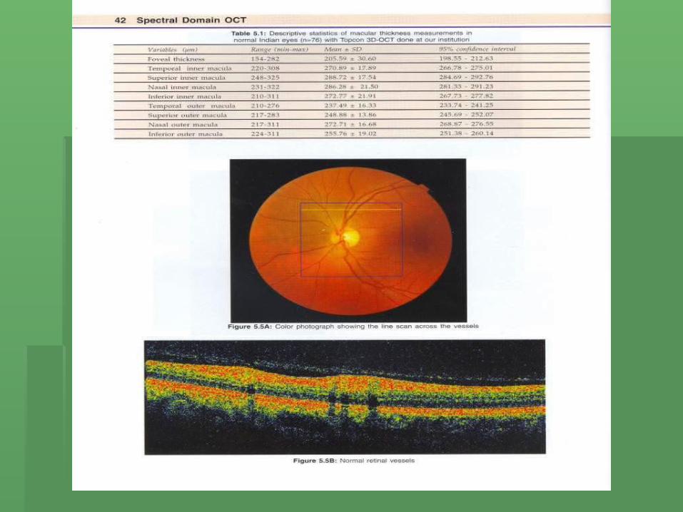
Retinal thickness
Macular thickness with
Topcon 3D –OCT ( Indian eyes )
Foveael thickness 154-282 micron
Other parts 210 -311 micron


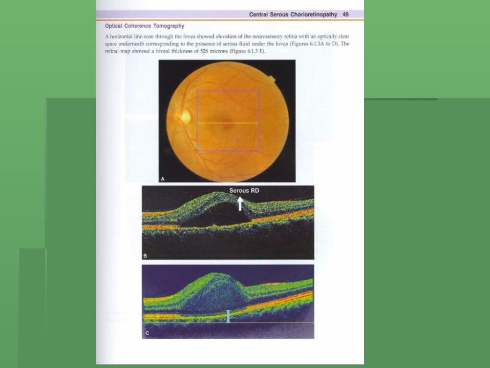
CSR
Men 6-10 times more often than it affects women.
After 50 years usually its bilateral
Age-20-55 years
Leakage from choriocapillaries through the RPE

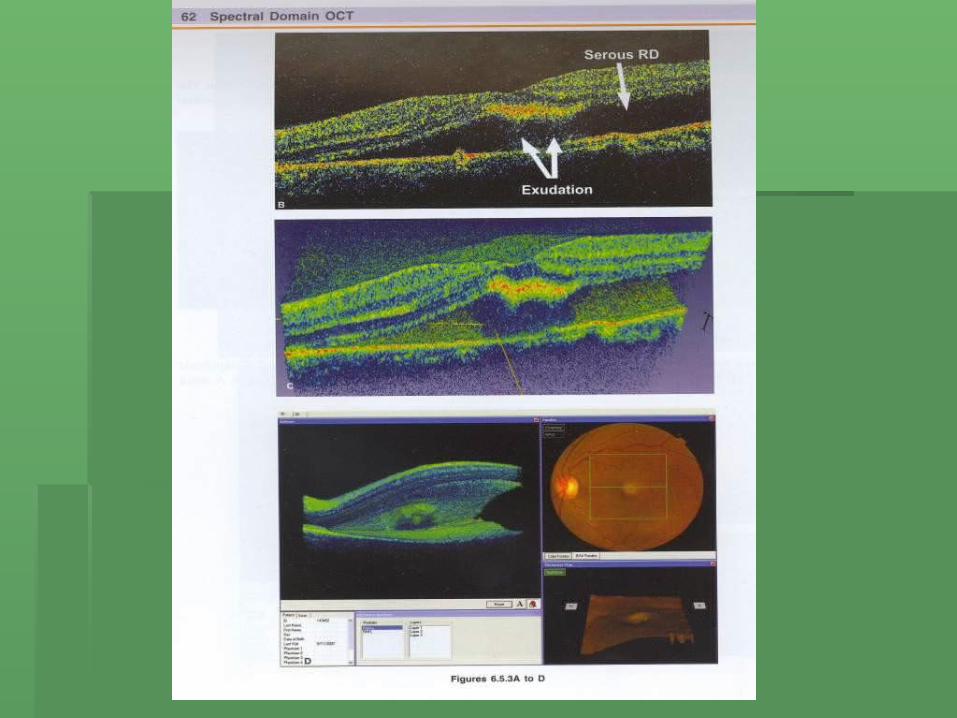
1- Central SerousChorioretinopathy

First case of CSR


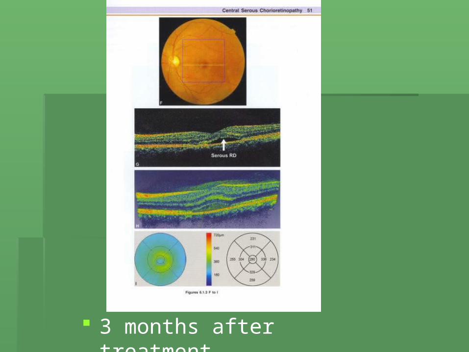
3 months after treatment

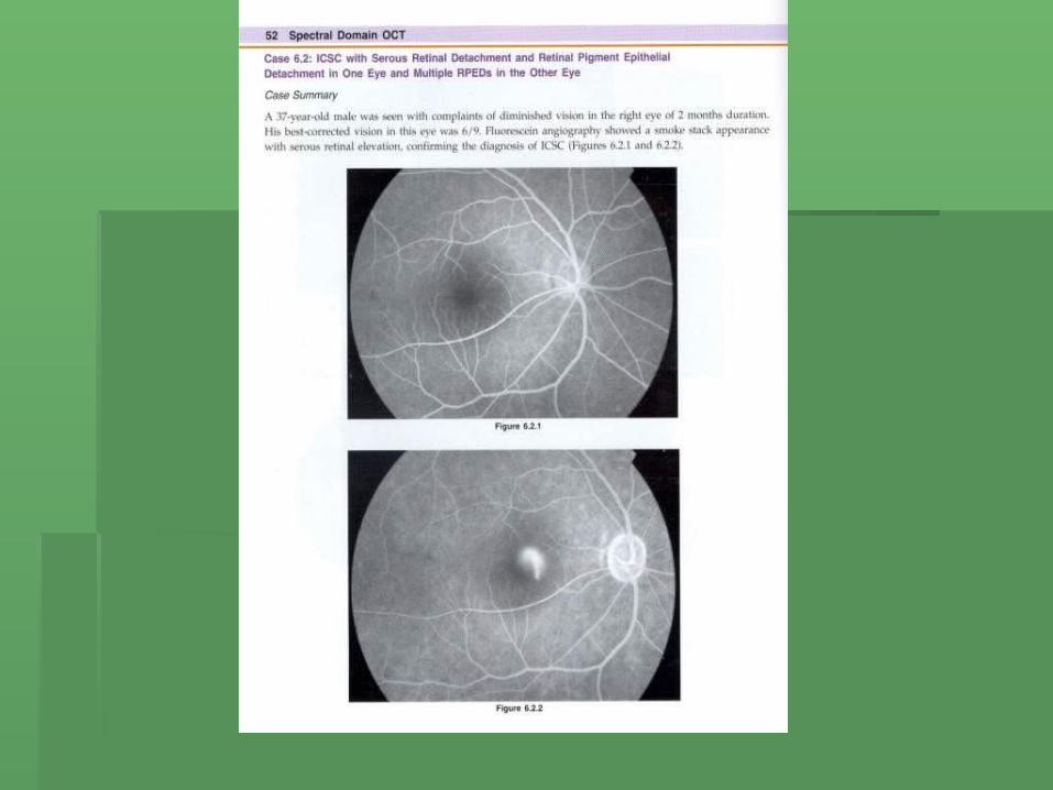
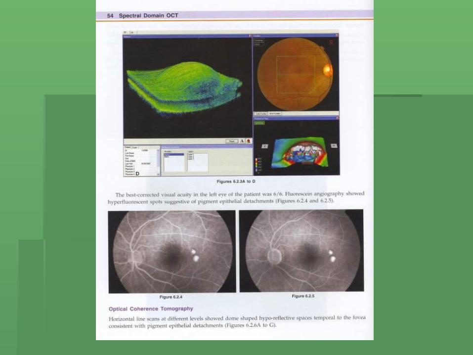
CSR Case no 2

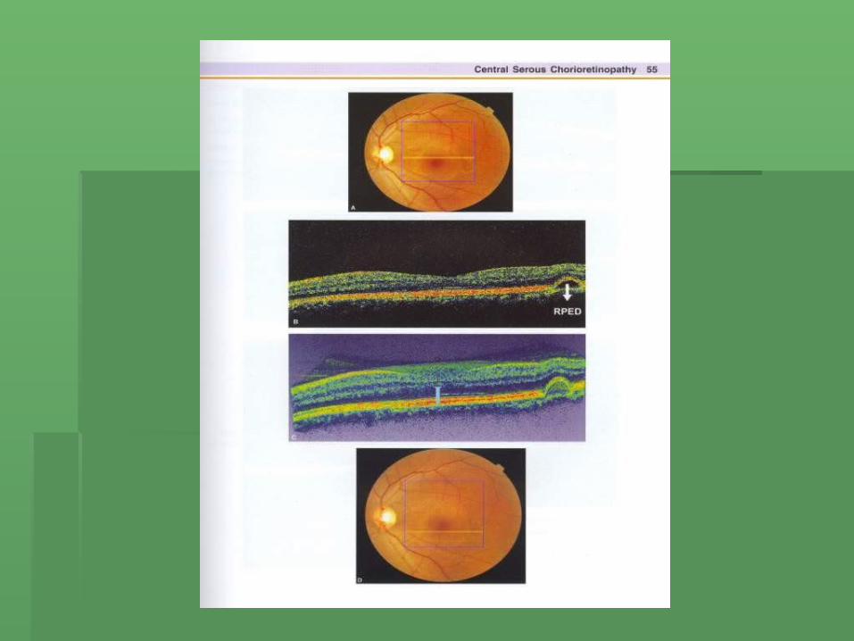

CSRCase no 3


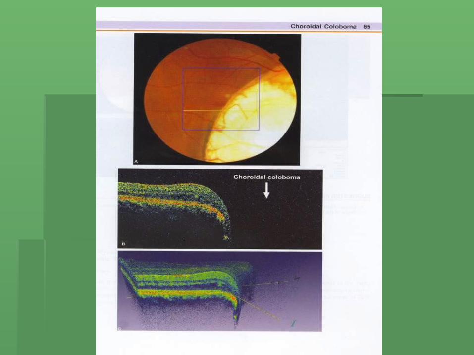
2 - Choroidal Coloboma


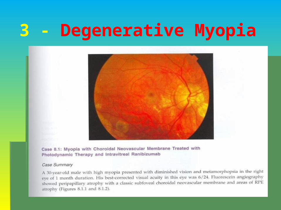
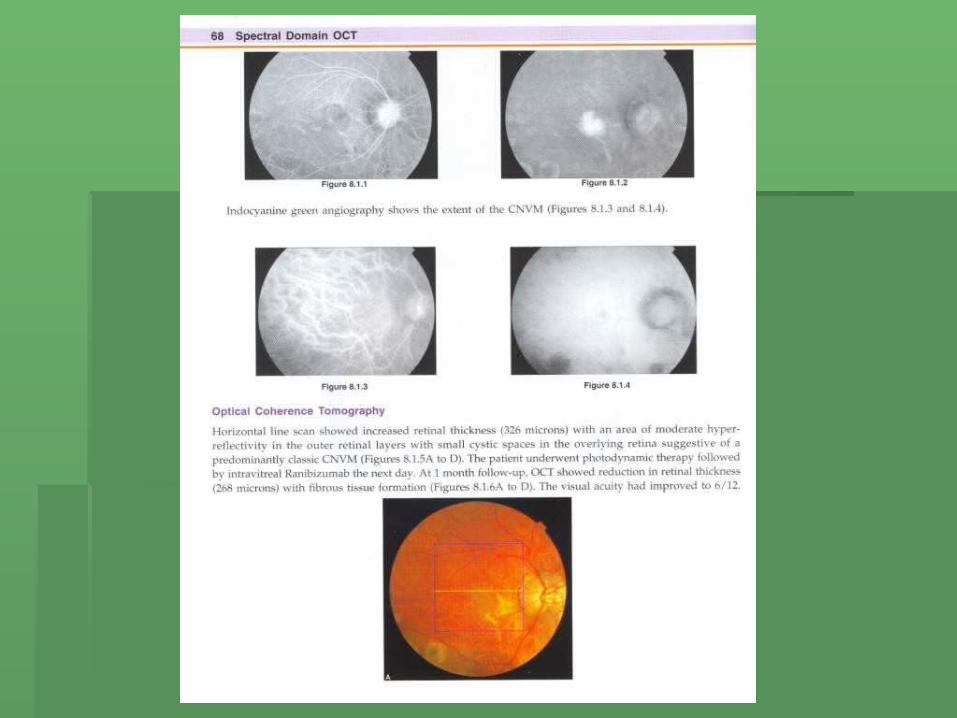
3 - Degenerative Myopia


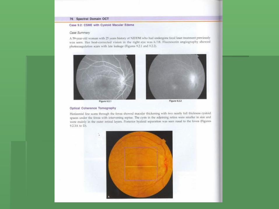
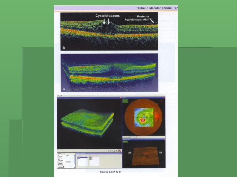
4- Diabetic Macular Edema

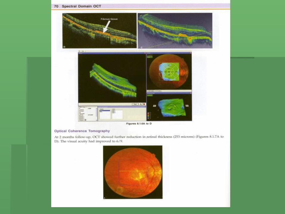
Diabetic Macular Edema
Case no 1


Cystoid Macular Edema
Case no 2
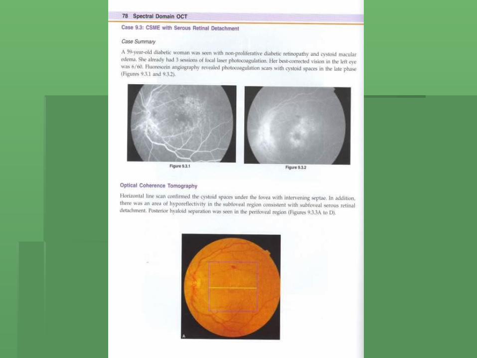
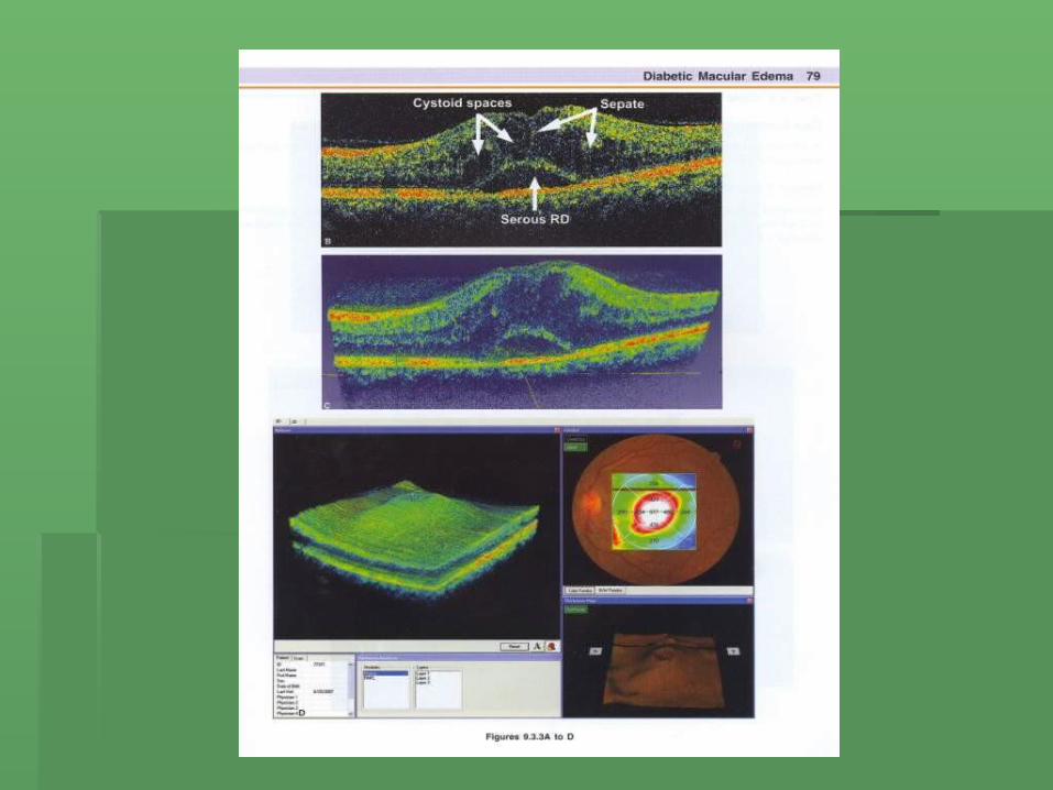
CSME with Serous Retinal
Detahment(Cystoid Macular Edema)
Case no 3

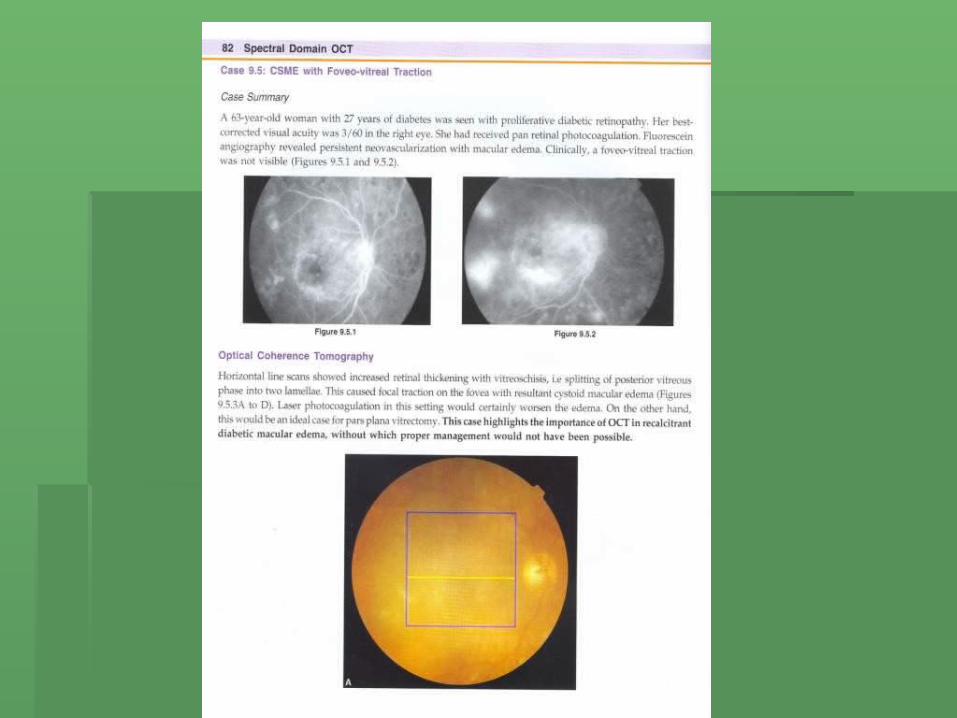
CSME with Fovea Vitreal Traction


Intravitreal Triamcinolone Acetonide in Diabetic Macular Edema (4mg)
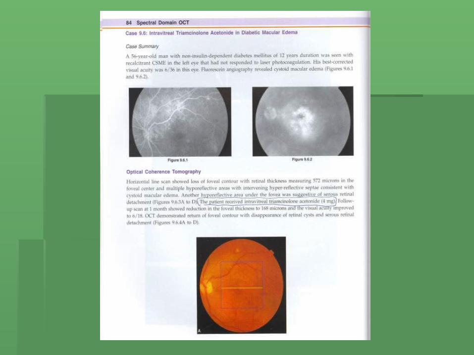

Pre-treatment

Post treatment (4mg Triamcinolone)


5- Epiretinal Membranes

Case no 1


پوزش عرض ضمنباالی حجم بدلیل
LECTUER ادامهپذیر امکان اسالیدها
نیاز صورت در نمیباشدواحد به لطفا ادامه بهمرکز بصری و سمعیفیض درمانی آموزشیشماره با یا و مراجعه
تلفن 03114476010
تماس 392داخلی نمائید حاصل
تشکر با