oct interpretation in retinal disease - home - … - oct interpretation...macular hole erm with...
TRANSCRIPT

9/23/2017
1
OCT Interpretation in
Retinal Disease
Executive Clinical Director – Retina and Macula Specialists
Jay M. Haynie, OD, FAAO
Financial Disclosure
I have received honoraria or am on the advisory board for the following companies:
Carl Zeiss Meditec
Advanced Ocular Care
Arctic DX – Macula Risk
Genentech
Lampa Advisory
OCT Cross section of retinal
LayersOCT Evolution:
Thickness algorithms differ by system
Stratus:
includes SRF
stops at CNV
and RPE
Cirrus and Topcon:
Includes SRF and
CNV
Stops at RPE
Spectralis:
Include SRF, CNV,
PED
Stops at Bruch’s
membrane
Duke Reading Center/CA Toth

9/23/2017
2
Outer Retinal Complex
“Bright Bands”
1.External Limiting Membrane = ELM
2. ISOS / Photoreceptor integrity line
3./4. Retinal Pigment Epithelium
Complex
5. Bruch’s Membrane
Beautiful images, but…
OCT Artifacts
Look for Signal
Strength
Opacities lower it!
Dry Eye
Cloudy
Cornea/Lens
Vitreous
Densities
OCT Artifacts
Dry Eyes
OCT ArtifactsSmall pupils

9/23/2017
3
• Images are only as good as the technician
taking them
• Images depend on good fixation
• If something doesn’t look right, dilate the
patient and take a look clinically
VitreoMacular Traction
Macular hole
ERM with lamellar macular hole
Epi Retinal Membrane (ERM) Epiretinal membrane
2 weeks post op

9/23/2017
4
Interpretation of OCT
Differentiate between inner and outer
retinal disease as first step…..
Interpretation of OCT
Differentiate between inner and outer
retinal disease as first step…..
Inner retina (most common)
Vascular disease – HTN and DM
Interpretation of OCT
Differentiate between inner and outer
retinal disease as first step…..
Outer retina
Disease of the photoreceptors
Disease of the RPE
AMD, CSR and dystrophic disease
73 year old man with a new
scotoma OS
Poorly controlled diabetes, HTN
BRAO
Sudden loss of central vision

9/23/2017
5
CRAOSudden loss of central vision
Referred with diagnosis of AMD
MacroaneurysmBRVO with
CME
55 year old man with sudden onset
of decreased vision
Rhegmatogenous retinal
detachment
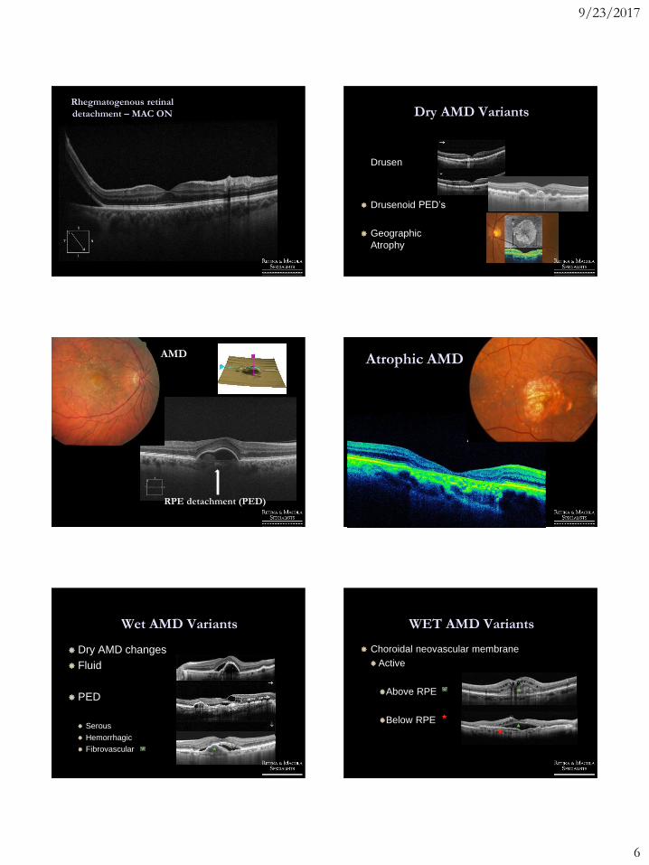
9/23/2017
6
Rhegmatogenous retinal
detachment – MAC ON Dry AMD Variants
Drusen
Drusenoid PED’s
Geographic
Atrophy
AMD
RPE detachment (PED)
Atrophic AMD
Wet AMD Variants
Dry AMD changes
Fluid
PED
Serous
Hemorrhagic
Fibrovascular
WET AMD Variants
Choroidal neovascular membrane
Active
Above RPE
Below RPE
9/23/2017
7
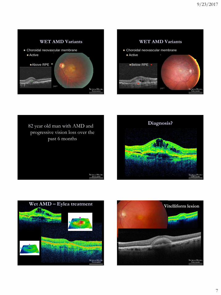
WET AMD Variants
Choroidal neovascular membrane
Active
Above RPE
WET AMD Variants
Choroidal neovascular membrane
Active
Below RPE
82 year old man with AMD and
progressive vision loss over the
past 6 months
Diagnosis?
Wet AMD – Eylea treatment Vitelliform lesion
9/23/2017
8
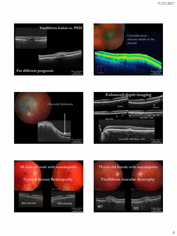
Vitelliform lesion vs. PED
Far different prognosis
Choroidal nevus –
obscures details of the
choroid
Choroidal MelanomaStratus
Cirrus
SpectralisSpectralis - EDI
Enhanced-depth imaging
Cirrus 5000 – 100X Raster - EDI
350 microns 450 microns
Central Serous Retinopathy
69 year old male with maculopathy 74 year old female with maculopathy
467 322
Vitelliform macular dystrophy
9/23/2017
9
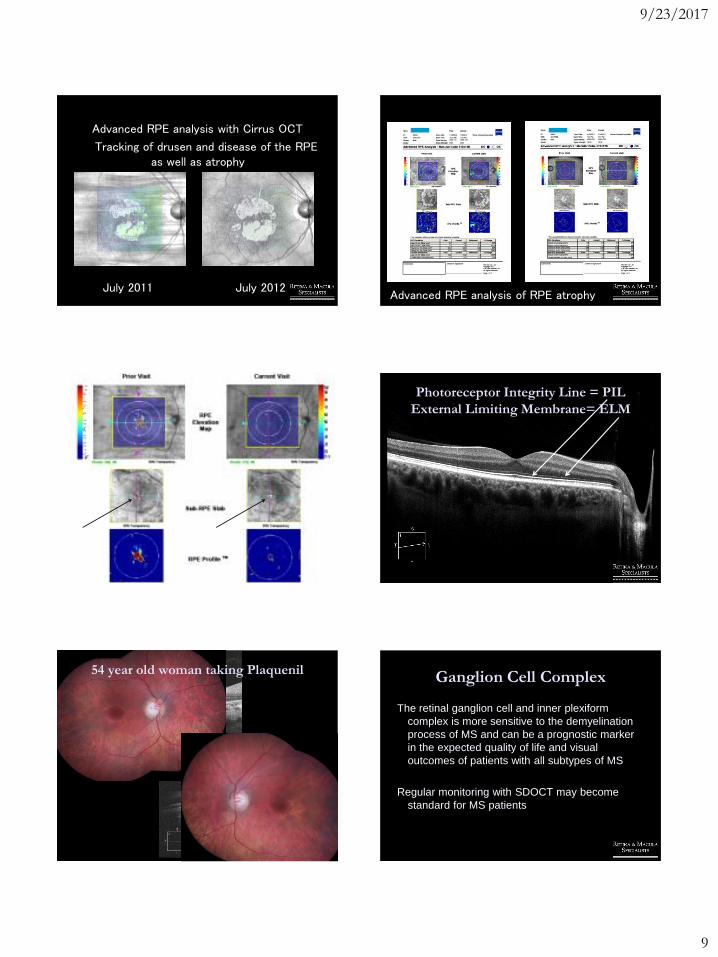
Advanced RPE analysis with Cirrus OCT
Tracking of drusen and disease of the RPE as well as atrophy
July 2011 July 2012Advanced RPE analysis of RPE atrophy
68 year old with AMD and new vision loss OD Photoreceptor Integrity Line = PIL
External Limiting Membrane= ELM
54 year old woman taking PlaquenilGanglion Cell Complex
The retinal ganglion cell and inner plexiform
complex is more sensitive to the demyelination
process of MS and can be a prognostic marker
in the expected quality of life and visual
outcomes of patients with all subtypes of MS
Regular monitoring with SDOCT may become
standard for MS patients

9/23/2017
10
Ganglion Cell Complex
Disease entities (neurogenic) that will affect the
ganglion cell complex will be seen by OD’s and it
is becoming more common to share these
patients with Neurology.
Alzheimers/Dementia Stroke
Multiple Sclerosis Glaucoma
Parkinsons Disease ADHD
Myasthenia Gravis
Ganglion Cell Complex
In my experience these conditions will present with
variable vision loss when the clinical
examination, standard SD OCT scans, visual
fields, pupils etc are intact initially.
When things don’t make sense….
LOOK at the Ganglion Cell Complex
Ganglion Cell Complex
MS Myasthenia Gravis
Ganglion Cell Complex
Parkinson Disease ADHD
What’s Coming
Latest OCT Devices = Fourier Domain Detection
Spectral Domain SD OCT (2006)
70,000 A scans/sec
Swept Source SS OCT (Research only)
250,000 - 400,000 A scans/sec Better Light
Sources
Enhanced Depth Imaging (EDI)
What’s Coming
Video OCT
Measure retinal blood flow
Better software
Quantify GA, RPED, CNVM, CSR.......

9/23/2017
11
Traditional Angiography
versus
OCT angiography
Will this change what we
do?
Traditional Angiography
Images
OCT Angiography (OCTA)
Images
OCT Angiography (OCTA)
Images
AngioPlex OCT
Angiography from ZEISS
• new
• non-invasive
• microvasculature
• imaging
technology
AngioPlex OCT Angiography allows visualization of both perfused
vasculature and vascular abnormalities of the retina without the need
of contrast.US_31_150_0016I_REVB
AngioPlex OCT
Angiography from ZEISS
•AngioPlex Technology detects motion of scattering
particles such as red-blood cells within sequential
OCT B-scans performed repeatedly at the same location of the retina.
B-scan
y0
y2
y3
y
1
y
Fast axis
x
AngioPlex Technology
AngioPlex Maps consist of reconstruction
of the perfused microvasculature within
the retina and choroid.
AngioPlex Maps
US_31_150_0016I_REVB
9/23/2017
12
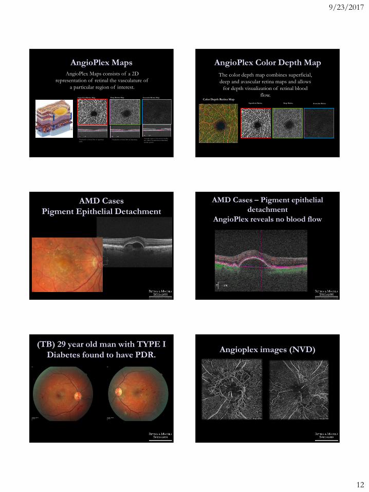
AngioPlex Maps
Superficial Retina Map
Visualization of blood flow in superficial
retina.
Deep Retina Map
Visualization of blood flow in deep retina.
Avascular Retina Map
Avascular region of the retina in healthy
eyes. Allows for detection of abnormal
vascular growth.
AngioPlex Maps consists of a 2D
representation of retinal the vasculature of
a particular region of interest.
1
2
3
AngioPlex Color Depth Map
Superficial Retina Deep Retina Avascular Retina
Color Depth Retina Map
The color depth map combines superficial,
deep and avascular retina maps and allows
for depth visualization of retinal blood
flow.
AMD Cases
Pigment Epithelial Detachment
AMD Cases – Pigment epithelial
detachment
AngioPlex reveals no blood flow
(TB) 29 year old man with TYPE I
Diabetes found to have PDR.Angioplex images (NVD)

9/23/2017
13
AngioPlex versus FA imaging Diabetic MA’s and Ischemia
Not appreciated clinically
Microaneurysm lesions in Diabetes Diabetic MA’s and Ischemia
Not appreciated clinically
PED with occult CNV - Neovascular AMD Neovascular AMD
