objectives: 1. to ensure good section cutting and frozen section. 2. to overcome troubleshooters...
TRANSCRIPT

Lab 3: Section Cutting and
Frozen Section

Objectives :
1. To ensure good section cutting and frozen section.
2. To overcome troubleshooters during section
cutting and frozen section.
3. To familiarize the staff with the equipment used
for section cutting and frozen section.

Overview:
Tissues are sectioned using a microtome.
Turn on the water bath and check that the temp is 35-37ºC.
Use fresh deionized water (DEPC treated water must be used if in situ hybridization will be performed on the sections).
Blocks to be sectioned.

Place a fresh blade on the microtome; blades may be used
to section up to 10 blocks, but replace if sectioning becomes
problematic.
Insert the block into the microtome chuck so the wax block
faces the blade and is aligned in the vertical plane.
Cont.

Face the block by cutting it down to the desired tissue plane and
discard the paraffin ribbon.
If the block is ribboning well then cut another four sections and pick
them up with forceps or a fine paint brush and float them on the surface
of the 37ºC water bath.
Float the sections onto the surface of clean glass slides.
If the block is not ribboning well then place it back on the ice block to
cool off firm up the wax.
If the specimens fragment when placed on the water bath then it may
be too hot.
Cont…
Place the slides with paraffin sections on the
warming block in a 65°C oven for 20 minutes
(so the wax just starts to melt) to bond the tissue
to the glass.
Slides can be stored overnight at room
temperature.

Devices for cutting sections
Microtome
VibratomeCryostat

Cont…
Most sectioning in routine histopathology
departments is done with a microtome producing
sections of ~3μm thickness, from tissue that has been
embedded in wax.
A number of devices are available for cutting
sections.

Microtomes
These are mechanical devices for cutting uniform
sections of tissue of appropriate thickness.
Ultramicrotome >>>> 50-100nm

Types of microtome
1. Hand microtomes
2. Rocking microtome.
3. Rotary microtome.
4. Freezing microtome.

Cont.
5. Base sledge microtome.
6. Vibrating knife microtome.
7. Sliding microtome.

Rotary microtome
Designed for cutting celloidin-embedded tissue
blocks.
The Knife or blade is stationary, specimen slides
under it during sectioning.
Also used for paraffin-wax embedded sections.

Section adhesives
An adhesive is a substance which can be smeared on
to the slides so that the sections stick well to the slides.
Most of the tissue sections which are adequately thin
and thoroughly dried without any air bubble trapped
under them do not require an adhesive, as in case of
routine H and E staining.

Cont…
But for histochemical methods requiring alkaline solutions, e.g.
ammonia tend to remove sections from slide for such cases
adhesive is required.
Also adhesive is required for tissues like brain, spinal cord,
blood clot, decalcified tissues which have a tendency to detach
themselves from the slide.
Tissue impregnated with ester wax also requires section
adhesive.
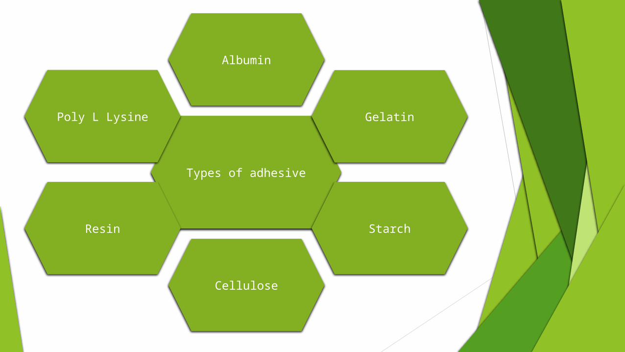
Types of adhesive
Albumin
Gelatin
Starch
Cellulose
Resin
Poly L Lysine

Common Problems with Sectioning Cutting Problems:
1. Cut on an angle:
Angled cuts can be identified in the following ways: The section or cells within it are oval in shape.
One can focus through several cell layers in one
area of the section.
Part of the section appears to be “smeared”.

Cont…
2. Cut too thin: This problem can be identified if a section
has a part missing, and/or it is not completely round.
3. Cut too thick: This problem can be identified if
boundaries within the section appear exceptionally thick, or
if you are able to focus through several cell layers across the
whole section.
Cont…
4. Sections cut in half: The problem here is that there are
too many sections in the drop of water, and convection is
bringing them back towards the razor where they are
being cut in half.
Frozen Section
A thin slice of tissue that is cut from a frozen specimen and
is often used for rapid microscopic diagnosis section and a
histologic section of tissue that has been frozen by exposure
to dry ice.
The frozen section procedure is a pathological laboratory
procedure to perform rapid microscopic analysis of a
specimen. It is used most often in on cological surgery.
Cont…
The technical name for this procedure is cryosection.
The quality of the slides produced by frozen section is
of lower quality than formalin fixed, wax embedded
tissue processing.
While diagnosis can be rendered in many cases, fixed
tissue processing is preferred in many conditions for
more accurate diagnosis.
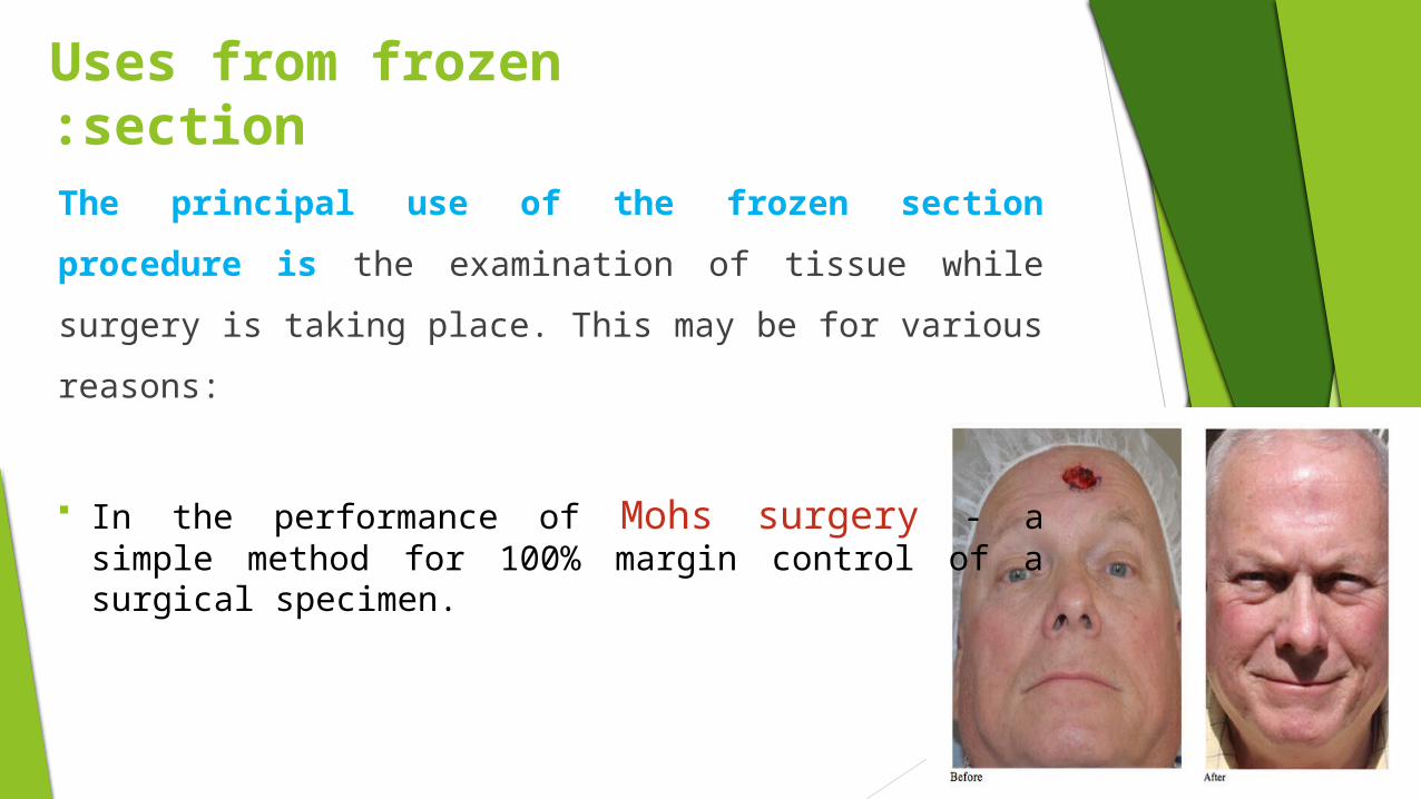
Uses from frozen section:
The principal use of the frozen section procedure is the
examination of tissue while surgery is taking place. This
may be for various reasons:
In the performance of Mohs surgery - a simple method for 100% margin control of a surgical specimen.
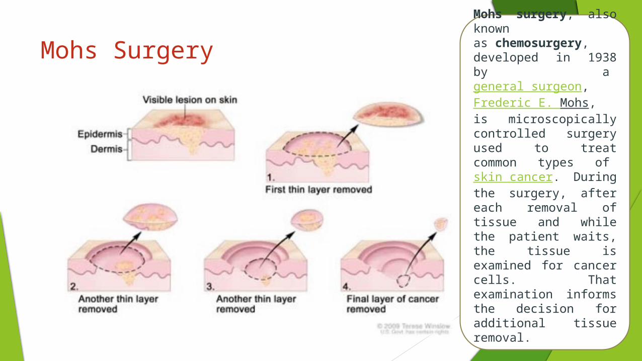
Mohs Surgery Mohs surgery, also known as chemosurgery, developed in 1938 by a general surgeon, Frederic E. Mohs, is microscopically controlled surgery used to treat common types of skin cancer. During the surgery, after each removal of tissue and while the patient waits, the tissue is examined for cancer cells. That examination informs the decision for additional tissue removal.

Cont. If a tumor appears to have metastasized, a
sample of the suspected metastasis is sent for cryo section to confirm its identity.
If the tumor has metastasized, surgery is usually not curative, and the surgeon will choose a more conservative surgery, or no resection at all.
Cont…
If a tumor has been resected but it is unclear whether the
surgical margin is free of tumor, an intraoperative
consultation is requested to assess the need to make a
further resection for clear margins.
In a sentinel node procedure, a sentinel node containing
tumor tissue prompts a further lymph node dissection,
while a benign node will avoid such a procedure.

Cont.
If surgery is explorative, rapid examination of a lesion
might help identify the possible cause of a patient's
symptoms.
Rarely, cryo sections are used to detect the presence of
substances lost in the traditional histology technique, for
example lipids. They can also be used to detect some
antigens masked by formalin.
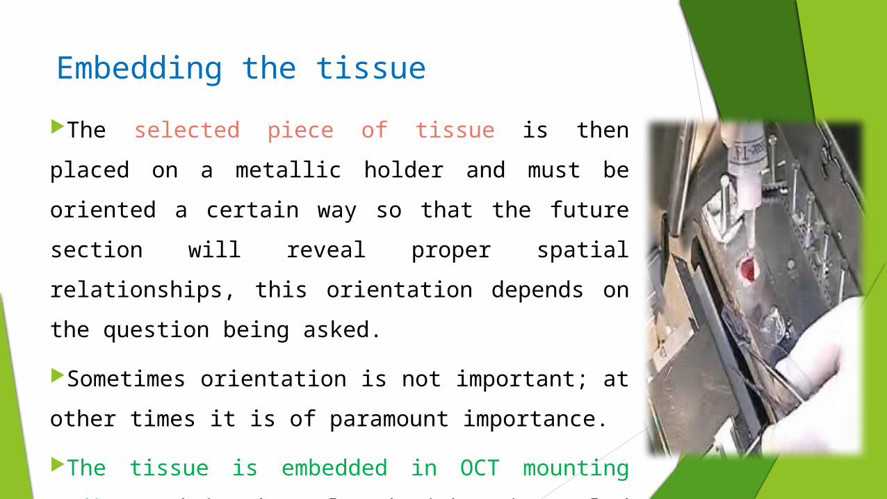
Embedding the tissue
The selected piece of tissue is then placed on a metallic holder
and must be oriented a certain way so that the future section will
reveal proper spatial relationships, this orientation depends on
the question being asked.
Sometimes orientation is not important; at other times it is of
paramount importance.
The tissue is embedded in OCT mounting medium and is then
placed either in cooled 2-methyl butane or the cryostat machine
where it is properly frozen.

Cryostat: The machine, which cuts the tissue, is the
cryostat. Certain things should be routinely checked in the
operation of this machine:
a) Temperature: The temperature should be at -20°F for
most tissues.
For tissues with a large fat component, -40°F is optimal.
This temperature is critical for optimal sectioning.

Cont…
Too high, i.e., -10°F and the tissue will not
stay frozen and firm and will not cut crisp.
Too cold, i.e., -50°F and the tissue will
crumble and become powder. The Ideal tissue
should cut like butter, smooth and in one piece.
b) Blade sharpness and angle: The blade should be sharp and should
be changed approximately once every 2 weeks.
A dull blade cuts dull.
Equally important is the blade angle. There is an optimal angle between
blade and tissue:
Too steep an angle and the tissue will crumble like it was too cold.
Too shallow, then two things will happen. The section will
alternately skip and not cut and then it will cut, but too thick.

Methodology

GOOD LUCK