an audit of frozen section consultations in a tertiary
TRANSCRIPT

IP Journal of Diagnostic Pathology and Oncology 4 (2019) 193–199
Content available at: iponlinejournal.com
IP Journal of Diagnostic Pathology and Oncology
Journal homepage: www.innovativepublication.com
Original Research Article
An audit of frozen section consultations in a tertiary care center
Nithin Diwagar1, Ganthimathy Sekhar1,*1Dept. of Pathology, Saveetha Medical College, Chennai, Tamil Nadu, India
A R T I C L E I N F O
Article history:Received 01-07-2019Accepted 17-09-2019Available online 20-09-2019
Keywords:AuditAccuracy rateDiscordance rateFrozen section
A B S T R A C T
Frozen section technique is a valuable tool used to rapidly prepare slides from tissue for microscopicinterpretation, in surgical pathology; frozen sections are routinely used for rapid intra-operative diagnosis,providing guidance for the surgeons. An audit of this vital procedure helps to identify the efficiency,diagnostic error rates, and cause of the error and also preventive measures which can be taken to avoidthose errors. Ours was a retrospective study undertaken at Saveetha medical college, Chennai. In our studyout of the 160 cases, 154 cases were concordant with the final histopathological diagnosis and 6 cases werediscordant, which gives an accuracy rate of 96% and a discordance rate of 4%. Discordance was primarilydue to interpretation errors and errors due to limited sampling during frozen section. Knowledge of thepitfalls and causes of diagnostic errors are important in improving the accuracy of this process. Regular,periodic audits will go a long way in addressing this issue.
© 2019 Published by Innovative Publication.
1. Introduction
Intraoperative consultations in the form of frozen sectionplay a vital role in the surgical management by avoidingextensive or second surgeries. The surgeon’s appropriatemanagement and decision taking on the operating tabledepends on a frozen report that is as accurate as possible.Therefore it is critical to determine efficiency and audit thefrozen section performance periodically. Audit also servesas an internal quality control, which will improve the qualityof the service provided to the patient
2. Materials and Methods
This was a retrospective study undertaken at Saveethamedical college, Chennai. Complete enumerative samplingmethod was used. The records of 160 frozen sections andthe corresponding final formalin fixed paraffin embedded(FFPE) histopathology reports during the study period of 2years (2016–2018) were compared and analyzed.
In all the cases the frozen section analysis had beendone on fresh tissue samples. After gross examination,
* Corresponding author.E-mail address: [email protected] (G. Sekhar).
the appropriate numbers of bits were taken depending onthe nature of the tissue followed by snap freezing andsectioning of tissues using cryostat (Model: LEICA CM1520). 4 microns thin sections were prepared, stained andre viewed bysenior experienced pathologist and the reportswere conveyed to the operating surgeon. The time takenfrom receiving the specimen to conveying the diagnosisto the surgeon was noted in all cases. The remaininggross specimen and the frozen bits were fixed overnightin 10% neutral buffered formalin and processed routinely.The routine histopathology slides were reviewed by seniorexperienced pathologists and the final report was given.
The reports of the frozen section and FFPE tissuesections were compared and the results were categorizedinto concordant, discordant. The slides of the discordantcases were reviewed and the causes of discrepancywerenoted. The diagnostic accuracy and diagnostic errorrates were calculated.
3. Results
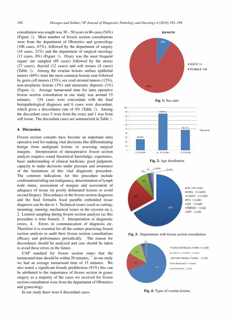
Out of 160 cases analyzed 146(91%) were from femalepatients and 14(9%) were from male patients (Figure 1).The most frequent age group in which frozen section
https://doi.org/10.18231/j.jdpo.2019.0412581-3714/© 2019 Published by Innovative Publication. 193
194 Diwagar and Sekhar / IP Journal of Diagnostic Pathology and Oncology 4 (2019) 193–199
consultation was sought was 30 – 50 years in 86 cases (54%)(Figure 2). Most number of frozen section consultationswere from the department of Obstetrics and gynecology(106 cases, 67%), followed by the department of surgery(34 cases, 21%) and the department of surgical oncology(13 cases, 8%) (Figure 3). Ovary was the most frequentorgan/ site sampled (89 cases) followed by the uterus(27 cases), thyroid (12 cases) and soft tissues (4 cases)(Table 1). Among the ovarian lesions surface epithelialtumors (69%) were the most common lesions sent followedby germ cell tumors (15%), sex cord stromal tumors (12%),non-neoplastic lesions (3%) and metastatic deposits (1%)(Figure 4). Average turnaround time for intra operativefrozen section consultation in our study was around 15minutes. 154 cases were concordant with the finalhistopathological diagnosis and 6 cases were discordant,which gives a discordance rate of 4% (Table 2). Amongthe discordant cases 5 were from the ovary and 1 was fromsoft tissue. The discordant cases are summarized in Table 3.
4. Discussion
Frozen section consults have become an important intraoperative tool for making vital decisions like differentiatingbenign from malignant lesions or assessing surgicalmargins. Interpretation of intraoperative frozen sectionanalysis requires sound theoretical knowledge, experience,basic understanding of clinical medicine, good judgment,capacity to make decisions under pressure and awarenessof the limitations of this vital diagnostic procedure.The common indications for this procedure includeconfirmation/ruling out malignancy, determination of lymphnode status, assessment of margins and assessment ofadequacy of tissue (in poorly delineated lesions to avoidsecond biopsy). Discordance in the frozen section diagnosisand the final formalin fixed paraffin embedded tissuediagnosis can be due to 1. Technical issues (such as cutting,mounting, staining, mechanical issues in the cryostat etc.),2. Limited sampling during frozen section analysis (as thisprocedure is time bound), 3. Interpretation or diagnosticerrors, 4. Errors in communication of diagnosis etc.Therefore it is essential for all the centers practicing frozensection analysis to audit their frozen section consultationsefficacy and performance periodically. The reason fordiscordance should be analyzed and care should be takento avoid these errors in the future.
CAP standard for frozen section states that theturnaround time should be within 20 minutes,
1,2in our study
we had an average turnaround time of 15 minutes. Wealso noted a significant female predilection (91%) this canbe attributed to the importance of frozen section in gynecsurgery as a majority of the cases we received for frozensection consultation were from the department of Obstetricsand gynecology.
In our study there were 6 discordant cases.
Fig. 1: Sex ratio
Fig. 2: Age distribution
Fig. 3: Departments with frozen section consultation
Fig. 4: Types of ovarian lesions

Diwagar and Sekhar / IP Journal of Diagnostic Pathology and Oncology 4 (2019) 193–199 195
Table 1: Site/organ sampled for frozen section
Site No. of cases % of cases No. of discordant cases Organ wiseaccuracy
OVARY 89 56% 5 94.4%UTERUS 27 17% 0 100%
THYROID 12 8% 0 100%SOFT TISSUE 4 4% 1 75%
CERVICAL NODE 3 2% 0 100%SALIVARY GLAND 3 2% 0 100%INGUINAL NODE 2 1% 0 100%GALLBLADDER 2 1% 0 100%
INTESTINE 2 1% 0 100%CNS 2 1% 0 100%
TONGUE 2 1% 0 100%FALLOPIAN TUBE 1 0.50% 0 100%
BREAST 1 0.50% 0 100%FALCIFORM LIGAMENT 1 0.50% 0 100%
KIDNEY 1 0.50% 0 100%PERIPANCREATIC NODE 1 0.50% 0 100%
OMENTUM 1 0.50% 0 100%PANCREAS 1 0.50% 0 100%
PARATHYROID 1 0.50% 0 100%PERIGASTRIC NODE 1 0.50% 0 100%
SACRAL MASS 1 0.50% 0 100%SKIN 1 0.50% 0 100%
STOMACH 1 0.50% 0 100%TOTAL 160 100% 6 96%
Table 2: Discordance rates
Total No. of Cases 160No of discordant cases 6% Of concordant cases 96%% Of discordant cases 4%
Table 3: Comparison of discordance rates with other studies
Authors Study period (Year) Number of cases Concordance rate Discordance Rate%Patil P et al 3 2 100 96.9 3.1Roy S et al 4 9 months 327 97.6 2.4
Shreshtha S et al 5 5 404 94.6 5.4Chbani et al 6 1 261 95 5
A. SathishSelvakumar etal 7
5 132 94.7 5.3
Present study 2 160 96 4
Table 4: Discordant cases
No Site Frozen diagnosis Benign/malignant HPE diagnosis Benign/malignant1 Ovary Mucinous cystadenoma Benign Benign cystic
teratomaBenign
2 Ovary Borderline mucinous tumor Borderline Mucinouscarcinoma
Malignant
3 Ovary Serous cystadenofibroma Benign Serous borderlinetumor
Borderline
4 Ovary Borderline serous tumor Borderline Serouscystadenofibroma
Benign
5 Ovary Sertoli leydig cell tumor Malignant High gradeserous
Malignant
6 Soft tissue Benign spindle cell lesion Benign Leiomyosarcoma Malignant

196 Diwagar and Sekhar / IP Journal of Diagnostic Pathology and Oncology 4 (2019) 193–199
Fig. 5: [ Case 1 ] A – Frozen section, H&E, 40x: ovarian cyst wall lined by mucin ous epithelium – Mucinous cystadenoma.. B –FFPE section, H&E, 40x : Foci of squamous epithelium – Benign cystic teratoma. [Case 2]C - Frozen section, H&E, 10x : F eatures ofborderline mucinous tumor. D - FFPE section H&E, 10x : Evidence of invasion was noted – Mucinous carcinoma. [Case 3] E - Frozensection, H&E, 1 0x : Shows predominant areas of congestion and hemorrhage – Serous cystadenofibroma. F - FFPE section H&E, 10x :Tiny viable area showing features of borderline serous tumor.

Diwagar and Sekhar / IP Journal of Diagnostic Pathology and Oncology 4 (2019) 193–199 197
Fig. 6: [Case 4]A – Frozen section, H&E, 10x Cyst wall lined by cuboidal to columnar cells with areas of stratification and mild atypia –Borderline serous tumor. B – FFPE section, H&E, 1 0x: fibrous cyst wall lined by cuboidal to low columnar ciliated epithelium - Serouscystadenofibroma. [Case 5]C - Frozen section, H&E, 10x : sheets and cords of neoplastic cells with abundant eosinophilic cytoplasm andpale nuclei resembling leydig cells – sertoli-leydig cell tumor. D - FFPE section H&E, 10x : Features of high grade serous carcinoma.[Case6] E - Frozen section, H&E, 40x : fascicles and interlacing bundles of spindle cells with mild nucleomegaly and no evidence ofmitosis, pleomorphism and necrosis – Benign spindle cell lesion. F - FFPE section H&E, 40x : Mitosis and nuclear pleomorphism isevident – Malignant spindle cell lesion.

198 Diwagar and Sekhar / IP Journal of Diagnostic Pathology and Oncology 4 (2019) 193–199
5. Case 1
Reported as mucinous cystadenoma in frozen section butturned out to be a benign cystic teratoma in final FFPEtissue report. The reason for this discordance was dueto limited sampling of the gross specimen during frozensection analysis. Upon extensive sampling in formalin fixedtissue there were focal areas of neural tissue, squamousepithelium and smooth muscle bundles which prompted usto change the diagnosis.
6. Case 2
Reported asborderline mucinous tumor in frozen section butwas signed out to be a mucinous carcinoma in final FFPEtissue report. This discordance was also due to limitedsampling in frozen sections. Subsequent extensive samplingin formalin fixed tissue revealed areas of invasion.
7. Case 3
Reported as serous cystadenofibroma in frozen section butturned out to be a borderline serous tumor in final FFPEtissue report. Grossly the specimen of ovary receivedfor frozen section analysis underwent torsion and washemorrhagic, Microscopy showed predominant areas ofcongestion and hemorrhage, and one area showed cyst linedby single layer of cuboidal epithelium. Upon extensivesampling in formalin fixed tissue there was a tiny viable areashowing features of borderline serous tumor.
8. Case 4
Reported as serous borderline tumor in frozen section butturned out to be a serous cystadenofibroma in final FFPEtissue report. Discordance was probably because the frozensection diagnosis was based on the gross findings such asnumerous papillary excrescences which were reminiscentof borderline serous tumor; microscopy showed areas ofstratification with mild atypia. Moreover CA 125 wasmildly elevated. Tangential sectioning artifact of the frozensections caused pseudo stratification of cells. Ice crystalartifacts and poor morphology of the cells also contributedto the discordance.
9. Case 5
Reported as sertoli-leydig cell tumor in frozen section butturned out to be a high grade serous carcinoma in final FFPEtissue report. Frozen section slides showed sheets and cordsof neoplastic cells with abundant eosinophilic cytoplasmand pale nuclei resembling leydig cells. Clinically too,the patient had some features of virilisation. All thesefinding prompted the diagnosis of sertoli-leydig cell tumor.Discordance was due to poor morphology of the cells infrozen sections which led to an interpretation error.
10. Case 6
Reported as Benign spindle cell lesion in frozen section butturned out to be a malignant spindle cell tumor in the finalFFPE tissue report. Features which are vital to make adiagnosis of malignant spindle cell lesion such as mitosis,nuclear pleomorphism, and necrosis were not appreciatedin the frozen sections. Here too, the discordance was dueto poor morphology in frozen sections which led to aninterpretation error.
In our study discordance was primarily due tointerpretation errors and errors due to limited samplingduring frozen section. Interpretation error was alsoattributed to sectioning artifacts (freezing and folding),lack/poor quality of morphological details and presence ofthe diagnostic features in only in the deeper sections. Theaccuracy rate of frozen section analysis reported in theliterature ranges from 92% to 97.98 %.
5,8,9Our study had
an accuracy rate of 96% which was comparable with otherstudies.
4–6,10
11. Conclusion
Frozen sections are invaluable in directing the course ofpatient management during surgeries. However knowledgeof the pitfalls and causes of diagnostic errors are importantin improving the accuracy of this process. Regular, periodicaudits will go a long way in addressing this issue.
12. Source of Funding
None.
13. Conflict of Interest
None.
References1. Vimal M. A study on accuracy of frozen section diagnosis and
turnaround time. Int J Health Sci Res. 2015;5(12):138–42.2. Hatami H, Mohsenifar ZH, Alavi SN. The Diagnostic Accuracy of
Frozen Section Compared to Permanent Section: A Single CenterStudy in Iran. Iran J Pathol. 2015;10(4):295–9.
3. Patil P, Shukla S, Bhake A. Hiwale K Accuracy of frozen sectionanalysis in correlation with surgical pathology diagnosis. Int J ResMed Sci. 2015;3:399–404.
4. Somak R, Anil VP, Rajiv D, Samuel AY, Susan MK, et al. FrozenSection Diagnosis: Is There Discordance Between What PathologistsSay and What Surgeons Hear? Am J Clin Pathol. 2013;140(3):363–9.
5. Shrestha S, Basyal R, Pathak TSS, Lee M, Dhakal H, et al.Comparatve Study of Frozen Secton Diagnoses with Histopathology.Post Graduate Med NAMS. 2009;9(2):1–5.
6. Chbani L, Mohamed S, Harmouch T, Fatemi H, Amarti A. Qualityassessment of intraoperative frozen sections: An analysis of 261consecutive cases in a resource limited area: Morocco. ; 2012,.
7. Selvakumar AS, Rajalakshmi V, Sundaram KM. Intraoperative frozensection consultation- an audit in a tertiary care hospital. Ind JPatholOncol. 2018;5(3):421–8.
8. Farah-Klibi F, Neji O, Ferjaoui M, Zaouche A, Koubaa A, et al.Accuracy of frozen section diagnosis: an analysis of 1695 consecutive

Diwagar and Sekhar / IP Journal of Diagnostic Pathology and Oncology 4 (2019) 193–199 199
cases. Tunis Med. 2008;86(7):693–7.9. Preeti A, Sameer G, Kulranjan S, Abhinav SA, Preeti R, et al. Intra-
Operative Frozen Sections: Experience at A Tertiary Care Centre.Asian Pac J Cancer Pre. 2016;17(12):5057–61.
10. Mishra S, Gupta M, Bharat V, Bansal R. Qualitative ComparativeStudy of Frozen Section with Routine Histological Technique.National Journal of Laboratory Medicine. 2016;2:44–50.
Author biography
Nithin Diwagar Post Graduate
Ganthimathy Sekhar Professor
Cite this article: Diwagar N, Sekhar G. An audit of frozen sectionconsultations in a tertiary care center. J Diagn Pathol Oncol2019;4(3):193-199.