nysora - the new york school of regional anesthesia...
TRANSCRIPT

10/28/10 11:09 PMNYSORA - The New York School of Regional Anesthesia - Pediatric Epidural and Caudal Analgesia and Anesthesia in Children
Page 1 of 37http://www.nysora.com/regional_anesthesia/sub-specialties/pediatric_ane…atric_epidural_and_caudal_analgesia_and_anesthesia_in_childr.html?print
2009-03-14 16:04:00
Copyright NYSORA © 1996, 2008 NYSORA.com
Loading Image...
Pediatric Epidural and Caudal Analgesiaand Anesthesia in Children
Epiduralanalgesiahas manybeneficialeffects inthepediatricpatient
population. In clinical practice, it is commonly used to augment generalanesthesia and to manage postoperative pain.
TABLE OF CONTENTS (click here to expand)
Epidural Blockade For Pediatric Surgery (General Aspects)IntroductionAnatomical ConsiderationsConsiderations For Choosing Local Anesthetic Solution ForEpidural And Caudal Anesthesia And AnalgesiaSelection Of Epidural Local Anesthetic SolutionsAdjuvants To Local Anesthetics Solutions
10/28/10 11:09 PMNYSORA - The New York School of Regional Anesthesia - Pediatric Epidural and Caudal Analgesia and Anesthesia in Children
Page 2 of 37http://www.nysora.com/regional_anesthesia/sub-specialties/pediatric_ane…atric_epidural_and_caudal_analgesia_and_anesthesia_in_childr.html?print
Complications Associated With Epidural And CaudalAnalgesia
Epidural Block TechniqueIntroductionConfirmation Of Proper Epidural Needle/Catheter PlacementEpidural ECG TechniqueContinuous Caudal Epidural To Lumbar Or Thoracic SpaceLumbar Epidural AnesthesiaThoracic Epidural Analgesia
Managing Epidural Infusions Postoperatively
Epidural Blockade for pediatric surgery(General Aspects)
Introduction
Epidural analgesia has many beneficial effects in the pediatric patientpopulation. In clinical practice, it is commonly used to augment generalanesthesia and to manage postoperative pain. Effective postoperative painrelief from epidural analgesia has numerous benefits including earlierambulation, rapid weaning from ventilators, reduced time spent in acatabolic state and lowered circulating stress hormone levels.1 Preciseplacement of epidural needles and catheters for single-shot and continuousepidural anesthesia ensures the dermatomes involved in the surgicalprocedure are selectively blocked, allowing for lower doses of localanesthetics and sparing of unnecessary blockade in the regions whereblockade is not desired. 2-4.
Anatomical Considerations
Significant anatomic differences in comparison with adults, should beconsidered while utilizing regional anesthesia in children. For instance, inneonates and infants, the conus medullaris is located lower in the spinalcolumn (at approximately the L3 vertebra) compared to adults where it issituated at approximately the L1 vertebra. This dissimilarity is a result ofdifferent rates of growth between the spinal cord and the bony vertebral
10/28/10 11:09 PMNYSORA - The New York School of Regional Anesthesia - Pediatric Epidural and Caudal Analgesia and Anesthesia in Children
Page 3 of 37http://www.nysora.com/regional_anesthesia/sub-specialties/pediatric_ane…atric_epidural_and_caudal_analgesia_and_anesthesia_in_childr.html?print
column in infants. However, at approximately 1 year of age the conusmedullaris reaches similar L1 level as in an adult. The sacrum of children isalso more narrow and flat compared to the adult population. At birth, thesacral plate, which is formed by five sacral vertebrae, is not completelyossified and continues to fuse until approximately 8 years of age. Theincomplete fusion of the sacral vertebral arch forms the sacral hiatus. Thecaudal epidural space can be accessed easily in infants and children throughthe sacral hiatus. Due to the continuous development of the sacral canalroof, there is considerable variation in the sacral hiatus. In children, thesacral hiatus is located more cephalad compared to adults. Therefore,caution is warranted when placing caudal blocks in infants as the dura mayend more caudad thereby increasing the risk of accidental dural puncture. Ithas also been suggested that the epidural fat is less densely packed inchildren than in adults.5 This loosely packed epidural fat may facilitate notonly the spread of local anesthestic, but it may also allow the unimpededadvancement of epidural catheters from the caudal epidural space to thelumbar and thoracic level.
Clinical Pearls
In the neonate the intercristal line bisects L5 (cf L4 or L3/4interspace in the adult) and the spinal cord ends at L3 infirst year of life (cf L1 in the adult).As a general rule the epidural space will be found at 1mm/kg of body weight, however, there is considerableindividual variation.
Considerations for Choosing Local Anesthetic Solution forEpidural and Caudal Anesthesia and Analgesia
Newer local anesthetics with favorable potencies, durations of effect anddecreased toxicity profiles have been introduced in the past decade. Localanesthetic concentration and volume are important factors in determiningthe density and level of blockade. Since most pediatric patients receiveepidural analgesia in conjunction with a general anesthetic, the mainpurpose of the epidural catheter is to deliver sufficient local anestheticsolution for effective intraoperative and postoperative analgesia. Knowledgeof total drug dose is important to avoid local anesthetic toxicity, particularlyin pediatric patients.
Clinical Pearls

10/28/10 11:09 PMNYSORA - The New York School of Regional Anesthesia - Pediatric Epidural and Caudal Analgesia and Anesthesia in Children
Page 4 of 37http://www.nysora.com/regional_anesthesia/sub-specialties/pediatric_ane…atric_epidural_and_caudal_analgesia_and_anesthesia_in_childr.html?print
High concentrations of local anesthetics such as 0.5%bupivacaine or 0.5% ropivacaine are rarely used in pediatricpopulationInstead, larger volumes of more dilute local anesthetic aremore commonly used to cover multiple dermatomes.
A more detailed description of local anesthetics solutions, theircharacteristics and toxic potential has been described elsewhere in this text.As a general rule, however, high concentrations of local anesthetics such as0.5% bupivacaine or 0.5% ropivacaine are seldom used in pediatricpopulation particularly in the epidural space. Instead, larger volumes ofmore dilute local anesthetic are more commonly used to cover multipledermatomes. Opioids prolong the duration of analgesia of local anesthetic,but have also been associated with unacceptable side effects, particularly inpediatric outpatients. Various non-opioid adjuncts like clonidine and alpha-2 agonist offer more favorable side effect profiles; however relatively littleinformation is available regarding their use pediatric patients.
Selection of epidural local anesthetic solutions
Clinical Pearls
In pediatric population, body weight is a better correlatethan patient age in predicting spread of local anestheticfollowing a caudal block.For caudal use, the optimum concentration of bupivacaineis 0.125-0.175%.The maximal safe dose of bupivacaine is 2.5 mg/kg. to 4mg/kgFor continuous epidural infusion, bupivacaine 0.2 mg/kg/hfor neonates and 0.4 mg/kg/h for older children is oftenused.For a single-shot caudal block, a bolus of 1 ml/kg of 0.2%ropivacaine is recommended.A continuous infusion of 0.2 mg/kg/hr of 0.1% ropivacainein infants and 0.4 mg/kg per hour in older children for 48hrs, has been shown to be effective and safe regimen
Bupivacaine and ropivacaine are the two most commonly used localanesthetics for neuraxial anesthesia in children. Lidocaine is not often used

10/28/10 11:09 PMNYSORA - The New York School of Regional Anesthesia - Pediatric Epidural and Caudal Analgesia and Anesthesia in Children
Page 5 of 37http://www.nysora.com/regional_anesthesia/sub-specialties/pediatric_ane…atric_epidural_and_caudal_analgesia_and_anesthesia_in_childr.html?print
because of its short duration of action and excessive motor block. Bodyweight is usually a better correlate than patient age in predicting spread oflocal anesthetic following a caudal block.58 The maximal safe dose ofbupivacaine is 2.5 mg/kg. to 4 mg/kg59 For caudal use, the optimumconcentration of bupivacaine is 0.125-0.175%.60 Compared with the 0.25%preparation, this concentration provides a similar duration of postoperativeanalgesia (4 to 8 hours) but with less motor blockade.60 Some cliniciansprefer administering doses on a volume per weight basis. A dose of 1.0mL/kg of a dilute solution such as 0.125% bupivacaine to a maximumvolume of 30 mL can reliably provide T10 sensory block without exceedingmaximum levels recommended in the literature.6 Higher doses such as 1.25mL/kg, or even 1.5 mL/kg, may be administered to provide a more cephaladblock without the risk of local anesthetic toxicity.6 For continuous epiduralinfusion, a commonly accepted dosage guideline of bupivacaine is 0.2mg/kg/h for neonates and 0.4 mg/kg/h for older children.7 Cumulativetoxicity is a concern even at lower rates of local anesthetic solutioninfusions. 3 The alternate use of 2-chloroprocaine may be well tolerated byneonates.61
Newer local anesthetic agents include the levo-entiomers ropivacaine andlevobupivacaine. Ropivacaine has a higher therapeutic index than the olderlocal anesthetic bupivacaine.62-65 At low concentrations, ropivacaine mayproduce less motor block and comparable analgesia when compared tobupivacaine with decreased incidence of cardiac and central nervous systemtoxicity.6 Due to its possible vasoconstricting properties, ropivacaine mayundergo slower systemic absorption than bupivacaine.66,67 This may haveclinical implications when a prolonged local anesthetic infusion is used inchildren with impaired hepatic function.68 For a single-shot caudal block, abolus of 1 ml/kg of 0.2% ropivacaine is recommended.69,70 An infusion of0.2 mg/kg/hr of 0.1% ropivacaine in infants and 0.4 mg/kg per hour inolder children lasting no longer than 48 hrs, has also been shown to beeffective and safe.70
Levobupivacaine, the S (-)-isomer of bupivacaine, is less likely to causemyocardial depression and fatal arrhythmias and is also less toxic to thecentral nervous system than racemic bupivacaine. A dose of 0.8 ml/kg of0.25% levobupivacine injected caudally provides analgesia in childrenhaving penile or groin surgery.71 For continuous epidural infusions, thedose for levobupivacaine is similar to racemic bupivacaine.7
Adjuvants to local anesthetics solutions

10/28/10 11:09 PMNYSORA - The New York School of Regional Anesthesia - Pediatric Epidural and Caudal Analgesia and Anesthesia in Children
Page 6 of 37http://www.nysora.com/regional_anesthesia/sub-specialties/pediatric_ane…atric_epidural_and_caudal_analgesia_and_anesthesia_in_childr.html?print
Adjuvants may be used to prolong the duration of blockade, particularly forsingle-shot caudal epidural blocks.72 Single-shot caudal block is mainlyused for ambulatory surgery. The major problem associated with thistechnique is the limited duration of analgesia and unwanted motorblockade. Recent research has focused on trying to resolve these problemswith the addition of various adjuvants.
(i) Epinephrine: The most commonly used adjuvant for single-shot caudalanesthesia is epinephrine in a concentration of 1:200,000. Epinephrine hasthe added benefit of serving as a marker for an inadvertent intravascularinjection.
(ii) Opioids: Epidural opioids may enhance and prolong analgesia.However, opioid use in an ambulatory setting may not be advisable due tothe potential for respiratory depression and other unfavorable side effects(e.g. nausea and vomiting, itching, urinary retention).59 As a result, the useof caudal epidural opioids in children should be restricted to special clinicalsituations.73-75 Fentanyl has been used with desirable effects for epiduralanalgesia in adults for a number of years. Whether there is benefit forfentanyl as an additive in children undergoing single-shot caudal blockadeis still debated amongst clinicians.76,77 One study found an increasedincidence of nausea and vomiting when fentanyl was added to the localanesthetic solution for a single-shot caudal block.77 A dose of 2 µg/kg offentanyl for single-shot caudal anesthesia along with the standard localanesthetic solution has been recommended for more extensive or painfulprocedures or in patients who have a urinary catheter in the postoperativeperiod. The addition of 1 µg/mL to 2 µg/mL of fentanyl to 0.1% bupivacainefor continuous epidural infusions has also been used with success inneonates and children in a well monitored inpatient setting.15
(iii) Clonidine: Clonidine, an alpha-2 agonist, acts by stimulatingdescending noradrenergic medullo-spinal pathways which inhibits therelease of nociceptive neurotransmitters in the dorsal horn of the spinalcord. The addition of clonidine (1 to 5 µg/kg) can improve the analgesiceffect of local anesthetics for single-shot caudal blockade as well as prolongits duration of action without the unwanted side effects of epiduralopioids.78 For continuous epidural infusions clonidine 0.1 µg/kg/h has beenused with good effect.79 It should be cautioned that higher doses have beenassociated with sedation and hemodynamic instability in the form ofhypotension and bradycardia, and doses as low as 2 µg/kg have beenassociated with postoperative sedation.80 In addition, epidural clonidine

10/28/10 11:09 PMNYSORA - The New York School of Regional Anesthesia - Pediatric Epidural and Caudal Analgesia and Anesthesia in Children
Page 7 of 37http://www.nysora.com/regional_anesthesia/sub-specialties/pediatric_ane…atric_epidural_and_caudal_analgesia_and_anesthesia_in_childr.html?print
blunts the ventilatory response to increasing levels of end-tidal carbondioxide (PCO2). Although respiratory depression does not appear to be acommon problem,81 apnea has been reported in a term neonate whoreceived a caudal block consisting of 1 mL/kg of 0.2% ropivacaine withclonidine 2 µg/kg.82 Caution should be exercised while using clonidine invery young infants due to the sedation and hypotension that may ensue.
(iv) Ketamine: The addition of ketamine or S-ketamine to single-shotcaudal block prolongs the analgesic effect of local anesthetics. The mindisadvantage of ketamine are its psychomimetic effects. However, at lowdoses ( 0.25-0.5 mg/kg), ketamine is effective without noticeable behavioralside effects.78 Ketamine 1 mg/kg can also be used as an effective caudalanalgesic solely without the addition of local anesthetic solution.83,84 Thecombination of S(+)-ketamine (0.5- 1 mg/kg) and clonidine (1 or 2 µg/kg)has shown to provide effective analgesia after inguinal herniotomy inchildren with prolonged duration of effect (>20 hours) without any adverseCNS effects or motor impairment.84,85 However, the safety of ketamine forcentral neuraxial block has been questioned, particularly with the racemicformulations that contain preservatives. Results from a small clinical trialand case series indicate a single bolus administration of preservative-free S-ketamine appears to be safe and efective.7,59 Regardless, these reports lackstatistical power and detailed postoperative evaluations to draw definitiveconclusions regarding the safety of ketamine for neuraxial use. Anadditional concern regarding use of ketamine in neonates relate to acontroversial series of animal studies that suggest ketamine can produceapoptotic neurodegeneration in the developing brain.86,87 Other infantanimal studies have demonstrated that ketamine may have aneuroprotective effect.88,89 Nevertheless, many anesthesiologists arehesitant to introduce caudal S-ketamine into their routine clinical practiceand it is unlikely ketamine will be widely adopted in countries wherepreservative-free formulas are not available.
(v) Midazolam: Epidural midazolam (50 µg/kg), when used alone, producespostoperative analgesia without motor weakness or behavioral changes.78This is due to its ability to inhibit GABA receptors in the spinal cord. Whenadded to local anesthetic solutions, midazolam can prolong the duration ofanalgesia but this effect has not been consistently demonstrated.90 Similarto ketamine, the safety of midazolam for neuraxial use has not beenestablished and a preservative-free formulation is not universallyavailable.59
(vi) Neostigmine: Neostigmine (2 µg/kg) alone produces postoperative

10/28/10 11:09 PMNYSORA - The New York School of Regional Anesthesia - Pediatric Epidural and Caudal Analgesia and Anesthesia in Children
Page 8 of 37http://www.nysora.com/regional_anesthesia/sub-specialties/pediatric_ane…atric_epidural_and_caudal_analgesia_and_anesthesia_in_childr.html?print
analgesia by inhibiting the breakdown of acetylcholine at muscarinicreceptors in the dorsal horn.1 When combined with bupivacaine, asignificant synergistic effect is observed. The addition of neostigmine (2µg/kg) to 0.25% bupivacaine prolongs the duration of analgesia from 5 to20 hours after hypospadias repair.1,91 However, it is associated with anunacceptably high incidence of vomiting (20-30%).91 This will likelypreclude its use particularly in an ambulatory setting. Preservative–freeneostigmine has not been widely available and has limited applications inpediatric regional anesthesia.
Complications Associated with Epidurial and Caudal Analgesia
Neurologic injury
Major complications from either single-shot or continuousepidural blocks are rare if proper technique is employed.33,34A large prospective study, which summarized data from over15,000 central blocks in children, reported no incidence ofpermanent neurologic injuries and concluded that theincidence of complications is rare.92 However, three infantdeaths and two other incidences of paraplegia and quadriplegiawere reported in another large retrospective report publishedin 1995 with over 24,000 epidural blocks in children.93 Thisstudy also reported two cases of transient paraesthesia.93Although the overall risk seems very low, devastatingcomplications from direct damage to the spinal cord can occurduring direct thoracic and high lumbar epidural needleplacement. Since the placement of epidural needles/cathetersare usually performed under sedation or general anesthesia, thefact that unconscious patients are unable to report pain orparesthesias (the currently accepted warning sign of needleencroachment on the spinal cord) raises concern.43,48-50Recently, a case report described a spinal cord injury afterplacing single shot thoracic epidural under general anesthesiafor appendectomy.52 This case reports highlights the need forclinicians to routinely assess risk/benefit ratio of placing directthoracic epidurals for less extensive surgery. Thoracic and highlumbar epidural catheter placement in particular should belimited to extensive thoracic and abdominal procedures andshould be performed by anesthesiologists with experience inthoracic epidural placement. Before using a direct thoracic

10/28/10 11:09 PMNYSORA - The New York School of Regional Anesthesia - Pediatric Epidural and Caudal Analgesia and Anesthesia in Children
Page 9 of 37http://www.nysora.com/regional_anesthesia/sub-specialties/pediatric_ane…atric_epidural_and_caudal_analgesia_and_anesthesia_in_childr.html?print
approach in patients less than 2 years old, some prefer to makean attempt in threading the epidural catheter from the lumbaror caudal space with a proper epidural confirmation technique.
Epidural hematoma
Epidural hematoma associated with epidural analgesia isextremely rare. This may be because anticoagulation protocolsare rarely indicated during the perioperative period in pediatricpatients. Nonetheless, epidural analgesia should be avoided inpatients with clinically significant coagulopathy orthrombocytopenia. The guidelines for use of epiduralanesthesia in anticoagulated adult patients should probablyalso be applied in pediatric patients.
Infection
Compared to lumbar epidural catheters, there is some concernregarding catheter infection with the prolonged use of caudallyplaced catheters due to the proximity of the sacral hiatus to therectum. Although studies have not found clinical evidence ofhigher infection rates with the caudal approach, bacterialcolonization has been reported as higher. Staphylococcusepidermidis is the predominant microorganism colonized onthe skin and catheters of lumbar and caudal epidurals.94Gram-negative bacteria has also been demonstrated on the tipsof the caudal catheter. 94 While the overall infection rateassociated with caudal epidural catheters appears to be quitelow, there have been isolated case reports of infection related toepidural catheters in children. Even with widely used single-shot caudal blocks, infection such as sacral osteomyelitis canstill occur.95 Perforation of the rectum may occur if the caudalneedle is angled too steeply.96 To reduce the risk ofcontamination by stool and urine techniques such as cathetertunneling or fixing the catheter with occlusive dressing in acephalad direction can be used.15,97 A strict aseptic techniqueincluding the use of a sterile closed infusion system should alsobe employed and care should be taken to avoid local tissuetrauma. Daily inspection of the dressing and entry site are alsoimportant.

10/28/10 11:09 PMNYSORA - The New York School of Regional Anesthesia - Pediatric Epidural and Caudal Analgesia and Anesthesia in Children
Page 10 of 37http://www.nysora.com/regional_anesthesia/sub-specialties/pediatric_ane…tric_epidural_and_caudal_analgesia_and_anesthesia_in_childr.html?print
Dural puncture and post-dural headache
Dural puncture during caudal epidural analgesia is uncommonif caution is taken to avoid advancing the needle too far into thesacral canal. Treatment for post-dural puncture headache(PDPH) include bed rest, oral or intravenous hydration, simpleanalgesia such as regular acetaminophen, non-steroidal anti-inflammatory agents, and anti-emetics. Bed rest, althoughrelieving the severity of the headache, has no effect on theincidence or duration of PDPH. Hydration should bemaintained in order to continue CSF production and to avoiddehydration which may alleviate symptoms. Simple analgesicscan be all that is required until there is spontaneous resolutionof symptoms. In adults, caffeine has been used for bothprophylaxis and treatment for PDPH. Caffeine causes cerebralvasoconstriction by blocking adenosine receptors, which dilatevessels when activated. Reducing cerebral blood flow decreasesthe amount of blood in the brain and may lessen the traction onpain sensitive intracranial structures, relieving PDPH.98Caffeine is not frequently used in children for relief of PDPHand an optimal dose is not known. Side effects are usually mildand may include nausea, insomnia, restlessness andlightheadedness.
The use of epidural blood patch (EPB) to treat PDPH has beenused with success in adults since 1960.99 There are now manyreports of its successful use in children as well.99 EPB isthought to be effective through the formation of a gelatinouscover over the dural hole by the injected blood. In the shortterm, EPB seals the hole and relieves CSF hypotension both bymass effect from CSF cranial displacement and by increasingthe intracranial volume and pressure.100 Actual healing takesplace over the longer term. In children it is recommended thatapproximately 0.3 mL/kg is injected , in the awake or mildlysedated patient if possible, in order to detect the appearance ofradicular symptoms.
Hemodynamic effects and total spinal anesthesia
Significant changes in blood pressure are uncommon inpediatric patients after the proper administration epidural
10/28/10 11:09 PMNYSORA - The New York School of Regional Anesthesia - Pediatric Epidural and Caudal Analgesia and Anesthesia in Children
Page 11 of 37http://www.nysora.com/regional_anesthesia/sub-specialties/pediatric_ane…tric_epidural_and_caudal_analgesia_and_anesthesia_in_childr.html?print
analgesia. A high sympathetic single-shot caudal block to T6had no significant changes in heart rate, cardiac index andblood pressure in children.101,102 Even when thoracicepidural block is combined with general anesthesia,cardiovascular stability is usually maintained in otherwisehealthy pediatric patients. Hypotension should promptanesthesiologists to immediately rule out a total spinal and/orintravascular injection leading to local anesthetic toxicity. Oncethese complications are ruled out, other causes such ashydration status, intravascular filling pressure, inotropic state,and the depth of anesthesia should be assessed. If a total spinaloccurs, supportive measures have to provided until the effect ofthe block have dissipated. However, in the event of life-threatening extensions of total spinals and if attemptedsupportive measures are neither effective nor an option,cerebrospinal lavage can be considered as a last maneuver. Arecent case report, suggested that 20 mL to 30 mL of CSF canbe withdrawn and replaced with 30 to 40 ml of preservative-free normal saline, Ringer’s lactate or Plasma-lyte via theepidural catheter.103 It is believed this intervention maypossibly shorten the recovery times, minimize potentialneurotoxic insult and reduce the incidence of postduralpuncture. In light of the limited experiences and informationon cerebrospinal lavage, the potential risks and benefits shouldbe evaluated on a case-by-case basis before using thistechnique.
Local anesthetic toxicity
Local anesthetic toxicity often stems from accidentalintravascular injection into epidural blood vessels. Thiscomplication can often be avoided by using careful aspirationand test dosing, Table 1.
Table 1: Test-Dosing for Epidural Blockade
Recomendations
1. Use test dosing routinely, even while recognizing that testdosing with all available agents is not 100% sensitive. Inaddition, because the true incidence of intravascularplacement is relatively low, most of the positive tests (heart
10/28/10 11:09 PMNYSORA - The New York School of Regional Anesthesia - Pediatric Epidural and Caudal Analgesia and Anesthesia in Children
Page 12 of 37http://www.nysora.com/regional_anesthesia/sub-specialties/pediatric_ane…tric_epidural_and_caudal_analgesia_and_anesthesia_in_childr.html?print
rate increases) will be false positives. When there is aborderline response, repeating the test dose may increasethe specificity and sensitivity.
2. Continuously monitor the ECG and cycle the blood pressurecuff repeatedly. With epinephrine-containing solutions, ifthe heart rate does not increase, an increase in bloodpressure should also raise suspicion of intravascularplacement.
3. Avoid performing test dosing when the child is in a verylight plane of anesthesia or when there is stimulation (e.g.repositioning the patient on the operating table,instrumentation of the airway, incision, etc.). Performingthe test dose under these conditions increases the likelihoodof false-positive, stimulation-induced increases in heartrate or blood pressure.
4. Following the test dose, the remainder of the full doseshould be administered incrementally. Incremental dosingand continuous monitoring helps increase the odds thatintravascular placement will be detected and furtherinjection will be halted before full cardio-depressant dosesare administered.
For single shot caudal, this is more likely to occur whenneedles are advanced too far into the caudal canal or whensharp-tipped needles are used.104 For continuous epiduralinfusion, neonates and very young infants are at greater risk forlocal anesthetic toxicity.3 Seizures have been reported inchildren receiving continuous infusions of localanesthetics.2,105 This can be avoided by using dilute solutionsof local anesthetics (≤ 0.125% bupivacaine) and by followingcurrent dosing recommendations (see local anestheticsection).106 More importantly, vigilant monitoring during theadministration of epidural analgesia should be priority.
Other adverse effects
In a retrospective review based on a prospective collected datafrom 286 pediatric patients; pruritus (26.1%), nausea andvomiting (16.9%), and urinary retention (20.8%) were the mostcommon side effects encountered during epidural anesthesia

10/28/10 11:09 PMNYSORA - The New York School of Regional Anesthesia - Pediatric Epidural and Caudal Analgesia and Anesthesia in Children
Page 13 of 37http://www.nysora.com/regional_anesthesia/sub-specialties/pediatric_ane…tric_epidural_and_caudal_analgesia_and_anesthesia_in_childr.html?print
using an infusion of bupivacaine and fentanyl infusion.15Sedation and excessive block each occurred in less than 2% ofpatients. The incidence of respiratory depression was 4.2%, butthe administration of naloxone, for severe respiratorydepression, was never necessary. Table 2 summarizes therecommended treatment for the common adverse effects.
Table 2: Side-effects of epidural analgesia and suggestedtreatment
A. Itching
1. Exclude and/or fix other remediable causes2. Low-dose naloxone infusions or partial agonist-antagonists
(nalbuphine) are both more effective and less sedating thanantihistamines
3. If itching persists despite naloxone or nalbuphine, considersubstituting clonidine for opioid in the epidural infusion.
B. Nausea
1. Exclude and/or fix other remediable causes2. 5-HT antagonists, e.g. ondansetron, dolasetron3. Low-dose naloxone infusions or nalbuphine4. Substitute clonidine for opioids in epidural infusion
C. Ileus and bowel dysfunction
1. Exclude and/or fix other remediable causes2. Laxatives, if not otherwise contraindicated3. Substitute clonidine for opioids in epidural infusion4. Low-dose naloxone infusions or nalbuphine5. Peripherally or enterally-constrained opioid antagonists,
including methylnaltrexone or alvimopan (investigational)
D. Sedation or hypoventilation
Exclude and/or fix other remediable causes
1. Depending on severity, reduce or hold dosing of opioids orclonidine
2. Awaken, stimulate, encourage deep breathing3. If severe, consider naloxone or assisted ventilation as
needed
10/28/10 11:09 PMNYSORA - The New York School of Regional Anesthesia - Pediatric Epidural and Caudal Analgesia and Anesthesia in Children
Page 14 of 37http://www.nysora.com/regional_anesthesia/sub-specialties/pediatric_ane…tric_epidural_and_caudal_analgesia_and_anesthesia_in_childr.html?print
E. Urinary retention
1. Exclude and/or fix other remediable causes2. Avoid use of anticholinergics or antihistaminics if
alternatives are available3. Low-dose naloxone infusions or nalbuphine4. Bladder catheterization5. Selective alpha-1a antagonists such as Flomax6. Substitute clonidine for opioids in the epidural infusion
Epidural Block Technique
Introduction
Epidural analgesia can be delivered via a single-shot or a continuousinfusion technique. These needles and catheter can be inserted at the caudal,lumbar or thoracic level. Aspiration tests and test doses indicate possibleinadvertent intravascular or intrathecal needle/catheter placement. Othernew advances in the field of epidural analgesia have focused on accuratelypositioning continuous epidural catheters. Epidural stimulation, epiduralECG and ultrasound techniques have been developed in addition toconventional x-ray imaging to assist with accurate epidural needle/catheterplacement.
Confirmation of Proper Epidural Needle/Catheter Placement
Aspiration/test dose
An aspiration test performed prior to local anesthetic injectionis used to avoid a total spinal and intravascular injection.However, a negative aspiration of blood or cerebrospinal fluid(CSF) should not be considered as an absolute indicator ofproper needle and catheter placement.6 The specificity of ECGchanges, (i.e. >25% increase in T wave) following the injectionof an epinephrine test dose (0.5 µg/kg), on the other hand, canhelp predict intravascular injection.7,8 When used, the ECGshould be continuously monitored while injecting localanesthetic via the caudal space, Table 3.

10/28/10 11:09 PMNYSORA - The New York School of Regional Anesthesia - Pediatric Epidural and Caudal Analgesia and Anesthesia in Children
Page 15 of 37http://www.nysora.com/regional_anesthesia/sub-specialties/pediatric_ane…tric_epidural_and_caudal_analgesia_and_anesthesia_in_childr.html?print
Table 3: Electrical stimulation test
CatheterLocation
MotorResponse
Current
Subcutaneous None >10 mA
SubduralBilateral(manysegments)
< 1 mA
SubarachnoidUnilateral orBilateral
< 1 mA
Epidural space
Against NerveRoot
Unilateral < 1 mA
NonintravascularUnilateral orBilateral
1-10 mA (threshold currentincrease after local anestheticinjected)
IntravascularUnilateral orBilateral
1-10 mA (no change in thresholdcurrent after local anestheticinjected)
Radiographic methods
X-ray imaging in conjunction with a contrasting agentprecisely identifies the tip of the catheter at a specific spinallevel.9 However, without contrast, a radiograph will not be ableto distinguish inadvertent intrathecal or subdural catheterplacement from proper epidural placement. In addition,standard x-ray does not allow the anesthesiologist to adjust theposition of the catheter during insertion unless fluoroscopy isutilized. While fluoroscopy permits the real-time monitoringand adjustment of advancing catheters, it requires additionalset-up, incurs increased expense, and increases a patient’sexposure to ionizing radiation. As a result, fluoroscopy is notroutinely used and is usually limited to difficult and/or specialcircumstances such as long-term epidural catheter placement

10/28/10 11:09 PMNYSORA - The New York School of Regional Anesthesia - Pediatric Epidural and Caudal Analgesia and Anesthesia in Children
Page 16 of 37http://www.nysora.com/regional_anesthesia/sub-specialties/pediatric_ane…tric_epidural_and_caudal_analgesia_and_anesthesia_in_childr.html?print
for cancer pain.
Ultrasound-guided techniques
Ultrasound allows the real-time visualization of anatomicalstructures and offers the potential to guide epidural needle andcatheter placement. Ultrasound can be beneficial for guidingperipheral nerve block placement in both in adultpatients,10,11 and in children. Although the images producedby ultrasound can be used to guide caudal needle placement,they may be of limited value in older children.12,13Calcification of the posterior vertebral bodies in childrengreater than 6 months prevents reliable imaging of `the spinalcord.13 At the present time, ultrasound guidance can be helpfulfor caudal and epidural blocks only in infants and smallchildren, as the sacrum and vertebrae are not fully ossified.
Epidural stimulation test
Recently, the use of low current electrical stimulation has beensuggested to monitor and guide the position of the of theepidural catheter during insertion.14,15 The epiduralstimulation test (Table 3) can be used confirms epiduralcatheter placement through stimulation of the spinal nerveroots (not the spinal cord) with low electrical current conductedthrough normal saline in the epidural space via an electricallyconducting catheter.14
Table 3: Confirmation of the Epidural Catheter PositionIntraoperatively (while the patient is under generalanesthesia)
1. Radiography with contrast2. Electrical stimulation3. ECG4. Ultrasonography (infant)
Postoperatively (while the patient is awake, whether ornot they can give verbal responses)
1. Electrical stimulation2. Radiography with contrast

10/28/10 11:09 PMNYSORA - The New York School of Regional Anesthesia - Pediatric Epidural and Caudal Analgesia and Anesthesia in Children
Page 17 of 37http://www.nysora.com/regional_anesthesia/sub-specialties/pediatric_ane…tric_epidural_and_caudal_analgesia_and_anesthesia_in_childr.html?print
Figure 1. Epidural stimulation test:Equipment. The stimulating catheterset-up requires the cathode lead (blackfor block) of the nerve stimulator to beconnected to the epidural catheter viaan electrode adapter while the anode
3. Chloroprocaine test: Incremental dosing of chloroprocaine3% solution to demonstrate analgesia (by self-report orbehavioral measures as appropriate) and signs of segmentaleffect
1. lumbar catheter tip: at least partial sensory and motor blockade in bothlegswarming of the volar surface of the toes
2. lower thoracic catheter tip:reduced strength in hip flexionreduced abdominal skin reflexessome reduction in heart rate and blood pressure
3. upper thoracic catheter tip: some reduction in heart rate and blood pressurewarming of the volar surface of the handsunilateral or bilateral Horner’s syndrome
4. Dosing is given in 4 increments at 60 second intervalsaccording to body weight: 0-10 kg - 0.2 ml/kg increments (0.8 ml/kg total)10-25 kg - 0.15 ml/kg increments (0.6 ml/kg total) 25-40 kg - 0.1 ml/kg increments (0.4 ml/kg total)> 40 kg - 0.075 ml/kg increments (0.3 ml/kg total, to amaximum of 20 mls)
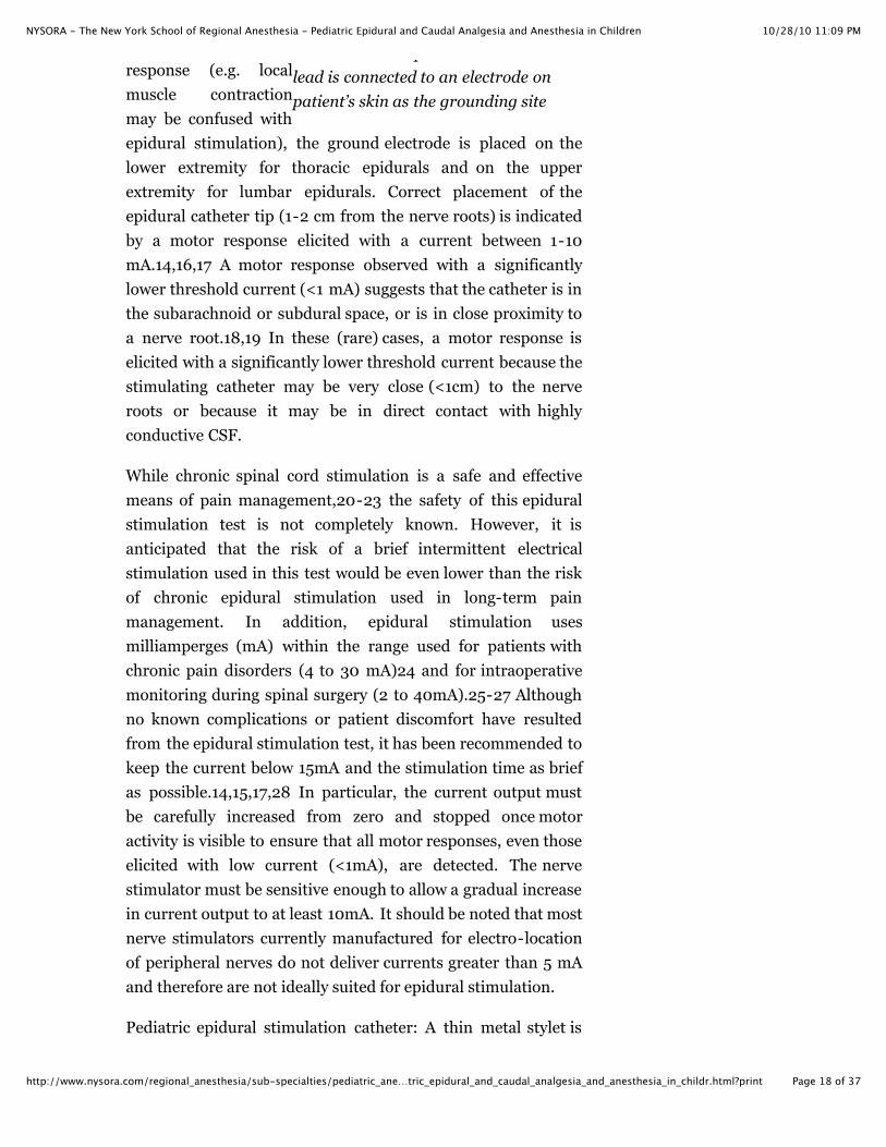
The stimulatingcatheter set-uprequires the cathodelead (black for block)of the nerve stimulatorto be connected to theepidural catheter viaan electrode adapterwhile the anode lead isconnected to anelectrode on patient’sskin as the groundingsite, Figure 1. Toavoidmisinterpretation ofthe stimulation
10/28/10 11:09 PMNYSORA - The New York School of Regional Anesthesia - Pediatric Epidural and Caudal Analgesia and Anesthesia in Children
Page 18 of 37http://www.nysora.com/regional_anesthesia/sub-specialties/pediatric_ane…tric_epidural_and_caudal_analgesia_and_anesthesia_in_childr.html?print
an electrode adapter while the anodelead is connected to an electrode onpatient’s skin as the grounding site
response (e.g. localmuscle contractionmay be confused withepidural stimulation), the ground electrode is placed on thelower extremity for thoracic epidurals and on the upperextremity for lumbar epidurals. Correct placement of theepidural catheter tip (1-2 cm from the nerve roots) is indicatedby a motor response elicited with a current between 1-10mA.14,16,17 A motor response observed with a significantlylower threshold current (<1 mA) suggests that the catheter is inthe subarachnoid or subdural space, or is in close proximity toa nerve root.18,19 In these (rare) cases, a motor response iselicited with a significantly lower threshold current because thestimulating catheter may be very close (<1cm) to the nerveroots or because it may be in direct contact with highlyconductive CSF.
While chronic spinal cord stimulation is a safe and effectivemeans of pain management,20-23 the safety of this epiduralstimulation test is not completely known. However, it isanticipated that the risk of a brief intermittent electricalstimulation used in this test would be even lower than the riskof chronic epidural stimulation used in long-term painmanagement. In addition, epidural stimulation usesmilliamperges (mA) within the range used for patients withchronic pain disorders (4 to 30 mA)24 and for intraoperativemonitoring during spinal surgery (2 to 40mA).25-27 Althoughno known complications or patient discomfort have resultedfrom the epidural stimulation test, it has been recommended tokeep the current below 15mA and the stimulation time as briefas possible.14,15,17,28 In particular, the current output mustbe carefully increased from zero and stopped once motoractivity is visible to ensure that all motor responses, even thoseelicited with low current (<1mA), are detected. The nervestimulator must be sensitive enough to allow a gradual increasein current output to at least 10mA. It should be noted that mostnerve stimulators currently manufactured for electro-locationof peripheral nerves do not deliver currents greater than 5 mAand therefore are not ideally suited for epidural stimulation.
Pediatric epidural stimulation catheter: A thin metal stylet is

10/28/10 11:09 PMNYSORA - The New York School of Regional Anesthesia - Pediatric Epidural and Caudal Analgesia and Anesthesia in Children
Page 19 of 37http://www.nysora.com/regional_anesthesia/sub-specialties/pediatric_ane…tric_epidural_and_caudal_analgesia_and_anesthesia_in_childr.html?print
essential for effective threading of the epidural catheter from alower spinal level to the target upper spinal level. A stylettedcatheter has a soft and flexible tip and is made from a softpolyurethane polymer.29,15 The stylet of the epidural catheterends 10 mm proximal to the tip which allows the tip of thecatheter to fold back on itself in a “J” configuration duringinsertion (Arrow International™). This feature allows retentionof the soft and blunted tip of the catheter while the stylet wireprovides stiffness for ease of advancement within the epiduralspace. For monitoring advancement, elicited muscle twitchesare observed from the lower limbs to the intercostal muscles asthe catheter is advanced cranially. This minimizes the concernsof the catheter coiling or kinking by immediately identifyingthese events at the time of insertion, allowing for any necessaryadjustments.15,28 The absence of muscle twitches or resistanceto the advancing epidural catheter may be indicative of a curledor kinked catheter. Epidural stimulation test relies on a smallelectrical current being transmitted through a conducting fluidinjected into the epidural space. An ionic solution such asnormal saline is used as the priming solution for the catheter.Normal saline dissociates into ions that are sufficient foreffective electrical conduction over a short distance. The longlength of the epidural catheter or any air lock within its lumenincreases the resistance to current flow. Consequently, thelumen of the catheter must contain a metal element to reducethe impedance of the conducting solutions and to ensureproper conduction of electricity through the entire length of thecatheter..14,16,17,30 Many commercial epidural catheters withmetal elements are now available through a number of majormanufacturers and can be used for the purpose of epiduralstimulation test.30
Epidural ECG technique
One disadvantage of the epidural stimulation technique is that it cannot beperformed reliably if any significant clinical neuromuscular blockade ispresent or local anesthetics have been administered in the epidural space.To overcome this limitation, an alternative monitoring technique usingelectrocardiograph (ECG) monitoring has been suggested.31,32 Usingepidural ECG monitoring lead, the anatomical position of the epiduralcatheter is determined by comparing the ECG signal from the tip of the
10/28/10 11:09 PMNYSORA - The New York School of Regional Anesthesia - Pediatric Epidural and Caudal Analgesia and Anesthesia in Children
Page 20 of 37http://www.nysora.com/regional_anesthesia/sub-specialties/pediatric_ane…tric_epidural_and_caudal_analgesia_and_anesthesia_in_childr.html?print
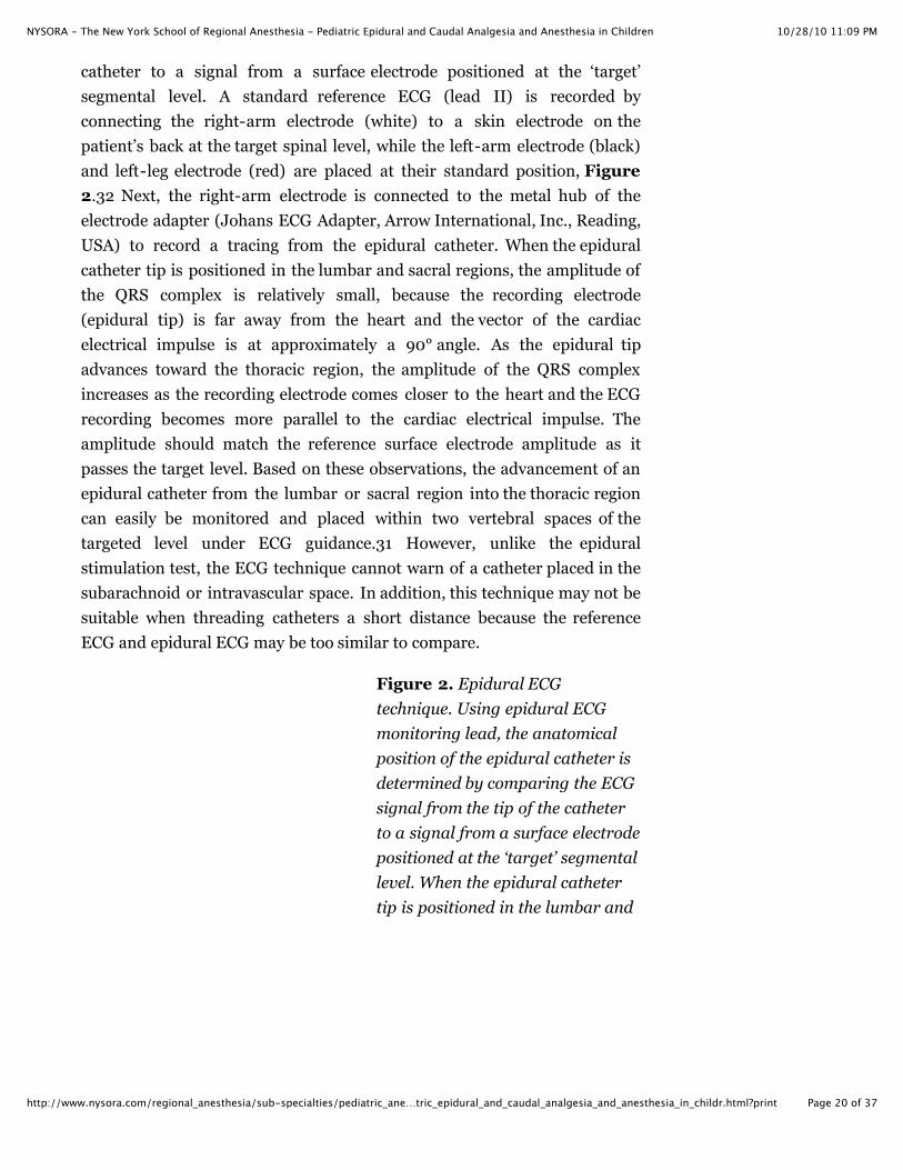
catheter to a signal from a surface electrode positioned at the ‘target’segmental level. A standard reference ECG (lead II) is recorded byconnecting the right-arm electrode (white) to a skin electrode on thepatient’s back at the target spinal level, while the left-arm electrode (black)and left-leg electrode (red) are placed at their standard position, Figure2.32 Next, the right-arm electrode is connected to the metal hub of theelectrode adapter (Johans ECG Adapter, Arrow International, Inc., Reading,USA) to record a tracing from the epidural catheter. When the epiduralcatheter tip is positioned in the lumbar and sacral regions, the amplitude ofthe QRS complex is relatively small, because the recording electrode(epidural tip) is far away from the heart and the vector of the cardiacelectrical impulse is at approximately a 90° angle. As the epidural tipadvances toward the thoracic region, the amplitude of the QRS complexincreases as the recording electrode comes closer to the heart and the ECGrecording becomes more parallel to the cardiac electrical impulse. Theamplitude should match the reference surface electrode amplitude as itpasses the target level. Based on these observations, the advancement of anepidural catheter from the lumbar or sacral region into the thoracic regioncan easily be monitored and placed within two vertebral spaces of thetargeted level under ECG guidance.31 However, unlike the epiduralstimulation test, the ECG technique cannot warn of a catheter placed in thesubarachnoid or intravascular space. In addition, this technique may not besuitable when threading catheters a short distance because the referenceECG and epidural ECG may be too similar to compare.
Figure 2. Epidural ECGtechnique. Using epidural ECGmonitoring lead, the anatomicalposition of the epidural catheter isdetermined by comparing the ECGsignal from the tip of the catheterto a signal from a surface electrodepositioned at the ‘target’ segmentallevel. When the epidural cathetertip is positioned in the lumbar and

10/28/10 11:09 PMNYSORA - The New York School of Regional Anesthesia - Pediatric Epidural and Caudal Analgesia and Anesthesia in Children
Page 21 of 37http://www.nysora.com/regional_anesthesia/sub-specialties/pediatric_ane…tric_epidural_and_caudal_analgesia_and_anesthesia_in_childr.html?print
sacral regions, the amplitude of theQRS complex is relatively small,because the recording electrode(epidural tip) is far away from theheart and the vector of the cardiacelectrical impulse is atapproximately a 90° angle. As theepidural tip advances toward thethoracic region, the amplitude ofthe QRS complex increases as therecording electrode comes closer tothe heart and the ECG recordingbecomes more parallel to thecardiac electrical impulse. Theamplitude should match thereference surface electrodeamplitude as it passes the targetlevel.
Technique
Several epidural techniques currently used in children will bedescribed in this chapter. The most common types of epiduralanalgesia are (A) caudal analgesia which constitutes the mostcommonly used regional technique in children; (B) lumbarepidural analgesia and (C) thoracic epidural analgesia
Single-shot caudal technique
Single shot caudal epidural blockade (‘kiddy caudals’) is widelyused to provide perioperative analgesia in pediatric practice. Asa single injection, it offers a reliable and effective block forpatients undergoing urological, general and orthopedic surgeryinvolving the lower abdomen and lower limbs. A single-shotcaudal epidural may not be suitable for every case as it has alimited dermatomal distribution and a short duration of action.New local anesthetics and adjuvants, as well as continuouscatheter approaches may overcome these limitations.
Choice of needle for caudal analgesia

10/28/10 11:09 PMNYSORA - The New York School of Regional Anesthesia - Pediatric Epidural and Caudal Analgesia and Anesthesia in Children
Page 22 of 37http://www.nysora.com/regional_anesthesia/sub-specialties/pediatric_ane…tric_epidural_and_caudal_analgesia_and_anesthesia_in_childr.html?print
A variety of needles are available for single-shot caudalblockade. The size or type of needle does not appear to affectthe rate of success or the incidence of complications of caudalblockade. Short-bevel 22 gauge needles (< 4 cm in length) withstylets are believed to offer a better tactile sensation when thesacrococcygeal ligament is punctured.33 Theoretically, the useof a styletted needle may reduce the risk of introducing adermal plug into the caudal space, although an epidermal cellgraft tumor in the epidural space has yet to be reported. Theuse of 22-gauge Angiocath® is also advocated because with theadvancement of these catheters into the caudal space mayindicate proper positioning.34 There is also indications that itis easier to detect intravascular placement and interosseousplacement with angiocatheters.6 To avoid tissue coring withthese angiocatheters, the needle must be removed before anyinjection is made.35
Technique for performing caudal epidural block
Patients are placed either in a lateral decubitus position withthe knees drawn up to the chest or in a prone position with aroll under the hips for caudal epidural block placement,Figure 3. Following proper positioning, the landmarks forcaudal epidural block are easily identified in children. Afterinitially identifying the coccyx and continuing to palpate in themidline in a cephalad fashion, the sacral cornua can be felt oneither side of the midline approximately one centimeter apart,Figure 4. The sacral hiatus is felt as a depression between twobony prominences of the sacral cornua. Under sterileconditions, the needle is inserted and advanced into the sacralhiatus at approximately a 70-degree angle to the skin until adistinctive “pop” is felt as the sacrococcygeal ligament ispunctured, Figure 5. Following this puncture, the angle of theneedle should be reduced to approximately 20 to 30 degreeswhile the needle is advanced 2 to 4 mm into the caudal canal. Ifusing an angiocatheter (Figure 6), the plastic catheter of theneedle should easily advance into the caudal epidural space.Any advancement past this point is not recommended as therisk of an inadvertent dural puncture increases significantly.

10/28/10 11:09 PMNYSORA - The New York School of Regional Anesthesia - Pediatric Epidural and Caudal Analgesia and Anesthesia in Children
Page 23 of 37http://www.nysora.com/regional_anesthesia/sub-specialties/pediatric_ane…tric_epidural_and_caudal_analgesia_and_anesthesia_in_childr.html?print
Figure 3. PatientPositioning. Shown is leftlateral position with hipsmaximally flexed
Figure 4. Landmarks forcaudal anesthesia. Shownare posterior superioriliac spines (two fingers)which form equalateraltriangle with sacralcornua (single finger)
Figure 5. Needleadvancement in caudalblock. Cannula isadvanced in a cephaladdirection. Occasionally, apop is felt as thesacrococcygeal ligamentis penetrated. At thispoint the cannula isadvanced a few cm off theneedle.
Figure 6. Cannulaplacement. Easy passageof the cannula confirmscorrect placement.
10/28/10 11:09 PMNYSORA - The New York School of Regional Anesthesia - Pediatric Epidural and Caudal Analgesia and Anesthesia in Children
Page 24 of 37http://www.nysora.com/regional_anesthesia/sub-specialties/pediatric_ane…tric_epidural_and_caudal_analgesia_and_anesthesia_in_childr.html?print
Clinical Pearls
Posterior superior iliac spines and sacral hiatus formequalateral triangleSacral Cornua either side of hiatus (0.5-1.0 cm apart)Dural sac extends to S4 in the infant less than 1 year (S2 inthe adult)
Confirmation of needle placement
The classic “pop”, felt as sacrococcygeal membrane is pierced isusually sought for proper caudal needle placement. Theabsence of subcutaneous bulging and the lack of resistanceupon injection of local anesthetic are additional signs of properneedle placement, Figure 7. Aspiration of the needle shouldbe clear of blood and CSF and a negative response to a testdose of epinephrine should be also used to rule out intrathecaland intravascular placement, Figure 8. Other tests to confirmproper needle placement include the “whoosh” test, the“swoosh” test, and the use of nerve stimulation.36,37 The“whoosh” test requires the injection of 2.5 ml of air through thecaudal needle, with a “whoosh” being heard with a stethoscopeplaced over the thoracolumbar spine. However, this can lead toa patchy block. More importantly, it can cause a venous airembolism if the needle is inserted into an epidural vesselespecially in small infants. The “swoosh” technique avoidsthese problems by injecting local anesthetic or saline in place ofair but the benefit of confirming needle placement prior to localanesthetic injection is lost. Excessive saline injection maydilute subsequent local anesthetic injections and lead to aninadequate block. When using nerve stimulation, proper needleplacement is confirmed by motor activity in the anal sphincterwith 1-10 mA of current through an insulated needle.37 Thesensitivity and specificity of predicting proper needleplacement approaches 100% with this approach, although therequirement for an insulated sheathed needle limits its use.37Furthermore, most insulated needles lack a stylet and may bemore expensive than standard non-insulated needles.Ultrasound has been used to provide real-time images to guideneedles into the caudal space.12 Other predictors of accurate

10/28/10 11:09 PMNYSORA - The New York School of Regional Anesthesia - Pediatric Epidural and Caudal Analgesia and Anesthesia in Children
Page 25 of 37http://www.nysora.com/regional_anesthesia/sub-specialties/pediatric_ane…tric_epidural_and_caudal_analgesia_and_anesthesia_in_childr.html?print
block placement following the injection of local anesthetic havebeen attempted. Relaxation of the anal sphincter predictssuccessful caudal blockade, 38but pupillary reflex dilation andskin temperature changes are not clinically useful.39
Figure 7. The cannula is stabilizedwith the left hand while the localanesthetic syringe is connected andsubsequently injected in divided doses.The EKG is monitored during injectionfor an increase in heart reate of 10beats/min or a 20% change in T waveamplitude. The reliability of these signswithout EKG strip monitoring remainsuntested. The area of skin immediatelyover the sacrum should be visible toobserve for inadvertent subcutaneousinjection.
Figure 8. Bloody tap. In the infantshown, an epidural vein isinadvertently cannulated as evidencedby the free flow of venous blood. Thecannula is consequently removed andthe process repeated.
Clinical Pearls
Formulae exist for the volume of local anaesthetic requiredto achieve a given dermatomal spread. In practice, a dose of1ml/kg of 0.25% bupivacaine with epinephrine will give fourhours of postoperative analgesia with a low incidence ofmotor block.The only additives that have been shown to prolonganalgesia without increasing side effects are:
Clonidine 1-2 mcg/kg (approximately 8 hourspostoperative analgesia)Ketamine (preservative free) 0.5 mg/kg (up to 12

10/28/10 11:09 PMNYSORA - The New York School of Regional Anesthesia - Pediatric Epidural and Caudal Analgesia and Anesthesia in Children
Page 26 of 37http://www.nysora.com/regional_anesthesia/sub-specialties/pediatric_ane…tric_epidural_and_caudal_analgesia_and_anesthesia_in_childr.html?print
hours postoperative analgesia)These agents have been shown to prolong the time to firstanalgesic following minor surgery. In our experience, afterthe local anaesthetic block has warn off, these agentsprovide only mild analgesia.
Continuous caudal epidural to lumbar or thoracic space
Continuous caudal epidural analgesia overcomes the limited duration andsegmental effect of a single-shot technique. Caudal catheters advanced to alumbar or thoracic level can be used for surgery involving dermatomesabove T10. This technique may carry a smaller risk of dural puncture orspinal cord trauma than a direct thoracic epidural approach.40
Technique
The technique for needle insertion for continuous caudalanalgesia is very similar to the single-shot caudal approach. Anintravenous catheter (an 18-gauge angiocatheter for a 20-gauge epidural catheter or a 16-gauge angiocatheter for a 19gauge epidural catheter) or an 18-gauge Crawford needle isinserted through the sacrococcygeal ligament as described forthe single-shot technique. The complete angiocatheter with theneedle set should then be advanced no more than 1 cm into thesacral canal. After withdrawing the metal needle, the plasticsheath is gently advanced completely into the caudal space.This allows the epidural catheter to easily pass through theplastic sheath. The appropriate length of epidural catheter ismeasured against the back of the child from sacral to the targetspinal level or approximate dermatomal coverage required forthe surgical procedure. The epidural catheter is then advancedcarefully from the caudal space to the target level. Minorresistance to the passage of the catheter can usually beovercome by simple flexion or extension of the patient’svertebral column and /or by simultaneously injecting normalsaline through the advancing specialized stimulation epiduralcatheter (Epidural Positioning System using Tsui test, ArrowInternational Inc., USA™).15 The location of the catheter tipshould be verified using an objective test described as in theprevious section (radiography,9 nerve stimulation,15

10/28/10 11:09 PMNYSORA - The New York School of Regional Anesthesia - Pediatric Epidural and Caudal Analgesia and Anesthesia in Children
Page 27 of 37http://www.nysora.com/regional_anesthesia/sub-specialties/pediatric_ane…tric_epidural_and_caudal_analgesia_and_anesthesia_in_childr.html?print
electrocardiography,31,32 or ultrasound12). While some maycriticize these techniques as cumbersome or redundant, thesetests are valuable teaching aids and may avoid extensive followup on patients with inadequate analgesia as a result of poorlysituated catheters. Studies have suggested that caudal catheterplacement should be limited to patients under 1 year of age dueto the development of a lumbar curve during infancypreventing easy cephalad advancement of the catheter.9However, recent reports have demonstrated that cephaladadvancement is possible in older children using epiduralstimulation.15,41,42 The improved success rate in olderchildren has been attributed to the use of a styletted catheterwhich allows the simultaneous injecting of saline duringadvancement, and, more importantly, to the stimulation testwhich monitors the advancement of the catheter tip.15
Clinical Pearls
Advantages of a cannula over a needle are;Confidence of placement if the cannula slides off the needleeasilyPossibly reduced intraosseous injection riskPossibly reduced intravascular injection riskPossibly reduced dural puncture risk
Lumbar epidural anesthesia
Lumbar epidural analgesia is commonly used for continuous infusions andis rarely employed as a single-shot technique. A direct lumbar approach isprimarily indicated for providing pain control during and following lowerextremity surgery. Lumbar epidural placement, particularly in youngchildren, is performed after the induction of general anesthesia. However,this approach may also be performed awake in a select group of cooperativechildren and adolescents. The risk/benefit ratio of inserting thoracicepidural catheters in children under general anesthesia iscontroversial.41,43 Although this issue is not as controversial for lumbarepidural analgesia as thoracic epidural analgesia,43 caution should beexercised whenever performing lumbar epidural analgesia above the level ofspinal cord to avoid direct needle trauma.
Technique for placement of lumbar epidural analgesia

10/28/10 11:09 PMNYSORA - The New York School of Regional Anesthesia - Pediatric Epidural and Caudal Analgesia and Anesthesia in Children
Page 28 of 37http://www.nysora.com/regional_anesthesia/sub-specialties/pediatric_ane…tric_epidural_and_caudal_analgesia_and_anesthesia_in_childr.html?print
A midline approach to lumbar epidural needle placement ispreferred. Identification of the epidural space is commonlyachieved by loss of resistance (LOR) to saline. LOR to airshould be avoided due to the risk of introducing a venous airembolism particularly in neonates and infants. Children shouldbe positioned in the lateral decubitus position for direct lumbarepidural placement, Figure 9. An 18-gauge Tuohy needleswith a 20-gauge epidural catheter is often used in children,Figure 10, Figure 11. Although identification of theintervertebral space and ligamentum flavum in most pediatricpatients is easy, the ligamentum flavum can be less tensile inchildren and hence a distinctive “pop” may not be easily feltwhen penetrating this layer. In addition, the distance from theskin to the epidural space can be shallow. Formulas forestimating the distance from skin to epidural space distancehave been proposed.44-46 (Table:4) Formulae are only aguidelines and will change depending on the angle ofplacement of the epidural needle.
Figure 9. Landmarks forepidural anesthesia insmall children
The landmarks aresimilar in adultpopulation except that theintercristal line bisectsL5. In this child the L1spinous process ismarked with an arrow.
Figure 10. Epidural

10/28/10 11:09 PMNYSORA - The New York School of Regional Anesthesia - Pediatric Epidural and Caudal Analgesia and Anesthesia in Children
Page 29 of 37http://www.nysora.com/regional_anesthesia/sub-specialties/pediatric_ane…tric_epidural_and_caudal_analgesia_and_anesthesia_in_childr.html?print
Figure 10. Epiduralanesthesia in children:Hand position
Patient in the left lateralposition. Left hand indexand middle finger eitherside of chosen interspace.Right hand holds needlehub.
Figure 11. Epiduralanesthesia in children:Needle Advancement
Needle is advanced withstylet in place untilinterspinous ligament isreached. Stylet isremoved and saline filledloss of resistance syringeconnected to needle. Bothplunger and needlecontinuously advanced.Initially an increase inresistance is felt as theligamentum flavum isentered before a loss ofresistance. Thesesensations are very subtlein the small infant.
Table 4: Formula for depth of epidural space from skin
1. Rough estimate 1 mm/kg body weight2. Depth(cm) = 1 + 0.15 X age (years)3. Depth (cm) = 0.8 + 0.05 X weight (kg)4. Mean depth in neonates= 1 cm
Lumbar to thoracic approach
Catheters placed via the lumbar route may be advanced cephalad to thoracic

10/28/10 11:09 PMNYSORA - The New York School of Regional Anesthesia - Pediatric Epidural and Caudal Analgesia and Anesthesia in Children
Page 30 of 37http://www.nysora.com/regional_anesthesia/sub-specialties/pediatric_ane…tric_epidural_and_caudal_analgesia_and_anesthesia_in_childr.html?print
vertebral levels, Figure 12-16. Similar to the problems encountered whenadvancing catheters in the caudal space in older children, significantresistance also prevents the easy advancement of lumbar epidural cathetersto the thoracic levels Despite favorable results using stimulation via a caudalapproach, there has been only one recent case report demonstrating thesuccessful placement of a thoracic epidural catheter via the lumbar routewith epidural stimulation guidance.47 Further research and study iswarranted for using the stimulating technique for this approach.
Figure 12. Epiduralanesthesia in children:Catheter insertion
Catheter advancement isassociated with greaterresistance than in theadult. The catheterstabilizing attachmentmay help (not used here).
Figure 13. Epiduralanesthesia in children:Preventing the leakage
Preventing the leakage oflocal anesthetic inpediatric patients isimportant because thiscan comprise asignificant percentage ofthe total drug delivered.The puncture site can besealed using severalmethods, one of which iswith ‘Liquid Bandage’(Johnson and Johnson)using the suppliedproduct applicator.

10/28/10 11:09 PMNYSORA - The New York School of Regional Anesthesia - Pediatric Epidural and Caudal Analgesia and Anesthesia in Children
Page 31 of 37http://www.nysora.com/regional_anesthesia/sub-specialties/pediatric_ane…tric_epidural_and_caudal_analgesia_and_anesthesia_in_childr.html?print
Figure 14. Epiduralanesthesia in children:Securing the catheter
Tincture of benzoin isapplied to improveadhesion of the fixationdevice.
Figure 15. Epiduralanesthesia in children:Securing the catheter
Epidural catheters thatare not secured well insmall children dislodgevery easily. The deviceused here is the SimmsPortex ‘lockit’ device. Inthe small child/infantallowance should bemade for the relativelysmall distance betweenadjacent vertebrae.Leaving 3 cm of catheterin the epidural spacemeans the tip of thecatheter may be threesegments higher (orlower) than the needleinsertion point. In thischild the epidural spacewas located 2 cm deep tothe skin. Leaving thecatheter at 5 cm (3cm inspace) will result in thecatheter being situated atapproximately T10 (threesegments above T12/L1interspace).

10/28/10 11:09 PMNYSORA - The New York School of Regional Anesthesia - Pediatric Epidural and Caudal Analgesia and Anesthesia in Children
Page 32 of 37http://www.nysora.com/regional_anesthesia/sub-specialties/pediatric_ane…tric_epidural_and_caudal_analgesia_and_anesthesia_in_childr.html?print
Figure 16. Epiduralanesthesia in children:Securing the catheter
The epidural fixationdevice is covered with aclear occlusive dressing
Clinical Pearls
Various formulae exist for calculating the volume of localanesthetic required to block a given number of segments.Because sympathetic blockade is well tolerated in childrenwith very little change in both heart rate and bloodpressure, in practice, (following an appropriate test dose) abolus of 0.5-1.0 ml/kg of 0.25% bupivacaine isadministered to establish the block.For postoperative analgesia, the most common agent usedis a combination of bupivicaine 0.125% with fentanyl 2mcg/ml at the following rates.
Age > 3 months 0.20-0.35 ml/kg/hr (<0.4mg/kg/hrbupivacaine)Age < 3 months 0.1-0.15 ml/kg/hr (<0.2 mg/kg/hrbupivacaine)
In preschool age children and especially infants,irritability/agitation may occur despite an apparently wellfunctioning epidural. This is most likely the result of the IVline, nasogastric tube, urinary catheter or even the hospitalenvironment. Satisfactory sedation can be achieved witheither,
IV boluses of morphine 25 mcg/kg as required orAdding clonidine 0.5 mcg/ml to the epidural mixture
PCEA (Age > 7) may provide less motor block than aninfusion only prescription without compromising analgesia.
Infusion 0.15 ml/kg/hrBolus 0.07 ml/kg lockout 20 minutes
Thoracic epidural analgesia

10/28/10 11:09 PMNYSORA - The New York School of Regional Anesthesia - Pediatric Epidural and Caudal Analgesia and Anesthesia in Children
Page 33 of 37http://www.nysora.com/regional_anesthesia/sub-specialties/pediatric_ane…tric_epidural_and_caudal_analgesia_and_anesthesia_in_childr.html?print
Controversy exists concerning the safety of placing thoracic epidurals underheavy sedation or general anesthesia, as unconscious patients are unable toreport symptoms that may warn the anesthesiologist of potentialneurological complications.43,48-50 Direct needle trauma to the spinal cordduring epidural insertion is rare but can cause devastating complications.Recent reports have detailed cases of direct needle trauma to the spinal cordduring epidural placement in both awake and anesthetized patients.51-53The advancement of catheters from the lumbar and caudal epidural spacesto the thoracic level can be alternative approach. However, for reasonspoorly understood, the advancement of catheters in the epidural spacebecomes increasingly difficult with advancing age. The reason for this ispoorly understood but it has been suggested that the increase in resistanceto catheter advancement parallels the development of the lumbarcurvature.9 Direct placement of thoracic epidural catheters are still used butmore commonly at tertiary care centers limited for extensive proceduresinvolving thoracic and abdominal surgery with well trained personnel. Arecent study in pediatric patients suggested that electrical stimulationapplied to an advancing epidural needle may be used as an additional safetymeasure to warn of needle proximity to the intrathecal space, spinal cord ornerve root.54 This study demonstrated that the mean current necessary toelicit a motor response with insulated needles in the epidural space is muchhigher than that in the intrathecal space (5.2 � 2.4 mA versus 0.6 � 0.3mA, respectively).54 Individually, electrical stimulation and LOR have theirlimitations, but together, both these techniques may compensate for eachother’s weaknesses to facilitate optimal needle placement. A similar conceptusing electrophysiological monitoring is common practice in spinal surgery,but currently there is no clear evidence that electrical stimulation woulddirectly benefit thoracic epidural placement.55 This concept is still in itsinfancy and further research is warranted.
Clinical Pearls
Because children require a significantly higher volume/doseof local anesthetic compared to adults to achieve the samedermatome spread, it is important to have the tip of thecatheter at the intended surgical site.Intended high thoracic catheter advancement from alumbar insertion site is rarely successful.Thoracic epidural insertion should only be performed bypractitioners experienced with pediatric lumbar epidural

10/28/10 11:09 PMNYSORA - The New York School of Regional Anesthesia - Pediatric Epidural and Caudal Analgesia and Anesthesia in Children
Page 34 of 37http://www.nysora.com/regional_anesthesia/sub-specialties/pediatric_ane…tric_epidural_and_caudal_analgesia_and_anesthesia_in_childr.html?print
technique
Technique
Epidural needle insertion in pediatric patients can beperformed at any thoracic interspace using either a midline orparamedian approach. The paramedian approach is preferredin adults while a midline approach is often used in children.
Midline approach
Using midline approach, insertion of the needle iseasier at the lower thoracic level (T10 to T12) thanat the mid-thoracic (T4 to T7) level. The lowerborder of the shoulder blade, which is level with7th thoracic vertebra, is commonly used as ananatomical landmark. After the patient is placedin the lateral decubitus position, the spinuousprocess of the targeted vertebral level is identified.A 20-gauge Tuohy epidural needle is theninserted at the interspace at a cephalad angle ofapproximately 70 degree to the longitudinal axisof the spine (Figure 4). Continuous resistanceshould be felt as the needle is inserted through thesupraspinous and interspinous ligaments. Inpediatric patients, the resistance met at theligamentum flavum may not be noticeablydifferent from the other ligaments. The thoracicepidural space is identified with loss of resistanceto saline. The advantage of the midline approachis that its technique is very similar to lumbarepidural insertion with the needle angulated onlyin one plane.
Paramedian approach
he paramedian approach permits entry to theepidural space at any spinal level. This approach isusually performed with patients in the lateraldecubitus position. Right-handed clinicians mayprefer to use a right paramedian approach because
10/28/10 11:09 PMNYSORA - The New York School of Regional Anesthesia - Pediatric Epidural and Caudal Analgesia and Anesthesia in Children
Page 35 of 37http://www.nysora.com/regional_anesthesia/sub-specialties/pediatric_ane…tric_epidural_and_caudal_analgesia_and_anesthesia_in_childr.html?print
it increases the working space and it may facilitateneedle placement with the patient on the sameside as the needle. The needle is initially insertednext to the spinal process and slowly advanced ina direction perpendicular to the skin until laminais contacted. It is important to take note of laminadepth as it provides an estimated depth of theepidural space from the skin.56,57 (Figure 5)The needle is redirected medial before beinginclined cephalad toward the interspace. Again,LOR to saline will identify the epidural space.Many believe that this approach requires moreskill and experience as the needle must be angledin 2 planes (i.e. medially and cephahad). Thus,anesthesiologists with extensive experience andconfidence in epidural analgesia should performthis technique.
Managing Epidural Infusions Postoperatively
In order for epidural analgesia to be effective and safe systematic and regulatedapproaches to patient care must be practiced. A dedicated pediatric acute pain team,consisting of anesthesiologists and nurses, is vital to ensure standardized assessments ofpain, vigilant patient monitoring and the proper treatment of any adverse effects.Recommendations for epidural troubleshooting and managing inadequate analgesia aresummarized in Table 5. Precise placement of epidural needles and catheters is the keyto successful epidural analgesia. This requires employing the use of a reliable method toconfirm the location of the catheter tip (e.g. x-ray, epidural stimulation). The use ofopioids or alpha-2 agonists in the epidural space may lead to better analgesia. An averagelength of epidural infusion is about 72 hours although it may be necessary to continue theinfusion for longer periods especially in children with complicated medical histories andprolonged need for analgesia. A team of dedicated personnel with a focus on painmanagement should care for these children. When plans to discontinue the epiduralinfusion are in place, an oral opioid should always be administered to enable the patientto continue to have excellent analgesia. Finally the success of the process is based onproperly written orders which are a crucial part of executing adequate analgesia. Asample of an order sheet for managing epidural analgesia is attached, Table 6.
Table 5: Tips for improving epidural analgesia

10/28/10 11:09 PMNYSORA - The New York School of Regional Anesthesia - Pediatric Epidural and Caudal Analgesia and Anesthesia in Children
Page 36 of 37http://www.nysora.com/regional_anesthesia/sub-specialties/pediatric_ane…tric_epidural_and_caudal_analgesia_and_anesthesia_in_childr.html?print
Inadequate Analgesia
If there is any doubt about the adequacy of analgesia or theposition of the catheter, prove the catheter’s position usingone of the approaches outlined in Table 4.The use ofchloroprocaine can provide catheter tip position and rapidlyproviding analgesia in most cases.If the epidural infusion does not already include either anopioid or clonidine or S(+) ketamine along with the localanesthetic, consider inclusion of one of these additives,unless there are specific contraindications or unmanageableside-effects.Use bolus dosing to produce analgesia relatively promptly(while staying within maximum allowable local anestheticdosing parameters). Simply increasing an infusion rate by10 or 20% will require hours to reach a steady state, andwill subject the patient to prolonged pain.Titrate the local anesthetic upwards into the acceptablerange, if this has not been done already, unless specificallycontraindicated or unless limited by motor or autonomicblockade.For adolescents and young adults with thoracic catheter tipsfor upper abdominal or thoracic surgery, if there isinadequate analgesia when the local anesthetic infusion istitrated upwards to more than 12 ml/hour, consider nextincreasing the local anesthetic concentration, e.g. increasingbupivacaine to 0.15%, while maintaining a maximum hourlydosing within the 0.4 mg/kg/hr limit.If the catheter tip is in lumbar dermatomes and the surgeryis thoracic or upper abdominal, add a hydrophilic opioid,such as hydromorphone or morphine, unless there arespecific contraindications.If the catheter is in the epidural space, but the block ispreferentially one-sided to the wrong side, consideraddition of a hydrophilic opioid, either hydromorphone ormorphine, unless there are specific contraindications.
References

10/28/10 11:09 PMNYSORA - The New York School of Regional Anesthesia - Pediatric Epidural and Caudal Analgesia and Anesthesia in Children
Page 37 of 37http://www.nysora.com/regional_anesthesia/sub-specialties/pediatric_ane…tric_epidural_and_caudal_analgesia_and_anesthesia_in_childr.html?print
1. Bainbridge W: Analgesia in children by spinal injection with a report of a newmethod of sterilization of the injection fluid. Med Rec 1900; 58: 937-40
Back to Pediatrics