nutrition, digestion, and absorption 39. chapter 39 nutrition, digestion, and absorption key...
TRANSCRIPT
Nutrition, Digestion, and Absorption
39
Chapter 39 Nutrition, Digestion, and Absorption
Key Concepts
• 39.1 Food Provides Energy and Nutrients
• 39.2 Digestive Systems Break Down Macromolecules
• 39.3 The Vertebrate Digestive System Is a Tubular Gut with Accessory Glands
• 39.4 Food Intake and Metabolism Are Regulated
Chapter 39 Opening Question
Why are some ethnic groups of humans much more prone to obesity and diabetes than other groups?
Concept 39.1 Food Provides Energy and Nutrients
Animals are heterotrophs and derive their nutrition by eating other organisms.
Autotrophs use solar energy or inorganic chemical energy to synthesize their necessary nutrients.
Heterotrophs depend on this synthesis and have adapted to take advantage of it.
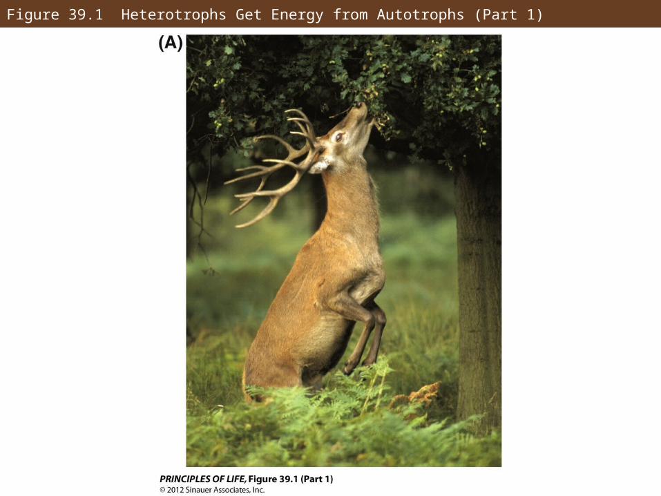
Figure 39.1 Heterotrophs Get Energy from Autotrophs (Part 1)
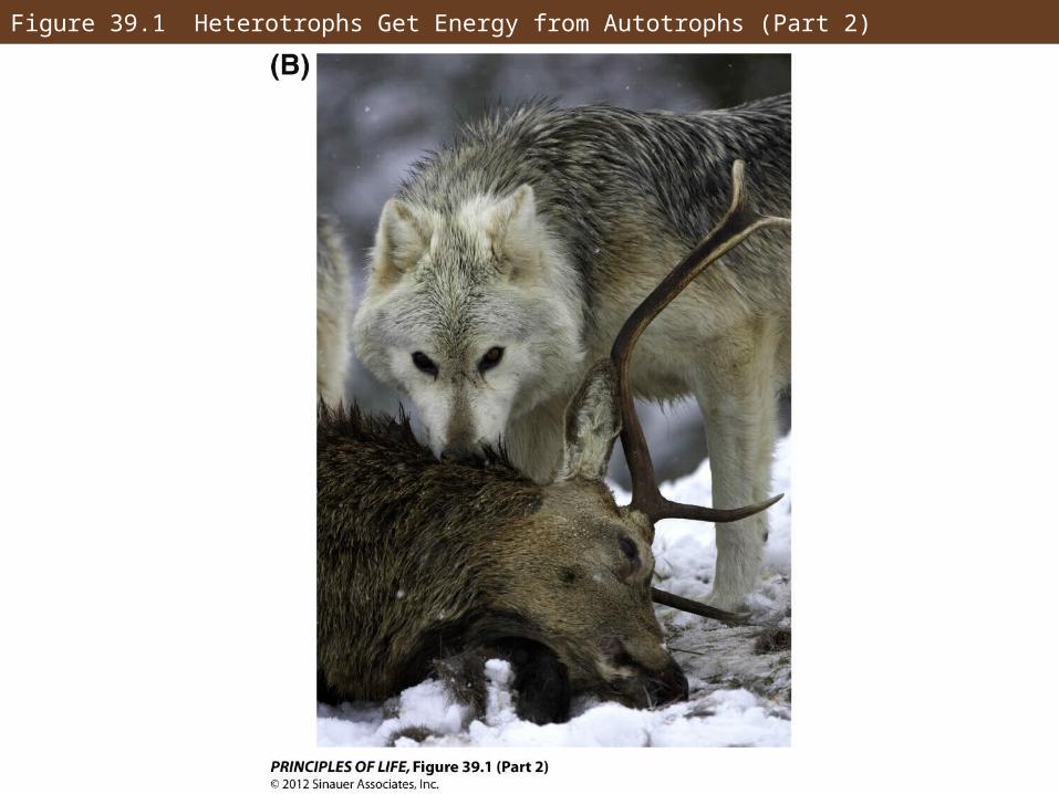
Figure 39.1 Heterotrophs Get Energy from Autotrophs (Part 2)
Concept 39.1 Food Provides Energy and Nutrients
Energy needs can be measured.
Measures of heat energy:
A calorie is the amount of heat needed to raise 1 gram of water 1°C
A kilocalorie (kcal) = 1,000 calories
The Calorie (Cal) is the same as a kilocalorie and = 1,000 calories
A joule = 0.239 calories
Concept 39.1 Food Provides Energy and Nutrients
The metabolic rate measures energy needs of an animal that are met by food intake and digestion.
Foods that provide energy are fats, carbohydrates, and proteins.
Basal metabolic rate (BMR) is the minimum energy needed to sustain life while an animal is at rest.
Concept 39.1 Food Provides Energy and Nutrients
Animals must store food between meals.
Carbohydrates are stored in liver and muscle cells as glycogen—enough for about a day’s energy needs.
Most fuel is stored as fat—stores more energy per gram and with little water, which makes it more compact.
Concept 39.1 Food Provides Energy and Nutrients
Essential nutrients are required but cannot be synthesized.
Macronutrients—nutrients required in large amounts, like calcium
Micronutrients—nutrients required in tiny amounts, like iron
Amino acids are the building blocks of proteins.
Each species has essential amino acids that they cannot synthesize.
Concept 39.1 Food Provides Energy and Nutrients
Complementary diets can supply all eight essential amino acids for adult humans.
Humans must also obtain essential fatty acids.
Linoleic acid is one that helps synthesize other unsaturated fatty acids.
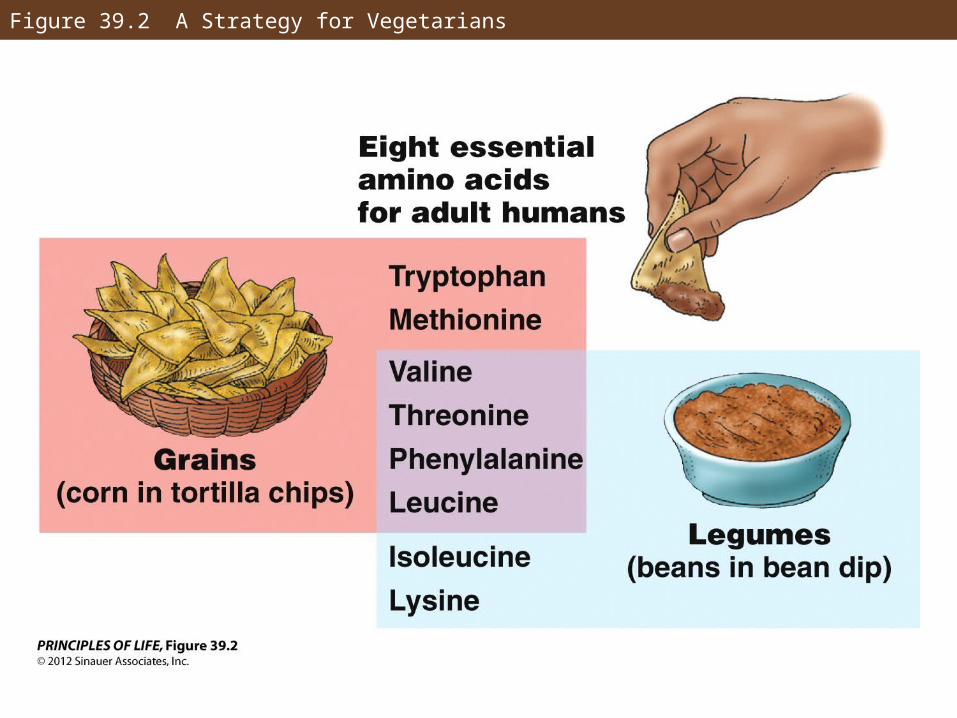
Figure 39.2 A Strategy for Vegetarians
Concept 39.1 Food Provides Energy and Nutrients
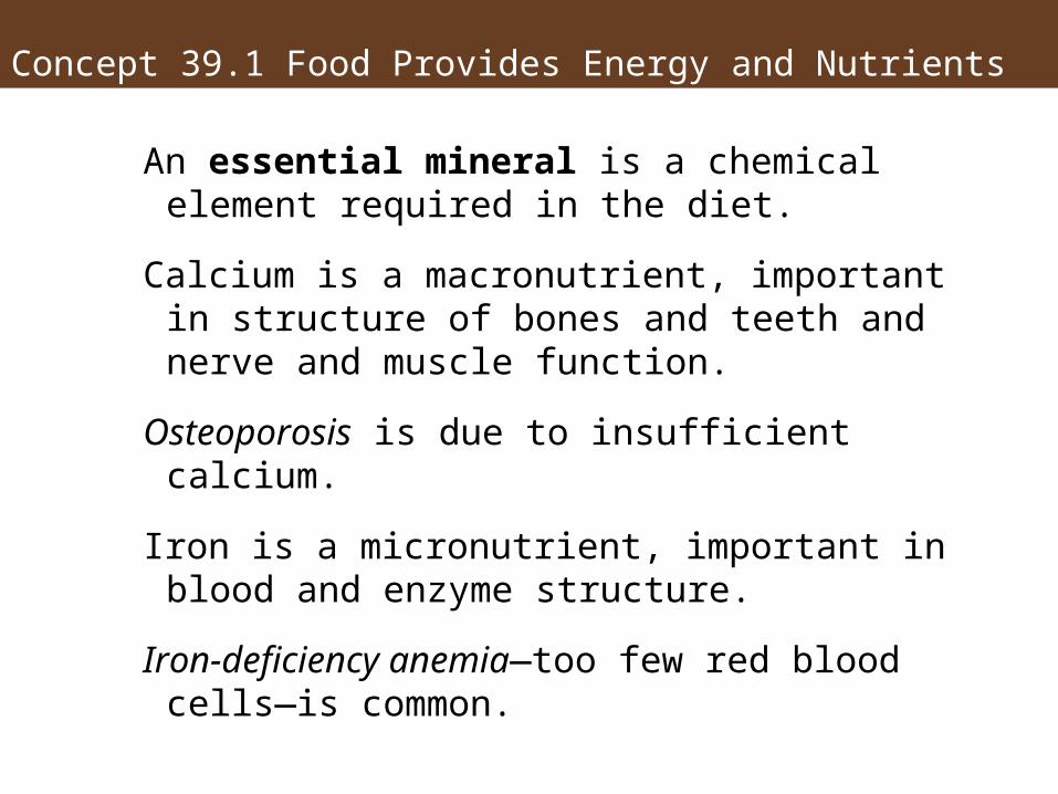
An essential mineral is a chemical element required in the diet.
Calcium is a macronutrient, important in structure of bones and teeth and nerve and muscle function.
Osteoporosis is due to insufficient calcium.
Iron is a micronutrient, important in blood and enzyme structure.
Iron-deficiency anemia—too few red blood cells—is common.
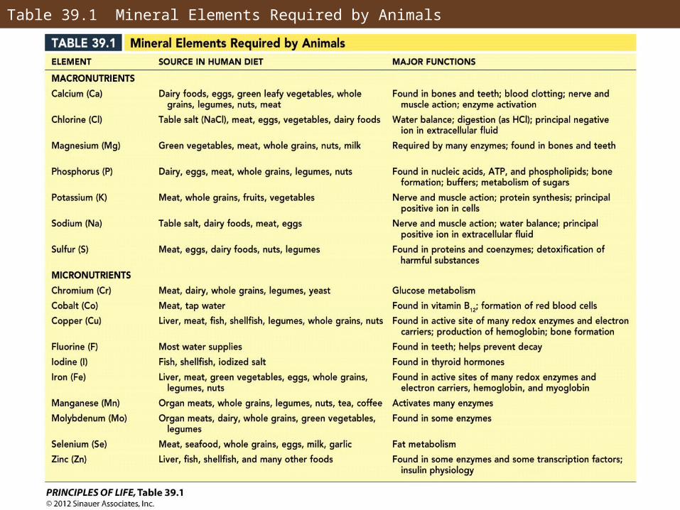
Table 39.1 Mineral Elements Required by Animals
Concept 39.1 Food Provides Energy and Nutrients
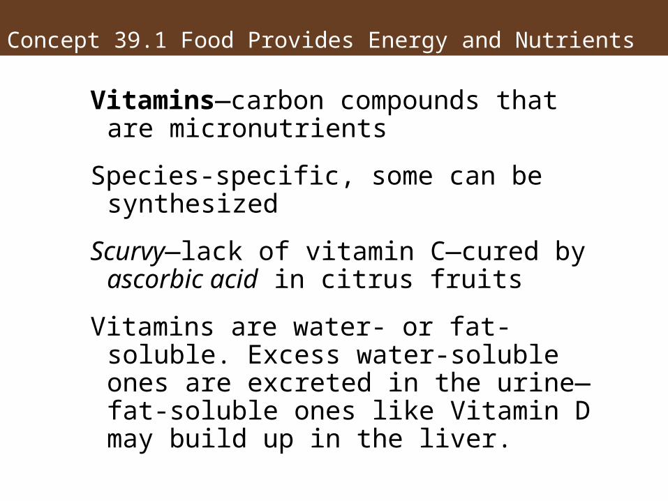
Vitamins—carbon compounds that are micronutrients
Species-specific, some can be synthesized
Scurvy—lack of vitamin C—cured by ascorbic acid in citrus fruits
Vitamins are water- or fat-soluble. Excess water-soluble ones are excreted in the urine—fat-soluble ones like Vitamin D may build up in the liver.
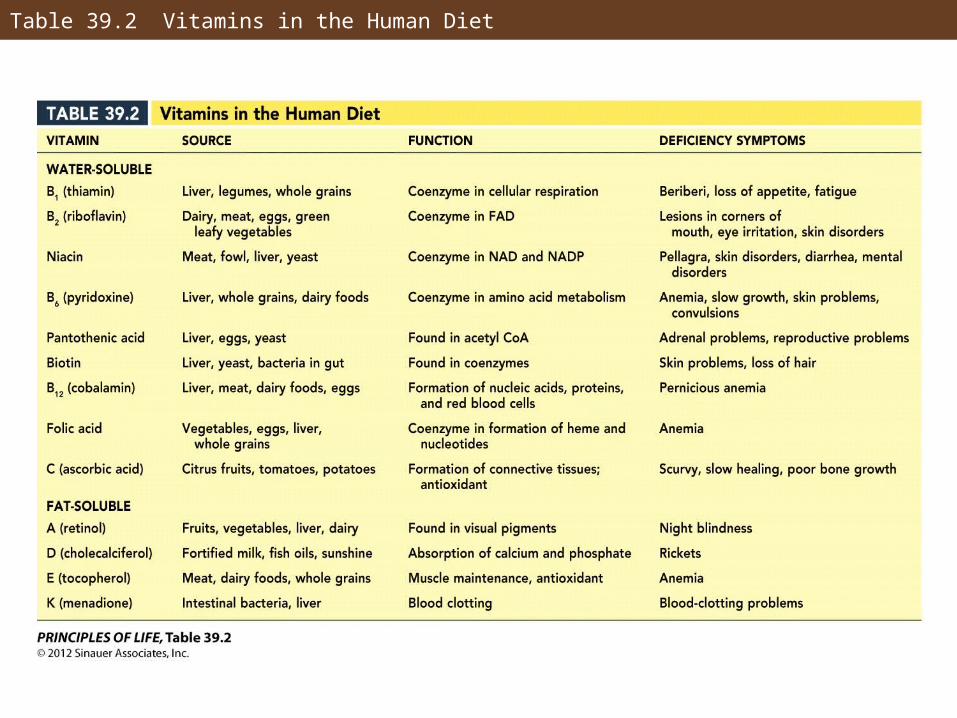
Table 39.2 Vitamins in the Human Diet
Concept 39.1 Food Provides Energy and Nutrients
Nutrient deficiency leads to malnutrition—chronic malnutrition leads to a deficiency disease:
• Scurvy and anemia
• Beriberi—due to deficiency of B1
• Goiter and hypothyroidism—due to iodine deficiency
Concept 39.2 Digestive Systems Break Down Macromolecules
The function of the digestive system, or gut, is to break down food into molecules for absorption.
Food is broken down through hydrolysis by enzymes produced by the digestive system.
Enzymes are classified by the food they break down: proteases, carbohydrases, peptidases, lipases, and nucleases.
Concept 39.2 Digestive Systems Break Down Macromolecules
Digestion usually begins in a body cavity.
Gastrovascular cavities connect to the outside through a single opening.
Example: A jellyfish stings prey and brings it into its gastrovascular cavity—enzymes break down food, and undigested particles are released through the same opening.
Concept 39.2 Digestive Systems Break Down Macromolecules
Tubular guts have an opening at each end:
A mouth takes in food, molecules are digested, and wastes are eliminated through the anus.
In most animals the gut can be divided into three sections: foregut, midgut, and hindgut.
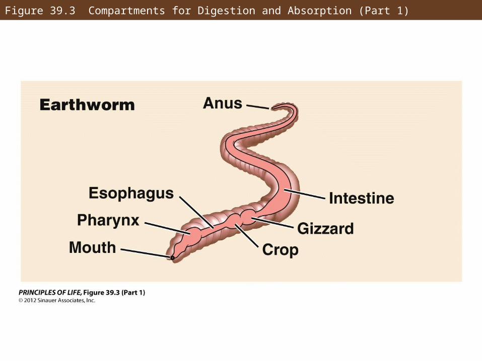
Figure 39.3 Compartments for Digestion and Absorption (Part 1)
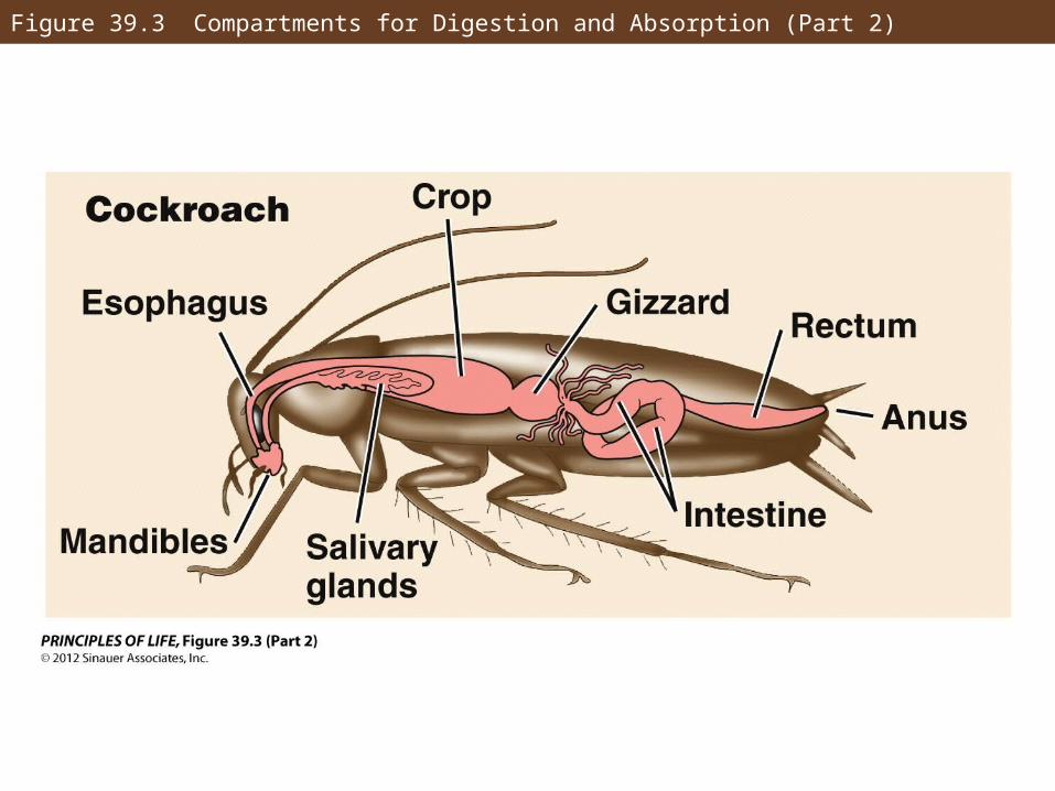
Figure 39.3 Compartments for Digestion and Absorption (Part 2)
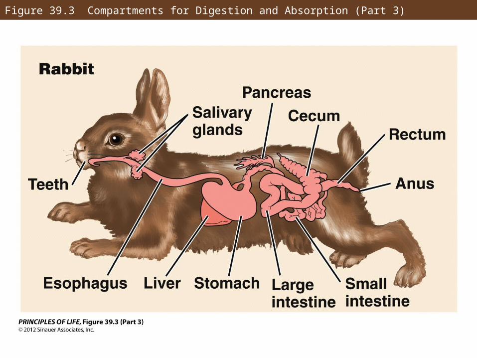
Figure 39.3 Compartments for Digestion and Absorption (Part 3)
Concept 39.2 Digestive Systems Break Down Macromolecules
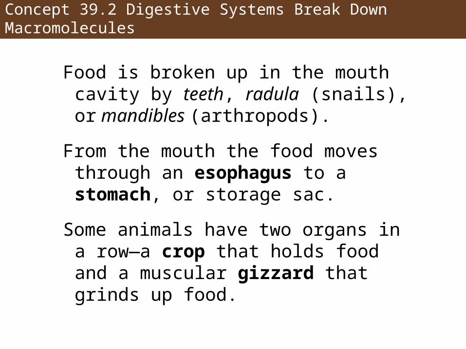
Food is broken up in the mouth cavity by teeth, radula (snails), or mandibles (arthropods).
From the mouth the food moves through an esophagus to a stomach, or storage sac.
Some animals have two organs in a row—a crop that holds food and a muscular gizzard that grinds up food.
Concept 39.2 Digestive Systems Break Down Macromolecules
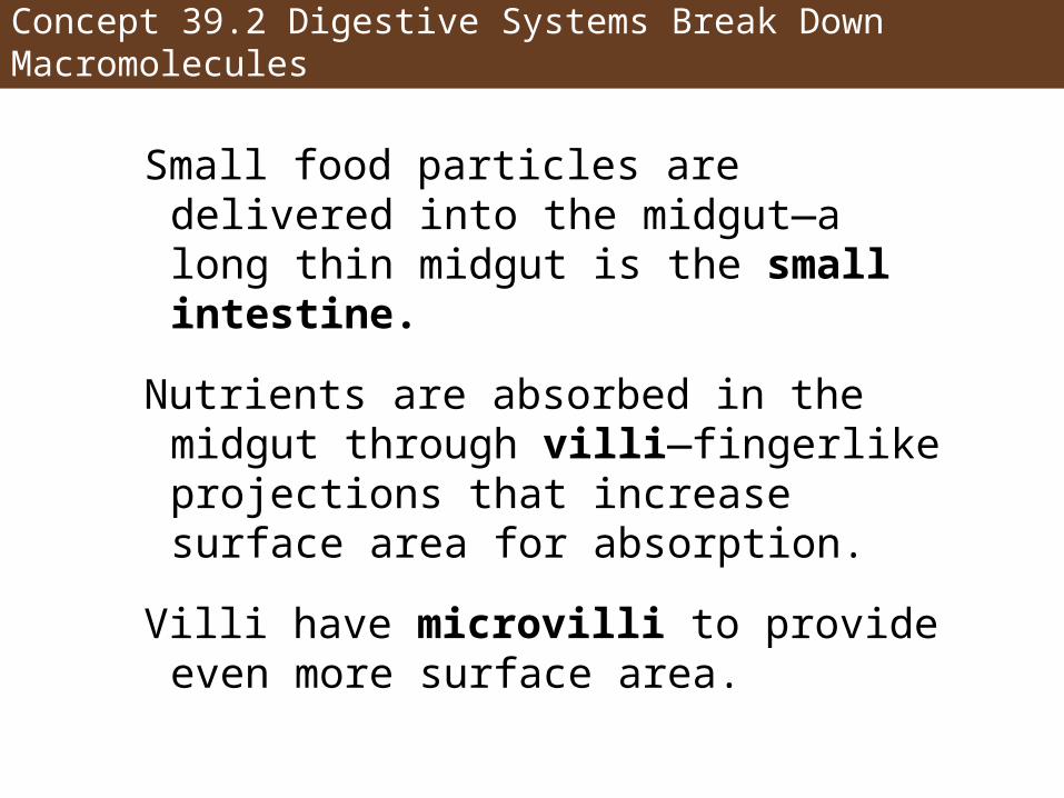
Small food particles are delivered into the midgut—a long thin midgut is the small intestine.
Nutrients are absorbed in the midgut through villi—fingerlike projections that increase surface area for absorption.
Villi have microvilli to provide even more surface area.
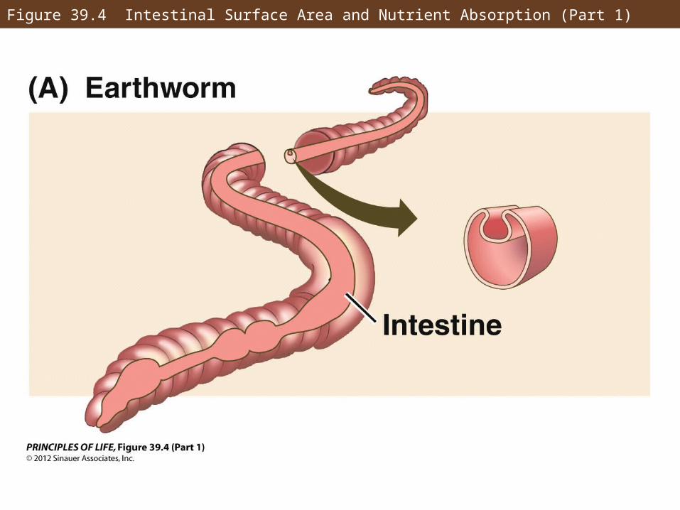
Figure 39.4 Intestinal Surface Area and Nutrient Absorption (Part 1)
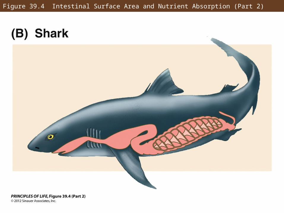
Figure 39.4 Intestinal Surface Area and Nutrient Absorption (Part 2)
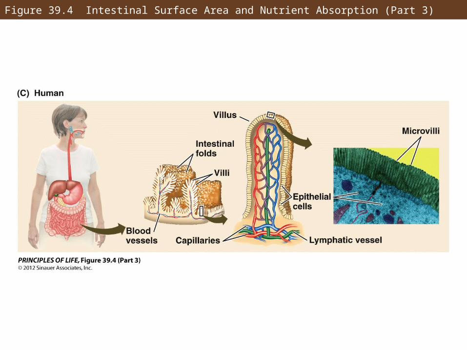
Figure 39.4 Intestinal Surface Area and Nutrient Absorption (Part 3)
Concept 39.2 Digestive Systems Break Down Macromolecules
The hindgut, or large intestine, recovers ions and water and stores undigested waste as feces.
The end of the digestive tract is the anus.
In birds, amphibians and reptiles, the cloaca expels both urinary and digestive wastes.
Most digestive tracts contain symbiotic bacteria.
Concept 39.2 Digestive Systems Break Down Macromolecules
Heterotrophs can be classified by how they acquire food:
Saprobes (decomposers) absorb nutrients from dead organic matter.
Detritivores actively feed on dead organic matter.
Predators feed on living organisms.
Concept 39.2 Digestive Systems Break Down Macromolecules
Predators feed on other animals:
• Herbivores—consume plants
• Carnivores—prey on animals
• Omnivores—prey on both
• Filter feeders filter small organisms from an aquatic environment
• Fluid feeders include mosquitoes
Concept 39.2 Digestive Systems Break Down Macromolecules
Mammalian teeth have shapes and organization adapted to specific diets:
• Incisors—used for cutting, chopping, or gnawing
• Canines—for stabbing, gripping, or ripping
• Molars or premolars—shearing, crushing, or grinding
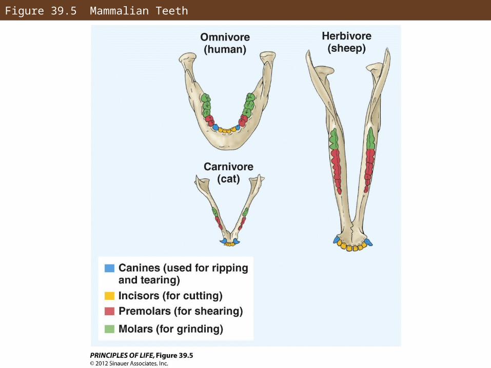
Figure 39.5 Mammalian Teeth
Concept 39.2 Digestive Systems Break Down Macromolecules
Diet affects the size of the digestive system.
Carnivores have short digestive tracts, because meat is easy to digest.
Herbivores have longer digestive tracts, often with compartments for cellulose-digesting bacteria to aid in plant digestion.
Traits in humans indicate we are omnivores.
Concept 39.3 The Vertebrate Digestive System Is a Tubular Gut with Accessory Glands
The vertebrate digestive system is a tubular gut, running from mouth to anus.
It has several accessory glands, including liver and pancreas.
Processes occur in sequence in different sections.
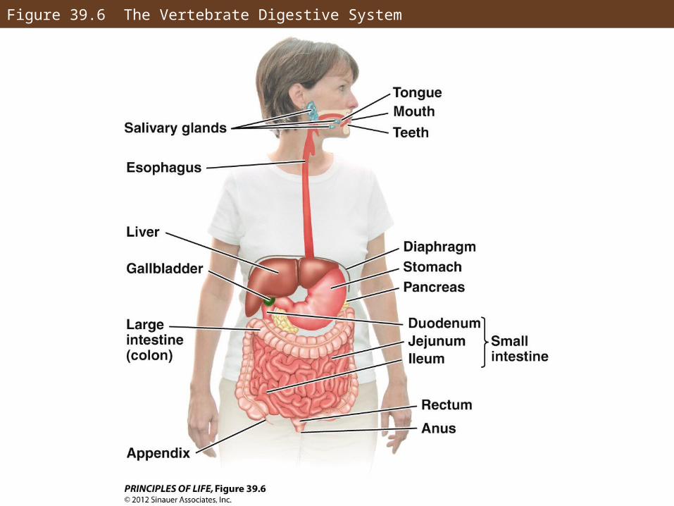
Figure 39.6 The Vertebrate Digestive System
Concept 39.3 The Vertebrate Digestive System Is a Tubular Gut with Accessory Glands
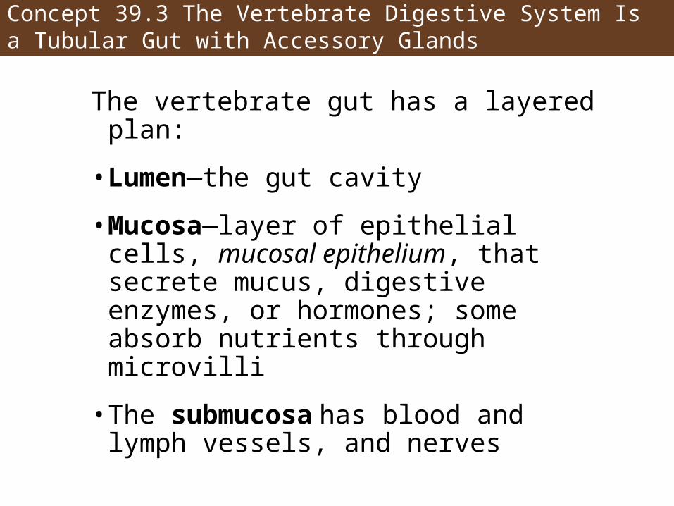
The vertebrate gut has a layered plan:
• Lumen—the gut cavity
• Mucosa—layer of epithelial cells, mucosal epithelium, that secrete mucus, digestive enzymes, or hormones; some absorb nutrients through microvilli
• The submucosa has blood and lymph vessels, and nerves
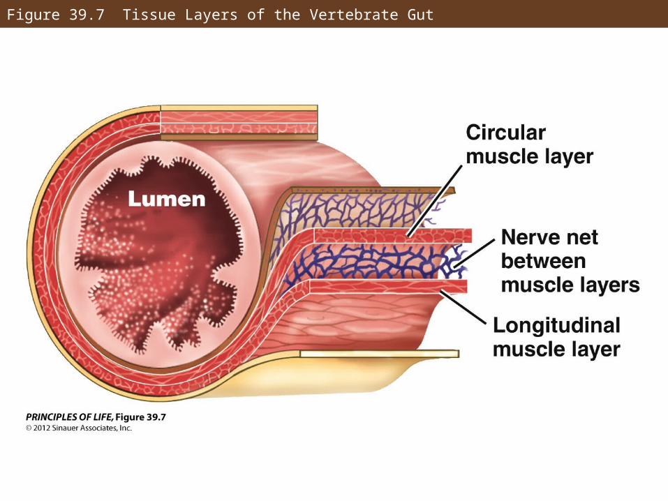
Figure 39.7 Tissue Layers of the Vertebrate Gut
Concept 39.3 The Vertebrate Digestive System Is a Tubular Gut with Accessory Glands
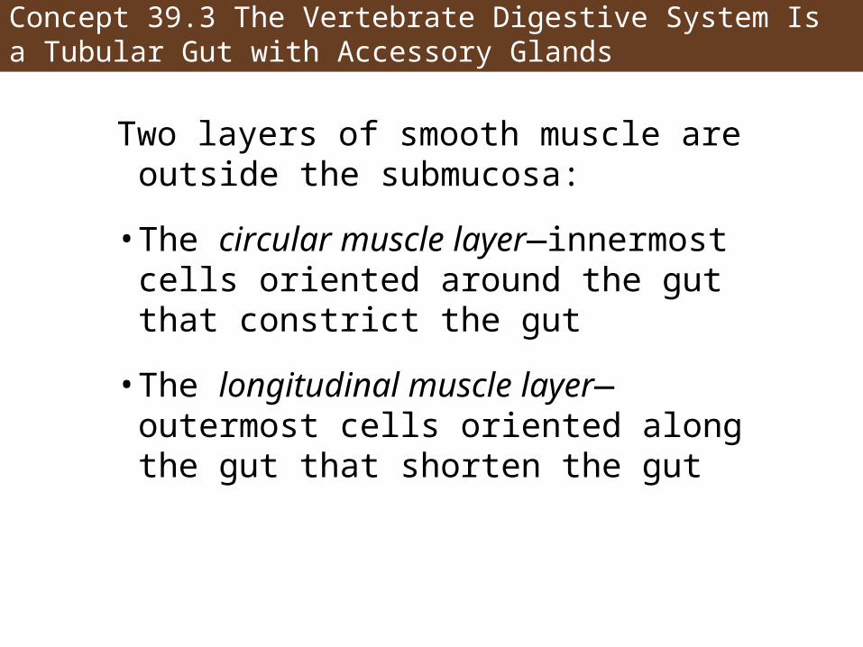
Two layers of smooth muscle are outside the submucosa:
• The circular muscle layer—innermost cells oriented around the gut that constrict the gut
• The longitudinal muscle layer—outermost cells oriented along the gut that shorten the gut
Concept 39.3 The Vertebrate Digestive System Is a Tubular Gut with Accessory Glands
Between layers of smooth muscle are nerves that coordinate movement of the gut.
Nerves in the enteric nervous system only form synapses with other nerves in the network.
The peritoneum is a membrane that surrounds the gut and lines the wall of the cavity.
Concept 39.3 The Vertebrate Digestive System Is a Tubular Gut with Accessory Glands
In mammals, digestion begins in the mouth—chewing mixes food with saliva, containing amylase to digest starch.
A mouthful of food is a bolus—when swallowed it passes into the esophagus.
Food is kept out of the trachea by the epiglottis, which closes off the larynx.
The bolus moves toward the stomach through peristalsis, coordinated by an anticipatory wave of relaxation.
Concept 39.3 The Vertebrate Digestive System Is a Tubular Gut with Accessory Glands
The stomach holds large amounts of food, breaks it up, and begins protein digestion.
Gastric pits in the stomach are lined with three types of secretory cells:
• Chief cells
• Parietal cells
• Mucus-secreting cells
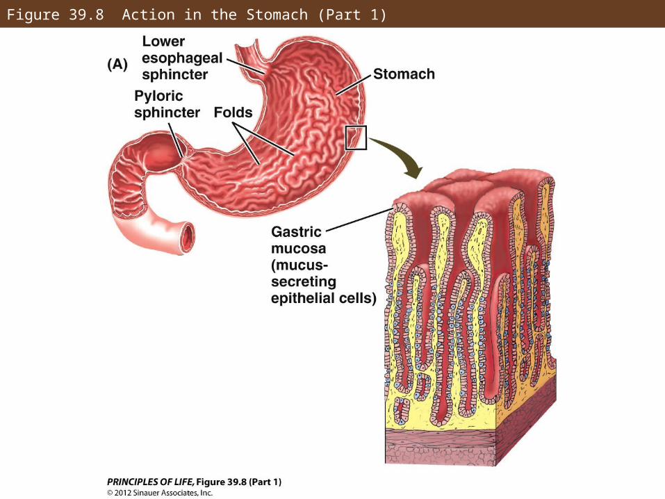
Figure 39.8 Action in the Stomach (Part 1)
Concept 39.3 The Vertebrate Digestive System Is a Tubular Gut with Accessory Glands
Chief cells secrete pepsinogen, the inactive form of a proteolytic enzyme, pepsin.
Parietal cells secrete hydrochloric acid (HCl), which kills ingested organisms, breaks down food, and activates pepsin.
Newly active pepsin activates other pepsinogen molecules—a process called autocatalysis.
Mucus-secreting cells protect the stomach from the formation of ulcers.
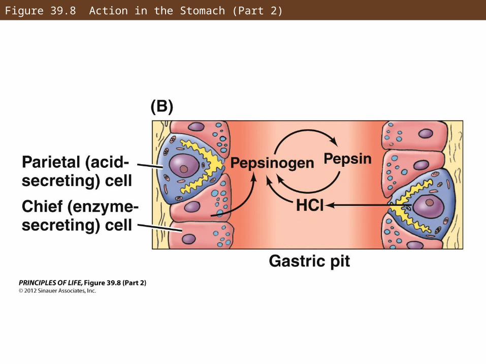
Figure 39.8 Action in the Stomach (Part 2)
Concept 39.3 The Vertebrate Digestive System Is a Tubular Gut with Accessory Glands
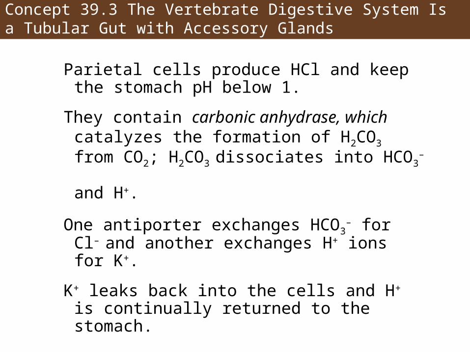
Parietal cells produce HCl and keep the stomach pH below 1.
They contain carbonic anhydrase, which catalyzes the formation of H2CO3 from CO2; H2CO3 dissociates into HCO3
– and H+.
One antiporter exchanges HCO3– for Cl– and
another exchanges H+ ions for K+.
K+ leaks back into the cells and H+ is continually returned to the stomach.
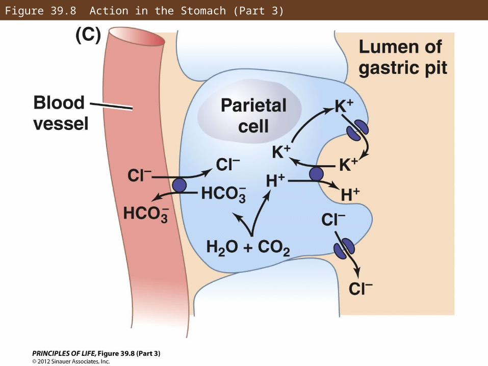
Figure 39.8 Action in the Stomach (Part 3)
Concept 39.3 The Vertebrate Digestive System Is a Tubular Gut with Accessory Glands
Smooth muscle contractions in the stomach create chyme—a mixture of gastric juice and partly digested food.
The stomach walls contract and move chyme to the bottom of the stomach.
The pyloric sphincter allows small amounts to enter the small intestine.
Concept 39.3 The Vertebrate Digestive System Is a Tubular Gut with Accessory Glands
Ruminants have four-chambered stomachs:
The rumen and reticulum contain microorganisms that metabolize the cellulose into nutrients for the host.
Food then travels to the omasum where it is concentrated by water absorption.
The abomasum is the true stomach.
Microorganisms are also digested by the host and provide protein.
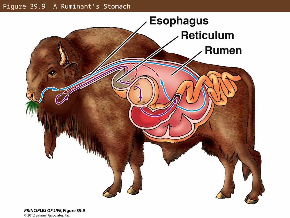
Figure 39.9 A Ruminant’s Stomach
Concept 39.3 The Vertebrate Digestive System Is a Tubular Gut with Accessory Glands
Most chemical digestion occurs in the small intestine.
The small intestine has three sections:
• Duodenum—the initial section and site of most digestion
• Jejunum and ileum—carry out most absorption
Concept 39.3 The Vertebrate Digestive System Is a Tubular Gut with Accessory Glands
The duodenum contains epithelial cells that produce enzymes:
• Peptidases cleave peptides into absorbable amino acids
• Other enzymes cleave disaccharides into monosaccharides
• Lipases that digest fats
Concept 39.3 The Vertebrate Digestive System Is a Tubular Gut with Accessory Glands
The liver produces and secretes bile that is stored in the gallbladder.
Bile flows to the duodenum via the common bile duct.
Bile’s function is to emulsify fat—bile contains bile salts that prevent fat molecules from sticking together.
Greater surface area exposes more of the fat molecule to the lipases for digestion.
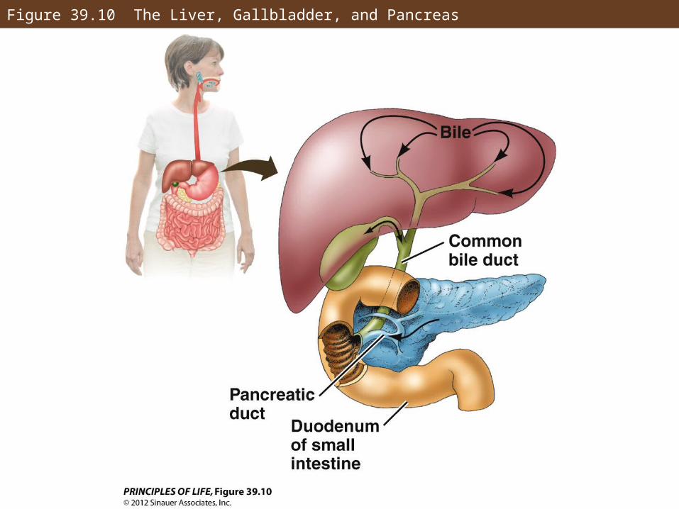
Figure 39.10 The Liver, Gallbladder, and Pancreas
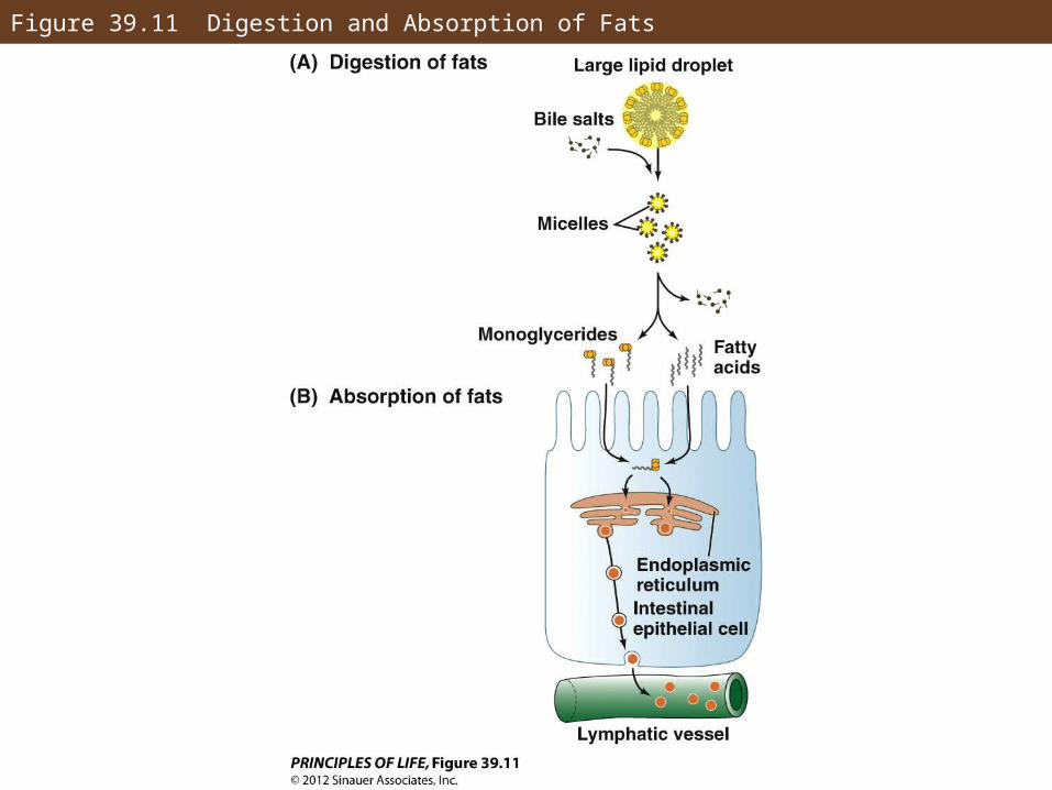
Figure 39.11 Digestion and Absorption of Fats
Concept 39.3 The Vertebrate Digestive System Is a Tubular Gut with Accessory Glands
The pancreas is an endocrine (hormone-releasing) and an exocrine (digestive juice-secreting) gland.
It produces insulin, glucagon, and digestive enzymes.
The pancreas secretes enzymes as zymogens.
Concept 39.3 The Vertebrate Digestive System Is a Tubular Gut with Accessory Glands
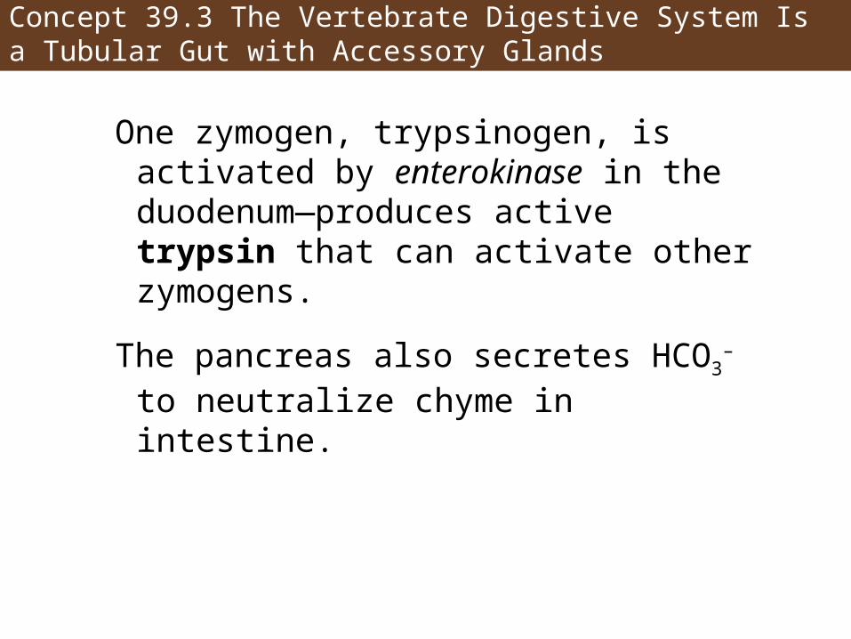
One zymogen, trypsinogen, is activated by enterokinase in the duodenum—produces active trypsin that can activate other zymogens.
The pancreas also secretes HCO3– to
neutralize chyme in intestine.
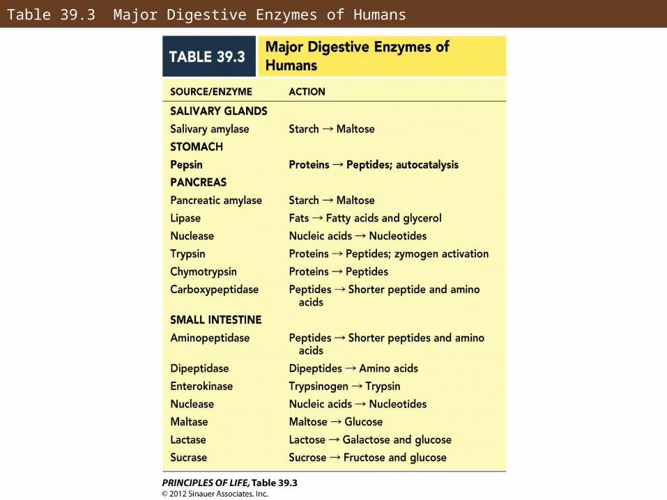
Table 39.3 Major Digestive Enzymes of Humans
Concept 39.3 The Vertebrate Digestive System Is a Tubular Gut with Accessory Glands
Breakdown of complex molecules continues in the jejunum.
Absorption of the breakdown products takes place in the jejunum and ileum.
The microvilli absorb nutrients and inorganic ions through several methods.
• Na+ and other ions are actively transported —sometimes with “hitchhikers.”
• Water moving through spaces between cells can carry nutrients.
Concept 39.3 The Vertebrate Digestive System Is a Tubular Gut with Accessory Glands
• Lipid-soluble fats pass through the villi membrane.
Fats are then re-formed into chylomicrons with a protein coat that makes them soluble in water.
Chylomicrons flow through the lymphatic system and then enter the blood stream through the thoracic ducts.

Figure 39.11 Digestion and Absorption of Fats
Concept 39.3 The Vertebrate Digestive System Is a Tubular Gut with Accessory Glands
Blood leaving the digestive tract goes to the liver via the hepatic portal vein.
Liver cells absorb nutrients and store them or convert them for use.
Contents of the small intestine pass into the large intestine, or colon, that absorbs water and ions, and produces feces, which are stored in the rectum.
Too much water absorption in the colon leads to constipation; too little leads to diarrhea.
Concept 39.4 Food Intake and Metabolism Are Regulated
Animals do not eat continuously and are in one of two states:
Absorptive state—the period after a meal when food is in the gut and nutrients are absorbed
Postabsorptive state—stomach and small intestine are empty and metabolism runs on stored nutrients
Concept 39.4 Food Intake and Metabolism Are Regulated
Digestion is governed by neuronal and hormonal controls.
The enteric nervous system is able to coordinate digestion.
Many hormones regulate the digestive tract and its accessory organs.
Concept 39.4 Food Intake and Metabolism Are Regulated
Digestive hormones and sites of production:
• Gastrin—released by the stomach into the blood when food is present
• Secretin—produced when chyme arrives in the duodenum, causes pancreas to secrete bicarbonate ions
• Cholecystokinin (CCK)—released by duodenal cells, causes gallbladder to release bile, stimulates pancreas, slows stomach
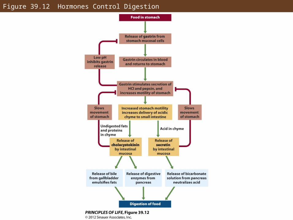
Figure 39.12 Hormones Control Digestion
Concept 39.4 Food Intake and Metabolism Are Regulated
Insulin is the major hormone in blood glucose regulation.
Insulin is released by β cells of the islets of Langerhans in the pancreas when blood glucose rises.
Glucose transporters are inserted into cell membranes in response to insulin—more glucose can move in to cells.
Concept 39.4 Food Intake and Metabolism Are Regulated
Blood glucose falls in the postabsorptive period.
Insulin release is lower and glucose uptake is slowed.
If glucose level is very low, glucagon is released and causes the liver to break down glycogen and begin gluconeogenesis.
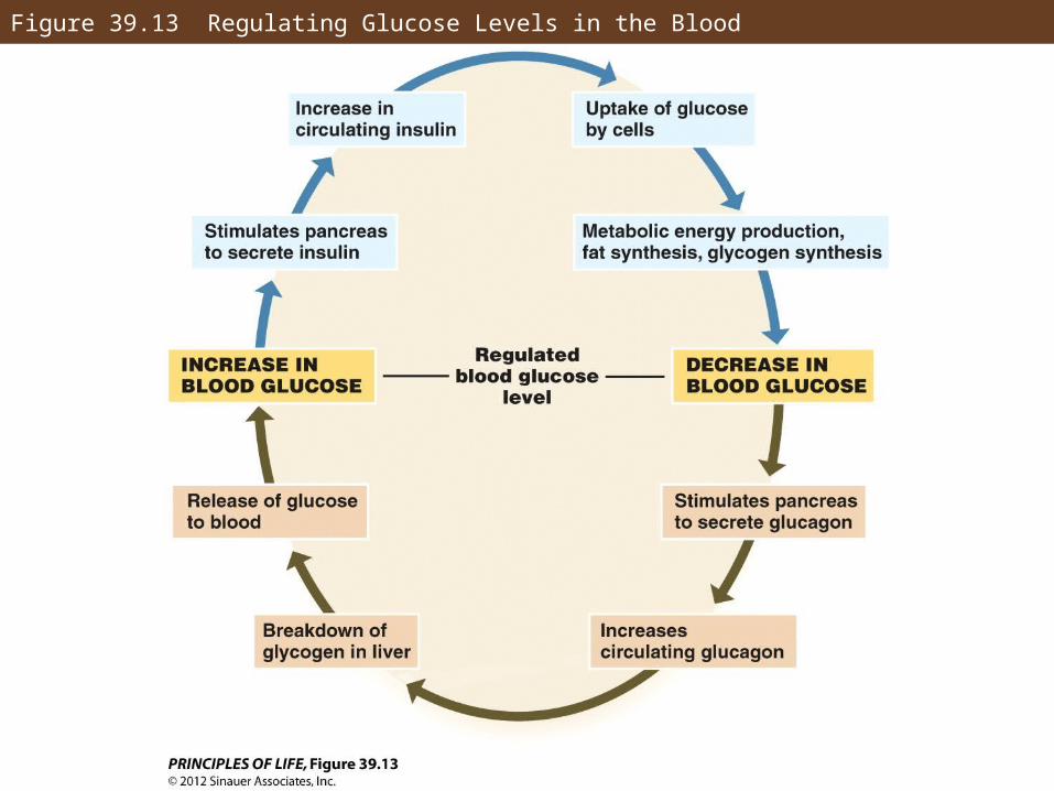
Figure 39.13 Regulating Glucose Levels in the Blood
Concept 39.4 Food Intake and Metabolism Are Regulated
Diabetes mellitus is a lack of insulin or the inability to respond to insulin.
Type I is the lack of cells that produce insulin, also called juvenile diabetes—treated by supplemental insulin.
Type II, adult diabetes, is a poor response to insulin and cells are unable to absorb enough to meet metabolic needs—treated by changes in diet and lifestyle.
Concept 39.4 Food Intake and Metabolism Are Regulated
The liver coordinates changes between states.
During absorptive state the liver stores fuel as glycogen and fats, and synthesizes blood plasma proteins.
During postabsorptive state the liver can deliver nutrients into the blood from its reserves.
Concept 39.4 Food Intake and Metabolism Are Regulated
Lipoproteins aid in fat transport and are produced in the liver:
• High-density lipoproteins (HDLs) remove cholesterol from tissue and carry it to liver
• Low-density lipoproteins (LDLs) transport cholesterol in body
• Very-low-density lipoproteins (VLDLs) transport triglycerides to fat cells
Concept 39.4 Food Intake and Metabolism Are Regulated
The hypothalamus provides signals of hunger or satiety and governs how much food is eaten.
Leptin is a hormone produced by adipose cells with receptors in the hypothalamus.
Leptin provides feedback about body fat to the brain—the more fat a cell contains, the more leptin it produces.
Concept 39.4 Food Intake and Metabolism Are Regulated
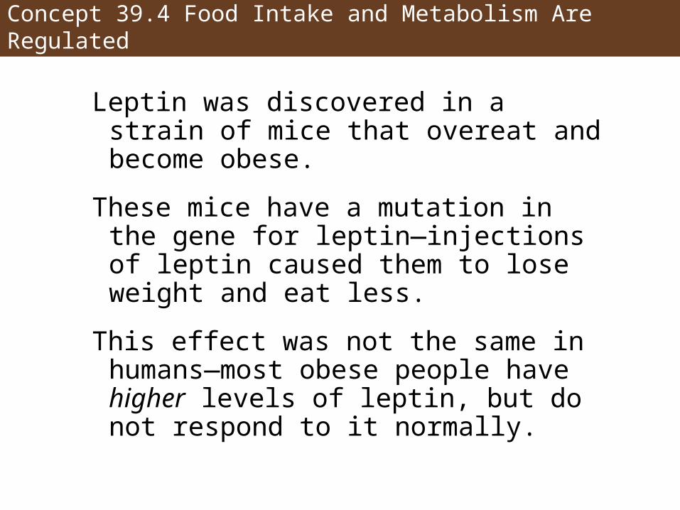
Leptin was discovered in a strain of mice that overeat and become obese.
These mice have a mutation in the gene for leptin—injections of leptin caused them to lose weight and eat less.
This effect was not the same in humans—most obese people have higher levels of leptin, but do not respond to it normally.
Concept 39.4 Food Intake and Metabolism Are Regulated
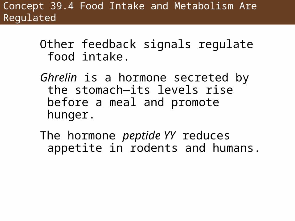
Other feedback signals regulate food intake.
Ghrelin is a hormone secreted by the stomach—its levels rise before a meal and promote hunger.
The hormone peptide YY reduces appetite in rodents and humans.
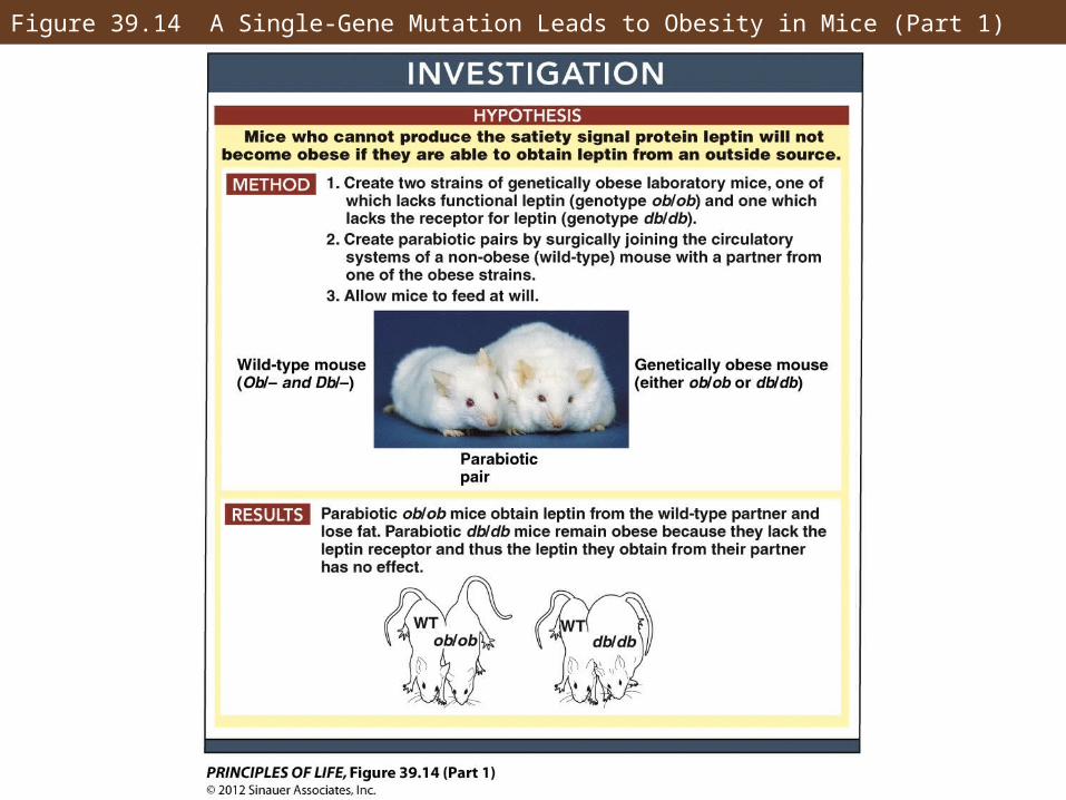
Figure 39.14 A Single-Gene Mutation Leads to Obesity in Mice (Part 1)
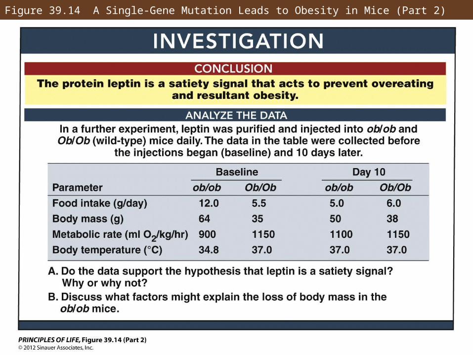
Figure 39.14 A Single-Gene Mutation Leads to Obesity in Mice (Part 2)
Answer to Opening Question
Genetics and lifestyle both contribute to the Pimas’ diabetes and obesity.
The Arizona Pimas have a much greater insulin response to glucose than people of other heritage do—one effect of that is increased fat synthesis.
Their lifestyle as compared to the Mexican Pimas is much more sedentary, with a poorer diet.