digestion & absorption chapter 3. outline digestive tracts the 4 stages ingestion digestion ...
TRANSCRIPT
DIGESTION & ABSORPTION
Chapter 3
Outline
Digestive Tracts The 4 Stages
Ingestion Digestion Absorption Egestion
Nutrition
Overview: Digestion and Absorption
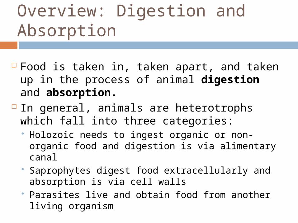
Food is taken in, taken apart, and taken up in the process of animal digestion and absorption.
In general, animals are heterotrophs which fall into three categories: Holozoic needs to ingest organic or non-organic
food and digestion is via alimentary canal Saprophytes digest food extracellularly and
absorption is via cell walls Parasites live and obtain food from another
living organism
The 4 Main Stages : Ingestion
Ingestion is the act of eating
Four types of feeders: Suspension Feeders
Substrate Feeders
Fluid Feeders
Bulk feeders
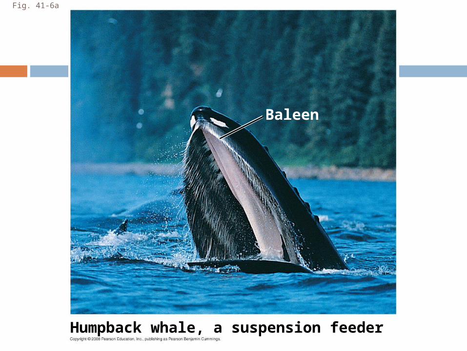
Fig. 41-6a
Humpback whale, a suspension feeder
Baleen
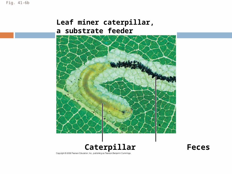
Fig. 41-6b
Leaf miner caterpillar,a substrate feeder
Caterpillar Feces
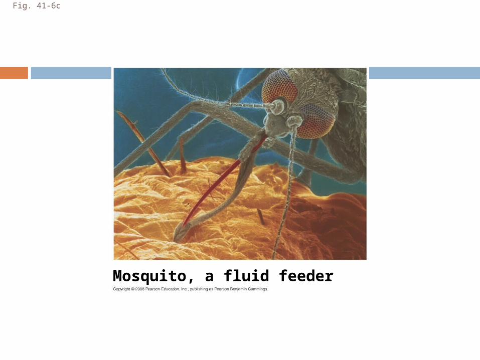
Fig. 41-6c
Mosquito, a fluid feeder
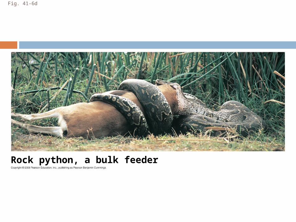
Fig. 41-6d
Rock python, a bulk feeder
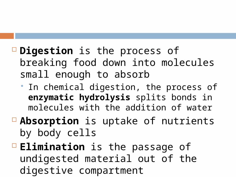
Digestion is the process of breaking food down into molecules small enough to absorb In chemical digestion, the process of
enzymatic hydrolysis splits bonds in molecules with the addition of water
Absorption is uptake of nutrients by body cells
Elimination is the passage of undigested material out of the digestive compartment
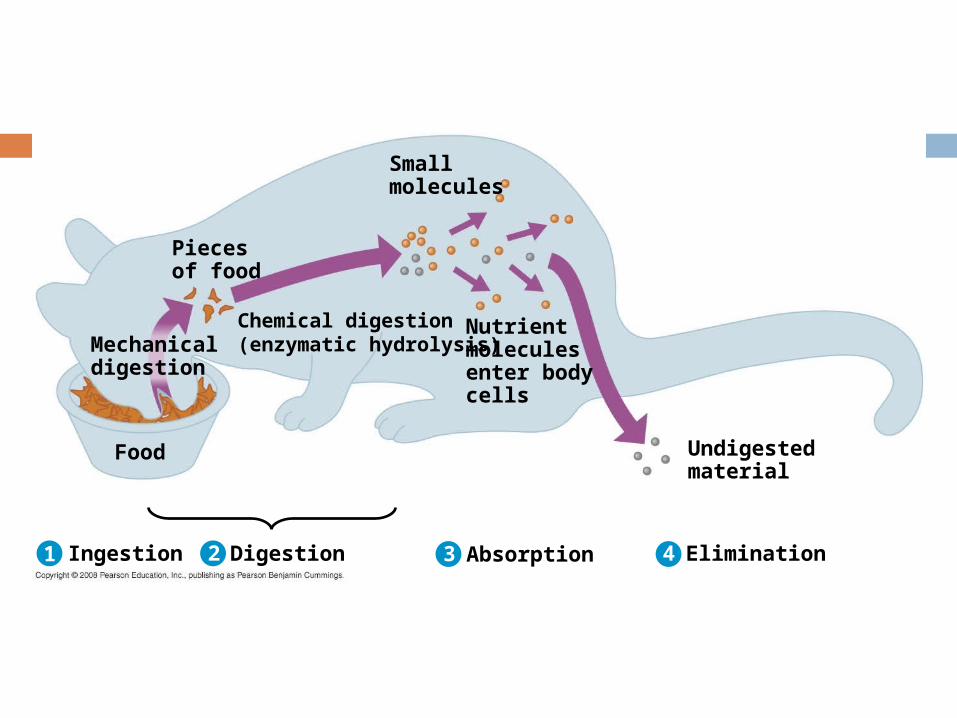
Ingestion Digestion Absorption Elimination
Undigestedmaterial
Chemical digestion(enzymatic hydrolysis)
Nutrientmoleculesenter bodycells
Smallmolecules
Mechanicaldigestion
Food
Piecesof food
1 2 3 4
Digestive Compartments
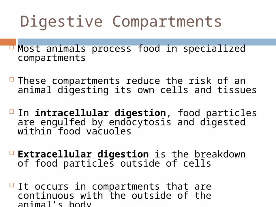
Most animals process food in specialized compartments
These compartments reduce the risk of an animal digesting its own cells and tissues
In intracellular digestion, food particles are engulfed by endocytosis and digested within food vacuoles
Extracellular digestion is the breakdown of food particles outside of cells
It occurs in compartments that are continuous with the outside of the animal’s body
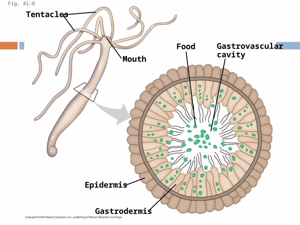
Fig. 41-8
Gastrovascularcavity
Food
Epidermis
Mouth
Tentacles
Gastrodermis
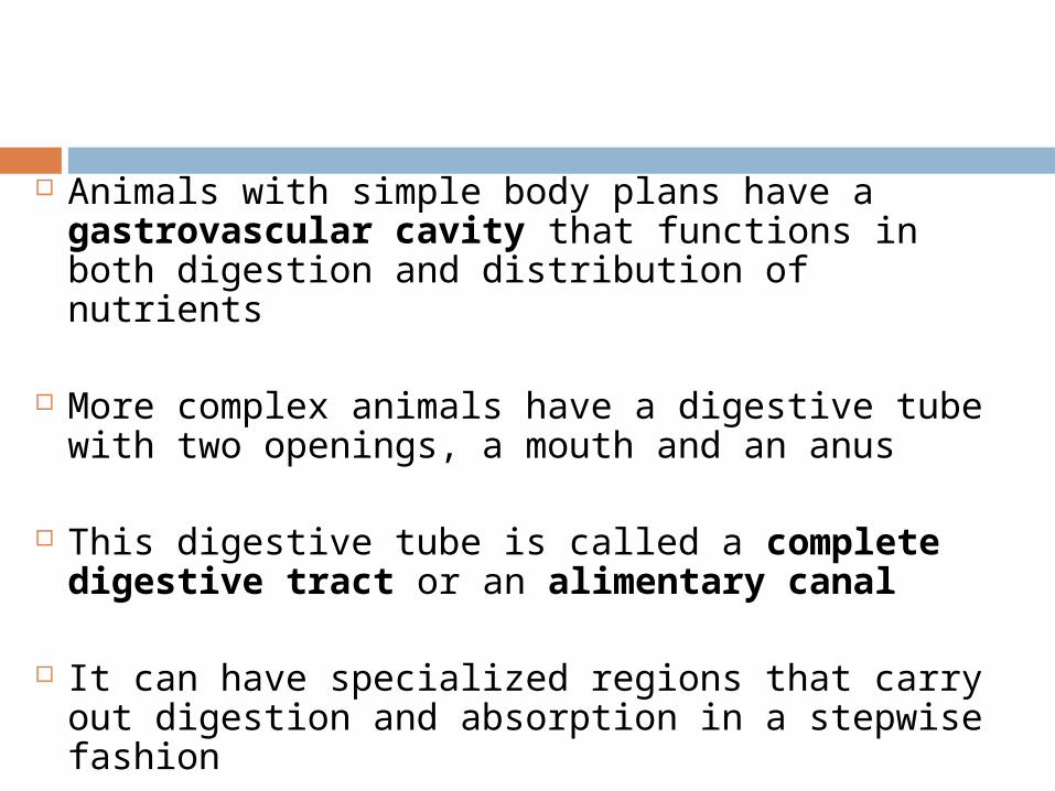
Animals with simple body plans have a gastrovascular cavity that functions in both digestion and distribution of nutrients
More complex animals have a digestive tube with two openings, a mouth and an anus
This digestive tube is called a complete digestive tract or an alimentary canal
It can have specialized regions that carry out digestion and absorption in a stepwise fashion
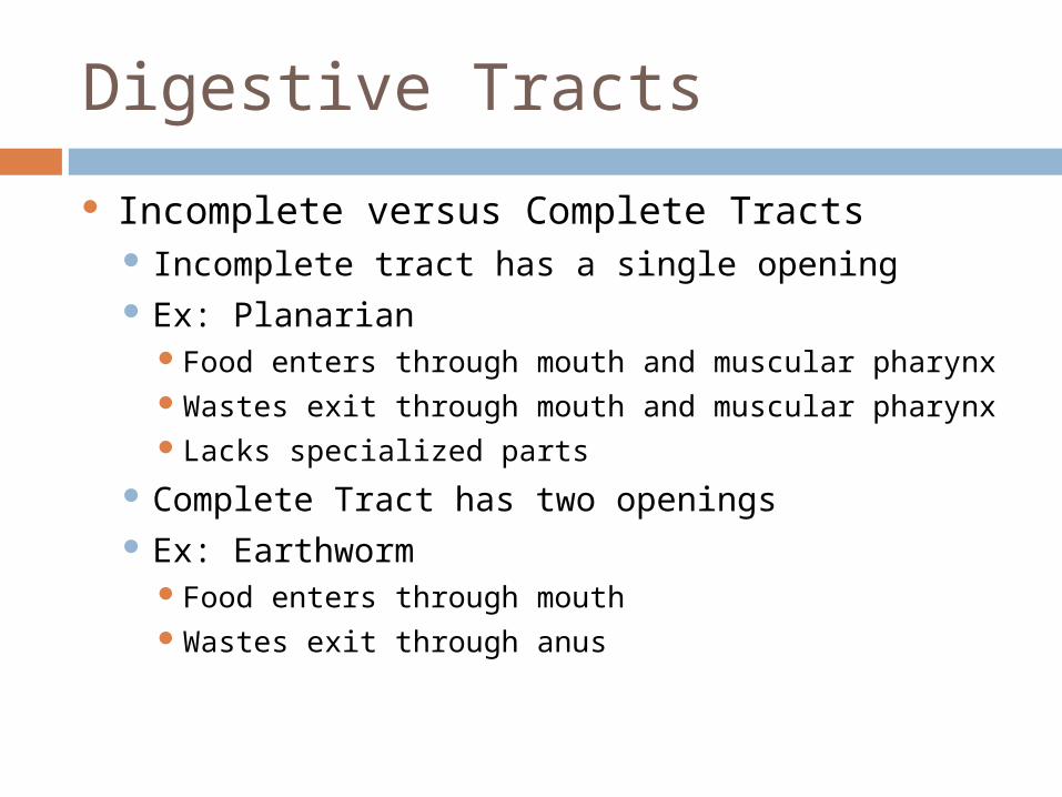
Digestive Tracts
Incomplete versus Complete Tracts Incomplete tract has a single opening Ex: Planarian
Food enters through mouth and muscular pharynxWastes exit through mouth and muscular pharynxLacks specialized parts
Complete Tract has two openings Ex: Earthworm
Food enters through mouthWastes exit through anus
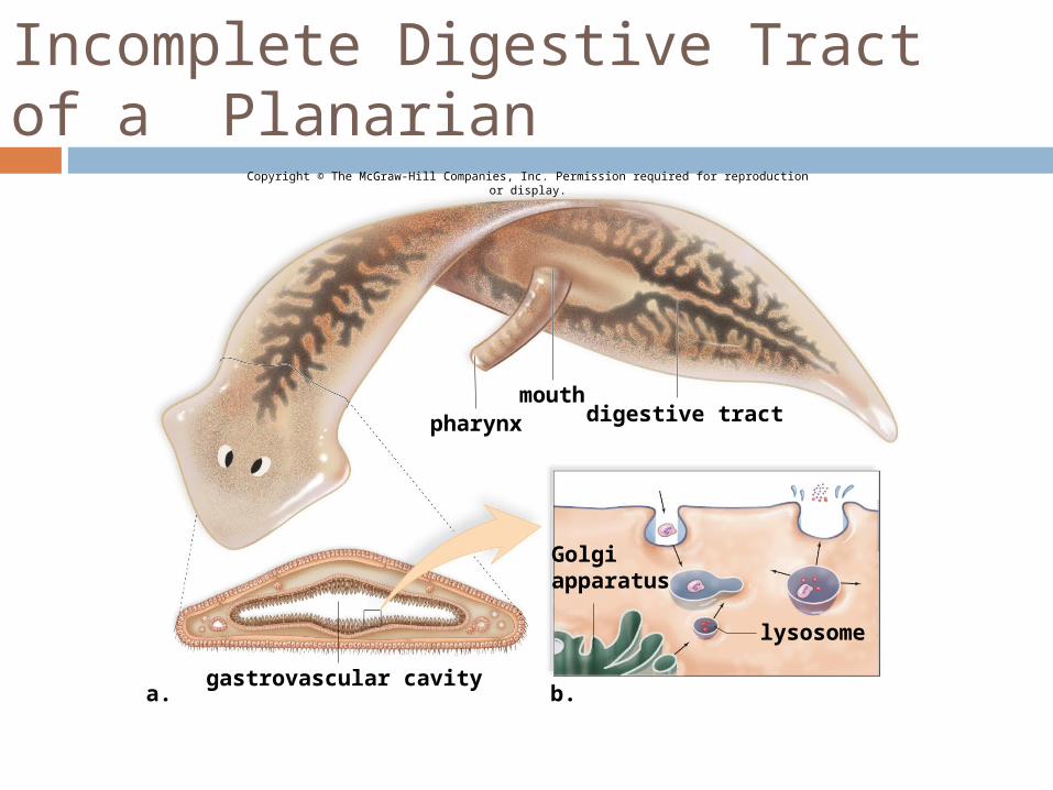
Incomplete Digestive Tract of a Planarian
gastrovascular cavity
lysosome
mouthpharynx digestive tract
a. b.
Golgiapparatus
Copyright © The McGraw-Hill Companies, Inc. Permission required for reproduction or display.
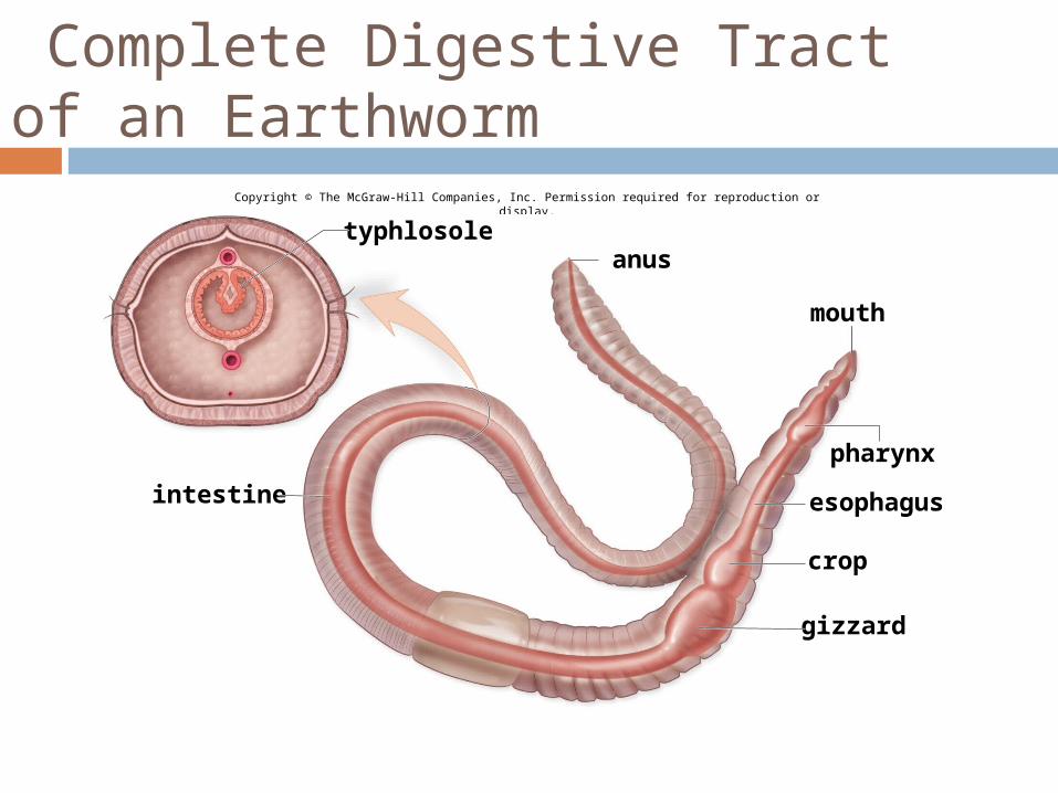
Complete Digestive Tract of an Earthworm
Copyright © The McGraw-Hill Companies, Inc. Permission required for reproduction or display.
anus
intestine
crop
gizzard
pharynx
mouth
esophagus
typhlosole
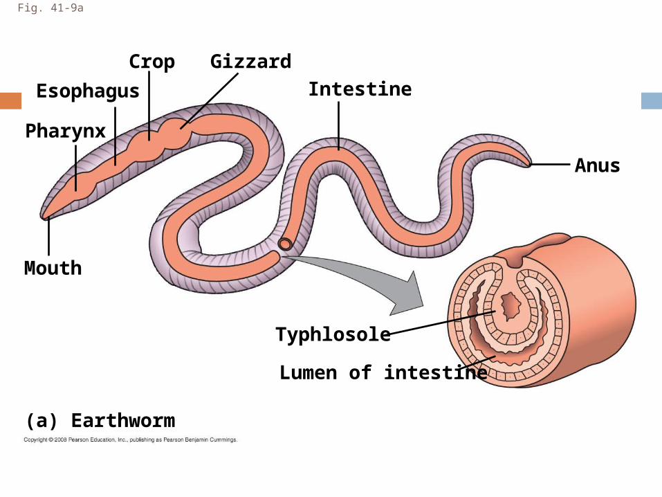
Fig. 41-9a
Esophagus
Mouth
Pharynx
Crop Gizzard
Typhlosole
Intestine
Lumen of intestine
Anus
(a) Earthworm
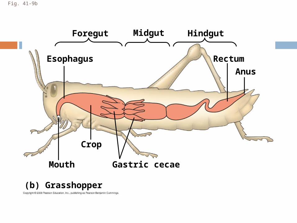
Fig. 41-9b
(b) Grasshopper
Foregut
Mouth
Crop
Gastric cecae
Esophagus Rectum
Anus
Midgut Hindgut

Fig. 41-9c
(c) Bird
StomachGizzard
Intestine
Esophagus
Anus
Crop
Mouth
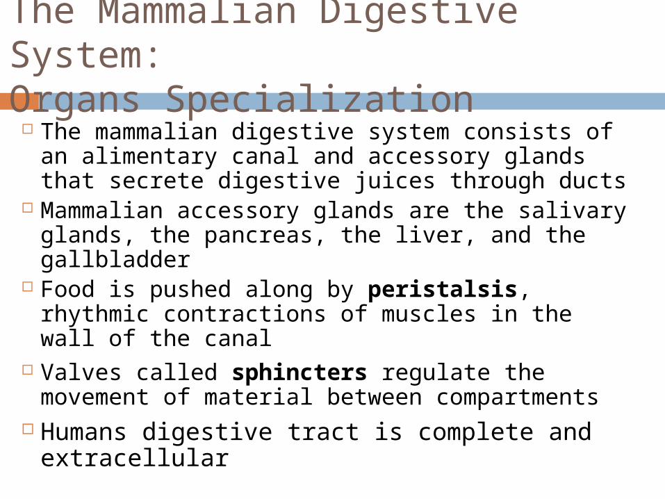
The Mammalian Digestive System: Organs Specialization
The mammalian digestive system consists of an alimentary canal and accessory glands that secrete digestive juices through ducts
Mammalian accessory glands are the salivary glands, the pancreas, the liver, and the gallbladder
Food is pushed along by peristalsis, rhythmic contractions of muscles in the wall of the canal
Valves called sphincters regulate the movement of material between compartments
Humans digestive tract is complete and extracellular
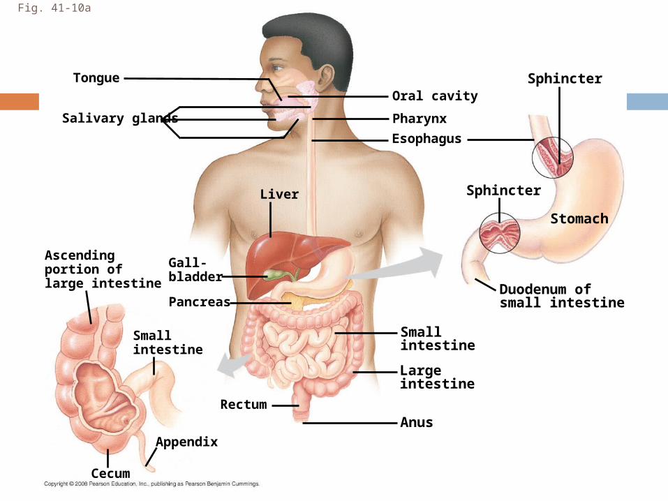
Fig. 41-10a
Cecum
Anus
Ascendingportion oflarge intestine
Gall-bladder
Smallintestine
Largeintestine
Smallintestine
Rectum
Pancreas
Liver
Salivary glands
TongueOral cavity
Pharynx
Esophagus
Sphincter
Stomach
Sphincter
Duodenum ofsmall intestine
Appendix
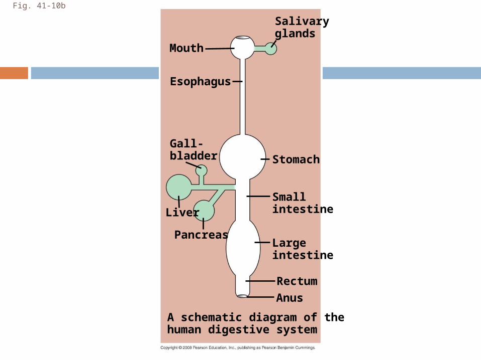
Fig. 41-10b
Anus
Liver
Pancreas
Smallintestine
Largeintestine
Rectum
StomachGall-bladder
A schematic diagram of thehuman digestive system
Esophagus
Salivaryglands
Mouth
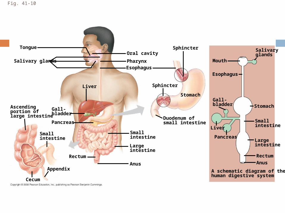
Fig. 41-10
Cecum
Anus Anus
Ascendingportion oflarge intestine
Gall-bladder
Smallintestine
Largeintestine
Smallintestine
Rectum
Pancreas
Liver
Salivary glands
TongueOral cavity
PharynxEsophagus
Sphincter
Stomach
Sphincter
Duodenum ofsmall intestine
Appendix
Liver
Pancreas
Smallintestine
Largeintestine
Rectum
StomachGall-bladder
A schematic diagram of thehuman digestive system
Esophagus
Salivaryglands
Mouth
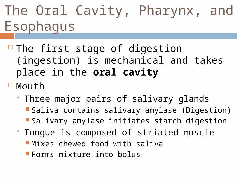
The Oral Cavity, Pharynx, and Esophagus The first stage of digestion (ingestion) is
mechanical and takes place in the oral cavity
Mouth Three major pairs of salivary glands
Saliva contains salivary amylase (Digestion) Salivary amylase initiates starch digestion
Tongue is composed of striated muscle Mixes chewed food with saliva Forms mixture into bolus
25
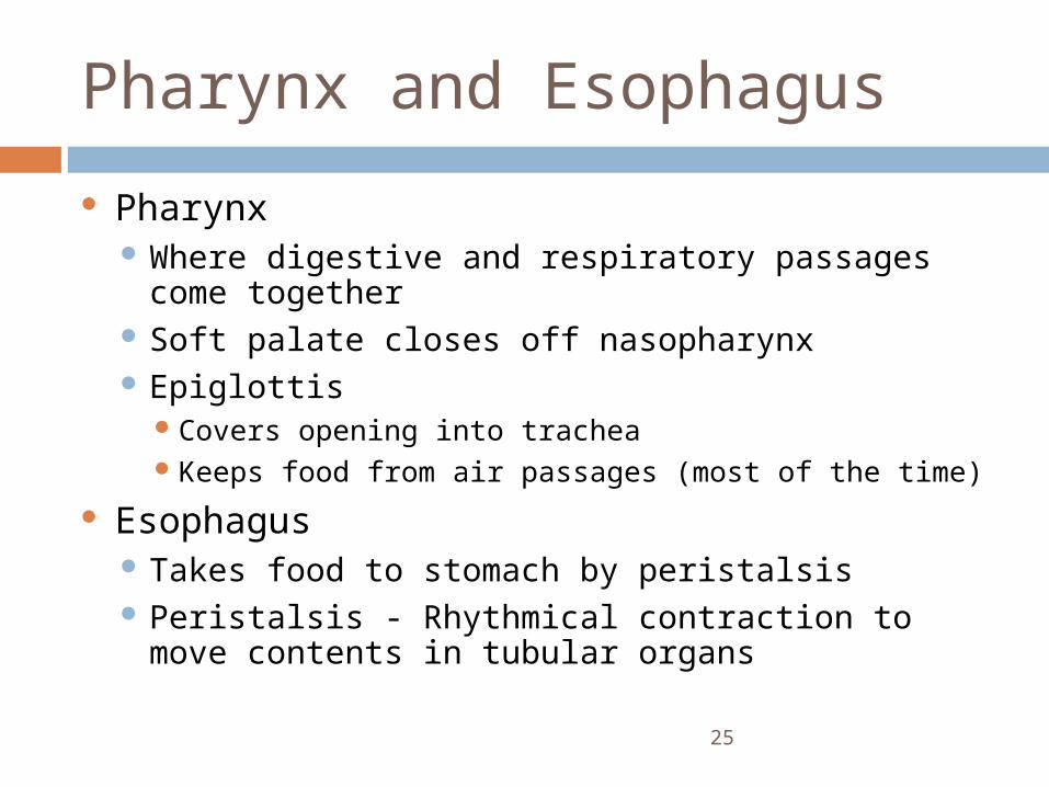
Pharynx and Esophagus
Pharynx Where digestive and respiratory passages
come together Soft palate closes off nasopharynx Epiglottis
Covers opening into tracheaKeeps food from air passages (most of the time)
Esophagus Takes food to stomach by peristalsis Peristalsis - Rhythmical contraction to move
contents in tubular organs
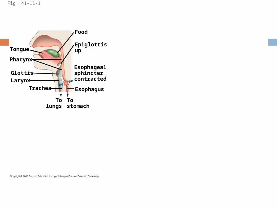
Fig. 41-11-1
LarynxTrachea
Epiglottisup
Pharynx
Tongue
Glottis
Esophagus
Esophagealsphinctercontracted
Food
Tostomach
Tolungs
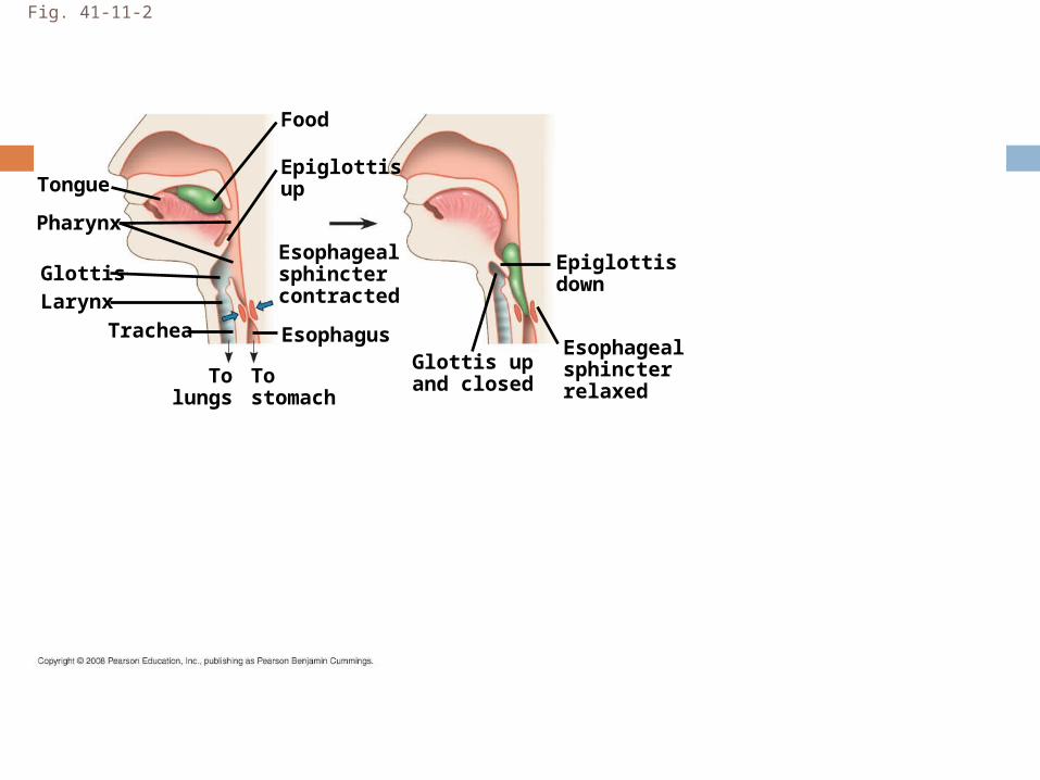
Fig. 41-11-2
LarynxTrachea
Epiglottisup
Pharynx
Tongue
Glottis
Esophagus
Esophagealsphinctercontracted
Food
Tostomach
Tolungs
Epiglottisdown
Esophagealsphincterrelaxed
Glottis upand closed
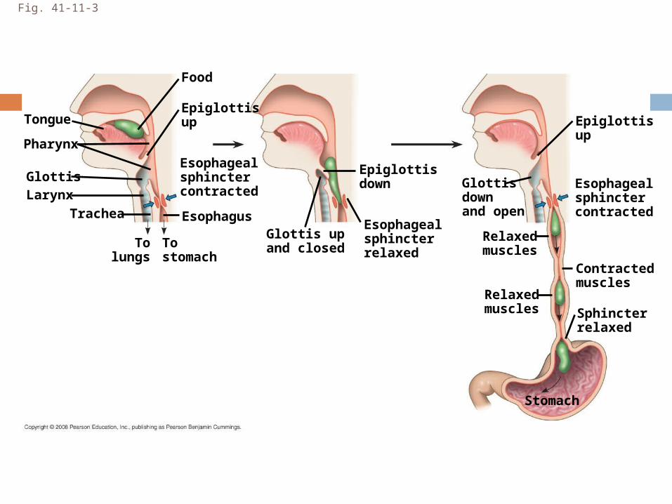
Fig. 41-11-3
LarynxTrachea
Epiglottisup
Pharynx
Tongue
Glottis
Esophagus
Esophagealsphinctercontracted
Food
Tostomach
Tolungs
Epiglottisdown
Esophagealsphincterrelaxed
Glottis upand closed
Epiglottisup
Esophagealsphinctercontracted
Sphincterrelaxed
Relaxedmuscles
Contractedmuscles
Relaxedmuscles
Stomach
Glottisdownand open
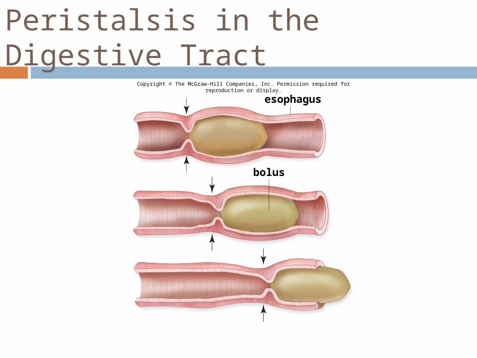
Peristalsis in the Digestive Tract
Copyright © The McGraw-Hill Companies, Inc. Permission required for reproduction or display.
esophagus
bolus
Stage 2: Digestion (Stomach)
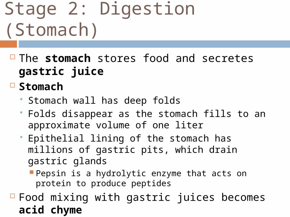
The stomach stores food and secretes gastric juice
Stomach Stomach wall has deep folds Folds disappear as the stomach fills to an
approximate volume of one liter Epithelial lining of the stomach has millions of
gastric pits, which drain gastric glands Pepsin is a hydrolytic enzyme that acts on protein
to produce peptides Food mixing with gastric juices becomes
acid chyme
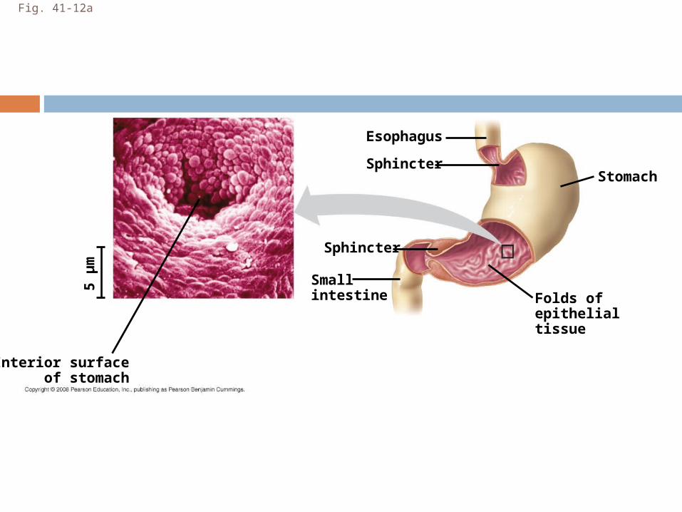
Fig. 41-12a
Esophagus
Small intestine
Stomach
Sphincter
Folds ofepithelialtissue
Sphincter
5 µ
m
Interior surfaceof stomach
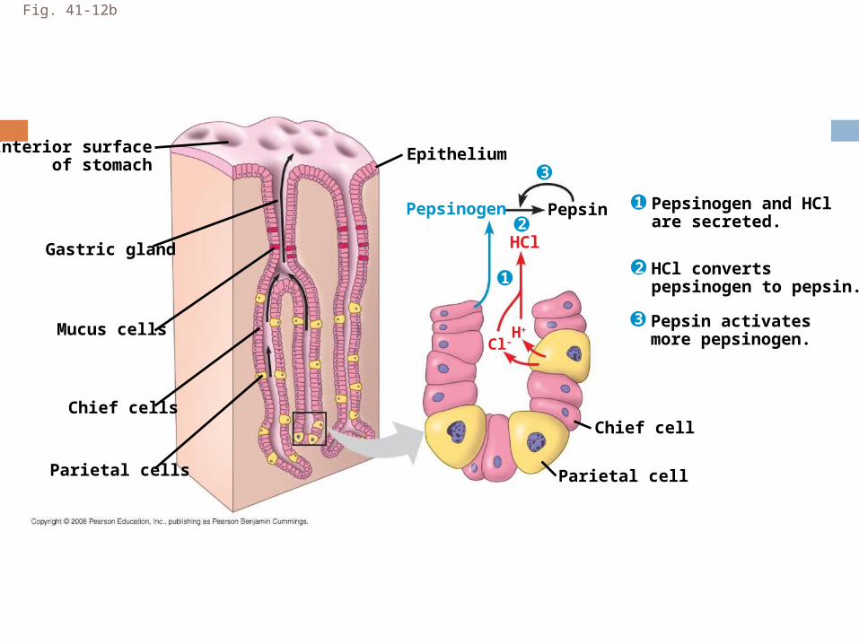
Fig. 41-12b
Interior surfaceof stomach
Chief cells
Epithelium
Parietal cell
Pepsinogen and HClare secreted.
HCl convertspepsinogen to pepsin.
Pepsin activatesmore pepsinogen.
Chief cell
PepsinPepsinogen
HCl
H+
Cl–
Parietal cells
Mucus cells
Gastric gland2
3
1
1
2
3
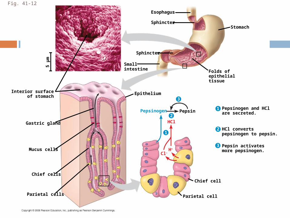
Fig. 41-12
Interior surfaceof stomach
Esophagus
Chief cells
Small intestine
Epithelium
Stomach
Sphincter
Parietal cell
Pepsinogen and HClare secreted.
HCl convertspepsinogen to pepsin.
Pepsin activatesmore pepsinogen.
Chief cell
Folds ofepithelialtissue
Pepsin
Sphincter
Pepsinogen
HCl
H+
Cl–
Parietal cells
Mucus cells
Gastric gland
1
2
2
3
3
1
5 µ
m
STAGE 2: Digestion (Small Intestine)
The small intestine is the longest section of the alimentary canal
It is the major organ of digestion and absorption
The first portion of the small intestine is the duodenum, where acid chyme from the stomach mixes with digestive juices from the pancreas, liver, gallbladder, and the small intestine itself

Stomach
GallbladderLiver
Duodenum ofsmall intestine
Bile
Pancreas
Duodenum: The Pancreas
The pancreas produces proteases trypsin and chymotrypsin, protein-digesting enzymes that are activated after entering the duodenum
Its solution is alkaline and neutralizes the acidic chyme
The pancreas produces proteases trypsin and chymotrypsin, protein-digesting enzymes that are activated after entering the duodenum
Its solution is alkaline and neutralizes the acidic chyme
Duodenum: The Liver
In the small intestine, bile aids in digestion and absorption of fats
Bile is made in the liver and stored in the gallbladder
Bile contains bile salts which break up fat into fat droplets via emulsification
Helps maintain glucose concentration in blood by converting excess into glycogen
Small Intestine
The epithelial lining of the duodenum, called the brush border, produces several digestive enzymes
Enzymatic digestion is completed as peristalsis moves the chyme and digestive juices along the small intestine
Most digestion occurs in the duodenum; the jejunum and ileum function mainly in absorption of nutrients and water
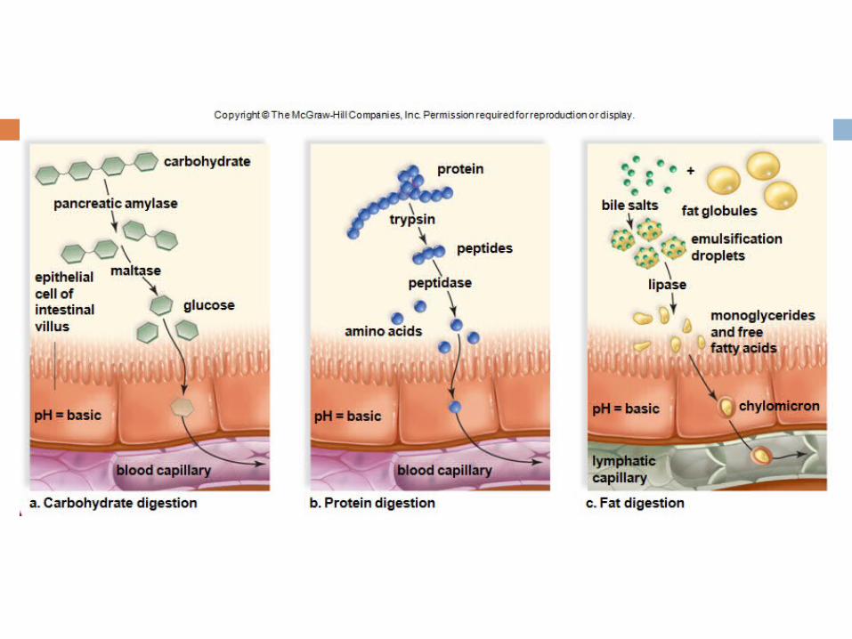
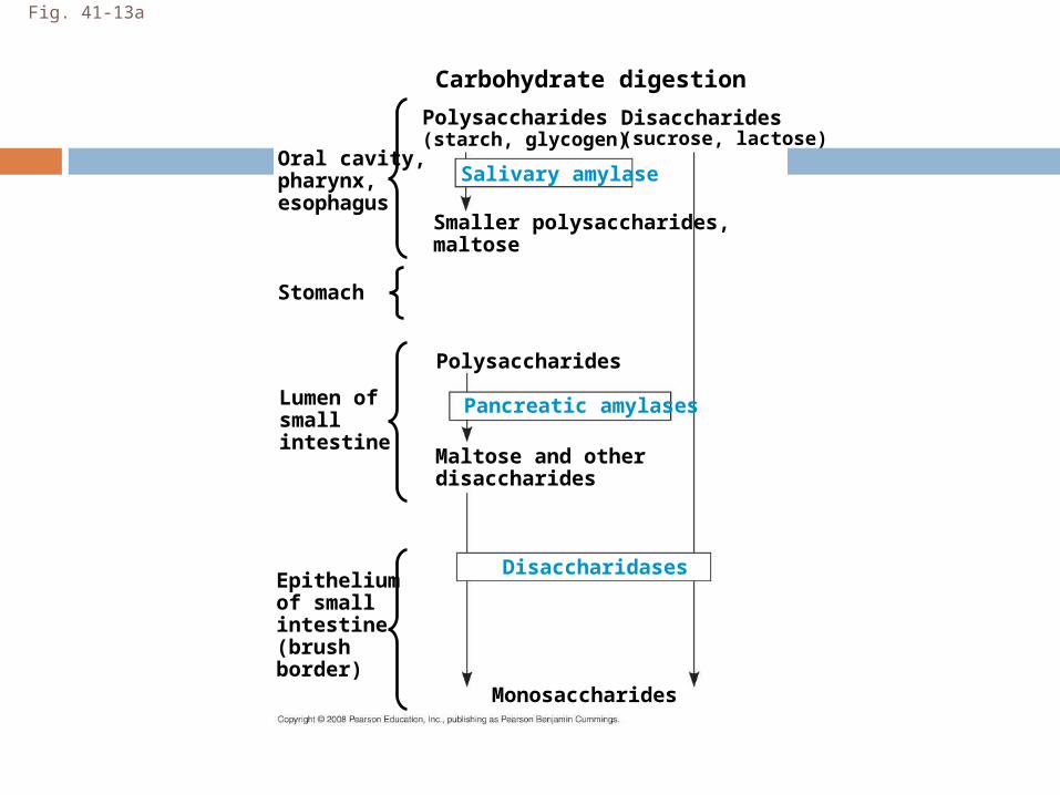
Fig. 41-13a
Oral cavity,pharynx,esophagus
Stomach
Lumen ofsmall intestine
Epitheliumof smallintestine(brushborder)
Carbohydrate digestion
Polysaccharides
Smaller polysaccharides,maltose
Polysaccharides
Maltose and otherdisaccharides
Disaccharides
Pancreatic amylases
Salivary amylase
Disaccharidases
Monosaccharides
(starch, glycogen)(sucrose, lactose)
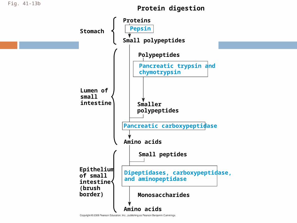
Fig. 41-13b
Stomach
Lumen ofsmall intestine
Epitheliumof smallintestine(brushborder)
Protein digestion
Proteins
Polypeptides
Smallerpolypeptides
Pancreatic trypsin andchymotrypsin
Pepsin
Dipeptidases, carboxypeptidase,and aminopeptidase
Monosaccharides
Small polypeptides
Amino acids
Pancreatic carboxypeptidase
Amino acids
Small peptides
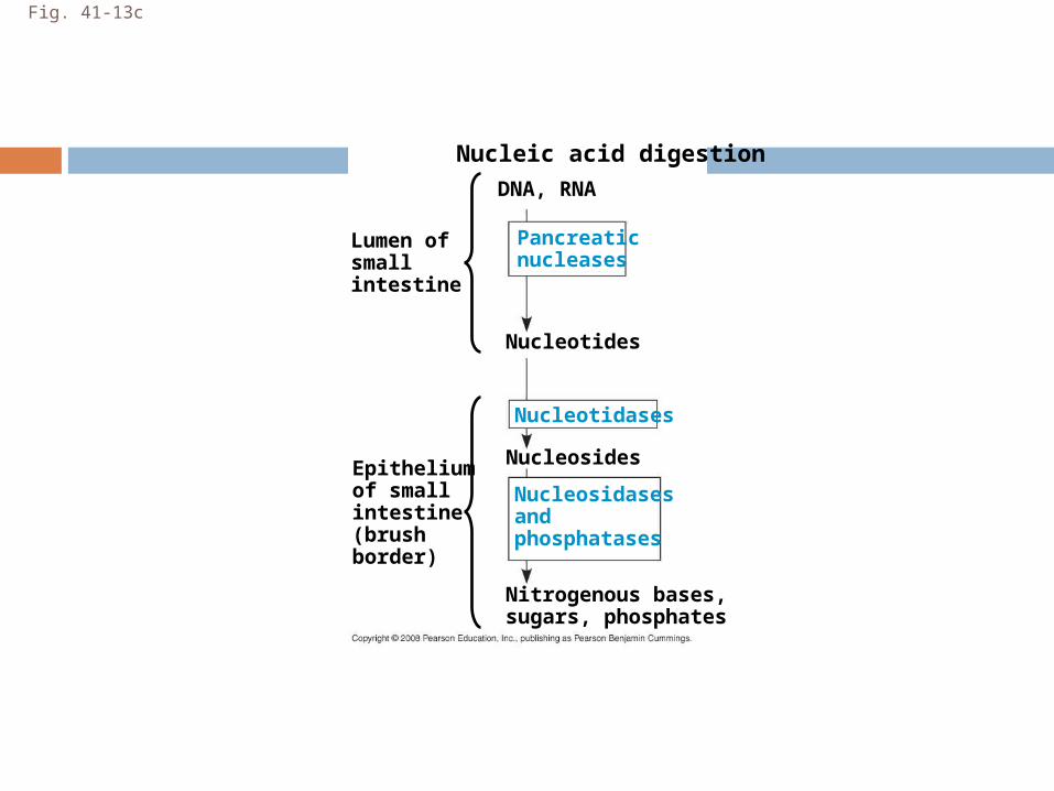
Fig. 41-13c
Lumen ofsmall intestine
Epitheliumof smallintestine(brushborder)
Nucleic acid digestion
DNA, RNA
Nucleotides
Pancreaticnucleases
Nucleosidasesandphosphatases
Nucleosides
Nucleotidases
Nitrogenous bases,sugars, phosphates
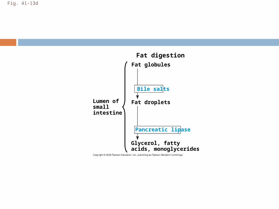
Fig. 41-13d
Lumen ofsmall intestine
Fat digestion
Fat globules
Fat droplets
Pancreatic lipase
Bile salts
Glycerol, fattyacids, monoglycerides
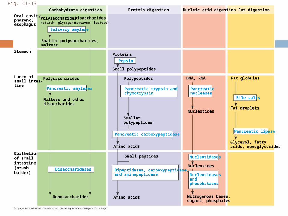
Fig. 41-13
Oral cavity,pharynx,esophagus
Stomach
Lumen ofsmall intes-tine
Epitheliumof smallintestine(brushborder)
Carbohydrate digestion
Polysaccharides
Smaller polysaccharides,maltose
Polysaccharides
Maltose and otherdisaccharides
Disaccharides
Protein digestion Nucleic acid digestion Fat digestion
Proteins
Small polypeptides
Pepsin
Pancreatic amylases
Salivary amylase
Disaccharidases
Monosaccharides
Small peptides
Amino acids
Amino acids
Polypeptides
Smallerpolypeptides
Pancreatic trypsin andchymotrypsin
Pancreatic carboxypeptidase
Dipeptidases, carboxypeptidase,and aminopeptidase
DNA, RNA
Pancreatic nucleases
Fat globules
NucleotidesFat droplets
Nucleosides
Nitrogenous bases,sugars, phosphates
Nucleotidases
Nucleosidasesandphosphatases
Glycerol, fattyacids, monoglycerides
Bile salts
Pancreatic lipase
(starch, glycogen) (sucrose, lactose)
STAGE 3: Absorption (Small Intestines)
The small intestine has a huge surface area, due to villi and microvilli that are exposed to the intestinal lumen
The enormous microvillar surface greatly increases the rate of nutrient absorption
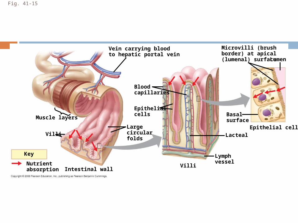
Fig. 41-15
Muscle layers
Microvilli (brushborder) at apical(lumenal) surface
Vein carrying bloodto hepatic portal vein
Villi
Intestinal wall
Key
Nutrientabsorption
Largecircularfolds
Bloodcapillaries
Epithelialcells
Villi
Lymphvessel
Basal surface
LactealEpithelial cells
Lumen
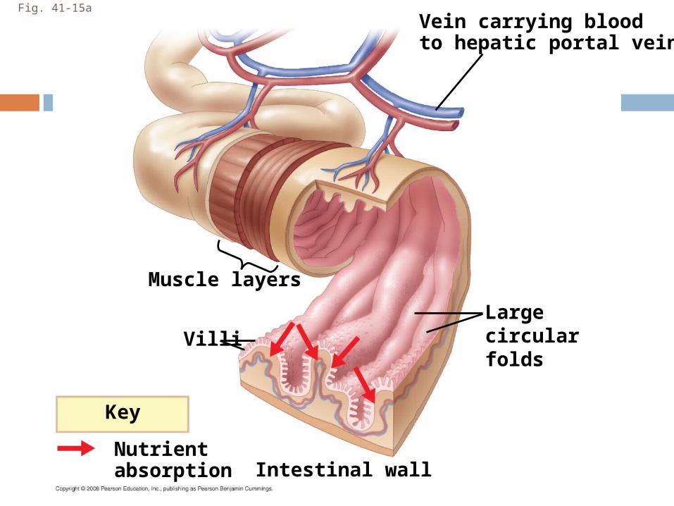
Fig. 41-15a
Muscle layers
Vein carrying bloodto hepatic portal vein
Villi
Intestinal wall
Key
Nutrientabsorption
Largecircularfolds
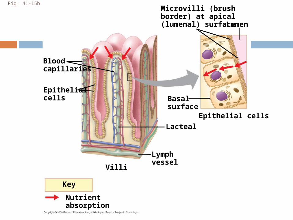
Fig. 41-15bMicrovilli (brushborder) at apical(lumenal) surface
Key
Nutrientabsorption
Bloodcapillaries
Epithelialcells
Villi
Lymphvessel
Basal surface
Lacteal
Epithelial cells
Lumen
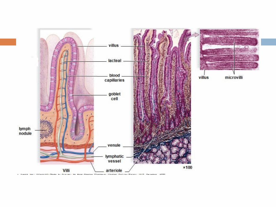
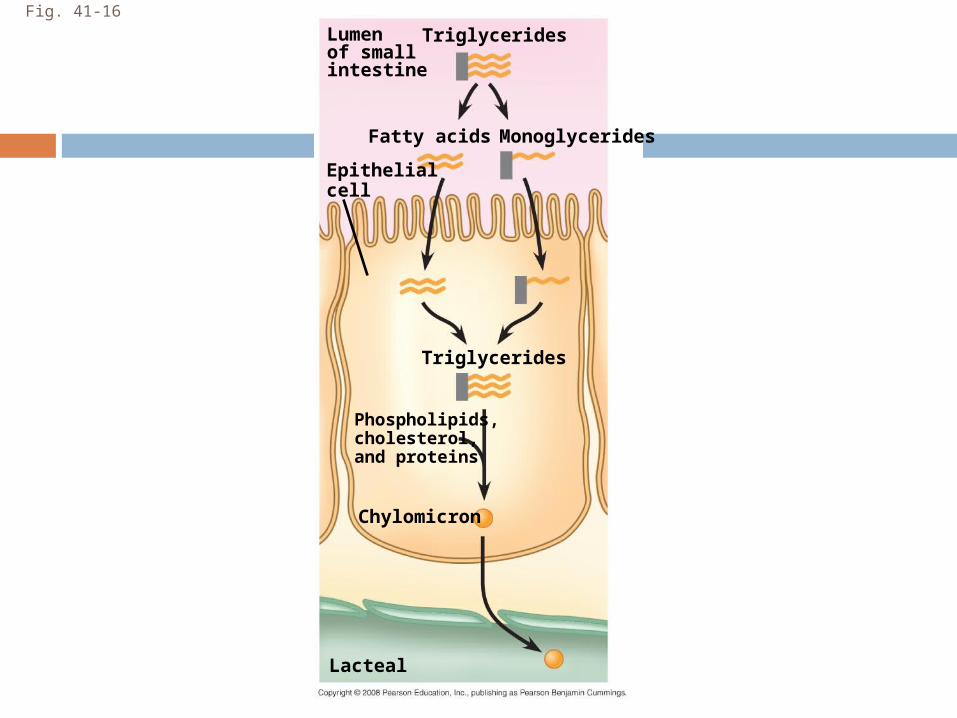
Fig. 41-16
Lumenof smallintestine
Lacteal
Chylomicron
Phospholipids,cholesterol,and proteins
Triglycerides
Monoglycerides
Triglycerides
Fatty acids
Epithelialcell
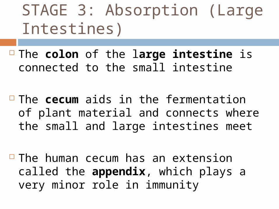
STAGE 3: Absorption (Large Intestines)
The colon of the large intestine is connected to the small intestine
The cecum aids in the fermentation of plant material and connects where the small and large intestines meet
The human cecum has an extension called the appendix, which plays a very minor role in immunity
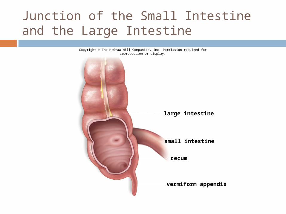
Junction of the Small Intestine and the Large Intestine
Copyright © The McGraw-Hill Companies, Inc. Permission required for reproduction or display.
large intestine
small intestine
vermiform appendix
cecum
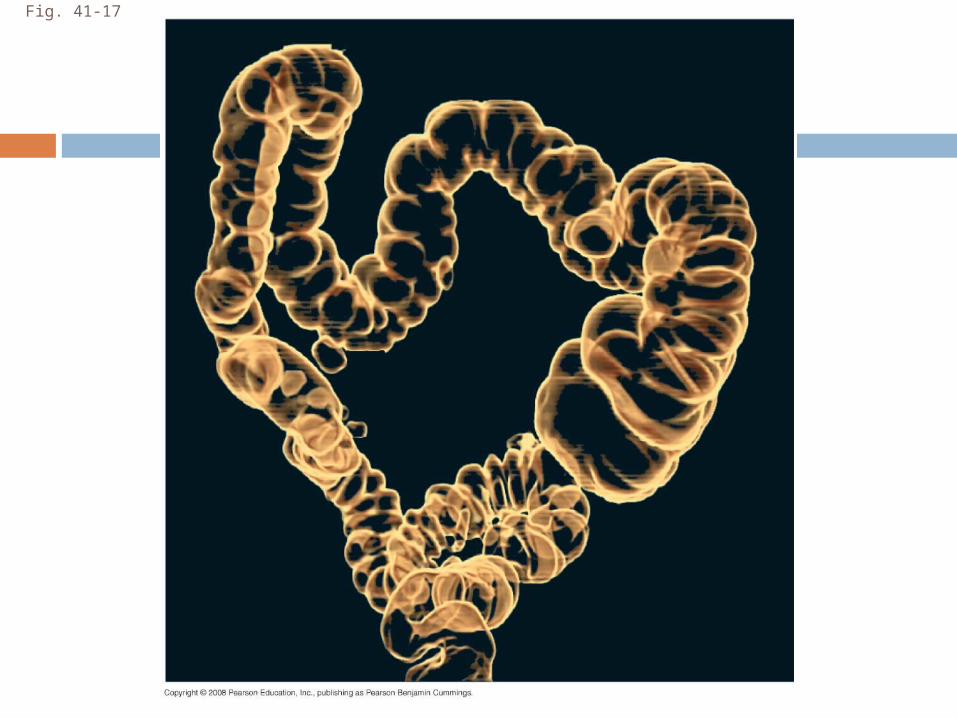
Fig. 41-17
A major function of the colon is to recover water that has entered the alimentary canal
Wastes of the digestive tract, the feces, become more solid as they move through the colon
Feces are stored in the rectum until they can be eliminated
Two sphincters between the rectum and anus control bowel movements
Feces pass through the rectum and exit via the anal canal where the opening is call the anus.
The colon houses strains of the enterobacteria Escherichia coli, some of which produce vitamins
STAGE 4: Egestion (Large Intestines)