noninvasive ventilation in the treatment of sleep-related breathing disorders: concise clinical...
TRANSCRIPT
REVIEW
Noninvasive ventilation in the treatment of sleep-related breathingdisorders: concise clinical review
Antonello Nicolini • Paolo Banfi • Cornelius Barlascini •
Gianluca Ferraioli • Agata Lax • Bruna Grecchi
Received: 27 September 2012 / Accepted: 29 April 2014
� Springer-Verlag Italia 2014
Abstract Noninvasive mechanical ventilation (NIPPV)
was originally used in patients with acute respiratory
compromises or exacerbations of chronic respiratory dis-
eases, as an alternative to the endotracheal tube. Over the
past 30 years NIPPV has been also used during the night in
patients with stable chronic lung disease such as obstruc-
tive sleep apnea, the overlap syndrome (COPD and
obstructive sleep apnea), neuromuscular disorders, obesity-
hypoventilation syndrome, and in other conditions such as
sleep disorders associated with congestive heart failure
(Cheyne–Stokes respiration). In this review we discuss the
different types of NIPPV, the specific conditions in which
they can be used and the indications, recommendations,
and evidence supporting the efficacy of NIPPV. Obstruc-
tive sleep apnea syndrome (OSA) is characterized com-
monly by instability of upper airway during sleep,
reduction or elimination of airflow, daytime hyper som-
nolence, and sleep disruption. It is a risk factor for car-
diovascular and cerebrovascular disorders including
hypertension, myocardial infarction and stroke. Optimizing
patient acceptance and adherence to noninvasive ventila-
tion treatment is challenging. The treatment of sleep-rela-
ted disorders is a life-threatening condition. The optimal
level of treatment should be determinated in a sleep labo-
ratory. Side effects directly affecting the patient’s adher-
ence to treatment are known. The most common are
nasopharyngeal symptoms including increased congestion
and rhinorrea; these effects are related to reduced humidity
of inspired gas. Humidification of delivered gas may
improve these symptoms. Sleep specialists should review
the results of objective testing with the patient and educate
the patient concerning the nature of the disorder and
treatment options. General education on the impact of
weight loss, sleep position, alcohol avoidance, risk factor
modification, and medication effects should be discussed.
The patient should be counseled on the risks and man-
agement of drowsy driving. Patient education should
optimally be delivered as a part of a multidisciplinary
chronic disease management team.
Keywords Sleep-related respiratory disorders �Noninvasive ventilation � Continuous positive airway
pressure � Bi-level positive airway pressure
Introduction
Noninvasive mechanical ventilation (NIPPV) was origi-
nally used in patients with acute respiratory compromises
or exacerbations of chronic respiratory diseases, as an
alternative to the endotracheal tube. Over the past 30 years
NIPPV has been also used during the night in patients with
stable chronic lung disease such as obstructive sleep apnea,
the overlap syndrome (COPD and obstructive sleep apnea),
A. Nicolini (&)
Respiratory Diseases Unit, Hospital of Sestri Levante,
Via Terzi 43, 16039 Sestri Levante, Italy
e-mail: [email protected]
P. Banfi � A. Lax
Neuromuscular Diseases Unit, Don Gnocchi Foundation, Milan,
Italy
C. Barlascini
Department of Forensic Medicine, ASL4� Chiavarese, Chiavari,
Italy
G. Ferraioli
Emergency Department, ASL4� Chiavarese, Chiavari, Italy
B. Grecchi
Rehabilitation Department, ASL4� Chiavarese, Chiavari, Italy
123
J Med Pers
DOI 10.1007/s12682-014-0176-3
neuromuscular disorders, obesity-hypoventilation syn-
drome, and in other conditions such as sleep disorders
associated with congestive heart failure (Cheyne–Stokes
respiration) [1]. In this review we discuss the different
types of NIPPV, the specific conditions in which they can
be used and the indications, recommendations and evi-
dence supporting the efficacy of NIPPV.
Specific conditions for noninvasive ventilation
Obstructive sleep apnea–hypopnea syndrome (OSA)
The obstructive apnea–hypopnea syndrome has an inci-
dence of 2 % in women and 4 % in men. It is characterized
by recurrent episodes of partial (hypopnea) or complete
(apnea) obstruction of the upper airway during sleep and is
associated with episodes of arousal and/or oxyhemoglobin
desaturation [3].
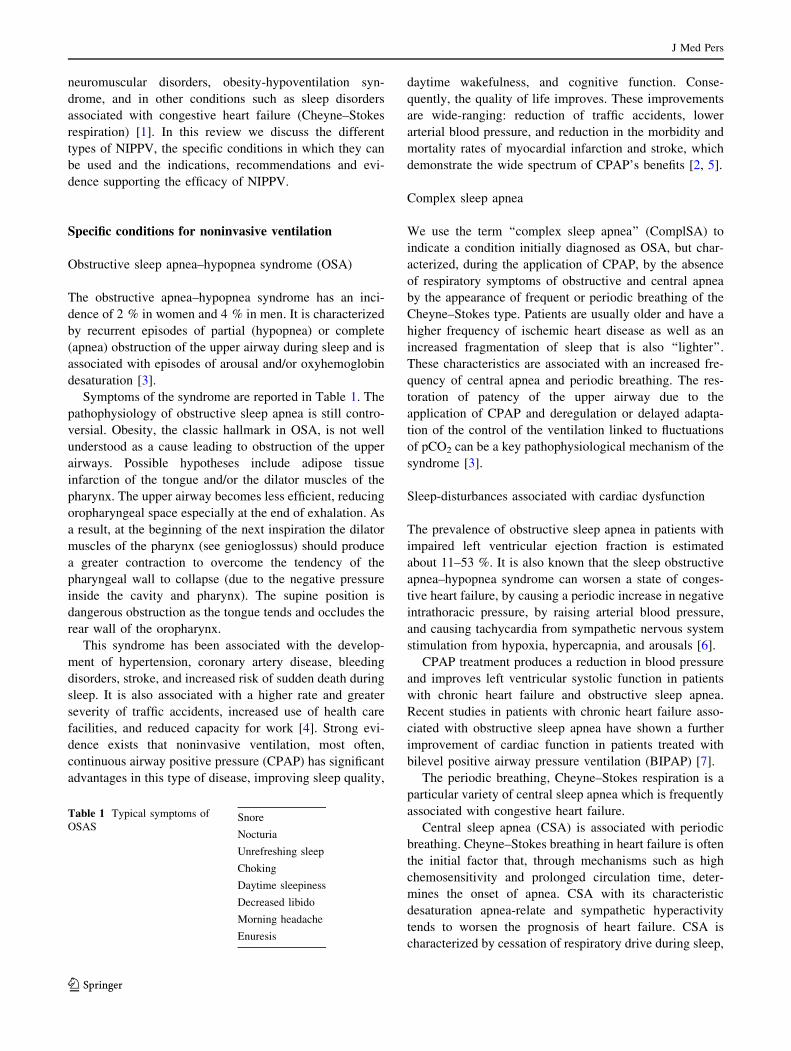
Symptoms of the syndrome are reported in Table 1. The
pathophysiology of obstructive sleep apnea is still contro-
versial. Obesity, the classic hallmark in OSA, is not well
understood as a cause leading to obstruction of the upper
airways. Possible hypotheses include adipose tissue
infarction of the tongue and/or the dilator muscles of the
pharynx. The upper airway becomes less efficient, reducing
oropharyngeal space especially at the end of exhalation. As
a result, at the beginning of the next inspiration the dilator
muscles of the pharynx (see genioglossus) should produce
a greater contraction to overcome the tendency of the
pharyngeal wall to collapse (due to the negative pressure
inside the cavity and pharynx). The supine position is
dangerous obstruction as the tongue tends and occludes the
rear wall of the oropharynx.
This syndrome has been associated with the develop-
ment of hypertension, coronary artery disease, bleeding
disorders, stroke, and increased risk of sudden death during
sleep. It is also associated with a higher rate and greater
severity of traffic accidents, increased use of health care
facilities, and reduced capacity for work [4]. Strong evi-
dence exists that noninvasive ventilation, most often,
continuous airway positive pressure (CPAP) has significant
advantages in this type of disease, improving sleep quality,
daytime wakefulness, and cognitive function. Conse-
quently, the quality of life improves. These improvements
are wide-ranging: reduction of traffic accidents, lower
arterial blood pressure, and reduction in the morbidity and
mortality rates of myocardial infarction and stroke, which
demonstrate the wide spectrum of CPAP’s benefits [2, 5].
Complex sleep apnea
We use the term ‘‘complex sleep apnea’’ (ComplSA) to
indicate a condition initially diagnosed as OSA, but char-
acterized, during the application of CPAP, by the absence
of respiratory symptoms of obstructive and central apnea
by the appearance of frequent or periodic breathing of the
Cheyne–Stokes type. Patients are usually older and have a
higher frequency of ischemic heart disease as well as an
increased fragmentation of sleep that is also ‘‘lighter’’.
These characteristics are associated with an increased fre-
quency of central apnea and periodic breathing. The res-
toration of patency of the upper airway due to the
application of CPAP and deregulation or delayed adapta-
tion of the control of the ventilation linked to fluctuations
of pCO2 can be a key pathophysiological mechanism of the
syndrome [3].
Sleep-disturbances associated with cardiac dysfunction
The prevalence of obstructive sleep apnea in patients with
impaired left ventricular ejection fraction is estimated
about 11–53 %. It is also known that the sleep obstructive
apnea–hypopnea syndrome can worsen a state of conges-
tive heart failure, by causing a periodic increase in negative
intrathoracic pressure, by raising arterial blood pressure,
and causing tachycardia from sympathetic nervous system
stimulation from hypoxia, hypercapnia, and arousals [6].
CPAP treatment produces a reduction in blood pressure
and improves left ventricular systolic function in patients
with chronic heart failure and obstructive sleep apnea.
Recent studies in patients with chronic heart failure asso-
ciated with obstructive sleep apnea have shown a further
improvement of cardiac function in patients treated with
bilevel positive airway pressure ventilation (BIPAP) [7].
The periodic breathing, Cheyne–Stokes respiration is a
particular variety of central sleep apnea which is frequently
associated with congestive heart failure.
Central sleep apnea (CSA) is associated with periodic
breathing. Cheyne–Stokes breathing in heart failure is often
the initial factor that, through mechanisms such as high
chemosensitivity and prolonged circulation time, deter-
mines the onset of apnea. CSA with its characteristic
desaturation apnea-relate and sympathetic hyperactivity
tends to worsen the prognosis of heart failure. CSA is
characterized by cessation of respiratory drive during sleep,
Table 1 Typical symptoms of
OSASSnore
Nocturia
Unrefreshing sleep
Choking
Daytime sleepiness
Decreased libido
Morning headache
Enuresis
J Med Pers
123

cessation of airflow obstruction, and impaired gas
exchange. Unlike the OSA in which there is a respiratory
effort to overcome the resistance of the upper airway, CSA
is characterized by the absence of respiratory movement
due to the cessation of ventilation. In the heart failure
patients, the onset of apnea occurs by a redistribution
of blood volume from the lower limbs to pulmonary cir-
culation that is mainly triggered by the supine position.
Stimulation of pulmonary vagal receptors causes hyper-
ventilation which results in hypocapnia.
When the value decreases below the hypocapnic apneic
threshold, stimulation of the bulbar center ceases, inspira-
tory drive stops, and apnea occurs. In patients with chronic
heart failure, the prolonged circulation time due to the
reduction in cardiac output leads to a delay of feedback
between chemoreceptors and bulbar centers resulting in
hyperventilation and respiratory greater instability. The
main risk factors for CSA are male sex, hypocapnia, atrial
fibrillation, and advanced age. CPAP and BIPAP can fail to
correct this category of apneas; therefore, a servo-assisted
mode (ASV or adaptive servo ventilation) is recommended
[8].
Obesity-hypoventilation syndrome
Obesity hypoventilation refers to a syndrome including
daytime hypercapnia (PCO2 [45 mmHg) in obese people
in which no other cause of hypoventilation is present. Its
prevalence among patients with obstructive sleep apnea is
20–30 % and is greater in extremely obese patients (BMI
[40). Approximately 10 % of patients with obesity-
hypoventilation syndrome do not have sleep apnea–hypo-
pnea syndrome. Additionally, nocturnal hypoxemia and
diurnal hypercapnia persist in about 40 % of these patients
after the treatment with CPAP eliminated apnea. Factors
other than sleep apnea contribute to the development of
obesity-hypoventilation syndrome associated with the
persistence of daytime hypercapnia: these include body
mass index and apnea–hypopnea index, mean overnight
oxygen saturation, and the severity of restrictive ventilatory
syndrome. BIPAP therapy may be useful in those patients
in whom CPAP has failed or gave unsatisfactory results.
The average volume-assured pressure support ventilation
seems to be able to lower PCO2 in a superior manner
compared to BIPAP, but it is not able to further improve
the oxygenation, sleep quality, or quality of life [9].
Neuromuscular and chest wall disorders
Noninvasive ventilation has been used in patients with
progressive neuromuscular disease or serious abnormalities
of the thoracic cage, with recognized benefits, including an
improved survival rate and an improved quality of life. The
benefits of noninvasive ventilation in this type of patient
includes improvements on daytime levels of blood gas
(including hypercapnia), a reduction in the oxygen cost of
breathing, an increase in the ventilatory response to
increased carbon dioxide, and improved lung compliance
[10].
Chronic obstructive pulmonary disease and sleep apnea
(overlap syndrome)
Sleep-disordered breathing (mainly obstructive sleep
apnea) and chronic obstructive pulmonary disease (COPD)
are the most common lung diseases: a large number of
patients have both disorders and hence the term ‘‘overlap
syndrome’’. The overlap syndrome was first described by
Flenley in 1985 as a combination of COPD and obstructive
apnea–hypopnea syndrome. The coexistence of these
conditions can lead to severe episodes of desaturation
during sleep, thus increasing the risk of hypoxemia, day-
time hypercapnia, and pulmonary hypertension. Noninva-
sive ventilation may be useful in patients with overlap
syndrome, but there are no controlled studies [11, 12].
Clinical criteria for initiating noninvasive ventilation
The presence of symptoms and physiological markers of
hypoventilation are useful in identifying the clinical
severity; moreover, these factors relate to therapeutic
decision-making, especially initiating nocturnal noninva-
sive ventilation [1]. In a typical ‘‘progressive disease’’ two
successive steps occur:
1. Initial phase of nocturnal hypoventilation reversible
during waking hours, associated with few or no clinical
symptoms.
2. Nocturnal and daylight hypoventilation associated with
clinical symptoms which shows a reduced respiratory
reserve.
The continuous sleep monitoring of pCO2 and O2 sat-
uration values are necessary to document the presence of
nocturnal hypoventilation which may be present in all the
stages of sleep (in some cases only during REM sleep).
Daytime hypoventilation is defined by reduced values of
arterial oxygen tension (PaO2), high levels of arterial car-
bon dioxide tension (PaCO2), and/or high serum bicar-
bonate levels with a relatively normal pH. Chronic daytime
hypoventilation is an important indicator always associated
with nocturnal hypoventilation. In the presence of daytime
hypoventilation, polysomnography is done to exclude
obstructive or central apnea. Clinical symptoms, although
modest, should be evaluated carefully, because they are
very important determining disease severity and prognosis
J Med Pers
123
as well defining the need for noninvasive ventilation.
Pulmonary function tests may be helpful in defining the
reduction of lung function, but they have a low predictive
value for patients with sleep-related hypoventilation.
However, in patients with neuromuscular disease, there is a
good correlation between lung function and nocturnal
hypoventilation: it has been shown that hypoventilation
during REM only or during all sleep stages or in the day-
time appears, respectively, with supine inspiratory vital
capacities of \40, 25, or 12 % of predicted values [1, 13].
Types of noninvasive ventilation
CPAP (continuous positive airway pressure)
CPAP is currently the most widely used mode of nonin-
vasive ventilation in the treatment of obstructive sleep-
disordered breathing and of disordered breathing associated
with chronic heart failure. It consists in the application of a
constant level of positive pressure during spontaneous
breathing. The mechanism of action of CPAP includes a
series of actions on pathophysiological mechanisms:
(a) It prevents intermittent narrowing and collapse of the
airways in patients with obstructive sleep apnea–
hypopnea syndrome.
(b) It counteracts auto-positive end-expiratory pressure,
which reduces respiratory muscles load, the work of
breathing and daytime PaCO2 in patients with
overlap syndrome.
(c) It improves lung function, particularly the functional
residual capacity, daytime gas exchange in patients
with obstructive sleep apnea–hypopnea syndrome.
(d) It improves systolic function of the left ventricle in
patients with heart failure coexisting with obstruc-
tive sleep apnea–hypopnea syndrome [2].
Auto-CPAP (automatic adjustment of the level
of continuous positive airway pressure)
Auto-CPAP (APAP) is delivered via a self-titrating CPAP
device, which uses algorithms to detect variations in the
degree of obstruction and adjusts the pressure level to restore
normal breathing. Auto-CPAP compensates for factors that
modify the upper airway collapsibility, such as body posture
during sleep, stage of sleep, use of alcohol, and drugs that
affect upper airway muscle tone [2]. It is not recommended in
the diagnosis of OSA and titrating continuous pressure
during split-night. The auto-CPAP can be used during pol-
ysomnography or cardiorespiratory monitoring to titrate a
single pressure value to be used later with fixed CPAP for
treatment of OSA in patients without comorbid conditions.
The use of auto-CPAP is reserved only for those patients with
sleep apnea syndrome only present during REM or respira-
tory events related to position, in whom constraining posi-
tional maneuvers are poorly tolerated [14].
Servo-assisted ventilation (ASV-adaptive servo-
ventilation)
The servo-assisted ventilation (ASV) has been developed for
the treatment of Cheyne–Stokes respiration-central apnea
syndrome in patients with chronic heart failure who have a
breathing pattern characterized by periods of crescendo–
decrescendo change in tidal volume with possible interca-
lated episodes of central apnea or hypopnea. This more
complex device can use patient expiratory positive airway
pressure (EPAP) level sufficient to control the obstructive
apnea. The device then automatically adjusts the inspiratory
pressure support for each inspiration within a prespecified
range, to maintain a moving-target ventilation set at 90 % of
the patient’s recent average ventilation. The aim is the sta-
bilization of breathing patterns and reduction in the respi-
ratory alkalosis that can trigger apnea reentry cycles [15].
BIPAP (Bilevel positive airway pressure)
Bilevel positive airway pressure (BIPAP) is also used for
sleep-related disorders (including those associated with
chronic heart failure), but its main indication is in patho-
logical conditions associated with hypoventilation. The BI-
PAP devices deliver a higher pressure during inspiration
(IPAP––inspiratory positive airway pressure) and a lower
pressure during expiration (EPAP––expiratory positive air-
way pressure). The gradient between IPAP and EPAP
(pressure support ventilation) is crucial in maintaining ade-
quate alveolar ventilation and reducing PaCO2. The IPAP
acts also in reducing the work of breathing and fatigue,
reducing the workload of respiratory muscles; EPAP has the
function of maintaining the patency of the upper airway, to
control obstructive apnea and to improve the functional
residual capacity. BIPAP is now proposed for the type of
patients who require high expiratory pressures to control
obstructive sleep apnea–hypopnea, but who cannot tolerate
exhaling against a high-fixed CPAP pressure [16]. Other
indications of BIPAP are the treatment of coexisting central
apnea or hypoventilation, the obesity-hypoventilation syn-
drome that cannot have a complete correction of the hypoxic
state with only CPAP, the overlap syndrome and neuro-
muscular disorders. Although the patient should be able to
maintain spontaneous breathing, it is used to set a back-up
rate option for those patients whose ventilation during sleep
may be particularly impaired (neuromuscular disorders,
complex sleep apnea, central apnea in chronic heart failure,
obesity-hypoventilation syndrome) [2, 16].
J Med Pers
123
Average volume-assured pressure support ventilation
(AVAPS)
Average volume-assured pressure support ventilation
(AVAPS) is used in patients with chronic hypoventilation
and in particular with obesity hypoventilation syndrome,
neuromuscular diseases, and sometimes, in chronic
obstructive pulmonary disease. In this mode a target tidal
volume is set; the device adjusts the pressure support which
to reach that set tidal volume. The advantage of this mode
(a cross between the volume and pressure-assisted mode) is
that it guarantees a delivered tidal volume adjusted despite
variability in the patient effort, airway resistance, and lung
or chest wall compliance. A particular benefit of this mode
is that it may be modified as the disease progresses, (as it
occurs in neuromuscular disorders such as amyotrophic
lateral sclerosis) [2].
Management of NIPPV
Initiation and settings in case of nocturnal ventilation
The main objective of the use of noninvasive ventilation is
the correction of blood gas values near ‘‘normal’’ with the
least possible discomfort or sleep disturbance. Even if there
is no absolute recommendation, it is good practice to
proceed in three successive steps. The first step is to choose
and adjust the ventilator settings while the patient is awake,
assuring physiological adequacy and patient comfort for at
least 1 or 2 h. In the second step the clinician should
evaluate the adequacy of the settings when sleeping during
a nap and a night’s sleep. Different options, according to
the resources available in each center, are used. A full
polysomnography recording oxygen saturation (SpO2) and
transcutaneous pCO2 (PtcCO2) or end-tidal (PetCO2), flow,
tidal volume, airway pressure, rib cage and abdomen
excursion as well as sleep-staging allows a complete
assessment (gold standard). When the resources are not
available fewer parameters may be used. The minimum
required is recording SpO2 on room air, assessing that the
normalization of SpO2 accompanies the normalization, or
at least the improvement in PaCO2.
The second step relates to patient tolerance, comfort,
changes in sleep quality, and well-being; these data should
be obtained. The third step consists of looking for reduction
in PaCO2 and augmentation of PaO2 without dyspnea
during the day in free ventilation after several nights of
NIPPV. This is done to confirm that the settings are ade-
quate for the patient’s needs [1, 17].
If the results are not satisfactory, changes must be made
to the settings. One may also change the type of mask and
ventilator. At the beginning a starting level of pressure
support of 10 cmH2O is recommended. Continuing the
adaptation, the pressure level can progressively be
increased to achieve evidence of improvement. Pressure
support higher than 20 cmH2O is rarely necessary [1, 17,
18]. A back-up frequency set close to the spontaneous
frequency of the patient during sleep is reasonable sub-
stitute to inspiratory trigger failure to avoid central apnea
induced by transitory but repeated hyperventilation passing
the apnea threshold. When employing a volume-preset
ventilator, the initial suggested setting may be established
by adjusting the frequency of ventilator-delivered breaths
so that it approximates the patient’s spontaneous breathing
frequency during sleep, an inspiratory time/total breathing
time between 0.33 and 0.5, and a relatively high tidal
volume of around 10–15 ml/kg to insure sufficient tidal
volume in case of leaks [1].
Supplemental oxygen (O2) will be added to the venti-
lator circuit, immediately near the interface or hooked to a
respirator, especially in those patients who require oxygen
during the daytime (COPD, cystic fibrosis, bronchiectasis).
In the absence of obstructive pulmonary disease, the
addition of O2 to the ventilation circuit may be justified
only to maintain an acceptable level of PaO2 during sleep
and only after all the parameters have been optimized [19].
Follow-up
Clinical follow-up and arterial blood gases should be
conducted at least twice a year. The recordings during
sleep, (possibly identical to those performed for the adap-
tation to noninvasive ventilation), are useful. At any time,
when there are indications of unsatisfactory results such as
the recurrence of clinical symptoms and/or signs of hypo-
ventilation on arterial blood gases, inadequate noninvasive
ventilation should be suspected, and a complete objective
assessment of ventilation during sleep with polysomnog-
raphy must be undertaken.
When the noninvasive ventilation is not proven to be
optimal, a change of ventilation modality and/or parame-
ters of the ventilator and/or a revision of the interface may
be indicated. In case of disease progression one should
consider increasing the duration of ventilation during the
day. The interfaces need to be regularly checked and
modified or adapted to changing needs of the patient [1].
Management of complications and side effects
Air leaks during ventilation
The major potential adverse effect is the loss of effec-
tiveness of the ventilation and, therefore, the potential
fragmentation of sleep. A variety of more or less effective
measures have been suggested to tackle the problem of
J Med Pers
123
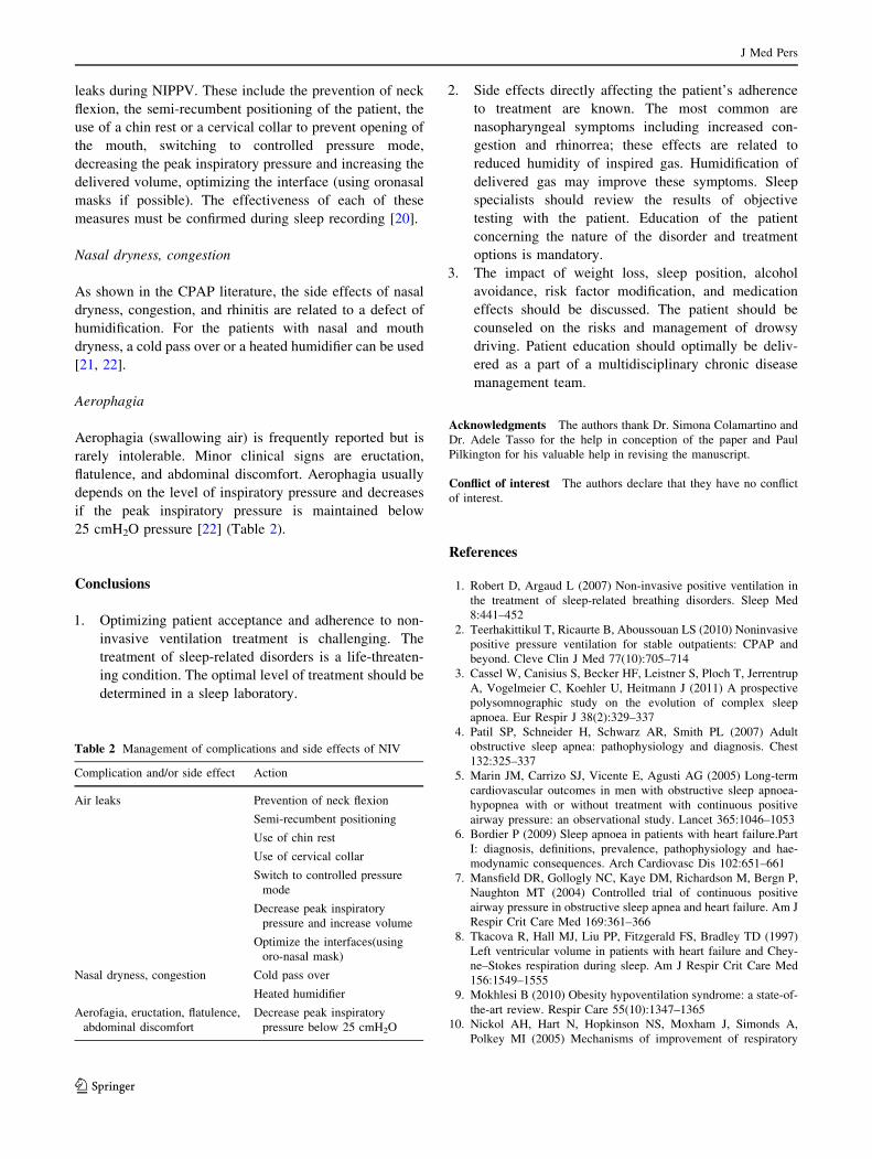
leaks during NIPPV. These include the prevention of neck
flexion, the semi-recumbent positioning of the patient, the
use of a chin rest or a cervical collar to prevent opening of
the mouth, switching to controlled pressure mode,
decreasing the peak inspiratory pressure and increasing the
delivered volume, optimizing the interface (using oronasal
masks if possible). The effectiveness of each of these
measures must be confirmed during sleep recording [20].
Nasal dryness, congestion
As shown in the CPAP literature, the side effects of nasal
dryness, congestion, and rhinitis are related to a defect of
humidification. For the patients with nasal and mouth
dryness, a cold pass over or a heated humidifier can be used
[21, 22].
Aerophagia
Aerophagia (swallowing air) is frequently reported but is
rarely intolerable. Minor clinical signs are eructation,
flatulence, and abdominal discomfort. Aerophagia usually
depends on the level of inspiratory pressure and decreases
if the peak inspiratory pressure is maintained below
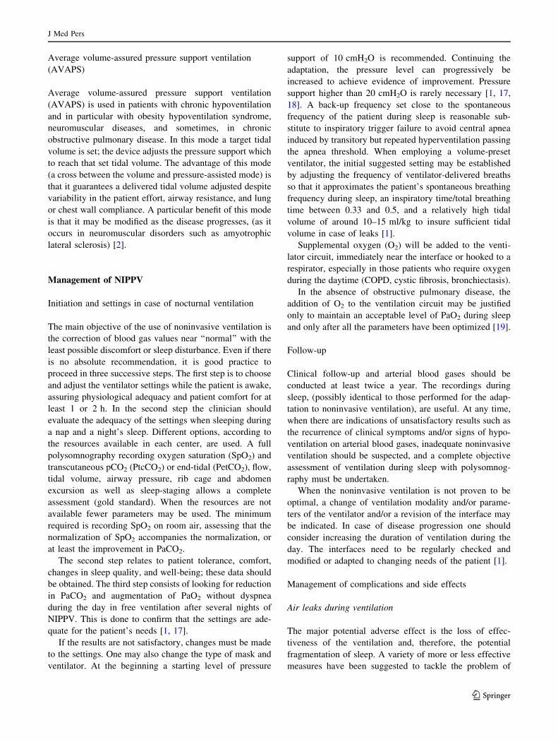
25 cmH2O pressure [22] (Table 2).
Conclusions
1. Optimizing patient acceptance and adherence to non-
invasive ventilation treatment is challenging. The
treatment of sleep-related disorders is a life-threaten-
ing condition. The optimal level of treatment should be
determined in a sleep laboratory.
2. Side effects directly affecting the patient’s adherence
to treatment are known. The most common are
nasopharyngeal symptoms including increased con-
gestion and rhinorrea; these effects are related to
reduced humidity of inspired gas. Humidification of
delivered gas may improve these symptoms. Sleep
specialists should review the results of objective
testing with the patient. Education of the patient
concerning the nature of the disorder and treatment
options is mandatory.
3. The impact of weight loss, sleep position, alcohol
avoidance, risk factor modification, and medication
effects should be discussed. The patient should be
counseled on the risks and management of drowsy
driving. Patient education should optimally be deliv-
ered as a part of a multidisciplinary chronic disease
management team.
Acknowledgments The authors thank Dr. Simona Colamartino and
Dr. Adele Tasso for the help in conception of the paper and Paul
Pilkington for his valuable help in revising the manuscript.
Conflict of interest The authors declare that they have no conflict
of interest.
References
1. Robert D, Argaud L (2007) Non-invasive positive ventilation in
the treatment of sleep-related breathing disorders. Sleep Med
8:441–452
2. Teerhakittikul T, Ricaurte B, Aboussouan LS (2010) Noninvasive
positive pressure ventilation for stable outpatients: CPAP and
beyond. Cleve Clin J Med 77(10):705–714
3. Cassel W, Canisius S, Becker HF, Leistner S, Ploch T, Jerrentrup
A, Vogelmeier C, Koehler U, Heitmann J (2011) A prospective
polysomnographic study on the evolution of complex sleep
apnoea. Eur Respir J 38(2):329–337
4. Patil SP, Schneider H, Schwarz AR, Smith PL (2007) Adult
obstructive sleep apnea: pathophysiology and diagnosis. Chest
132:325–337
5. Marin JM, Carrizo SJ, Vicente E, Agusti AG (2005) Long-term
cardiovascular outcomes in men with obstructive sleep apnoea-
hypopnea with or without treatment with continuous positive
airway pressure: an observational study. Lancet 365:1046–1053
6. Bordier P (2009) Sleep apnoea in patients with heart failure.Part
I: diagnosis, definitions, prevalence, pathophysiology and hae-
modynamic consequences. Arch Cardiovasc Dis 102:651–661
7. Mansfield DR, Gollogly NC, Kaye DM, Richardson M, Bergn P,
Naughton MT (2004) Controlled trial of continuous positive
airway pressure in obstructive sleep apnea and heart failure. Am J
Respir Crit Care Med 169:361–366
8. Tkacova R, Hall MJ, Liu PP, Fitzgerald FS, Bradley TD (1997)
Left ventricular volume in patients with heart failure and Chey-
ne–Stokes respiration during sleep. Am J Respir Crit Care Med
156:1549–1555
9. Mokhlesi B (2010) Obesity hypoventilation syndrome: a state-of-
the-art review. Respir Care 55(10):1347–1365
10. Nickol AH, Hart N, Hopkinson NS, Moxham J, Simonds A,
Polkey MI (2005) Mechanisms of improvement of respiratory
Table 2 Management of complications and side effects of NIV
Complication and/or side effect Action
Air leaks Prevention of neck flexion
Semi-recumbent positioning
Use of chin rest
Use of cervical collar
Switch to controlled pressure
mode
Decrease peak inspiratory
pressure and increase volume
Optimize the interfaces(using
oro-nasal mask)
Nasal dryness, congestion Cold pass over
Heated humidifier
Aerofagia, eructation, flatulence,
abdominal discomfort
Decrease peak inspiratory
pressure below 25 cmH2O
J Med Pers
123
failure in patients with restrictive thoracic disease treated with
non-invasive ventilation. Thorax 60:754–760
11. Hill NS (2004) Noninvasive ventilation for chronic obstructive
pulmonary disease. Respir Care 49:72–87
12. McEvoy RD, Pierce RJ, Hillman D, Australian Trial of Non-
invasive Ventilation in Chronic Airflow Limitation (AVCAL)
Study Group (2009) Nocturnal non-invasive nasal ventilation in
stable hypercapnic COPD: a randomized controlled trial. Thorax
64:561–566
13. Lo Coco D, Marchese S, Corrao S, Cettina Pesco M, La bella V,
Piccoli F et al (2006) Development of chronic hypoventilation in
amyotrophic lateral sclerosis patients. Respir Med 100:1028–1036
14. Meurice JC, Cornette A, Philip-Joet F, ANTADIR ‘‘PPC’’
Working Group (2007) Evaluation of autoCPAP devices in home
treatment of sleep apnea–hypopnea syndrome. Sleep Med
8:159–164
15. Teschler H, Dohring J, Wang YM, Berthon-Jones M (2001)
Adaptive pressure support servo-ventilation: a novel treatment for
Cheyne–Stokes respiration in heart failure. Am J Respir Crit Care
Med 164:614–619
16. Kushida CA, Littner MR, Hirshkowitz M, American Academy of
Sleep Medicine (2006) Practice parameters for the use of con-
tinuous and bilevel positive airway pressure devices to treat adult
patients with sleep related patients breathing disorders. Sleep
29:375–380
17. Epstein LJ, Kristo D, Strollo PJ Jr, Friedman N, Malhotra A, Patil
SP, Ramar K, Rogers R, Schwab RJ, Weaver EM, Weinstein MD,
Adult Obstructive Sleep Apnea Task Force of the American
Academy of Sleep Medicine (2009) Clinical guideline for the
evaluation, management and long-term care of obstructive sleep
apnea in adults. J Clin Sleep Disord 5(3):263–276
18. Rosario IC (2011) Obstructive sleep apnea: a review and update.
Minn Med 94(11):44–48
19. Schwartz AR, Kacmarek RM, Hess DR (2004) Factors affecting
oxygen delivery with bi-level positive airway pressure. Respir
care 49:270–275
20. Teschler H, Stampa J, Ragette R, Konietzko N, Berthon-Jones M
(1999) Effect of mouth leak on effectiveness of nasal bilevel
ventilator assistance and sleep architecture. Eur Respir J
14:1251–1257
21. Randerath WJ, Meier J, Genger H, Domanski U, Ruhle KH (2002)
Efficiency of cold passover and heated humidification under con-
tinuous positive airway pressure. Eur Respir J 20:183–186
22. Hill NS (2000) Complications of noninvasive ventilation.
Respiratory Care 45:480–481
J Med Pers
123