nicotine exploits a copi-mediated process for chaperone ...drenan/henderson_jgenphysiol... · copi...
TRANSCRIPT

R e s e a r c h A r t i c l e
The Rockefeller University Press $30.00J. Gen. Physiol. Vol. 143 No. 1 51–66www.jgp.org/cgi/doi/10.1085/jgp.201311102 51
I N T R O D U C T I O N
One of the earliest discoveries of chronic nicotine expo-sure was the observation of an increased number of nicotinic acetylcholine receptors (nAChRs; termed up-regulation) (Marks et al., 1983; Schwartz and Kellar, 1983; Breese et al., 1997; Mamede et al., 2007; Nashmi et al., 2007). Classically, up-regulation of nAChRs has been defined as an increase in nAChR protein, as iden-tified by increased agonist binding (Marks et al., 1983; Benwell et al., 1988; Flores et al., 1992; Peng et al., 1994), but we now define up-regulation as a change in receptor number, stoichiometry, and trafficking (Lester et al., 2009; Miwa et al., 2011). Often, an increase in total binding as opposed to cell surface binding has been reported. Therefore, up-regulation of nAChRs in-volves an increase in nAChR abundance throughout the cell (ER, Golgi, etc.) and not exclusively on the plasma membrane (PM). There have been several hy-potheses regarding the mechanism of up-regulation, but a consensus is emerging that nicotine acts inside the cell to enhance a critical step(s) in the maturation process
Correspondence to Henry A. Lester: L e s t e r @ C a l t e c h . e d u Abbreviations used in this paper: COPI, coat protein complex I; ERES,
ER exit sites; nAChR, nicotinic acetylcholine receptor; Neuro-2a, neuroblas-toma 2a; NFRET, normalized Förster resonance energy transfer; PM, plasma membrane; PMID, PM integrated density; ROI, regions of interest; SEP, supercliptic-pHluorin; SNc, substantia nigra pars compacta; TIRFM, total internal reflection fluorescence microscopy; VTA, ventral tegmental area.
of nAChRs (Sallette et al., 2005). This intracellular en-hancement process has been characterized as pharma-cological chaperoning (Kuryatov et al., 2005; Lester et al., 2009), and it occurs at the nanomolar concentrations thought to persist in the brain for hours after a person smokes. Research from many laboratories indicates that up-regulation of 42 nAChRs occurs through a similar process in human brains, animal brains, cultured neu-rons, and clonal transfected cell lines (Nashmi et al., 2007; Mukhin et al., 2008; Miwa et al., 2011; Srinivasan et al., 2011; Lester et al., 2012).
Up-regulation of nAChRs in response to chronic nic-otine plays a major role in nicotine dependence and, perhaps, in the inverse correlation between a person’s history of tobacco use and his or her susceptibility to Parkinson’s disease (Ritz et al., 2007; Koob, 2009). Indi-vidual deletions of the 4, 6, or 2 nAChR subunits are sufficient to block the self-administration of nicotine in mice (Pons et al., 2008), whereas the selective reexpres-sion of these deleted subunits in the ventral tegmental area (VTA) is sufficient to reinstate self-administration of nicotine (Pons et al., 2008; Brunzell et al., 2010).
Nicotine exploits a COPI-mediated process for chaperone-mediated up-regulation of its receptors
Brandon J. Henderson,1 Rahul Srinivasan,1 Weston A. Nichols,1 Crystal N. Dilworth,1 Diana F. Gutierrez,1 Elisha D.W. Mackey,1 Sheri McKinney,1 Ryan M. Drenan,2 Christopher I. Richards,3 and Henry A. Lester1
1Division of Biology and Biological Engineering, California Institute of Technology, Pasadena, CA 911252Department of Medicinal Chemistry and Molecular Pharmacology, Purdue University, West Lafayette, IN 479073Department of Chemistry, University of Kentucky, Lexington, KY 40506
Chronic exposure to nicotine up-regulates high sensitivity nicotinic acetylcholine receptors (nAChRs) in the brain. This up-regulation partially underlies addiction and may also contribute to protection against Parkinson’s disease. nAChRs containing the 6 subunit (6* nAChRs) are expressed in neurons in several brain regions, but comparatively little is known about the effect of chronic nicotine on these nAChRs. We report here that nicotine up-regulates 6* nAChRs in several mouse brain regions (substantia nigra pars compacta, ventral tegmental area, medial habenula, and superior colliculus) and in neuroblastoma 2a cells. We present evidence that a coat protein complex I (COPI)-mediated process mediates this up-regulation of 6* or 4* nAChRs but does not participate in basal trafficking. We show that 623 nAChR up-regulation is prevented by mutating a putative COPI-binding motif in the 3 sub-unit or by inhibiting COPI. Similarly, a COPI-dependent process is required for up-regulation of 42 nAChRs by chronic nicotine but not for basal trafficking. Mutation of the putative COPI-binding motif or inhibition of COPI also results in reduced normalized Förster resonance energy transfer between 623 nAChRs and COP subunits. The discovery that nicotine exploits a COPI-dependent process to chaperone high sensitivity nAChRs is novel and suggests that this may be a common mechanism in the up-regulation of nAChRs in response to chronic nicotine.
© 2014 Henderson et al. This article is distributed under the terms of an Attribution– Noncommercial–Share Alike–No Mirror Sites license for the first six months after the publi-cation date (see http://www.rupress.org/terms). After six months it is available under a Creative Commons License (Attribution–Noncommercial–Share Alike 3.0 Unported license, as described at http://creativecommons.org/licenses/by-nc-sa/3.0/).
The
Jour
nal o
f G
ener
al P
hysi
olo
gy
on June 17, 2014jgp.rupress.org
Dow
nloaded from
Published December 30, 2013
http://jgp.rupress.org/content/suppl/2013/12/20/jgp.201311102.DC2.html http://jgp.rupress.org/content/suppl/2013/12/20/jgp.201311102.DC1.html Supplemental Material can be found at:
on June 17, 2014jgp.rupress.org
Dow
nloaded from
Published December 30, 2013 on June 17, 2014
jgp.rupress.orgD
ownloaded from
Published December 30, 2013
on June 17, 2014jgp.rupress.org
Dow
nloaded from
Published December 30, 2013 on June 17, 2014
jgp.rupress.orgD
ownloaded from
Published December 30, 2013

52 Nicotine exploits COPI for up-regulation
M A T E R I A L S A N D M E T H O D S
ReagentsThe -COP antibody (rabbit polyclonal) was obtained from Abcam (AB2899). CI-976 and ()-nicotine hydrogen tartrate were ob-tained from Sigma-Aldrich.
Mice and chronic nicotine administrationThe construction of 6-GFP mice has been described previously (Mackey et al., 2012). All experiments were conducted in accor-dance with the guidelines for care and use of animals provided by the National Institutes of Health, and protocols were approved by the Institutional Animal Care and Use Committee at the Califor-nia Institute of Technology.
Chronic nicotine administrationMice were kept on a standard 12-h light/dark cycle at 22°C and given food and water ad libitum. Chronic nicotine or saline was administered to mice using miniosmotic pumps (model 2002; Alzet). On the day of pump implantation, saline or ()-nicotine hydrogen tartrate (Sigma-Aldrich) was prepared freshly and loaded into the pump to deliver nicotine at 2 mg/kg/h or 0.4 mg/kg/h. This concentration provides maximal nAChR up-regulation and a peripheral blood concentration that is near the peak concentra-tion found in the blood of human smokers (Marks et al., 2004). Surgical procedures for the pump implantation have been re-ported previously (Nashmi et al., 2007).
Cell cultureMouse neuroblastoma 2a (Neuro-2a) cells were cultured using standard techniques (Xiao et al., 2011). Cells were plated by add-ing 90,000 cells to poly-d-lysine–coated 35-mm glass-bottom imag-ing dishes (MatTek Corporation) and cultured in a humidified incubator (37°C, 95% air, 5% CO2). Cells were transfected with 500 ng of each nAChR subunit plasmid and 250 ng GalT-mCherry or Sec24D-mCherry for assays. Plasmids were mixed with 250 µl Opti-MEM. Lipofectamine-2000 was separately added to 250 µl Opti-MEM. After 5 min at 24°C, DNA and Lipofectamine solu-tions were mixed together and incubated for 25 min at 24°C. The solutions were then added to preplated Neuro-2a cells and incu-bated for 24 h. After 24 h, the Opti-MEM was removed and re-placed with growth medium. 50 or 500 nM of filter-sterilized nicotine was added after replacing the Opti-MEM with standard culture medium (623 nAChRs). For 42 nAChRs, 100 nM nicotine was used for 48 h (nicotine was added at the time of transfection and then replenished when the media was changed). 20 µM CI-976 was added with nicotine 24 h before imaging. Cells were imaged 48 h after transfection.
Patch-clamp recordingsFor patch-clamp electrophysiology, 50,000 Neuro-2a cells were plated onto sterilized 12-mm glass coverslips (Deckgläser), placed in 35-mm culture dishes, and cultured in a humidified incubator (37°C, 95% air, 5% CO2). Recorded cells were visualized with an inverted fluorescence microscope (IX71; Olympus) in either bright field or fluorescence (eGFP) mode using a high pressure Hg lamp (HB-10103AF; Nikon). Electrophysiological signals were recorded with an amplifier (Axopatch-1D; Axon Instruments), analogue- to-digital converter (Digidata 1440A; Axon Instruments), and soft-ware (pClamp 10.0; Axon Instruments). Patch pipettes were filled with solution containing (mM): 135 K gluconate, 5 KCl, 5 EGTA, 0.5 CaCl2, 10 HEPES, 2 Mg-ATP, and 0.1 GTP (pH was adjusted to 7.2 with Tris-base, and osmolarity was adjusted to 280–300 mOsm with sucrose). The resistance of patch pipettes was 2–4 MΩ for whole-cell recordings. Junction potential was nulled just before
Furthermore, nicotine self-administration can be blocked by the selective antagonism of 6* (*, IUPHAR nomen-clature, “other subunits may be present”) (Jackson et al., 2009) or 4* (Yoshimura et al., 2007) nAChRs. From this, it is clear that the nAChRs mediating nico-tine addiction include those that contain 4, 6, and 2 subunits (Picciotto et al., 1998; Tapper et al., 2004; Pons et al., 2008).
Despite the clarity of 42 nAChR up-regulation, there have been conflicting reports of up-regulation, down-regulation, or no change in response to chronic nicotine for 6* nAChRs (Tumkosit et al., 2006; Perez et al., 2008; Walsh et al., 2008). Here, we used mice ex-pressing 6–enhanced green fluorescence protein (eGFP) nAChR subunits to test for in vivo up-regulation of 6* nAChRs using concentrations of nicotine com-parable to those produced by smoking in humans. We determined that this up-regulation occurs in all four brain areas that robustly express 6* nAChRs: the VTA, the substantia nigra pars compacta (SNc), the medial habenula, and the superior colliculus.
We then used 6 subunits tagged with supercliptic-pHluorin (SEP), a pH-sensitive eGFP analogue, expressed in neuroblastoma cells to analyze the mechanism of 6* nAChR up-regulation. SEP has been used to study vesi-cle dynamics and synaptic delivery (Miesenböck et al., 1998; Mani and Ryan, 2009) as well as the trafficking of AMPA and GABA receptors (Jacob et al., 2005; Jaskolski et al., 2009; Lin et al., 2009; Araki et al., 2010), and we have used 4-SEP* to study nAChR trafficking after chronic exposure to nicotine (Richards et al., 2011). Here, we show that nicotine-induced up-regulation fails to occur with inhibition of coat protein complex I (COPI)-mediated retrograde traffic from the Golgi to the ER (either through the mutation of a putative COPI-binding motif [3-KKK] or through the use of the COPI inhibitor CI-976). COPI is a protein complex that coats vesicles for retrograde transport of proteins from the cis end of the Golgi to the ER (Brandizzi and Barlowe, 2013). COPI is a heptameric protein com-plex, and di-lysine–trafficking motifs, including KKxx and KxKxx, facilitate protein binding to COPI (Jackson et al., 2012; Spang, 2013). Inhibition of COPI-mediated trafficking resulted in a decrease of nAChR density in the ER and a complementary increase of nAChR den-sity in the Golgi. Inhibition of COPI had no effect on basal insertion of nAChRs in the PM but prevented their up-regulation during chronic nicotine treatment. Therefore, we propose that the cycling of nAChRs be-tween the Golgi and ER (via COPI) is necessary for the up-regulation of 42 and 623 nAChRs. Our data suggest that this may be a common mechanism of nAChR up-regulation by nicotine, and that the manipula-tion of Golgi–ER cycling may lead to novel therapeutic strategies for nicotine cessation or neuroprotection against Parkinson’s disease.
on June 17, 2014jgp.rupress.org
Dow
nloaded from
Published December 30, 2013

Henderson et al. 53
Insertion events were quantified using methods similar to those published previously (Richards et al., 2011). TIRFM measurements to detect membrane insertion events were performed with con-secutive 200-ms frames. Insertion events were defined as punctate regions of fluorescence appearing at the membrane. Insertion event data are represented in terms of insertion per 10 µm2 of area per minute of observation.
Spectral confocal imagingImaging experiments were performed in live Neuro-2a cells at 37°C in a stage-mounted culture dish incubator (Warner Instru-ments). For immunostaining, fixed Neuro-2a cells were imaged. A C1si laser-scanning confocal microscope (Eclipse; Nikon) equipped with a 60× 1.2 NA VC Plan Apo water objective and 32 photomul-tiplier tubes was used for confocal imaging. Before an imaging session, cell culture medium was replaced with phenol red–free CO2-independent Leibovitz (L-15) medium (Invitrogen). eGFP and mCherry were excited at 488 and 561 nm, respectively. For each cell, we focused on a plane containing the most ER exit sites (ERES), Golgi bodies, or COPI vesicles, and sequential images of eGFP and mCherry fluorescence were obtained. eGFP and mCherry fluorescence emission spectra were captured and images were unmixed using standard spectra acquired from cells express-ing eGFP, mCherry, or Alexa Fluor 555 alone. For quantification, ERES, Golgi, or COPI regions of interest (ROI) were manually de-marcated using intensity-based thresholding, and the raw inte-grated densities were measured for each cell using ImageJ. The total Sec24D fluorescence in ERES and the total GalT fluorescence per cell were quantified. Error bars for measurements indicate the SEM, and p-values are based on a two-tailed t test. The figures show a single imaging session that is representative of at least three ses-sions performed on separate days with similar or identical results.
For direct fluorescence imaging of in vivo 6-GFP* nAChRs, images were acquired with the 60× 1.2 NA VC Plan Apo water objective at wavelengths between 496 and 561 nm using a 488-nm line of an argon laser. Images were collected at 12-bit intensity over 512 × 512 pixels, a resolution of 2.5 nm, a 61.3-µm pinhole, and a pixel dwell time of 8.4 µs. Autofluorescence was separated from GFP fluorescence using techniques that have been published previously (Nashmi et al., 2007). Image analysis of cell counts and mean intensities were done with ImageJ.
Normalized Förster resonance energy transfer (NFRET)The general methods for Förster resonance energy transfer from sensitized acceptor emission have been described previously (Srinivasan et al., 2011, 2012a). For these studies, Neuro-2a cells were transfected with 6-mCherry, 2wt, COP-GFP, and 3wt or 3AAA nAChR subunits. Cells transfected with either 6-mCherry or COP-GFP alone were included in all imaging sessions to con-trol for pixel saturation and spectral bleedthrough. Live cells were imaged with a C1si laser-scanning confocal microscope (Eclipse; Nikon). During acquisition of images, cells were focused on a plane where the COP-GFP fluorescence most resembled the pattern for the endogenous COPI staining (see Fig. 4 E). GFP and mCherry images were acquired with 488- and 561-nm laser lines, and images were linearly unmixed by using reference spec-tra. Reference spectra were acquired from Neuro-2a cells trans-fected with 6-mCherry or COP-GFP during the same imaging session. After linear unmixing, NFRET was calculated using the PixFRET ImageJ plug-in as described previously (Moss et al., 2009; Srinivasan et al., 2011, 2012a).
Plasmid constructsMouse 6 with a C-terminal fusion of an SEP tag was constructed by PCR amplification using the forward primer 5-CATGGTTGG-CTGGTATGATCAGTAAAGGAGAAGAACTT-3 and the reverse primer 5-ATGGATGAACTATACAAATAGGGAATAGCGGCACCT-3,
forming a gigaseal. All recordings were done at 24°C. Data were sampled at 10 kHz and filtered at 2 kHz for whole-cell recordings. Acetylcholine was dissolved in extracellular solution containing (mM) 140 NaCl, 5 KCl, 2 CaCl2, 1 MgCl2, 10 HEPES, and 10 glu-cose (320 mOsm, pH set to 7.3 with Tris-base), and were puffed (0.3 s, 20 psi) onto voltage-clamped Neuro-2a cells (holding po-tential [VH] of 50 mV). To avoid receptor desensitization by re-petitive ACh application, we applied ACh at 3-min intervals and continually perfused the recording chamber with extracellular solution. For concentration response studies, we used a rapid su-perfusion system with 500-ms puffs of agonist (Octaflow II; ALA Scientific Instruments).
Immunostaining and antibodiesFor immunostaining, cultured Neuro-2a cells were fixed with 4% paraformaldehyde (15 min), permeabilized in 0.01% Triton X-100 (5 min), and blocked with 10% goat serum (30 min). After two PBS washes, the appropriate primary antibody (1:500) in 1% goat serum was applied for 1 h at 24°C. Cells were washed three times with PBS and incubated with 1% goat serum containing second-ary antibody (1:2,000) for 1 h at 24°C. Cells were washed three times with PBS and imaged immediately. Immunostaining used -COP rabbit polyclonal antibody as the primary (antibody 2899; Abcam) and Alexa Fluor 555–labeled secondary (goat anti–rabbit; Invitrogen) antibodies.
Midbrain 6-GFP neurons existed in a high enough density to be imaged without staining (direct/inherent fluorescence). Im-munohistochemical techniques were required for observing 6-GFP fluorescence in other regions of the mouse brain. 20-µm brain sections on slides were rinsed twice for 10 min with PBS and then permeabilized with 0.5% Triton X-100 in PBS for 1 h. Brain sections were then blocked with 4% goat serum (Jackson Immuno-Research Laboratories, Inc.) in PBS for 45 min. The primary antibody was diluted in 4% goat serum in PBS and incubated overnight at 24°C. Brain sections were then washed three times for 15 min with PBS, and the secondary antibody was diluted in 4% goat serum in PBS and incubated for 1 h at 24°C. Finally, brain sections were washed three times for 15 min with PBS and mounted with Vectashield.
Total internal reflection fluorescence microscopy (TIRFM)Cultured Neuro-2a cells were imaged live at 37°C in a stage-mounted culture dish incubator (Warner Instruments) using methods similar to TIRFM assays in cultured cortical neurons (Richards et al., 2011). TIRFM enables the visualization of fluo-rescently labeled intracellular molecules within 250 nm of the cell-coverslip interface. TIRFM images were obtained using an in-verted fluorescence microscope (IX81; Olympus) equipped with a 100× 1.45 NA oil objective (PlanApo; Olympus) and a stepper motor (Thorlabs) to control the position of the fiber optic and TIRFM evanescent field illumination. Just before imaging, growth medium was exchanged for extracellular solution (150 mM NaCl, 4 mM KCl, 10 mM HEPES, 2 mM MgCl2, 2 mM CaCl2, and 10 mM glucose), adjusted to the appropriate pH (5.4 or 7.4). SEP was excited at 488 nm with an air-cooled argon laser (IMA101040ALS; Melles Griot). Images were captured with a back-illuminated EMCCD camera (iXon DU-897; Andor). We acidified the imaging dish by perfusing the bath, normally at pH 7.4, with an otherwise identical solution adjusted to pH 5.4. The PM integrated density (PMID) was determined by taking an initial TIRFM image of each cell at pH 7.4, followed by acidification of the solution and a sub-sequent low pH image (pH 5.4). Low pH images were used to demarcate ER-localized nAChRs, which were subtracted from the total footprint to determine the PMID of nAChRs. The figures show a single imaging session that is representative of at least three sessions performed on separate days with similar or identi-cal results.
on June 17, 2014jgp.rupress.org
Dow
nloaded from
Published December 30, 2013

54 Nicotine exploits COPI for up-regulation
R E S U L T S
In vivo up-regulation of 6* nAChRs with chronic nicotineWe used a mouse line containing GFP-labeled 6* nAChRs to study the up-regulation of 6* nAChRs (Mackey et al., 2012). 6-GFP nAChR subunits assemble appropriately with other nonlabeled nAChR subunits, traffic properly, and function in electrophysiological as-says in a manner comparable to that of WT nAChRs (Mackey et al., 2012).
Direct fluorescence of 6-GFP* nAChRs was apparent in the VTA and SNc of saline- or nicotine-treated mice (Fig. 1 A). The fluorescence intensity of 6-GFP* nAChRs in the VTA and SNc of mice administered nico-tine at 2 mg/kg/h for 10 d was increased twofold com-pared with that of saline-treated mice (P < 0.005; Fig. 1, B1 and C1). In addition, we used cumulative percentage plots to compare saline and nicotine treatments (Fig. 1, B2 and C2). Cumulative percentage plots are similar to a cumulative frequency analysis and allow us to compare the full range of 6-GFP fluorescence intensities between saline and nicotine treatments. In all four brain regions, we can see that the percentage of low, mid, and high 6-GFP fluorescence intensities are shifted to the right
which overlap with sequences within the C-terminal end of the 6-coding sequence. This PCR product was then cloned directly into the vector containing the 6 gene using Pfu-Turbo polymerase. The mouse 23 nAChR subunit was constructed by PCR amplifica-tion using the forward primer 5-ATGGCCCGGTGCTCC AACTC-3 and the reverse primer 5-CCTGCCCTCAGACTGT GGTG-3. The mouse 32 nAChR subunit was constructed by PCR amplification using the forward primer 5-ATGACAGGC TTCCTACGGGT-3 and the reverse primer 5-ACGGTCCTG GTGTTCTACCT-3. Mouse 3AAA (the KKK mutated to AAA) and the 23[AAA] were constructed by PCR amplification using the forward primer 5-AAATTTCCAG-GGGCTGCAGCCCAGACTC CTA CC-3 and the reverse primer 5-CCCCCTCACGGTTCCCTTACTCTCCGT-3.
Online supplemental materialFig. S1 illustrates that the presence of 3 subunits increased the PMID of 64 nAChRs but is not required for the up-regulation of 64* nAChRs. Fig. S2 presents a schematic of the various chime-ras and mutant nAChR subunits that were used in this paper. Fig. S3 shows the electrophysiological characterization of 623WT and 623AAA nAChRs after treatment with nicotine. Here, we show that functional up-regulation accompanies the increase in PMID for 623WT nAChRs when treated with nicotine, but there is no functional up-regulation with 623AAA nAChRs. Fig. S4 illustrates that treatment with CI-976 produces an increase of nAChRs in the Golgi but does not increase export from the ER. Fig. S5 shows that CI-976 treatment does not affect basal PMID of nAChRs. The online supplemental material is available at http://www.jgp.org/ cgi/content/full/jgp.201311102/DC1.
Figure 1. Chronic nicotine up-regulates 6-GFP* nAChRs in four brain regions. (A) Direct fluorescence imaging of SNc and VTA in mice expressing 6-GFP subunits after treatment with saline or nicotine (2 mg/kg/h, 10 d). (D1 and E1) Immunohisto-chemical imaging of medial ha-benula and superior colliculus in mice expressing 6-GFP sub-units after treatment with saline or nicotine. For all images, mon-tages were compiled from indi-vidual 60× images. (A and E1) Bars, 100 µm. (D1) Bars, 20 µm. (B1 and C1) Quantification of 6-GFP intensities in SNc and VTA of saline- and nicotine-treated mice (n = 6 mice). (D2 and E2) Quantification of 6-GFP inten-sities in medial habenula and superior colliculus of saline- and nictotine-treated mice (n = 6 mice). (D3 and E3) Quantifica-tion of 6-GFP immunofluores-cence of medial habenula and superior colliculus neurons in saline- and nicotine-treated mice (n = 4–5 mice). (B2, C2, D3, and E3) Cumulative percentage plots show changes in 6-GFP fluores-cence in all brain regions with chronic nicotine. ***, P < 0.005; *, P < 0.05.
on June 17, 2014jgp.rupress.org
Dow
nloaded from
Published December 30, 2013

Henderson et al. 55
(Asokan and Cho, 2002; Jaskolski et al., 2009). When ex-posed to a neutral buffer (pH 7.4) both ER- and PM-resident nAChRs are fluorescent. Upon perfusing an acidic buffer (pH 5.4), PM-resident nAChR fluores-cence is quenched, whereas the ER component remains fluorescent. Thus, SEP allows the differentiation of re-ceptors within the ER from those in the PM. As in a previous study using SEP tagged to 4 nAChRs (Rich-ards et al., 2011), we fused SEP to the C terminus of 6 subunits so that it was positioned on the luminal side of organelles in the secretory pathway.
nAChRs with eGFP subunits have little or no func-tional difference (i.e., peak current amplitude, decay time) from WT subunits (Drenan et al., 2008; Richards et al., 2011; Srinivasan et al., 2011, 2012b; Xiao et al., 2011) and, indeed, we found no detectable functional difference between 6-SEP23 nAChRs and 6-eGFP23 nAChRs (Fig. 3). Inclusion of the 3 sub-unit increased ACh-induced peak currents by threefold compared with those obtained with 6-eGFP2 nAChRs (Fig. 3 B; P < 0.05). Inclusion of the 3 sub-unit also reduced the concentration of ACh at which we observed maximal peak amplitude from 300 µM (6-eGFP2) to 3 µM (6-eGFP23) (Fig. 3). Con-centration–response studies of ACh and ()-nicotine on 6-eGFP23 nAChRs yielded EC50 values of 0.31 ± 0.10 µM and 0.12 ± 0.08 µM, respectively (Fig. 3, C and D), which agree with those obtained with linked con-catamers containing (62)23 nAChRs (Kuryatov and Lindstrom, 2011).
(higher intensity) after chronic nicotine treatment (Fig. 1, B2 and C2). Nicotine at 0.4 mg/kg/h also increased 6-GFP fluorescence intensity in VTA (60.8 ± 10.9% increase) and SNc (70.0 ± 1.4% increase) neurons (P < 0.05 com-pared with saline; Fig. 2). Similar to 2 mg/kg/h nicotine treatments, the 0.4-mg/kg/h nicotine treatment shifted the full range of 6-GFP fluorescence intensities to the right in cumulative percentage plots (Fig. 2, D1 and D2).
2 mg/kg/h nicotine also increased the fluorescence intensity of medial habenula and superior colliculus 6-GFP* nAChRs (81% [P < 0.005] and 68% [P < 0.05], respectively; Fig. 1, D and E). In cumulative percentage plots, we can also see that the full range of 6-GFP fluo-rescence intensities is shifted to the left after chronic nicotine treatment (Fig. 1, D1 and D2).
6-SEP* nAChRs assemble functional receptors, and 3 subunits increase 6* nAChR functionTo perform a systematic study on 6* nAChR up-regulation, we used an expression system, Neuro-2a cells, which has been optimized to avoid many artifacts of “overexpressing” PM proteins. The appearance of functional PM proteins in this system is linear and un-saturated as a function of parameters such as level of transfected cDNAs, trafficking machinery, oligomeriza-tion, and anchoring (Moss et al., 2009).
SEP undergoes 488-nm excitation at neutral pH (7.4) but not under acidic conditions (Richards et al., 2011). 6-SEP* nAChRs in the Golgi and secretory vesicles (6.7 and <6.5 pH, respectively) are not fluorescent
Figure 2. Low concentrations of nicotine (0.4 mg/kg/h) up-regulate 6-GFP* nAChRs in midbrain neurons. (A and B) Direct fluo-r escence imaging of VTA and SNc neurons in mice expressing 6-GFP subunits after treatment with saline or nicotine (0.4 mg/kg/h, 10 d). Bars, 100 µm. (C1 and C2) Quantification of 6-GFP intensities of neurons in saline- or nicotine-treated mice (n = 4 mice). (D1 and D2) Cumulative percentage plots show changes in 6-GFP fluorescence in the SNc and VTA with chronic nicotine (0.4 mg/kg/h; n = 4 mice). *, P < 0.05.
on June 17, 2014jgp.rupress.org
Dow
nloaded from
Published December 30, 2013

56 Nicotine exploits COPI for up-regulation
As reported previously (Tumkosit et al., 2006), 623 nAChRs had a higher density on the PM than 62 nAChRs; the PMID of 623 nAChRs was three times that of 62 nAChRs (P < 0.0001; Fig. 4 B). We also found a 1.5-fold increase in the PMID of 643 nAChRs compared with that of 6-SEP4* nAChRs (Fig. S1). We found that, when expressed in Neuro-2a
Incorporation of 3 subunits increases the density of 6* nAChRs at the PMWe used TIRFM and 6-SEP* nAChRs transfected in Neuro-2A cells to quantify the density of receptors in the peripheral ER versus the PM. Fig. 4 A shows TIRFM images of Neuro-2a cells expressing 6-SEP2 or 6-SEP23 nAChRs at neutral (7.4) and acidic (5.4) pH.
Figure 3. The 6-SEP nAChRs are functional. Representative whole-cell ACh-induced currents of (A1) 6-SEP23 nAChRs (3 µM ACh). (A2) 6-eGFP23 nAChRs (3 µM ACh) and (A3) 6-eGFP2 nAChRs (300 µM ACh). (B) Comparison of whole-cell ACh-induced current amplitudes of 6-SEP23, 6-eGFP23, and 6-eGFP2 nAChRs (n = 7–11). (C) Concentration–response of ACh on 6-eGFP23 nAChRs (n = 15). (D) Concentration-response of ()-nicotine on -eGFP23 nAChRs (n = 14). *, P < 0.05.
Figure 4. The 3 subunit in-creases the density of 6* nAChRs at the PM and is nec-essary for the nicotine-induced up-regulation of 62* nAChRs. (A, C, and E) Representative TIRFM images of Neuro-2a cells transfected with 6-SEP and a combination of 2 with or with-out 3 nAChR subunits at basic (pH 7.4) and acidic (pH 5.4) conditions. Nicotine was added at listed concentrations 24 h before imaging. (B, D, and F) PMID for 6-SEP* nAChRs. Bars, 10 µm (n = 19–23). Data are mean values ± SEM. n.s., not sig-nificant; ***, P < 0.0001.
on June 17, 2014jgp.rupress.org
Dow
nloaded from
Published December 30, 2013

Henderson et al. 57
nAChRs (Fig. 5, C and D). These data correlated with the observed nicotine-induced changes of PM intensities for 6-SEP23 and 6-SEP2 nAChRs. Specifically, we note a similarity between the increase in PMID and rate of insertion of 6-SEP23 nAChRs (both are an ap-proximate threefold increase) (Fig. 5, C and D). This sug-gests that the up-regulation of 623 nAChRs is caused primarily by increased insertion into the PM, with no major contribution from decreased internalization of receptors from the PM (Lomazzo et al., 2011).
Despite the different PMIDs between 6-SEP23 and 6-SEP2 nAChRs, they showed the same rate of insertion under basal conditions (12 ins/min). There-fore, the difference in the PM density of 6-SEP23 and 6-SEP2 nAChRs may be caused by differential rates of internalization or stability of these nAChR sub-types at the PM, suggesting that the 3 nAChR subunit may provide a stabilizing effect.
A KKK motif within the 3 M3–M4 loop governs the up-regulation of 623 nAChRsOur data suggest that 3 subunits markedly influence the trafficking and dynamics of 62* nAChRs. We next sought to identify specific motifs within 3 subunits that may play a role in the trafficking and dynamics of 62* nAChRs.
We engineered a series of chimeras: (a) a chimera with a 3 M3–M4 loop replacing the 2 M3–M4 loop in a 2 subunit (23) and (b) a chimera with a 2 M3–M4 loop replacing the 3 M3–M4 loop in a 3 subunit (32) (see Fig. S2). 6-SEP223, 6-SEP23, 6-SEP232, and 6-SEP32 nAChRs exhibited typical TIRFM foot-prints and PM-ER fluorescence that were similar to 6-SEP2* nAChRs (Fig. 6). 6-SEP223 and 6-SEP23
cells, 6-SEP3 nAChRs resided almost entirely in the ER (94.5 ± 2.2% ER localization; not depicted). Collec-tively, these data show that the 3 subunits, combined with 2 or 4 nAChR subunits, increase the density of 6* nAChRs on the PM.
Confirmation that 3 subunits enhance nicotine-induced up-regulation of 62* nAChRsWe next assessed the effect of chronic nicotine on 62* nAChRs. We examined 6* nAChRs exposed to 50 or 500 nM nicotine for 24 h. 50- and 500-nM nicotine con-centrations, respectively, mimic the steady-state and peak concentrations of nicotine found in the brains of smokers (Marks et al., 2004). 50 nM nicotine also equals the plasma concentration produced by 14–21-mg nico-tine patches (Schnoll et al., 2013). We observed a signifi-cant (P < 0.0001) increase in the PMID of 6-SEP23 nAChRs after treatment with 50 nM nicotine, but a sig-nificant (P < 0.0001) decrease in PMID after 500-nM nicotine treatment (Fig. 4, C and D). 6-SEP2 nAChRs showed no change in PMID after 50-nM nicotine treat-ment and a decrease in PMID after 500-nM nicotine treatment (Fig. 4, E and F). 50 or 500 nM nicotine in-duced a greater than threefold increase in 6-SEP4* nAChRs PMIDs in the presence or absence of the 3 subunit (Fig. S1).
Dynamic visualization of SEP nAChRs enables us to see when nAChRs arrive at the PM, as they appear as a region of punctate fluorescence (Richards et al., 2011; Fig. 5, A and B1–3). We quantified the frequency of these vesicular insertion events at the PM (Fig. 5). After expo-sure to 50 nM nicotine, we observed a threefold increase in the insertion events of 6-SEP23 nAChRs (P < 0.05) and no change in the insertion events of 6-SEP2
Figure 5. Visualization of membrane insertion events. (A) TIRFM image of a Neuro-2a cell transfected with 6-SEP23 nAChRs and excited with a 488-nm laser. Images were taken with a frame rate of 200 ms. (B1) Frame directly preceding an insertion event at the PM. (B2) Frame showing the insertion and (B3) the frame directly after the observation of the insertion event. The white circle marks the location of the insertion. (C and D) Frequency of insertion events for 6-SEP2* nAChRs without or with nicotine treatment (50 nM, 24 h) calculated as insertion events per minute (n = 4). Bars, 10 µm. Data are mean values ± SEM. n.s., not sig-nificant; *, P < 0.05.
on June 17, 2014jgp.rupress.org
Dow
nloaded from
Published December 30, 2013

58 Nicotine exploits COPI for up-regulation
nAChRs (Fig. S3). 6-eGFP23 or 6-eGFP23AAA nAChRs did not show a difference in ACh-induced cur-rents under basal conditions. These results correlated with the observed PMIDs of 6-SEP23 and 6-SEP23AAA nAChRs (Fig. 6).
Nicotine up-regulates nAChRs by binding to and chap-eroning ER-localized receptors, which are then trafficked to the PM (Srinivasan et al., 2011). The formation of ad-ditional ERES with nicotine is a direct indicator of an event immediately after the nAChR chaperoning pro cess (Srinivasan et al., 2011). To assess the effect of nicotine on 623AAA nAChR chaperoning, we quantified the formation of condensed ERES (Fig. 7, A and B). Neuro-2a cells were cotransfected with either 6-eGFP23 or 6-eGFP23AAA nAChRs and Sec24D-mCherry (a marker for active ERES) (Srinivasan et al., 2011). 50 nM nicotine caused a significant (P < 0.005) threefold in-crease of ERES in 6-eGFP23 nAChRs but no change in ERES with 6-eGFP23AAA nAChRs (Fig. 7 B).
The 3 KKK motif inhibits up-regulation by preventing retrograde Golgi to ER nAChR transportWe observed a decrease in the ER density of 3-AAA nAChRs compared with WT nAChRs (Fig. 6, C and D) and hypothesized that loss of the KKK (COPI-binding motif) disrupted trafficking between the Golgi and ER. To test this hypothesis, we studied how WT and 3-AAA nAChRs localized within the Golgi. Fig. 7 (C and D) shows colocalization of 6-eGFP23 and 6-eGFP23AAA nAChRs with the Golgi marker GalT-mCherry. 3AAA subunits caused a significant increase in the Pearson
nAChRs up-regulated with 50 nM nicotine, whereas 6-SEP32 and 6-SEP232 nAChRs showed either no change or a decrease in the PMID with 50 nM nicotine, suggesting that the 3 subunit M3–M4 domain specifi-cally regulates nicotine-induced up-regulation but may not control the basal trafficking of 6* nAChRs (Fig. 6, A2 and B2).
Unique among nAChR subunits, the 3 nAChR sub-unit possesses a KKK motif within its M3–M4 loop. Di-lysine trafficking motifs, including KKxx and KxKxx, facilitate protein binding to COPI, a component of the Golgi–ER retrograde transport machinery (Jackson et al., 2012). Although these motifs typically occur at cytoplasmic C termini, the KKK found in the 3 subunit satisfies both of these consensus motifs. Thus, we hypoth-esized that the KKK may act as a COPI recognition site. We mutated the 381KKK383 motif in the 3 M3–M4 loop to 381AAA383 to create 23[AAA] and 3AAA nAChR sub-units. Mutant nAChRs exhibited a typical TIRFM foot-print and showed comparable overall nAChR expression to WT receptors (Fig. 6, C and D). We observed a sig-nificant decrease in the number of ER resident 6-SEP223[AAA] and 6-SEP23AAA nAChRs (Fig. 6, C1 and D1). In addition, the 6-SEP223[AAA] and 6-SEP23AAA nAChRs did not increase the PMID when treated with 50 nM nicotine (Fig. 5, C and D), suggest-ing that the KKK motif was essential for the nicotine-induced up-regulation of 6-SEP2* nAChRs. Similarly, we found a 44% increase in peak ACh-induced currents for 6-eGFP23 nAChRs after treatment with 50 nM nicotine (24 h) but no increase for 6-eGFP23AAA
Figure 6. The 3 KKK motif participates in nAChR up-regulation. (A1, B1, C1, and D1) Representative TIRFM images of Neuro-2a cells transfected with 6-SEP and cotransfected with combinations of 2, 23, 32, 23[AAA], and 3AAA subunits. 50 nM nicotine was added 24 h after transfection. Bars, 10 µm. (A2, B2, C2, and D2) PMID for nAChRs (n = 11–27). ***, P < 0.0001 (ANOVA).
on June 17, 2014jgp.rupress.org
Dow
nloaded from
Published December 30, 2013

Henderson et al. 59
Upon treatment with 50 nM nicotine, we observed a twofold increase in the 6-eGFP23 nAChR density in COPI vesicles when compared with non-nicotine–treated conditions. In contrast, there was no increase in 6-eGFP23AAA nAChRs (Fig. 7 F1). This suggests that after chronic nicotine treatment, an increase of 6* nAChRs in COPI vesicles accompanies the up-regula-tion observed on the PM. Collectively, these data show that a defect in the recognition between COPI vesicles and 6-eGFP23AAA nAChRs results in decreased ret-rograde movement of these nAChRs back into the ER.
The COPI inhibitor CI-976 blocks up-regulation but not basal trafficking of 623 and 42 nAChRsOur data suggest that Golgi–ER cycling of 623 nAChRs is necessary for up-regulation during chronic exposure to nicotine. Next, we investigated whether this also applies to other subtypes. For these experiments, we used CI-976, which inhibits COPI-mediated retro-grade trafficking at concentrations ≥20 µM (Yang et al., 2005, 2011). Inhibition of COPII vesicle budding occurs at a somewhat higher concentration (50 µM) (Brown et al., 2008).
correlation coefficient for 6-eGFP and GalT-mCherry colocalization (Fig. 7 D1; P < 0.0001) and a significant (P < 0.01) increase in the 6-eGFP23AAA nAChR den-sity when compared with 6-eGFP23 nAChRs, within the Golgi (Fig. 7 D2). Therefore, the removal of the KKK motif increased the density of 623 nAChRs in the Golgi compartment.
To determine whether COPI Golgi–ER trafficking is deficient, we assessed colocalization by immunostaining for endogenous COPI in Neuro-2a cells transfected with 6-eGFP23 and 6-eGFP23AAA nAChRs (Fig. 7, E, F1, and F2). We found that 6-eGFP23AAA nAChRs exhibited a significant, twofold decrease in density in COPI vesicles when compared with 6-eGFP23 nAChRs (Fig. 7 F1; P < 0.01). This was accompanied by a significant decrease in the pixel-based Pearson corre-lation coefficient among WT and 3-AAA nAChRs (P < 0.0001; Fig. 7 F2). Despite this decrease, we observed no difference in the 6-eGFP* whole-cell fluorescence intensity between 6-eGFP23 nAChRs and 6-eGFP23AAA nAChRs (20.2 ± 3.1 and 18.3 ± 4.8 RFUs [millions], respectively). Thus, the potential contribution from degradation is unlikely.
Figure 7. 3-AAA nAChRs show increased density in Golgi despite no change in ER export. Confocal image of 6-eGFP23 and Sec24D-mCherry (A), 6-eGFP23 with Golgi marker GalT-mCherry (C), and 6-eGFP23 nAChRs stained for endogenous -COP (E). Quanti-fication of: Sec24D fluorescence in ERES per cell for 6-eGFP23WT and 6-eGFP23AAA nAChRs (B) (n = 21–31); Pearson correlation be-tween 6-eGFP* and GalT-mCherry fluorescence for 6-eGFP23WT and 6-eGFP23AAA nAChRs (D1) (n = 30–35); 6-eGFP* intensity in Golgi for 6-eGFP23WT and 6-eGFP23AAA nAChRs (D2) (n = 30–35); 6-eGFP fluorescence in COPI vesicles for 6-eGFP23WT and 6-eGFP23AAA nAChRs (F1) (n = 23–25); and Pearson correlation for the colocalization of 6-eGFP* fluorescence with COPI fluores-cence in 6-eGFP23WT and 6-eGFP23AAA nAChRs (F2) (n = 23–24). Bars, 5 µm. *, P < 0.01; **, P < 0.005; ***, P < 0.0001 (ANOVA).
on June 17, 2014jgp.rupress.org
Dow
nloaded from
Published December 30, 2013

60 Nicotine exploits COPI for up-regulation
either 44 or 42-DM nAChRs, it would indicate an inhibitory effect on COPI-mediated anterograde traffic. We observed no change in the PMID of 44 or 42-DM nAChRs after CI-976 treatment (Fig. S5). We also note (again) that COPI did not change the level of basal ERES (Fig. S4 A), arguing against the idea that exit from the ER is affected by CI-976. Collectively, these data suggest that CI-976 effects are exerted on COPI’s retrograde pathway only.
623 nAChRs treated with CI-976 produced simi-lar trends compared to 623AAA nAChRs (compare Figs. 6 D2 to 8 B1). As usual, 50 nM nicotine treatment produced a significant (P < 0.005) increase in the PMID of 6-SEP23 nAChRs (Fig. 8 B1). Treatment with 20 µM CI-976 alone did not differ from the basal condi-tions seen in 6-SEP23 nAChRs (no change in PMID), but when cells were coexposed to CI-976 and nicotine, up-regulation of 6-SEP23 nAChRs was blocked (Fig. 8 B1). Previous analyses show that 100 nM nicotine is an appropriate concentration for the study of 4-SEP2 nAChR up-regulation (Richards et al., 2011; Srinivasan et al., 2011). We observed a significant (P < 0.05) increase in the 4-SEP2 nAChR PMID with
First, we assessed the effects of 20 µM CI-976 on ERES (Fig. S4 A). CI-976 did not affect the basal number of ERES in 6-GFP23 nAChRs but did prevent the in-crease in ERES with chronic nicotine treatment (50 nM). This is the same trend that was observed with 6-GFP23AAA nAChRs (see Fig. 6 D2). With CI-976 treat-ment, we also noted a significant (P < 0.005) increase in the 6-GFP density in the Golgi (Fig. S4 B) and a signifi-cant (P < 0.05) increase in the Pearson correlation coef-ficient for colocalization of 6-eGFP and GalT-mCherry (Fig. S4 C). These data mimic the results seen with 6-GFP23AAA nAChRs (Fig. 7) and suggest that CI-976 can be used to study the inhibition of Golgi–ER cycling in nAChRs.
COPI may play a role in anterograde transport in ad-dition to its role in retrograde transport (Yang et al., 2011). We conducted experiments to determine whether the concentration of CI-976 we used inhibited COPI-mediated anterograde traffic of nAChRs. We studied 44 and 42enhanced-ER-export (also called 42-DM nAChRs), which both have a high rate of passage from ER to the PM (Richards et al., 2011; Srinivasan et al., 2011). We reasoned that if CI-976 reduced the PMID of
Figure 8. Blocking Golgi–ER cycling prevents up-regulation of nAChRs. (A) Representative TIRFM images of 4-SEP2 (right) or 6-SEP23 (left) nAChR subunits. Nicotine (50 nM 6-SEP23 nAChRs or 100 nM 4-SEP2 nAChRs) and/or 20 µM CI-796 were added 24 h before imaging. Bars, 10 µm. Quantification of PMID for 6-SEP23 nAChRs (B1) or 4-SEP2 nAChRs (B2) in Neuro-2a cells with drug treat-ments: no drug, nicotine, CI-976, or combined (n = 17–23). *, P < 0.05; ***, P < 0.0001 (ANOVA).
on June 17, 2014jgp.rupress.org
Dow
nloaded from
Published December 30, 2013
Henderson et al. 61
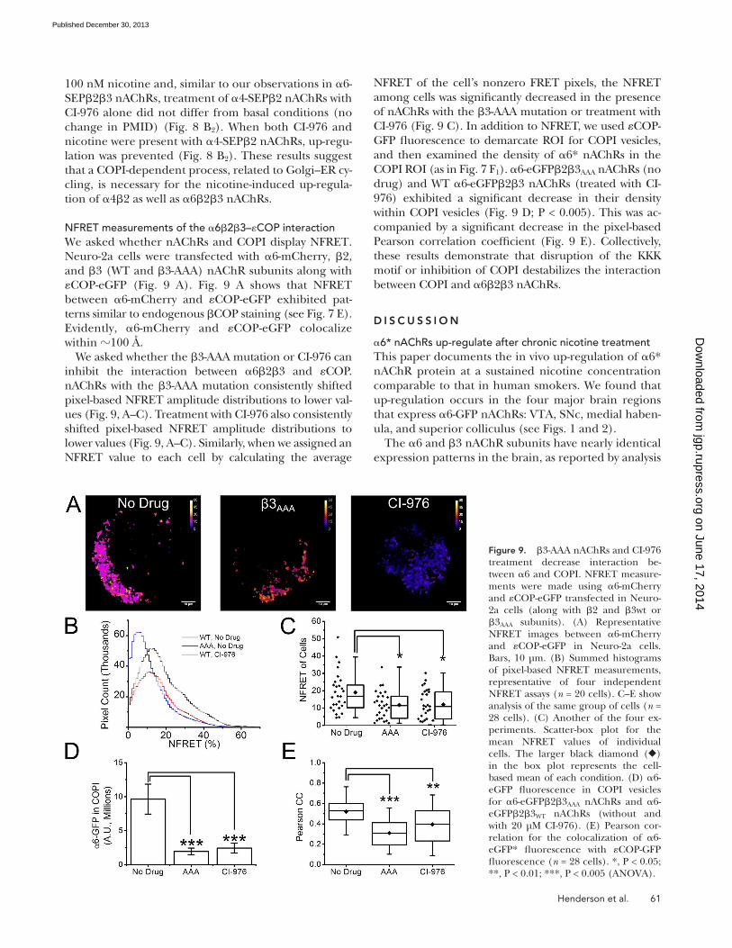
NFRET of the cell’s nonzero FRET pixels, the NFRET among cells was significantly decreased in the presence of nAChRs with the 3-AAA mutation or treatment with CI-976 (Fig. 9 C). In addition to NFRET, we used COP-GFP fluorescence to demarcate ROI for COPI vesicles, and then examined the density of 6* nAChRs in the COPI ROI (as in Fig. 7 F1). 6-eGFP23AAA nAChRs (no drug) and WT 6-eGFP23 nAChRs (treated with CI-976) exhibited a significant decrease in their density within COPI vesicles (Fig. 9 D; P < 0.005). This was ac-companied by a significant decrease in the pixel-based Pearson correlation coefficient (Fig. 9 E). Collectively, these results demonstrate that disruption of the KKK motif or inhibition of COPI destabilizes the interaction between COPI and 623 nAChRs.
D I S C U S S I O N
6* nAChRs up-regulate after chronic nicotine treatmentThis paper documents the in vivo up-regulation of 6* nAChR protein at a sustained nicotine concentration comparable to that in human smokers. We found that up-regulation occurs in the four major brain regions that express 6-GFP nAChRs: VTA, SNc, medial haben-ula, and superior colliculus (see Figs. 1 and 2).
The 6 and 3 nAChR subunits have nearly identical expression patterns in the brain, as reported by analysis
100 nM nicotine and, similar to our observations in 6-SEP23 nAChRs, treatment of 4-SEP2 nAChRs with CI-976 alone did not differ from basal conditions (no change in PMID) (Fig. 8 B2). When both CI-976 and nicotine were present with 4-SEP2 nAChRs, up-regu-lation was prevented (Fig. 8 B2). These results suggest that a COPI-dependent process, related to Golgi–ER cy-cling, is necessary for the nicotine-induced up-regula-tion of 42 as well as 623 nAChRs.
NFRET measurements of the 623–COP interactionWe asked whether nAChRs and COPI display NFRET. Neuro-2a cells were transfected with 6-mCherry, 2, and 3 (WT and 3-AAA) nAChR subunits along with COP-eGFP (Fig. 9 A). Fig. 9 A shows that NFRET between 6-mCherry and COP-eGFP exhibited pat-terns similar to endogenous COP staining (see Fig. 7 E). Evidently, 6-mCherry and COP-eGFP colocalize within 100 Å.
We asked whether the 3-AAA mutation or CI-976 can inhibit the interaction between 623 and COP. nAChRs with the 3-AAA mutation consistently shifted pixel-based NFRET amplitude distributions to lower val-ues (Fig. 9, A–C). Treatment with CI-976 also consistently shifted pixel-based NFRET amplitude distributions to lower values (Fig. 9, A–C). Similarly, when we assigned an NFRET value to each cell by calculating the average
Figure 9. 3-AAA nAChRs and CI-976 treatment decrease interaction be-tween 6 and COPI. NFRET measure-ments were made using 6-mCherry and COP-eGFP transfected in Neuro-2a cells (along with 2 and 3wt or 3AAA subunits). (A) Representative NFRET images between 6-mCherry and COP-eGFP in Neuro-2a cells. Bars, 10 µm. (B) Summed histograms of pixel-based NFRET measurements, representative of four independent NFRET assays (n = 20 cells). C–E show analysis of the same group of cells (n = 28 cells). (C) Another of the four ex-periments. Scatter-box plot for the mean NFRET values of individual cells. The larger black diamond () in the box plot represents the cell-based mean of each condition. (D) 6-eGFP fluorescence in COPI vesicles for 6-eGFP23AAA nAChRs and 6-eGFP23WT nAChRs (without and with 20 µM CI-976). (E) Pearson cor-relation for the colocalization of 6-eGFP* fluorescence with COP-GFP fluorescence (n = 28 cells). *, P < 0.05; **, P < 0.01; ***, P < 0.005 (ANOVA).
on June 17, 2014jgp.rupress.org
Dow
nloaded from
Published December 30, 2013

62 Nicotine exploits COPI for up-regulation
in the contribution of 6* nAChRs, as identified by bind-ing or dopamine release assays (Lai et al., 2005; Perry et al., 2007; Exley et al., 2013). Several factors may contrib-ute to these disparate findings. First, the up-regulation of 6* nAChRs we observed was in midbrain (VTA and SNc) dopaminergic neurons, whereas the studies show-ing a decrease in 6* nAChR contribution were com-pleted in the striatal regions. We have confirmed previously that 6-GFP* nAChRs in the VTA and SNc are localized exclusively in dopaminergic neurons (Mackey et al., 2012). The difference in striatal (down-regulation) and midbrain (up-regulation) may be explained by a different population of nAChRs (64*, 6[non4]*, 63*, and/or 6[non3]*). Perez et al. (2008) have shown that although 6(non4)* nAChRs up-regulate, 64* nAChRs down-regulate. We, along with others (Tumkosit et al., 2006), have shown that 6* nAChRs may require the 3 subunit to up-regulate. Therefore, the population of nAChRs residing in the somata of midbrain dopaminergic neurons may be composed of
of mRNA and autoradiography (Le Novère et al., 1996; Azam et al., 2002; Baddick and Marks, 2011). In heter-ologous systems, 623 nAChRs show better function (higher density on the PM, larger peak current re-sponse) than 62. Therefore, it is appropriate to study effects of chronic nicotine on 62* nAChRs in the context of coexpressed 3 subunits. We found that, at concentrations mimicking the steady-state levels found in human smokers and during nicotine replacement therapy (50 nM nicotine), 6-SEP23 nAChRs up-reg-ulate whereas 6-SEP2 nAChRs failed to up-regulate. The 3 subunit thus appears necessary for the up-regu-lation of 6-SEP2* nAChRs. This agrees well with Tumkosit et al. (2006), who showed that 3 nAChR subunits promote nicotine-induced up-regulation of 6* nAChRs.
Our observations of 6* up-regulation agree with some previously published work (Parker et al., 2004; Perez et al., 2008; Walsh et al., 2008). Other studies found that chronic nicotine treatment leads to a decrease
Figure 10. A schematic representation of selective pharmacological chaperoning of nAChRs in response to chronic nicotine. (A) Nicotine interconverts within milliseconds between the protonated membrane-imper-meant form and the neutral membrane-per-meant form. The latter enters cells and enters the ER (red arrow). Here, the charged form pharmacologically chaperones nAChRs so that increased numbers exit toward the Golgi via the COPII pathway, eventually resulting in receptor up-regulation at the PM. Thus, pharmacological chaperoning by nicotine is thought to underlie the process of nAChR up-regulation by chronic nicotine. (B) This study finds that nicotine-mediated up-regu-lation also depends on the COPI machinery involved in ER to Golgi and Golgi to ER trans-port of nAChRs. One possible explanation for the COPI dependence is that nicotine enters additional organelles and binds more extensively than previously thought within the early exocytic pathway, shown by the ad-ditional red arrows leading to both COPI and COPII vesicles, ERGIC, and cis-Golgi. See also the animation, “Nicotine Up-regulates nAChRs”: http://www.jgp.org/cgi/content/full/jgp.201311102/DC2.
on June 17, 2014jgp.rupress.org
Dow
nloaded from
Published December 30, 2013

Henderson et al. 63
(see Fig. 7, E, F1, and F2). Furthermore, 3-AAA nAChRs shifted the NFRET distribution to lower values com-pared with WT nAChRs (see Fig. 9). Collectively, these data suggest that the disruption of the KKK motif re-sults in deficient retrograde transport of nAChRs be-tween the Golgi and ER. Although one cannot draw specific conclusions about the locus of interference in the cyclic processes involving ER, COPII, cis-Golgi, and COPI (Orci et al., 1997), the data suggest that this dis-rupted cycling from the Golgi back to the ER prevents up-regulation during exposure to nicotine.
A COPI-dependent process, such as Golgi–ER cycling, is necessary for the up-regulation of nAChRsWe asked whether Golgi–ER cycling plays a role in the other case of nAChRs (42) that up-regulate at smoking-relevant concentrations of nicotine. Because the 3 KKK motif is unique in nAChR subunits, we exploited a more general inhibitor approach, CI-976, to block COPI func-tion. On 623 nAChRs, CI-976 produced results that resembled the pattern observed for 3-WT and 3-AAA nAChRs (compare Figs. 8 B1 with 6 D2). Inhibiting Golgi–ER cycling prevented up-regulation of 623 and 42 nAChRs after nicotine treatment. This suggests that the cycling of nAChRs between the Golgi and ER is necessary for the up-regulation of nAChRs on the PM.
Previous reports indicate that nAChR up-regulation is initiated within intracellular compartments and that the locus is partially in the ER (see Fig. 10) (Kuryatov et al., 2005; Sallette et al., 2005; Lester et al., 2009; Miwa et al., 2011; Srinivasan et al., 2011). The data presented here suggest novel aspects of the up-regulation of nAChRs, related to general observations that secretory pathways have both regulated and constitutive components. The increased nAChR density on the PM results from in-creased ER export of 623 nAChRs (increase in ERES/COPII, leading to increased insertion events). Previous data show that this also occurs in 42 nAChRs (Richards et al., 2011; Srinivasan et al., 2011) (depicted in Fig. 10). We show here that chronic nicotine also increases the number of nAChRs in COPI vesicles (Fig. 7 F1). This suggests that in addition to an increase in ER export, there is also an increase of retrograde movement of nAChRs from the Golgi to the ER. Possibly, 623 and 42 nAChRs are continually cycled between the ER and Golgi through COPI and COPII. This cycling maintains a population of nAChRs in the ER that may be neces-sary for up-regulation. In the absence of nicotine, dis-ruption of Golgi–ER cycling (either through the 3-AAA mutants or use of CI-976) does not affect PM nAChR density (Figs. 8 B1 and 6 D2). However, when Golgi–ER cycling is inhibited and nicotine is present, nicotine can-not produce up-regulated PM nAChRs.
How does nicotine exploit a COPI process to up-reg-ulate PM nAChRs? There are three nonmutually exclu-sive possibilities. (1) Decreased retrieval of nAChRs
more 6(non4)3* nAChRs than 64* or 6(non 3)* nAChRs. However, there may be more 64* or 6(non3)* nAChRs on these neurons’ dopaminergic terminals in the striatum and accumbens. This is sup-ported by a report that there are more 64* nAChRs on dopaminergic terminals (in striatum and accum-bens) than there are on dopaminergic cell bodies (in VTA and SNc) (Champtiaux et al., 2003).
Second, differing concentrations of nicotine were used in the different studies. The doses of nicotine we used in mice (2 mg/kg/h and 0.4 mg/kg/h) produce plasma concentrations of <500 and <100 nM nicotine, respectively (Marks et al., 2004). Perry et al. (2007) used a dose (6 mg/kg/day) in rats that produces >500 nM plasma nicotine (Nguyen et al., 2004). Our data show that 500 nM nicotine decreased the density of 6* nAChRs on the PM (Fig. 4, D and F). Therefore, the differences observed in our current work and in that of others (Lai et al., 2005; Perry et al., 2007; Exley et al., 2013) may be caused by the concentration of nicotine used.
The KKK motif of 3 nAChR subunit suggests that a COPI-mediated retrograde pathway participates in up-regulationA KKK motif appears in the M3–M4 intracellular loop of the 3 nAChR subunit. COPI binds to di-lysine motifs (KKxx or KxKxx) (Jackson et al., 2012; Ma and Goldberg, 2013), and the KKK of the 3 subunit satisfies both mo-tifs. Measurements of FRET between 623 nAChRs and COP indicate that the two molecules approach with 100 Å, and this interaction is decreased by mutat-ing the 3-KKK sequence to 3-AAA (Fig. 9). This sug-gests that one of these di-lysine motifs in 3 mediates a direct interaction between nAChRs and COPI.
We hypothesized that COPI likely plays a role in the retrograde transport of 623 nAChRs from the Golgi back to the ER. 6* nAChRs with a 3-AAA in place of 3-KKK failed to up-regulate on the PM in the presence of nicotine. We found that the ER localization of the 6-SEP23AAA nAChRs was decreased compared with 6-SEP23 nAChRs, suggesting that the 3-AAA motif changed the distribution of nAChRs among organelles in the early secretory pathway. Indeed, we found that the 6-SEP23AAA nAChRs had a significantly higher localization within the Golgi than the 6-SEP23 nAChRs (Fig. 7, C, D1, and D2).
When we add the fact that the number of ERES does not differ between the 623 and 623AAA nAChRs in basal conditions (e.g., no nicotine) (Fig. 7, A and B), we can conclude that the export (via COPII vesicles) of nAChRs is not affected. Thus, 623AAA nAChRs are exported from the ER at a rate similar to that of 623 nAChRs. Because export (COPII mediated) is not affected, this suggests that retrieval (COPI mediated) is respon-sible for the disrupted trafficking. This was confirmed when we observed significantly fewer 623AAA nAChRs in COPI vesicles when compared with 623 nAChRs
on June 17, 2014jgp.rupress.org
Dow
nloaded from
Published December 30, 2013

64 Nicotine exploits COPI for up-regulation
The authors have no conflicting financial interests.
Edward N. Pugh Jr. served as editor.
Submitted: 16 September 2013Accepted: 6 December 2013
R E F E R E N C E SAraki, Y., D.T. Lin, and R.L. Huganir. 2010. Plasma membrane in-
sertion of the AMPA receptor GluA2 subunit is regulated by NSF binding and Q/R editing of the ion pore. Proc. Natl. Acad. Sci. USA. 107:11080–11085. http://dx.doi.org/10.1073/pnas.1006584107
Asokan, A., and M.J. Cho. 2002. Exploitation of intracellular pH gradients in the cellular delivery of macromolecules. J. Pharm. Sci. 91:903–913. http://dx.doi.org/10.1002/jps.10095
Azam, L., U.H. Winzer-Serhan, Y. Chen, and F.M. Leslie. 2002. Expression of neuronal nicotinic acetylcholine receptor subunit mRNAs within midbrain dopamine neurons. J. Comp. Neurol. 444:260–274. http://dx.doi.org/10.1002/cne.10138
Baddick, C.G., and M.J. Marks. 2011. An autoradiographic survey of mouse brain nicotinic acetylcholine receptors defined by null mutants. Biochem. Pharmacol. 82:828–841. http://dx.doi.org/10.1016/j.bcp.2011.04.019
Benwell, M.E., D.J. Balfour, and J.M. Anderson. 1988. Evidence that tobacco smoking increases the density of ()-[3H]nicotine binding sites in human brain. J. Neurochem. 50:1243–1247. http://dx.doi.org/10.1111/j.1471-4159.1988.tb10600.x
Brandizzi, F., and C. Barlowe. 2013. Organization of the ER-Golgi interface for membrane traffic control. Nat. Rev. Mol. Cell Biol. 14:382–392. http://dx.doi.org/10.1038/nrm3588
Breese, C.R., M.J. Marks, J. Logel, C.E. Adams, B. Sullivan, A.C. Collins, and S. Leonard. 1997. Effect of smoking history on [3H]nicotine binding in human postmortem brain. J. Pharmacol. Exp. Ther. 282:7–13.
Brown, W.J., H. Plutner, D. Drecktrah, B.L. Judson, and W.E. Balch. 2008. The lysophospholipid acyltransferase antagonist CI-976 inhibits a late step in COPII vesicle budding. Traffic. 9:786–797. http://dx.doi.org/10.1111/j.1600-0854.2008.00711.x
Brunzell, D.H., K.E. Boschen, E.S. Hendrick, P.M. Beardsley, and J.M. McIntosh. 2010. -conotoxin MII-sensitive nicotinic acetylcho-line receptors in the nucleus accumbens shell regulate progressive ratio responding maintained by nicotine. Neuropsychopharmacology. 35:665–673. http://dx.doi.org/10.1038/npp.2009.171
Champtiaux, N., C. Gotti, M. Cordero-Erausquin, D.J. David, C. Przybylski, C. Léna, F. Clementi, M. Moretti, F.M. Rossi, N. Le Novère, et al. 2003. Subunit composition of functional nicotinic receptors in dopaminergic neurons investigated with knock-out mice. J. Neurosci. 23:7820–7829.
Drenan, R.M., R. Nashmi, P.I. Imoukhuede, H. Just, S. McKinney, and H.A. Lester. 2008. Subcellular trafficking, pentameric assem-bly, and subunit stoichiometry of neuronal nicotinic acetylcholine receptors containing fluorescently labeled 6 and 3 subunits. Mol. Pharmacol. 73:27–41. http://dx.doi.org/10.1124/mol.107.039180
Exley, R., M.A. Clements, H. Hartung, J.M. McIntosh, M. Franklin, I. Bermudez, and S.J. Cragg. 2013. Striatal dopamine transmis-sion is reduced after chronic nicotine with a decrease in 6-nico-tinic receptor control in nucleus accumbens. Eur. J. Neurosci. 38:3036–3043. http://dx.doi.org/10.1111/ejn.12298.
Flores, C.M., S.W. Rogers, L.A. Pabreza, B.B. Wolfe, and K.J. Kellar. 1992. A subtype of nicotinic cholinergic receptor in rat brain is composed of 4 and 2 subunits and is up-regulated by chronic nicotine treatment. Mol. Pharmacol. 41:31–37.
Jackson, K.J., J.M. McIntosh, D.H. Brunzell, S.S. Sanjakdar, and M.I. Damaj. 2009. The role of alpha6-containing nicotinic acetylcho-line receptors in nicotine reward and withdrawal. J. Pharmacol. Exp. Ther. 331:547–554. http://dx.doi.org/10.1124/jpet.109.155457
from the Golgi produces an increased number of Golgi-resident nAChRs but a reduced number of ER-resident nAChRs. Individual nAChRs therefore spend a smaller fraction of time in the ER. Perhaps this renders them less available for pharmacological chaperoning by nico-tine (Fig. 10 A). Such chaperoning seems to be rela-tively inefficient, even though it takes place at nicotine concentrations 10–100 times lower than that required for activation of PM nAChRs. This inefficiency could occur because binding of nicotine must await the dis-sociation of a competing endogenous chaperone protein such as lynx1 (Miwa et al., 2012). (2) Pharmacological chaperoning by nicotine does stabilize pentameric nAChRs in the ER (Whiteaker et al., 1998; Sallette et al., 2005) but is nonetheless a pathological process that could block or prevent one or more modifications re-quired for efficient exit from the Golgi to the PM. If the nAChRs chaperoned by nicotine fail to pass the quality control checks in the Golgi, they would be targeted for retrieval back to the ER via a COPI-dependent process. (3) An intriguing possibility is that nicotine may enter additional organelles within the early exocytotic path-way. Nicotine might remain bound to nAChRs while the nAChRs reside in COPI and/or COPII vesicles (Fig. 10 B). Processes related to pharmacological chaperoning, termed “matchmaking,” “escorting,” or “abduction,” might occur in these vesicles (Lester et al., 2012). Be-cause the ER and cis-Golgi have nearly neutral lumenal pH, acid trapping of nicotine (Jia et al., 2003; Lester et al., 2009) probably plays little role in the processes studied here.
Golgi–ER cycling as a new mechanistic target for drug discoveryDiscovery of the role played by Golgi–ER cycling in both 623 and 42 nAChRs may eventually provide ad-ditional avenues for nicotine addiction therapies. A hy-pothetical small molecule designed to bind to the KKK and disrupt COPI binding would prevent the Golgi–ER cycling in a manner similar to observations in this work. This inhibition of Golgi–ER cycling would prevent the up-regulation of 3-containing nAChRs and may pro-duce effects similar to many antagonists that have shown clinical promise (Watkins et al., 1999; Rauhut et al., 2003; Yoshimura et al., 2007). In the search for potential therapeutics, the selective expression pattern of 623 nAChRs to just a few neuronal populations would render the targeting of 3 more selective than a drug that in-hibits Golgi–ER cycling in general.
We thank Jennifer Lippincott-Schwartz for kindly providing COP-GFP subunits for our studies, Rell Parker for comments, and the Barbara Wold laboratory for providing cryosectioning tools.
This work was supported by National Institutes of Health (grants AG033954, DA017279, DA019375, DA030396, NS034407, and DA033721) and by the California Tobacco-Related Disease Research Program (grant 17RT0127). Louis and Janet Fletcher provided partial funding for the TIRF microscope.
on June 17, 2014jgp.rupress.org
Dow
nloaded from
Published December 30, 2013

Henderson et al. 65
Mani, M., and T.A. Ryan. 2009. Live imaging of synaptic vesicle release and retrieval in dopaminergic neurons. Front Neural Circuits. 3:3.
Marks, M.J., J.B. Burch, and A.C. Collins. 1983. Effects of chronic nicotine infusion on tolerance development and nicotinic recep-tors. J. Pharmacol. Exp. Ther. 226:817–825.
Marks, M.J., P.P. Rowell, J.Z. Cao, S.R. Grady, S.E. McCallum, and A.C. Collins. 2004. Subsets of acetylcholine-stimulated 86Rb+ efflux and [125I]-epibatidine binding sites in C57BL/6 mouse brain are differentially affected by chronic nicotine treatment. Neuropharmacology. 46:1141–1157. http://dx.doi.org/10.1016/j.neuropharm.2004.02.009
Miesenböck, G., D.A. De Angelis, and J.E. Rothman. 1998. Visualizing secretion and synaptic transmission with pH-sensitive green fluorescent proteins. Nature. 394:192–195. http://dx.doi.org/10.1038/28190
Miwa, J.M., R. Freedman, and H.A. Lester. 2011. Neural systems gov-erned by nicotinic acetylcholine receptors: emerging hypotheses. Neuron. 70:20–33. http://dx.doi.org/10.1016/j.neuron.2011.03.014
Miwa, J.M., H.A. Lester, and A. Walz. 2012. Optimizing cholinergic tone through lynx modulators of nicotinic receptors: implica-tions for plasticity and nicotine addiction. Physiology (Bethesda). 27:187–199. http://dx.doi.org/10.1152/physiol.00002.2012
Moss, F.J., P.I. Imoukhuede, K. Scott, J. Hu, J.L. Jankowsky, M.W. Quick, and H.A. Lester. 2009. GABA transporter function, oligo-merization state, and anchoring: Correlates with subcellularly re-solved FRET. J. Gen. Physiol. 134:489–521. http://dx.doi.org/10.1085/jgp.200910314
Mukhin, A.G., A.S. Kimes, S.I. Chefer, J.A. Matochik, C.S. Contoreggi, A.G. Horti, D.B. Vaupel, O. Pavlova, and E.A. Stein. 2008. Greater nicotinic acetylcholine receptor density in smokers than in nonsmokers: a PET study with 2-18F-FA-85380. J. Nucl. Med. 49:1628–1635. http://dx.doi.org/10.2967/jnumed.108.050716
Nashmi, R., C. Xiao, P. Deshpande, S. McKinney, S.R. Grady, P. Whiteaker, Q. Huang, T. McClure-Begley, J.M. Lindstrom, C. Labarca, et al. 2007. Chronic nicotine cell specifically upregulates functional 4* nicotinic receptors: basis for both tolerance in midbrain and en-hanced long-term potentiation in perforant path. J. Neurosci. 27:8202–8218. http://dx.doi.org/10.1523/JNEUROSCI.2199-07.2007
Nguyen, H.N., B.A. Rasmussen, and D.C. Perry. 2004. Binding and functional activity of nicotinic cholinergic receptors in selected rat brain regions are increased following long-term but not short-term nicotine treatment. J. Neurochem. 90:40–49. http://dx.doi.org/10.1111/j.1471-4159.2004.02482.x
Orci, L., M. Stamnes, M. Ravazzola, M. Amherdt, A. Perrelet, T.H. Söllner, and J.E. Rothman. 1997. Bidirectional transport by distinct populations of COPI-coated vesicles. Cell. 90:335–349. http://dx.doi.org/10.1016/S0092-8674(00)80341-4
Parker, S.L., Y. Fu, K. McAllen, J. Luo, J.M. McIntosh, J.M. Lindstrom, and B.M. Sharp. 2004. Up-regulation of brain nico-tinic acetylcholine receptors in the rat during long-term self-administration of nicotine: disproportionate increase of the 6 subunit. Mol. Pharmacol. 65:611–622. http://dx.doi.org/10.1124/mol.65.3.611
Peng, X., V. Gerzanich, R. Anand, P.J. Whiting, and J. Lindstrom. 1994. Nicotine-induced increase in neuronal nicotinic receptors results from a decrease in the rate of receptor turnover. Mol. Pharmacol. 46:523–530.
Perez, X.A., T. Bordia, J.M. McIntosh, S.R. Grady, and M. Quik. 2008. Long-term nicotine treatment differentially regulates striatal 642* and 6(non4)2* nAChR expression and function. Mol. Pharmacol. 74:844–853. http://dx.doi.org/10.1124/mol.108.048843
Perry, D.C., D. Mao, A.B. Gold, J.M. McIntosh, J.C. Pezzullo, and K.J. Kellar. 2007. Chronic nicotine differentially regulates 6- and 3-containing nicotinic cholinergic receptors in rat brain. J. Pharmacol. Exp. Ther. 322:306–315. http://dx.doi.org/10.1124/jpet.107.121228
Jackson, L.P., M. Lewis, H.M. Kent, M.A. Edeling, P.R. Evans, R. Duden, and D.J. Owen. 2012. Molecular basis for recognition of dilysine trafficking motifs by COPI. Dev. Cell. 23:1255–1262. http://dx.doi.org/10.1016/j.devcel.2012.10.017
Jacob, T.C., Y.D. Bogdanov, C. Magnus, R.S. Saliba, J.T. Kittler, P.G. Haydon, and S.J. Moss. 2005. Gephyrin regulates the cell surface dynamics of synaptic GABAA receptors. J. Neurosci. 25:10469–10478. http://dx.doi.org/10.1523/JNEUROSCI.2267-05.2005
Jaskolski, F., B. Mayo-Martin, D. Jane, and J.M. Henley. 2009. Dynamin-dependent membrane drift recruits AMPA receptors to dendritic spines. J. Biol. Chem. 284:12491–12503. http://dx.doi.org/10.1074/jbc.M808401200
Jia, L., K. Flotildes, M. Li, and B.N. Cohen. 2003. Nicotine trapping causes the persistent desensitization of 42 nicotinic receptors ex-pressed in oocytes. J. Neurochem. 84:753–766. http://dx.doi.org/10.1046/j.1471-4159.2003.01578.x
Koob, G.F. 2009. New dimensions in human laboratory models of addiction. Addict. Biol. 14:1–8. http://dx.doi.org/10.1111/j.1369-1600.2008.00127.x
Kuryatov, A., and J. Lindstrom. 2011. Expression of functional human 623* acetylcholine receptors in Xenopus laevis oocytes achieved through subunit chimeras and concatamers. Mol. Pharmacol. 79:126–140. http://dx.doi.org/10.1124/mol.110.066159
Kuryatov, A., J. Luo, J. Cooper, and J. Lindstrom. 2005. Nicotine acts as a pharmacological chaperone to up-regulate human 42 acetylcholine receptors. Mol. Pharmacol. 68:1839–1851.
Lai, A., N. Parameswaran, M. Khwaja, P. Whiteaker, J.M. Lindstrom, H. Fan, J.M. McIntosh, S.R. Grady, and M. Quik. 2005. Long-term nicotine treatment decreases striatal 6* nicotinic acetylcholine receptor sites and function in mice. Mol. Pharmacol. 67:1639–1647. http://dx.doi.org/10.1124/mol.104.006429
Le Novère, N., M. Zoli, and J.P. Changeux. 1996. Neuronal nico-tinic receptor 6 subunit mRNA is selectively concentrated in catecholaminergic nuclei of the rat brain. Eur. J. Neurosci. 8:2428–2439. http://dx.doi.org/10.1111/j.1460-9568.1996.tb01206.x
Lester, H.A., C. Xiao, R. Srinivasan, C.D. Son, J. Miwa, R. Pantoja, M.R. Banghart, D.A. Dougherty, A.M. Goate, and J.C. Wang. 2009. Nicotine is a selective pharmacological chaperone of acetylcholine receptor number and stoichiometry. Implications for drug discovery. AAPS J. 11:167–177. http://dx.doi.org/10.1208/s12248-009-9090-7
Lester, H.A., J.M. Miwa, and R. Srinivasan. 2012. Psychiatric drugs bind to classical targets within early exocytotic pathways: thera-peutic effects. Biol. Psychiatry. 72:907–915. http://dx.doi.org/10.1016/j.biopsych.2012.05.020
Lin, D.T., Y. Makino, K. Sharma, T. Hayashi, R. Neve, K. Takamiya, and R.L. Huganir. 2009. Regulation of AMPA receptor extrasyn-aptic insertion by 4.1N, phosphorylation and palmitoylation. Nat. Neurosci. 12:879–887. http://dx.doi.org/10.1038/nn.2351
Lomazzo, E., G.P. Hussmann, B.B. Wolfe, R.P. Yasuda, D.C. Perry, and K.J. Kellar. 2011. Effects of chronic nicotine on heteromeric neuro-nal nicotinic receptors in rat primary cultured neurons. J. Neurochem. 119:153–164. http://dx.doi.org/10.1111/j.1471-4159.2011.07408.x
Ma, W., and J. Goldberg. 2013. Rules for the recognition of dilysine retrieval motifs by coatomer. EMBO J. 32:926–937. http://dx.doi.org/10.1038/emboj.2013.41
Mackey, E.D., S.E. Engle, M.R. Kim, H.C. O’Neill, C.R. Wageman, N.E. Patzlaff, Y. Wang, S.R. Grady, J.M. McIntosh, M.J. Marks, et al. 2012. 6* nicotinic acetylcholine receptor expression and function in a visual salience circuit. J. Neurosci. 32:10226–10237. http://dx.doi.org/10.1523/JNEUROSCI.0007-12.2012
Mamede, M., K. Ishizu, M. Ueda, T. Mukai, Y. Iida, H. Kawashima, H. Fukuyama, K. Togashi, and H. Saji. 2007. Temporal change in human nicotinic acetylcholine receptor after smoking cessation: 5IA SPECT study. J. Nucl. Med. 48:1829–1835. http://dx.doi.org/10.2967/jnumed.107.043471
on June 17, 2014jgp.rupress.org
Dow
nloaded from
Published December 30, 2013

66 Nicotine exploits COPI for up-regulation
Picciotto, M.R., M. Zoli, R. Rimondini, C. Léna, L.M. Marubio, E.M. Pich, K. Fuxe, and J.P. Changeux. 1998. Acetylcholine receptors con-taining the 2 subunit are involved in the reinforcing properties of nicotine. Nature. 391:173–177. http://dx.doi.org/10.1038/34413
Pons, S., L. Fattore, G. Cossu, S. Tolu, E. Porcu, J.M. McIntosh, J.P. Changeux, U. Maskos, and W. Fratta. 2008. Crucial role of 4 and 6 nicotinic acetylcholine receptor subunits from ventral tegmental area in systemic nicotine self-administration. J. Neurosci. 28:12318–12327. http://dx.doi.org/10.1523/JNEUROSCI.3918-08.2008
Rauhut, A.S., N. Neugebauer, L.P. Dwoskin, and M.T. Bardo. 2003. Effect of bupropion on nicotine self-administration in rats. Psychopharmacology (Berl.). 169:1–9. http://dx.doi.org/10.1007/s00213-003-1450-x
Richards, C.I., R. Srinivasan, C. Xiao, E.D. Mackey, J.M. Miwa, and H.A. Lester. 2011. Trafficking of 4* nicotinic receptors revealed by superecliptic phluorin: effects of a 4 amyotrophic lateral sclerosis-associated mutation and chronic exposure to nicotine. J. Biol. Chem. 286:31241–31249. http://dx.doi.org/10.1074/jbc.M111.256024
Ritz, B., A. Ascherio, H. Checkoway, K.S. Marder, L.M. Nelson, W.A. Rocca, G.W. Ross, D. Strickland, S.K. Van Den Eeden, and J. Gorell. 2007. Pooled analysis of tobacco use and risk of Parkinson disease. Arch. Neurol. 64:990–997. http://dx.doi.org/10.1001/archneur.64.7.990
Sallette, J., S. Pons, A. Devillers-Thiery, M. Soudant, L. Prado de Carvalho, J.P. Changeux, and P.J. Corringer. 2005. Nicotine upregulates its own receptors through enhanced intracellular maturation. Neuron. 46:595–607. http://dx.doi.org/10.1016/j.neuron.2005.03.029
Schnoll, R.A., E.P. Wileyto, F.T. Leone, R.F. Tyndale, and N.L. Benowitz. 2013. High dose transdermal nicotine for fast metaboliz-ers of nicotine: a proof of concept placebo-controlled trial. Nicotine Tob. Res. 15:348–354. http://dx.doi.org/10.1093/ntr/nts129
Schwartz, R.D., and K.J. Kellar. 1983. Nicotinic cholinergic receptor binding sites in the brain: regulation in vivo. Science. 220:214–216. http://dx.doi.org/10.1126/science.6828889
Spang, A. 2013. Traffic COPs: rules of detection. EMBO J. 32:915–916. http://dx.doi.org/10.1038/emboj.2013.57
Srinivasan, R., R. Pantoja, F.J. Moss, E.D.W. Mackey, C.D. Son, J. Miwa, and H.A. Lester. 2011. Nicotine up-regulates 42 nicotinic recep-tors and ER exit sites via stoichiometry-dependent chaperoning. J. Gen. Physiol. 137:59–79. http://dx.doi.org/10.1085/jgp.201010532
Srinivasan, R., C.I. Richards, C. Dilworth, F.J. Moss, D.A. Dougherty, and H.A. Lester. 2012a. Förster resonance energy transfer (FRET) correlates of altered subunit stoichiometry in Cys-loop receptors, exemplified by nicotinic 42. Int. J. Mol. Sci. 13:10022–10040. http://dx.doi.org/10.3390/ijms130810022
Srinivasan, R., C.I. Richards, C. Xiao, D. Rhee, R. Pantoja, D.A. Dougherty, J.M. Miwa, and H.A. Lester. 2012b. Pharmacological chaperoning of nicotinic acetylcholine receptors reduces the en-doplasmic reticulum stress response. Mol. Pharmacol. 81:759–769. http://dx.doi.org/10.1124/mol.112.077792
Tapper, A.R., S.L. McKinney, R. Nashmi, J. Schwarz, P. Deshpande, C. Labarca, P. Whiteaker, M.J. Marks, A.C. Collins, and H.A. Lester. 2004. Nicotine activation of 4* receptors: sufficient for reward, tolerance, and sensitization. Science. 306:1029–1032. http://dx.doi.org/10.1126/science.1099420
Tumkosit, P., A. Kuryatov, J. Luo, and J. Lindstrom. 2006. 3 subunits promote expression and nicotine-induced up-regulation of human nicotinic 6* nicotinic acetylcholine receptors expressed in trans-fected cell lines. Mol. Pharmacol. 70:1358–1368. http://dx.doi.org/10.1124/mol.106.027326
Walsh, H., A.P. Govind, R. Mastro, J.C. Hoda, D. Bertrand, Y. Vallejo, and W.N. Green. 2008. Up-regulation of nicotinic receptors by nic-otine varies with receptor subtype. J. Biol. Chem. 283:6022–6032. http://dx.doi.org/10.1074/jbc.M703432200
Watkins, S.S., M.P. Epping-Jordan, G.F. Koob, and A. Markou. 1999. Blockade of nicotine self-administration with nicotinic antagonists in rats. Pharmacol. Biochem. Behav. 62:743–751. http://dx.doi.org/10.1016/S0091-3057(98)00226-3
Whiteaker, P., C.G. Sharples, and S. Wonnacott. 1998. Agonist-induced up-regulation of 42 nicotinic acetylcholine receptors in M10 cells: pharmacological and spatial definition. Mol. Pharmacol. 53:950–962.
Xiao, C., R. Srinivasan, R.M. Drenan, E.D. Mackey, J.M. McIntosh, and H.A. Lester. 2011. Characterizing functional 62 nicotinic acetylcholine receptors in vitro: mutant 2 subunits improve mem-brane expression, and fluorescent proteins reveal responsive cells. Biochem. Pharmacol. 82:852–861. http://dx.doi.org/10.1016/j.bcp.2011.05.005
Yang, J.S., S.Y. Lee, S. Spanò, H. Gad, L. Zhang, Z. Nie, M. Bonazzi, D. Corda, A. Luini, and V.W. Hsu. 2005. A role for BARS at the fis-sion step of COPI vesicle formation from Golgi membrane. EMBO J. 24:4133–4143. http://dx.doi.org/10.1038/sj.emboj.7600873
Yang, J.S., C. Valente, R.S. Polishchuk, G. Turacchio, E. Layre, D.B. Moody, C.C. Leslie, M.H. Gelb, W.J. Brown, D. Corda, et al. 2011. COPI acts in both vesicular and tubular transport. Nat. Cell Biol. 13:996–1003. http://dx.doi.org/10.1038/ncb2273
Yoshimura, R.F., D.J. Hogenkamp, W.Y. Li, M.B. Tran, J.D. Belluzzi, E.R. Whittemore, F.M. Leslie, and K.W. Gee. 2007. Negative al-losteric modulation of nicotinic acetylcholine receptors blocks nicotine self-administration in rats. J. Pharmacol. Exp. Ther. 323:907–915. http://dx.doi.org/10.1124/jpet.107.128751
on June 17, 2014jgp.rupress.org
Dow
nloaded from
Published December 30, 2013

� Henderson�et�al. S1�of�20
jgp
TH
E j
OU
RN
AL
OF
gE
NE
RA
L p
HY
SIO
LO
gY
Figure S1. 3 subunits increase 64* nAChR density on PM. (A, C, and E) Representative TIRF images of Neuro-2a cells transfected with 6-SEP and a combination of 4 with or without 3 nAChR subunits at basic (pH 7.4) and acidic (pH 5.4) conditions. Nicotine was added at the listed concentrations (24 h). (B1, D1, and F1) PMID for 6-SEP* nAChRs. Number of imaged cells is indicated in parenthe-ses. Bars, 10 µm. Data are mean values ± SEM. n.s., not significant; *, P < 0.05; **, P < 0.005; ***, P < 0.0001.
O N L I N E S U p p L E m E N TA L m AT E R I A L
Henderson et al., http://www.jgp.org/cgi/content/full/jgp.201311102/DC1

S2� Nicotine�exploits�COPI�for�up-regulation
Figure S2. Schematic representation of nAChR chimeras and mutants. 3 and 2 nAChR subunits are designated blue and red, respec-tively. Numbers in diagrams correspond to transmembrane domains 1–4.
Figure S3. Functional up-regulation is observed in WT 623 nAChRs but not 623AAA nAChRs. (A) 6-eGFP23 and 6-eGFP23AAA nAChR currents elicited by 3 µM ACh in the absence or presence of nicotine treatment (50 nM nicotine, 24 h). (B) 6-eGFP23 nAChRs displayed a functional up-regulation of peak currents when treated chronically with nicotine, but 6-eGFP23AAA nAChRs did not display a functional up-regulation after chronic treatment with nicotine. *, P < 0.05.

� Henderson�et�al. S3�of�20
Figure S4. Blocking Golgi–ER cycling with CI-976 produces an increase in Golgi without an increase in ER export. Quantification of Sec24D fluorescence in ERES (A), 6-eGFP* intensity in Golgi cell sections (B), and Pearson correlation coefficients between 6-eGFP* and GalT-mCherry (Golgi marker) for 6-eGFP23 nAChRs (C).
Figure S5. CI-976 does not affect basal PM density of high ER export nAChRs. (A1 and A2) Representative TIRF images of Neuro-2a cells transfected with 4-SEP and cotransfected with either 4 or 2-DM nAChR subunits. 20 µM CI-976 was added 24 h before imaging. Cells were imaged at basic (pH 7.4) and acidic (pH 5.4) conditions. Bars, 10 µm. (B1 and B2) PMID for nAChRs. Numbers of imaged cells is indicated in parentheses.