anterograde microtubule transport drives microtubule ...€¦ · anterograde microtubule transport...
TRANSCRIPT
Molecular Biology of the CellVol. 20, 2943–2953, June 15, 2009
Anterograde Microtubule Transport Drives MicrotubuleBending in LLC-PK1 Epithelial CellsAndrew D. Bicek,*† Erkan Tuzel,†‡ Aleksey Demtchouk,* Maruti Uppalapati,§William O. Hancock,§ Daniel M. Kroll,� and David J. Odde*
*Department of Biomedical Engineering and ‡Institute for Mathematics and Its Applications, University ofMinnesota, Minneapolis, MN 55455; §Department of Bioengineering, The Pennsylvania State University,University Park, PA 16802; and �Department of Physics, North Dakota State University, Fargo, ND 58105
Submitted September 5, 2008; Revised February 23, 2009; Accepted April 16, 2009Monitoring Editor: Erika Holzbaur
Microtubules (MTs) have been proposed to act mechanically as compressive struts that resist both actomyosin contractileforces and their own polymerization forces to mechanically stabilize cell shape. To identify the origin of MT bending, wedirectly observed MT bending and F-actin transport dynamics in the periphery of LLC-PK1 epithelial cells. We found thatF-actin is nearly stationary in these cells even as MTs are deformed, demonstrating that MT bending is not driven byactomyosin contractility. Furthermore, the inhibition of myosin II activity through the use of blebbistatin results inmicrotubules that are still dynamically bending. In addition, as determined by fluorescent speckle microscopy, MTpolymerization rarely results, if ever, in bending. We suppressed dynamic instability using nocodazole, and we observedno qualitative change in the MT bending dynamics. Bending most often results from anterograde transport of proximalportions of the MT toward a nearly stationary distal tip. Interestingly, we found that in an in vitro kinesin-MT glidingassay, MTs buckle in a similar manner. To make quantitative comparisons, we measured curvature distributions ofobserved MTs and found that the in vivo and in vitro curvature distributions agree quantitatively. In addition, themeasured MT curvature distribution is not Gaussian, as expected for a thermally driven semiflexible polymer, indicatingthat thermal forces play a minor role in MT bending. We conclude that many of the known mechanisms of MT deformation,such as polymerization and acto-myosin contractility, play an inconsequential role in mediating MT bending in LLC-PK1 cellsand that MT-based molecular motors likely generate most of the strain energy stored in the MT lattice. The results argueagainst models in which MTs play a major mechanical role in LLC-PK1 cells and instead favor a model in which mechanicalforces control the spatial distribution of the MT array.
INTRODUCTION
Microtubules (MTs) are self-assembling linear polymers thatserve a range of functions within the cell, including trans-port and positioning of intracellular cargo via coupling tomolecular motors. The mechanical stiffness of MTs has beenproposed to play an important role in the mechanics of thecell (Wang et al., 1993, 2001; Ingber et al., 2000; Howard, 2001;Stamenovic et al., 2001; Ingber, 2003; Brangwynne et al.,2006). This hypothesis derives in part from the observationthat in interphase animal tissue cells, MTs are commonlyobserved to form a radial array, which qualitatively resem-bles the spokes of a bicycle wheel. MTs are therefore ideallypositioned to act as rigid struts that mechanically bear com-pressive loads in the cell to oppose and balance the tensionalforces (Buxbaum and Heidemann, 1988; Ingber, 1993; Wanget al., 1993, 2001; Stamenovic et al., 2001; Ingber, 2003). Theability of MTs to resist compressive loading has recentlybeen proposed to be enhanced by lateral reinforcementfrom the actin cytoskeleton, which could make MTs even
more effective in their putative role as compressive struts(Brangwynne et al., 2006). MTs are indeed mechanically stiffstructures, with a “flexural rigidity” (EI)—a measure ofbending resistance—of �21 pN-m2, �300 times larger thanthat of an actin filament (Gittes et al., 1993; Howard, 2001;VanBuren et al., 2005). Note, however, that this value hasonly been measured in vitro; it remains a formal possibilitythat MTs are softer in living cells due to the presence of MTbinding proteins or lattice defects.
Despite their mechanical stiffness, MTs in living cells areoften highly buckled, consistent with the model predictionthat MTs bear compressive loads (Wang et al., 1993, 2001;Ingber et al., 2000; Stamenovic et al., 2001; Ingber, 2003;Brangwynne et al., 2006). There have been several mecha-nisms suggested to cause the deformation of MTs. Theseinclude MT polymerization against a stationary distal tip(Dogterom and Yurke, 1997), acto-myosin contractility(Forscher and Smith, 1988; Waterman-Storer and Salmon,1997; Zhou et al., 2002) and transport (Schaefer et al., 2002),and buckling of MTs due to direct interactions with molec-ular motors such as dynein or kinesin (Koonce et al., 1999;Burakov et al., 2003; Dujardin et al., 2003; Malikov et al., 2004;Baas et al., 2005, 2006; Brito et al., 2005; Ferenz and Wads-worth, 2007), all of which may act alone or together in thepresence of thermal fluctuations.
It is well known that MTs can exert pushing forces duringpolymerization. During mitosis, for example, it is believedthat assembling MTs can exert pushing forces on chromo-
This article was published online ahead of print in MBC in Press(http://www.molbiolcell.org/cgi/doi/10.1091/mbc.E08–09–0909)on April 29, 2009.† These authors equally contributed to this work.
Address correspondence to: David J. Odde ([email protected]).
Abbreviations used: MT, microtubule.
© 2009 by The American Society for Cell Biology 2943
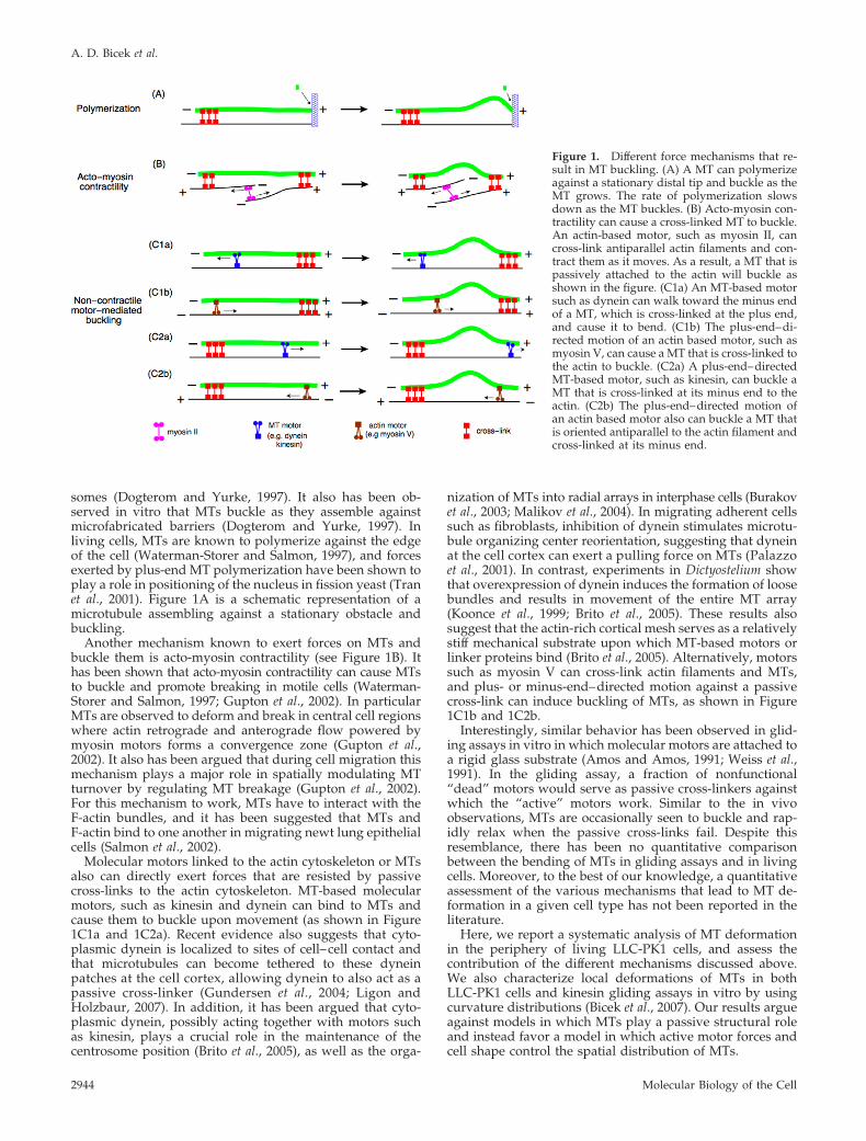
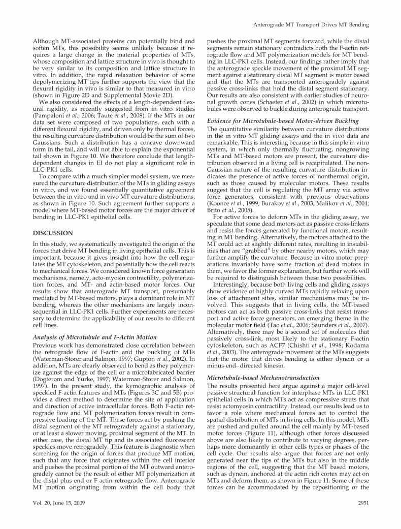
somes (Dogterom and Yurke, 1997). It also has been ob-served in vitro that MTs buckle as they assemble againstmicrofabricated barriers (Dogterom and Yurke, 1997). Inliving cells, MTs are known to polymerize against the edgeof the cell (Waterman-Storer and Salmon, 1997), and forcesexerted by plus-end MT polymerization have been shown toplay a role in positioning of the nucleus in fission yeast (Tranet al., 2001). Figure 1A is a schematic representation of amicrotubule assembling against a stationary obstacle andbuckling.
Another mechanism known to exert forces on MTs andbuckle them is acto-myosin contractility (see Figure 1B). Ithas been shown that acto-myosin contractility can cause MTsto buckle and promote breaking in motile cells (Waterman-Storer and Salmon, 1997; Gupton et al., 2002). In particularMTs are observed to deform and break in central cell regionswhere actin retrograde and anterograde flow powered bymyosin motors forms a convergence zone (Gupton et al.,2002). It also has been argued that during cell migration thismechanism plays a major role in spatially modulating MTturnover by regulating MT breakage (Gupton et al., 2002).For this mechanism to work, MTs have to interact with theF-actin bundles, and it has been suggested that MTs andF-actin bind to one another in migrating newt lung epithelialcells (Salmon et al., 2002).
Molecular motors linked to the actin cytoskeleton or MTsalso can directly exert forces that are resisted by passivecross-links to the actin cytoskeleton. MT-based molecularmotors, such as kinesin and dynein can bind to MTs andcause them to buckle upon movement (as shown in Figure1C1a and 1C2a). Recent evidence also suggests that cyto-plasmic dynein is localized to sites of cell–cell contact andthat microtubules can become tethered to these dyneinpatches at the cell cortex, allowing dynein to also act as apassive cross-linker (Gundersen et al., 2004; Ligon andHolzbaur, 2007). In addition, it has been argued that cyto-plasmic dynein, possibly acting together with motors suchas kinesin, plays a crucial role in the maintenance of thecentrosome position (Brito et al., 2005), as well as the orga-
nization of MTs into radial arrays in interphase cells (Burakovet al., 2003; Malikov et al., 2004). In migrating adherent cellssuch as fibroblasts, inhibition of dynein stimulates microtu-bule organizing center reorientation, suggesting that dyneinat the cell cortex can exert a pulling force on MTs (Palazzoet al., 2001). In contrast, experiments in Dictyostelium showthat overexpression of dynein induces the formation of loosebundles and results in movement of the entire MT array(Koonce et al., 1999; Brito et al., 2005). These results alsosuggest that the actin-rich cortical mesh serves as a relativelystiff mechanical substrate upon which MT-based motors orlinker proteins bind (Brito et al., 2005). Alternatively, motorssuch as myosin V can cross-link actin filaments and MTs,and plus- or minus-end–directed motion against a passivecross-link can induce buckling of MTs, as shown in Figure1C1b and 1C2b.
Interestingly, similar behavior has been observed in glid-ing assays in vitro in which molecular motors are attached toa rigid glass substrate (Amos and Amos, 1991; Weiss et al.,1991). In the gliding assay, a fraction of nonfunctional“dead” motors would serve as passive cross-linkers againstwhich the “active” motors work. Similar to the in vivoobservations, MTs are occasionally seen to buckle and rap-idly relax when the passive cross-links fail. Despite thisresemblance, there has been no quantitative comparisonbetween the bending of MTs in gliding assays and in livingcells. Moreover, to the best of our knowledge, a quantitativeassessment of the various mechanisms that lead to MT de-formation in a given cell type has not been reported in theliterature.
Here, we report a systematic analysis of MT deformationin the periphery of living LLC-PK1 cells, and assess thecontribution of the different mechanisms discussed above.We also characterize local deformations of MTs in bothLLC-PK1 cells and kinesin gliding assays in vitro by usingcurvature distributions (Bicek et al., 2007). Our results argueagainst models in which MTs play a passive structural roleand instead favor a model in which active motor forces andcell shape control the spatial distribution of MTs.
Figure 1. Different force mechanisms that re-sult in MT buckling. (A) A MT can polymerizeagainst a stationary distal tip and buckle as theMT grows. The rate of polymerization slowsdown as the MT buckles. (B) Acto-myosin con-tractility can cause a cross-linked MT to buckle.An actin-based motor, such as myosin II, cancross-link antiparallel actin filaments and con-tract them as it moves. As a result, a MT that ispassively attached to the actin will buckle asshown in the figure. (C1a) An MT-based motorsuch as dynein can walk toward the minus endof a MT, which is cross-linked at the plus end,and cause it to bend. (C1b) The plus-end–di-rected motion of an actin based motor, such asmyosin V, can cause a MT that is cross-linked tothe actin to buckle. (C2a) A plus-end–directedMT-based motor, such as kinesin, can buckle aMT that is cross-linked at its minus end to theactin. (C2b) The plus-end–directed motion ofan actin based motor also can buckle a MT thatis oriented antiparallel to the actin filament andcross-linked at its minus end.
A. D. Bicek et al.
Molecular Biology of the Cell2944
MATERIALS AND METHODS
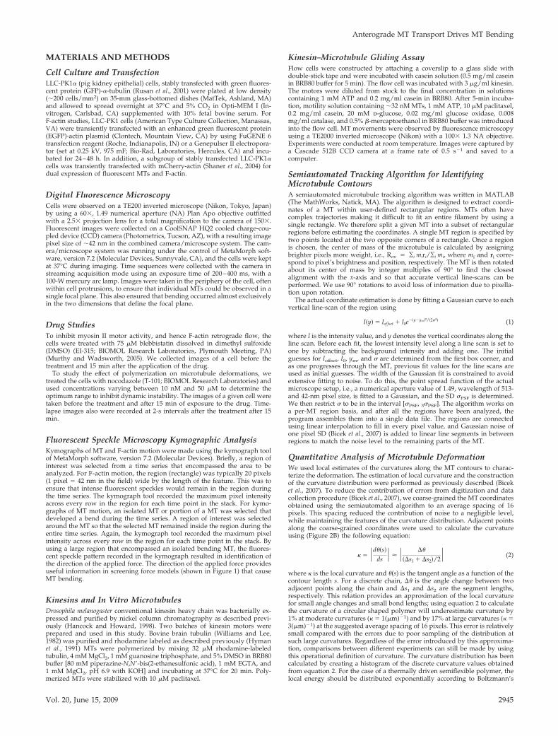
Cell Culture and TransfectionLLC-PK1� (pig kidney epithelial) cells, stably transfected with green fluores-cent protein (GFP)-�-tubulin (Rusan et al., 2001) were plated at low density(�200 cells/mm2) on 35-mm glass-bottomed dishes (MatTek, Ashland, MA)and allowed to spread overnight at 37°C and 5% CO2 in Opti-MEM I (In-vitrogen, Carlsbad, CA) supplemented with 10% fetal bovine serum. ForF-actin studies, LLC-PK1 cells (American Type Culture Collection, Manassas,VA) were transiently transfected with an enhanced green fluorescent protein(EGFP)-actin plasmid (Clontech, Mountain View, CA) by using FuGENE 6transfection reagent (Roche, Indianapolis, IN) or a Genepulser II electropora-tor (set at 0.25 kV, 975 mF; Bio-Rad, Laboratories, Hercules, CA) and incu-bated for 24–48 h. In addition, a subgroup of stably transfected LLC-PK1�cells was transiently transfected with mCherry-actin (Shaner et al., 2004) fordual expression of fluorescent MTs and F-actin.
Digital Fluorescence MicroscopyCells were observed on a TE200 inverted microscope (Nikon, Tokyo, Japan)by using a 60�, 1.49 numerical aperture (NA) Plan Apo objective outfittedwith a 2.5� projection lens for a total magnification to the camera of 150�.Fluorescent images were collected on a CoolSNAP HQ2 cooled charge-cou-pled device (CCD) camera (Photometrics, Tucson, AZ), with a resulting imagepixel size of �42 nm in the combined camera/microscope system. The cam-era/microscope system was running under the control of MetaMorph soft-ware, version 7.2 (Molecular Devices, Sunnyvale, CA), and the cells were keptat 37°C during imaging. Time sequences were collected with the camera instreaming acquisition mode using an exposure time of 200–400 ms, with a100-W mercury arc lamp. Images were taken in the periphery of the cell, oftenwithin cell protrusions, to ensure that individual MTs could be observed in asingle focal plane. This also ensured that bending occurred almost exclusivelyin the two dimensions that define the focal plane.
Drug StudiesTo inhibit myosin II motor activity, and hence F-actin retrograde flow, thecells were treated with 75 �M blebbistatin dissolved in dimethyl sulfoxide(DMSO) (EI-315; BIOMOL Research Laboratories, Plymouth Meeting, PA)(Murthy and Wadsworth, 2005). We collected images of a cell before thetreatment and 15 min after the application of the drug.
To study the effect of polymerization on microtubule deformations, wetreated the cells with nocodazole (T-101; BIOMOL Research Laboratories) andused concentrations varying between 10 nM and 50 �M to determine theoptimum range to inhibit dynamic instability. The images of a given cell weretaken before the treatment and after 15 min of exposure to the drug. Time-lapse images also were recorded at 2-s intervals after the treatment after 15min.
Fluorescent Speckle Microscopy Kymographic AnalysisKymographs of MT and F-actin motion were made using the kymograph toolof MetaMorph software, version 7.2 (Molecular Devices). Briefly, a region ofinterest was selected from a time series that encompassed the area to beanalyzed. For F-actin motion, the region (rectangle) was typically 20 pixels(1 pixel � 42 nm in the field) wide by the length of the feature. This was toensure that intense fluorescent speckles would remain in the region duringthe time series. The kymograph tool recorded the maximum pixel intensityacross every row in the region for each time point in the stack. For kymo-graphs of MT motion, an isolated MT or portion of a MT was selected thatdeveloped a bend during the time series. A region of interest was selectedaround the MT so that the selected MT remained inside the region during theentire time series. Again, the kymograph tool recorded the maximum pixelintensity across every row in the region for each time point in the stack. Byusing a large region that encompassed an isolated bending MT, the fluores-cent speckle pattern recorded in the kymograph resulted in identification ofthe direction of the applied force. The direction of the applied force providesuseful information in screening force models (shown in Figure 1) that causeMT bending.
Kinesins and In Vitro MicrotubulesDrosophila melanogaster conventional kinesin heavy chain was bacterially ex-pressed and purified by nickel column chromatography as described previ-ously (Hancock and Howard, 1998). Two batches of kinesin motors wereprepared and used in this study. Bovine brain tubulin (Williams and Lee,1982) was purified and rhodamine labeled as described previously (Hymanet al., 1991) MTs were polymerized by mixing 32 �M rhodamine-labeledtubulin, 4 mM MgCl2, 1 mM guanosine triphosphate, and 5% DMSO in BRB80buffer [80 mM piperazine-N,N�-bis(2-ethanesulfonic acid), 1 mM EGTA, and1 mM MgCl2, pH 6.9 with KOH] and incubating at 37°C for 20 min. Poly-merized MTs were stabilized with 10 �M paclitaxel.
Kinesin–Microtubule Gliding AssayFlow cells were constructed by attaching a coverslip to a glass slide withdouble-stick tape and were incubated with casein solution (0.5 mg/ml caseinin BRB80 buffer for 5 min). The flow cell was incubated with 3 �g/ml kinesin.The motors were diluted from stock to the final concentration in solutionscontaining 1 mM ATP and 0.2 mg/ml casein in BRB80. After 5-min incuba-tion, motility solution containing �32 nM MTs, 1 mM ATP, 10 �M paclitaxel,0.2 mg/ml casein, 20 mM d-glucose, 0.02 mg/ml glucose oxidase, 0.008mg/ml catalase, and 0.5% �-mercaptoethanol in BRB80 buffer was introducedinto the flow cell. MT movements were observed by fluorescence microscopyusing a TE2000 inverted microscope (Nikon) with a 100� 1.3 NA objective.Experiments were conducted at room temperature. Images were captured bya Cascade 512B CCD camera at a frame rate of 0.5 s�1 and saved to acomputer.
Semiautomated Tracking Algorithm for IdentifyingMicrotubule ContoursA semiautomated microtubule tracking algorithm was written in MATLAB(The MathWorks, Natick, MA). The algorithm is designed to extract coordi-nates of a MT within user-defined rectangular regions. MTs often havecomplex trajectories making it difficult to fit an entire filament by using asingle rectangle. We therefore split a given MT into a subset of rectangularregions before estimating the coordinates. A single MT region is specified bytwo points located at the two opposite corners of a rectangle. Once a regionis chosen, the center of mass of the microtubule is calculated by assigningbrighter pixels more weight, i.e., Rcm � ¥i miri/¥i mi, where mi and ri corre-spond to pixel’s brightness and position, respectively. The MT is then rotatedabout its center of mass by integer multiples of 90° to find the closestalignment with the x-axis and so that accurate vertical line-scans can beperformed. We use 90° rotations to avoid loss of information due to pixella-tion upon rotation.
The actual coordinate estimation is done by fitting a Gaussian curve to eachvertical line-scan of the region using
I�y� � Iof fset � I0e��y�ym�2/�2�2� (1)
where I is the intensity value, and y denotes the vertical coordinates along theline scan. Before each fit, the lowest intensity level along a line scan is set toone by subtracting the background intensity and adding one. The initialguesses for Ioffset, I0, ym, and � are determined from the first box corner, andas one progresses through the MT, previous fit values for the line scans areused as initial guesses. The width of the Gaussian fit is constrained to avoidextensive fitting to noise. To do this, the point spread function of the actualmicroscope setup, i.e., a numerical aperture value of 1.49, wavelength of 513-and 42-nm pixel size, is fitted to a Gaussian, and the SD �PSF is determined.We then restrict � to be in the interval [�PSF, 3�PSF]. The algorithm works ona per-MT region basis, and after all the regions have been analyzed, theprogram assembles them into a single data file. The regions are connectedusing linear interpolation to fill in every pixel value, and Gaussian noise ofone pixel SD (Bicek et al., 2007) is added to linear line segments in betweenregions to match the noise level to the remaining parts of the MT.
Quantitative Analysis of Microtubule DeformationWe used local estimates of the curvatures along the MT contours to charac-terize the deformation. The estimation of local curvature and the constructionof the curvature distribution were performed as previously described (Biceket al., 2007). To reduce the contribution of errors from digitization and datacollection procedure (Bicek et al., 2007), we coarse-grained the MT coordinatesobtained using the semiautomated algorithm to an average spacing of 16pixels. This spacing reduced the contribution of noise to a negligible level,while maintaining the features of the curvature distribution. Adjacent pointsalong the coarse-grained coordinates were used to calculate the curvatureusing (Figure 2B) the following equation:
� � �d��s�ds� � � ��
��s1 � �s2�/2� (2)
where � is the local curvature and �(s) is the tangent angle as a function of thecontour length s. For a discrete chain, �� is the angle change between twoadjacent points along the chain and �s1 and �s2 are the segment lengths,respectively. This relation provides an approximation of the local curvaturefor small angle changes and small bond lengths; using equation 2 to calculatethe curvature of a circular shaped polymer will underestimate curvature by1% at moderate curvatures (� � 1(�m)�1) and by 17% at large curvatures (� �3(�m)�1) at the suggested average spacing of 16 pixels. This error is relativelysmall compared with the errors due to poor sampling of the distribution atsuch large curvatures. Regardless of the error introduced by this approxima-tion, comparisons between different experiments can still be made by usingthis operational definition of curvature. The curvature distribution has beencalculated by creating a histogram of the discrete curvature values obtainedfrom equation 2. For the case of a thermally driven semiflexible polymer, thelocal energy should be distributed exponentially according to Boltzmann’s
Anterograde MT Transport Drives MT Bending
Vol. 20, June 15, 2009 2945
law. Because energy is proportional to the curvature squared, the curvaturedistribution is Gaussian. The mean of the Gaussian is zero for polymers withzero mean curvature and the variance is inversely proportional to the persis-tence length (Bicek et al., 2007). In our previous work (Bicek et al., 2007), wehave shown that the curvature distribution of a discretized thermally drivensemiflexible polymer chain is Gaussian as long as the segment length is muchsmaller than the persistence length of the chain.
RESULTS
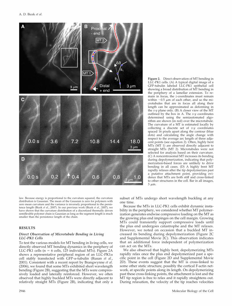
Direct Observation of Microtubule Bending in LivingLLC-PK1 CellsTo test the various models for MT bending in living cells, wedirectly observed MT bending dynamics in the periphery ofLLC-PK1 cells (n � 6 cells, 125 individual MTs). Figure 2Ashows a representative peripheral region of an LLC-PK1�cell stably transfected with GFP-�-tubulin (Rusan et al.,2001). Consistent with a recent report by Brangwynne et al.(2006), we found that some MTs exhibited short wavelengthbending (Figure 2B), suggesting that the MTs were compres-sively loaded and laterally reinforced. However, we oftenobserved that highly buckled MTs were directly adjacent torelatively straight MTs (Figure 2B), indicating that only a
subset of MTs undergo short wavelength buckling at anyone time.
Because the MTs in LLC-PK1 cells exhibit dynamic insta-bility in the periphery, we considered whether MT polymer-ization generates endwise compressive loading on the MT asthe growing plus end impinges on the cell margin. GrowingMTs could transiently support compressive loads untilthe plus end undergoes catastrophe and the MT relaxes.However, we noted on occasion that a buckled MT in-creased its bending during depolymerization (Figure 2Cand Supplemental Movie 2C). This observation indicatesthat an additional force independent of polymerizationcan act on the MTs.
We also observed that highly bent, depolymerizing MTsrapidly relax once the plus end depolymerized past a spe-cific point in the cell (Figure 2D and Supplemental Movie2D). These events suggest that the MT is cross-linked tosome other static structure, possibly the cortical F-actin net-work, at specific points along its length. On depolymerizingpast these cross-linking points, the attachment is lost and theMT tip region is free to relax and it rapidly straightens out.During relaxation, the velocity of the tip reaches velocities
Figure 2. Direct observation of MT bending inLLC-PK1 cells. (A) A typical digital image of aGFP-tubulin labeled LLC-PK1 epithelial cellshowing a broad distribution of MT bending inthe periphery of a lamellar extension. To re-main in focus, the z-coordinates must remainwithin �0.5 �m of each other, and so the mi-crotubules that are in focus all along theirlength can be approximated as deforming inthe x-y plane only. (B) A closer view of the MToutlined by the box in A. The x-y coordinatesdetermined using the semiautomated algo-rithm are shown (in red) over the microtubule.The curvature of a MT is estimated locally bycollecting a discrete set of x-y coordinatesspaced 16 pixels apart along the contour (bluedots) and calculating the angle change withrespect to the average arc length of three adja-cent points (see equation 2). Often, highly bentMTs (MT 1) are observed directly adjacent tostraight MTs (MT 2). Microtubules were notselected for analysis based on their curvature.(C) A noncentrosomal MT increases its bendingduring depolymerization, indicating that poly-merization-based forces are unlikely to drivebending in all cases. (D) A highly bent MTrapidly relaxes after the tip depolymerizes pasta putative attachment point, providing evi-dence that MTs are both stiff and cross-linkedto other structures in the cell. Bar in all images,3 �m.
A. D. Bicek et al.
Molecular Biology of the Cell2946
greater than 5 �m/s, suggesting that the MT is indeed stiff(relaxation time of the bend shown in Figure 2D is 0.2 s).This observation also suggests the idea that MT cross-linksare important in sustaining MT bending.
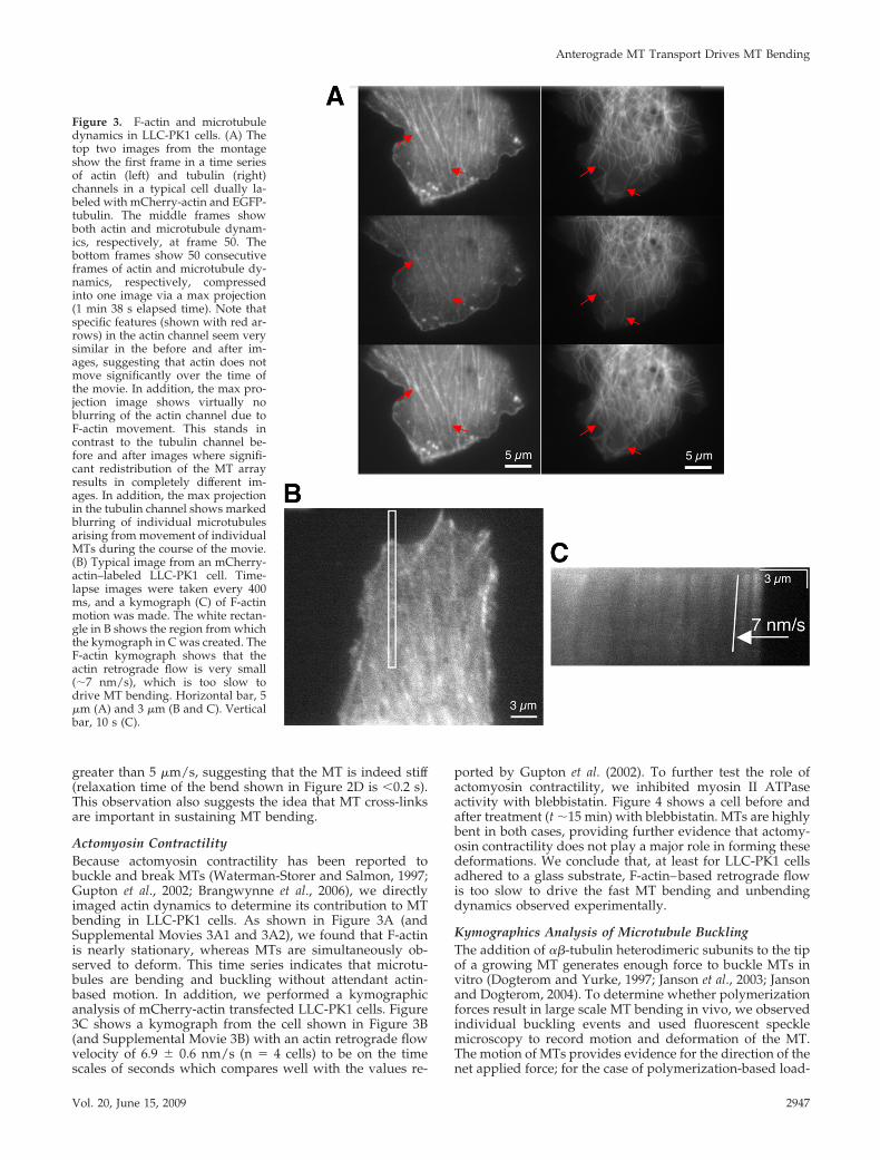
Actomyosin ContractilityBecause actomyosin contractility has been reported tobuckle and break MTs (Waterman-Storer and Salmon, 1997;Gupton et al., 2002; Brangwynne et al., 2006), we directlyimaged actin dynamics to determine its contribution to MTbending in LLC-PK1 cells. As shown in Figure 3A (andSupplemental Movies 3A1 and 3A2), we found that F-actinis nearly stationary, whereas MTs are simultaneously ob-served to deform. This time series indicates that microtu-bules are bending and buckling without attendant actin-based motion. In addition, we performed a kymographicanalysis of mCherry-actin transfected LLC-PK1 cells. Figure3C shows a kymograph from the cell shown in Figure 3B(and Supplemental Movie 3B) with an actin retrograde flowvelocity of 6.9 0.6 nm/s (n � 4 cells) to be on the timescales of seconds which compares well with the values re-
ported by Gupton et al. (2002). To further test the role ofactomyosin contractility, we inhibited myosin II ATPaseactivity with blebbistatin. Figure 4 shows a cell before andafter treatment (t �15 min) with blebbistatin. MTs are highlybent in both cases, providing further evidence that actomy-osin contractility does not play a major role in forming thesedeformations. We conclude that, at least for LLC-PK1 cellsadhered to a glass substrate, F-actin–based retrograde flowis too slow to drive the fast MT bending and unbendingdynamics observed experimentally.
Kymographics Analysis of Microtubule BucklingThe addition of ��-tubulin heterodimeric subunits to the tipof a growing MT generates enough force to buckle MTs invitro (Dogterom and Yurke, 1997; Janson et al., 2003; Jansonand Dogterom, 2004). To determine whether polymerizationforces result in large scale MT bending in vivo, we observedindividual buckling events and used fluorescent specklemicroscopy to record motion and deformation of the MT.The motion of MTs provides evidence for the direction of thenet applied force; for the case of polymerization-based load-
Figure 3. F-actin and microtubuledynamics in LLC-PK1 cells. (A) Thetop two images from the montageshow the first frame in a time seriesof actin (left) and tubulin (right)channels in a typical cell dually la-beled with mCherry-actin and EGFP-tubulin. The middle frames showboth actin and microtubule dynam-ics, respectively, at frame 50. Thebottom frames show 50 consecutiveframes of actin and microtubule dy-namics, respectively, compressedinto one image via a max projection(1 min 38 s elapsed time). Note thatspecific features (shown with red ar-rows) in the actin channel seem verysimilar in the before and after im-ages, suggesting that actin does notmove significantly over the time ofthe movie. In addition, the max pro-jection image shows virtually noblurring of the actin channel due toF-actin movement. This stands incontrast to the tubulin channel be-fore and after images where signifi-cant redistribution of the MT arrayresults in completely different im-ages. In addition, the max projectionin the tubulin channel shows markedblurring of individual microtubulesarising from movement of individualMTs during the course of the movie.(B) Typical image from an mCherry-actin–labeled LLC-PK1 cell. Time-lapse images were taken every 400ms, and a kymograph (C) of F-actinmotion was made. The white rectan-gle in B shows the region from whichthe kymograph in C was created. TheF-actin kymograph shows that theactin retrograde flow is very small(�7 nm/s), which is too slow todrive MT bending. Horizontal bar, 5�m (A) and 3 �m (B and C). Verticalbar, 10 s (C).
Anterograde MT Transport Drives MT Bending
Vol. 20, June 15, 2009 2947
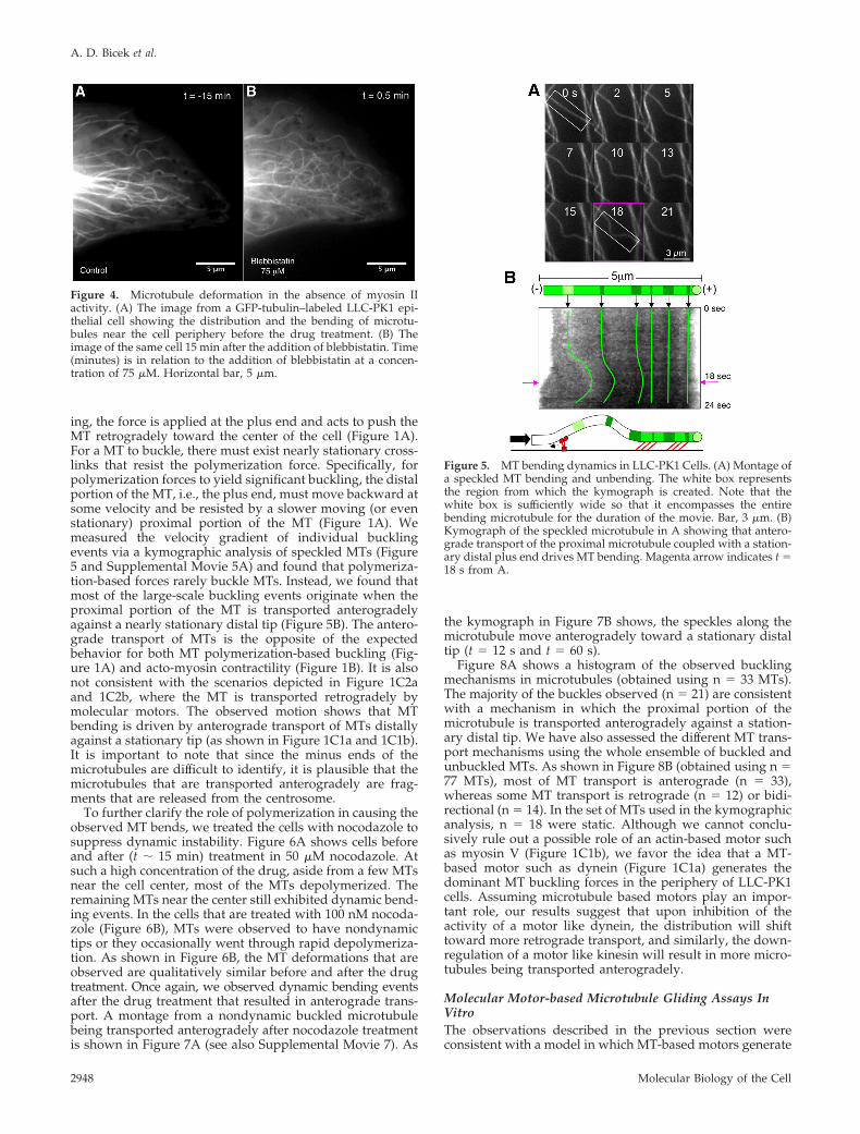
ing, the force is applied at the plus end and acts to push theMT retrogradely toward the center of the cell (Figure 1A).For a MT to buckle, there must exist nearly stationary cross-links that resist the polymerization force. Specifically, forpolymerization forces to yield significant buckling, the distalportion of the MT, i.e., the plus end, must move backward atsome velocity and be resisted by a slower moving (or evenstationary) proximal portion of the MT (Figure 1A). Wemeasured the velocity gradient of individual bucklingevents via a kymographic analysis of speckled MTs (Figure5 and Supplemental Movie 5A) and found that polymeriza-tion-based forces rarely buckle MTs. Instead, we found thatmost of the large-scale buckling events originate when theproximal portion of the MT is transported anterogradelyagainst a nearly stationary distal tip (Figure 5B). The antero-grade transport of MTs is the opposite of the expectedbehavior for both MT polymerization-based buckling (Fig-ure 1A) and acto-myosin contractility (Figure 1B). It is alsonot consistent with the scenarios depicted in Figure 1C2aand 1C2b, where the MT is transported retrogradely bymolecular motors. The observed motion shows that MTbending is driven by anterograde transport of MTs distallyagainst a stationary tip (as shown in Figure 1C1a and 1C1b).It is important to note that since the minus ends of themicrotubules are difficult to identify, it is plausible that themicrotubules that are transported anterogradely are frag-ments that are released from the centrosome.
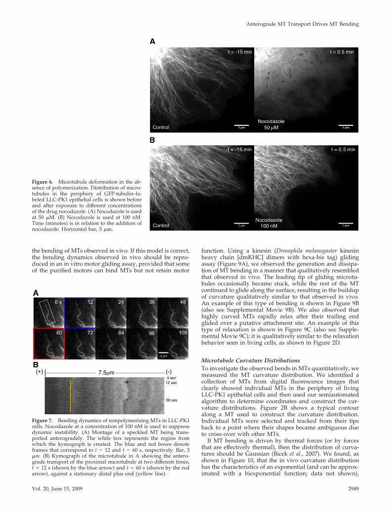
To further clarify the role of polymerization in causing theobserved MT bends, we treated the cells with nocodazole tosuppress dynamic instability. Figure 6A shows cells beforeand after (t � 15 min) treatment in 50 �M nocodazole. Atsuch a high concentration of the drug, aside from a few MTsnear the cell center, most of the MTs depolymerized. Theremaining MTs near the center still exhibited dynamic bend-ing events. In the cells that are treated with 100 nM nocoda-zole (Figure 6B), MTs were observed to have nondynamictips or they occasionally went through rapid depolymeriza-tion. As shown in Figure 6B, the MT deformations that areobserved are qualitatively similar before and after the drugtreatment. Once again, we observed dynamic bending eventsafter the drug treatment that resulted in anterograde trans-port. A montage from a nondynamic buckled microtubulebeing transported anterogradely after nocodazole treatmentis shown in Figure 7A (see also Supplemental Movie 7). As
the kymograph in Figure 7B shows, the speckles along themicrotubule move anterogradely toward a stationary distaltip (t � 12 s and t � 60 s).
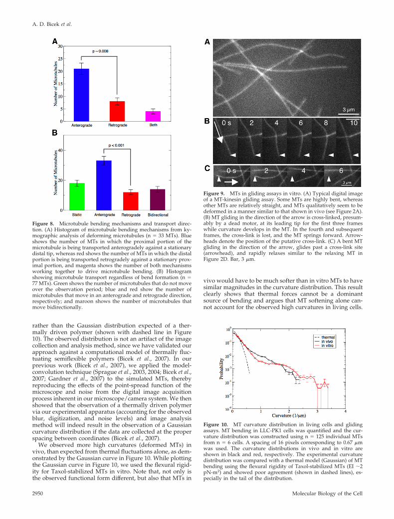
Figure 8A shows a histogram of the observed bucklingmechanisms in microtubules (obtained using n � 33 MTs).The majority of the buckles observed (n � 21) are consistentwith a mechanism in which the proximal portion of themicrotubule is transported anterogradely against a station-ary distal tip. We have also assessed the different MT trans-port mechanisms using the whole ensemble of buckled andunbuckled MTs. As shown in Figure 8B (obtained using n �77 MTs), most of MT transport is anterograde (n � 33),whereas some MT transport is retrograde (n � 12) or bidi-rectional (n � 14). In the set of MTs used in the kymographicanalysis, n � 18 were static. Although we cannot conclu-sively rule out a possible role of an actin-based motor suchas myosin V (Figure 1C1b), we favor the idea that a MT-based motor such as dynein (Figure 1C1a) generates thedominant MT buckling forces in the periphery of LLC-PK1cells. Assuming microtubule based motors play an impor-tant role, our results suggest that upon inhibition of theactivity of a motor like dynein, the distribution will shifttoward more retrograde transport, and similarly, the down-regulation of a motor like kinesin will result in more micro-tubules being transported anterogradely.
Molecular Motor-based Microtubule Gliding Assays InVitroThe observations described in the previous section wereconsistent with a model in which MT-based motors generate
Figure 4. Microtubule deformation in the absence of myosin IIactivity. (A) The image from a GFP-tubulin–labeled LLC-PK1 epi-thelial cell showing the distribution and the bending of microtu-bules near the cell periphery before the drug treatment. (B) Theimage of the same cell 15 min after the addition of blebbistatin. Time(minutes) is in relation to the addition of blebbistatin at a concen-tration of 75 �M. Horizontal bar, 5 �m.
Figure 5. MT bending dynamics in LLC-PK1 Cells. (A) Montage ofa speckled MT bending and unbending. The white box representsthe region from which the kymograph is created. Note that thewhite box is sufficiently wide so that it encompasses the entirebending microtubule for the duration of the movie. Bar, 3 �m. (B)Kymograph of the speckled microtubule in A showing that antero-grade transport of the proximal microtubule coupled with a station-ary distal plus end drives MT bending. Magenta arrow indicates t �18 s from A.
A. D. Bicek et al.
Molecular Biology of the Cell2948
the bending of MTs observed in vivo. If this model is correct,the bending dynamics observed in vivo should be repro-duced in an in vitro motor gliding assay, provided that someof the purified motors can bind MTs but not retain motor
function. Using a kinesin (Drosophila melanogaster kinesinheavy chain [dmKHC] dimers with hexa-his tag) glidingassay (Figure 9A), we observed the generation and dissipa-tion of MT bending in a manner that qualitatively resembledthat observed in vivo. The leading tip of gliding microtu-bules occasionally became stuck, while the rest of the MTcontinued to glide along the surface, resulting in the buildupof curvature qualitatively similar to that observed in vivo.An example of this type of bending is shown in Figure 9B(also see Supplemental Movie 9B). We also observed thathighly curved MTs rapidly relax after their trailing endglided over a putative attachment site. An example of thistype of relaxation is shown in Figure 9C (also see Supple-mental Movie 9C); it is qualitatively similar to the relaxationbehavior seen in living cells, as shown in Figure 2D.
Microtubule Curvature DistributionsTo investigate the observed bends in MTs quantitatively, wemeasured the MT curvature distribution. We identified acollection of MTs from digital fluorescence images thatclearly showed individual MTs in the periphery of livingLLC-PK1 epithelial cells and then used our semiautomatedalgorithm to determine coordinates and construct the cur-vature distributions. Figure 2B shows a typical contouralong a MT used to construct the curvature distribution.Individual MTs were selected and tracked from their tipsback to a point where their shapes became ambiguous dueto cross-over with other MTs.
If MT bending is driven by thermal forces (or by forcesthat are effectively thermal), then the distribution of curva-tures should be Gaussian (Bicek et al., 2007). We found, asshown in Figure 10, that the in vivo curvature distributionhas the characteristics of an exponential (and can be approx-imated with a biexponential function; data not shown),
Figure 6. Microtubule deformation in the ab-sence of polymerization. Distribution of micro-tubules in the periphery of GFP-tubulin–la-beled LLC-PK1 epithelial cells is shown beforeand after exposure to different concentrationsof the drug nocodazole. (A) Nocodazole is usedat 50 �M. (B) Nocodazole is used at 100 nM.Time (minutes) is in relation to the addition ofnocodazole. Horizontal bar, 5 �m.
Figure 7. Bending dynamics of nonpolymerizing MTs in LLC-PK1cells. Nocodazole at a concentration of 100 nM is used to suppressdynamic instability. (A) Montage of a speckled MT being trans-ported anterogradely. The white box represents the region fromwhich the kymograph is created. The blue and red boxes denoteframes that correspond to t � 12 and t � 60 s, respectively. Bar, 3�m. (B) Kymograph of the microtubule in A showing the antero-grade transport of the proximal microtubule at two different times,t � 12 s (shown by the blue arrow) and t � 60 s (shown by the redarrow), against a stationary distal plus end (yellow line).
Anterograde MT Transport Drives MT Bending
Vol. 20, June 15, 2009 2949
rather than the Gaussian distribution expected of a ther-mally driven polymer (shown with dashed line in Figure10). The observed distribution is not an artifact of the imagecollection and analysis method, since we have validated ourapproach against a computational model of thermally fluc-tuating semiflexible polymers (Bicek et al., 2007). In ourprevious work (Bicek et al., 2007), we applied the model-convolution technique (Sprague et al., 2003, 2004; Bicek et al.,2007; Gardner et al., 2007) to the simulated MTs, therebyreproducing the effects of the point-spread function of themicroscope and noise from the digital image acquisitionprocess inherent in our microscope/camera system. We thenshowed that the observation of a thermally driven polymervia our experimental apparatus (accounting for the observedblur, digitization, and noise levels) and image analysismethod will indeed result in the observation of a Gaussiancurvature distribution if the data are collected at the properspacing between coordinates (Bicek et al., 2007).
We observed more high curvatures (deformed MTs) invivo, than expected from thermal fluctuations alone, as dem-onstrated by the Gaussian curve in Figure 10. While plottingthe Gaussian curve in Figure 10, we used the flexural rigid-ity for Taxol-stabilized MTs in vitro. Note that, not only isthe observed functional form different, but also that MTs in
vivo would have to be much softer than in vitro MTs to havesimilar magnitudes in the curvature distribution. This resultclearly shows that thermal forces cannot be a dominantsource of bending and argues that MT softening alone can-not account for the observed high curvatures in living cells.
Figure 8. Microtubule bending mechanisms and transport direc-tion. (A) Histogram of microtubule bending mechanisms from ky-mographic analysis of deforming microtubules (n � 33 MTs). Blueshows the number of MTs in which the proximal portion of themicrotubule is being transported anterogradely against a stationarydistal tip, whereas red shows the number of MTs in which the distalportion is being transported retrogradely against a stationary prox-imal portion, and magenta shows the number of both mechanismsworking together to drive microtubule bending. (B) Histogramshowing microtubule transport regardless of bend formation (n �77 MTs). Green shows the number of microtubules that do not moveover the observation period; blue and red show the number ofmicrotubules that move in an anterograde and retrograde direction,respectively; and maroon shows the number of microtubules thatmove bidirectionally.
Figure 9. MTs in gliding assays in vitro. (A) Typical digital imageof a MT-kinesin gliding assay. Some MTs are highly bent, whereasother MTs are relatively straight, and MTs qualitatively seem to bedeformed in a manner similar to that shown in vivo (see Figure 2A).(B) MT gliding in the direction of the arrow is cross-linked, presum-ably by a dead motor, at its leading tip for the first three frameswhile curvature develops in the MT. In the fourth and subsequentframes, the cross-link is lost, and the MT springs forward. Arrow-heads denote the position of the putative cross-link. (C) A bent MTgliding in the direction of the arrow, glides past a cross-link site(arrowhead), and rapidly relaxes similar to the relaxing MT inFigure 2D. Bar, 3 �m.
Figure 10. MT curvature distribution in living cells and glidingassays. MT bending in LLC-PK1 cells was quantified and the cur-vature distribution was constructed using n � 125 individual MTsfrom n � 6 cells. A spacing of 16 pixels corresponding to 0.67 �mwas used. The curvature distributions in vivo and in vitro areshown in black and red, respectively. The experimental curvaturedistribution was compared with a thermal model (Gaussian) of MTbending using the flexural rigidity of Taxol-stabilized MTs (EI �2pN-m2) and showed poor agreement (shown in dashed lines), es-pecially in the tail of the distribution.
A. D. Bicek et al.
Molecular Biology of the Cell2950
Although MT-associated proteins can potentially bind andsoften MTs, this possibility seems unlikely because it re-quires a large change in the material properties of MTs,whose composition and lattice structure in vivo is thought tobe very similar to its composition and lattice structure invitro. In addition, the rapid relaxation behavior of somedepolymerizing MT tips further supports the view that theflexural rigidity in vivo is similar to that measured in vitro(shown in Figure 2D and Supplemental Movie 2D).
We also considered the effects of a length-dependent flex-ural rigidity, as recently suggested from in vitro studies(Pampaloni et al., 2006; Taute et al., 2008). If the MTs in ourdata set were composed of two populations, each with adifferent flexural rigidity, and driven only by thermal forces,the resulting curvature distribution would be the sum of twoGaussians. Such a distribution has a concave downwardform in the tail, and will not able to explain the exponentialtail shown in Figure 10. We therefore conclude that length-dependent changes in EI do not play a significant role inLLC-PK1 cells.
To compare with a much simpler model system, we mea-sured the curvature distribution of the MTs in gliding assaysin vitro, and we found essentially quantitative agreementbetween the in vitro and in vivo MT curvature distributions,as shown in Figure 10. Such agreement further supports amodel where MT-based motor forces are the major driver ofbending in LLC-PK1 epithelial cells.
DISCUSSION
In this study, we systematically investigated the origin of theforces that drive MT bending in living epithelial cells. This isimportant, because it gives insight into how the cell regu-lates the MT cytoskeleton, and potentially how the cell reactsto mechanical forces. We considered known force generationmechanisms, namely, acto-myosin contractility, polymeriza-tion forces, and MT- and actin-based motor forces. Ourresults show that anterograde MT transport, presumablymediated by MT-based motors, plays a dominant role in MTbending, whereas the other mechanisms are largely incon-sequential in LLC-PK1 cells. Further experiments are neces-sary to determine the applicability of our results to differentcell lines.
Analysis of Microtubule and F-Actin MotionPrevious work has demonstrated close correlation betweenthe retrograde flow of F-actin and the buckling of MTs(Waterman-Storer and Salmon, 1997; Gupton et al., 2002). Inaddition, MTs are clearly observed to bend as they polymer-ize against the edge of the cell or a microfabricated barrier(Dogterom and Yurke, 1997; Waterman-Storer and Salmon,1997). In the present study, the kymographic analysis ofspeckled F-actin features and MTs (Figures 3C and 5B) pro-vides a direct method to determine the site of applicationand direction of active intracellular forces. Both F-actin ret-rograde flow and MT polymerization forces result in com-pressive loading of the MT. These forces act by pushing thedistal segment of the MT retrogradely against a stationary,or at least a slower moving, proximal segment of the MT. Ineither case, the distal MT tip and its associated fluorescentspeckles move retrogradely. This feature is diagnostic whenscreening for the origin of forces that produce MT motion,such that any force that originates within the cell interiorand pushes the proximal portion of the MT outward antero-gradely cannot be the result of either MT polymerization atthe distal plus end or F-actin retrograde flow. AnterogradeMT motion originating from within the cell body that
pushes the proximal MT segments forward, while the distalsegments remain stationary contradicts both the F-actin ret-rograde flow and MT polymerization models for MT bend-ing in LLC-PK1 cells. Instead, our findings rather imply thatthe anterograde speckle movement of the proximal MT seg-ment against a stationary distal MT segment is motor basedand that the MTs are transported anterogradely againstpassive cross-links that hold the distal segment stationary.Our results are also consistent with earlier studies of neuro-nal growth cones (Schaefer et al., 2002) in which microtu-bules were observed to buckle during anterograde transport.
Evidence for Microtubule-based Motor-driven BucklingThe quantitative similarity between curvature distributionsin the in vitro MT gliding assays and the in vivo data areremarkable. This is interesting because in this simple in vitrosystem, in which only thermally fluctuating, nongrowingMTs and MT-based motors are present, the curvature dis-tribution observed in a living cell is recapitulated. The non-Gaussian nature of the resulting curvature distribution in-dicates the presence of active forces of nonthermal origin,such as those caused by molecular motors. These resultssuggest that the cell is regulating the MT array via activeforce generators, consistent with previous observations(Koonce et al., 1999; Burakov et al., 2003; Malikov et al., 2004;Brito et al., 2005).
For active forces to deform MTs in the gliding assay, wespeculate that some dead motors act as passive cross-linkersand resist the forces generated by functional motors, result-ing in MT bending. Alternatively, the motors attached to theMT could act at slightly different rates, resulting in instabil-ities that are “grabbed” by other nearby motors, which mayfurther amplify the curvature. Because in vitro motor prep-arations invariably have some fraction of dead motors inthem, we favor the former explanation, but further work willbe required to distinguish between these two possibilities.
Interestingly, because both living cells and gliding assaysshow evidence of highly curved MTs rapidly relaxing uponloss of attachment sites, similar mechanisms may be in-volved. This suggests that in living cells, the MT-basedmotors can act as both passive cross-links that resist trans-port and active force generators, an emerging theme in themolecular motor field (Tao et al., 2006; Saunders et al., 2007).Alternatively, there may be a second set of molecules thatpassively cross-link, most likely to the stationary F-actincytoskeleton, such as ACF7 (Chishti et al., 1998; Kodamaet al., 2003). The anterograde movement of the MTs suggeststhat the motor that drives bending is either dynein or aminus-end–directed kinesin.
Microtubule-based MechanotransductionThe results presented here argue against a major cell-levelpassive structural function for interphase MTs in LLC-PK1epithelial cells in which MTs act as compressive struts thatresist actomyosin contractility. Instead, our results lead us tofavor a role where mechanical forces act to control thespatial distribution of MTs in living cells. In this model, MTsare pushed and pulled around the cell mainly by MT-basedmotor forces (Figure 11), although other forces discussedabove are also likely to contribute to varying degrees, per-haps more dominantly in other cells types or phases of thecell cycle. Our results also argue that forces are not onlygenerated near the tips of the MTs but also in the middleregions of the cell, suggesting that the MT based motors,such as dynein, anchored at the actin rich cortex may act onMTs and deform them, as shown in Figure 11. Some of theseforces can be accommodated by the repositioning or the
Anterograde MT Transport Drives MT Bending
Vol. 20, June 15, 2009 2951
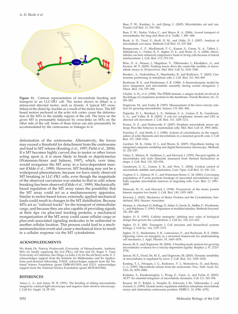
deformation of the centrosome. Alternatively, the forcesmay exceed a threshold for detachment from the centrosomeand lead to MT release (Keating et al., 1997; Piehl et al., 2004).If a MT becomes highly curved due to motor or other forcesacting upon it, it is more likely to break or depolymerize(Waterman-Storer and Salmon, 1997), which, over time,would reorganize the MT array in a force-dependent man-ner (Odde et al., 1999). However, MT breaking may not be awidespread phenomenon, because we have rarely observedMT breaking in LLC-PK1 cells, even though the magnitudeof the observed curvatures was similar to that in cells wherebreaking has been observed (Odde et al., 1999). Mechanicallybased regulation of the MT array raises the possibility thatthe MT array could act as a mechanosensory apparatus.Similar to motor-based forces, externally applied mechanicalloads could result in changes to the MT distribution. BecauseMTs act as “railroad tracks” for the transport of intracellularcargo, and because they are also capable of providing signalsat their tips via plus-end tracking proteins, a mechanicalreorganization of the MT array could cause cellular cargo orplus-end–associated signaling molecules to be redirected toanother cellular location. This process could lead to a mech-anotransduction event and cause a mechanical force to resultin a cellular response via the MT cytoskeleton.
ACKNOWLEDGMENTS
We thank Dr. Patricia Wadsworth (University of Massachusetts, Amherst,MA) for kindly supplying the LLC-PK1� cell line and Dr. Roger Y. Tsien(University of California, San Diego, La Jolla, CA) for the mCherry-actin. E. T.acknowledges support from the Institute for Mathematics and Its Applica-tions post-doctoral fellowship, D.M.K. acknowledges support from the Na-tional Science Foundation (grant DMR-0513393) and D.J.O. acknowledgessupport from the National Science Foundation (grant MCB-0615568).
REFERENCES
Amos, L. A., and Amos, W. B. (1991). The bending of sliding microtubulesimaged by confocal light microscopy and negative stain electron microscopy.J. Cell Sci. Suppl. 14, 95–101.
Baas, P. W., Karabay, A., and Qiang, L. (2005). Microtubules cut and run.Trends Cell Biol. 15, 518–524.
Baas, P. W., Nadar, Vidya, C., and Myers, K. A. (2006). Axonal transport ofmicrotubules: the long and short of it. Traffic 7, 490–498.
Bicek, A. D., Tuzel, E., Kroll, D. M., and Odde, D. J. (2007). Analysis ofmicrotubule curvature. Methods Cell Biol. 83, 237–268.
Brangwynne, C. P., MacKintosh, F. C., Kumar, S., Giesse, N. A., Talbot, J.,Mahadevan, L., Parker, K. P., Ingber, D. E., and Weitz, D. A. (2006). Micro-tubules can bear enhanced compressive loads in living cells because of lateralreinforcement. J. Cell. Biol. 173, 733–741.
Brito, D. A., Strauss, J., Magidson, V., Tikhonenko, I., Khodjakov, A., andKoonce, M. P. (2005). Pushing forces drive the comet-like motility of micro-tubule arrays in Dictyostelium. Mol. Biol. Cell 16, 3334–3340.
Burakov, A., Nadezhdina, E., Slepchenko, B., and Rodionov, V. (2003). Cen-trosome positioning in interphase cells. J. Cell. Biol. 162, 963–969.
Buxbaum, R. E., and Heidemann, S. R. (1988). A thermodynamic model forforce integration and microtubule assembly during axonal elongation. J.Theor. Biol. 134, 379–390.
Chishti, A. H., et al. (1998). The FERM domain: a unique module involved inthe linkage of cytoplasmic proteins to the membrane. Trends Biochem. Sci. 23,281–282.
Dogterom, M., and Yurke, B. (1997). Measurement of the force-velocity rela-tion for growing microtubules. Science 278, 856–860.
Dujardin, D. L., Barnhart, L. E., Stehman, S. A., Gomes, E. R., Gundersen,G. G., and Vallee, R. B. (2003). A role for cytoplasmic dynein and LIS1 indirected cell movement. J. Cell. Biol. 163, 1205–1211.
Ferenz, N. P., and Wadsworth, P. (2007). Prophase microtubule arrays un-dergo Flux-like behavior in mammalian cells. Mol. Biol. Cell 18, 3993–4002.
Forscher, P., and Smith, S. J. (1988). Actions of cytochalasins on the organi-zation of actin filaments and microtubules in a neuronal growth cone. J. CellBiol. 107, 1505–1516.
Gardner, M. K., Odde, D. J., and Bloom, K. (2007). Hypothesis testing viaintegrated computer modeling and digital fluorescence microscopy. Methods41, 232–237.
Gittes, F., Mickey, B., Nettleton, J., and Howard, J. (1993). Flexural rigidity ofmicrotubules and actin filaments measured from thermal fluctuations inshape. J. Cell. Biol. 120, 923–934.
Gundersen, G. G., Gomes, E. R., and Wen, Y. (2004). Cortical control ofmicrotubule stability and polarization. Curr. Opin. Cell Biol. 16, 106–112.
Gupton, S. L., Salmon, W. C., and Waterman-Storer, C. M. (2002). Convergingpopulations of F-actin promote breakage of associated microtubules to spa-tially regulate microtubule turnover in migrating cells. Curr. Biol. 12, 1891–1899.
Hancock, W. O., and Howard, J. (1998). Processivity of the motor proteinkinesin requires two heads. J. Cell. Biol. 140, 1395–1405.
Howard, J. (2001). Mechanics of Motor Proteins and the Cytoskeleton, Sun-derland, MA: Sinauer Associates.
Hyman, A., Drechsel, D., Kellogg, D., Salser, S., Sawin, K., Steffen, P., Wordeman,L., and Mitchison, T. (1991). Preparation of modified tubulins. Methods Enzymol.196, 478–485.
Ingber, D. E. (1993). Cellular tensegrity: defining new rules of biologicaldesign that govern the cytoskeleton. J. Cell Sci. 104, 613–627.
Ingber, D. E. 2003. Tensegrity I. Cell structure and hierarchical systemsbiology. J. Cell Sci. 116, 1157–1173.
Ingber, D. E., Heidemann, S. R., Lamoureux, P., and Buxbaum, R. E. (2000).Opposing views on tensegrity as a structural framework for understandingcell mechanics. J. Appl. Physiol. 89, 1663–1678.
Janson, M. E., and Dogterom, M. (2004). A bending mode analysis for growingmicrotubules: evidence for a velocity-dependent rigidity. Biophys. J. 87, 2723–2736.
Janson, M. E., Dood, De, M. E., and Dogterom, M. (2003). Dynamic instabilityof microtubules is regulated by force. J. Cell. Biol. 161, 1029–1034.
Keating, T. J., Peloquin, J. G., Rodionov, V. I., Momcilovic, D., and Borisy,G. G. (1997). Microtubule release from the centrosome. Proc. Natl. Acad. Sci.USA 94, 5078–5083.
Kodama, A., Karakesisoglou, I., Wong, E., Vaezi, A., and Fuchs, E. (2003).ACF7 An essential integrator of microtubule dynamics. Cell 115, 343–354.
Koonce, M. P., Kohler, J., Neujahr, R., Schwartz, J.-M., Tikhonenko, I., andGerisch, G. (1999). Dynein motor regulation stabilizes interphase microtubulearrays and determines centrosome position. EMBO J. 18, 6786–6792.
Figure 11. Cartoon representation of microtubule bending andtransport in an LLC-PK1 cell. The motor shown in (blue) is aminus-end–directed motor, such as dynein. A typical MT cross-linked at the distal tip, buckles as a result of the motor force. The MTbased motors anchored at the actin rich cortex cause the deforma-tion of the MTs in the middle regions of the cell. The force on thegiven MT is presumably balanced by cross-links on MTs on theother side of the cell. Some of these forces can also presumably beaccommodated by the centrosome or linkages to it.
A. D. Bicek et al.
Molecular Biology of the Cell2952
Ligon, L. A., and Holzbaur, E.L.F. (2007). Centrosome fragments and micro-tubules are transported asymmetrically away from division plane in an-aphase. Traffic 8, 808–819.
Malikov, V., Kashina, A., and Rodionov, V. (2004). Cytoplasmic dynein nu-cleates microtubules to organize them into radial arrays in vivo. Mol. Biol.Cell 15, 2742–2749.
Murthy, K., and Wadsworth, P. (2005). Myosin-II-dependent localization anddynamics of F-Actin during cytokinesis. Curr. Biol. 15, 724–731.
Odde, D. J., Ma, L., Briggs, H., DeMarco, A., and Kirschner, M. W. (1999).Microtubule bending and breaking in living fibroblast cells. J. Cell Sci. 112,3283–3288.
Palazzo, A. F., Joseph, H. L., Chen, Y.-J., Dujardin, D. L., Alberts, A. S., Pfister,K. K., Vallee, R. B., and Gundersen, G. G. (2001). Cdc42, dynein, and dynactinregulate MTOC reorientation independent of Rho-regulated microtubule sta-bilization. Curr. Biol. 11, 1536–1541.
Pampaloni, F., Lattanzi, G., Jonas, A., Surrey, T., Frey, E., and Florin, E.-L.(2006). Thermal fluctuations of grafted microtubules provide evidence of alength-dependent persistence length. Proc. Natl. Acad. Sci. USA 103, 10248–10253.
Piehl, M., Tulu, U. S., Wadsworth, P., and Cassimeris, L. (2004). Centrosomematuration: measurement of microtubule nucleation throughout the cell cycleby using GFP-tagged EB1. Proc. Natl. Acad. Sci. USA 101, 1584–1588.
Rusan, N. M., Fagerstrom, C. J., Yvon, A. M., and Wadsworth, P. (2001). Cellcycle dependent changes in microtubule dynamics in living cells expressinggreen fluorescent protein-alpha tubulin. Mol. Biol. Cell 12, 971–980.
Salmon, W. C., Adams, M. C., and Waterman-Storer, C. M. (2002). Dual-wavelength fluorescent speckle microscopy reveals coupling of microtubuleand actin movements in migrating cells. J. Cell. Biol. 158, 31–37.
Saunders, A. M., Powers, J., Strome, S., and Saxton, W. M. (2007). Kinesin-5acts as a brake in anaphase spindle elongation. Curr. Biol. 17, R453–R454.
Schaefer, A. W., Kabir, N., and Forscher, P. (2002). Filopodia and actin arcsguide the assembly and transport of two populations of microtubules withunique dynamic parameters in neuronal growth cones. J. Cell. Biol. 158,139–152.
Shaner, N. C., Campbell, R. E., Steinbach, P. A., Giepmans, B.N.G., Palmer,A. E., and Tsien, R. Y. (2004). Improved monomeric red, orange and yellowfluorescent proteins derived from Discosoma sp. red fluorescent protein. Nat.Biotechnol. 22, 1567–1572.
Sprague, B. L., Pearson, C. G., Maddox, P. S., Bloom, K. S., Salmon, E. D., andOdde, D. J. (2003). Mechanisms of microtubule-based kinetochore positioningin the yeast metaphase spindle. Biophys. J. 84, 3529–3546.
Sprague, B. L., Gardner, M. K., Pearson, C. G., Maddox, P. S., Bloom, K.,Salmon, E. D., and Odde, D. J. (2004). Model-convolution approach to mod-eling fluorescent protein dynamics. signals, systems and computers. Confer-ence Record of the Thirty-Eighth Asilomar Conference 2, 1821–1825.
Stamenovic, D., Mijailovich, S. M., Tolic-Norrelykke, I. M., Chen, J., andWang, N. (2001). Cell prestress. II. Contribution of microtubules. Am. J.Physiol. Cell Physiol. 282, C617–C624.
Tao, L., Mogilner, A., Civelekoglu-Scholey, G., Wollman, R., Evans, J., Stahlberg,H., and Scholey, J. M. (2006). A homotetrameric Kinesin-5, KLP61F, bundlesmicrotubules and antagonizes Ncd in motility assays. Curr. Biol. 16, 2293–2302.
Taute, K. M., Pampaloni, F., Frey, E., and Florin, E.-L. (2008). Microtubuledynamics depart from the wormlike chain model. Phys. Rev. Lett. 100, 028102
Tran, P. T., Marsh, L., Doye, V., Inoue, S., and Chang, F. (2001). A mechanismfor nuclear positioning in fission yeast based on microtubule pushing. J. Cell.Biol. 153, 397–411.
VanBuren, V., Cassimeris, L., and Odde, D. J. (2005). Mechanochemical modelof microtubule structure and self-assembly kinetics. Biophys. J. 89, 2911–2926.
Wang, N., Butler, J. P., and Ingber, D. E. (1993). Mechanotransduction acrossthe cell surface and through the cytoskeleton. Science 260, 1124–1127.
Wang, N., Naruse, K., Stamenovic, D., Fredberg, J. J., Mijailovich, S. M.,Tolic-Norrelykke, I. M., Polte, T., Mannix, R., and Ingber, D. E. (2001). Me-chanical behavior in living cells consistent with the tensegrity model. Proc.Natl. Acad. Sci. USA 98, 7765–7770.
Waterman-Storer, C. M., and Salmon, E. D. (1997). Actomyosin-based retro-grade flow of microtubules in the lamella of migrating epithelial cells influ-ences microtubule dynamic instability and turnover and is associated withmicrotubule breakage and treadmilling. J. Cell. Biol. 139, 417–434.
Weiss, D. G., Langford, G. M., Seitz-Tutter, D., and Maile, W. (1991). Analysisof the gliding, fishtailing and circling motions of native microtubules. ActaHistochem. Suppl. 41, 81–105.
Williams, Jr., R. C., and Lee, J. C. (1982). Preparation of tubulin from brain.Methods Enzymol. 85, 376–385.
Zhou, F. Q., Waterman-Storer, C. M., and Cohan, C. S. (2002). Focal loss ofactin bundles causes microtubule redistribution and growth cone turning.J. Cell. Biol. 157, 839–849.
Anterograde MT Transport Drives MT Bending
Vol. 20, June 15, 2009 2953