new perspectives in iron chelation therapy for the treatment of … · 2018-10-19 · iron are...
TRANSCRIPT
pharmaceuticals
Review
New Perspectives in Iron Chelation Therapy for theTreatment of Neurodegenerative Diseases
Marco T. Nuñez 1,* ID and Pedro Chana-Cuevas 2
1 Faculty of Sciences, Universidad de Chile, Las Palmeras 3425, Santiago 7800024, Chile2 Center for the Treatment of Movement Disorders, Universidad de Santiago de Chile, Belisario Prat 1597,
Santiago 83800000, Chile; [email protected]* Correspondence: [email protected]; Tel.: +562-29787360
Received: 25 June 2018; Accepted: 3 August 2018; Published: 19 October 2018�����������������
Abstract: Iron chelation has been introduced as a new therapeutic concept for the treatment ofneurodegenerative diseases with features of iron overload. At difference with iron chelators usedin systemic diseases, effective chelators for the treatment of neurodegenerative diseases must crossthe blood–brain barrier. Given the promissory but still inconclusive results obtained in clinical trialsof iron chelation therapy, it is reasonable to postulate that new compounds with properties thatextend beyond chelation should significantly improve these results. Desirable properties of a newgeneration of chelators include mitochondrial destination, the center of iron-reactive oxygen speciesinteraction, and the ability to quench free radicals produced by the Fenton reaction. In addition,these chelators should have moderate iron binding affinity, sufficient to chelate excessive incrementsof the labile iron pool, estimated in the micromolar range, but not high enough to disrupt physiologicaliron homeostasis. Moreover, candidate chelators should have selectivity for the targeted neuronaltype, to lessen unwanted secondary effects during long-term treatment. Here, on the basis ofa number of clinical trials, we discuss critically the current situation of iron chelation therapyfor the treatment of neurodegenerative diseases with an iron accumulation component. The listincludes Parkinson’s disease, Friedreich’s ataxia, pantothenate kinase-associated neurodegeneration,Huntington disease and Alzheimer’s disease. We also review the upsurge of new multifunctionaliron chelators that in the future may replace the conventional types as therapeutic agents for thetreatment of neurodegenerative diseases.
Keywords: neurodegeneration with brain iron accumulation; iron chelation therapy; multifunctionaliron chelators
1. Introduction
Iron content increases with age in several regions of the brain. Particularly, high levels of non-hemeiron are found in the globus pallidus, the red nucleus, substantia nigra, cortex and putamen; in contrast,the iron content of the medulla oblongata does not change with age whereas the iron content of thethalamus decreases from age 30 to 90 [1–3]. The causes underlying the increase in brain iron withage remain elusive. It is unclear whether this increase is a reflection of total body iron, since a reportshows that non-heme iron in the liver does not change with age [1], although body stores of iron,as determined by circulating ferritin levels, seem to increase with age [4].
Abundant evidence suggests that disturbed iron homeostasis and mitochondrial dysfunction playimportant roles in the development of an increasing number of neurodegenerative diseases [3,5–9].The occurrence of high iron content in brain areas susceptible to neurodegeneration, in conjunctionwith the known ability of iron to generate reactive oxygen species (ROS) and induce the formation ofprotein aggregates, provides a relevant seed mechanism for downstream events leading to the death of
Pharmaceuticals 2018, 11, 109; doi:10.3390/ph11040109 www.mdpi.com/journal/pharmaceuticals
Pharmaceuticals 2018, 11, 109 2 of 22
affected neurons. It has been postulated that the high iron–ROS–mitochondrial dysfunction eventsundergo a positive feedback loop that further fosters oxidative damage, protein aggregation and celldeath [3,6,8].
The use of iron chelators for the treatment of systemic diseases such as thalassemia major andhemochromatosis is already a proven therapeutic approach [10]. As a norm, chelator treatment in ironoverload patients induces substantial iron excretion and a negative iron balance [11–15]. On the basisof this experience, strategies to stop or slow neurodegenerative process with an iron accumulationcomponent are now being tested in therapeutic trials.
A review on the current evidence of the benefits and drawbacks of iron chelation therapy, and theanalysis of new compounds that could be used for the treatment of neurodegenerative diseases, follows.
2. Neurodegenerative Diseases with an Iron Accumulation Component
A wide variety of neurological diseases are characterized by the accumulation of iron indifferent areas of the central nervous system; these diseases include Parkinson’s disease (PD) andother parkinsonisms such as Lewy bodies dementia, progressive supranuclear palsy, corticobasaldegeneration [16–21], the Westfal variant of Huntington disease [22], Alzheimer’s disease (AD) [23–27],Friedreich’s ataxia [28], pantothenate kinase-associated neurodegeneration [29–31] and otherneuropathologies associated with brain iron accumulation [32–34]. From the pathophysiologicalstandpoint, different mechanisms are observed, so the clinical and therapeutic implications of ironaccumulation may be different for each individual disease process [2,35].
There is ample evidence linking iron to the pathology of idiopathic PD. A good review on thissubject can be found in the article by Moreau et al. [36]. Iron is particularly abundant in dopaminergicneurons, where it is needed for dopamine synthesis [37] and the production of energy throughthe electron transport chain [38,39]. In dopaminergic neurons, iron behaves as a double-edgedsword since it also participates in the production of the noxious hydroxyl radical during dopamineauto-oxidation. Moreover, the nonenzymatic oxidation of dopamine produces the leukoaminochromeo-semiquinone radical, which reacts with oxygen to generate superoxide anion [40–42]. Since underphysiological conditions iron reacts with superoxide and hydrogen peroxide [6], it is possible thatiron dyshomeostasis plays a fundamental role in mediating oxidative damage in dopaminergicneurons. Indeed, the hydroxyl radical, the most reactive ROS in living matter, is formed by theFenton reaction (Fe2+ + H2O2 → OH− + HO•), a nonenzymatic reaction that obeys mass action law.Hence, there is a direct relationship between the concentration of redox-active iron and the productionof hydroxyl radical.
The mechanisms of iron homeostasis that go awry in neurodegeneration form a cutting-edgequestion in metalloneurobiology [43]. Highly relevant to this point is the mitochondria–ironconnection. The iron homeostasis regulator iron regulatory protein 1 (IRP1) is activated in idiopathicPD. Postmortem brain tissue from PD patients displays increased IRP1 activity when compared totissue from control individuals [44]. Increased IRP1 activity was found also in the ipsilateral ventralmesencephalon of 6-OHDA-treated rats [44]. Studies performed in our laboratory showed that inSH-SY5Y cells, loss of mitochondrial function caused by inhibition of complex I results in decreasedFe–S cluster synthesis and increased IRP1 binding activity, accompanied by increased intracellulariron levels [45]. Further studies revealed that complex I inhibition is associated with increased levelsof iron uptake proteins, and decreased levels of the iron efflux transporter ferroportin 1 [46]. ComplexI inhibition also results in increased oxidative modifications and increased cysteine oxidation, whileIRP1 silencing abolishes the increase in 55Fe uptake activity and protects cells from death induced bycomplex I inhibition [46]. Thus, mitochondrial dysfunction initiates a positive feedback loop that alsocomprises increasing iron uptake and increased oxidative damage. In this view, iron accumulation,more than a primary cause, seems to be a consequence of other initiation factors.
An attractive hypothesis for the genesis of diseases with a redox-active metal accumulationcomponent is the metal-based neurodegeneration hypothesis [3]. According to this hypothesis,
Pharmaceuticals 2018, 11, 109 3 of 22
redox-active metal ions like iron and copper generate ROS that cause the peroxidation of membranephospholipids, which in turn leads to the formation of reactive aldehydes. Reactive aldehydes,together with other ROS, interact with proteins inducing their aggregation, which overwhelms thecellular protein degradation systems, resulting in their accumulation within intracellular inclusionbodies. Accordingly, this hypothesis suggests that protein aggregation occurs downstream of iron orcopper dyshomeostasis.
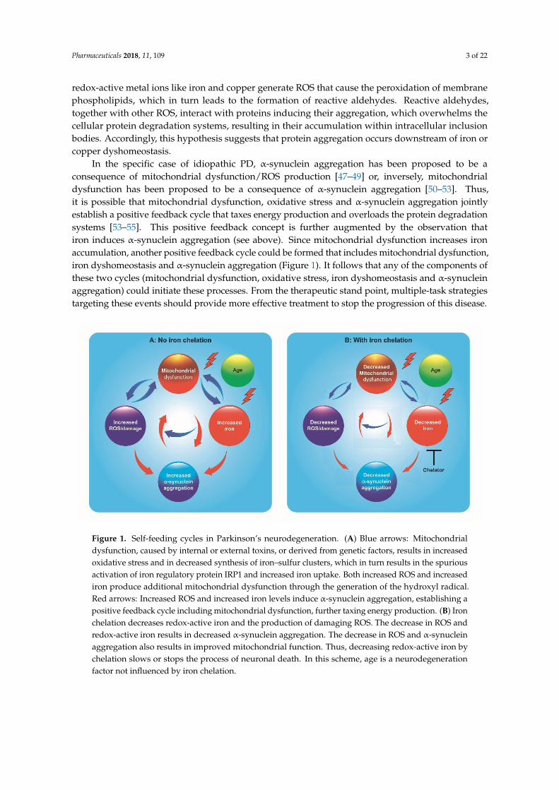
In the specific case of idiopathic PD, α-synuclein aggregation has been proposed to be aconsequence of mitochondrial dysfunction/ROS production [47–49] or, inversely, mitochondrialdysfunction has been proposed to be a consequence of α-synuclein aggregation [50–53]. Thus,it is possible that mitochondrial dysfunction, oxidative stress and α-synuclein aggregation jointlyestablish a positive feedback cycle that taxes energy production and overloads the protein degradationsystems [53–55]. This positive feedback concept is further augmented by the observation thatiron induces α-synuclein aggregation (see above). Since mitochondrial dysfunction increases ironaccumulation, another positive feedback cycle could be formed that includes mitochondrial dysfunction,iron dyshomeostasis and α-synuclein aggregation (Figure 1). It follows that any of the components ofthese two cycles (mitochondrial dysfunction, oxidative stress, iron dyshomeostasis and α-synucleinaggregation) could initiate these processes. From the therapeutic stand point, multiple-task strategiestargeting these events should provide more effective treatment to stop the progression of this disease.
Pharmaceuticals 2018, 11, x FOR PEER REVIEW 3 of 21
phospholipids, which in turn leads to the formation of reactive aldehydes. Reactive aldehydes, together with other ROS, interact with proteins inducing their aggregation, which overwhelms the cellular protein degradation systems, resulting in their accumulation within intracellular inclusion bodies. Accordingly, this hypothesis suggests that protein aggregation occurs downstream of iron or copper dyshomeostasis.
In the specific case of idiopathic PD, α-synuclein aggregation has been proposed to be a consequence of mitochondrial dysfunction/ROS production [47–49] or, inversely, mitochondrial dysfunction has been proposed to be a consequence of α-synuclein aggregation [50–53]. Thus, it is possible that mitochondrial dysfunction, oxidative stress and α-synuclein aggregation jointly establish a positive feedback cycle that taxes energy production and overloads the protein degradation systems [53–55]. This positive feedback concept is further augmented by the observation that iron induces α-synuclein aggregation (see above). Since mitochondrial dysfunction increases iron accumulation, another positive feedback cycle could be formed that includes mitochondrial dysfunction, iron dyshomeostasis and α-synuclein aggregation (Figure 1). It follows that any of the components of these two cycles (mitochondrial dysfunction, oxidative stress, iron dyshomeostasis and α-synuclein aggregation) could initiate these processes. From the therapeutic stand point, multiple-task strategies targeting these events should provide more effective treatment to stop the progression of this disease.
Figure 1. Self-feeding cycles in Parkinson’s neurodegeneration. (A) Blue arrows: Mitochondrial dysfunction, caused by internal or external toxins, or derived from genetic factors, results in increased oxidative stress and in decreased synthesis of iron–sulfur clusters, which in turn results in the spurious activation of iron regulatory protein IRP1 and increased iron uptake. Both increased ROS and increased iron produce additional mitochondrial dysfunction through the generation of the hydroxyl radical. Red arrows: Increased ROS and increased iron levels induce α-synuclein aggregation, establishing a positive feedback cycle including mitochondrial dysfunction, further taxing energy production. (B) Iron chelation decreases redox-active iron and the production of damaging ROS. The decrease in ROS and redox-active iron results in decreased α-synuclein aggregation. The decrease in ROS and α-synuclein aggregation also results in improved mitochondrial function. Thus, decreasing redox-active iron by chelation slows or stops the process of neuronal death. In this scheme, age is a neurodegeneration factor not influenced by iron chelation.
3. Clinical Trials Using Iron Chelation
Overwhelming evidence shows that iron accumulation in the brain may contribute to neurodegenerative processes, as shown in recent reviews [7,35,43,56,57]. A tempering view states that cellular iron homeostasis mechanisms are sufficient to limit its toxicity [58]. Nevertheless, excessive
Figure 1. Self-feeding cycles in Parkinson’s neurodegeneration. (A) Blue arrows: Mitochondrialdysfunction, caused by internal or external toxins, or derived from genetic factors, results in increasedoxidative stress and in decreased synthesis of iron–sulfur clusters, which in turn results in the spuriousactivation of iron regulatory protein IRP1 and increased iron uptake. Both increased ROS and increasediron produce additional mitochondrial dysfunction through the generation of the hydroxyl radical.Red arrows: Increased ROS and increased iron levels induce α-synuclein aggregation, establishing apositive feedback cycle including mitochondrial dysfunction, further taxing energy production. (B) Ironchelation decreases redox-active iron and the production of damaging ROS. The decrease in ROS andredox-active iron results in decreased α-synuclein aggregation. The decrease in ROS and α-synucleinaggregation also results in improved mitochondrial function. Thus, decreasing redox-active iron bychelation slows or stops the process of neuronal death. In this scheme, age is a neurodegenerationfactor not influenced by iron chelation.

Pharmaceuticals 2018, 11, 109 4 of 22
3. Clinical Trials Using Iron Chelation
Overwhelming evidence shows that iron accumulation in the brain may contribute toneurodegenerative processes, as shown in recent reviews [7,35,43,56,57]. A tempering view states thatcellular iron homeostasis mechanisms are sufficient to limit its toxicity [58]. Nevertheless, excessiveiron levels will increase hydroxyl radical generation, for which there are no cellular mechanisms tocounteract its noxious effects.
Successful experiences support the use of iron chelation therapy for the treatment of systemicdiseases with an iron accumulation component, such as thalassemia major, sickle cell disease andcardiomyopathy associated with hereditary hemochromatosis [59–67]. The chelators used in thesetherapies are deferoxamine, deferasirox, deferiprone and PBT2. Adherence to treatment by patientstreated with deferoxamine is low since, as it does not permeate the intestinal barrier, it must be injected.Oral chelators such as deferasirox and deferiprone have better treatment compliance. Deferasirox(Exjade®, Novartis Pharma AG, Basel, Switzerland) was the first oral chelator approved for humanuse in 2005, while Deferiprone (Ferriprox®, Apotex Inc., Toronto, ON, Canada) was approved in 2011.
Recently, iron chelation has been introduced as a new therapeutic concept for the treatment ofneurodegenerative diseases that have a component of metal ion accumulation [56,68–70]. Essentially,the iron chelator should be able to penetrate the cell membranes, as well as the blood–brain barrier,and should have the capacity to extract the chelated iron from the accumulation site and to transfer itto other biological acceptors such as circulating transferrin [68,71]. In addition, small doses of chelatorsmust be used in order to minimize side effects [72,73].
A search in https://clinicaltrials.gov indicated 12 ongoing or finished trials of iron chelation forthe treatment of neurodegenerative diseases: four trials for Parkinson’s disease, three for Friedreich’sataxia, two for amyotrophic lateral sclerosis, one for mild Alzheimer’s disease, one for pantothenatekinase-associated neurodegeneration and one for neurodegeneration with brain iron accumulation.A review on the results of finished trials with published results follows. A number of single-casereports that lack the appropriate controls will not be mentioned here (reviewed by Dusek et al. [70]).
3.1. Parkinson’s Disease
A randomized pilot clinical trial tested 40 patients with early-stage PD, tried with deferiproneversus placebo. A dose of 30 mg/kg/day, during a period of six months, produced a decreasein the iron content of the sustantia nigra, evaluated by T3 magnetic resonance [69]. A significantimprovement of the motor indicators of the progression of the disease was found. Nevertheless,once the treatment was suspended, iron accumulation reappeared, suggesting a reversal to thepathological state. In a second report of this same study, the usefulness of ceruloplasmin (CP) as abiomarker was emphasized, associating the low activity of this enzyme in Parkinson’s disease withiron overload in the substantia nigra [72]. It was found that after six to 12 months of deferipronetreatment, greater reductions in substantia nigra iron levels and Unified Parkinson’s Disease RatingScale (UPDRS) motor scores were obtained in patients with higher serum and cerebrospinal fluid levelsof CP-ferroxidase activity. A second stage of this project, under the acronym FAIRPARK, intends toenroll 338 participants (https://clinicaltrials.gov/ct2/show/NCT02655315?term=chelation&cond=Parkinson+Disease&draw=2&rank=1).
In another series reported in Clinical Trials by researchers from the Imperial College London,good tolerance to deferiprone was reported in patients with Parkinson’s disease, removing excess ironin dentate and caudate nucleus but with minimal effects on the symptoms of the disease [73].
In summary, the reported results on chelator treatment of Parkinson’s disease discussed above areencouraging in terms of a possible slowdown of the disease progression, granting the development offurther long-term trials.

Pharmaceuticals 2018, 11, 109 5 of 22
3.2. Friedreich’s Ataxia
This genetic disease presents a disorder of iron metabolism associated with chronic inflammationand iron accumulation at the level of the central nervous system, the peripheral system, themyocardium and the endocrine system [74]. It was suggested earlier that a reduction of ironaccumulation could be a therapeutic alternative for Friedreich’s ataxia patients [75–77]. Initialpilot studies in young patients with no overt cardiomyopathy showed that treatment with 20 to30 mg/kg/day of deferiprone significantly decreased iron accumulation in the dentate nucleus inthe cerebellum, while reducing neuropathy and ataxic gait. Nevertheless, the effects of this chelationtherapy on neurological symptoms remained controversial [78]. In a follow-up of this study, a one-yearopen-label extension, it was determined that deferiprone dosed at 40 mg/kg/day worsened ataxia,as indicated by a four-point mean increase in International Cooperative Ataxia Rating Scale (ICARS)scores in this group (unpublished data, compiled in [79]). As in the first study, results using a dose of20 mg/kg/day were inconclusive. Nevertheless, a significant reduction in cardiac hypertrophy was aninteresting side effect of deferiprone treatment [79].
A study of co-administration of deferiprone and the coenzyme Q10 analog idebenone reportedimprovement in heart hypertrophy parameters and iron deposits in the dentate nucleus, but noimprovements in ICARS [80]. Another study that used deferiprone in association with idebenone alsofailed to find significant improvement in neurological function. Nevertheless, an improvement in hearthypertrophy was reported as possible [81].
The largest series reported for the treatment of Friedreich’s ataxia enrolled 72 patients, who weretreated with varied doses of deferiprone, 20, 40, or 60 mg/kg/day in a six-month Phase-IIplacebo-controlled trial. The results confirmed the safety of deferiprone at doses lower than 20mg/kg/day, while the 60 mg/kg/day dose was discontinued due to worsening of ataxia in twopatients [82]. Patients receiving 20 mg/kg/day of deferiprone showed a decline in the left ventricularmass index but did not present changes in ICARS scores [82]. The decrease in cardiomyopathycorrelated with the decrease of iron in the cardiac muscle.
In a study with 13 Friedreich’s ataxia patients, a triple therapy with deferiprone plus idebenoneand riboflavin (both antioxidants) resulted in four patients discontinued due to adverse effects after15–39 months of therapy. Other parameters, like the annual worsening rate scale, the scale for theassessment and rating of ataxia scores and cardiac function did not present significant changes [83].The authors concluded that the benefits of this triple therapy are uncertain.
In summary, it has been established that therapeutic doses of deferiprone, 20 mg/kg/day, appearto be safe for long-term use for the treatment of Friedreich’s ataxia. In most trials, this treatmentproduced some improvement in heart function, but no improvement of the neurological symptomswere apparent. In addition, doses higher than 40 mg/kg/day seemed to worsen the disease. The effectsof long-term treatment at low doses on, for example, slowing or stopping disease progression, need tobe evaluated.
3.3. Neurodegeneration with Brain Iron Accumulation (NBIA) Disorders
Within the spectrum of iron deposition disorders there is a group of genetic diseasesthat have in common a syndrome of NBIA [32,84–86]. These disorders include pantothenatekinase-associated neurodegeneration (PKAN, previously known as Hallervorden-Spatzsyndrome) [87–90], PLA2G6 calcium-independent phospholipase A2 (PLAN) [91,92], infantileneuroaxonal dystrophy (INAD) [93,94], mitochondrial membrane protein-associated neurodegeneration(MPAN) [95–98], beta-propeller protein-associated neurodegeneration (BPAN) [99,100], CoA synthaseprotein-associated neurodegeneration (CoPAN) [101–103], fatty acid-2 hydroxylase-associatedneurodegeneration (FAHN) [104,105], Kufor–Rakeb disease [106–108], aceruloplasminemia [109,110]and neuroferritinopathy [111,112]. Of these diseases, iron chelation therapy has been tried inPKAN patients.
Pharmaceuticals 2018, 11, 109 6 of 22
The PKAN neurodegenerative condition is characterized by the presence of iron deposits atthe level of the basal ganglia, currently detected by MRI [86,113,114]. There have been several trialsoriented to the use of deferiprone for the treatment of PKAN patients [115–118]. In all trials, deferipronetreatment decreased iron accumulation in the ganglia, in addition to an improvement in the UnifiedParkinson’s Disease Rating Scale [115–118]. Although the series is still too small to establish definitiveconclusions, iron chelation may be a therapeutic option for the treatment of PKAN. Additionally, ina downside, treatment of a single BPAN patient with deferiprone had to be interrupted because ofworsening of the parkinsonian symptoms [116].
3.4. Huntington Disease
In a study with early/mid-stage Huntington disease patients, patients were subjected todaily doses (250 mg or 100 mg) of 5,7-dichloro-2-[(dimethylamino)methyl]quinolin-8-ol (PBT2) orplacebo [119]. After treatment for 26 weeks with this iron chelator, none or marginal improvementswere found in cognitive tests. The authors concluded that PBT2 was generally safe and well tolerated,but the evaluation of potential benefits remains uncertain and in need of further studies.
3.5. Alzheimer’s Disease
In an initial Phase-II trial with Alzheimer’s disease patients, treatment with the iron–copperchelator clioquinol resulted in stabilization of Alzheimer’s Disease Assessment Scale scores, comparedto placebo-treated controls. In addition, plasma Aβ1–42 levels declined in the clioquinol-treatedgroup [120]. In a subsequent Phase-II double-blind trial, it was found that patients treated withdoses of 250 mg PBT2 exhibited a significant reduction in cerebrospinal fluid Aβ1–42 concentrationcompared with those treated with placebo [121]. In addition, PBT2-treated patients showed significantimprovement in executive function and cognition tests. No serious secondary effects were reported bypatients receiving PBT2. The authors concluded that the findings of this study support larger-scaletesting of PBT2 in Alzheimer’s disease patients.
4. Potential Risks of Iron Chelation Therapy
Iron is vital to life, participating as an essential cofactor in essential cellular processes thatinclude oxygen transport, energy production, DNA synthesis, and a myriad of hydrolysis and redoxreactions [122]. Thus, therapeutic iron chelation may affect not only the intended target but alsoprocesses that are essential for cell function, generating cases in which “the cure is worse than thedisease”. Indeed, excessive iron depletion could result in cognitive decline, by decreasing the activityof enzymes and iron-containing complexes [122,123] and the synthesis of neurotransmitters, such asdopamine, norepinephrine and serotonin [37,124–126]. Fortunately, cognitive decline has not beenreported in clinical chelation trials for neurodegenerative diseases using therapeutic concentrations ofdeferasirox, deferiprone, PBT1 or PBT2 [115–119,121,127,128].
In thalassemia patients treated with high concentrations of deferiprone (75–99 mg/kg/day [129]),the most frequent adverse effects were arthritis, nausea and, more critically, agranulocytosis andneutropenia [130–133], whereas deferasirox was generally well tolerated, with mild gastrointestinaladverse effects [12,134]. A recent meta-analysis, which analyzed results from 34 studies with atotal of 2040 young patients with hemoglobinopathies, found increased transaminase adverse effects(between 3.9% and 31.3% in the different studies) and gastrointestinal complaints for both deferiprone(3.7–18.4%) and deferasirox (5.8–18.8%) [135]. Other effects included arthritis, nausea and, mostseriously, agranulocytosis in 0.6% to 4% of patients. This work concluded that there may be few butserious adverse reactions in performing iron chelation therapy with these chelators.
Another potential problem with iron chelation therapy is the depletion of other essential metalions, in particular Zn and Cu [131,136,137]. Accordingly, in a clinical trial of thalassemia withdeferiprone, four of eight patients who received treatment for one year showed increased excretion
Pharmaceuticals 2018, 11, 109 7 of 22
of Zn in the urine and subnormal values of zinc in the serum, associated with areas of dry skin anditching. These symptoms were resolved with zinc supplementation [136].
There are no reports in the literature indicating that iron chelation therapy causes copper depletion.This is possibly because intracellular copper is not free but it is strongly bound to chaperones [138,139].
From the previous analysis, it is clear that the concentration of the iron chelator should be finelytuned to achieve maximal effectiveness in removing excess redox-active iron, and at the same timeavoiding toxicity and other side effects.
5. New Multifunctional Iron/Copper Chelators with Therapeutic Capacity
The limited success of metal chelation therapy trials using deferiprone raises the need for novelmultifunctional agents, which in addition to decreasing iron accumulation, will have the capacity tointerfere with two or more symptoms of a given disease, thus improving the possibilities of stopping,and eventually reversing, the neurodegenerative process.
The question arises on what are the characteristics of an ideal compound for thetherapeutic treatment of the vicious cycle of mitochondrial dysfunction–iron accumulation–oxidativedamage–protein aggregation in neurodegenerative diseases (Figure 1). These basal properties include:(1) effectiveness via oral administration, which implies crossing the intestinal and blood–brain barrierwithout modification of its therapeutic properties. Compliance of treatment is better with oral drugsthan those injected on a daily basis during long-term treatments [140]; (2) low molecular weight andhigh membrane permeability. The compound must fulfill the rule of 5 of Lipinski, an empirical modelthat allows one to evaluate qualitatively how a chemical compound could be absorbed once it is orallyingested as a medicine [141]. Desirable additional properties include (3) free radical quenching capacityindependent of its iron chelation capacity. Free radical quenching should decrease lipid peroxidation,thus decreasing ferroptosis events [142]; (4) to undergo mitochondrial accumulation. The reason forthis last property is that both high concentrations of ROS and high concentrations of iron coexist inthe secluded space of the mitochondrion, which makes this organelle particularly prone to oxidativedamage [57,143,144]; (5) to have intermediate (µM) affinity and high specificity for iron, consequentwith chelation of the redox-active (labile) iron pool, estimated to be in the micromolar range [134,135];and (6) in addition, the chelator must have “shuttle” capacity, that is, the capacity to deliver excessiveintracellular iron to innocuous acceptors such as transferrin or ferritin [145].
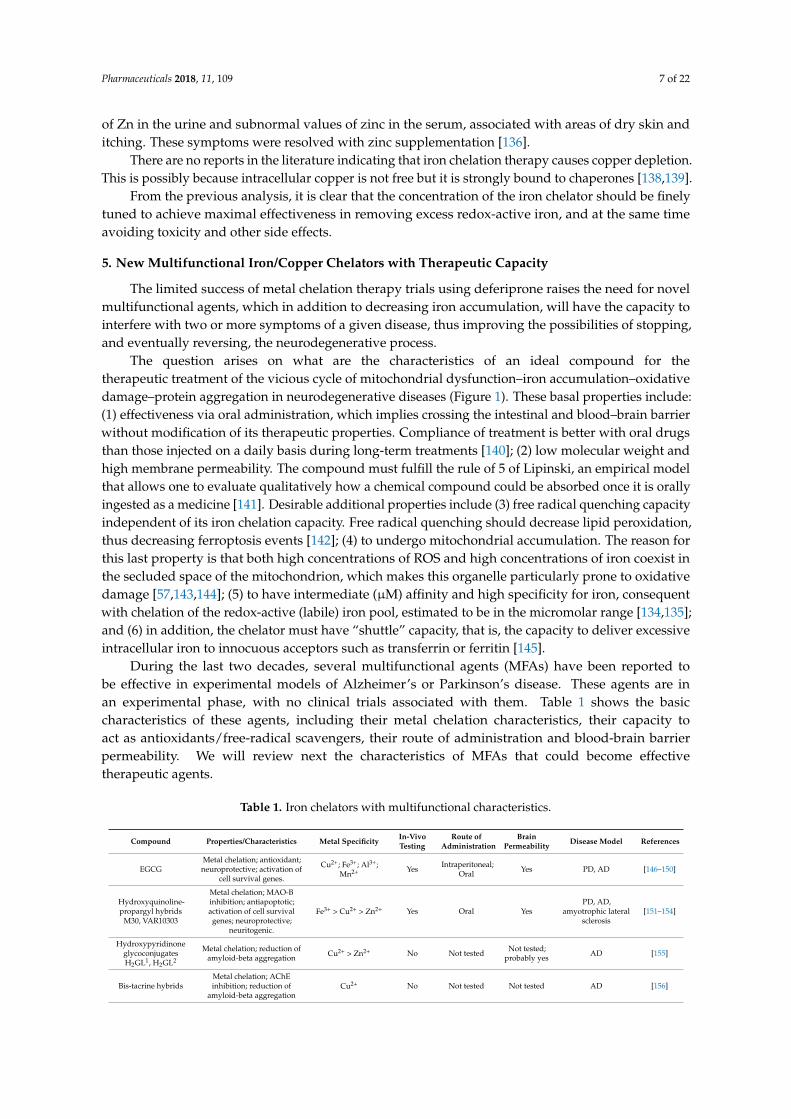
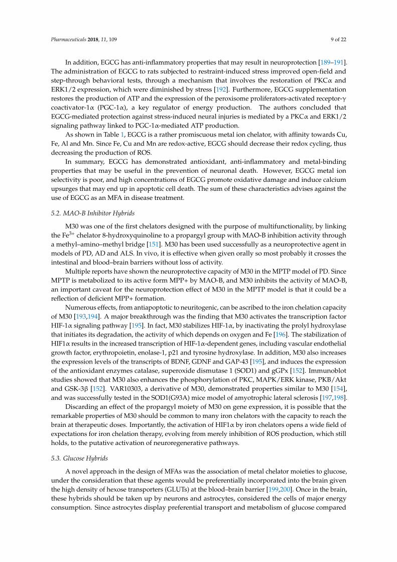
During the last two decades, several multifunctional agents (MFAs) have been reported tobe effective in experimental models of Alzheimer’s or Parkinson’s disease. These agents are inan experimental phase, with no clinical trials associated with them. Table 1 shows the basiccharacteristics of these agents, including their metal chelation characteristics, their capacity toact as antioxidants/free-radical scavengers, their route of administration and blood-brain barrierpermeability. We will review next the characteristics of MFAs that could become effectivetherapeutic agents.
Table 1. Iron chelators with multifunctional characteristics.
Compound Properties/Characteristics Metal Specificity In-VivoTesting
Route ofAdministration
BrainPermeability Disease Model References
EGCGMetal chelation; antioxidant;neuroprotective; activation of
cell survival genes.
Cu2+; Fe3+; Al3+;Mn2+ Yes Intraperitoneal;
Oral Yes PD, AD [146–150]
Hydroxyquinoline-propargyl hybrids
M30, VAR10303
Metal chelation; MAO-Binhibition; antiapoptotic;activation of cell survivalgenes; neuroprotective;
neuritogenic.
Fe3+ > Cu2+ > Zn2+ Yes Oral YesPD, AD,
amyotrophic lateralsclerosis
[151–154]
HydroxypyridinoneglycoconjugatesH2GL1, H2GL2
Metal chelation; reduction ofamyloid-beta aggregation Cu2+ > Zn2+ No Not tested Not tested;
probably yes AD [155]
Bis-tacrine hybridsMetal chelation; AChEinhibition; reduction of
amyloid-beta aggregationCu2+ No Not tested Not tested AD [156]

Pharmaceuticals 2018, 11, 109 8 of 22
Table 1. Cont.
Compound Properties/Characteristics Metal Specificity In-VivoTesting
Route ofAdministration
BrainPermeability Disease Model References
8-OH-Quinoline-tacrine hybrids
Metal chelation; AChEinhibition Cu2+ No Not tested Probably yes AD [157]
Benzylamine-tacrinehybrids
Metal chelation; AChEinhibition; inhibition of
amyloid-beta aggregation;moderate antioxidant activity
Cu2+; Fe2+; Zn2+ No Not tested Not tested AD [158]
Phenyl–benzimidazole-tacrine hybrid
AChE inhibition; metalchelation; inhibition of
Cu-induced amyloid-betaaggregation; free radical
scavenger
Cu2+; other metalsnot tested
No Not tested Not tested AD [159]
Coumarin-tacrinehybrid
Metal chelation; AChEinhibition; inhibition of
amyloid-beta aggregation; freeradical scavenger
Cu2+; other metalsnot tested
No Not tested Not tested AD [160]
Piperazine–8-OH-quinolone hybrids
Metal chelation; dopamineD2/D3 receptor agonists;
hydroxyl radical scavengerFe2+; Fe3+ Yes Subcutaneous Yes PD [161]
Dipyridyl-D2R/D3Ragonist hybrids
Metal chelation; dopamineD2/D3 receptor agonist;
antioxidant; neuroprotectiveFe2+ >>> Fe3+ Yes Intraperitoneal Yes PD [162,163]
Curcumin hybridsMetal chelation; antioxidant
activity; reduction ofamyloid-beta aggregation
Cu2+; Fe2+ No Not tested Not tested AD [164]
Benzyl–indanonehybrid compound 41
Metal chelation; antioxidantactivity; AChE inhibition;inhibition of amyloid-beta
aggregation
Cu2+ No Not tested Not tested AD [165]
Benzothiazole–linker–pyridinone hybrids
Metal chelation; antioxidantactivity; AChE inhibition;inhibition of amyloid-beta
aggregation
Fe3+ No Not tested Probably yes AD [166]
Clioquinol-selegilinehybrids
MAO-B inhibition; metalchelation; antioxidant activity Cu2+; Fe2+; Zn2+ No Not tested Probably yes PD [167]
Deferiprone-H3receptor antagonist
hybrid C5
H3R inhibition; metal chelation;antioxidant activity; reductionof amyloid-beta aggregation
Cu2+∼ Fe2+ >>>Zn2+ Yes Intraperitoneal Yes AD [168]
7,8-Dihydroxycoumarinderivative DHC12
Metal chelation; MAO-Binhibition; mitochondriotropic;
free radical scavenger;neuroprotective
Cu2+∼ Fe2+ > Zn2+
> Fe3+ Yes Oral Yes PD [169]
Coumarin–tris hybridCT51
Metal chelation; impedesFe2+/Fe3+cycling; antioxidant;mitochondriotropic; calcium
upsurge blocker
Fe2+ > Fe3+ No Not tested Not tested PD [170]
5.1. Epigallocatechin-3-Gallate (EGCG)
The pioneering work of Youdim’s group at Technion resulted in two of the best-characterizedMFAs: EGCG, a component of green tea, and M30, an N-propargyl-8-hydroxyquinoline hybrid.
Epidemiological reports associate tea consumption with positive effects in the central nervoussystem function, such as reduced dementia incidence, delayed PD onset and diminished cognitiveimpairment in the elderly (reviewed in [171]). Additional studies have shown the neuroprotectiveeffect of EGCG in the MPTP model of PD [146,149,150,172,173]. However, this neuroprotective effectmay be secondary to the inhibition of the dopamine transporter (DAT) by EGCG [148,174]. Indeed,MPTP is metabolized to 1-methyl-4-phenylpyridinium (MPP+) by MAO-B in astrocytes; after releasefrom astrocytes, MPP+ is transported into dopaminergic neurons via DAT [175,176]. Thus, ablation,or inhibition, of DAT results in neuroprotection against MPTP/MPP+ [177,178].
The protection exerted by EGCG probably involves direct scavenging of ROS such as superoxide,hydrogen peroxide and nitric oxide [179–181]. Nevertheless, the EGCG antioxidant effect is observedonly at low concentrations. In-vitro studies show that high concentrations (10–100 µM) of EGCGactually prompt pro-oxidant effects [182–186]. In rat hippocampal neurons, EGCG causes elevation ofintracellular calcium and ROS in a dose-dependent way [185,187]. Downstream, high calcium/ROSlevels were associated with reduction in the Bcl-2/Bax expression ratio, reduction of the mitochondrialmembrane potential and apoptotic cell death [185,188].
Pharmaceuticals 2018, 11, 109 9 of 22
In addition, EGCG has anti-inflammatory properties that may result in neuroprotection [189–191].The administration of EGCG to rats subjected to restraint-induced stress improved open-field andstep-through behavioral tests, through a mechanism that involves the restoration of PKCα andERK1/2 expression, which were diminished by stress [192]. Furthermore, EGCG supplementationrestores the production of ATP and the expression of the peroxisome proliferators-activated receptor-γcoactivator-1α (PGC-1α), a key regulator of energy production. The authors concluded thatEGCG-mediated protection against stress-induced neural injuries is mediated by a PKCα and ERK1/2signaling pathway linked to PGC-1α-mediated ATP production.
As shown in Table 1, EGCG is a rather promiscuous metal ion chelator, with affinity towards Cu,Fe, Al and Mn. Since Fe, Cu and Mn are redox-active, EGCG should decrease their redox cycling, thusdecreasing the production of ROS.
In summary, EGCG has demonstrated antioxidant, anti-inflammatory and metal-bindingproperties that may be useful in the prevention of neuronal death. However, EGCG metal ionselectivity is poor, and high concentrations of EGCG promote oxidative damage and induce calciumupsurges that may end up in apoptotic cell death. The sum of these characteristics advises against theuse of EGCG as an MFA in disease treatment.
5.2. MAO-B Inhibitor Hybrids
M30 was one of the first chelators designed with the purpose of multifunctionality, by linkingthe Fe3+ chelator 8-hydroxyquinoline to a propargyl group with MAO-B inhibition activity througha methyl–amino–methyl bridge [151]. M30 has been used successfully as a neuroprotective agent inmodels of PD, AD and ALS. In vivo, it is effective when given orally so most probably it crosses theintestinal and blood–brain barriers without loss of activity.
Multiple reports have shown the neuroprotective capacity of M30 in the MPTP model of PD. SinceMPTP is metabolized to its active form MPP+ by MAO-B, and M30 inhibits the activity of MAO-B,an important caveat for the neuroprotection effect of M30 in the MPTP model is that it could be areflection of deficient MPP+ formation.
Numerous effects, from antiapoptotic to neuritogenic, can be ascribed to the iron chelation capacityof M30 [193,194]. A major breakthrough was the finding that M30 activates the transcription factorHIF-1α signaling pathway [195]. In fact, M30 stabilizes HIF-1α, by inactivating the prolyl hydroxylasethat initiates its degradation, the activity of which depends on oxygen and Fe [196]. The stabilization ofHIF1α results in the increased transcription of HIF-1α-dependent genes, including vascular endothelialgrowth factor, erythropoietin, enolase-1, p21 and tyrosine hydroxylase. In addition, M30 also increasesthe expression levels of the transcripts of BDNF, GDNF and GAP-43 [195], and induces the expressionof the antioxidant enzymes catalase, superoxide dismutase 1 (SOD1) and gGPx [152]. Immunoblotstudies showed that M30 also enhances the phosphorylation of PKC, MAPK/ERK kinase, PKB/Aktand GSK-3β [152]. VAR10303, a derivative of M30, demonstrated properties similar to M30 [154],and was successfully tested in the SOD1(G93A) mice model of amyotrophic lateral sclerosis [197,198].
Discarding an effect of the propargyl moiety of M30 on gene expression, it is possible that theremarkable properties of M30 should be common to many iron chelators with the capacity to reach thebrain at therapeutic doses. Importantly, the activation of HIF1α by iron chelators opens a wide field ofexpectations for iron chelation therapy, evolving from merely inhibition of ROS production, which stillholds, to the putative activation of neuroregenerative pathways.
5.3. Glucose Hybrids
A novel approach in the design of MFAs was the association of metal chelator moieties to glucose,under the consideration that these agents would be preferentially incorporated into the brain giventhe high density of hexose transporters (GLUTs) at the blood–brain barrier [199,200]. Once in the brain,these hybrids should be taken up by neurons and astrocytes, considered the cells of major energyconsumption. Since astrocytes display preferential transport and metabolism of glucose compared

Pharmaceuticals 2018, 11, 109 10 of 22
to neurons [201], it would be expected that metal-hexose MFAs should preferentially accumulate inastrocytes. The hydroxypyridinone glycoconjugates H2GL1 and H2GL2 demonstrated substantialaffinity for Cu2+ and Zn2+, and both agents decreased Aβ1–40 aggregation induced by Cu and Zn.They also demonstrated antioxidant activity, probably through their phenolic moieties, capable ofquenching free radicals by a hydroquinone/quinone conversion [155]. No in-vivo testing of theseagents was reported, so their putative Fe3+-chelating and blood–brain barrier permeability propertiesremain undetermined.
5.4. Acetyl Cholinesterase Inhibitor Hybrids
Under the concept that inhibition of acetylcholinesterase (AChE) increases acetylcholine atcholinergic synapses, thus reducing the cognitive deficit [202,203], an approach for the putativetreatment of AD is the design and synthesis of MFAs with AChE inhibition activity. This particularstrategy is common for MFAs with a tacrine (a cholinesterase inhibitor) component [156–159](Table 1). None of these compounds have been tested for brain permeability, although tacrinederivatives, without an iron chelator component, are brain-effective under intravenous and intranasaladministrations [204]. At this stage, tests of effectiveness in animal models of AD are needed forfurther evaluation of their putative usefulness.
5.5. Dopamine Receptor Agonist Hybrids
19a, 19b and D-607 are dopamine D2/D3 receptor agonists and metal chelator hybrids, designedunder the notion that D2/D3 receptor agonists have been used for the treatment of both motorand psychiatric syndromes in PD [179]. The three compounds demonstrated iron chelation andantioxidant capacity in vitro [180,181]. In addition, 19b partly reversed hypolocomotion in reserpinizedrats, and reduced the rotational activity in a 6-OHDA/apomorphine model, thus demonstratingin-vivo neuroprotective activity [181]. D-607 was the product of a further D2/D3 agonist/chelatordevelopment by the same research group. The chelator moiety of D-607 was changed from 8-OHquinolone, a Fe3+ chelator, to bipyridyl, a Fe2+ chelator. D-607 was shown to suppress retinaldegeneration in a Drosophila melanogaster PD model that expresses α-synuclein A30P, a PD-causingvariant of the protein [180]. In addition, D-607 was shown to confer significant neuroprotectionin the mouse MPTP PD model under chronic MPTP administration [180]. Overall, the publishedevidence points to D-607 as a putative candidate for the pharmacological treatment of PD. A drawbackis that D-607 is administrated intraperitoneally, and not orally, which may decrease hypotheticalpatient compliance.
5.6. Curcumin Hybrids
Curcumin analogs are proposed as potential anti-AD drugs, based on the radicalscavenger [182,183] and metal chelator [184,185] properties of curcumin. Curcumin analogs A1–A10were tested for their radical quenching activity and their ability to reduce metal-induced amyloid-betaaggregation [186]. A1, A2, A3 and A4 presented good radical quenching capacity in SH-SY5Y cellswhile the capacity of A5, A6, A7, A8, A9 and A10 was weak. All analogs, with the exception ofA7, A8 and A10, presented the capacity to diminish amyloid beta self-aggregation at IC50s similarto curcumin. The authors reported Fe and Cu chelation capacity by A4 based on the red shift ofabsorbance peaks at 267 nm and 427 nm. Nevertheless, A4 lacks the two adjacent ketone groupsresponsible for metal binding by coumarin [184] and it does not have putative metal binding groupsin its structure, so the mechanism of iron binding is not apparent. No in-vivo studies were reported forthese curcumin analogs.
5.7. Benzothiazole–3-Hydroxy-4-Pyridine Hybrids
Another approach for MFAs is the design of constructs of benzothiazole and 3-hydroxy-4-pyridineconnected by a variable linker [83]. Given its hydrophobicity, benzothiazole has strong affinity for
Pharmaceuticals 2018, 11, 109 11 of 22
amyloid plaques [84,85] while the 3-hydroxy-4-pyridine moiety (deferiprone) has strong Fe3+ chelationcapacity. The linker between these two moieties was modelled in order to obtain AChE inhibitorycapacity [83]. Of the tested compounds, 2a and 2d formed Fe3+ chelates with affinities similar todeferiprone. The chelators displayed significant antioxidant properties, with compounds 1a, 1b, 1cand 2d having significant AChE inhibitory activity. Accordingly, these same hybrids presented goodinhibitory capacity towards Aβ42 self-aggregation, mostly above 40%. In addition, hybrid 2d inhibitedzinc-induced Aβ1–42 aggregation [83]. Overall, the hydroxypyridinone–spacer–benzothiazole hybridsappear as good candidate drugs for the treatment of AD, but in-vivo testing is needed beforefurther development.
5.8. MAO-B Inhibitors
On the basis of its moderate success in AD treatment [187], the MAO-B inhibitor selegiline hasbeen used for the design of MFAs directed to the treatment of AD [188,189]. The most promisingcompounds are selegiline–clioquinol hybrids, which combine MAO-B inhibition with metal chelationcapacity. Selegiline–clioquinol hybrids tested in vitro demonstrated inhibition of Cu-induced Aβ1–42aggregation, antioxidant activity and Cu2+, Fe2+ and Zn2+ chelation capacity [189]. Like other MFAs ina proof-of-concept step, demonstration of in-vivo effectiveness is needed.
5.9. Histamine H3 Receptor Antagonists
A different approach in the design of MFAs was the design of a histamine H3 receptorantagonist, 1-phenyl-3-hydroxy-4-pyridinone, and the 3-hydroxy-4-pyridinone iron chelator moiety ofdeferiprone [190], under the rationale that blocking the action of presynaptic H3 receptors producesincreased secretion of histamine and other excitatory neurotransmitters [191]. H3 antagonist treatmentresults in modest effects on cognitive function [192]. The most promising compound, 5c, displayed H3receptor antagonistic activity, free radical scavenging capacity, copper and iron chelation, and inhibitionof self- and Cu2+-induced Aβ1−42 aggregation [190]. After intraperitoneal administration to SpragueDawley rats, compound 5c demonstrated good blood–brain barrier penetration. In conclusion,the histamine H3 receptor antagonist–iron chelator hybrid 5c is brain-permeant and possesses fourfunctions applicable for the treatment of AD, which makes it a good therapeutic candidate.
5.10. Coumarin Hybrids
Two coumarin derivatives have been proposed as candidate drugs for the treatment ofneurodegenerative conditions with an iron accumulation component [193,194], based on the knownqualities of hydroxycoumarins as free radical quenchers [195] and metal chelator [196,197] agents.DHC12 is a 7,8-dihydroxycoumarin with an amino substituent group at position four of the coumarinring [193]. The molecule is small and quite simple; nevertheless, it has interesting neuroprotectivefeatures. DHC12 exhibited metal binding capacity for Fe2+ and Cu2+. DHC12 distributed tomitochondria, where it chelated the mitochondrial and cytoplasmic labile iron pool. In a cell model of PD,DHC12 protected cells from plasma membrane and mitochondrial oxidative damage. Oral administrationof DHC12 protected sustantia nigra neurons in the MPTP model of PD. On the whole, DHC12 emergesas a good candidate for further development as a PD treatment drug.
CT51 is a hybrid of 7-hydroxycoumarin linked through an acetomethyl group to tris(hydroxymethyl)aminomethane (tris). The hydroxyl group in the coumarin ring quenches free radicals [195] while thethree hydroxyl residues of tris provide metal binding capacity [198]. In vitro, CT51 exhibited selectiveFe2+ and Fe3+ binding with no apparent interaction with Cu2+, Zn2+ and other divalent cations. It alsodemonstrated free radical quenching capacity superior to Trolox. Interestingly, cyclic voltammetryanalysis revealed irreversible binding of Fe3+ to CT51, an important finding since stopping Fe2+/Fe3+
cycling in cells should prevent hydroxyl radical production fostered by oxygen and intracellularreductants [8]. In SH-SY5Y cells, CT51 distributed to both mitochondria and cytoplasm bound ironreversibly and protected against rotenone-induced oxidative damage, while in primary hippocampal

Pharmaceuticals 2018, 11, 109 12 of 22
neurons, CT51 largely prevented the increase in intracellular calcium levels produced by an agonist ofredox-sensitive RyR channels [194]. These capacities so-far demonstrated make CT51 a good therapeuticcandidate for the treatment of PD, although in-vivo efficacy needs to be demonstrated.
6. Conclusions
The aging of the world population introduces an ever-increasing burden of neurodegenerativediseases on public health systems worldwide. Among these diseases, Parkinson’s, Alzheimer’s andother diseases with an iron accumulation component are at the top of the list. Based on initial trialsusing the iron chelators deferiprone and PBT2, the metalloneurobiology community has reached theconclusion that therapies targeted to decrease the iron content in specific areas of the brain is a viablecourse of action to slow or stop the progress of these diseases.
Given the multifactoriality of the neurodegenerative process, the use of multifunctional ironchelators is a promising developmental avenue. As discussed in the text, additional properties offuture iron chelator drugs should comprise high selectivity for iron, free radical quenching capacity,mitochondrial distribution and the capacity to block protein aggregation. Several of the compoundsnow in experimental stages have one or more of these additional characteristics. Let us hope thatfurther research will provide treatments that are both effective and affordable for public health systems.
Author Contributions: M.T.N. and P.C.-C. wrote the paper.
Acknowledgments: M.T.N. and P.C.-C. were supported by FONDEF project 17I10095 from National Commissionof Science and Technology (CONICYT) of Chile. We thank Cecilia Hidalgo for the critical reviewing of this paper.
Conflicts of Interest: The authors declare no conflicts of interest.
References
1. Hallgren, B.; Sourander, P. The effect of age on the non-haemin iron in the human brain. J. Neurochem. 1958,3, 41–51. [CrossRef] [PubMed]
2. Zecca, L.; Youdim, M.B.; Riederer, P.; Connor, J.R.; Crichton, R.R. Iron, brain ageing and neurodegenerativedisorders. Nat. Rev. Neurosci. 2004, 5, 863–873. [CrossRef] [PubMed]
3. Crichton, R.R.; Dexter, D.T.; Ward, R.J. Brain iron metabolism and its perturbation in neurological diseases.J. Neural Transm. 2011, 118, 301–314. [CrossRef] [PubMed]
4. Garry, P.J.; Goodwin, J.S.; Hunt, W.C. Iron status and anemia in the elderly: New findings and a review ofprevious studies. J. Am. Geriatr. Soc. 1983, 31, 389–399. [CrossRef] [PubMed]
5. Ward, R.J.; Zucca, F.A.; Duyn, J.H.; Crichton, R.R.; Zecca, L. The role of iron in brain ageing andneurodegenerative disorders. Lancet Neurol. 2014, 13, 1045–1060. [CrossRef]
6. Nunez, M.T.; Urrutia, P.; Mena, N.; Aguirre, P.; Tapia, V.; Salazar, J. Iron toxicity in neurodegeneration.Biometals 2012, 25, 761–776. [CrossRef] [PubMed]
7. Apostolakis, S.; Kypraiou, A.M. Iron in neurodegenerative disorders: Being in the wrong place at the wrongtime? Rev. Neurosci. 2017, 28, 893–911. [CrossRef] [PubMed]
8. Munoz, Y.; Carrasco, C.M.; Campos, J.D.; Aguirre, P.; Nunez, M.T. Parkinson’s Disease: The Mitochondria-Iron Link. Parkinson Dis. 2016, 2016, 7049108.
9. Carocci, A.; Catalano, A.; Sinicropi, M.S.; Genchi, G. Oxidative stress and neurodegeneration: The involvementof iron. Biometals 2018. [CrossRef] [PubMed]
10. Kolnagou, A.; Kontoghiorghe, C.N.; Kontoghiorghes, G.J. New targeted therapies and diagnostic methodsfor iron overload diseases. Front. Biosci. 2018, 10, 1–20.
11. Kwiatkowski, J.L. Management of transfusional iron overload—Differential properties and efficacy of ironchelating agents. J. Blood Med. 2011, 2, 135–149. [CrossRef] [PubMed]
12. Poggiali, E.; Cassinerio, E.; Zanaboni, L.; Cappellini, M.D. An update on iron chelation therapy. Blood Transfus.2012, 10, 411–422. [PubMed]
13. Saliba, A.N.; Harb, A.R.; Taher, A.T. Iron chelation therapy in transfusion-dependent thalassemia patients:Current strategies and future directions. J. Blood Med. 2015, 6, 197–209. [PubMed]
Pharmaceuticals 2018, 11, 109 13 of 22
14. Flaten, T.P.; Aaseth, J.; Andersen, O.; Kontoghiorghes, G.J. Iron mobilization using chelation and phlebotomy.J. Trace Elem. Med. Boil. 2012, 26, 127–130. [CrossRef] [PubMed]
15. Cohen, A.; Martin, M.; Schwartz, E. Depletion of excessive liver iron stores with desferrioxamine. Br. J.Haematol. 1984, 58, 369–373. [CrossRef] [PubMed]
16. Lee, J.H.; Han, Y.H.; Kang, B.M.; Mun, C.W.; Lee, S.J.; Baik, S.K. Quantitative assessment of subcorticalatrophy and iron content in progressive supranuclear palsy and parkinsonian variant of multiple systematrophy. J. Neurol. 2013, 260, 2094–2101. [CrossRef] [PubMed]
17. Youdim, M.B.; Ben-Shachar, D.; Riederer, P. Is Parkinson’s disease a progressive siderosis of substantia nigraresulting in iron and melanin induced neurodegeneration? Acta Neurol. Scand. Suppl. 1989, 126, 47–54.[CrossRef] [PubMed]
18. Savoiardo, M. Differential diagnosis of Parkinson’s disease and atypical parkinsonian disorders by magneticresonance imaging. Neurol. Sci. 2003, 24 (Suppl. S1), S35–S37. [CrossRef] [PubMed]
19. Lee, S.H.; Lyoo, C.H.; Ahn, S.J.; Rinne, J.O.; Lee, M.S. Brain regional iron contents in progressive supranuclearpalsy. Park. Relat. Disord. 2017, 45, 28–32. [CrossRef] [PubMed]
20. Fernandez, B.; Ferrer, I.; Gil, F.; Hilfiker, S. Biomonitorization of iron accumulation in the substantia nigrafrom Lewy body disease patients. Toxicol. Rep. 2017, 4, 188–193. [CrossRef] [PubMed]
21. Martin-Bastida, A.; Lao-Kaim, N.P.; Loane, C.; Politis, M.; Roussakis, A.A.; Valle-Guzman, N.;Kefalopoulou, Z.; Paul-Visse, G.; Widner, H.; Xing, Y.; et al. Motor associations of iron accumulationin deep grey matter nuclei in Parkinson’s disease: A cross-sectional study of iron-related magnetic resonanceimaging susceptibility. Eur. J. Neurol. 2017, 24, 357–365. [CrossRef] [PubMed]
22. Bartzokis, G.; Cummings, J.; Perlman, S.; Hance, D.B.; Mintz, J. Increased basal ganglia iron levels inHuntington disease. Arch. Neurol. 1999, 56, 569–574. [CrossRef] [PubMed]
23. Smith, M.A.; Harris, P.L.; Sayre, L.M.; Perry, G. Iron accumulation in Alzheimer disease is a source ofredox-generated free radicals. Proc. Natl. Acad. Sci. USA 1997, 94, 9866–9868. [CrossRef] [PubMed]
24. Perry, G.; Taddeo, M.A.; Petersen, R.B.; Castellani, R.J.; Harris, P.L.; Siedlak, S.L.; Cash, A.D.; Liu, Q.;Nunomura, A.; Atwood, C.S.; et al. Adventiously-bound redox active iron and copper are at the center ofoxidative damage in Alzheimer disease. Biometals 2003, 16, 77–81. [CrossRef] [PubMed]
25. Lane, D.J.R.; Ayton, S.; Bush, A.I. Iron and Alzheimer’s Disease: An Update on Emerging Mechanisms.J. Alzheimer’s Dis. 2018, 64, S379–S395. [CrossRef] [PubMed]
26. Bartzokis, G.; Sultzer, D.; Cummings, J.; Holt, L.E.; Hance, D.B.; Henderson, V.W.; Mintz, J. In vivo evaluationof brain iron in Alzheimer disease using magnetic resonance imaging. Arch. Gen. Psychiatry 2000, 57, 47–53.[CrossRef] [PubMed]
27. Bulk, M.; Abdelmoula, W.M.; Nabuurs, R.J.A.; van der Graaf, L.M.; Mulders, C.W.H.; Mulder, A.A.; Jost, C.R.;Koster, A.J.; van Buchem, M.A.; Natte, R.; et al. Postmortem MRI and histology demonstrate differential ironaccumulation and cortical myelin organization in early- and late-onset Alzheimer’s disease. Neurobiol. Aging2018, 62, 231–242. [CrossRef] [PubMed]
28. Chiang, S.; Kovacevic, Z.; Sahni, S.; Lane, D.J.; Merlot, A.M.; Kalinowski, D.S.; Huang, M.L.; Richardson, D.R.Frataxin and the molecular mechanism of mitochondrial iron-loading in Friedreich’s ataxia. Clin. Sci. 2016,130, 853–870. [CrossRef] [PubMed]
29. Fermin-Delgado, R.; Roa-Sanchez, P.; Speckter, H.; Perez-Then, E.; Rivera-Mejia, D.; Foerster, B.; Stoeter, P.Involvement of globus pallidus and midbrain nuclei in pantothenate kinase-associated neurodegeneration:Measurement of T2 and T2* time. Clin. Neuroradiol. 2013, 23, 11–15. [CrossRef] [PubMed]
30. Rossi, D.; De Grandis, E.; Barzaghi, C.; Mascaretti, M.; Garavaglia, B.; Zanotto, E.; Morana, G.; Biancheri, R.Early-onset neurodegeneration with brain iron accumulation due to PANK2 mutation. Brain Dev. 2012, 34,536–538. [CrossRef] [PubMed]
31. Swaiman, K.F. Hallervorden-Spatz syndrome and brain iron metabolism. Arch. Neurol. 1991, 48, 1285–1293.[CrossRef] [PubMed]
32. Hayflick, S.J.; Kurian, M.A.; Hogarth, P. Neurodegeneration with brain iron accumulation. Handb. Clin.Neurol. 2018, 147, 293–305. [PubMed]
33. Poplawska-Domaszewicz, K.; Florczak-Wyspianska, J.; Kozubski, W. Update on neurodegeneration withbrain iron accumulation. Neurol. I Neurochir. Polska 2014, 48, 206–213. [CrossRef] [PubMed]
34. Tonekaboni, S.H.; Mollamohammadi, M. Neurodegeneration with brain iron accumulation: An overview.Iran. J. Child Neurol. 2014, 8, 1–8. [PubMed]
Pharmaceuticals 2018, 11, 109 14 of 22
35. Wiethoff, S.; Houlden, H. Neurodegeneration with brain iron accumulation. Handb. Clin. Neurol. 2017, 145,157–166. [PubMed]
36. Moreau, C.; Duce, J.A.; Rascol, O.; Devedjian, J.C.; Berg, D.; Dexter, D.; Cabantchik, Z.I.; Bush, A.I.; Devos, D.Iron as a therapeutic target for Parkinson’s disease. Mov. Disord. 2018, 33, 568–574. [CrossRef] [PubMed]
37. Lehmann, W.D.; Heinrich, H.C. Impaired phenylalanine-tyrosine conversion in patients with iron-deficiencyanemia studied by a L-(2H5)phenylalanine-loading test. Am. J. Clin. Nutr. 1986, 44, 468–474. [CrossRef][PubMed]
38. Ohnishi, T. Iron-sulfur clusters/semiquinones in complex I. Biochim. Biophys. Acta 1998, 1364, 186–206.[CrossRef]
39. Stiban, J.; So, M.; Kaguni, L.S. Iron-Sulfur Clusters in Mitochondrial Metabolism: Multifaceted Roles of aSimple Cofactor. Biochem. 2016, 81, 1066–1080. [CrossRef] [PubMed]
40. Segura-Aguilar, J.; Metodiewa, D.; Welch, C.J. Metabolic activation of dopamine o-quinones too-semiquinones by NADPH cytochrome P450 reductase may play an important role in oxidative stress andapoptotic effects. Biochim. Biophys. Acta 1998, 1381, 1–6. [CrossRef]
41. Arriagada, C.; Paris, I.; Sanchez de las Matas, M.J.; Martinez-Alvarado, P.; Cardenas, S.; Castaneda, P.;Graumann, R.; Perez-Pastene, C.; Olea-Azar, C.; Couve, E.; et al. On the neurotoxicity mechanism ofleukoaminochrome o-semiquinone radical derived from dopamine oxidation: Mitochondria damage,necrosis, and hydroxyl radical formation. Neurobiol. Dis. 2004, 16, 468–477. [CrossRef] [PubMed]
42. Zoccarato, F.; Toscano, P.; Alexandre, A. Dopamine-derived dopaminochrome promotes H(2)O(2) release atmitochondrial complex I: Stimulation by rotenone, control by Ca(2+), and relevance to Parkinson disease.J. Biol. Chem. 2005, 280, 15587–15594. [CrossRef] [PubMed]
43. Uranga, R.M.; Salvador, G.A. Unraveling the Burden of Iron in Neurodegeneration: Intersections withAmyloid Beta Peptide Pathology. Oxidative Med. Cell. Longev. 2018, 2018, 2850341. [CrossRef] [PubMed]
44. Salazar, J.; Mena, N.; Núñez, M.T. Iron dyshomeostasis in Parkinson’s disease. J. Neural Transm. Suppl. 2006,205–213.
45. Mena, N.P.; Bulteau, A.L.; Salazar, J.; Hirsch, E.C.; Núñez, M.T. Effect of mitochondrial complex I inhibitionon Fe-S cluster protein activity. Biochem. Biophys. Res. Commun. 2011, 409, 241–246. [CrossRef] [PubMed]
46. Urrutia, P.J.; Aguirre, P.; Tapia, V.; Carrasco, C.M.; Mena, N.P.; Nunez, M.T. Cell death induced bymitochondrial complex I inhibition is mediated by Iron Regulatory Protein 1. Biochim. Biophys. Acta 2017.[CrossRef] [PubMed]
47. Lee, S.J. alpha-synuclein aggregation: A link between mitochondrial defects and Parkinson’s disease?Antioxid. Redox Signal. 2003, 5, 337–348. [CrossRef] [PubMed]
48. Betarbet, R.; Canet-Aviles, R.M.; Sherer, T.B.; Mastroberardino, P.G.; McLendon, C.; Kim, J.H.; Lund, S.;Na, H.M.; Taylor, G.; Bence, N.F.; et al. Intersecting pathways to neurodegeneration in Parkinson’sdisease: Effects of the pesticide rotenone on DJ-1, alpha-synuclein, and the ubiquitin-proteasome system.Neurobiol. Dis. 2006, 22, 404–420. [CrossRef] [PubMed]
49. Esteves, A.R.; Arduino, D.M.; Silva, D.F.; Oliveira, C.R.; Cardoso, S.M. Mitochondrial Dysfunction: The Roadto Alpha-Synuclein Oligomerization in PD. Parkinson Dis. 2011, 2011, 693761. [CrossRef] [PubMed]
50. Karmacharya, M.B.; Hada, B.; Park, S.R.; Choi, B.H. Low-Intensity Ultrasound Decreases alpha-SynucleinAggregation via Attenuation of Mitochondrial Reactive Oxygen Species in MPP(+)-Treated PC12 Cells.Mol. Neurobiol. 2017, 54, 6235–6244. [CrossRef] [PubMed]
51. Hashimoto, M.; Rockenstein, E.; Crews, L.; Masliah, E. Role of protein aggregation in mitochondrialdysfunction and neurodegeneration in Alzheimer’s and Parkinson’s diseases. Neuromol. Med. 2003, 4, 21–36.[CrossRef]
52. Reeve, A.K.; Ludtmann, M.H.; Angelova, P.R.; Simcox, E.M.; Horrocks, M.H.; Klenerman, D.; Gandhi, S.;Turnbull, D.M.; Abramov, A.Y. Aggregated alpha-synuclein and complex I deficiency: Exploration of theirrelationship in differentiated neurons. Cell Death Dis. 2015, 6, e1820. [CrossRef] [PubMed]
53. Rocha, E.M.; De Miranda, B.; Sanders, L.H. Alpha-synuclein: Pathology, mitochondrial dysfunction andneuroinflammation in Parkinson’s disease. Neurobiol. Dis. 2018, 109, 249–257. [CrossRef] [PubMed]
54. Faustini, G.; Bono, F.; Valerio, A.; Pizzi, M.; Spano, P.; Bellucci, A. Mitochondria and alpha-Synuclein: Friendsor Foes in the Pathogenesis of Parkinson’s Disease? Genes 2017, 8, 377. [CrossRef] [PubMed]
55. Mullin, S.; Schapira, A. alpha-Synuclein and mitochondrial dysfunction in Parkinson’s disease. Mol. Neurobiol.2013, 47, 587–597. [CrossRef] [PubMed]
Pharmaceuticals 2018, 11, 109 15 of 22
56. Belaidi, A.A.; Bush, A.I. Iron neurochemistry in Alzheimer’s disease and Parkinson’s disease: Targets fortherapeutics. J. Neurochem. 2015. [CrossRef] [PubMed]
57. Mena, N.P.; Urrutia, P.J.; Lourido, F.; Carrasco, C.M.; Núñez, M.T. Mitochondrial iron homeostasis and itsdysfunctions in neurodegenerative disorders. Mitochondrion 2015, 21, 92–105. [CrossRef] [PubMed]
58. Eid, R.; Arab, N.T.; Greenwood, M.T. Iron mediated toxicity and programmed cell death: A review and are-examination of existing paradigms. Biochim. Biophys. Acta 2017, 1864, 399–430. [CrossRef] [PubMed]
59. Murphy, C.J.; Oudit, G.Y. Iron-overload cardiomyopathy: Pathophysiology, diagnosis, and treatment. J. Card.Fail. 2010, 16, 888–900. [CrossRef] [PubMed]
60. Fisher, S.A.; Brunskill, S.J.; Doree, C.; Chowdhury, O.; Gooding, S.; Roberts, D.J. Oral deferiprone for ironchelation in people with thalassaemia. Cochrane Database Syst. Rev. 2013. [CrossRef] [PubMed]
61. Gulati, V.; Harikrishnan, P.; Palaniswamy, C.; Aronow, W.S.; Jain, D.; Frishman, W.H. Cardiac involvement inhemochromatosis. Cardiol. Rev. 2014, 22, 56–68. [CrossRef] [PubMed]
62. Brissot, P. Optimizing the diagnosis and the treatment of iron overload diseases. Expert Rev. Gastroenterol.Hepatol. 2016, 10, 359–370. [CrossRef] [PubMed]
63. Belmont, A.; Kwiatkowski, J.L. Deferiprone for the treatment of transfusional iron overload in thalassemia.Expert Rev. Hematol. 2017, 10, 493–503. [CrossRef] [PubMed]
64. Bollig, C.; Schell, L.K.; Rucker, G.; Allert, R.; Motschall, E.; Niemeyer, C.M.; Bassler, D.; Meerpohl, J.J.Deferasirox for managing iron overload in people with thalassaemia. Cochrane Database Syst. Rev. 2017, 8,Cd007476. [CrossRef] [PubMed]
65. Aydinok, Y. Iron Chelation Therapy as a Modality of Management. Hematol. Oncol. Clin. N. Am. 2018, 32,261–275. [CrossRef] [PubMed]
66. Diez-Lopez, C.; Comin-Colet, J.; Gonzalez-Costello, J. Iron overload cardiomyopathy: From diagnosis tomanagement. Curr. Opin. Cardiol. 2018, 33, 334–340. [CrossRef] [PubMed]
67. Ballas, S.K.; Zeidan, A.M.; Duong, V.H.; DeVeaux, M.; Heeney, M.M. The effect of iron chelation therapyon overall survival in sickle cell disease and beta-thalassemia: A systematic review. Am. J. Hematol. 2018.[CrossRef] [PubMed]
68. Ward, R.J.; Dexter, D.T.; Crichton, R.R. Neurodegenerative diseases and therapeutic strategies using ironchelators. J. Trace Elem. Med. Boil. 2015, 31, 267–273. [CrossRef] [PubMed]
69. Devos, D.; Moreau, C.; Devedjian, J.C.; Kluza, J.; Petrault, M.; Laloux, C.; Jonneaux, A.; Ryckewaert, G.;Garçon, G.; Rouaix, N.; et al. Targeting chelatable iron as a therapeutic modality in Parkinson’s disease.Antioxid. Redox Signal. 2014, 21, 195–210. [CrossRef] [PubMed]
70. Dusek, P.; Schneider, S.A.; Aaseth, J. Iron chelation in the treatment of neurodegenerative diseases. J. TraceElem. Med. Boil. 2016. [CrossRef] [PubMed]
71. Shvartsman, M.; Kikkeri, R.; Shanzer, A.; Cabantchik, Z.I. Non-transferrin-bound iron reaches mitochondriaby a chelator-inaccessible mechanism: Biological and clinical implications. Am. J. Physiol. Cell Physiol. 2007,293, C1383–C1394. [CrossRef] [PubMed]
72. Grolez, G.; Moreau, C.; Sablonniere, B.; Garcon, G.; Devedjian, J.C.; Meguig, S.; Gele, P.; Delmaire, C.;Bordet, R.; Defebvre, L.; et al. Ceruloplasmin activity and iron chelation treatment of patients withParkinson’s disease. BMC Neurol. 2015, 15, 74. [CrossRef] [PubMed]
73. Martin-Bastida, A.; Ward, R.J.; Newbould, R.; Piccini, P.; Sharp, D.; Kabba, C.; Patel, M.C.; Spino, M.;Connelly, J.; Tricta, F.; et al. Brain iron chelation by deferiprone in a phase 2 randomised double-blindedplacebo controlled clinical trial in Parkinson’s disease. Sci. Rep. 2017, 7, 1398. [CrossRef] [PubMed]
74. Pandolfo, M. Friedreich’s ataxia: Clinical aspects and pathogenesis. Semin. Neurol. 1999, 19, 311–321. [CrossRef][PubMed]
75. Gordon, N. Friedreich’s ataxia and iron metabolism. Brain Dev. 2000, 22, 465–468. [CrossRef]76. Richardson, D.R.; Mouralian, C.; Ponka, P.; Becker, E. Development of potential iron chelators for the
treatment of Friedreich’s ataxia: Ligands that mobilize mitochondrial iron. Biochim. Biophys. Acta 2001, 1536,133–140. [CrossRef]
77. Richardson, D.R. Friedreich’s ataxia: Iron chelators that target the mitochondrion as a therapeutic strategy?Expert Opin. Investig. Drugs 2003, 12, 235–245. [CrossRef] [PubMed]
78. Boddaert, N.; Le Quan Sang, K.H.; Rotig, A.; Leroy-Willig, A.; Gallet, S.; Brunelle, F.; Sidi, D.; Thalabard, J.C.;Munnich, A.; Cabantchik, Z.I. Selective iron chelation in Friedreich ataxia: Biologic and clinical implications.Blood 2007, 110, 401–408. [CrossRef] [PubMed]
Pharmaceuticals 2018, 11, 109 16 of 22
79. Pandolfo, M.; Hausmann, L. Deferiprone for the treatment of Friedreich’s ataxia. J. Neurochem. 2013, 126(Suppl. S1), 142–146. [CrossRef] [PubMed]
80. Velasco-Sanchez, D.; Aracil, A.; Montero, R.; Mas, A.; Jimenez, L.; O’Callaghan, M.; Tondo, M.; Capdevila, A.;Blanch, J.; Artuch, R.; et al. Combined therapy with idebenone and deferiprone in patients with Friedreich’sataxia. Cerebellum 2011, 10, 1–8. [CrossRef] [PubMed]
81. Elincx-Benizri, S.; Glik, A.; Merkel, D.; Arad, M.; Freimark, D.; Kozlova, E.; Cabantchik, I.; Hassin-Baer, S.Clinical Experience With Deferiprone Treatment for Friedreich Ataxia. J. Child Neurol. 2016, 31, 1036–1040.[CrossRef] [PubMed]
82. Pandolfo, M.; Arpa, J.; Delatycki, M.B.; Le Quan Sang, K.H.; Mariotti, C.; Munnich, A.; Sanz-Gallego, I.;Tai, G.; Tarnopolsky, M.A.; Taroni, F.; et al. Deferiprone in Friedreich ataxia: A 6-month randomizedcontrolled trial. Ann. Neurol. 2014, 76, 509–521. [CrossRef] [PubMed]
83. Arpa, J.; Sanz-Gallego, I.; Rodriguez-de-Rivera, F.J.; Dominguez-Melcon, F.J.; Prefasi, D.; Oliva-Navarro, J.;Moreno-Yanguela, M. Triple therapy with deferiprone, idebenone and riboflavin in Friedreich’sataxia—Open-label trial. Acta Neurol. Scand. 2014, 129, 32–40. [CrossRef] [PubMed]
84. Limongi, J.C. Neurodegeneration with brain iron accumulation. Arq. Neuro-Psiquiatr. 2016, 74, 517–518.[CrossRef] [PubMed]
85. Meyer, E.; Kurian, M.A.; Hayflick, S.J. Neurodegeneration with Brain Iron Accumulation: Genetic Diversityand Pathophysiological Mechanisms. Annu. Rev Genom. Hum. Genet. 2015, 16, 257–279. [CrossRef] [PubMed]
86. Salomao, R.P.; Pedroso, J.L.; Gama, M.T.; Dutra, L.A.; Maciel, R.H.; Godeiro-Junior, C.; Chien, H.F.; Teive, H.A.;Cardoso, F.; Barsottini, O.G. A diagnostic approach for neurodegeneration with brain iron accumulation:Clinical features, genetics and brain imaging. Arq. Neuro-Psiquiatr. 2016, 74, 587–596. [CrossRef] [PubMed]
87. Gregory, A.; Hayflick, S.J. Pantothenate Kinase-Associated Neurodegeneration. In Genereviews((R));Adam, M.P., Ardinger, H.H., Pagon, R.A., Wallace, S.E., Bean, L.J.H., Stephens, K., Amemiya, A., Eds.;University of Washington: Seattle, WA, USA, 2018.
88. Vakili, S.; Drew, A.L.; Von Schuching, S.; Becker, D.; Zeman, W. Hallervorden-Spatz syndrome. Arch. Neurol.1977, 34, 729–738. [CrossRef] [PubMed]
89. Schaffert, D.A.; Johnsen, S.D.; Johnson, P.C.; Drayer, B.P. Magnetic resonance imaging in pathologicallyproven Hallervorden-Spatz disease. Neurology 1989, 39, 440–442. [CrossRef] [PubMed]
90. Koeppen, A.H.; Dickson, A.C. Iron in the Hallervorden-Spatz syndrome. Pediatr. Neurol. 2001, 25, 148–155.[CrossRef]
91. Gregory, A.; Kurian, M.A.; Maher, E.R.; Hogarth, P.; Hayflick, S.J. PLA2G6-Associated Neurodegeneration.In Genereviews((R)); Adam, M.P., Ardinger, H.H., Pagon, R.A., Wallace, S.E., Bean, L.J.H., Stephens, K.,Amemiya, A., Eds.; University of Washington: Seattle, WA, USA, 1993.
92. Kurian, M.A.; Hayflick, S.J. Pantothenate kinase-associated neurodegeneration (PKAN) andPLA2G6-associated neurodegeneration (PLAN): Review of two major neurodegeneration with brain ironaccumulation (NBIA) phenotypes. Int. Rev. Neurobiol. 2013, 110, 49–71. [PubMed]
93. Simonati, A.; Trevisan, C.; Salviati, A.; Rizzuto, N. Neuroaxonal dystrophy with dystonia and pallidalinvolvement. Neuropediatrics 1999, 30, 151–154. [CrossRef] [PubMed]
94. Wakabayashi, K.; Fukushima, T.; Koide, R.; Horikawa, Y.; Hasegawa, M.; Watanabe, Y.; Noda, T.; Eguchi, I.;Morita, T.; Yoshimoto, M.; et al. Juvenile-onset generalized neuroaxonal dystrophy (Hallervorden-Spatzdisease) with diffuse neurofibrillary and lewy body pathology. Acta Neuropathol. 2000, 99, 331–336. [CrossRef][PubMed]
95. Hogarth, P.; Gregory, A.; Kruer, M.C.; Sanford, L.; Wagoner, W.; Natowicz, M.R.; Egel, R.T.; Subramony, S.H.;Goldman, J.G.; Berry-Kravis, E.; et al. New NBIA subtype: Genetic, clinical, pathologic, and radiographicfeatures of MPAN. Neurology 2013, 80, 268–275. [CrossRef] [PubMed]
96. Schulte, E.C.; Claussen, M.C.; Jochim, A.; Haack, T.; Hartig, M.; Hempel, M.; Prokisch, H.; Haun-Junger, U.;Winkelmann, J.; Hemmer, B.; et al. Mitochondrial membrane protein associated neurodegenration: A novelvariant of neurodegeneration with brain iron accumulation. Mov. Disord. 2013, 28, 224–227. [CrossRef] [PubMed]
97. Hartig, M.; Prokisch, H.; Meitinger, T.; Klopstock, T. Mitochondrial membrane protein-associatedneurodegeneration (MPAN). Int. Rev. Neurobiol. 2013, 110, 73–84. [PubMed]
98. Deutschlander, A.; Konno, T.; Ross, O.A. Mitochondrial membrane protein-associated neurodegeneration.Parkinsonism Relat. Disord. 2017, 39, 1–3. [CrossRef] [PubMed]
Pharmaceuticals 2018, 11, 109 17 of 22
99. Haack, T.B.; Hogarth, P.; Gregory, A.; Prokisch, H.; Hayflick, S.J. BPAN: The only X-linked dominant NBIAdisorder. Int. Rev. Neurobiol. 2013, 110, 85–90. [PubMed]
100. Verhoeven, W.M.; Egger, J.I.; Koolen, D.A.; Yntema, H.; Olgiati, S.; Breedveld, G.J.; Bonifati, V.; van deWarrenburg, B.P. Beta-propeller protein-associated neurodegeneration (BPAN), a rare form of NBIA: Novelmutations and neuropsychiatric phenotype in three adult patients. Parkinsonism Relat. Disord. 2014, 20,332–336. [CrossRef] [PubMed]
101. Evers, C.; Seitz, A.; Assmann, B.; Opladen, T.; Karch, S.; Hinderhofer, K.; Granzow, M.; Paramasivam, N.;Eils, R.; Diessl, N.; et al. Diagnosis of CoPAN by whole exome sequencing: Waking up a sleeping tiger’s eye.Am. J. Med. Genet. Part A 2017. [CrossRef] [PubMed]
102. Annesi, G.; Gagliardi, M.; Iannello, G.; Quattrone, A.; Iannello, G.; Quattrone, A. Mutational analysis ofCOASY in an Italian patient with NBIA. Parkinsonism Relat. Disord. 2016, 28, 150–151. [CrossRef] [PubMed]
103. Dusi, S.; Valletta, L.; Haack, T.B.; Tsuchiya, Y.; Venco, P.; Pasqualato, S.; Goffrini, P.; Tigano, M.;Demchenko, N.; Wieland, T.; et al. Exome sequence reveals mutations in CoA synthase as a cause ofneurodegeneration with brain iron accumulation. Am. J. Hum. Genet. 2014, 94, 11–22. [CrossRef] [PubMed]
104. Kruer, M.C.; Paisan-Ruiz, C.; Boddaert, N.; Yoon, M.Y.; Hama, H.; Gregory, A.; Malandrini, A.; Woltjer, R.L.;Munnich, A.; Gobin, S.; et al. Defective FA2H leads to a novel form of neurodegeneration with brain ironaccumulation (NBIA). Ann. Neurol. 2010, 68, 611–618. [CrossRef] [PubMed]
105. Garone, C.; Pippucci, T.; Cordelli, D.M.; Zuntini, R.; Castegnaro, G.; Marconi, C.; Graziano, C.; Marchiani, V.;Verrotti, A.; Seri, M.; et al. FA2H-related disorders: A novel c.270+3A>T splice-site mutation leads to acomplex neurodegenerative phenotype. Dev. Med. Child Neurol. 2011, 53, 958–961. [CrossRef] [PubMed]
106. Najim al-Din, A.S.; Wriekat, A.; Mubaidin, A.; Dasouki, M.; Hiari, M. Pallido-pyramidal degeneration,supranuclear upgaze paresis and dementia: Kufor-Rakeb syndrome. Acta Neurol. Scand. 1994, 89, 347–352.[CrossRef] [PubMed]
107. Ramirez, A.; Heimbach, A.; Grundemann, J.; Stiller, B.; Hampshire, D.; Cid, L.P.; Goebel, I.; Mubaidin, A.F.;Wriekat, A.L.; Roeper, J.; et al. Hereditary parkinsonism with dementia is caused by mutations in ATP13A2,encoding a lysosomal type 5 P-type ATPase. Nat. Genet. 2006, 38, 1184–1191. [CrossRef] [PubMed]
108. Schneider, S.A.; Paisan-Ruiz, C.; Quinn, N.P.; Lees, A.J.; Houlden, H.; Hardy, J.; Bhatia, K.P. ATP13A2mutations (PARK9) cause neurodegeneration with brain iron accumulation. Mov. Disord. 2010, 25, 979–984.[CrossRef] [PubMed]
109. Yazaki, M.; Yoshida, K.; Nakamura, A.; Furihata, K.; Yonekawa, M.; Okabe, T.; Yamashita, N.; Ohta, M.;Ikeda, S. A novel splicing mutation in the ceruloplasmin gene responsible for hereditary ceruloplasmindeficiency with hemosiderosis. J. Neurol. Sci. 1998, 156, 30–34. [CrossRef]
110. Nittis, T.; Gitlin, J.D. The copper-iron connection: Hereditary aceruloplasminemia. Semin. Hematol. 2002, 39,282–289. [CrossRef] [PubMed]
111. Curtis, A.R.; Fey, C.; Morris, C.M.; Bindoff, L.A.; Ince, P.G.; Chinnery, P.F.; Coulthard, A.; Jackson, M.J.;Jackson, A.P.; McHale, D.P.; et al. Mutation in the gene encoding ferritin light polypeptide causes dominantadult-onset basal ganglia disease. Nat. Genet. 2001, 28, 350–354. [CrossRef] [PubMed]
112. Levi, S.; Rovida, E. Neuroferritinopathy: From ferritin structure modification to pathogenetic mechanism.Neurobiol. Dis. 2015, 81, 134–143. [CrossRef] [PubMed]
113. McNeill, A.; Birchall, D.; Hayflick, S.J.; Gregory, A.; Schenk, J.F.; Zimmerman, E.A.; Shang, H.; Miyajima, H.;Chinnery, P.F. T2* and FSE MRI distinguishes four subtypes of neurodegeneration with brain ironaccumulation. Neurology 2008, 70, 1614–1619. [CrossRef] [PubMed]
114. Singh, P.; Saggar, K.; Kaur, M.; Pannu, D.S. Magnetic resonance imaging in pantothenate kinase-2-associatedneurodegeneration. J. Pediatr. Neurosci. 2012, 7, 27–29. [CrossRef] [PubMed]
115. Zorzi, G.; Zibordi, F.; Chiapparini, L.; Bertini, E.; Russo, L.; Piga, A.; Longo, F.; Garavaglia, B.;Aquino, D.; Savoiardo, M.; et al. Iron-related MRI images in patients with pantothenate kinase-associatedneurodegeneration (PKAN) treated with deferiprone: Results of a phase II pilot trial. Mov. Disord. 2011, 26,1756–1759. [CrossRef] [PubMed]
116. Abbruzzese, G.; Cossu, G.; Balocco, M.; Marchese, R.; Murgia, D.; Melis, M.; Galanello, R.; Barella, S.;Matta, G.; Ruffinengo, U.; et al. A pilot trial of deferiprone for neurodegeneration with brain ironaccumulation. Haematologica 2011, 96, 1708–1711. [CrossRef] [PubMed]

Pharmaceuticals 2018, 11, 109 18 of 22
117. Cossu, G.; Abbruzzese, G.; Matta, G.; Murgia, D.; Melis, M.; Ricchi, V.; Galanello, R.; Barella, S.; Origa, R.;Balocco, M.; et al. Efficacy and safety of deferiprone for the treatment of pantothenate kinase-associatedneurodegeneration (PKAN) and neurodegeneration with brain iron accumulation (NBIA): Results from afour years follow-up. Parkinsonism Relat. Disord. 2014, 20, 651–654. [CrossRef] [PubMed]
118. Rohani, M.; Razmeh, S.; Shahidi, G.A.; Alizadeh, E.; Orooji, M. A pilot trial of deferiprone in pantothenatekinase-associated neurodegeneration patients. Neurol. Int. 2017, 9, 7279. [CrossRef] [PubMed]
119. Investigators, H.S.G.R.H. Safety, tolerability, and efficacy of PBT2 in Huntington’s disease: A phase 2,randomised, double-blind, placebo-controlled trial. Lancet Neurol. 2015, 14, 39–47.
120. Ritchie, C.W.; Bush, A.I.; Mackinnon, A.; Macfarlane, S.; Mastwyk, M.; MacGregor, L.; Kiers, L.; Cherny, R.;Li, Q.X.; Tammer, A.; et al. Metal-protein attenuation with iodochlorhydroxyquin (clioquinol) targetingAbeta amyloid deposition and toxicity in Alzheimer disease: A pilot phase 2 clinical trial. Arch. Neurol. 2003,60, 1685–1691. [CrossRef] [PubMed]
121. Lannfelt, L.; Blennow, K.; Zetterberg, H.; Batsman, S.; Ames, D.; Harrison, J.; Masters, C.L.; Targum, S.;Bush, A.I.; Murdoch, R.; et al. Safety, efficacy, and biomarker findings of PBT2 in targeting Abeta as amodifying therapy for Alzheimer’s disease: A phase IIa, double-blind, randomised, placebo-controlled trial.Lancet Neurol. 2008, 7, 779–786. [CrossRef]
122. Winter, W.E.; Bazydlo, L.A.; Harris, N.S. The molecular biology of human iron metabolism. Lab. Med. 2014,45, 92–102. [CrossRef] [PubMed]
123. Kontoghiorghes, G.J.; Kolnagou, A.; Peng, C.T.; Shah, S.V.; Aessopos, A. Safety issues of iron chelationtherapy in patients with normal range iron stores including thalassaemia, neurodegenerative, renal andinfectious diseases. Expert Opin. Drug Saf. 2010, 9, 201–206. [CrossRef] [PubMed]
124. Hasegawa, E.; Takeshige, K.; Oishi, T.; Murai, Y.; Minakami, S. 1-Methyl-4-phenylpyridinium (MPP+)induces NADH-dependent superoxide formation and enhances NADH-dependent lipid peroxidation inbovine heart submitochondrial particles. Biochem. Biophys. Res. Commun. 1990, 170, 1049–1055. [CrossRef]
125. Shukla, A.; Agarwal, K.N.; Shukla, G.S. Latent iron deficiency alters gamma-aminobutyric acid and glutamatemetabolism in rat brain. Experientia 1989, 45, 343–345. [CrossRef] [PubMed]
126. Li, D. Effects of iron deficiency on iron distribution and gamma-aminobutyric acid (GABA) metabolism inyoung rat brain tissues. Hokkaido Igaku Zasshi 1998, 73, 215–225. [PubMed]
127. Fonderico, M.; Laudisi, M.; Andreasi, N.G.; Bigoni, S.; Lamperti, C.; Panteghini, C.; Garavaglia, B.;Carecchio, M.; Emanuele, E.A.; Forni, G.L.; et al. Patient Affected by Beta-Propeller Protein-AssociatedNeurodegeneration: A Therapeutic Attempt with Iron Chelation Therapy. Front. Neurol. 2017, 8, 385.[CrossRef] [PubMed]
128. Faux, N.G.; Ritchie, C.W.; Gunn, A.; Rembach, A.; Tsatsanis, A.; Bedo, J.; Harrison, J.; Lannfelt, L.; Blennow, K.;Zetterberg, H.; et al. PBT2 rapidly improves cognition in Alzheimer’s Disease: Additional phase II analyses.J. Alzheimer’s Dis. 2010, 20, 509–516. [CrossRef] [PubMed]
129. Kwiatkowski, J.L. Current recommendations for chelation for transfusion-dependent thalassemia. Ann. N. Y.Acad. Sci. 2016, 1368, 107–114. [CrossRef] [PubMed]
130. Porter, J.B. A risk-benefit assessment of iron-chelation therapy. Drug Saf. 1997, 17, 407–421. [CrossRef] [PubMed]131. Galanello, R. Deferiprone in the treatment of transfusion-dependent thalassemia: A review and perspective.
Ther. Clin. Risk Manag. 2007, 3, 795–805. [PubMed]132. Cohen, A.R.; Galanello, R.; Piga, A.; De Sanctis, V.; Tricta, F. Safety and effectiveness of long-term therapy
with the oral iron chelator deferiprone. Blood 2003, 102, 1583–1587. [CrossRef] [PubMed]133. Henter, J.I.; Karlen, J. Fatal agranulocytosis after deferiprone therapy in a child with Diamond-Blackfan
anemia. Blood 2007, 109, 5157–5159. [CrossRef] [PubMed]134. Cappellini, M.D.; Cohen, A.; Piga, A.; Bejaoui, M.; Perrotta, S.; Agaoglu, L.; Aydinok, Y.; Kattamis, A.;
Kilinc, Y.; Porter, J.; et al. A phase 3 study of deferasirox (ICL670), a once-daily oral iron chelator, in patientswith beta-thalassemia. Blood 2006, 107, 3455–3462. [CrossRef] [PubMed]
135. Botzenhardt, S.; Li, N.; Chan, E.W.; Sing, C.W.; Wong, I.C.; Neubert, A. Safety profiles of iron chelators inyoung patients with haemoglobinopathies. Eur. J. Haematol. 2017, 98, 198–217. [CrossRef] [PubMed]
136. al-Refaie, F.N.; Wonke, B.; Wickens, D.G.; Aydinok, Y.; Fielding, A.; Hoffbrand, A.V. Zinc concentrationin patients with iron overload receiving oral iron chelator 1,2-dimethyl-3-hydroxypyrid-4-one ordesferrioxamine. J. Clin. Pathol. 1994, 47, 657–660. [CrossRef] [PubMed]
Pharmaceuticals 2018, 11, 109 19 of 22
137. Crisponi, G.; Nurchi, V.M.; Crespo-Alonso, M.; Sanna, G.; Zoroddu, M.A.; Alberti, G.; Biesuz, R. A SpeciationStudy on the Perturbing Effects of Iron Chelators on the Homeostasis of Essential Metal Ions. PLoS ONE2015, 10, e0133050. [CrossRef] [PubMed]
138. Lalioti, V.; Muruais, G.; Tsuchiya, Y.; Pulido, D.; Sandoval, I.V. Molecular mechanisms of copper homeostasis.Front. Biosci. 2009, 14, 4878–4903. [CrossRef]
139. Rae, T.D.; Schmidt, P.J.; Pufahl, R.A.; Culotta, V.C.; O’Halloran, T.V. Undetectable intracellular free copper:The requirement of a copper chaperone for superoxide dismutase. Science 1999, 284, 805–808. [CrossRef][PubMed]
140. Singh, R.; Singh, S.; Lillard, J.W., Jr. Past, present, and future technologies for oral delivery of therapeuticproteins. J. Pharm. Sci. 2008, 97, 2497–2523. [CrossRef] [PubMed]
141. Lipinski, C.A.; Lombardo, F.; Dominy, B.W.; Feeney, P.J. Experimental and computational approaches toestimate solubility and permeability in drug discovery and development settings. Adv. Drug Deliv. Rev. 2001,46, 3–26. [CrossRef]
142. Yang, W.S.; Stockwell, B.R. Ferroptosis: Death by Lipid Peroxidation. Trends Cell Boil. 2016, 26, 165–176.[CrossRef] [PubMed]
143. Aoun, M.; Tiranti, V. Mitochondria: A crossroads for lipid metabolism defect in neurodegeneration withbrain iron accumulation diseases. Int. J. Biochem. Cell Boil. 2015, 63, 25–31. [CrossRef] [PubMed]
144. Urrutia, P.J.; Mena, N.P.; Núñez, M.T. The interplay between iron accumulation, mitochondrial dysfunction,and inflammation during the execution step of neurodegenerative disorders. Front. Pharmacol. 2014, 5, 38.[CrossRef] [PubMed]
145. Sohn, Y.S.; Breuer, W.; Munnich, A.; Cabantchik, Z.I. Redistribution of accumulated cell iron: A modality ofchelation with therapeutic implications. Blood 2008, 111, 1690–1699. [CrossRef] [PubMed]
146. Levites, Y.; Weinreb, O.; Maor, G.; Youdim, M.B.; Mandel, S. Green tea polyphenol (−)-epigallocatechin-3-gallate prevents N-methyl-4-phenyl-1,2,3,6-tetrahydropyridine-induced dopaminergic neurodegeneration.J. Neurochem. 2001, 78, 1073–1082. [CrossRef] [PubMed]
147. Mandel, S.; Maor, G.; Youdim, M.B. Iron and alpha-synuclein in the substantia nigra of MPTP-treated mice:Effect of neuroprotective drugs R-apomorphine and green tea polyphenol (−)-epigallocatechin-3-gallate.J. Mol. Neurosci. 2004, 24, 401–416. [CrossRef]
148. Pan, T.; Fei, J.; Zhou, X.; Jankovic, J.; Le, W. Effects of green tea polyphenols on dopamine uptake and onMPP+-induced dopamine neuron injury. Life Sci. 2003, 72, 1073–1083. [CrossRef]
149. Li, R.; Peng, N.; Du, F.; Li, X.P.; Le, W.D. Epigallocatechin gallate protects dopaminergic neurons against1-methyl-4-phenyl-1,2,3,6-tetrahydropyridine-induced neurotoxicity by inhibiting microglial cell activation.Nan Fang Yi Ke Da Xue Xue Bao 2006, 26, 376–380. [PubMed]
150. Chen, M.; Wang, T.; Yue, F.; Li, X.; Wang, P.; Li, Y.; Chan, P.; Yu, S. Tea polyphenols alleviate motorimpairments, dopaminergic neuronal injury, and cerebral alpha-synuclein aggregation in MPTP-intoxicatedparkinsonian monkeys. Neuroscience 2015, 286, 383–392. [CrossRef] [PubMed]
151. Zheng, H.; Weiner, L.M.; Bar-Am, O.; Epsztejn, S.; Cabantchik, Z.I.; Warshawsky, A.; Youdim, M.B.;Fridkin, M. Design, synthesis, and evaluation of novel bifunctional iron-chelators as potential agentsfor neuroprotection in Alzheimer’s, Parkinson’s, and other neurodegenerative diseases. Bioorg. Med. Chem.2005, 13, 773–783. [CrossRef] [PubMed]
152. Kupershmidt, L.; Weinreb, O.; Amit, T.; Mandel, S.; Bar-Am, O.; Youdim, M.B. Novel molecular targets ofthe neuroprotective/neurorescue multimodal iron chelating drug M30 in the mouse brain. Neuroscience 2011,189, 345–358. [CrossRef] [PubMed]
153. Kupershmidt, L.; Amit, T.; Bar-Am, O.; Weinreb, O.; Youdim, M.B. Multi-target, neuroprotective andneurorestorative M30 improves cognitive impairment and reduces Alzheimer’s-like neuropathology andage-related alterations in mice. Mol. Neurobiol. 2012, 46, 217–220. [CrossRef] [PubMed]
154. Bar-Am, O.; Amit, T.; Kupershmidt, L.; Aluf, Y.; Mechlovich, D.; Kabha, H.; Danovitch, L.; Zurawski, V.R.;Youdim, M.B.; Weinreb, O. Neuroprotective and neurorestorative activities of a novel iron chelator-brainselective monoamine oxidase-A/monoamine oxidase-B inhibitor in animal models of Parkinson’s diseaseand aging. Neurobiol. Aging 2015, 36, 1529–1542. [CrossRef] [PubMed]

Pharmaceuticals 2018, 11, 109 20 of 22
155. Storr, T.; Merkel, M.; Song-Zhao, G.X.; Scott, L.E.; Green, D.E.; Bowen, M.L.; Thompson, K.H.; Patrick, B.O.;Schugar, H.J.; Orvig, C. Synthesis, characterization, and metal coordinating ability of multifunctionalcarbohydrate-containing compounds for Alzheimer’s therapy. J. Am. Chem. Soc. 2007, 129, 7453–7463. [CrossRef][PubMed]
156. Bolognesi, M.L.; Cavalli, A.; Valgimigli, L.; Bartolini, M.; Rosini, M.; Andrisano, V.; Recanatini, M.; Melchiorre, C.Multi-target-directed drug design strategy: From a dual binding site acetylcholinesterase inhibitor to atrifunctional compound against Alzheimer’s disease. J. Med. Chem. 2007, 50, 6446–6449. [CrossRef] [PubMed]
157. Fernandez-Bachiller, M.I.; Perez, C.; Gonzalez-Munoz, G.C.; Conde, S.; Lopez, M.G.; Villarroya, M.;Garcia, A.G.; Rodriguez-Franco, M.I. Novel tacrine-8-hydroxyquinoline hybrids as multifunctional agents forthe treatment of Alzheimer’s disease, with neuroprotective, cholinergic, antioxidant, and copper-complexingproperties. J. Med. Chem. 2010, 53, 4927–4937. [CrossRef] [PubMed]
158. Mao, F.; Huang, L.; Luo, Z.; Liu, A.; Lu, C.; Xie, Z.; Li, X. O-Hydroxyl- or o-amino benzylamine-tacrinehybrids: Multifunctional biometals chelators, antioxidants, and inhibitors of cholinesterase activity andamyloid-beta aggregation. Bioorg. Med. Chem. 2012, 20, 5884–5892. [CrossRef] [PubMed]
159. Hiremathad, A.; Keri, R.S.; Esteves, A.R.; Cardoso, S.M.; Chaves, S.; Santos, M.A. NovelTacrine-Hydroxyphenylbenzimidazole hybrids as potential multitarget drug candidates for Alzheimer’sdisease. Eur. J. Med. Chem. 2018, 148, 255–267. [CrossRef] [PubMed]
160. Hamulakova, S.; Poprac, P.; Jomova, K.; Brezova, V.; Lauro, P.; Drostinova, L.; Jun, D.; Sepsova, V.;Hrabinova, M.; Soukup, O.; et al. Targeting copper(II)-induced oxidative stress and the acetylcholinesterasesystem in Alzheimer’s disease using multifunctional tacrine-coumarin hybrid molecules. J. Inorg. Biochem.2016, 161, 52–62. [CrossRef] [PubMed]
161. Ghosh, B.; Antonio, T.; Reith, M.E.; Dutta, A.K. Discovery of 4-(4-(2-((5-Hydroxy-1,2,3,4-tetrahydronaphthalen-2-yl)(propyl)amino)ethyl)piperazin-1-yl)quinolin-8-ol and its analogues as highly potent dopamine D2/D3agonists and as iron chelator: In vivo activity indicates potential application in symptomatic andneuroprotective therapy for Parkinson’s disease. J. Med. Chem. 2010, 53, 2114–2125. [PubMed]
162. Das, B.; Kandegedara, A.; Xu, L.; Antonio, T.; Stemmler, T.; Reith, M.E.A.; Dutta, A.K. A Novel Iron(II)Preferring Dopamine Agonist Chelator as Potential Symptomatic and Neuroprotective Therapeutic Agentfor Parkinson’s Disease. ACS Chem. Neurosci. 2017, 8, 723–730. [CrossRef] [PubMed]
163. Das, B.; Rajagopalan, S.; Joshi, G.S.; Xu, L.; Luo, D.; Andersen, J.K.; Todi, S.V.; Dutta, A.K. A novel iron(II) preferring dopamine agonist chelator D-607 significantly suppresses alpha-syn- and MPTP-inducedtoxicities in vivo. Neuropharmacology 2017, 123, 88–99. [CrossRef] [PubMed]
164. Chen, S.Y.; Chen, Y.; Li, Y.P.; Chen, S.H.; Tan, J.H.; Ou, T.M.; Gu, L.Q.; Huang, Z.S. Design, synthesis, andbiological evaluation of curcumin analogues as multifunctional agents for the treatment of Alzheimer’sdisease. Bioorg. Med. Chem. 2011, 19, 5596–5604. [CrossRef] [PubMed]
165. Huang, L.; Lu, C.; Sun, Y.; Mao, F.; Luo, Z.; Su, T.; Jiang, H.; Shan, W.; Li, X. Multitarget-directedbenzylideneindanone derivatives: Anti-beta-amyloid (Abeta) aggregation, antioxidant, metal chelation, andmonoamine oxidase B (MAO-B) inhibition properties against Alzheimer’s disease. J. Med. Chem. 2012, 55,8483–8492. [CrossRef] [PubMed]
166. Nunes, A.; Marques, S.M.; Quintanova, C.; Silva, D.F.; Cardoso, S.M.; Chaves, S.; Santos, M.A. Multifunctionaliron-chelators with protective roles against neurodegenerative diseases. Dalton Trans. 2013, 42, 6058–6073.[CrossRef] [PubMed]
167. Xie, S.; Chen, J.; Li, X.; Su, T.; Wang, Y.; Wang, Z.; Huang, L.; Li, X. Synthesis and evaluation of selegilinederivatives as monoamine oxidase inhibitor, antioxidant and metal chelator against Alzheimer’s disease.Bioorg. Med. Chem. 2015, 23, 3722–3729. [CrossRef] [PubMed]
168. Sheng, R.; Tang, L.; Jiang, L.; Hong, L.; Shi, Y.; Zhou, N.; Hu, Y. Novel 1-Phenyl-3-hydroxy-4-pyridinoneDerivatives as Multifunctional Agents for the Therapy of Alzheimer’s Disease. ACS Chem. Neurosci. 2016, 7,69–81. [CrossRef] [PubMed]
169. Aguirre, P.; Garcia-Beltran, O.; Tapia, V.; Munoz, Y.; Cassels, B.K.; Nunez, M.T. Neuroprotective effect of anew 7,8-dihydroxycoumarin-based Fe2+/Cu2+ chelator in cell and animal models of Parkinson’s disease.ACS Chem. Neurosci. 2017, 12, 178–185. [CrossRef] [PubMed]
170. Garcia-Beltran, O.; Mena, N.P.; Aguirre, P.; Barriga-Gonzalez, G.; Galdamez, A.; Nagles, E.; Adasme, T.;Hidalgo, C.; Nunez, M.T. Development of an iron-selective antioxidant probe with protective effects onneuronal function. PLoS ONE 2017, 12, e0189043. [CrossRef] [PubMed]
Pharmaceuticals 2018, 11, 109 21 of 22
171. Chen, S.Q.; Wang, Z.S.; Ma, Y.X.; Zhang, W.; Lu, J.L.; Liang, Y.R.; Zheng, X.Q. Neuroprotective Effectsand Mechanisms of Tea Bioactive Components in Neurodegenerative Diseases. Molecules 2018, 23, 512.[CrossRef] [PubMed]
172. Mandel, S.; Reznichenko, L.; Amit, T.; Youdim, M.B. Green tea polyphenol (−)-epigallocatechin-3-gallateprotects rat PC12 cells from apoptosis induced by serum withdrawal independent of P13-Akt pathway.Neurotox. Res. 2003, 5, 419–424. [CrossRef] [PubMed]
173. Xu, Q.; Langley, M.; Kanthasamy, A.G.; Reddy, M.B. Epigallocatechin Gallate Has a Neurorescue Effect in aMouse Model of Parkinson Disease. J. Nutr. 2017, 147, 1926–1931. [CrossRef] [PubMed]
174. Li, R.; Peng, N.; Li, X.P.; Le, W.D. (−)-Epigallocatechin gallate regulates dopamine transporter internalizationvia protein kinase C-dependent pathway. Brain Res. 2006, 1097, 85–89. [CrossRef] [PubMed]
175. Miller, G.W.; Gainetdinov, R.R.; Levey, A.I.; Caron, M.G. Dopamine transporters and neuronal injury.Trends Pharmacol. Sci. 1999, 20, 424–429. [CrossRef]
176. McKinley, E.T.; Baranowski, T.C.; Blavo, D.O.; Cato, C.; Doan, T.N.; Rubinstein, A.L. Neuroprotection ofMPTP-induced toxicity in zebrafish dopaminergic neurons. Brain Res. Mol. Brain Res. 2005, 141, 128–137.[CrossRef] [PubMed]
177. Gainetdinov, R.R.; Fumagalli, F.; Jones, S.R.; Caron, M.G. Dopamine transporter is required for in vivo MPTPneurotoxicity: Evidence from mice lacking the transporter. J. Neurochem. 1997, 69, 1322–1325. [CrossRef][PubMed]
178. Bezard, E.; Gross, C.E.; Fournier, M.C.; Dovero, S.; Bloch, B.; Jaber, M. Absence of MPTP-induced neuronaldeath in mice lacking the dopamine transporter. Exp. Neurol. 1999, 155, 268–273. [CrossRef] [PubMed]
179. Wei, H.; Zhang, X.; Zhao, J.F.; Wang, Z.Y.; Bickers, D.; Lebwohl, M. Scavenging of hydrogen peroxide andinhibition of ultraviolet light-induced oxidative DNA damage by aqueous extracts from green and blackteas. Free. Radic. Boil. Med. 1999, 26, 1427–1435. [CrossRef]
180. Nakagawa, T.; Yokozawa, T. Direct scavenging of nitric oxide and superoxide by green tea. Food Chem.Toxicol. 2002, 40, 1745–1750. [CrossRef]
181. Frei, B.; Higdon, J.V. Antioxidant activity of tea polyphenols in vivo: Evidence from animal studies. J. Nutr.2003, 133, 3275s–3284s. [CrossRef] [PubMed]
182. Raza, H.; John, A. Green tea polyphenol epigallocatechin-3-gallate differentially modulates oxidative stressin PC12 cell compartments. Toxicol. Appl. Pharmacol. 2005, 207, 212–220. [CrossRef] [PubMed]
183. Noda, C.; He, J.; Takano, T.; Tanaka, C.; Kondo, T.; Tohyama, K.; Yamamura, H.; Tohyama, Y. Induction ofapoptosis by epigallocatechin-3-gallate in human lymphoblastoid B cells. Biochem. Biophys. Res. Commun.2007, 362, 951–957. [CrossRef] [PubMed]
184. Hsuuw, Y.D.; Chan, W.H. Epigallocatechin gallate dose-dependently induces apoptosis or necrosis in humanMCF-7 cells. Ann. N. Y. Acad. Sci. 2007, 1095, 428–440. [CrossRef] [PubMed]
185. Yin, S.T.; Tang, M.L.; Deng, H.M.; Xing, T.R.; Chen, J.T.; Wang, H.L.; Ruan, D.Y. Epigallocatechin-3-gallateinduced primary cultures of rat hippocampal neurons death linked to calcium overload and oxidative stress.Naunyn-Schmiedeberg’s Arch. Pharmacol. 2009, 379, 551–564. [CrossRef] [PubMed]
186. Suh, K.S.; Chon, S.; Oh, S.; Kim, S.W.; Kim, J.W.; Kim, Y.S.; Woo, J.T. Prooxidative effects of green teapolyphenol (−)-epigallocatechin-3-gallate on the HIT-T15 pancreatic beta cell line. Cell Boil. Toxicol. 2010, 26,189–199. [CrossRef] [PubMed]
187. Wang, J.H.; Cheng, J.; Li, C.R.; Ye, M.; Ma, Z.; Cai, F. Modulation of Ca(2)(+) signals by epigallocatechin-3-gallate(EGCG) in cultured rat hippocampal neurons. Int. J. Mol. Sci. 2011, 12, 742–754. [CrossRef] [PubMed]
188. Qanungo, S.; Das, M.; Haldar, S.; Basu, A. Epigallocatechin-3-gallate induces mitochondrial membranedepolarization and caspase-dependent apoptosis in pancreatic cancer cells. Carcinogenesis 2005, 26, 958–967.[CrossRef] [PubMed]
189. Cavet, M.E.; Harrington, K.L.; Vollmer, T.R.; Ward, K.W.; Zhang, J.Z. Anti-inflammatory and anti-oxidativeeffects of the green tea polyphenol epigallocatechin gallate in human corneal epithelial cells. Mol. Vis. 2011,17, 533–542. [PubMed]

Pharmaceuticals 2018, 11, 109 22 of 22
190. Wang, L.; Tian, X. Epigallocatechin-3-Gallate Protects against Homocysteine-Induced Brain Damage in Rats.Planta Med. 2018, 84, 34–41. [CrossRef] [PubMed]
191. Mandel, S.; Amit, T.; Bar-Am, O.; Youdim, M.B. Iron dysregulation in Alzheimer’s disease: Multimodalbrain permeable iron chelating drugs, possessing neuroprotective-neurorescue and amyloid precursorprotein-processing regulatory activities as therapeutic agents. Prog. Neurobiol. 2007, 82, 348–360. [CrossRef][PubMed]
192. Zhao, X.; Liu, F.; Jin, H.; Li, R.; Wang, Y.; Zhang, W.; Wang, H.; Chen, W. Involvement of PKCalpha andERK1/2 signaling pathways in EGCG’s protection against stress-induced neural injuries in Wistar rats.Neuroscience 2017, 346, 226–237. [CrossRef] [PubMed]
193. Youdim, M.B.; Kupershmidt, L.; Amit, T.; Weinreb, O. Promises of novel multi-target neuroprotective andneurorestorative drugs for Parkinson’s disease. Parkinsonism Relat. Disord. 2014, 20 (Suppl. S1), S132–S136.[CrossRef]
194. Amit, T.; Bar-Am, O.; Mechlovich, D.; Kupershmidt, L.; Youdim, M.B.H.; Weinreb, O. The novel multitargetiron chelating and propargylamine drug M30 affects APP regulation and processing activities in Alzheimer’sdisease models. Neuropharmacology 2017, 123, 359–367. [CrossRef] [PubMed]
195. Avramovich-Tirosh, Y.; Bar-Am, O.; Amit, T.; Youdim, M.B.; Weinreb, O. Up-regulation of hypoxia-induciblefactor (HIF)-1α and HIF-target genes in cortical neurons by the novel multifunctional iron chelatoranti-Alzheimer drug, M30. Curr. Alzheimer Res. 2010, 7, 300–306. [CrossRef] [PubMed]
196. Park, S.S.; Bae, I.; Lee, Y.J. Flavonoids-induced accumulation of hypoxia-inducible factor (HIF)-1alpha/2alphais mediated through chelation of iron. J. Cell Biochem. 2008, 103, 1989–1998. [CrossRef] [PubMed]
197. Golko-Perez, S.; Amit, T.; Bar-Am, O.; Youdim, M.B.; Weinreb, O. A novel iron chelator-radical scavengerameliorates motor dysfunction and improves life span and mitochondrial biogenesis in SOD1 G93A ALSmice. Neurotox. Res. 2017, 31, 230–244. [CrossRef] [PubMed]
198. Golko-Perez, S.; Amit, T.; Youdim, M.B.; Weinreb, O. Beneficial Effects of Multitarget Iron Chelator on CentralNervous System and Gastrocnemius Muscle in SOD1 G93A Transgenic ALS Mice. J. Mol. Neurosci. 2016, 59,504–510. [CrossRef] [PubMed]
199. Qutub, A.A.; Hunt, C.A. Glucose transport to the brain: A systems model. Brain Res. Rev. 2005, 49, 595–617.[CrossRef] [PubMed]
200. Schugar, H.; Green, D.E.; Bowen, M.L.; Scott, L.E.; Storr, T.; Bohmerle, K.; Thomas, F.; Allen, D.D.;Lockman, P.R.; Merkel, M.; et al. Combating Alzheimer’s disease with multifunctional molecules designedfor metal passivation. Angew. Chem. 2007, 46, 1716–1718. [CrossRef] [PubMed]
201. Jakoby, P.; Schmidt, E.; Ruminot, I.; Gutierrez, R.; Barros, L.F.; Deitmer, J.W. Higher transport and metabolismof glucose in astrocytes compared with neurons: A multiphoton study of hippocampal and cerebellar tissueslices. Cereb. Cortex 2014, 24, 222–231. [CrossRef] [PubMed]
202. Tumiatti, V.; Minarini, A.; Bolognesi, M.L.; Milelli, A.; Rosini, M.; Melchiorre, C. Tacrine derivatives andAlzheimer’s disease. Curr. Med. Chem. 2010, 17, 1825–1838. [CrossRef] [PubMed]
203. Giacobini, E. Modulation of brain acetylcholine levels with cholinesterase inhibitors as a treatment ofAlzheimer disease. Keio J. Med. 1987, 36, 381–391. [CrossRef] [PubMed]
204. Jogani, V.V.; Shah, P.J.; Mishra, P.; Mishra, A.K.; Misra, A.R. Intranasal mucoadhesive microemulsion oftacrine to improve brain targeting. Alzheimer Dis. Assoc. Disord. 2008, 22, 116–124. [CrossRef] [PubMed]
© 2018 by the authors. Licensee MDPI, Basel, Switzerland. This article is an open accessarticle distributed under the terms and conditions of the Creative Commons Attribution(CC BY) license (http://creativecommons.org/licenses/by/4.0/).