new dye-sensitized solar cells obtained from extracted bracts of
TRANSCRIPT

Int. J. Mol. Sci. 2011, 12, 5565-5576; doi:10.3390/ijms12095565
International Journal of
Molecular Sciences ISSN 1422-0067
www.mdpi.com/journal/ijms
Article
New Dye-Sensitized Solar Cells Obtained from Extracted Bracts of Bougainvillea Glabra and Spectabilis Betalain Pigments by Different Purification Processes
Angel Ramon Hernandez-Martinez 1, Miriam Estevez 1, Susana Vargas 1, Fracisco Quintanilla 2
and Rogelio Rodriguez 1,2,*
1 Centro de Física Aplicada y Tecnología Avanzada, Universidad Nacional Autónoma de México,
Campus Juriquilla, Boulevard Juriquilla No. 3001, CP 76230, Juriquilla, Querétaro, Mexico;
E-Mails: [email protected] (A.R.H.-M.); [email protected] (M.E.);
[email protected] (S.V.) 2 Ciencias de la Salud, Universidad del Valle de México, Campus Querétaro,
Boulevard Villas del Mesón No. 1000, CP 76230, Juriquilla, Querétaro, Mexico;
E-Mail: [email protected]
* Author to whom correspondence should be addressed; E-Mail: [email protected];
Tel.: +01-55-5623-4153; Fax: +01-55-5623-4165.
Received: 24 May 2011; in revised form: 3 August 2011 / Accepted: 17 August 2011 /
Published: 30 August 2011
Abstract: The performance of a new dye-sensitized solar cell (DSSC) based in a natural
dye extracted from the Bougainvillea spectabilis’ bracts, is reported. The performance of
this solar cell was compared with cells prepared using extract of the Bougainvillea glabra
and mixture of both extracts; in both cases the pigments were betalains, obtained from
Reddish-purple extract. These dyes were purified to different extents and used for the
construction of solar cells that were electrically characterized. The materials were
characterized using FTIR and UV-Vis. Solar cells were assembled using TiO2 thin film on
indium tin oxide (ITO)-coated glass; a mesoporous film was sensitized with the
Bougainvillea extracts. The obtained solar energy conversion efficiency was of 0.48% with
a current density JSC of 2.29 mA/cm2 using an irradiation of 100 mW/cm2 at 25 °C.
Keywords: dye-sensitized solar cell; betalain; natural dyes; solar energy; dip coating
OPEN ACCESS

Int. J. Mol. Sci. 2011, 12
5566
1. Introduction
Dye-sensitized solar cells (DSSCs) are the third generation of photovoltaic devices for the
conversion of visible light in electric energy. These new types of solar cells are based on the
photosensitization produced by the dyes on wide band-gap mesoporous metal oxide semiconductors;
this sensitization is produced by the dye absorption of part of the visible light spectrum. One aspect of
these DSSCs photocells that is particularly attractive, is the low cost of the solar energy conversion
into electricity; this is possible mainly due to the use of inexpensive materials and the relative ease of
the fabrication processes [1–3].
Recent studies have shown that metal oxides such as ZnO [4–6], SnO2 [4,7,8], Nb2O5 [4,9], but
mainly TiO2, have been successfully used as photo-anode when a dye is absorbed in the interior of the
porous layer [10]. The performance of DSSCs can be understood as a competition between two
principal redox processes: electrons injection with rate constants of the order of picoseconds (10−15 to
10−12 s) and the regeneration of the oxidized dye with rate constants of the order of nanoseconds
(10−7 to 10−9 s) [4]. The injected electrons are transported through the TiO2 film to a transparent
electrode, while a redox-active electrolyte of I−/I3− is used to reduce the dye cation charge and
transport the resulting positive charge to a counter-electrode; however, before this, the photo-induced
electron injection from the sensitizer dye to the TiO2 film conduction band, initiates the charge
separation [10,11]. In this sense, the sensitized dye acts as the photo-driven electron pump of
the device.
Several inorganic, organic and hybrid compounds have been investigated as sensitizer, including
porphyrins [12], phtalocyanines [13,14], platinum complexes [15], fluorescent dyes [16], among
others. Ru-based complexes sensitizers have been widely used because they have better efficiency and
high durability. However these advantages are offset by their high expense and the tendency to
undergo degradation in presence of water [17].
The use of natural pigments as sensitizing dye for the conversion of solar energy in electricity is
very interesting because, on one hand they enhance the economical aspect and on the other, produce
significant benefits from the environmental point of view [18,19]. Natural pigments extracted from
fruits and vegetables [20–22], such as chlorophyll and anthocyanins, have been extensively investigated
as DSSCs sensitizer.
Two recent reports focused on the study of betalain pigments as DSSCs dyes sensitizer [17,20].
These pigments are present in flowers petals [17,20,23], fruits [17,18,24], leaves, stems and roots [20]
of the Caryophyllales plants; they have high molar extinction coefficients in the visible region and
pH-dependent redox properties [17,23,24]. The betalain pigments derived from betalamic acid are
divided in two subgroups: the red betacyanins with maximum absorptivity at λ ≈ 535 nm and the
yellow betaxanthins with maximum absorptivity at λ ≈ 480 nm. The schematic structures of betacyanin
(red-purple): (a) betanin; (b) betanidin; (c) indicaxanthin (yellow-orange); and (d) betalamic acid, are
reported in Figure 1 [17]. As shown in this figure, both dyes contain carboxylic groups that facilitate
the link to the TiO2 surface. Betalain can be easily obtained from Bougainvillea bracts plants. The
plants bracts are characterized by the presence of betacyanins [25] and betaxanthins [26]. These plants
often grow in temperate climates having small flowers enclosed by large, brilliant red or purple bracts
(modified leaves). However, there are relatively few reports about the use of betalain compounds as

Int. J. Mol. Sci. 2011, 12
5567
DSSCs sensitizer dyes, and most of them used beet [17] and Prickly pear fruit [20] as sources of
these betalain compounds. Other sources of betalain as Pitaya (Stenocereus thurberi), Garambullo
(Myrtillocactus geometrizans), Bougainvillea and Gooseberry, have been studied as food dye
additives, however their performance has not been profoundly studied as cell sensitizers dyes. A recent
study reports the performance of betalain extracts obtained from the Prickly pear fruit as a sensitizer
which was compared with the Bougainvillea extract [20]. Except for this study, no more reports were
found about the use of Bougainvillea as a source for these sensitizing dyes, although this offers more
advantages over the Prickly pear fruit and the beet. The Genus Bougainvillea consist of about fourteen
shrubby species indigenous of South America with inconspicuous flowers enclosed by showy bracts
whose color ranges from white, yellow, orange, various shades of red to purple and violet [27].
Figure 1. Schematic structures of betacyanin (a) Betanin R = β-D-glucose; (b) Betanidin
R = H; (c) Indicaxanthin; (d) Betalamic Acid.
In a previous work [28] it has been reported, using paper electrophoresis and chromatography, the
presence of two betacyanins in Bougainvillea spectabilis and four in Bougainvillea glabra. Later, by
the use of chromatography on polyamide powder and high-voltage electrophoresis, some authors [29]
found eleven violet-red pigments (bougainvillein-v’s) in Bougainvillea glabra with lambda max from
522 to 551 nm, the main difference between the pigments found in the Bougainvillea and another
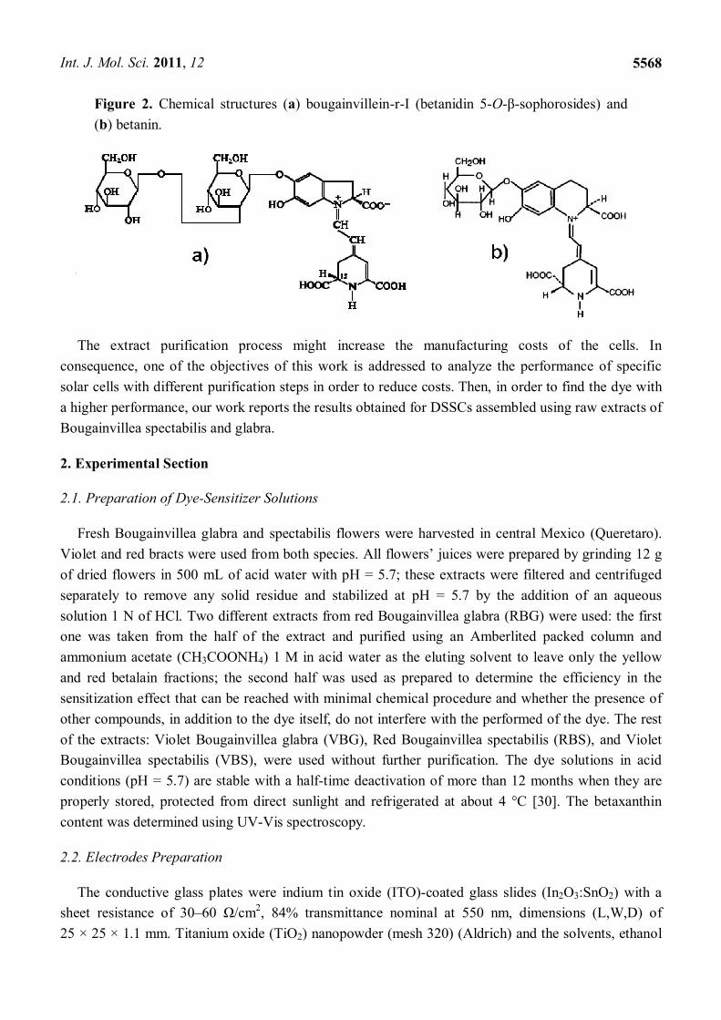
source of betalain pigments is the saccharide type present in the betanidin; Figure 2 shows the
molecular structure of (a) bougainvillein-r-I (betanidin 5-O-β-sophorosides) [23] and (b) betanin [24].
The two main varieties of Bougainvillea, Bougainvillea glabra (Nyctainaceae) and Bougainvillea
spectabilis, are easily available and low cost ornamental plants. In some cases they are used in
traditional medicine [25], but usually they do not have commercial or nutritious use; so there is no
conflict for their use in energy production. Additionally, they flourish all year.
Int. J. Mol. Sci. 2011, 12
5568
Figure 2. Chemical structures (a) bougainvillein-r-I (betanidin 5-O-β-sophorosides) and
(b) betanin.
The extract purification process might increase the manufacturing costs of the cells. In
consequence, one of the objectives of this work is addressed to analyze the performance of specific
solar cells with different purification steps in order to reduce costs. Then, in order to find the dye with
a higher performance, our work reports the results obtained for DSSCs assembled using raw extracts of
Bougainvillea spectabilis and glabra.
2. Experimental Section
2.1. Preparation of Dye-Sensitizer Solutions
Fresh Bougainvillea glabra and spectabilis flowers were harvested in central Mexico (Queretaro).
Violet and red bracts were used from both species. All flowers’ juices were prepared by grinding 12 g
of dried flowers in 500 mL of acid water with pH = 5.7; these extracts were filtered and centrifuged
separately to remove any solid residue and stabilized at pH = 5.7 by the addition of an aqueous
solution 1 N of HCl. Two different extracts from red Bougainvillea glabra (RBG) were used: the first
one was taken from the half of the extract and purified using an Amberlited packed column and
ammonium acetate (CH3COONH4) 1 M in acid water as the eluting solvent to leave only the yellow
and red betalain fractions; the second half was used as prepared to determine the efficiency in the
sensitization effect that can be reached with minimal chemical procedure and whether the presence of
other compounds, in addition to the dye itself, do not interfere with the performed of the dye. The rest
of the extracts: Violet Bougainvillea glabra (VBG), Red Bougainvillea spectabilis (RBS), and Violet
Bougainvillea spectabilis (VBS), were used without further purification. The dye solutions in acid
conditions (pH = 5.7) are stable with a half-time deactivation of more than 12 months when they are
properly stored, protected from direct sunlight and refrigerated at about 4 °C [30]. The betaxanthin
content was determined using UV-Vis spectroscopy.
2.2. Electrodes Preparation
The conductive glass plates were indium tin oxide (ITO)-coated glass slides (In2O3:SnO2) with a
sheet resistance of 30–60 Ω/cm2, 84% transmittance nominal at 550 nm, dimensions (L,W,D) of
25 × 25 × 1.1 mm. Titanium oxide (TiO2) nanopowder (mesh 320) (Aldrich) and the solvents, ethanol

Int. J. Mol. Sci. 2011, 12
5569
and acetonitrile (Aldrich), were analytic grade; they were used as received. ITO substrates were
ultrasonically cleaned in an ethanol-water mixture for 30 min and then heated at 450 °C during 30 min
prior to film deposition. The photo-anodes were prepared by depositing two TiO2 films on the cleaned
ITO glass: for the first one the ITO glass was immersed in a sol-gel solution containing a mixture of
titanium isopropoxide, water and isopropanol at concentration 2:1:25 vol.; the immersion was at
constant speed (1.5 cm/min) [31]; the coated glass was heated at 450 °C during 30 min producing a
densified TiO2 thin film; this isolate the dye-activated porous TiO2 layer from the conductor glass.
Subsequently two edges of the ITO glass plate were covered with adhesive tape (Scotch 3M) to control
the thickness of the film; finally a TiO2 paste was spread uniformly on the substrate by sliding a glass
rod along the tape spacer. The TiO2 paste was prepared by mixing 3.0 g of TiO2 nano-powder, 10 mL
of nitric acid 0.1 N and 4 mL of polyethylene glycol; this suspension was stirred in a closed glass
container for 24 h to obtain a smooth paste with the appropriate viscosity. The film was heated at
450 °C for 60 min resulting in a mesoporous film with a thickness of around 8–10 μm and opaque. The
TiO2 photo-anodes were first soaked for 12 h in HCl and then immersed in the natural dye solutions
for one night at room temperature, according to published procedures [17]. Later, the photo-anodes
were rinsed with distilled water and ethanol and dried. Carbon coated counter electrodes were prepared
following a procedure reported elsewhere [32].
2.3. DSSC Assembling
An electrolyte solution was prepared as reported elsewhere [17]: 0.1 M of I2 was mixed with
0.05 M of LiI and 0.05 M of 3-methoxypropionitrile in 50 mL of acetonitrile (C2H3N) and stirring for
60 min. This electrolyte solution was poured in the mesoporous TiO2 film which was previously
prepared using paraffin-film as framework to seal the cells to prevent evaporation of the liquids. The
counter electrode was pressed against the impregnated anode and clamped firmly in a sandwich
configuration. No leaks (solvent evaporation) were detected.
2.4. Characterization
The topology of the TiO2 films was obtained using a scanning electron microscope (SEM) Jeol
JSM-6060LV operated at 20 kV in secondary electron mode with different magnifications; the samples
were covered with a gold film. The particle size and the particle size distribution of the commercial
TiO2 particles were determined using a light scattering apparatus Brookhaven Instruments model
BI200SM, equipped with a high speed digital correlator PCI-BI9000AT, a solid-state photon detector
and a He-Ne laser of 35 mW Melles Griot 9167EB-1 as a light source. The crystalline structure of the
TiO2 thin film was determined using a diffractometer Rigaku model Miniflex+ equipped with a
radiation source of 1.54 Å and the angle 2θ was varied from 5° to 80° at a scan of
2°/min. Absorption spectra were obtained using a UV-Vis spectrometer Genesys 2PC. The DSSCs
were illuminated using a 75 W Halogen lamp with an incident power of about 100 mW/cm2 in an
illumination area of 0.16 cm2; UV and IR filter glasses were used in front of the sample. Photocurrent
and photovoltage were measured using a Keithley 2400 source meter and the incident light power was
determined using a Powermeter Thor Labs S130A (0–300 mw) and a Spectrometer Ocean
Optics HR 400.

Int. J. Mol. Sci. 2011, 12
5570
3. Results and Discussion
3.1. Absorption Spectra
Figure 3 shows the absorption spectra of RBS and RBG extracts, both diluted in water at pH = 5.7.
In both cases two peaks were found: the first one around 480 nm (480 nm for RBS and 481 nm for
RBG) can be associated to the presence of indicaxanthin, while the second one at 535 nm is
attributable to the betanin or the bougainvillein-r.
Figure 3. Absorption spectra of red Bougainvillea spectabilis (RBS) extract (thick line)
and red Bougainvillea glabra (RBG) extract (thin line), both diluted in water at pH = 5.7.
Red Bougainvillea
0
0.1
0.2
0.3
0.4
0.5
0.6
0.7
0.8
0.9
1
300 340 380 420 460 500 540 580 620 660 700Wavelength (nm)
Re
l. A
bso
rban
ce
0.540
535
0.595
481.0
0.703
480.0 0.625
535.0
The indicaxanthin concentration in µM was estimated from the absorbance peaks at 481 and 535
nm using the expression [24,33]:
482 536(Indicaxanthin) 23.8 7.7A A
The extracted and diluted VBS and VBG solutions were analyzed with the spectrophotometer at
wavelengths between 260 and 700 nm. The absorption spectrum showed two major absorption peaks at
310 and 535 nm for VBS extract and at 307 and 547 nm for VBG extract (Figure 4). Absorbance peaks
at 300 and 535 nm are characteristic absorptions for red violet betalain group, betacyanin [34], for
bougainvillein-r the absorbance peaks are 280, 320 and 541 nm [23].
The substrates were immersed for 12 h in a 1 M HCl solution to facilitate the absorption of
betacyanins on betaxanthins; it is known [17] that the betacyanins absorption is inhibited by the
presence of indicaxina. A dip in HCl results in the protonation of the betalainic carboxyl group from its
anionic form, promoting its absorption in the TiO2 film.

Int. J. Mol. Sci. 2011, 12
5571
Figure 4. Absorption spectra of violet Bougainvillea spectabilis (VBS) extract (thick line)
and violet Bougainvillea glabra (VBG) extract (thin line), both diluted in water at pH = 5.7.
0.0
0.2
0.4
0.6
0.8
1.0
1.2
1.4
1.6
300 340 380 420 460 500 540 580 620 660 700
Re
l. A
bs
orb
an
ce
Wavelength (nm)
Violet Bougainvillea
0.367547
0.948307.0
0.716535.0
310.0
3.2. Structure and Surface Characteristic
The particle size and the particle size distribution of the commercial TiO2 particles were determined
by dynamic light scattering (DLS) resulting of (285 ± 15) nm. The TiO2 surface morphology is shown
in Figure 5a,b. Figure 5a shows a compact (no pores) structure of the first thin layer of TiO2 obtained
by immersing the ITO plates in a titanium alkoxide sol-gel precursor. This thin film acts as an insulator
between the ITO layer and the dye-activated porous TiO2 layer. Some authors [20] have proved the
usefulness of the compact layer to achieve a higher efficiency in the performance of the DSSC. On the
other side, Figure 5b shows the surface of the thick TiO2 coating obtained by the screen-printing
technique; here it is possible to see a homogeneous mesoporous surface formed by the TiO2
nanoparticles. The nanoporous structure is useful because it has a high effective surface area where the
molecules of the Bougainvillea extracts can be linked.
Figure 5. SEM images (a) TiO2 compact layer overlying conductive glass, and (b) second
nanoporous TiO2 layer formed by screen-printing technique.
(a) (b)

Int. J. Mol. Sci. 2011, 12
5572
3.3. Photoelectrochemistry
Five different Bougainvillea extracts were investigated. The first four correspond to extracts
obtained from: RBG, VBG, RBS and VBS. They were centrifuged, filtered and used fresh without
further purification, as explained above. The fifth one was Red Bougainvillea Spectabilis extract
freshly centrifuged and purified by a packed column to remove the bulk of free sucrose compounds;
this extract was named PRBS.
Table 1 summarize the results for the five extracts; in all cases low currents were obtained, but
comparatively higher compared to those obtained by other authors [17]; this probably partially
originated as a result of using ITO-glass with sheet resistance of 30–60 Ohm cm−2 instead of Fluorine
doped tin oxide (FTO) glass with sheet resistance of 10–15 Ohm cm−2 as reported elsewhere [20]. In this
table the short-circuit photocurrent density JSC, the current density at maximum power JMP, the open-
circuit voltage VOC, the maximum power Voltage VMP, the maximum power PM, the theoretical power
PT, the fill factor FF, and the energy conversion efficiency η, are reported.
Table 1. Electrical characterization of all prepared cells.
Dye Source * Jsc
(mA/cm2) JMP
(mA/cm2) VOC (V)
VMP (V)
PM (µW)
PT (µW)
FF η
(%)
RBG 2.344 1.966 0.26 0.231 0.45 0.60 0.74 0.45
VBG 1.860 1.568 0.23 0.199 0.31 0.43 0.71 0.31
RBS 2.291 2.083 0.28 0.229 0.48 0.63 0.76 0.48
VBS 1.881 1.552 0.25 0.226 0.35 0.48 0.73 0.35
PRBS 2.327 2.12 0.26 0.228 0.48 0.61 0.79 0.49
* Red Bougainvillea glabra (RBS), Violet Bougainvillea glabra (VBG), Red Bougainvillea
spectabilis (RBS), Violet Bougainvillea spectabilis (VBS), and Purified Red Bougainvillea
glabra (PRBS).
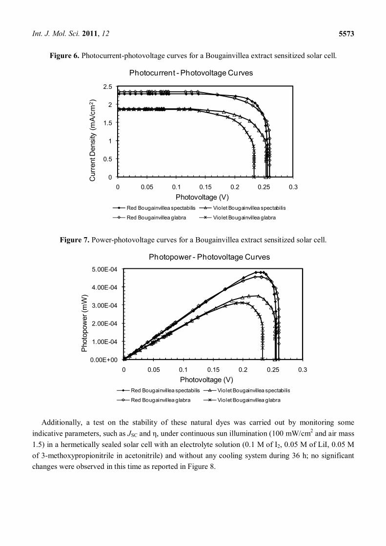
It was found that regardless of the species of Bougainvillea used, the extract with the best
performance parameters comes from the red bracts (RBG and RBS) as shown in Figure 6. This is
because these extracts have both betaxanthin and betacyanins; each with absorptions at different
wavelengths, helping the cell to capture photons of two different energies. Figure 7 shows the power
curves obtained in terms of voltage; it is possible to observe again the improved sensitivity provided
by red bracts compared to the violet ones.
Bougainvillea extracts displayed interesting photoelectrochemical performance parameters: JSC
close to 2.3 mA/cm2; VOC near to 0.26 V; an excellent fill factor of 0.74; and η in the neighborhood of
0.49% using an electrolyte composed of 0.1 M I2/0.05 M LiI/0.05 M 3-methoxypropionitrile in
acetonitrile (C2H3N). The application of a compact TiO2 under-layer (i.e., overcrowding layer) was
necessary for enhancing the cell performance, as compared with previously reported yields [17,20]
(Table 1). The filling factor found in this research is promising because, to our knowledge, it is among
the highest so far reported with raw natural dyes.
Int. J. Mol. Sci. 2011, 12
5573
Figure 6. Photocurrent-photovoltage curves for a Bougainvillea extract sensitized solar cell.
0
0.5
1
1.5
2
2.5
0 0.05 0.1 0.15 0.2 0.25 0.3
Red Bougainvillea spectabilis Violet Bougainvillea spectabilis
Red Bougainvillea glabra Violet Bougainvillea glabra
Curr
entD
ensi
ty (
mA
/cm
2)
Photovoltage (V)
Photocurrent - Photovoltage Curves
Figure 7. Power-photovoltage curves for a Bougainvillea extract sensitized solar cell.
0.00E+00
1.00E-04
2.00E-04
3.00E-04
4.00E-04
5.00E-04
0 0.05 0.1 0.15 0.2 0.25 0.3
Red Bougainvillea spectabilis Violet Bougainvillea spectabilis
Red Bougainvillea glabra Violet Bougainvillea glabra
Photo
pow
er(m
W)
Photovoltage (V)
Photopower - Photovoltage Curves
Additionally, a test on the stability of these natural dyes was carried out by monitoring some
indicative parameters, such as JSC and η, under continuous sun illumination (100 mW/cm2 and air mass
1.5) in a hermetically sealed solar cell with an electrolyte solution (0.1 M of I2, 0.05 M of LiI, 0.05 M
of 3-methoxypropionitrile in acetonitrile) and without any cooling system during 36 h; no significant
changes were observed in this time as reported in Figure 8.

Int. J. Mol. Sci. 2011, 12
5574
Figure 8. Time dependence of the efficiency for Bougainvillea extract sensitized solar cell.
Efficient Vs time
0.1
0.2
0.3
0.4
0.5
0 6 12 18 24 30 36
Hours
n (
%)
4. Conclusions
The Bougainvillea spectabilis and glabra extracts were studied as natural dyes for DSSCs. The
results are consistent with those reported previously for Bougainvillea; however a higher FF was
obtained due to the presence of the insulating TiO2 layer; additionally an electrolyte with less vapor
pressure increases the lifetime of the solar cell. One important finding was that the mixture of
betaxanthin and betacyanin produces a better conversion than betacyanin alone: the absorption at
different wavelengths of betaxanthin and betacyanin increases the absorption of photons of different
energies. A thorough study of the ratio betaxanthin-betacyanin in order to obtain the highest efficiency
in these dye-sensitized solar cells is required. It was also found that the use of a highly purified extract
does not represent a significant increment in cell efficiency, but it is possible that the presence of other
compounds than the dye itself, e.g., sucrose, can affect the lifetime of the solar cell. As each extract
from a particular source will have a different compound combination or different betalain compounds,
it is important to characterize the extract from different sources in order to find the best efficiency at
the lowest cost. In this particular case, a simple purification process to separate sucrose free
compounds produced a device with the lowest cost and highest efficiency.
Acknowledgments
The authors are in debt to: A. del Real for the SEM micro-images, D. Rangel for instrumental
support. One of the authors (AHM) is in debt for the economical support from DGAPA, UNAM.
References
1. Grätzel, M. Solar energy conversion by dye-sensitized photovoltaic cells. Inorg. Chem. 2005, 44,
6841–6851.

Int. J. Mol. Sci. 2011, 12
5575
2. Gómez-Ortíz, N.M.; Vázquez-Maldonado, I.A.; Pérez-Espadas, A.R.; Mena-Rejón, G.J.;
Azamar-Barrios, J.A.; Oskam, G. Dye-sensitized solar cells with natural dyes extracted from
achiote seeds. Solar Energy Mater. Solar Cells 2010, 94, 40–44.
3. O’Regan, B.; Grätzel, M. A low-cost, high-efficiency solar cell based on dye sensitized colloidal
TiO2 films. Nature 1991, 353, 737–740.
4. Caramori, S.; Cristino, V.; Boaretto, R.; Argazzi, R.; Bignozzi, C.A.; Di Carlo, A. New
components for dye-sensitized solar cells. Int. J. Photoenergy 2010, 2010, 1–16.
5. Saito, M.; Fujihara, S. Large photocurrent generation in dye-sensitized ZnO solar cells. Energy
Environ. Sci. 2008, 1, 280–283.
6. Quintana, M.; Edvinsson, T.; Hagfeldt, A.; Boschloo, G. Comparison of dye-sensitized ZnO and TiO2
solar cells: Studies of charge transport and carrier lifetime. J. Phys. Chem. C 2007, 111, 1035–1041.
7. Fukai, Y.; Kondo, Y.; Mori, S.; Suzuki, E. Highly efficient dye-sensitized SnO2 solar cells having
sufficient electron diffusion length. Electrochem. Commun. 2007, 9, 1439–1443.
8. Bauer, C.; Boschloo, G.; Mukhtar, E.; Hagfeldt, A. Ultrafast studies of electron injection in Ru
dye sensitized SnO2 nanocrystalline thin film. Int. J. Photoenergy 2002, 4, 17–20.
9. Sayama, K.; Sugihara, H.; Arakawa, H. Photoelectrochemical properties of a porous Nb2O5
electrode sensitized by a ruthenium dye. Chem. Mater. 1998, 10, 3825–3832.
10. Nazeeruddin, M.K.; Péchy, P.; Renouard, T.; Zakeeruddin, S.M.; Humphry-Baker, R.; Comte, P.;
Liska, P.; Cevey, L.; Costa, E.; Shklover, V.; et al. Engineering of efficient panchromatic
sensitizers for nanocrystalline TiO2-based solar cells. J. Am. Chem. Soc. 2001, 123, 1613–1624.
11. Hagfeldt, A.; Grätzel, M. Molecular photovoltaics. Acc. Chem. Res. 2000, 33, 269–277.
12. Odobel, F.; Blart, E.; Lagrée, M. Porphyrin dyes for TiO2 sensitization. J. Mater. Chem. 2003, 13,
502–510.
13. Nazeeruddin, M.K.; Humphry-Baker, R.; Grätzel, M.; Wohrle, D.; Schnurpfeil, G.; Schneider, G.;
Hirth, A.; Trombach, N. Efficient near-IR sensitization of nanocrystalline TiO2 films by zinc and
aluminum phthalocyanines. J. Porphyrins Phthalocyanines 1999, 3, 230–237.
14. He, J.; Hagfeldt, A.; Lindquist, S.E. Phthalocyanine-Sensitized nanostructured TiO2 electrodes
prepared by a novel anchoring method. Langmuir 2001, 17, 2743–2747.
15. Islam, A.; Sugihara, H.; Hara, K.; Singh, L.P.; Katoh, R.; Yanagida, M.; Takahashi, Y.;
Murata, S.; Arakawa, H. Dye sensitization of nanocrystalline titanium dioxide with square planar
platinum(II) diimine dithiolate complexes. Inorg. Chem. 2001, 40, 5371–5380.
16. Rehm, J.M.; McLendon, G.L.; Nagasawa, Y.; Yoshihara, K.; Moser, J.; Grätzel, M. Femtosecond
electron-transfer dynamics at a sensitizing dye-semiconductor (TiO2) interface. J. Phys. Chem.
1996, 100, 9577–9588.
17. Zhang, D.; Lanier, S.M.; Downing, J.A.; Avent, J.L.; Lum, J.; McHale, J.L. Betalain pigments for
dye-sensitized solar cells. Photochem. Photobiol. A 2008, 195, 72–80.
18. Kay, A.; Graetzel, M. Photosensitization of titania solar cells with chlorophyll derivatives and
related natural porphyrins. J. Phys. Chem. 1993, 97, 6272–6277.
19. Nazeeruddin, M.K.; Kay, A.; Rodicio, I.; Humphry-Baker, R.; Mueller, E.; Liska, P.;
Vlachopoulos, N.; Graetzel, M. Conversion of light to electricity by cis-X2bis(2,2′-bipyridyl-4,4′-
dicarboxylate)ruthenium(II) charge-transfer sensitizers (X = Cl-, Br-, I-, CN-, and SCN-) on
nanocrystalline TiO2 electrodes. J. Am. Chem. Soc. 1993, 115, 6382–6390.

Int. J. Mol. Sci. 2011, 12
5576
20. Calogero, G.; Di Marco, G.; Cazzanti, S.; Caramori, S.; Argazzi, R.; Di Carlo, A.D.; Bignozzi, C.A.
Efficient dye-sensitized solar cells using red turnip and purple wild sicilian prickly pear fruits. Int.
J. Mol. Sci. 2010, 11, 254–267.
21. Garcia, C.G.; Polo, A.S.; Murakami Iha, N.Y. Fruit extracts and ruthenium polypyridinic dyes for
sensitization of TiO2 in photoelectrochemical solar cells. J. Photochem. Photobiol. A 2003, 160,
87–91.
22. Calogero, G.; Di Marco, G. Red Sicilian orange and purple eggplant fruits as natural sensitizers
for dye-sensitized solar cells. Solar Energy Mater. Solar Cells 2008, 92, 1341–1346.
23. Piattelli, M.; Imperato, F. Betacyanins from Bougainvillea. Phytochemistry 1970, 9, 455–458.
24. Butera, D.; Tesoriere, L.; Di Gaudio, F.; Bongiorno, A.; Allegra, M.; Pintaudi, A.M.; Kohen, R.;
Livrea, M.A. Antioxidant activities of Sicilian prickly pear (Opuntia ficus indica) fruit extracts
and reducing properties of its betalains: Betanin and indicaxanthin. J. Agric. Food Chem. 2002,
50, 6895–6901.
25. Simon, A.; Tóth, G.; Duddeck, H.; Soliman, H.S.M.; Mahmoud, I.I.; Samir, H. Glycosides from
Bougainvillea Glabra. Nat. Prod. Res. 2006, 20, 63–67.
26. Piattelli, M.; Minale, L.; Nicolaus, R.A. Pigments of centrospermae—V.: Betaxanthins from
Mirabilis jalapa L. Phytochemistry 1965, 4, 817–823.
27. Impellizzeri, G.; Piattelli, M.; Sciuto, S. Acylated betacyanins from Drosanthemum floribundum.
Phytochemistry 1973, 12, 2295–2296.
28. Wyler, H.; Dreiding, A.S. Über Betacyane, die stickstoffhaltigen Farbstoffe der Centrospermen.
Vorläufige Mitteilung. Cell. Mol. Life Sci. 1961, 17, 23–25.
29. Piattelli, M.; Imperato, F. Pigments of Bougainvillea glabra. Phytochemistry 1970, 9, 2557–2560.
30. Castellar, R.; Obón, J.M.; Alacid, M.; Fernández-lopez, J.A. Color properties and stability of
betacyanins from Opuntia fruits. J. Agric. Food Chem. 2003, 51, 2772–2776.
31. Su, C.; Sheu, T.K.; Chang, Y.T.; Wan, M.A.; Feng, M.C.; Hung, W.C. Preparation of ITO thin
films by Sol-Gel process and their characterizations. Synth. Metals 2005, 153, 9–12.
32. Smestad, G.P. Education and solar conversion: Demonstrating electron transfer. Solar Energy
Mater. Solar Cells 1998, 55, 157–178.
33. Tesoriere, L.; Allegra, M.; Butera, D.; Livrea, M.A. Absorption, excretion, and distribution of
dietary antioxidant betalains in LDLs: Potential health effects of betalains in humans. Am. J. Clin.
Nutr. 2004, 80, 941–945.
34. Sánchez, F.D.; Lopez, E.M.S.; Kerstupp, S.F.; Ibarra, R.V.; Scheinvar, L. Colorant extraction
from red prickly pear (Opuntia lasiacantha) for food application. J. Environ. Agric. Food Chem.
2006, 5, 1330–1337.
© 2011 by the authors; licensee MDPI, Basel, Switzerland. This article is an open access article
distributed under the terms and conditions of the Creative Commons Attribution license
(http://creativecommons.org/licenses/by/3.0/).