neuroprotection with a brain-penetrating biologic tumor...
TRANSCRIPT

Neuroprotection with a Brain-Penetrating Biologic TumorNecrosis Factor Inhibitor
Qing-Hui Zhou, Rachita Sumbria, Eric Ka-Wai Hui, Jeff Zhiqiang Lu, Ruben J. Boado,and William M. PardridgeDepartment of Medicine (Q.-H.Z., R.S., R.J.B., W.M.P.), University of California, Los Angeles, California; and ArmaGenTechnologies, Inc. (E.K.-W.H., J.Z.L., R.J.B.) Santa Monica, California
Received July 7, 2011; accepted August 9, 2011
ABSTRACTBiologic tumor necrosis factor (TNF)-� inhibitors do not crossthe blood-brain barrier (BBB). A BBB-penetrating TNF-� inhib-itor was engineered by fusion of the extracellular domain of thetype II human TNF receptor (TNFR) to the carboxyl terminus ofthe heavy chain of a mouse/rat chimeric monoclonal antibody(MAb) against the mouse transferrin receptor (TfR), and thisfusion protein is designated cTfRMAb-TNFR. The cTfRMAb-TNFR fusion protein and etanercept bound human TNF-� withhigh affinity and KD values of 374 � 77 and 280 � 80 pM,respectively. Neuroprotection in brain in vivo after intravenousadministration of the fusion protein was examined in a mousemodel of Parkinson’s disease. Mice were also treated withsaline or a non-BBB-penetrating TNF decoy receptor, etaner-cept. After intracerebral injection of the nigral-striatal toxin,6-hydroxydopamine, mice were treated every other day for
3 weeks. Treatment with the cTfRMAb-TNFR fusion proteincaused an 83% decrease in apomorphine-induced rotation, a67% decrease in amphetamine-induced rotation, a 82% in-crease in vibrissae-elicited forelimb placing, and a 130% in-crease in striatal tyrosine hydroxylase (TH) enzyme activity.In contrast, chronic treatment with etanercept, which does notcross the BBB, had no effect on neurobehavior or striatal THenzyme activity. A bridging enzyme-linked immunosorbent as-say specific for the cTfRMAb-TNFR fusion protein showed thatthe immune response generated in the mice was low titer. Inconclusion, a biologic TNF inhibitor is neuroprotective afterintravenous administration in a mouse model of neurodegen-eration, providing that the TNF decoy receptor is reengineeredto cross the BBB.
Introduction
Tumor necrosis factor (TNF)-� is a proinflammatory cyto-kine in peripheral tissues, and the leading TNF-�-inhibitors(TNFIs) are biologic drugs, including etanercept, a TNF de-coy receptor-Fc fusion protein, infliximab, a chimeric anti-TNF-� monoclonal antibody (MAb), and adalimumab, a hu-man anti-TNF-� MAb (Tansey and Szymkowski, 2009).TNF-� also plays a pathologic role in brain disorders, includ-ing Parkinson’s disease (PD) (McCoy et al., 2006), Alzhei-mer’s disease (He et al., 2007), and depression (Himmerich etal., 2008). However, the biologic TNFIs cannot be developed
for treatment of the brain, because these large moleculedrugs do not cross the blood-brain barrier (BBB).
Biologic TNFIs can be reengineered for BBB penetrationby engineering fusion proteins of the TNFI and a BBB mo-lecular Trojan horse (MTH) (Pardridge, 2010). The latter is apeptide or peptidomimetic MAb that traverses the BBB viatransport on an endogenous receptor-mediated transport sys-tem, such as the insulin receptor or the transferrin receptor(TfR). The most potent BBB MTH is a genetically engineeredMAb against the human insulin receptor (HIR) (Boado et al.,2007). A fusion protein of the HIRMAb and the extracellulardomain (ECD) of the type II TNF receptor (TNFR) has beenengineered and is designated the HIRMAb-TNFR fusion pro-tein (Hui et al., 2009). The brain uptake of the HIRMAb-TNFR fusion protein in the rhesus monkey is high, �3% ofinjected dose (ID)/brain, whereas etanercept does not crossthe BBB in vivo (Boado et al., 2010). However, the HIRMAbonly cross-reacts with the insulin receptor of the Old World
This work was supported by the National Institutes of Health NationalInstitute of Neurological Disorders and Stroke [Grant R01-NS065917].
Article, publication date, and citation information can be found athttp://jpet.aspetjournals.org.
doi:10.1124/jpet.111.185876.
ABBREVIATIONS: TNF, tumor necrosis factor; TNFI, tumor necrosis inhibitor; MAb, monoclonal antibody; PD, Parkinson’s disease; BBB,blood-brain barrier; MTH, molecular Trojan horse; TfR, transferrin receptor; HIR, human insulin receptor; ECD, extracellular domain; TNFR, tumornecrosis factor receptor; ID, injected dose; cTfRMAb, chimeric MAb against mouse TfR; TH, tyrosine hydroxylase; ELISA, enzyme-linkedimmunosorbent assay; CHO, Chinese hamster ovary; ANOVA, analysis of variance; AD, Alzheimer’s disease; PBS, phosphate-buffered saline;PBSB, PBS containing 1% bovine serum albumin.
0022-3565/11/3392-618–623$25.00THE JOURNAL OF PHARMACOLOGY AND EXPERIMENTAL THERAPEUTICS Vol. 339, No. 2Copyright © 2011 by The American Society for Pharmacology and Experimental Therapeutics 185876/3724032JPET 339:618–623, 2011 Printed in U.S.A.
618
at ASPE
T Journals on June 12, 2018
jpet.aspetjournals.orgD
ownloaded from

primate, such as the rhesus monkey (Pardridge et al., 1995),and does not cross-react with the insulin receptor of NewWorld primates or lower animals. There is no known MAbagainst the mouse insulin receptor that can be used as a BBBMTH in the mouse. Therefore, a surrogate MTH for themouse has been engineered, which is a chimeric MAb againstthe mouse TfR, designated the cTfRMAb (Boado et al., 2009).A fusion protein of the cTfRMAb and the ECD of the TNFR-IIhas been engineered (Zhou et al., 2011a), and the structure ofthe cTfRMAb-TNFR fusion protein is shown in Fig. 1A. Thedecoy receptor is fused to the carboxyl terminus of the heavychain of the IgG part of the fusion protein. In contrast, for theengineering of etanercept (Peppel et al., 1991), the TNFRdecoy receptor is fused to the amino terminus of the heavychain of the Fc fragment (Fig. 1B).
The present investigation tests the neuroprotective actionsof both the cTfRMAb-TNFR fusion protein and etanercept inthe 6-hydroxydopamine mouse model of PD. The fusion pro-teins are administered every other day over a 3-week periodby intravenous injection in the tail vein. Neuroprotection isassessed with three assays of neurobehavior, with a striataltyrosine hydroxylase (TH) enzyme activity, and with THimmunocytochemistry. The level of immune response in micecaused by chronic administration of the cTfRMAb-TNFR fu-sion protein is assessed with a bridging ELISA.
Materials and MethodsMouse Parkinson’s Disease Model and Treatment. All pro-
cedures were approved by the UCLA Animal Research Committee.Adult male C57BL/6J mice (The Jackson Laboratory, Bar Harbor,ME) weighing 25 to 32 g were anesthetized with ketamine (100mg/kg) and xylazine (10 mg/kg) intraperitoneally. Animals receiveda unilateral intracerebral injection of a total of 12 �g of6-hydroxydopamine�HBr (Sigma-Aldrich, St. Louis, MO) dissolved in0.02% ascorbic acid in 0.9% saline. The 6-hydroxydopamine (6 �g in2 �l) was injected into the right striatum at two locations as de-scribed previously (Fu et al., 2010). The toxin was injected into thestriatum at sites with the following stereotaxic coordinates: �1.0mm relative to bregma, 2.1 mm relative to midline, and 2.9 mmbelow the skull surface (site 1); and �0.3 mm relative to bregma, 2.3mm relative to midline, and 2.9 mm below the skull surface (site 2).Mice were treated intravenously with one of three treatment drugs:saline (n � 10 mice); etanercept, 1.0 mg/kg (n � 10 mice); or the
cTfRMAb-TNFR fusion protein, 1.0 mg/kg (n � 10 mice), every 2days over the following 3 weeks, with the first dose given 1 h aftertoxin injection into the brain. Treatment drugs were injected intra-venously via the tail vein in a volume of 50 �l/mouse. The mice wereeuthanized at 3 weeks after toxin administration for measurement ofstriatal TH enzyme activity.
Etanercept (Enbrel) was obtained from the UCLA Pharmacy. ThecTfRMAb-TNFR fusion protein was purified by protein G affinitychromatography of serum-free medium conditioned by stably trans-fected Chinese hamster ovary (CHO) cells as described previously(Zhou et al., 2011a). The 235-amino acid extracellular domain of thetype II human TNFR, minus the signal peptide, was fused to thecarboxyl terminus of the heavy chain of the cTfRMAb (Fig. 1A) asdescribed previously (Zhou et al., 2011a). The fusion protein wasformulated in 0.01 M sodium acetate-buffered saline (pH 6.5) andwas stored either sterile-filtered at 4°C or at �70°C. The molecularmass of the cTfRMAb-TNFR fusion protein is 195,200 Da (Zhou etal., 2011a), whereas the molecular mass of etanercept is 51,200 Da.Therefore, at a systemic dose of each fusion protein of 1 mg/kg, anearly 4-fold molar excess of etanercept was administered.
TNF-� Radioreceptor Assay. The saturable binding of humanTNF-� to either etanercept or to the cTfRMAb-TNFR fusion proteinwas determined with a radioreceptor assay as described previously(Hui et al., 2009). For TNF-� binding to either the cTfRMAb-TNFRfusion protein or to a mouse IgG1 negative control, a goat anti-mouseIgG1 Fc antibody (Bethyl Laboratories, Montgomery, TX) was platedin 96-well plates (0.2 �g/well). For TNF-� binding to either etaner-cept or to a human IgG1 negative control, a mouse anti-human IgG1Fc antibody (Invitrogen, Carlsbad, CA) was plated in 96-well plates(0.2 �g/well). The fusion protein or negative control antibody wasplated (100 ng/well), followed by a 1-h incubation at room tempera-ture. The wells were then washed with phosphate-buffered saline(PBS), followed by the addition of 100 �l/well of a comixture of125I-human TNF-� (specific activity � 91 �Ci/�g; PerkinElmer Lifeand Analytical Sciences, Waltham, MA) at a concentration of 0.01�Ci/well (0.1 �Ci/ml; 1.1 ng/ml; 60 pM) and various concentrations ofunlabeled human TNF-�, followed by a 3-h incubation at room tem-perature. The wells were washed, and bound radioactivity was de-termined as described previously (Hui et al., 2009). The half-satura-tion constant, KD, of TNF-� binding to the cTfRMAb-TNFR oretanercept fusion protein was determined by nonlinear regressionanalysis using BMDP2007e software (Statistical Solutions, Cork,Ireland), after fitting of binding data to the following equation:bound � [(Bmax)(C)]/(KD � C), where Bmax is the maximal bindingand C � the concentration of TNF-�.
Behavioral Testing. Beginning 1 week after the toxin adminis-tration, mice were tested weekly for apomorphine- and amphet-amine-induced rotation, which was performed on separate days, asdescribed previously (Fu et al., 2010). A vibrissae-elicited forelimb-placing trial in the mice was performed at the end of the 3 weeks oftreatment (Fu et al., 2010).
Tyrosine Hydroxylase Enzyme Activity. Homogenates ofmouse brain striatum (left and right side) and frontal cortex wereprepared with a Polytron homogenizer in 5 mM KPO4-0.1% TritonX-100 (pH 6.3) followed by centrifugation. After an aliquot wasremoved for measurement of protein with the bicinchoninic acidassay, dithiothreitol was added to the supernatant to 1 mM, and thesupernatant was stored at �70°C until assay. The TH enzyme ac-tivity in the supernatant was measured with [3,5-3H]L-tyrosine(PerkinElmer Life and Analytical Sciences) as substrate. The purityof the [3,5-3H]L-tyrosine was assessed by thin-layer chromatography.TH enzyme activity converts [3,5-3H]L-tyrosine to L-DOPA and[3H]water. The [3H]water product was separated from the [3H]ty-rosine substrate with a charcoal separation technique, as describedpreviously (Fu et al., 2010). Any residual [3H]water present in the[3,5-3H]L-tyrosine was accounted for with determinations of assayblanks in each assay. The assay was validated with [3H]water(PerkinElmer Life and Analytical Sciences), which showed that the
Fig. 1. A, the cTfRMAb-TNFR fusion protein is formed by fusion of theECD of the type II human TNFR to the carboxyl terminus of each heavychain of the cTfRMAb. B, the etanercept fusion protein is formed byfusion of the same ECD of the type II human TNFR to the aminoterminus of the Fc fragment of human IgG1 heavy chain.
Brain-Penetrating TNF-� Decoy Receptor 619
at ASPE
T Journals on June 12, 2018
jpet.aspetjournals.orgD
ownloaded from

[3H]water was 100% recovered in the supernatant after removal ofamino acid by the charcoal. TH enzyme activity was measured at37°C for 30 min and is expressed as picomoles per hour per milligramof protein.
Tyrosine Hydroxylase Immunocytochemistry. The brain wasremoved and coronal blocks were frozen in powdered dry ice, followedby embedding in Tissue Tek OCT medium, and refrozen, and blockswere stored at �70°C. Frozen sections (20-�m thickness) were pre-pared at �20°C on a Micron Instruments (San Marcos, CA) cryostat.Sections were fixed in ice-cold acetone-methanol (1:1) at �20°C for20 min. Immune staining was performed with an affinity-purifiedrabbit antibody against rat TH, which cross-reacts with all forms ofmammalian TH (Pel-Freez, Rogers, AR), which was diluted 1:1000 inPBS with 0.3% Triton X-100 and 3% horse serum. The secondaryantibody was 2 �g/ml biotinylated horse anti-rabbit IgG (VectorLaboratories, Burlingame, CA). Immune detection was performedwith the ImmPACT DAB kit (Vector Laboratories) using diamino-benzidine. The sections were not counterstained and were scannedwith a UMAX PowerLook III scanner with transparency adapter.Striatal immunostaining on the lesioned and nonlesioned side wasquantitated by determination of optical density using Image soft-ware (version 1.62; National Institutes of Health, Bethesda, MD).The entire striatum on both the lesioned and nonlesioned side wasoutlined for measurement of density of staining. In addition, thecortex was outlined as a measure of the background, and the corticaldensity was subtracted from the striatal density to yield the back-ground-corrected density of striatal immunostaining.
Immunity ELISA. The presence of anti-cTfRMAb-TNFR fusionprotein antibodies in mouse plasma was measured with a two-sitesandwich ELISA described previously (Zhou et al., 2011b). ThecTfRMAb-TNFR fusion protein is used as the capture reagent, andbiotinylated cTfRMAb-TNFR fusion protein is used as the detectorreagent. The mouse plasma was diluted 1:50 in PBS. The capturereagent was plated overnight at 4°C in 96-well plates at 100 �l (250ng)/well in 0.05 M NaHCO3/pH 8.3. The wells were blocked with PBScontaining 1% bovine serum albumin (PBSB), followed by the addi-tion of 100 �l/well of the 1:50 dilution of mouse plasma. After a60-min incubation at 37°C, the wells were washed with PBSB, andthe wells were incubated with biotinylated cTfRMAb-TNFR fusionprotein (25 ng/well) for 60 min. The wells were washed with PBSB,followed by incubation with 100 �l (500 ng/well) of a streptavidin-peroxidase conjugate (Vector Laboratories) for 30 min at room tem-perature. The wells were washed with PBSB, and 100 �l/well o-phen-ylenediamine-H2O2 developing solution (Sigma-Aldrich) was addedfor a 15-min incubation in the dark at room temperature. The reac-tion was stopped by the addition of 100 �l/well 1 M HCl, followed bythe measurement of absorbance at 492 and 650 nm. The A650 was
subtracted from the A492. The (A492 � A650) for the PBSB blank wasthen subtracted from the (A492 � A650) for the sample.
The cTfRMAb-TNFR fusion protein was biotinylated with sulfo-biotin-LC-LC-N-hydroxysuccinimide, where LC � long-chain (PierceChemical). The biotinylation of the cTfRMAb-TNFR fusion proteinwas confirmed by SDS-polyacrylamide gel electrophoresis and West-ern blotting, for which the blot was probed with avidin and biotin-ylated peroxidase. The nonbiotinylated cTfRMAb-TNFR fusion pro-tein was tested as a negative control.
Statistical Analysis. Statistical differences between saline-, et-anercept-, and fusion protein-treated mice were determined withanalysis of variance (ANOVA) with Bonferroni correction. Statisticaldifferences between striatal immunostaining on the lesioned andnonlesioned side were determined with Student’s t test.
ResultsTNF� bound to both the cTfRMAb-TNFR fusion protein
and to etanercept with high affinity and KD values of 374 �77 and 280 � 80 pM, respectively (Fig. 2).
Mice tolerated the chronic treatment with either etaner-cept or the cTfRMAb-TNFR fusion protein, no mice exhibitedsigns of immune reaction to the study drugs, and no weightloss was observed in any of the treatment groups. Treatmentof PD mice with chronic intravenous cTfRMAb-TNFR fusionprotein resulted in a 75 to 83% reduction in apomorphine-induced rotation compared with that in saline-treated mice,whereas etanercept had no significant effect on drug-inducedrotation (Fig. 3). Treatment of PD mice with chronic intrave-nous cTfRMAb-TNFR fusion protein caused a 45 to 67%reduction in amphetamine-induced rotation compared withthat in saline-treated mice, whereas etanercept had no sig-nificant effect on drug-induced rotation (Fig. 4). Treatment ofPD mice with chronic intravenous cTfRMAb-TNFR fusionprotein caused a 82% increase in the vibrissae-elicited fore-limb placing score compared with that for the saline-treatedmice, whereas etanercept had no significant effect on fore-limb placing (Fig. 5). Treatment of PD mice with chroniccTfRMAb-TNFR fusion protein caused a 130% increase instriatal TH enzyme activity on the lesioned (right) side, rel-ative to that in saline-treated mice, whereas etanercept hadno significant effect on striatal TH enzyme activity (Fig. 6).
Fig. 2. Radioreceptor assay shows saturation of binding of human TNF-�to either the cTfRMAb-TNFR fusion protein or to etanercept. The bindingdissociation constant, KD, was determined by nonlinear regression anal-ysis. The horizontal line at 1.5% binding represents the nonspecific bind-ing observed when either human IgG1 or mouse IgG1 was plated in lieuof the fusion protein.
Fig. 3. Contralateral rotation (over 20 min) after the administration ofapomorphine to PD mice treated with saline, etanercept, or the cTfRMAb-TNFR fusion protein at 1, 2, and 3 weeks after toxin injection. Data aremeans � S.E. (n � 10 mice/group). Statistical differences from the saline-treated animals: �, p � 0.01 at weeks 1, 2, and 3, as determined byANOVA with Bonferroni correction.
620 Zhou et al.
at ASPE
T Journals on June 12, 2018
jpet.aspetjournals.orgD
ownloaded from
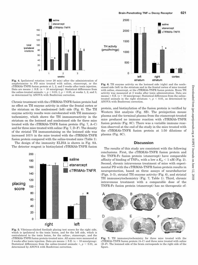
Chronic treatment with the cTfRMAb-TNFR fusion protein hadno effect on TH enzyme activity in either the frontal cortex orthe striatum on the nonlesioned (left) side (Fig. 6). The THenzyme activity results were corroborated with TH immunocy-tochemistry, which shows the TH immunoreactivity in thestriatum on the lesioned and nonlesioned side for three micetreated with the cTfRMAb-TNFR fusion protein (Fig. 7, A–C)and for three mice treated with saline (Fig. 7, D–F). The densityof the striatal TH immunostaining on the lesioned side wasincreased 101% in the mice treated with the cTfRMAb-TNFRfusion protein compared with the saline-treated mice (Table 1).
The design of the immunity ELISA is shown in Fig. 8A.The detector reagent is biotinylated cTfRMAb-TNFR fusion
protein, and biotinylation of the fusion protein is verified byWestern blot analysis (Fig. 8B). The preinjection mouseplasma and the terminal plasma from the etanercept-treatedmice produced no immune reaction with cTfRMAb-TNFRfusion protein (Fig. 8C). There was a variable immune reac-tion observed at the end of the study in the mice treated withthe cTfRMAb-TNFR fusion protein at 1:50 dilutions ofplasma (Fig. 8C).
DiscussionThe results of this study are consistent with the following
conclusions. First, the cTfRMAb-TNFR fusion protein andthe TNFR-Fc fusion protein (etanercept) have comparableaffinity of binding of TNF�, with a low a KD � 1 nM (Fig. 2).Second, chronic intravenous treatment of mice with experi-mental PD with the cTfRMAb-TNFR fusion protein results inneuroprotection, based on three assays of neurobehavior(Figs. 3–5), striatal TH enzyme activity (Fig. 6), and striatalTH immunocytochemistry (Fig. 7; Table 1). Third, chronicintravenous treatment with a comparable dose of theTNFR-Fc fusion protein (etanercept) has no therapeutic ef-
Fig. 5. Vibrissae-elicited forelimb placing test scores for the right side,which is ipsilateral to the toxin lesion, and for the left side, which iscontralateral to the toxin lesion, for the saline-, etanercept-, and thecTfRMAb-TNFR fusion protein-treated mice. All scores were measured at3 weeks after toxin injection. Data are means � S.E. (n � 10 mice/group).Statistical differences from the saline-treated animals: �, p � 0.01, asdetermined by ANOVA with Bonferroni correction.
Fig. 7. TH immunocytochemistry for three mice treated with thecTfRMAb-TNFR fusion protein (A–C) and three mice treated with saline(D–F). The lesioned side of the brain corresponds to the right side of thefigure.
Fig. 4. Ipsilateral rotation (over 20 min) after the administration ofamphetamine to PD mice treated with saline, etanercept, or thecTfRMAb-TNFR fusion protein at 1, 2, and 3 weeks after toxin injection.Data are means � S.E. (n � 10 mice/group). Statistical differences fromthe saline-treated animals: �, p � 0.01; ‡, p � 0.05, at weeks 1, 2, and 3,as determined by ANOVA with Bonferroni correction.
Fig. 6. TH enzyme activity on the lesioned side (right) and the nonle-sioned side (left) in the striatum and in the frontal cortex of mice treatedwith saline, etanercept, or the cTfRMAb-TNFR fusion protein. Brain THactivity was measured at 3 weeks after toxin administration. Data aremeans � S.E. (n � 10 mice/group). Statistical differences from the saline-treated animals in the right striatum: �, p � 0.01, as determined byANOVA with Bonferroni correction.
Brain-Penetrating TNF-� Decoy Receptor 621
at ASPE
T Journals on June 12, 2018
jpet.aspetjournals.orgD
ownloaded from

fect in experimental PD (Figs. 3–7). Fourth, chronic treat-ment of mice with the cTfRMAb-TNFR fusion protein causesa low-titer immune response against the cTfRMAb part of thefusion protein (Fig. 8).
IgG-decoy receptor fusion proteins are formed by fusion ofthe carboxyl terminus of the decoy receptor to the aminoterminus of the IgG heavy chain (Scallon et al., 1995), asshown for etanercept in Fig. 1B. The engineering of thecTfRMAb-TNFR fusion protein is a departure from all priorIgG-decoy receptor fusion proteins in that the amino termi-nus of the TNFR-II ECD is fused to the carboxyl terminus ofthe antibody heavy chain (Fig. 1A). Despite this novel struc-ture, the affinity of the cTfRMAb-TNFR fusion protein forTNF-� is high and is comparable to the binding affinity ofetanercept (Fig. 2). The KD of cTfRMAb-TNFR fusion proteinbinding of human TNF-�, 374 � 77 pM (Fig. 2), is comparableto the KD of TNF-� binding to the intact membrane receptor,which is 0.3 to 0.4 nM (Morita et al., 2001). The humanTNFR-II receptor binds mouse TNF-� to the same extent ashuman TNF-� (Scallon et al., 2002). In addition to TNF-�,the cTfRMAb-TNFR fusion protein binds the murine BBBTfR with high affinity, and the brain uptake of the fusionprotein in the mouse after intravenous injection is high,2.8 � 0.5% ID/g brain (Zhou et al., 2011a). In contrast, thebrain uptake of an IgG that does not cross the BBB is 0.06%ID/g in the mouse (Lee et al., 2000). Given a brain uptake of2.8% ID/g, the brain concentration of the cTfRMAb-TNFRfusion protein is 840 ng/g brain or approximately 6 nM, withthe systemic injection dose of 1 mg/kg used in these studies.Because the cerebral concentration of TNF-� increases toapproximately 0.5 nM in excitotoxic conditions (Shohami etal., 1994), the 1 mg/kg dose of fusion protein is sufficient tosequester the TNF-� produced in brain.
TNF-� induces apoptosis (Chen and Goeddel, 2002) and iselevated in the brains of patients with PD (Machado et al.,2011). The important role played by TNF-� in the pathogen-
esis of experimental PD was demonstrated in knockout mice.Deletion of the TNFR in mice produced resistance to PD-inducing neurotoxins such as 1-methyl-4-phenyl-1,2,3,6-tet-rahydropyridine (Sriram et al., 2002; Ferger et al., 2004). Theintracerebral injection of a dominant-negative TNF� mutantprotein is neuroprotective in experimental PD (McCoy et al.,2006). Intracerebral injection of the TNFI was necessary,because large-molecule TNFIs do not cross the BBB. Thepresent study shows that a BBB-penetrating TNFI, thecTfRMAb-TNFR fusion protein, is neuroprotective in exper-imental PD in the mouse after intravenous administration.Chronic treatment of 6-hydroxydopamine-injected mice withintravenous cTfRMAb-TNFR fusion protein causes an im-provement in three assays of neurobehavior (Figs. 3–5) and a130% increase in TH enzyme activity in the striatum on thelesioned side (Fig. 6), which correlates with an increase instriatal immunoreactive TH as observed with immunocyto-chemistry (Fig. 7; Table 1).
Chronic treatment of PD mice with intravenous etanercepthas no therapeutic effect on neurobehavior (Figs. 3–5) orstriatal TH enzyme activity (Fig. 6). The lack of therapeuticeffect of intravenous etanercept is consistent with prior workshowing that etanercept does not cross the BBB (Boado et al.,2010). Owing to the lack of BBB penetration, it was neces-sary to administer etanercept by direct injection into thespinal cord in the rat model of spinal cord injury (Marchandet al., 2009). There is evidence for focal BBB disruption intoxin-induced PD, and mice with knockout of the TNFR haveless BBB disruption after 1-methyl-4-phenyl-1,2,3,6-tetrahy-dropyridine administration (Zhao et al., 2007). Therefore,treatment of PD mice with the cTfRMAb-TNFR fusion pro-tein would be expected to reduce BBB disruption mediatedby TNF-�. However, any disruption of the BBB that occurs inthe 6-hydroxydopamine model in the mouse is insufficient toenable brain penetration of a therapeutic agent such as et-anercept, because chronic dosing of PD mice with etanerceptis not therapeutic (Figs. 3–6).
Chronic dosing of mice with the cTfRMAb-TNFR fusionprotein causes an immune response against the fusion pro-tein, whereas chronic administration of etanercept in micecauses no immune response (Fig. 8). This finding suggeststhat the immune response is directed against the cTfRMAbpart of the antibody. However, the immune response againstthe cTfRMAb-TNFR fusion protein is low-titer and producesan average optical density reading of 0.2 with a 1:50 dilution
Fig. 8. A, structure of the two-site ELISA fordetection of antibodies against the cTfRMAb-TNFR fusion protein. The cTfRMAb-TNFRfusion protein is used as the capture reagent,and the biotinylated cTfRMAb-TNFR fusionprotein is used as the detector reagent, alongwith a complex of streptavidin (SA) andhorseradish peroxidase (HRP); the biotinmoiety is designated, B. B, Western blot ofnonbiotinylated cTfRMAb-TNFR fusion pro-tein (lane 1) and biotinylated cTfRMAb-TNFR fusion protein (lane 2) with a conju-gate of avidin and biotinylated peroxidase.C, absorbance at 1:50 dilutions of mouseplasma taken preinjection or postinjection af-ter 3 weeks of either intravenous injectionswith either etanercept or the cTfRMAb-TNFR fusion protein. Data are shown for all10 mice in each group.
TABLE 1Scanning densitometry of striatal TH immunoreactivity
TreatmentDensity of Striatal Staining
Lesioned Nonlesioned
cTfRMAb-TNFR 0.167 � 0.028* 0.290 � 0.062Saline 0.083 � 0.009 0.260 � 0.006
* P � 0.05, difference from saline treatment. Striatal density was corrected forbackground density over cortex.
622 Zhou et al.
at ASPE
T Journals on June 12, 2018
jpet.aspetjournals.orgD
ownloaded from

of 100 �l of plasma (Fig. 8). The low-titer immune response inthis study equates to 0.1 optical density/�l plasma. This titeris 100-fold lower than the immune response against biologicdrugs that neutralize therapeutic effects of the drug (Dicksonet al., 2008). Moreover, recent work has shown that thelow-titer immune response produced with chronic treatmentwith cTfRMAb fusion proteins has no neutralizing effect onthe TfR in vivo (Zhou et al., 2011b). The rate of brain uptakeof fusion protein-treated mice and saline-treated mice is com-parable at the end of 12 weeks of twice weekly treatmentwith 2 mg/kg doses of cTfRMAb fusion protein (Zhou et al.,2011b). The low-titer, non-neutralizing immune response tothe fusion protein may be related to the presence of specificamino acid sequences within the constant region of the heavychain that induce T-cell immune tolerance (De Groot et al.,2008). In addition, the fusion protein is produced in CHOcells, which secrete fucosylated glycoproteins, and fucosy-lated glycoproteins have minimal effects on antibody depen-dent cytotoxicity (Kanda et al., 2006). Etanercept produced inCHO cells is fucosylated and does not induce antibody-depen-dent cytotoxicity (Shoji-Hosaka et al., 2006).
In summary, neurodegenerative disease of brain has aninflammatory component triggered in part by TNF-�. Cen-tral nervous system disease should be treatable with biologicTNF inhibitors similar to the use of these agents for inflam-mation in peripheral conditions (Tansey and Szymkowski,2009). However, the biologic TNF inhibitors, such as theTNFR decoy receptor, do not cross the BBB (Boado et al.,2010). Systemic etanercept administration has no therapeu-tic effect in the mouse model of PD, and this is attributed tothe lack of etanercept transport across the BBB. However,the same TNF-� decoy receptor that forms etanerceptis therapeutic after intravenous injection in a mouse model ofPD, providing that the TNF� decoy receptor is reengineeredto cross the BBB. Fusion of the TNF-� decoy receptor to thecTfRMAb, which acts as a BBB molecular Trojan horse, cre-ates a new IgG-TNFR fusion protein. The Trojan horse-TNFR fusion protein both binds the BBB TfR, to enable brainpenetration from blood, and binds TNF-�, to inhibit the ac-tion of this inflammatory cytokine in brain behind the BBB.
Acknowledgments
Winnie Tai and Phuong Tram provided expert technical assistance.
Authorship Contributions
Participated in research design: Zhou, Sumbria, Hui, Lu, Boado,and Pardridge.
Conducted experiments: Zhou, Sumbria, Hui, Lu, and Boado.Performed data analysis: Zhou, Sumbria, Hui, Lu, Boado, and
Pardridge.Wrote or contributed to the writing of the manuscript: Zhou, Sum-
bria, Hui, Lu, Boado, and Pardridge.
ReferencesBoado RJ, Hui EK, Lu JZ, and Pardridge WM (2010) Drug targeting of erythropoi-
etin across the primate blood-brain barrier with an IgG molecular Trojan horse.J Pharmacol Exp Ther 333:961–969.
Boado RJ, Zhang Y, Wang Y, and Pardridge WM (2009) Engineering and expressionof a chimeric transferrin receptor monoclonal antibody for blood-brain barrierdelivery in the mouse. Biotechnol Bioeng 102:1251–1258.
Boado RJ, Zhang Y, Zhang Y, and Pardridge WM (2007) Humanization of anti-human insulin receptor antibody for drug targeting across the human blood-brainbarrier. Biotechnol Bioeng 96:381–391.
Chen G and Goeddel DV (2002) TNF-R1 signaling: a beautiful pathway. Science296:1634–1635.
De Groot AS, Moise L, McMurry JA, Wambre E, Van Overtvelt L, Moingeon P, ScottDW, and Martin W (2008) Activation of natural regulatory T cells by IgG Fc-derived peptide “Tregitopes.” Blood 112:3303–3311.
Dickson P, Peinovich M, McEntee M, Lester T, Le S, Krieger A, Manuel H, JabagatC, Passage M, and Kakkis ED (2008) Immune tolerance improves the efficacy ofenzyme replacement therapy in canine mucopolysaccharidosis I. J Clin Invest118:2868–2876.
Ferger B, Leng A, Mura A, Hengerer B, and Feldon J (2004) Genetic ablation oftumor necrosis factor-� (TNF-�) and pharmacological inhibition of TNF-synthesisattenuates MPTP toxicity in mouse striatum. J Neurochem 89:822–833.
Fu A, Zhou QH, Hui EK, Lu JZ, Boado RJ, and Pardridge WM (2010) Intravenoustreatment of experimental Parkinson’s disease in the mouse with an IgG-GDNFfusion protein that penetrates the blood-brain barrier. Brain Res 1352:208–213.
He P, Zhong Z, Lindholm K, Berning L, Lee W, Lemere C, Staufenbiel M, Li R, andShen Y (2007) Deletion of tumor necrosis factor death receptor inhibits amyloidbeta generation and prevents learning and memory deficits in Alzheimer’s mice.J Cell Biol 178:829–841.
Himmerich H, Fulda S, Linseisen J, Seiler H, Wolfram G, Himmerich S, Gedrich K,Kloiber S, Lucae S, Ising M, et al. (2008) Depression, comorbidities and the TNF-�system. Eur Psychiatry 23:421–429.
Hui EK, Boado RJ, and Pardridge WM (2009) Tumor necrosis factor receptor-IgGfusion protein for targeted-drug delivery across the human blood-brain barrier.Mol Pharm 6:1536–1543.
Kanda Y, Yamane-Ohnuki N, Sakai N, Yamano K, Nakano R, Inoue M, Misaka H,Iida S, Wakitani M, Konno Y, et al. (2006) Comparison of cell lines for stableproduction of fucose-negative antibodies with enhanced ADCC. Biotechnol Bioeng94:680–688.
Lee HJ, Engelhardt B, Lesley J, Bickel U, and Pardridge WM (2000) Targeting ratanti-mouse transferrin receptor monoclonal antibodies through blood-brain bar-rier in mouse. J Pharmacol Exp Ther 292:1048–1052.
Machado A, Herrera AJ, Venero JL, Santiago M, De Pablos RM, Villaran RF,Espinosa-Oliva AM, Arguelles S, Sarmiento M, Delgado-Cortes MJ, et al. (2011)Peripheral inflammation increases the damage in animal models of nigrostriataldopaminergic neurodegeneration: possible implication in Parkinson’s disease in-cidence. Parkinsons Dis 2011:393769.
Marchand F, Tsantoulas C, Singh D, Grist J, Clark AK, Bradbury EJ, and McMahonSB (2009) Effects of Etanercept and Minocycline in a rat model of spinal cordinjury. Eur J Pain 13:673–681.
McCoy MK, Martinez TN, Ruhn KA, Szymkowski DE, Smith CG, Botterman BR,Tansey KE, and Tansey MG (2006) Blocking soluble tumor necrosis factor signal-ing with dominant-negative tumor necrosis factor inhibitor attenuates loss ofdopaminergic neurons in models of Parkinson’s disease. J Neurosci 26:9365–9375.
Morita C, Horiuchi T, Tsukamoto H, Hatta N, Kikuchi Y, Arinobu Y, Otsuka T,Sawabe T, Harashima S, Nagasawa K, et al. (2001) Association of tumor necrosisfactor receptor type II polymorphism 196R with systemic lupus erythematosus inthe Japanese: molecular and functional analysis. Arthritis Rheum 44:2819–2827.
Pardridge WM (2010) Biopharmaceutical drug targeting to the brain. J Drug Target18:157–167.
Pardridge WM, Kang YS, Buciak JL, and Yang J (1995) Human insulin receptormonoclonal antibody undergoes high affinity binding to human brain capillaries invitro and rapid transcytosis through the blood-brain barrier in vivo in the primate.Pharm Res 12:807–816.
Peppel K, Crawford D, and Beutler B (1991) A tumor necrosis factor (TNF) receptor-IgG heavy chain chimeric protein as a bivalent antagonist of TNF activity. J ExpMed 174:1483–1489.
Scallon B, Cai A, Solowski N, Rosenberg A, Song XY, Shealy D, and Wagner C (2002)Binding and functional comparisons of two types of tumor necrosis factor antag-onists. J Pharmacol Exp Ther 301:418–426.
Scallon BJ, Trinh H, Nedelman M, Brennan FM, Feldmann M, and Ghrayeb J (1995)Functional comparisons of different tumour necrosis factor receptor/IgG fusionproteins. Cytokine 7:759–770.
Shohami E, Novikov M, Bass R, Yamin A, and Gallily R (1994) Closed head injurytriggers early production of TNF � and IL-6 by brain tissue. J Cereb Blood FlowMetab 14:615–619.
Shoji-Hosaka E, Kobayashi Y, Wakitani M, Uchida K, Niwa R, Nakamura K, andShitara K (2006) Enhanced Fc-dependent cellular cytotoxicity of Fc fusion proteinsderived from TNF receptor II and LFA-3 by fucose removal from Asn-linkedoligosaccharides. J Biochem 140:777–783.
Sriram K, Matheson JM, Benkovic SA, Miller DB, Luster MI, and O’Callaghan JP(2002) Mice deficient in TNF receptors are protected against dopaminergic neu-rotoxicity: implications for Parkinson’s disease. FASEB J 16:1474–1476.
Tansey MG and Szymkowski DE (2009) The TNF superfamily in 2009: new path-ways, new indications, and new drugs. Drug Discov Today 14:1082–1088.
Zhao C, Ling Z, Newman MB, Bhatia A, and Carvey PM (2007) TNF-� knockout andminocycline treatment attenuates blood-brain barrier leakage in MPTP-treatedmice. Neurobiol Dis 26:36–46.
Zhou QH, Boado RJ, Hui EK, Lu JZ, and Pardridge WM (2011a) Brain-penetratingtumor necrosis factor decoy receptor in the mouse. Drug Metab Dispos 39:71–76.
Zhou QH, Boado RJ, Hui EK, Lu JZ, and Pardridge WM (2011b) Chronic dosing ofmice with a transferrin receptor monoclonal antibody-glial-derived neurotrophicfactor fusion protein. Drug Metab Dispos 39:1149–1154.
Address correspondence to: Dr. William M. Pardridge, UCLA Warren Hall 13-164, 900 Veteran Ave., Los Angeles, CA 90024. E-mail: [email protected]
Brain-Penetrating TNF-� Decoy Receptor 623
at ASPE
T Journals on June 12, 2018
jpet.aspetjournals.orgD
ownloaded from