neuroprotection in traumatic brain injury: a complex struggle
TRANSCRIPT

CC
Neuroprotection in traumatic br
ain injury: a complex struggleagainst the biology of natureJoost W. Schouten
Purpose of review
Translating the efficacy of neuroprotective agents in
experimental traumatic brain injury to clinical benefit has
proven an extremely complex and, to date, unsuccessful
undertaking. The focus of this review is on neuroprotective
agents that have recently been evaluated in clinical trials
and are currently under clinical evaluation, as well as on
those that appear promising and are likely to undergo
clinical evaluation in the near future.
Recent findings
Excitatory neurotransmitter blockage and magnesium have
recently been evaluated in phase III clinical trials, but
showed no neuroprotective efficacy. Cyclosporin A,
erythropoietin, progesterone and bradykinin antagonists are
currently under clinical investigation, and appear promising.
Summary
Traumatic brain injury is a complex disease, and
development of clinically effective neuroprotective agents is
a difficult task. Experimental traumatic brain injury has
provided numerous promising compounds, but to date
these have not been translated into successful clinical trials.
Continued research efforts are required to identify and test
new neuroprotective agents, to develop a better
understanding of the sequential activity of pathophysiologic
mechanisms, and to improve the design and analysis of
clinical trials, thereby optimizing chances for showing
benefit in future clinical trials.
Keywords
clinical trial, head injury, neuroprotection, neurotrauma,
traumatic brain injury
Curr Opin Crit Care 13:134–142. � 2007 Lippincott Williams & Wilkins.
Department of Neurosurgery, Erasmus Medical Center, Rotterdam, TheNetherlands
Correspondence to Joost W. Schouten, MD, Department of Neurosurgery, ErasmusMedical Center, Postbus 2040, 3000 CA, Rotterdam, The NetherlandsE-mail: [email protected]
Current Opinion in Critical Care 2007, 13:134–142
Abbreviations
CSF c
opyrigopyrig
134
erebrospinal fluid
iNOS in ducible nitric oxide synthase NMDA N -methyl-D-aspartic acid NOS n itric oxide synthase TBI tr aumatic brain injury� 2007 Lippincott Williams & Wilkins1070-5295
ht © Lippincott Williams & Wilkins. Unauthoht © Lippincott Williams & Wilkins. Unautho
IntroductionTraumatic brain injury (TBI) is a major cause of death
and disability worldwide, leading to immense personal
suffering to victims and relatives, and high costs to
society. Injuries are the leading cause of death between
the ages of 15 and 44, and head trauma accounts for the
majority of all trauma deaths. Today, at least 11.5 million
people live with TBI-related disability, impairment,
complaint or handicap in Europe (6.2 million) and the
USA (5.3 million) alone [1��,2].
Depending on the severity of injury, the medical
management of brain-injured patients currently
includes specialized prehospital care, clinical (inten-
sive) care, and, for some, long-term rehabilitation, but
lacks clinically proven effective management with
neuroprotective agents to limit pathophysiologic
cascades or enhance repair. The enormous burden of
TBI, however, clearly supports the need for such
neuroprotective agents. Translating promising exper-
imental results into clinical benefit has proven an
extremely complex issue. First, although many patho-
physiologic cascades inducing secondary damage have
been identified, it remains uncertain which of these are
active in individual patients and at what time after
injury. Moreover, many pathways may initially have
detrimental effects, but at later stages can be protective.
Second, clinical trials have suffered from inadequacy
in their design and analysis, not in the least part due
to heterogeneity of the population and variability in
treatment approaches [3].
Rationale for treatment: primary andsecondary injuryBrain trauma results in brain damage and dysfunction
from both primary injury (due to biomechanical effects)
and subsequent secondary damage due to activation of
pathophysiologic cascades. These are further aggravated
by secondary insults. Early detection of such secondary
insults, including intracranial insults (e.g. mass lesions,
increased intracranial pressure) and systemic insults
(e.g. hypoxia, hypotension), followed by appropriate
intervention currently forms the basis of clinical
management [4].
Secondary damage consists of seemingly innumerable
complex biochemical and cellular pathways that influ-
ence progression of the primary injury. The primary goal
of neuroprotection is to prevent and/or reduce secondary
rized reproduction of this article is prohibited.rized reproduction of this article is prohibited.

C
Neuroprotection in traumatic brain injury Schouten 135
damage and to enhance repair [5]. Over the past decades
our understanding of the pathophysiology of TBI has
greatly increased and based on this understanding
numerous pharmacological therapies have been
developed, tested and proven effective in the treatment
of experimental TBI. To date, however, promising
experimental results have not been translated into
successful clinical trials, and hence the cornerstone of
management of TBI patients remains the prevention of
initial injury and the minimization or reversal of second-
ary insults. The excitement about the new knowledge of
the neurobiology and neuropharmacology of TBI should
not detract from the absolute importance of correcting
hypoxia, hypotension, raised intracranial pressure and
other causes of secondary ischemic insult [6,7]. On the
other hand, we should not be discouraged by negative
results and difficulties in previous clinical trials, but
continue our search for effective neuroprotective drugs
for TBI patients in order to further improve outcome of
this devastating disease.
Neuroprotective strategiesThe complex pathobiology of TBI offers numerous tar-
gets for potential neuroprotective agents. Many of these
have been or will be investigated in experimental models
of TBI (Table 1 [8��,9–13]). The few that made it into
opyright © Lippincott Williams & Wilkins. Unauth
Table 1 Neuroprotective strategies evaluated in experimental traum
Pharmacological target Remarks
Excitatory amino acids Numerous compounds have been evadifferent pharmacological profiles (e
Calcium channels Extensively studied, also in clinical TBinjury seems to limit further clinical
Scavenging oxygen radicals Tirilazad Mesylate, PEG-SOD and Lubcompounds are at least promising i
Inflammation A double-edged sword in TBI, both dis a high potential target for neuropand nitroxides.
Caspases Caspases are important enzymes in amatter of debate whether apoptosis
Calpains Calcium-dependent proteases involveTBI reduce damage to fiber tracts,
Hormonal treatment Progesterone is currently being evaluapast. Experimental compounds attrathyrotropin-releasing hormone and t
Neurotransmission Widespread changes in neurotransmiserotonin, histamine, g-aminobutyricpotential interest following TBI. Cogwhich might benefit from this appro
Neurotrophic factors These growth/survival factors effectiveTBI. Many questions about dosage,
Coagulation Recombinant human factor VII has befollowing TBI, relate to outcome, antreatment of microvascular thrombo
Anticonvulsants Seizures occur frequently following TBacute administration can be neurop
Immunophilin ligands Cyclosporin A is currently being evaluinvestigation.
Minocycline Minocycline is a broad-spectrum antibOthers In experimental research additional ho
improvement of axonal outgrowth aclinical trial in pediatric TBI has bee
TBI, traumatic brain injury. Neuroprotective strategies are discussed more e
clinical trials join a growing list of neuroprotective agents
without proven clinical benefit (Table 2 [14–24]). The
focus of this review is on neuroprotective agents that
have recently been evaluated in clinical trials and are
currently under clinical evaluation, as well as on those
that appear promising and are likely to undergo clinical
evaluation in the near future.
Excitatory neurotransmitter antagonismDisturbances in neurotransmitter concentration occur
frequently following TBI. Excitotoxicity refers to an
excessive release of excitatory neurotransmitters
(primarily glutamate) initiating various pathophysiologic
processes including excessive calcium influx in neurons,
resulting in neuronal cell death [10]. High concentrations
of extracellular glutamate have been demonstrated in
both experimental models and clinical patients with TBI.
Experimental research has elucidated many aspects of
excitotoxicity and identified a number of glutamate
antagonists acting either pre- or postsynaptically on
N-methyl-D-aspartic acid (NMDA), a-amino-3-hydroxy-
5-methyl-4-isoxazolyl-propionic acid (AMPA)/kainate or
metabotropic receptors, in a competitive, noncompetitive
or modulating way. However, glutamate receptors are of
utmost importance to normal functioning, so antagonism
of excessive excitotoxic activity must be achieved
orized reproduction of this article is prohibited.
atic brain injury
luated and reviewed elsewhere [8��,10]. New compounds with.g. memantine) require further experimental evaluation.I (Nimodipine and SNX-111); the short time frame followinguse.eluzole have been clinically evaluated; many new
n experimental TBI.etrimental and beneficial. The massive inflammatory responserotection, with special attention for NO inhibitors, nitrones
poptotic cell death known to occur following TBI. It is however still ais a good or bad thing compared to necrosis following TBI.
d in cytoskeletal remodeling. Calpain inhibitors in experimentaland therefore are of major interest in axonal injury.ted in a clinical trial. Steroids have been extensively studied in thecting a lot of attention are dehydroepiandrosterone,heir analogs.tters occur following TBI. All compounds interfering in cathecholamine,
acid (GABA) and acetylcholine metabolism are therefore ofnitive problems and depression frequently present following TBI,ach, although a rationale for more acute administration exists.ly reduce apoptosis and improve functional outcome in experimentaltime-window and route of administration remain to be answered.
en evaluated in a clinical trial. Coagulation disorders are commond will be a hot topic for future research. Controversies regardingsis and progressive hemorrhagic contusions require attention [11].I, and anticonvulsants may reduce early seizures. In addition,
rotective [12,13].ated in a clinical trial, other compounds are under experimental
iotic, shown to be neuroprotective in experimental studies.t topics far from translation into clinical trials are neurogenesis,nd stem-cell transplantation, although for the latter a smalln initiated.
xtensively in [8��,9].

Copyright © Lippincott Williams & Wilkins. Unauthorized reproduction of this article is prohibited.
136 Neuroscience
Ta
ble
2D
isa
pp
oin
tin
gre
su
lts
of
tria
lso
nn
eu
rop
rote
cti
ve
ag
en
tsin
tra
um
ati
cb
rain
inju
ry
Stu
dy
and
agen
tS
tud
yp
op
ulat
ion
No
.o
fp
atie
nts
Sta
rto
ftr
eatm
ent
Yea
ro
fst
udy
Sta
tus
Pub
lishe
dR
esul
t
Bra
dyk
inin
inhi
biti
on
Bra
dyc
or/
CP
-01
27�
GC
S3
–8
13
9�
12
h1
99
6C
om
ple
ted
[14
]1
2%
imp
rove
men
tin
favo
rab
leo
utco
me
(P¼
0.2
6)
Cal
cium
-med
iate
dd
amag
eH
ITI
Nim
od
ipin
e�N
ot
ob
eyin
gco
mm
and
s3
51
24
h1
98
7–
19
89
Co
mp
lete
d[1
5]
No
sig
nific
ant
effe
ct
HIT
IIN
imo
dip
ine�
No
to
bey
ing
com
man
ds
85
21
2h
of
not
ob
eyin
gco
mm
and
sw
ithin
24
ho
fin
jury
19
89
–1
99
1C
om
ple
ted
[16
]N
osi
gni
fican
tef
fect
ove
rall
po
pul
atio
n
HIT
IIIN
imo
dip
ine
Tra
umat
icsu
bar
achn
oid
hem
orr
hag
e1
23
�1
2h
19
94
Co
mp
lete
d[1
7]
Sig
nific
ant
red
uctio
nin
unfa
vora
ble
out
com
eH
ITin
trav
eno
usN
imo
dip
ine
GC
S<
15þ
trau
mat
icsu
bar
achn
oid
hem
orr
hag
e5
92
�1
2h
19
97
–1
99
9C
om
ple
ted
No
No
sig
nific
ant
effe
ct
Par
keD
avis
/SN
X-1
11
GC
S4
–8
23
7�
12
h1
99
7–
19
98
Ter
min
ated
No
Hig
her
mo
rtal
ityG
luta
mat
eex
cito
toxi
city
Elip
rod
ilst
udy
GC
S4
–8
45
2�
12
h1
99
3–
19
95
Co
mp
lete
dN
oN
osi
gni
fican
tef
fect
rep
ort
edS
elfo
tel�
GC
S4
–8
69
3�
8h
and
with
in4
ho
fad
mis
sion
19
94
–1
99
6T
erm
inat
ed[1
8]
No
sig
nific
ant
effe
ct
Cer
esta
t/A
ptig
anely
GC
S4
–8
;G
CS
3if
pup
ilsre
activ
e5
32
�8
h1
99
6–
19
97
Ter
min
ated
No
No
sig
nific
ant
effe
ct
Sap
hir/� D
-CP
P-e
neN
ot
ob
eyin
gco
mm
and
s,�
one
reac
tive
pup
il9
24
�1
2h
19
95
–1
99
7C
om
ple
ted
No
No
sig
nific
ant
effe
ctre
po
rted
Pfiz
er/C
P-1
01
60
6G
CS
4–
84
16
�8
h1
99
7–
20
00
Co
mp
lete
dN
oN
osi
gni
fican
tef
fect
Lip
idp
ero
xid
atio
n/fr
eera
dic
ald
amag
eP
EG
-SO
D�
GC
S�
81
56
2W
ithin
8h
19
93
–1
99
5C
om
ple
ted
[19
],in
par
tN
osi
gni
fican
td
iffer
ence
Tiri
laza
dd
om
estic
tria
l�G
CS�
8,
70
%;
GC
S9
–1
2,
30
%1
15
5�
4h
19
91
–1
99
4T
erm
inat
edN
oN
osi
gni
fican
tef
fect
rep
ort
ed
Tiri
laza
din
tern
atio
naltr
ial�
GC
S�
8,
85
%;
GC
S9
–1
2,
15
%1
12
0�
4h
19
92
–1
99
4C
om
ple
ted
[20
]N
osi
gni
fican
tef
fect
Ste
roid
sT
riam
cini
lone
ster
oid
tria
lS
ever
ehe
adin
jury
,no
tfu
rthe
rd
efine
d3
96
�4
h1
98
5–
19
90
Co
mp
lete
d[2
1]
No
sig
nific
ant
effe
ct
Dex
amet
haso
nem
ega
do
setr
ial
GC
S<
13
30
0�
3h
19
86
-19
89
Co
mp
lete
d[2
2]
No
sig
nific
ant
effe
ct
CR
AS
H/s
tero
idtr
ialy
GC
S<
15
10
00
8�
8h
20
00
–2
00
4T
erm
inat
ed[2
3,2
4]
Hig
her
mo
rtal
ityM
ultip
leac
tions
Pha
rmo
s/d
exan
abin
oly
Mo
tor
sco
re2
–5þ
CT
abno
rmal
ities
86
1�
6h
20
00
–2
00
4C
om
ple
ted
[27� ]
No
sig
nific
ant
effe
ct
Mag
nesi
umG
CS
3–
12
and
/or
intr
acra
nial
surg
ery
49
9�
8h
19
98
–2
00
4C
om
ple
ted
[32� ]
Po
ore
ro
utco
me
atlo
wd
ose
;hi
ghe
rm
ort
ality
athi
gh
do
se
CT
,co
mp
uted
tom
og
rap
hy;
GC
S,
Gla
sgo
wC
om
aS
core
.�
Cur
rent
lyin
clud
edin
the
IMP
AC
Td
atab
ase.
yP
lann
edfo
rin
clus
ion
inth
eIM
PA
CT
dat
abas
e.

C
Neuroprotection in traumatic brain injury Schouten 137
without interference in normal function [25]. Some
highly neuroprotective NMDA antagonists have not
been evaluated in clinical trials because of concerns of
psychotropic side effects whereas for other compounds
trials were terminated prematurely due to excess
mortality in concomitant stroke trials. Recently,
Traxoprodil, a second-generation NMDA antagonist that
selectively targets NMDA receptors containing the
NR2B subunit, has been evaluated in a clinical trial.
Traxoprodil treatment was well tolerated and, although
not statistically significant, resulted in increased
favorable outcome and reduced mortality, which was
more pronounced in the more severe subset of patients
[26]. Dexanabinol is a synthetic cannabinoid devoid of
psychotropic activity, but with strong neuroprotective
potential due to antioxidant, anti-inflammatory and
antiexcitotoxic properties. This compound was recently
evaluated in a phase III trial and found safe, but not
efficacious in the treatment of TBI [27�]. Efficacy of
blocking excitotoxic responses following TBI as well as
other insults to the central nervous system, to date,
remains unproven [28,29]. Termination of trials before
definitive evidence could be obtained, incomplete pub-
lication of data and underpowered studies limit definitive
conclusions for this group of neuroprotective drugs.
New drugs with different pharmacological profiles are
currently under investigation in experimental TBI and
show promising results. Translation into clinical trials
should only occur in well designed trials based on what
we have learned from previous trials.
MagnesiumMagnesium plays an important role in normal cellular
functioning, and has demonstrated neuroprotective
properties in experimental studies in models of cerebral
ischemia as well as TBI. Magnesium treatment results in
reduction of cerebral edema and neuronal cell death, and
attenuated motor impairment and cognitive dysfunction
following experimental TBI [30,31]. One of the see-
mingly great advantages of magnesium, besides being
inexpensive and widely available, is its multidirectional
effect. Where other neuroprotective compounds usually
interfere with just one pathophysiological mechanism,
magnesium exerts its neuroprotective effects among
others by noncompetitive NMDA receptor blockade,
inhibition of presynaptic excitatory neurotransmitter
release, suppression of cortical spreading depression,
and blockade of voltage-gated calcium channels. Despite
the solid amount of experimental evidence concerning
the neuroprotective effects of magnesium, a recently
completed randomized double-blind trial evaluating
the efficacy of a 5-day continuous magnesium adminis-
tration in 499 patients with moderate or severe TBI
was unable to show neuroprotective effects and even
indicated a possibility of harm [32�]. The absence of
efficacy is consistent with a recently reported stroke trial
opyright © Lippincott Williams & Wilkins. Unauth
[33]. Possible statistical and methodological causes
(e.g. lack of power to detect differences) of these negative
results have not been identified. Even though magnes-
ium administration (dose, start time, duration, concen-
tration) in these studies was based on positive preclinical
data, and targeted serum levels of magnesium were
achieved, availability of magnesium in cerebrospinal fluid
(CSF) or cerebral extracellular fluid might be a concern
[34–36]. Further studies to elucidate the relationships
between total and ionized concentrations of magnesium
in serum and CSF at different times following clinical
TBI may hopefully provide an explanation for the nega-
tive results of recent clinical trials.
Mitochondrial dysfunctionMitochondria, as the centers of aerobic metabolism,
show marked dysfunction following experimental and
clinical TBI, contributing to cell death through several
mechanisms. Increased mitochondrial calcium results in
decreased ATP production and generation of reactive
oxygen species as well as increased permeability of
the inner mitochondrial membrane. The opening of the
mitochondrial permeability transition pore is responsible
for mitochondrial swelling and membrane rupture,
resulting in cell death. Mitochondrial dysfunction can
be attenuated by inhibitors of mitochondrial permeability
transition such as cyclosporin A and its derivatives [37].
Based on preclinical data cyclosporin A has been evaluated
in two phase II clinical trials, and was found to improve
cerebral perfusion pressure and cerebral metabolism, as
evaluated with microdialysis. Cyclosporin A is considered
safe in TBI patients, and its CSF pharmacokinetics in the
injured central nervous system have been elucidated,
supporting the initiative for a phase III clinical trial, which
is currently being designed [38�,39,40].
In experimental research, blockage of N-type voltage-
gated calcium channels by ziconotide (SNX-111) has
been shown to induce partial restoration of mitochondrial
function, but a clinical trial was terminated prematurely
because of increased mortality in the treatment group.
Newly developed, more selective N-type voltage-gated
calcium-channel blockers like SNX-185 have better bio-
availability, and appear neuroprotective in experimental
models, but will need additional preclinical evaluation
[41].
ErythropoietinErythropoietin is a kidney-derived cytokine regulating
hematopoiesis, and has recently been recognized as being
neuroprotective. Abundant expression of erythropoietin
and its receptor in most of the cell types in the central
nervous system exists, and in response to hypoxia or
excitotoxicity this expression is increased, suggesting a
central role in endogenous protection from deleterious
stimuli [42]. Erythropoietin has been shown to be
orized reproduction of this article is prohibited.

C
138 Neuroscience
neuroprotective in experimental models of stroke, and
following experimental TBI treatment with erythropoie-
tin leads to decreased lesion volume and improved func-
tional outcome, possibly by limiting the inflammatory
reaction [43]. Based on these experimental data clinical
trials were initiated and the safety of erythropoietin
administration in stroke patients was confirmed. A double-
blind proof-of-concept trial showed no adverse events,
and suggested improved functional outcome in erythro-
poietin-treated patients [44]. These results prompted
further clinical research, which is currently being con-
ducted as a multicenter phase II/III trial in stroke patients,
as well as an additional pharmacokinetic study evaluating
CSF erythropoietin following systemic administration
[45]. In TBI, a randomized phase II clinical trial is
currently ongoing in Wisconsin, USA. This trial focuses
primarily on moderate TBI patients, and instead of using
the Glasgow Outcome Score evaluates neuronal cell-death
markers as a primary outcome measure.
HormonesThere has been considerable debate about sex differences
in outcome following TBI, but a large meta-analysis
suggests that no differences in outcome between men
and women exist in outcome following TBI [46]. This
debate, however, together with gender difference in
treatment response and outcome in experimental TBI,
stimulated research directed at the role of sex steroids
following TBI. Both progesterone and estrogen may exert
neuroprotective effects [47]. Based on experimental
research, progesterone is thought to exert its neuroprotec-
tive effects through a variety of mechanisms including
a decrease of edema formation due to changes in the
blood–brain barrier and modulation of g-aminobutyric
acid (GABA)-ergic neurotransmission resulting in
decreased excitotoxicity. In addition progesterone inhibits
apoptosis and reduces gliosis and the posttraumatic
inflammatory response [48,49]. Allopregnanolone, a
metabolite of progesterone, has also been shown to be
neuroprotective, and might even be more effective than
progesterone [50]. Both progesterone and allopregnano-
lone improve neuronal survival and functional recovery
following experimental TBI [51].
In experimental TBI estrogen has shown to possess
antioxidant and antiapoptotic properties, and improves
cerebral blood flow. However, estrogen supplementation
in females increased mortality following experimental
brain injury [47], and most studies evaluating estrogen
used pretreatment paradigms, raising questions about the
value of estrogen in clinical TBI.
The wealth of experimental data on neuroprotective
effects of progesterone together with adequate pharma-
cokinetic studies [52] resulted in a phase II clinical trial
which concluded that no serious adverse events occurred
opyright © Lippincott Williams & Wilkins. Unautho
due to progesterone administration in TBI patients. Even
more interesting was the observation that, in moderate
TBI survivors treated with progesterone, the outcome
was better than in those treated with placebo [53]. A
large multicenter study to prove what this study suggests
has been initiated by the authors.
Bradykinin antagonistsIncreased production of kinins has been reported following
brain trauma, and their interaction with the constitutive
B2-bradykinin receptor has been shown to be important in
the development of postinjury inflammation-induced sec-
ondary damage. Specific inhibition of the B2-bradykinin
receptor is considered a promising strategy for neurorotec-
tion. Experimental data support this strategy, and recently
a phase I clinical trial to investigate the pharmacokinetics
of Anatibant, a selective potent bradykinin receptor
antagonist, was conducted and published [54]. Currently
a phase II safety study is being conducted on 500 patients
with TBI.
Nitric oxide and inhibitors of nitric oxidesynthasesNitric oxide is a key factor in the development of second-
ary injury, regulating the dilation of blood vessels and
acting as chemotoxin during inflammatory processes.
Nitric oxide is synthesized from L-arginine by the enzyme
nitric oxide synthase (NOS), of which four isoforms have
been identified. Three of these are constitutive and
one inducible [55]. The three constitutive isoforms are
neuronal NOS, endothelial NOS and mitochondrial NOS.
The fourth isoform is the inducible NOS (iNOS), which
is induced under pathological conditions [55–57].
In excess, nitric oxide is potentially neurotoxic because it
contributes to excitotoxic neuronal death, generates cyto-
toxic peroxynitrites, damages DNA directly, inhibits
DNA synthesis, inhibits mitochondrial respiration, and
has been associated with apoptotic cell death [58]. In
addition to producing nitric oxide, NOSs are also known
to produce considerable amounts of the free radicals
superoxide and hydrogen peroxide. This occurs particu-
larly when substrate levels fall below those required to
saturate the enzyme. This mechanism is called uncou-
pling of NOS [59,60]. Following TBI, nitric oxide syn-
thesis is also activated by inflammation, which is initiated
by both primary and secondary injuries. Proinflammatory
cytokines can induce iNOS, thereby promoting persist-
ent iNOS over-activation for several days after injury [61].
iNOS is mainly expressed in macrophages, microglia and
infiltrating neutrophils recruited from the blood and thus
has a substantially greater capacity to synthesize nitric
oxide than endothelial NOS [58,61]. During the course of
the pathophysiological process triggered by TBI, nitric
oxide accumulates in the brain immediately after injury,
as well as several hours or days later.
rized reproduction of this article is prohibited.

C
Neuroprotection in traumatic brain injury Schouten 139
Various studies have shown that nitric oxide and the NOS
pathways are involved, both positively and negatively, in
the secondary injury cascade following injury. These path-
ways, and the recognition of the importance of nitric oxide
in modulating regional cerebral blood flow, indicate a
promising treatment target. Overall, experimental studies
with inhibitors of NOS have shown beneficial effects, in
particular with inhibitors of neuronal NOS and iNOS [8��].
Other studies, however, failed to show benefit and a few
even show deleterious effects (see [8��] for references).
These differences may reflect differential effects of agents
investigated on the different isoforms of NOS as well as
time-dependent influences. A new drug currently under
investigation is the compound VAS203, a structural analog
of 5678-tetrahydro-l-biopterin (BH4), the endogenous
cofactor of NOS, and one of the most potent inhibitors
of NOSs discovered so far. This compound competitively
displaces BH4 from NOS and thus inhibits the formation
of nitric oxide, but does not interact with the binding site of
the substrate (L-arginine) [62]. Further, VAS203 is capable
of reducing uncoupling of NOS, with an additional effect,
the inhibition of increased superoxide production.
Translational neuroprotection:neuroprotection from bench to bedsideThe neuroprotective agents that fail in clinical trials have
all been proven effective in experimental models. One
could question whether the current models of TBI
adequately mimic human TBI. Many aspects of human
TBI, either focal or diffuse, are reflected by the different
experimental models, but experimental models cannot
reproduce the entire heterogeneous spectrum of clinical
TBI [63]. Additional concerns exist about the severity of
experimental injuries as well as ultra-early or even
pretreatment paradigms in experimental TBI. Extrapol-
ation of results obtained in animals to clinical setting will
remain problematic.
In ‘early’ TBI trials patient inclusion was primarily based
on Glasgow Coma Score on admission (Table 2). The
actual presence of the pathophysiological mechanism that
was targeted with the neuroprotective compound that was
studied, however, has hardly or never been confirmed. The
presence of certain pathophysiological mechanisms in
patients should be the basis of inclusion in a clinical trial
evaluating the compound that interferes with this
mechanism in a hopefully beneficial manner. In other
words: diffuse axonal injury patients and subdural hema-
toma patients should not be included in the same trial even
though their Glasgow Coma Scores on admission are
identical. We should select and include patients in trials
that are likely to benefit from the evaluated treatment [64].
Similarly, mild and moderate TBI may be considered
completely different diseases than severe TBI. Unsuc-
cessful clinical trials in which the subpopulation of
opyright © Lippincott Williams & Wilkins. Unauth
moderately injured patients responded to therapy (e.g.
[53]) raise the question of whether trials should not focus
more on moderate or even mild TBI patients whose
brains actually are ‘salvageable’. This population remains
poorly represented in clinical trials, but constitutes the
majority of all TBI worldwide.
To date, both clinical trials and experimental research
have focused on the evaluation of a single potential
neuroprotective compound at a time when it is con-
sidered unlikely that one ‘magic bullet’ will improve
outcome in all subtypes of TBI. Serious consideration
should be given to the possibility of combination thera-
pies in which multiple compounds are administered
sequentially, each in their own appropriate therapeutic
time window.
Another major concern in clinical trials is the uncertainty
about pharmacokinetics in the TBI patient, which are
considerably different from the normal physiological
situation. In recent phase III trials drug administration
was based on specifically designed phase II trials
[32�,40]. More detailed pharmacokinetic studies are
advocated by some, in which, in addition to the target,
serum concentrations in CSF or cerebral extracellular
fluid should be monitored [34,57]. In this development,
cerebral microdialysis is likely to become an increasingly
important monitoring tool for diagnosis (presence of a
pathophysiological mechanism in a patient), drug
monitoring (measurement of drug concentration in the
target organ) and outcome (surrogate outcome markers)
in clinical TBI [65].
OutcomePharmaceutical clinical trials in TBI, to date, have been
largely unsuccessful [66��]. To improve the quality of
future clinical trials it is imperative that all data, including
negative data, are published. Unfortunately, studies with
negative results have not always been pulished (Table 2).
In addition, trials have been prematurely terminated by
sponsors because of interim analysis or (primarily psy-
chomimetic) adverse effects in concurrent stroke trials.
The question remains whether adverse effects leading to
termination of clinical trials in stroke should have the
same consequence in TBI trials because the average TBI
patient, in contrast to the average stroke patient, is
younger and – more importantly – comatose and fully
sedated at the time of drug administration.
The primary end point in most TBI trials to date has been
the dichotomized Glasgow Outcome Score (favorable/
unfavorable). The use of this scale, together with the
hypothesis of most trials that a 10% increase in favorable
outcome is considered a positive result, has led to
negative trial results. Although reported as negative,
many trials showed some improvement in outcome,
orized reproduction of this article is prohibited.

C
140 Neuroscience
but this was not statistically significant, indicating that
neither efficacy nor inefficacy of the tested compound
was proven.
In the recently published Dexanabinol trial, a new stat-
istical method to reduce the effect of differences in initial
prognostic risk factors on outcome analysis has been
introduced. This so-called sliding dichotomy reduces
the effect of differences in initial prognostic risk on
outcome analysis, and thereby improves statistical power
[27�,67]. Somewhat similar, an improved outcome
measure was introduced in the recently published
magnesium trial. A composite outcome measure consist-
ing of mortality, seizures, functional measures and
neuropsychological tests was used as a primary outcome
in this study, and is suggested to be more sensitive for the
detection of a treatment effect [32�]. The value of such
composite outcomes, however, remains to be further
evaluated in additional clinical trials.
In addition to clinical outcome markers there is an
argument that surrogate outcome markers, like intracra-
nial pressure, improved lactate/pyruvate ratio, or other
biochemical markers, can be used to reflect therapeutic
efficacy. Such surrogate outcome markers, however,
would have to correlate with outcome, and this has not
been proven to date, even for the most studied surrogate
outcome marker, intracranial pressure.
A further complicating factor in clinical trials to date has
been the limited standardization of treatment. Analysis of
multicenter trials has shown heterogeneities in the
patient population and treatment approaches, which have
not been corrected for by covariate adjustment [68,69]. In
contrast, generalization of results from single-center trials
is not necessarily valid, as a result of differences in
treatment. Adherence to treatment protocols using evi-
dence-based guidelines is likely to reduce heterogeneity
in multicenter clinical trials [4].
ConclusionTBI is a major central-nervous-system disorder, with
enormous burden to individual patients and society.
Although extensive preclinical research has identified
numerous effective neuroprotective agents, none of
these agents has been proven to be effective in clinical
trials. Before translation into clinical trial, experimental
evidence should be strong, based on multiple exper-
iments, preferably in multiple models, and include
pharmacokinetic analysis. Successful translation of
compounds into clinical trials will probably require a
more mechanistic approach, in which only patients with
the proven presence of a certain pathophysiological
mechanism are included in trials evaluating a compound
that interferes with this particular mechanism. Extensive
pharmacokinetic evaluation of the potential neuropro-
opyright © Lippincott Williams & Wilkins. Unautho
tective agent in the injured brain should be required,
ensuring adequate tissue penetration once the agent is
studied in efficacy trials. A more sensitive analysis of
outcome in new types of clinical trials is advocated,
with an important role for surrogate outcome measures
as well as new types of outcome analysis. Further
standardization in treatment is likely to benefit from
further development of evidence-based treatment
guidelines. Implementation of these suggestions, even
though a complex challenge, is likely to improve the
chance that experimentally effective agents will show
positive results in future clinical trials.
References and recommended readingPapers of particular interest, published within the annual period of review, havebeen highlighted as:� of special interest�� of outstanding interest
Additional references related to this topic can also be found in the CurrentWorld Literature section in this issue (pp. 226–227).
1
��Tagliaferri F, Compagnone C, Korsic M, et al. A systematic review of braininjury epidemiology in Europe. Acta Neurochir (Wien) 2006; 148:255–268.
This paper summarizes the current knowledge on the epidemiology of TBI inEurope and serves to highlight the lack of standardized data.
2 Langlois JA, Rutland-Brown W, Wald MM. The epidemiology and impact oftraumatic brain injury: a brief overview. J Head Trauma Rehabil 2006;21:375–378.
3 Maas AI, Marmarou A, Murray GD, et al. Prognosis and clinical trial design intraumatic brain injury: the IMPACT� study. J Neurotrauma (in press).
4 Brain Trauma Foundation. Guidelines for the management of severe traumaticbrain injury. J Neurotrauma (in press).
5 Graham DI, McIntosh TK, Maxwell WL, Nicoll JA. Recent advances inneurotrauma. J Neuropathol Exp Neurol 2000; 59:641–651.
6 Teasdale GM, Graham DI. Craniocerebral trauma: protection and retrievalof the neuronal population after injury. Neurosurgery 1998; 43:723–737.
7 Narayan RK, Michel ME, Ansell B, et al. Clinical trials in head injury.J Neurotrauma 2002; 19:503–557.
8
��Marklund N, Bakshi A, Castelbuono DJ, et al. Evaluation of pharmacologicaltreatment strategies in traumatic brain injury. Curr Pharm Des 2006;12:1645–1680.
This is an excellent and extensive review of almost all preclinical research in TBIresearch.
9 Maas AI, Schouten JW, Teasdale GM. Neuroprotection. In: Reilly P, BullockMR, editors. Head Injury. London: Hodder Arnold Publishers; 2005. pp. 406–440.
10 Muir KW. Glutamate-based therapeutic approaches: clinical trials with NMDAantagonists. Curr Opin Pharm 2006; 6:53–60.
11 Stein SC, Smith DH. Coagulopathy in traumatic brain injury. Neurocrit Care2004; 1:479–488.
12 Hoover RC, Motta M, Davis J, et al. Differential effects of the anticonvulsanttopiramate on neurobehavioral and histological outcomes following traumaticbrain injury in rats. J Neurotrauma 2004; 21:501–512.
13 Willmore LJ. Antiepileptic drugs and neuroprotection: current status andfuture roles. Epilepsy Behav 2005; 7 (Suppl 3):S25–S28.
14 Marmarou A, Nichols J, Burgess J, et al. Effects of the bradykinin antagonistBradycor (deltibant, CP-1027) in severe traumatic brain injury: results of amulti-center, randomized, placebo-controlled trial. American Brain InjuryConsortium Study Group. J Neurotrauma 1999; 16:431–444.
15 Bailey I, Bell A, Gray J, et al. A trial of the effect of nimodipine on outcome afterhead injury. Acta Neurochir (Wien) 1991; 110:97–105.
16 The European Study Group on Nimodipine in Severe Head Injury. A multi-center trial of the efficacy of nimodipine on outcome after severe head injury.J Neurosurg 1994; 80:797–804.
17 Harders A, Kakarieka A, Braakman R. Traumatic subarachnoid hemorrhageand its treatment with nimodipine. German tSAH Study Group. J Neurosurg1996; 85:82–89.
rized reproduction of this article is prohibited.

C
Neuroprotection in traumatic brain injury Schouten 141
18 Morris GF, Bullock R, Marshall SB, et al. Failure of the competitive N-methyl-D-aspartate antagonist Selfotel (CGS 19755) in the treatment of severe headinjury: results of two phase III clinical trials. The Selfotel Investigators.J Neurosurg 1999; 91:737–743.
19 Young B, Runge JW, Waxman KS, et al. Effects of pegorgotein on neurologicoutcome of patients with severe head injury. A multicenter, randomizedcontrolled trial. JAMA 1996; 276:538–543.
20 Marshall LF, Maas AI, Marshall SB, et al. A multicenter trial on the efficacy ofusing tirilazad mesylate in cases of head injury. J Neurosurg 1998; 89:519–525.
21 Grumme T, Baethmann A, Kolodziejczyk D, et al. Treatment of patients withsevere head injury by triamcinolone: a prospective, controlled multicenterclinical trial of 396 cases. Res Exp Med (Berl) 1995; 195:217–229.
22 Gaab MR, Trost HA, Alcantara A, et al. ‘Ultrahigh’ dexamethasone in acutebrain injury. Results from a prospective randomized double-blind multicentertrial (GUDHIS). German Ultrahigh Dexamethasone Head Injury Study Group.Zentralbl Neurochir 1994; 55:135–143.
23 Roberts I, Yates D, Sandercock P, et al. Effect of intravenous corticosteroidson death within 14 days in 10008 adults with clinically significant head injury(MRC CRASH trial): randomised placebo-controlled trial. Lancet 2004;364:1321–1328.
24 Edwards P, Arango M, Balica L, et al. Final results of MRC CRASH,a randomised placebo-controlled trial of intravenous corticosteroid inadults with head injury-outcomes at 6 months. Lancet 2005; 365:1957–1959.
25 Chen HS, Lipton SA. The chemical biology of clinically tolerated NMDAreceptor antagonists. J Neurochem 2006; 97:1611–1626.
26 Yurkewicz L, Weaver J, Bullock MR, Marshall LF. The effect of the selectiveNMDA receptor antagonist traxoprodil in the treatment of traumatic braininjury. J Neurotrauma 2005; 22:1428–1443.
27
�Maas AI, Murray G, Henney H 3rd, et al. Efficacy and safety of dexanabinol insevere traumatic brain injury: results of a phase III randomised, placebo-controlled, clinical trial. Lancet Neurol 2006; 5:38–45.
This paper reports the results of one of the most recent phase III trials in TBI; this isthe first trial in TBI to use the sliding-dichotomy approach to improve outcomeanalysis.
28 Willis C, Lybrand S, Bellamy N. Excitatory amino acid inhibitors for traumaticbrain injury. Cochrane Database Syst Rev 2004; 1:CD003986.
29 Muir KW. Glutamate-based therapeutic approaches: clinical trials with NMDAantagonists. Curr Opin Pharmacol 2006; 6:53–60.
30 Bareyre FM, Saatman KE, Raghupathi R, McIntosh TK. Postinjury treatmentwith magnesium chloride attenuates cortical damage after traumatic braininjury in rats. J Neurotrauma 2000; 17:1029–1039.
31 van den Heuvel C, Vink R. The role of magnesium in traumatic brain injury. ClinCalcium 2004; 14:9–14.
32
�Temkin NR, Anderson GD, Winn HR, et al. Magnesium sulfate for neuropro-tection after traumatic brain injury: a randomized controlled trial. Lancet Neurol2007; 6:29–38.
This paper reports results of a single-center phase III trial of magnesium in TBI. Incontrast to general belief the results show that magnesium is not effective and mayeven have harmful effects.
33 Muir KW, Lees KR, Ford I, et al. Magnesium for acute stroke (IntravenousMagnesium Efficacy in Stroke trial): randomised controlled trial. Lancet 2004;363:439–445.
34 McKee JA, Brewer RP, Macy GE, et al. Analysis of the brain bioavailability ofperipherally administered magnesium sulfate: a study in humans with acutebrain injury undergoing prolonged induced hypermagnesemia. Crit Care Med2005; 33:661–666.
35 McKee JA, Brewer RP, Macy GE, et al. Magnesium neuroprotection is limitedin humans with acute brain injury. Neurocrit Care 2005; 2:342–351.
36 Sakamoto T, Takasu A, Saitoh D, et al. Ionized magnesium in the cerebrospinalfluid of patients with head injuries. J Trauma 2005; 58:1103–1109.
37 Sullivan PG, Rabchevsky AG, Waldmeier PC, Springer JE. Mitochondrialpermeability transition in CNS trauma: cause or effect of neuronal cell death?J Neurosci Res 2005; 79:231–239.
38
�Merenda A, Bullock R. Clinical treatments for mitochondrial dysfunctions afterbrain injury. Curr Opin Crit Care 2006; 12:90–96.
Recent review on the central role of mitochondria in the pathophysiology of TBI.
39 Mazzeo AT, Kunene NK, Gilman CB, et al. Severe human traumatic braininjury, but not cyclosporin A treatment, depresses activated T lymphocytesearly after injury. J Neurotrauma 2006; 23:962–975.
40 Empey PE, McNamara PJ, Young B, et al. Cyclosporin A disposition followingacute traumatic brain injury. J Neurotrauma 2006; 23:109–116.
opyright © Lippincott Williams & Wilkins. Unauth
41 Lee LL, Galo E, Lyeth BG, et al. Neuroprotection in the rat lateral fluidpercussion model of traumatic brain injury by SNX-185, an N-type voltage-gated calcium channel blocker. Exp Neurol 2004; 190:70–78.
42 Hasselblatt M, Ehrenreich H, Siren AL. The brain erythropoietin system and itspotential therapeutic exploitation in brain disease. J Neurosurg Anesth 2006;18:132–138.
43 Yatsiv I, Grigoriadis N, Simeonidou C, et al. Erythropoietin is neuroprotective,improves functional recovery, and reduces neuronal apoptosis and inflamma-tion in a rodent model of experimental closed head injury. FASEB J 2005;19:1701–1703.
44 Ehrenreich H, Hasselblatt M, Dembowski C, et al. Erythropoietin therapy foracute stroke is both safe and beneficial. Mol Med 2002; 8:495–505.
45 Xenocostas A, Cheung WK, Farrell F, et al. The pharmacokinetics oferythropoietin in the cerebrospinal fluid after intravenous administration ofrecombinant human erythropoietin. Eur J Clin Pharmacol 2005; 61:189–195.
46 Mushkudiani N, Engel DC, Steyerberg EW, et al. The prognostic valueof demographic characteristics in traumatic brain injury: results from theIMPACT� study. J Neurotrauma (in press).
47 Stein DG, Hoffman SW. Estrogen and progesterone as neuroprotectiveagents in the treatment of acute brain injuries. Pediatr Rehabil 2003;6:13–22.
48 Pettus EH, Wright DW, Stein DG, Hoffman SW. Progesterone treatmentinhibits the inflammatory agents that accompany traumatic brain injury. BrainRes 2005; 1049:112–119.
49 Robertson CL, Puskar A, Hoffman GE, et al. Physiologic progesteronereduces mitochondrial dysfunction and hippocampal cell loss after traumaticbrain injury in female rats. Exp Neurol 2006; 197:235–243.
50 He J, Hoffman SW, Stein DG. Allopregnanolone, a progesterone metabolite,enhances behavioral recovery and decreases neuronal loss after traumaticbrain injury. Restor Neurol Neurosci 2004; 22:19–31.
51 Djebaili M, Guo Q, Pettus EH, et al. The neurosteroids progesterone andallopregnanolone reduce cell death, gliosis, and functional deficits aftertraumatic brain injury in rats. J Neurotrauma 2005; 22:106–118.
52 Wright DW, Ritchie JC, Mullins RE, et al. Steady-state serum concentrationsof progesterone following continuous intravenous infusion in patients withacute moderate to severe traumatic brain injury. J Clin Pharmacol 2005;45:640–648.
53 Wright DW, Kellermann AL, Hertzberg VS, et al. ProTECT: A randomizedclinical trial of progesterone for acute traumatic brain injury. Ann Emerg Med2006 [epub ahead of print].
54 Marmarou A, Guy M, Murphey L, et al. A single dose, three-arm, placebo-controlled, phase I study of the bradykinin B2 receptor antagonist Anatibant(LF16-0687Ms) in patients with severe traumatic brain injury. J Neurotrauma2005; 22:1444–1455.
55 Wada K, Chatzipanteli K, Busto R, Dietrich WD. Role of nitric oxide intraumatic brain injury in the rat. J Neurosurg 1998; 89:807–818.
56 Cherian L, Hlatky R, Robertson CS. Nitric oxide in traumatic brain injury. BrainPathol 2004; 14:195–201.
57 Guix FX, Uribesalgo I, Coma M, Munoz FJ. The physiology and pathophysiol-ogy of nitric oxide in the brain. Prog Neurobiol 2005; 76:126–152.
58 Clark RSB, Kochanek PM, Obrist WD, et al. Cerebrospinal fluid and plasmanitrite and nitrate concentrations after head injury in humans. Crit Care Med1996; 24:1243–1251.
59 Pou S, Pou WS, Bredt DS, et al. Generation of superoxide by purified brainnitric oxide synthase. J Biol Chem 1992; 267:24173–24176.
60 Stuehr D, Pou S, Rosen GM. Oxygen reduction by nitric-oxide synthases.J Biol Chem 2001; 276:14533–14536.
61 Gahm C, Holmin S, Wiklund PN, et al. Neuroprotection by selective inhibitionof inducible nitric oxide synthase after experimental brain contusion. J Neuro-trauma 2006; 23:1343–1354.
62 Werner ER, Schmidt HH. Nitric oxide synthase inhibitors: pterin antagonistsand antipterins. In Handbook of experimental pharmacology. Berlin: SpringerVerlag; 2000: pp.137–157.
63 Morales DM, Marklund M, Lebold D, et al. Experimental models of traumaticbrain injury: do we really need to build a better mousetrap? Neuroscience2005; 136:971–989.
64 Doppenberg EM, Choi SC, Bullock R. Clinical trials in traumatic brain injury:lessons for the future. J Neurosurg Anesthesiol 2004; 16:87–94.
65 Hillered L, Persson L, Nilsson P, et al. Continuous monitoring of cerebralmetabolism in traumatic brain injury: a focus on cerebral microdialysis. CurrOpin Crit Care 2006; 12:112–118.
orized reproduction of this article is prohibited.

C
142 Neuroscience
66
��Wang KK, Larner SF, Robinson G, Hayes RL. Neuroprotection targets aftertraumatic brain injury. Curr Opin Neurol 2006; 19:514–519.
This is a brief review with the same topic as the current review. There is someoverlap in the drugs discussed, some points of view are different.
67 Murray GD, Barer D, Choi S, et al. Design and analysis of phase III trials withordered outcome scales: the concept of the sliding dichotomy. J Neurotrauma2005; 22:511–517.
opyright © Lippincott Williams & Wilkins. Unautho
68 Clifton GL, Miller ER, Choi SC, et al. Lack of effect of induction of hypothermiaafter acute brain injury. N Engl J Med 2001; 344:556–563.
69 Hukkelhoven CW, Steyerberg EW, Farace E, et al. Regional differences inpatient characteristics, case management, and outcomes in traumaticbrain injury: experience from the tirilazad trials. J Neurosurg 2002; 97:549–557.
rized reproduction of this article is prohibited.

� REVIEW ARTICLE
David C. Warltier, M.D., Ph.D., Editor
Anesthesiology 2006; 104:1293–318 © 2006 American Society of Anesthesiologists, Inc. Lippincott Williams & Wilkins, Inc.
Airway Management in Adults after Cervical Spine TraumaEdward T. Crosby, M.D., F.R.C.P.C.*
This article has been selected for the AnesthesiologyCME Program. After reading the article, go to http://www.asahq.org/journal-cme to take the test and apply forCategory 1 credit. Complete instructions may be found inthe CME section at the back of this issue.
Cervical spinal injury occurs in 2% of victims of blunt trau-ma; the incidence is increased if the Glasgow Coma Scale scoreis less than 8 or if there is a focal neurologic deficit. Immobili-zation of the spine after trauma is advocated as a standard ofcare. A three-view x-ray series supplemented with computedtomography imaging is an effective imaging strategy to rule outcervical spinal injury. Secondary neurologic injury occurs in2–10% of patients after cervical spinal injury; it seems to be aninevitable consequence of the primary injury in a subpopula-tion of patients. All airway interventions cause spinal move-ment; immobilization may have a modest effect in limitingspinal movement during airway maneuvers. Many anesthesiol-ogists state a preference for the fiberoptic bronchoscope tofacilitate airway management, although there is considerable,favorable experience with the direct laryngoscope in cervicalspinal injury patients. There are no outcome data that wouldsupport a recommendation for a particular practice option forairway management; a number of options seem appropriateand acceptable.
THE provision of acute medical care to patients withcervical spinal injuries (CSIs) is a complex, challenging,and rewarding task. It is also an anxiety-provoking en-deavor because care is provided in a milieu where thereis constant concern about medical interventions result-ing in the conversion of a spinal injury without neuro-logic sequelae to one in which the two are now concur-rent. It is also a topic of continuous debate because careproviders struggle in an environment of limited data andincomplete answers to try to craft clinical care para-digms designed to optimize preservation and return ofneurologic function, while minimizing the risk of creat-ing additional injury and neurologic compromise. Manyquestions regarding the initial care of these patients,particularly as they relate to airway management, remain
unresolved, but there has been great effort, energy, andenthusiasm expended during the past two decadessearching for these answers. This article reviews theliterature that has been generated on the topic of airwaymanagement after CSI, particularly that published in thepast 10 yr, identifying new areas of knowledge andevolving practice patterns. It also attempts to addressand resolve controversy surrounding areas of care thathave proven more contentious, most particularly the useof the direct laryngoscope to facilitate direct trachealintubation in these patients.
The Adult Cervical Spine: Stability, Injury,and Instability
Movement and Stability of the Upper Cervical SpineFlexion–extension occurs in the upper cervical spine
at both the atlanto-occipital and atlantoaxial articula-tions, and a combined 24° of motion may be achieved.1
Flexion is limited by contact between the odontoid pro-cess and the anterior border of the foramen magnum atthe atlanto-occipital articulation and by the tectorialmembrane and posterior elements at the Cl–C2 level.Extension is limited by the contact of the posterior archof the atlas with the occiput superiorly and with the archof the axis inferiorly. The distance from the posteriorarch of the atlas to the occiput is termed the atlanto-occipital gap, and a narrow atlanto-occipital gap hasbeen cited as being a cause of difficult intubation.2 Ni-chol and Zuck2 suggested that attempts to extend thehead in patients with a narrow atlanto-occipital gapresults in anterior bowing of the cervical spine, forwarddisplacement of the larynx, and a poor view duringlaryngoscopy. This concept, although offering an elegantanatomical explanation for the clinical experience ofdifficult laryngoscopy, has yet to be validated, and thetruth may be simpler. Calder et al.3 have reported thatlimited separation of the occiput from the atlas and theatlas from the axis yields an immobile upper spine andreduces both cervical spine extension and mouth open-ing, resulting in difficult direct laryngoscopy.
The ligaments contributing to the stability of the uppercomplex are the transverse, apical, and alar ligamentsas well as the superior terminations of the anterior andposterior longitudinal ligaments (fig. 1). In adults, thetransverse ligament normally allows no more than 3mm of anteroposterior translation between the dens and
* Professor.
Received from the Department of Anesthesiology, University of Ottawa, Ot-tawa, Ontario, Canada. Submitted for publication March 10, 2005. Accepted forpublication October 24, 2005. Support was provided solely from institutionaland/or departmental sources.
Address correspondence to Dr. Crosby: Department of Anesthesiology, TheOttawa Hospital–General Campus, Room 2600, 501 Smyth Road, Ottawa, On-tario, Canada, K1H 8L6. [email protected]. Individual article reprints may bepurchased through the Journal Web site, www.anesthesiology.org.
Anesthesiology, V 104, No 6, Jun 2006 1293

the anterior arch of the atlas. This may be measured onlateral radiographs of the neck and is termed the atlas–dens interval. If the transverse ligament alone is dis-rupted and the alar and apical ligaments remain intact,up to 5 mm of movement may be seen. If all the liga-ments have been disrupted, 10 mm or more of displace-ment may be seen. Destruction of these ligaments is acommon consequence of severe and long-standing rheu-matoid arthritis.4
Significant posterior displacement of the dens reducesthe space available for the spinal cord (SAC) in thevertebral column. The SAC is defined as the diameter ofthe spinal canal measured in the anteroposterior plane,at the Cl level, that is not occupied by the odontoidprocess. The SAC represents the area composed of bothcord and space. The area of the spinal canal at Cl may bedivided into one third odontoid, one third cord, and onethird “space.” The one third space allows for some en-croachment of the spinal lumen without cord compro-mise. However, when this margin of safety has beenexhausted, compression of neural elements will occur;persistent compression will eventually lead to myelopa-thy and neurologic deficit. The cord occupies a greaterproportion of the available SAC in the subaxial spine; atthe C6 level, approximately 75% of the SAC is occupiedby the cord.5
Movement and Stability of the Lower CervicalSpineA further 66° of flexion–extension may be achieved in
the lower cervical spine, with the C5–C7 segments con-tributing the largest component. There is an inverserelation between age and range of motion, i.e., as ageincreases, mobility decreases. However, most of the de-crease occurs at the C5–C7 motion segments, and thisusually does not have a significant impact on the ease ofdirect laryngoscopy. With the head in the standard sniff-ing position, the cervical spine below C5 is relativelystraight; there is increasing flexion from C4 to C2, andthe occipitoatlantoaxial complex is at or near full exten-sion.
In the lower cervical spine, the structures contributing
to stability include, from anterior to posterior, the ante-rior longitudinal ligament, the intervertebral discs, theposterior longitudinal ligament, the facet joints withtheir capsular ligaments and the intertransverse liga-ments, the interspinous ligament, and the supraspi-nous ligaments (fig. 2). The posterior longitudinal lig-ament and the structures anterior to it are grouped asthe anterior elements or anterior column (fig. 3). Theposterior elements or posterior column are thosegrouped behind the posterior ligament. Motion seg-ments are defined as two adjacent vertebrae and theintervening soft tissue elements.
Fig. 1. Ligaments of the atlantoaxial joint. View is from above,with the skull removed.
Fig. 2. The ligaments of the lower cervical spine, sagittal section.
Fig. 3. Schematic representation of the two column concept ofthe spine. From White AA III, Panjabi MM: Clinical biomechan-ics of the spine. Philadelphia, JB Lippincott, 1978; used withpermission.
1294 EDWARD T. CROSBY
Anesthesiology, V 104, No 6, Jun 2006

Cervical Spinal Instability after Injury: Mechanismsand ConsequencesWhite et al.6 have defined stability as “the ability of the
spine to limit its pattern of displacement under physio-logic loads so as not to allow damage or irritation of thespinal cord or nerve roots.” Instability occurs whenphysiologic loading causes patterns of vertebral displace-ment that jeopardize the spinal cord or nerve roots.7
Instability may result from congenital anomalies, ac-quired conditions related to chronic disease, and acutelyafter trauma. The following discussion will primarilyrelate to traumatic instability.
One element in the injured column must be preservedto achieve spinal stability. Clinically, to ensure a marginof safety, preservation of elements in the injured columncannot be assumed, and the spine must be considered tobe potentially unstable until proven otherwise. The an-terior column contributes more to the stability of thespine in extension, and the posterior column exerts itsmajor forces in flexion. Therefore, the anterior elementstend to be disrupted in hyperextension injuries, and theposterior elements tend to be disrupted in hyperflexioninjuries. With extreme flexion or extension or if either acompressive or rotational force is added, both columnsmay be disrupted.
Flexion injuries usually cause compression of the an-terior column and distraction of the posterior column(fig. 4).5 Pure flexion trauma may result in wedge frac-ture of the vertebral body without ligamentous injuries.These injuries are stable and are rarely associated withneurologic injuries. With more extreme trauma, ele-ments of the posterior column are disrupted as well, andfacet joint dislocation may result. These injuries are un-stable and are associated with a high incidence of corddamage. Flexion–rotation injuries also commonly dis-rupt the posterior ligamentous complex and may alsoproduce facet joint dislocation. They tend to be stableand are not usually associated with spinal cord injury,although cervical root injury is common. Hyperexten-sion injuries cause compression of the posterior column
and distraction of the anterior column (fig. 4). Hyperex-tension combined with compressive forces (e.g., divinginjury) may result in injury to the lateral vertebralmasses, pedicles, and laminae. Because both anterior andposterior columns are disrupted, this injury is unstableand is associated with a high incidence of cord injury.Violent hyperextension, with fracture of the pedicles ofC2 and forward movement of C2 on C3, produces atraumatic spondylolisthesis of the axis, or hangman’sfracture. The fracture is unstable, but the degree ofneurologic compromise is highly variable, because thebilateral pedicular fractures serve to decompress thespinal cord at the site of injury.
Burst fractures are caused by compressive loading ofthe vertex of the skull in the neutral position and are notas common as flexion–extension injuries. Compressionforces in the lower cervical spine result in the explosionof intervertebral disc material into the vertebral body.Depending on the magnitude of the compression load-ing and associated angulating forces, the resulting injuryranges from loss of vertebral body height with relativelyintact margins, to complete disruption of the vertebralbody. Posterior displacement (retropulsion) of commi-nuted fragments may result, producing cord injury; thespine is usually stable. Pure distraction injuries areuncommon but, if severe, may result in ligamentousdisruption causing both cord trauma and an unstablespine.
Determining Stability of the Cervical Spine afterInjuryBecause spinal instability usually results in vertebral
displacement, it may be detected in many instances byradiography. White and Panjabi8 identified the upperlimit of vertebral displacement and that which is beyondthe physiologic range. They concluded that a normaladult spine would not permit horizontal motion greaterthan 2.7 mm between vertebrae. Therefore, if horizontaldisplacement exceeding 3.5 mm (corrected for x-raymagnification) or 20% of the vertebral body width was
Fig. 4. Injuring force mechanisms and re-sulting lesions. In A, a compression hy-perextension force has resulted in dis-traction of the elements of the anteriorcolumn and compression of posteriorcolumn elements; an avulsion fracturefrom the anterior-inferior margin of thevertebral body (small arrow 2) and afracture of the articular process (smallarrow 1) have resulted. In B, a flexion(large arrow 2), compression (large ar-row 1) force has produced a wedge frac-ture of the vertebral body (small arrow2) and an incomplete disruption of theinterspinous and supraspinous liga-ments (small arrow 1).
1295AIRWAY MANAGEMENT AFTER CERVICAL SPINE INJURY
Anesthesiology, V 104, No 6, Jun 2006

found on lateral radiographs of the neck (or with flex-ion–extension views or dynamic fluoroscopy), this mo-tion was deemed abnormal and the spine was consid-ered unstable. With respect to angular displacement, theupper limit of physiologic angular displacement of avertebral body compared with adjacent vertebrae was11°. If there is greater angulation of the vertebra inquestion demonstrated on imaging studies, the spine isdeemed unstable at the site of the excessively rotatedvertebra.
The ligamentous structures, intervertebral discs, andosseous articulations have been extensively studied, andtheir major role in determining clinical stability has beendemonstrated.7 Although the muscles in the neck exertsome stabilizing forces, the contribution that they maketoward clinical stability has not been studied. The re-peated observation that secondary neurologic injuriesoccur frequently in spine-injured patients who are notimmobilized suggests that muscle splinting is not highlyprotective after injury.9,10
Not all cervical spine injuries result in clinical instabil-ity. Generally, fractures are considered to be clinicallyinsignificant if failing to identify them would be unlikelyto result in harm to the patient or, alternatively, recog-nizing the injury would prompt no specific treatment.Two groups have categorized, by expert consensus, anumber of injuries as not clinically important.11,12 TheNational Emergency X-Radiography Utilization Study(NEXUS) group identified the following injuries as notclinically significant: spinous process fractures, wedgecompression fractures with loss of 25% or less of bodyheight, isolated avulsion fractures without ligament in-jury, type 1 odontoid fractures, end-plate fractures, iso-lated osteophyte fractures, trabecular fractures, and iso-lated transverse process fractures.11 Similarly, theCanadian CT Head and Cervical Spine Study group iden-tified the following injuries as not significant: simpleosteophyte fractures, transverse process fractures, spi-nous process fractures, and compression fractures withloss of less than 25% of body height.12
Mechanisms of Spinal Cord InjuryThere are a number of mechanisms implicated in pri-
mary spinal cord injuries. Immediate neural damage mayresult from shear, compressive, ballistic, or distractingforces, which primarily avulse and devitalize tissues.Persistent cord compression from fracture–dislocationmay lead to ischemia. The cord may be injured by bonefragment or missile injury with resultant laceration, con-tusion or concussion.13 Secondary and progressive in-jury may also result from local perfusion deficits due tovascular compression by deranged anatomy (e.g., tissuedamage or edema) or from global perfusion compromisecaused by systemic hypotension. In addition, tissue hy-poxemia leading to secondary injury may also occur as aresult of hypoventilation caused by head or cord injury
or by primary lung trauma. Finally, there are multiplemechanisms at the cellular and subcellular level that mayresult in exacerbation of the injury resulting in an exten-sion of the clinical deficit.14
The impact of persistent cord compression and thebenefits of urgent decompression of injured cord havebeen assessed by a number of authors. Carlson et al.15
determined the relation between the duration of sus-tained spinal cord compression and the extent of spinalcord injury and the capacity for functional recovery afterimmediate decompression. Sixteen dogs underwent spi-nal cord compression for 30 or 180 min. Sustained cordcompression was associated with a gradual decline inthe amplitude of evoked potentials. Within 1 h of de-compression, dogs that had experienced 30 min of com-pression had recovery of the evoked potentials, but noanimal that had been subjected to 180 min of compres-sion had similar recovery. Motor tests demonstratedrapid recovery of hind-limb function in the 30-mingroup, but there was considerable impairment in the180-min group, and this impairment was persistent. In asimilar model, Delamarter et al.16 demonstrated thatneurologic recovery after 1 h of cord compression oc-curred after immediate decompression but not whencord compression persisted for 6 h or more.
Despite the basic science support for early decompres-sion after spinal cord injury, two recent reviews haveconcluded that the evidence supports decompression asa practice option only.17,18 The authors of these reviewsconcluded that the data assessing the impact of earlydecompression on neurologic outcomes was limited,consisted of primarily class III (case series, retrospectivereviews, and opinion) and limited class II (prospectivecohort studies or controlled studies with comparisoncohorts) evidence, and demonstrated a possible benefitto patients with incomplete injury only. Both early de-compression and conservative management were asso-ciated with neurologic improvement in some patientsand deterioration in others. Both groups of authors ac-knowledged the need for randomized, controlled trialsto better delineate the role of surgery in the managementof acute spinal cord injury.17,18
Biomechanics of the Spinal Cord and CanalFor proper functioning of the spinal cord, a minimum
canal lumen is required, both at rest and during move-ment. Cord compromise will result if the canal space isless than that required for cord function; neurologicinjury will occur if this reduction in canal space is per-sistent. The neurologic injury results from sustained me-chanical pressure on the cord leading to both anatomicaldeformation and ischemia. A reduction in canal size isoften seen with age-related changes in spinal anatomysuch as disc degeneration, osteophyte formation, hyper-trophy of the ligaments of the spinal column, and thevertebral subluxations common in the chronic arthriti-
1296 EDWARD T. CROSBY
Anesthesiology, V 104, No 6, Jun 2006

des. Canal size may also be reduced acutely with trau-matic injury to the spinal column. Although neurologicdeficits do not directly correlate with the degree ofposttraumatic reduction of the spinal canal, canal im-pingement is more commonly observed in patients withboth spinal injury and neurologic deficit than in patientswho do not have a deficit after spinal injury.19
The functional size of the spinal canal may be furtherreduced with movement. The spinal canal is a column ofrelatively fixed volume.20 As it lengthens, its cross-sec-tional area will be reduced, and as it is shortened, its areawill be increased; this behavior is termed the Poissoneffect. With flexion, the canal length is increased and itsarea is reduced; the cord is stretched. This occurs be-cause the axis of rotation of the spine is centered in thevertebral body.21 As the spine flexes, the rotation pointswill transcribe an arc; posterior spinal elements, includ-ing the canal, will also transcribe an arc, but that of alarger circle and will axially lengthen (fig. 5).22 ThePoisson effect dictates that both the lumen of the canaland the spinal cord will narrow as they lengthen. Thecord will tolerate a degree of elastic deformation whilemaintaining normal neurologic function.20 It may befurther stretched and deformed if there is a local anom-aly such as an osteophyte, prolapsed disc, or subluxedvertebral body projecting into the canal. These deforma-tions may, over time, result in the application of strainand shear forces to the cord and ultimately result inaxonal injury and myelopathy.23
With extension, the canal length is decreased and itsarea is increased; the cord is shortened. Again, this is aneffect of the axis of rotation being centered in the ver-tebral bodies and the posterior spinal elements includingthe canal now transcribing the arc of a smaller circle; thePoisson effect will dictate canal widening. However, theshortening and folding of the cord when the spine is inextension may result in a relative increase in the ratio ofcord size to canal lumen, despite the potential increasein the lumen. As well, there is posterior protrusion of thedisc annulus and buckling of the ligamentum flavum inextension, which may further reduce canal dimensionsand the space available for the cord at any given verte-
bral level. A number of age-related pathologic processes,including osteophyte formation and ossification of theposterior longitudinal ligament, may lead to further im-pingement on the canal lumen; these typically manifesta greater impact during spinal extension.
Ching et al.24 measured the impact of different posi-tioning on canal occlusion in a cervical spine burstfracture model. Extension increased the canal occlusionto levels normally associated with the onset of neuro-logic injury. Flexion did not result in a significant in-crease in canal occlusion. These observations run coun-terintuitive to what might be expected on the basis ofthe Poisson effect and are likely manifestations of boththe soft tissue buckling and bone fragment retropulsionwhich occur during extension. Prone positioning is alsooften associated with modest degrees of extension, andthere is evidence that canal stenosis is increased withpatients with cervical myelopathy who are positionedprone compared with supine positioning.25 Again, this islikely a manifestation of the soft tissue encroachment onthe spinal canal with extension and aggravated by thepreexistent canal compromise. The clinical relevance ofthese findings is that a persistent malposition of an ab-normal neck may result in a degree of cord compression.If the abnormality is modest, it is likely that the malpo-sition will need to be of greater magnitude and moreprolonged to cause harm; as the anatomical derange-ment is increased, the duration of positional stress re-quired to cause harm is shortened.15,26 Prone position-ing is also associated with increases in vena cavalpressures that may further reduce cord blood flow al-ready compromised by cord compression.27
Dominguez et al.28 reported the occurrence of irre-versible tetraplegia in a 21-yr-old woman without cervi-cal pathology whose neck was maintained in extremeflexion after tracheal reconstruction; a magnetic reso-nance imaging (MRI) study was consistent with cordinfarction. Deem et al.29 reported the occurrence ofquadriparesis in a 60-yr-old man with severe cervicalstenosis after thoracolumbar surgery in the prone posi-tion. The patient’s trachea was intubated, and he waspositioned prone while still awake; anesthesia was in-
Fig. 5. The Poisson effect: schematic rep-resentation. The axis of rotation is indi-cated by the small squares superimposedon the vertebral bodies. In the neutralposition (A), the gentle arc of the normallordotic curve is transcribed. In exten-sion (B), the elements posterior to thebodies, including the canal, transcribethe arc of a smaller circle than that of thevertebral bodies, indicated by the smallcircles. In flexion (C), the opposite effectis seen, and the arc of a larger circle istranscribed by the posterior elements.The Poisson effect dictates that as thelength increases (the arc is of a largercircle), the cross-sectional area (lumen)of the column will decrease.
1297AIRWAY MANAGEMENT AFTER CERVICAL SPINE INJURY
Anesthesiology, V 104, No 6, Jun 2006

duced after his cervical spinal positioning was ascer-tained to be near neutral, and neurologic examinationresults were deemed normal. When he awoke from an-esthesia after 6 h of surgery, there was evidence of acentral cord syndrome. The authors acknowledge thepossibility that, even though extreme degrees of flexionand extension were avoided, more subtle degrees ofmalpositioning may have been present. Unfortunately,cord injury may occur when positions detrimental tocanal architecture are persistent; the greater the degreeof underlying spinal pathology is, the lesser the magni-tude of malpositioning required to cause harm is. Theprone position may be especially threatening in theseinstances for the reasons already outlined.
Patients with severe cervical spondylosis may manifestsuch severe positional intolerance that they developsymptoms of cord compromise with degrees of malpo-sition that may be imperceptible to the caregivers. Milleret al.30 described exacerbation of neurologic symptomsin a 74-yr-old women with an osseous bar at C3–C4 whopresented with signs of cord compression and wasbooked for cervical laminectomy. On the first surgicaloccasion, after awake tracheal intubation accomplishedwith sedation, she was considerably weaker than beforeintubation. Surgery was cancelled, and her trachea wasextubated; her neurologic condition returned to baselinewithin 2 h. Four days later, she presented for surgery inhalo traction, and after sedation with intravenous diaze-pam, her neurologic condition deteriorated. A joint de-cision was made to induce anesthesia and proceed withtracheal intubation and surgical laminectomy at theC3–C5 spinal levels. Although she awoke with signs ofneurologic deterioration, she recovered to her baselinecondition by the fourth hour. The authors of this reportpostulated that the increased neurologic symptoms werean effect of the medications administered to facilitateawake intubation. Whether the drugs actually causeddeterioration in the patient’s neurologic status or madethe neurologic assessment less reliable is not certain.Equally unknown is whether, in general, patients mightbe more likely to overlook or underreport neurologicchanges that occur if they were sedated during awakeintubation. The reliability of a neurologic assessment ina sedated patient might be questioned, especially if oneis seeking evidence of subtle changes.
Bejjani et al.31 reported the case of a 54-yr-old womanwith cervical spondylosis and canal stenosis from C4 toC7 who developed signs of cord compression while herhead was restrained in a plastic head-holder for thepurpose of cerebral angiography. Approximately 45 minafter the procedure had begun, she reported neck painand upper extremity weakness; her symptoms were at-tributed to anxiety, and she was sedated. At the termi-nation of the procedure, she was hemiparetic on the leftside; an MRI study revealed a high-signal lesion consis-tent with edema. She recovered completely over the
next 6 weeks. The potential for general anesthesia topermit positioning for MRI in postures not tolerated byawake patients with resultant neurologic injury has alsobeen reported.32
Magnaes33 measured cerebral spinal fluid pressurewith the neck in the extended position for trachealintubation, in eight patients with a compromised spinalcanal due to cervical spondylosis. Pressures up to ap-proximately 140 cm H2O were recorded. Longitudinalskeletal traction with the tong placed frontally signifi-cantly reduced the pressure on the spinal cord in allpatients. This finding would suggest that there is likely abenefit, in terms of decreased intracanal pressures, inmaintaining the compromised cervical spine in as closeto the neutral position as possible at all times after injury.As has already been noted, it may be very difficult todetermine neutral position in some patients.
Persistent severe malpositioning at the extremes of thespinal range of motion has the potential to cause harmeven in the normal spine–cord complex. In patientswith disease processes that result in spinal canal com-promise, minor degrees of malpositioning may also re-sult in severe stress to the cord. If these positions areenforced, especially for prolonged periods, neurologicinjury may result. As well, the use of sedation or anes-thesia to allow patients to be maintained in positions thatare neurologically intolerable to them while awake mayalso result in neurologic injury.
Cervical Spine Trauma: Epidemiology andClinical Characteristics
The Incidence of Cervical Spinal Injury after BluntTraumaThe incidence of CSI in victims of blunt trauma is
estimated to be 0.9–3%, with a weighted average of1.8%.34 Many of these previously published studies eval-uating CSI after blunt trauma involved data from individ-ual institutions or limited populations of trauma victims;there have been few data available regarding injury pat-terns at a national level. A substudy of NEXUS wasdesigned to provide such data regarding the prevalence,spectrum, and distribution of CSI after blunt trauma.35 Atotal of 34,069 patients with blunt trauma undergoingcervical spine radiography at 21 US institutions wereenrolled. Consistent with past reports, 818 (2.4%) oftrauma victims had a total of 1,496 distinct CSIs. Thesecond cervical vertebra (C2) was the most commonlevel of injury (24.0% of all fractures), and 39.3% offractures occurred in the two lowest cervical vertebrae(C6, C7). The vertebral body was the most frequentanatomical site of fracture; nearly one third of all injuries(29.3%) were considered clinically insignificant.
1298 EDWARD T. CROSBY
Anesthesiology, V 104, No 6, Jun 2006
Cervical Spine Injury and AssociatedCraniocerebral TraumaAlthough it has been reported that patients with
craniocerebral trauma had an incidence of CSI similar tothat of the general trauma population, review of thelarge databases evolving at major trauma centers nowdispute this finding. Holly et al. 36 reviewed 447 consec-utive, moderately to severely head-injured patients pre-senting to two level l trauma centers. Twenty-four pa-tients (5.4%) had a CSI; patients with an initial GlasgowComa Scale (GCS) score less than 8 were more likely tosustain both a CSI and a cord injury than those withhigher scores. Demetriades et al.37 conducted a similarreview of all CSI patients admitted over a 5-yr period ata major trauma center. During the study period, therewere 14,755 admissions and 292 patients with CSI, foran overall incidence of 2.0%. Again, the incidence of CSIvaried with the GCS score, being 1.4% if the GCS scorewas 13–15, 6.8% when it was 9–12, and 10.2% when itwas less than 8. Hackl et al.38 used a large computerizeddatabase to assess the association between CSI and facialinjuries in 3,083 patients with facial injuries. Two hun-dred six (6.7%) of these patients had experienced aconcomitant CSI, an incidence substantially higher thanwould be expected after blunt trauma. Blackmore etal.39 reviewed their institutional experience with 472patients with trauma (168 with cervical fractures, 302without fractures) to delineate the clinical characteris-tics of trauma patients with cervical fracture. The clinicalpredictors of cervical spine injury included severe headinjury (odds ratio, 8.5; 95% confidence interval [CI],4–17) and focal neurologic deficit (odds ratio, 58; 95%CI, 12–283). In patients with head injury, those whowere persistently unconscious had an even higher like-lihood of spinal injury (odds ratio, 14; 95% CI, 6–35)than those with head injury who were not unconscious.Therefore, new evidence has emerged that consistentlysuggests a higher incidence of cervical injury in patientswho have experienced craniocerebral trauma, especiallyamong those with increasing severity of craniocerebralinjury as determined by low GCS score and unconscious-ness. The finding of a focal neurologic deficit has beenidentified as a highly important clinical finding predict-ing spinal injury.39
Systemic Injuries Associated with Cervical SpineInjuryThe majority of patients with CSI also have other
injuries; in only 20% of instances are traumatic injuriesrestricted to the cervical spine.40 Although 2–10% ofpatients with craniocerebral trauma have CSI, 25–50% ofpatients with CSI have an associated head injury. Patientswith additional injuries are more likely to experiencehypoxia and hypotension, both of which may not onlyprompt urgent airway intervention, but may also predis-pose to secondary neurologic injury. There is data to
suggest reduced neurologic recovery and increased mor-tality in cord-injured patients who have concurrent in-jury. It is not clear whether these patients experiencedmore severe primary injury or whether they are morelikely to experience secondary injury leading to thepoorer outcome.
Defining the Low-risk Trauma PatientThe National Emergency X-Radiography Utiliza-
tion Study. The majority of patients who have experi-enced a blunt traumatic injury do not have a CSI. Enor-mous resources are currently expended to clear thespine (determine the absence of injury when injury doesnot exist) in these patients. The NEXUS project at-tempted to derive a set of clinical criteria to identifyblunt trauma victims at low risk for CSI.41 The decisioninstrument required patients to meet five criteria to beclassified as having a low probability of injury: (1) nomidline cervical tenderness; (2) no focal neurologic def-icit; (3) normal alertness; (4) no intoxication; and (5) nopainful, distracting injury. Distracting injuries were de-fined as including long bone fractures; visceral injuriesrequiring surgical consultation; large lacerations; burns;degloving or crush injuries; or any injury that mightimpair the patient’s ability to participate in a generalphysical, mental, or neurologic examination. The deci-sion instrument was applied to 34,069 patients and iden-tified as high risk all but 8 of the 818 patients who hada CSI (sensitivity, 99%; 95% CI, 98–99.6%). The negativepredictive value was 99.8% (95% CI, 99.6 –100%),the specificity was 12.9%, and the positive predictivevalue was 2.7%. Only two of the eight patients missedby the screening protocol had a clinically significantinjury. In the NEXUS study, plain radiographs alonerevealed 932 injuries in 498 patients but missed 564injuries in 320 patients.42 The majority of missed in-juries (436 injuries in 237 patients) occurred in casesin which plain radiographs were interpreted as abnor-mal (but not diagnostic of injury) or inadequate. How-ever, 23 patients had 35 injuries (including three po-tentially unstable injuries) that were not visualized onadequate plain film imaging. In the absence of all fiveclinical risk factors identified by the NEXUS study aspredicting an increased risk of CSI, the likelihood of asignificant injury is low. The practice of withholdingimaging for patients who meet these exclusionarycriteria has been endorsed by recent neurosurgicalguidelines.43
The Canadian C-Spine Rule for Radiography afterTrauma. The Canadian CT Head and Cervical SpineStudy Group attempted to derive an optimally sensitiveclinical decision rule to allow for selectivity in the use ofradiography in alert and stable trauma patients.12 A pro-spective cohort study was conducted in 10 large Cana-dian hospitals and included 8,924 consecutive adult pa-tients presenting to emergency departments after
1299AIRWAY MANAGEMENT AFTER CERVICAL SPINE INJURY
Anesthesiology, V 104, No 6, Jun 2006

sustaining acute blunt trauma to the head or neck. Pa-tients were eligible for enrollment if they were alert(GCS of 15), if they had stable vitals signs, and if they hadeither neck pain after injury or had no neck pain butvisible injury above the clavicles after a dangerous mech-anism of injury. The patients were assessed using 20standardized clinical findings from the history, generalphysical examination, and an assessment of neurologicstatus. Patients then underwent diagnostic imaging atthe discretion of the treating physician; this imagingconsisted of a minimum of three views of the cervicalspine.
Among the study sample, 151 patients (1.7%) had animportant cervical injury. The resultant rule that wasderived comprises three questions: (1) Is there any high-risk factor present that mandates radiography? (2) Arethere low-risk factors that would allow a safe assessmentof a range of motion? and (3) Is the patient able toactively rotate the neck 45° to the left and to the right?When applied to the study population, the derived rulehad 100% sensitivity and 42.5% specificity for identifyingpatients with clinically important injuries. The rule alsoidentified 27 of 28 patients with clinically unimportantcervical injuries (primarily avulsion fractures), defined asthose not requiring stabilization or follow-up.
The NEXUS Low-Risk Criteria were compared prospec-tively with the Canadian C-Spine Rule in 8,283 patientspresenting to Canadian hospital emergency departmentsafter trauma.44 Two percent of patients had clinicallyimportant cervical injuries, and the C-Spine Rule wasboth more sensitive than the NEXUS criteria (99.4% vs.90.7%) and more specific (45.1% vs. 36.8%) for injury.The C-Spine Rule would have missed one patient, andthe NEXUS criteria would have missed 16 patients withimportant injuries.
Strategies to define a low-risk clinical population con-tinue to evolve. It must be emphasized that the primaryfocus and utility of these strategies is to allow for selec-tive use of diagnostic imaging in patients who have alow-risk of injury, thus reducing imaging use and patientexposure, conserving resources, and allowing for expe-dited and simplified care for this patient group. A criti-cism leveled at the NEXUS protocol is that applicationwould have a limited impact in reducing imaging be-cause only 12.9% of patients presenting after traumawould be deferred; most would not meet at least onedeferral criteria.45 Application of the C-Spine Rule wouldallow for the exclusion of 42.5% of trauma patients fromradiographic imaging. The original rationale for the der-ivation of the protocols, to provide more efficient careand conserve imaging resources, is satisfied to a verylimited degree by the NEXUS protocol but to a greaterdegree by the C-Spine Rule. Application of either proto-col will still demand imaging in a large portion of thetrauma patient population at low risk for CSI.
There will be a small population of patients presenting
for urgent surgical intervention after minor injury whoare fully evaluable using either the NEXUS criteria(12.9%) or the C-Spine Rule (42.5%); it is likely notnecessary to delay surgery to clear the cervical spine ofthese patients with detailed imaging. Unfortunately,many patients presenting for urgent operative interven-tions after trauma will manifest more severe injuries; itwill not be possible to clinically rule out injury in thispatient cohort, and they will still require diagnostic im-aging. As well, application of these protocols is compli-cated by the fact that there is a lack of agreement on thedefinitions of both distracting injury and intoxication.Failure to appreciate the degree of both distraction andintoxication may reduce the clinical index of suspicionfor injury, resulting in missed diagnosis.
Patterns of Practice in Evaluating and Clearing theCervical Spine after TraumaTwo authors have recently reported descriptions of
patterns of practice in the United States and the UnitedKingdom obtained through postal surveys regardingevaluation and clearance of the cervical spine after trau-ma.46,47 Grossmann et al.46 surveyed 165 US traumacenters and reported that between 26 and 73% hadwritten protocols for cervical spine clearance aftertrauma. It was more common for level I and academiccenters to have protocols. In most instances where aprotocol existed, it also described the radiographic ap-proach to clearance; most centers did not consider thateither computerized tomography (CT) or MRI was thestandard of care in this setting. The use of a five-viewseries was moderately prevalent in response to specificscenarios, and the problem of visualizing the cervicotho-racic junction was dealt with in most centers (68%) usingan axillary/swimmer’s radiographic view. For patientswith a head injury who are comatose or who havealtered mental status and who have normal plain films,21% of level II and 10% of level I centers advocatedremoval of the cervical collar without further testingbeyond a five-view series.
Jones et al.47 surveyed 27 United Kingdom neurosur-gical and spine injury units to determine the methods ofcervical spine clearance used in unconscious, adulttrauma patients and the point at which immobilizationwas discontinued. Most centers did not have either awritten protocol to perform clearance or one regardingdiscontinuing cervical immobilization (78%). All unitsrelied to some degree on plain radiography for clear-ance; 10 units (37%) performed only a single lateral viewas the initial evaluation, and the remainder performedtwo more views. Five units routinely used CT imaging,and 17 units (63%) made no use of CT to screen forcervical injury. If the initial investigations were normal,12 units (44%) would discontinue immobilization, and10 units continued it until the patient could be evaluated
1300 EDWARD T. CROSBY
Anesthesiology, V 104, No 6, Jun 2006

clinically irregardless as to the results of the screeningimaging.
The Eastern Association for the Surgery of Traumarecently reported the results of a survey of 31 largeAmerican and Canadian trauma centers.† Centers wereasked to identify their routine practice for determiningcervical spinal stability in obtunded or comatose patients.Twenty-four centers (77%) reported using three views ofthe cervical spine (lateral, odontoid, and antero-posteriorviews) supplemented by CT through suspicious or poorlyvisualized areas. Three centers (9.7%) relied on three viewsonly, and three centers (9.7%) added a swimmer’s view tovisualize the lower cervical spine and the cervicothoracicjunction.
There is considerable variation in the approach thatdifferent centers take in the performance of radiographicevaluation of at-risk patients, making the determinationthat the spine has been cleared, and reaching the deci-sion that immobilizing devices can be safely removed.The most common pattern of practice in North Ameri-can centers is to rely on multiple (at least three views)plain radiographs; the use of supplementary CT is alsocommon.
Radiographic Assessment after Blunt Trauma:Evolving a Best PracticeAn evaluative approach that would provide timely and
accurate assessment of cervical stability in patients whomay not be reliably examined clinically so that immobi-lizing devices can be safely removed is desirable. Thiswould minimize the potential for sequelae related toprolonged immobilization. The reader is referred tothree excellent reviews on the topic of evaluating andclearing the cervical spine in high-risk patients; thesereviews form the basis of the subsequent discus-sion.45,48,49
The cross-table lateral radiograph, of acceptable qual-ity and interpreted by an expert, will disclose the major-ity of injuries. However, the sensitivity of the cross-tableview is such that up to 20% of patients with cervicalinjury will have a normal study. Half of cross-table viewsare deemed inadequate to properly assess the entirecervical anatomy; injuries at both the craniocervical andthe cervicothoracic junctions are often not well visual-ized in the cross-table view. Too many injuries aremissed when only a cross-table view is used for it to beconsidered an acceptable study to rule out injury in ahigh-risk patient. The sensitivity of three views (cervicalseries) approximates 90%; the cervical series was longregarded as an acceptable radiologic evaluation in pa-tients deemed at risk for CSI. Similar technical concerns
apply to the cervical series as to the cross-table lateralview with respect to both anatomical limitations at thecervical junctions and inadequate studies being issues. Itis estimated that 1% of clinically important injuries willbe missed even with a technically adequate cervicalseries.
A three-view cervical series supplemented by CTthrough areas that are either difficult to visualize orsuspicious on plain radiography will detect most spinalinjuries. The negative predictive value of this combina-tion of studies is reported to be 99–100% in several classII and III evidence studies.45,48,49 In the obtunded pa-tient with a normal cervical series and appropriate sup-plemental CT of the cervical spine, the incidence ofsignificant spine injury is less than 1%. High-resolutionCT scanning with sagittal reconstruction of the entirecervical spine rather than directed scanning of only at-risk areas may be even more effective in capturing vir-tually all injuries.
The use of MRI in addition to plain radiography andsupplemental CT has been advocated to perform spinalclearance; the significance of a positive MRI study in thesetting of negative CT imaging is currently unclear be-cause many false-positive findings are reported withMRI. As well, MRI is less sensitive than CT for injuries inthe upper and posterior cervical spine. Shuster et al.50
studied the role of MRI in assessing the spines of patientswith persistent cervical pain and no motor deficits aftertrauma when the CT imaging was negative for injury.Ninety-three patients (3.4%) had a normal admissionmotor examination, a CT result negative for trauma, andpersistent cervical spine pain; they underwent MRI ex-amination. All MRI examinations were negative for clin-ically significant injury, and no patient subsequently ex-perienced a neurologic deterioration. Hogan51 assessedthe role of magnetic resonance imaging in 366 obtundedor unreliable patients who had normal CT imaging aftertrauma. Magnetic resonance images were negative foracute injury in 354 of 366 patients; the most commoninjury seen was a cervical cord contusion, identified in 7patients. Magnetic resonance images were also negativefor spinal ligament injuries in 362 of 366 patients; 4patients had ligament injuries, but in all cases, the injurywas limited to the ligaments of a single column. CT hadnegative predictive values of 98.9% for ligament injuryand 100% predictive value for unstable cervical injury;MRI identified a small number of patients with ligamentinjuries not diagnosed with CT, but none of these weredeemed to be unstable injuries.
In summary, in a patient at high risk for cervical injury,who cannot be evaluated clinically, a three-view cervicalseries supplemented by high-resolution CT scanningwith sagittal reconstruction will reduce the likelihood ofan occult fracture to less than 1%. After a technicallyadequate imaging series has been reviewed and clearedby a radiologist, it is prudent to remove cervical immo-
† Eastern Association for the Surgery of Trauma: Determination of cervicalspine stability in trauma patients. Winston-Salem, North Carolina, EAST, 2000.Available at: www.east.org/tpg/chap3u.pdf. Accessed October 27, 2005.
1301AIRWAY MANAGEMENT AFTER CERVICAL SPINE INJURY
Anesthesiology, V 104, No 6, Jun 2006

bilization. If there is evidence of a neurologic deficitreferable to the cervical spine despite the finding ofnormal cervical radiography and CT imaging, MRI shouldbe considered.
Spinal Ligament Injuries and Spinal Cord Injurywithout Radiographic AbnormalitySpinal ligament injuries are of particular concern be-
cause of the high incidence of resultant spinal instability,the potential for cord injury, and the hemodynamicinstability common at presentation in this subpopula-tion. In Demetriades’37 review of CSI patients admittedduring 5 yr to a major trauma center, 31 patients (10.6%)had a ligament injury (subluxation without fracture), and11 patients (3.8%) had an isolated spinal cord injurywithout fracture or subluxation (spinal cord injury with-out radiographic abnormality [SCIWORA]). Of the 31patients with ligament injury, one third required trachealintubation before clinical evaluation of the spine wascompleted. Of the 11 patients with spinal cord injurywithout radiographic abnormality, 27.3% required intu-bation before spinal evaluation occurred. The diagnosisof cord injury was made on admission in only 5 patients(45.5%) with spinal cord injury without radiographicabnormality. In 3 patients, the neurologic examinationon admission was normal, and neurologic deficits ap-peared a few hours later. In the remaining 3 patients (2intubated, 1 intoxicated), the diagnosis was missed ini-tially. Patients who required urgent airway interventionwere less likely to have had a complete neurologic eval-uation and were more likely to have neurologic injurythan those who did not require urgent interventions.Chiu et al.52 also investigated the incidence of cervicalspinal ligament injury in 14,577 blunt trauma victims. Sixhundred fourteen patients (4.2%) had CSI, and 87 (14%of CSI) had dislocation without evidence of fracture.There were 2,605 (18%) patients who could not beassessed for symptoms, and 143 (5.5%) of these unreli-able patients had a CSI; 129 (90%) had a fracture, and 14had no fracture.
Trauma patients with greater severity of injury aremore likely to have had a CSI; clinical evaluation is moredifficult in these patients, typically because of depressedconsciousness. Patients with ligament injury of the cer-vical spine without fractures frequently require urgentintubation, and not uncommonly, clinical evaluation iseither not possible or not complete at the time thatintervention is required; delay in the diagnosis of injuryis common in these patients.
Failure to Diagnose Cervical Spine Injury at InitialAssessment: Factors and ConsequencesPatients with decreased mental status from trauma,
alcohol, or drugs and patients with other painful ordistracting injuries have an unreliable history andphysical examination for CSI; patients with these char-
acteristics have spinal injuries that are also more likelyto be missed on initial presentation. The commonestreasons for missed diagnosis are failure to obtainradiographs, poor quality of the imaging study, ormisinterpretation of the radiographs.9,10 Inadequateradiographic studies are more likely in patients withhemodynamic compromise on admission or in thosepatients urgently requiring intervention for operativetreatment of associated injuries. Unfortunately, missedinjuries are often unstable, and secondary neurologiclesions occur in 10 –29% of patients whose injuries arenot diagnosed at initial evaluation.9,10 Failure to im-mobilize the spine in patients whose injuries aremissed at the initial assessment is considered to be aleading cause of secondary injury.
Poonnoose et al.53 conducted a detailed review of theexperience of a specialty spinal cord injury unit to de-termine both the incidence of missed injury and theclinical mismanagement that occurred in the setting ofmissed injury. The medical records of 569 patients withneurologic deficits secondary to traumatic spinal cordinjury were reviewed. In 52 instances (9.1%), the diag-nosis was initially missed, and 26 of these patients (50%)had evidence of neurologic deterioration after admissionto care. The median time to recognition of the injury was4 days. Therapeutic interventions were performed in 34patients that were deemed inappropriate to their condi-tion before the diagnosis was made. In 19 patients, therewere significant neurologic findings present on initialassessment, and in 7, the initial neurologic deficit wasminimal. Nine patients eventually developed paralysis,and 6 died with the deaths attributed to the delay indiagnosis. Again, the major cause for delayed diagnosiswas related to radiographic assessments: In 18 cases, theinitial images were of poor quality; in 11 patients, thearea of concern was not adequately visualized; in 10cases, an obvious fracture was missed; in 11 cases, facetjoint malalignment was not recognized; and in 10 cases,prevertebral hematoma went undetected. It was com-mon for the clinicians to consider the spine clearedwhen the radiographs “failed” to reveal injury and toattribute neurologic findings to either preexistent con-ditions (e.g., ankylosing spondylitis) or peripheral trau-matic injuries. As well, 7 patients with evidence of neu-rologic deficits were initially labeled as “hysterical” andnot managed as at-risk.
It is unfortunately the case that patients with CSI arefrequently not correctly diagnosed at the time of initialpresentation.9,10,53,54 This may occur in a small percent-age of CSI patients because the injury is a ligamentousone and the screening imaging seems on initial review tobe negative.37,54 However, it more commonly occursbecause there is a low index of suspicion for injurydespite high-risk mechanisms, inadequate radiographicstudies are deemed acceptable, and neurologic signs orsymptoms are either attributed to other causes or ig-
1302 EDWARD T. CROSBY
Anesthesiology, V 104, No 6, Jun 2006

nored entirely. Delayed diagnosis is associated with avery high incidence of secondary injury, and the magni-tude of that injury is often considerable.9,10,53,54
Secondary Neurologic Injury after Cervical SpineInjurySecondary injury may be precipitated in CSI victims
when management is suboptimal, and in particular whenthe injured spine is not immobilized. However, there isalso evidence that neurologic deterioration occurs afteracute injury despite appropriate management para-digms; the reported incidence of neurologic deteriora-tion in this setting ranges from 2 to 10%.55 Frankel56
reported the occurrence of an ascending myelopathy2–18 days after spinal cord injury despite appropriateclinical management. Only patients with ascension ofinjury level of at least four levels were included in thisanalysis; despite the high threshold for inclusion, thismagnitude of secondary injury occurred in 1% of 808patients admitted to the center. Frankel attributed thedeterioration to either vascular catastrophes (arterial in-sufficiency or venous thrombosis) or inflammation; thisreport predated MRI, so no imaging is available in thesepatients to support the clinical conjecture. Marshall etal.55 reported a prospective study assessing neurologicdeterioration in cord-injured patients conducted in fiveUS trauma centers. Deterioration occurred in 4.9% ofpatients and was consistent across the five centers. Al-though the deterioration was often associated with aspecific intervention (surgery in 4 patients, traction ap-plication in 3, halo vest application in 2, Stryker framerotation in 2, and rotobed rotation in 1), there was noevidence that these procedures were performed poorlyor that they could have been performed in an alteredfashion to prevent the deterioration. There were 375such interventions recorded among the 283 patients.The authors concluded that deterioration is an inevitableconsequence of providing care to cord-injured patientsand will occur in some patients despite acceptable carepractices.
Farmer et al.57 reported the experience of a US re-gional spinal cord center regarding neurologic deterio-ration after cervical cord injury. Deterioration was evi-dent in 1.84% of 1,031 patients assessed. The averagetime from injury to deterioration was 3.95 days, anddeteriorations were associated with early surgery (� 5days after injury), sepsis, ankylosing spondylitis, andtracheal intubation. Tracheal intubation was associatedwith two minor and two major deteriorations, but nofurther details were offered regarding this cohort; it ispossible that the intubation was necessitated by theneurologic deterioration and not the cause of it. In thepatients who experienced deterioration and survived,92% of patients eventually had improvement in theirneurologic status. Harrop et al.58 analyzed the cases of
12 of 186 patients (6%) with acute traumatic cord inju-ries who demonstrated neurologic ascension within 30days after injury. Three subgroups were defined: an earlydeterioration group who worsened within 24 h, a de-layed deterioration group (1–7 days), and a late (beyond7 days) deterioration group. Two patients in the lategroup had vertebral artery injury; vertebral artery injuryis common after midcervical injury, and its clinical sig-nificance is uncertain.59,60
Yablon et al.61 described 14 cases of ascending my-elopathy (involving 1–4 levels) that occurred in the first4 weeks after injury. These cases were attributed tospinal cord edema; MRI studies demonstrated evidenceof this as well as diffuse intrathecal hemorrhage. Be-langer et al.62 identified a similar occurrence of ascend-ing myelopathy, which they labeled as subacute post-traumatic ascending myelopathy, occurring within thefirst 2 weeks after injury. This syndrome occurred inthree patients who experienced neurologic deteriora-tion with a secondary injury ascending six or more levels(6, 9, and 17 levels) from the initial level after an un-eventful early course. No etiologic factors could be iden-tified. In all three patients, T2 weighted MRI studiesrevealed a high signal intensity located centrally withinthe cord and extending rostrally from the site of injury.T2-weighted images are sensitive to the presence ofedema and effectively distinguish pathologic from nor-mal tissue; the high signal intensity identified indicatesinjury and edema.
The above reports suggest that there is a progressivepostinjury course in some patients leading to a second-ary neurologic injury and ascension of injury level, some-times to a striking degree. In some instances, this dete-rioration has been associated with clinical interventions,including immobilization, traction, surgery, intubation,and sepsis. In other instances, no clear factors are asso-ciated, and in particular, both extrinsic cord compres-sion and vascular interruptions have been excluded. Thissyndrome, when witnessed early in the course afterinjury, has usually been attributed to vascular perturba-tions or cord edema and inflammation; MRI studies havebeen consistent with this attribution. More recent workhas also suggested a role for apoptosis in the causationand progression of ascending myelopathy.63 A diagnosisof ascending myelopathy must be considered when asecondary injury has occurred; there is natural tempta-tion to attribute the deterioration to temporally relatedclinical interventions but, in fact, these interventions arerarely associated with neurologic sequelae. Progressiveneurologic injury after CSI may be inevitable in somepatients because of pathophysiologic processes initiatedat the time of the application of the injuring forces andmay occur despite the provision of appropriate manage-ment paradigms and interventions.
1303AIRWAY MANAGEMENT AFTER CERVICAL SPINE INJURY
Anesthesiology, V 104, No 6, Jun 2006

Clinical Care of the Spine-injured Patient
Spinal Immobilization in Trauma Patients: TheOverviewDuring the past 30 yr, the neurologic status of spinal
cord–injured patients arriving in emergency depart-ments has dramatically improved, and the odds of dyingduring the first year after injury have been significantlyreduced.64,65 The improvement in the neurologic statusof patients has been attributed to improved initial careand retrieval systems, recognition of the importance ofinstituting prehospital spinal immobilization, maintain-ing immobilization until clearance is obtained or defini-tive therapy is applied, and hospital practices designedto prevent secondary injury. The routine use of spineimmobilization for all trauma patients, particularly thosewith a low likelihood of spinal injury, has been chal-lenged on the basis that it is unlikely that all patientsrescued from the scene of an accident or site of trau-matic injury require spine immobilization.66 A Cochranesystematic review also concluded that the impact ofimmobilization on mortality, neurologic injury, and spi-nal stability was uncertain and that direct evidence link-ing immobilization to improved outcomes was lacking.67
The Cochrane review further concluded that the poten-tial for immobilization to actually increase morbidity ormortality could not be excluded based on a review of theliterature. However, the current consensus among ex-perts remains that all patients with the potential for a CSIafter trauma should be treated with spinal column im-mobilization until injury has been excluded or definitivemanagement for CSI has been initiated.64
The benefits, consequences, and sequelae of spinalimmobilization in at-risk patients have been recentlyanalyzed, and the reader is referred to these reviews formore detailed discussions.64,68,69 The chief concern dur-ing the initial management of patients with potential CSIis that neurologic function may be further compromisedby pathologic motion of the injured vertebrae. Manage-ment of the potentially traumatized spine emphasizesthree principles: (1) restoration and maintenance of spi-nal alignment, (2) protection of the cord with preserva-tion of intact pathways, and (3) establishment of spinalstability. To achieve these principles, immobilization ofthe cervical spine before radiographic assessment andclearance is the accepted standard of care. The rationalebehind early immobilization is the prevention of neuro-logic injury in the patient with an unstable spine. Insti-tution of a clinical care paradigm that features immobi-lization as a core element has resulted in improvedneurologic outcomes in spine-injured patients during thepast three decades.64,65 Failure to immobilize in thecontext of missed or delayed diagnosis is also associatedwith an increased incidence of neurologic injury.9,10,53
Lack of immobilization has been cited as a cause ofneurologic deterioration among acutely injured trauma
patients being transported to medical facilities for defin-itive care.70
A number of complications to prolonged immobiliza-tion have been identified.64,68,69 Cutaneous ulcerations(pressure sores) are common, and the incidence in-creases when immobilization is prolonged beyond48–72 h. Airway management, central venous accessand line care, provision of oral care, enteral nutrition,and physiotherapy regimes are all made more difficultwhen immobilization must be maintained. The need formultiple staff to allow for safe positioning and transfer ofimmobilized patients makes barrier nursing more diffi-cult and may result in higher rates of cross-contamina-tion and infection in high-dependency units.
The application of cervical collars has also been asso-ciated with increased intracranial pressure (ICP) in bothinjured patients and healthy volunteers. Davies71 pro-spectively analyzed ICP in a series of injured patientstreated with a rigid collar. The ICP increased a mean of4.5 mmHg when the collar was firmly in place. Kolb72
also examined changes in ICP after the application of arigid Philadelphia collar in 20 adult patients. ICP aver-aged 17.68 cm H2O initially and increased to an averageof 20.15 cm H2O after collar placement. Although thedifference in ICP of 2.47 mm H2O was statistically sig-nificant, it remains uncertain that it has clinical rele-vance. Nonetheless, this modest increase in pressuremay be magnified in patients who already have increasedICP and poor intracranial compliance. The potential forcomplications should not discourage the use of immobi-lization where indicated. Rather, because many of thecomplications are time dependent, they should encour-age attempts to promptly assess the patient for cervicalinjury to expedite the discontinuance of immobilizationin those patients whose spines can be cleared.
Techniques and Devices for Preadmission SpinalImmobilizationThe position in which the injured spine should be
placed and held immobile, the “neutral position,” ispoorly defined. De Lorenzo et al.,73 in an MRI study of 19adults, found that 2 cm of occiput elevation produced afavorable increase in spinal canal/spinal cord ratio at theC5 and C6 levels, a region of frequent unstable cervicalspine injuries. Podolsky et al.74 evaluated the efficacy ofcervical spine immobilization techniques. Hard foam andhard plastic collars were better at limiting cervical spinemotion than soft foam collars, although the use of collarsalone did not effectively restrict spinal motion. The useof sandbag-tape immobilization was more effective atreducing spinal movement than any of the other individ-ual methods tested. Adding a Philadelphia collar to thesandbag–tape construct reduced neck extension but hadno effect on any other motion of the cervical spine.These authors found that sandbags and tape combinedwith a rigid cervical collar was the most effective con-
1304 EDWARD T. CROSBY
Anesthesiology, V 104, No 6, Jun 2006

struct of those evaluated to limit cervical spine motion,restricting movement to approximately 5% of the normalrange. The sandbag–tape–backboard–collar and varia-tions thereof have become the most commonly usedextrication and transport assembly in prehospital traumacare to provide spinal immobilization.
Bednar75 assessed the efficacy of soft, semirigid, andhard collars to immobilize the neck in a destabilizedelderly cadaver model. Bednar’s experiment involvedcreation of unstable motion segments at the C3–C4,C4–C5, or C5–C6 levels; isolated posterior column, com-bined column, and then anterior column injuries weresequentially assessed. Soft, semirigid, and rigid collarswere used in an attempt to restrict neck movements, andthen the spines were subjected to unrestrained gravita-tional forces with flexion, lateral side-bending, and ex-tension. The collars were not effective in reducing spinalmovement; in fact, there was evidence for increasedspinal movement. Bednar hypothesized that the in-creased movement resulted from the levering of themobile head and proximal cadaver neck over the collaredge. The model described allowed for the applicationof forces that would rarely be applied or permitted inclinical settings but did emphasize the very limited rolethat collars would play in limiting spinal movement if thespine were subjected to very hostile forces.
Goutcher and Lochhead76 measured maximal mouthopening (interincisor distance) in 52 volunteers, beforeand after the application of a semirigid cervical collar.Three collars were assessed: the Stifneck (Laerdal Medi-cal Corp., Wappinger’s Falls, NY), the Miami J (JeromeMedical, Moorestown, NJ) and the Philadelphia (Phila-delphia Cervical Collar Co., Thorofare, NJ). Applicationof a collar significantly reduced interincisor distancefrom a mean of 41 � 7 mm in the control state to 26 �8 mm with the Stifneck, 29 � 9 mm with the Miami J,and 29 � 9 mm with the Philadelphia. There was a widevariation between subjects, and a significant proportionhad an interincisor distance reduced to less than 20 mmafter application of the collar (Stifneck, 25%; Miami J,21%; Philadelphia, 21%). Goutcher and Lochhead con-cluded that the presence of a semirigid collar signifi-cantly reduced mouth opening and would likely ofteninterfere with airway management; removal of the ante-rior portion of the collar before attempts at trachealintubation was encouraged by these authors.
Manual In-line ImmobilizationThe goal of manual in-line immobilization (MILI) is to
apply sufficient forces to the head and neck to limit themovement which might result during medical interven-tions, most notably, airway management. MILI is typi-cally provided by an assistant positioned either at thehead of the bed or, alternatively, at the side of thestretcher facing the head of the bed. The patient ispositioned supine with the head and the neck in neutral
position. Assistants either grasp the mastoid processedwith their fingertips and cradle the occiput in the palmsof their hands (head-of-bed assistant) or cradle the mas-toids and grasp the occiput (side-of-bed assistant). WhenMILI is in place, the anterior portion of the cervical collarcan be removed to allow for greater mouth opening,facilitating airway interventions. During laryngoscopy,the assistant ideally applies forces that are equal in forceand opposite in direction to those being generated bythe laryngoscopist to keep the head and neck in theneutral position.
Avoiding traction forces during the application of MILImay be particularly important when there is a seriousligamentous injury resulting in gross spinal instability.Lennarson et al.77 noted excess distraction at the site ofa complete ligamentous injury when traction forceswere applied for the purposes of spinal stabilizationduring direct laryngoscopy. Similarly, Kaufmann et al.78
demonstrated that in-line traction applied for the pur-poses of radiographic evaluation resulted in spinal col-umn lengthening and distraction at the site of injury infour patients with ligamentous disruptions. Bivins etal.79 reported that traction applied during orotrachealintubation in four victims of blunt traumatic arrest withunstable spinal injuries resulted in both distraction andposterior subluxation at the fracture site. It is possiblethat the fracture site distraction that was observed re-sulted from application of traction forces not appropri-ately axially aligned.
Majernick et al.80 demonstrated that MILI reduced totalspinal movement during the process of laryngoscopyand tracheal intubation; movement was not reduced to asimilar degree by collars. Similarly, Watts et al.81 mea-sured a reduction of spinal movement with the applica-tion of MILI during tracheal intubation in patients withnormal spines during general anesthesia. However, Len-narson et al.82 were unable to demonstrate that applica-tion of MILI resulted in any significant reduction inmovement during intubation in a cadaver model with aposterior column injury. In a cadaver model with com-plete ligamentous instability, Lennarson et al.77 reportedthat application of MILI minimized distraction and angu-lation at the injured level but had no effect on subluxa-tion at the site of injury.
Manual in-line immobilization may be effective in re-ducing overall spinal movements recorded during airwaymaneuvers but may have lesser restraining effects at theactual point of injury. This may be because spinal move-ment is restricted by the weight of the torso at the caudalend and the MILI forces at the cephalad end but isunrestricted by any force at its cervical midpoint. It ispossible that application of traction forces during MILIwould also reduce midcervical movement in some pa-tients, but traction forces may also result in distraction atthe site of injury; the use of such forces during applica-
1305AIRWAY MANAGEMENT AFTER CERVICAL SPINE INJURY
Anesthesiology, V 104, No 6, Jun 2006

tion of in-line immobilization continues to be discour-aged.
Impact of MILI on the View Obtained atLaryngoscopyThe application of MILI during airway maneuvers may
result in decreases in overall spinal movement, but theevidence also suggests modest, if any, effect at individualmotion segments.77,82 However, the use of MILI mayhave lesser impact on the view obtained during directlaryngoscopy than relying on other immobilization tech-niques, such as axial traction or a cervical collar, tape,and sandbags. Heath83 examined the effect on laryngos-copy of two different immobilization techniques in 50patients. A grade 3 or 4 laryngoscopic view (partial or noview of the glottic structures) was obtained in 64% ofpatients immobilized with a collar, tape, and sandbagscompared with 22% of patients stabilized with MILI. Thelaryngeal view improved by one grade in 56% of patientsand by two grades in 10% when MILI was substituted forthe collar, tape, and sandbags. The main factor contrib-uting to the increased difficulty of laryngoscopy whenpatients were wearing cervical collars was reducedmouth opening. Gerling et al.84 reported the findings ofan analogous study using a cadaver model with a C5–C6destabilization and arrived at similar findings. MILI al-lowed less spinal movement than did cervical collarimmobilization during laryngoscopy and intubation andwas associated with improved laryngeal visualization.
Hastings and Wood85 measured the degree of headextension required to expose the arytenoid cartilagesand glottis and determined the impact of applied MILI.The subjects were 31 anesthetized patients (24 study, 7control) with normal cervical spines and Mallampati 1views on preoperative airway assessment. Two methodsof immobilization were assessed. Either axial tractionwas applied, wherein the assistant pulled the head in acaudal to cephalad direction as strongly as he or shethought was necessary to immobilize the neck, or forcewas applied to the head in a downward direction to holdthe head onto the table. Without stabilization, the bestview of the glottis was achieved with 10°–15° of headextension. Head immobilization reduced extension an-gles of 4°–5° compared with no stabilization, and it wasmore effective than axial traction immobilization in lim-iting extension. In 4 of the 24 study patients (17%), 2 ineach immobilization group, the laryngoscopic view de-teriorated from grade I or II to grade III with the appli-cation of immobilizing forces. Therefore, the use of MILIreduced the amount of head extension that was neces-sary for laryngoscopy but resulted in a poorer view in aportion of the patients studied.
Although MILI seems to have the least impact of allimmobilization techniques on airway management, itmay make direct laryngoscopy more difficult in somepatients than if no immobilizing forces are being applied.
Nolan and Wilson86 assessed the impact of MILI withcricoid pressure on the view obtained at laryngoscopy in157 normal patients and compared it with the viewobtained in the same patients while in the sniffing posi-tion. With application of MILI and cricoid pressure, theview remained the same in 86 patients (54.8%), wasworse by one grade in 56 (35.6%), and was worse by twogrades in 15 (9.5%). A grade 3 view (partial glottic view)was obtained in 34 restrained patients (21.6%) comparedwith 2 (1.3%) in the sniffing position. Wood et al.87 alsostudied the effect of cervical stabilization maneuvers onthe view obtained at laryngoscopy in 78 uninjured, elec-tive surgical patients and concluded that cervical immo-bilization commonly worsened laryngoscopic view. Theeffects of MILI on laryngeal view were in a similar direc-tion to those reported by Hastings but occurred morecommonly in Wood’s study. Anterior laryngeal or cricoidpressure often improved the view of the larynx whenthe neck was immobilized. Concern has been expressedin the past regarding the use of anterior cervical pressurein patients at risk for CSI, but Donaldson et al.88 reportedthat application of cricoid pressure did not result inmovement in an injured upper cervical spine in a ca-daver model.
Manual in-line immobilization may have lesser impacton airway interventions than do other forms of immobi-lization. The experience supports routinely removing atleast the anterior portion of collars to facilitate airwayinterventions provided that cervical spinal immobiliza-tion is maintained by MILI. Removal of the anteriorportion of the collar improves mouth opening and facil-itates airway management; reapplication of the mechan-ical immobilization should occur promptly when airwayinterventions are complete. MILI may increase laryngo-scopic grade in some patients; this may be counteredwith anterior laryngeal or cricoid pressure.
Spinal Movement during Airway InterventionsThe early biomechanical analyses of spinal motion typ-
ically used static radiography to determine the relationsbetween the vertebral elements of the cervical spine andto quantify spinal movements. Unfortunately, no stan-dardized technique of measurement has been used in theworks since published, which have evaluated spinalmovement during airway interventions. Both static radi-ography and dynamic fluoroscopy have been used; studyfindings have been reported movement as absolute dis-tances, relative distances (typically a percentage of ver-tebral body width), and degrees of motion and havefurther been categorized relative to individual motionsegments or upper and lower cervical spinal divisions orsummated across the entire cervical spine. There is alsolittle guidance available as to the clinical importance ofthe movements recorded, especially as they relate to theinjured spine. Those spinal movements that fall withinphysiologic ranges have usually been considered to be
1306 EDWARD T. CROSBY
Anesthesiology, V 104, No 6, Jun 2006

nonthreatening to the cord; whether they are in fact andremain so in a spine with a canal lumen already compro-mised by an acute, a chronic, or an acute superimposedon chronic anatomical derangement is by no meanscertain. Unfortunately, as we analyze the publishedworks, we typically find ourselves in the position ofcomparing the recorded results with physiologic normsand then drawing an empiric conclusion as to the po-tential risk of such movements.
The Effects of Basic Airway Maneuvers on theInjured Neck. Aprahamian89 studied the effect of bothairway maneuvers on a human cadaver, unstable spinemodel. The anterior and most of the posterior columnwere surgically disrupted; the interspinous and supraspi-nous ligaments were spared. Lateral cervical spine radio-graphs were taken during both basic and advanced air-way maneuvers. Basic maneuvers included chin lift, jawthrust, head tilt, and placement of both oral and esoph-ageal airways. Advanced maneuvers included placementof the following: an esophageal obturator airway; anorotracheal tube placed with both a straight and acurved laryngoscopic blade; and a nasotracheal tube,blindly placed. Chin lift and jaw thrust resulted in ex-pansion of the disc space more than 5 mm at the site ofinjury. When blind nasotracheal intubation was facili-tated by anterior pressure to stabilize the airway, 5 mmof posterior subluxation occurred at the site of injury.The other advanced airway maneuvers produced 3–4mm of disc space enlargement. The study was repeatedafter the application of both soft and semirigid cervicalcollars; collars did not effectively immobilize the neckfor either basic or advanced airway maneuvers.
Hauswald90 also determined the impact of basic airwaymaneuvers on cervical spine movement. Eight humantraumatic arrest victims were studied within 40 min ofdeath. All subjects were ventilated by mask, and theirtracheas were intubated orally with a direct laryngo-scope, over a lighted oral stylet and using a flexiblelaryngoscope, and nasally. Cinefluoroscopic measure-ment of maximum cervical displacement during eachprocedure was made with the subjects supine and im-mobilized by a hard collar, backboard, and tape. Themean maximum cervical spine displacement was foundto be 2.93 mm for mask ventilation, 1.51 mm for oralintubation, 1.85 mm for guided oral intubation, and 1.20mm for nasal intubation. Ventilation by mask causedmore cervical spine displacement than the other proce-dures studied. It was concluded that mask ventilationmoves the cervical spine more than any of the com-monly used methods of tracheal intubation.
Airway maneuvers will result in some degree of neckmovement, both in general and specifically at the sites ofinjury. The amounts of movement are small, typicallywell within physiologic ranges, and their impact onsecondary neurologic injury has not been defined. How-ever, as will be subsequently discussed, airway interven-
tions are frequently performed on at-risk trauma pa-tients, and there seems to be a very low incidence ofsecondary injury in these patients associated with airwayclinical interventions.
Cervical Spinal Movement during Direct Laryn-goscopy in Normal Patients. Sawin et al.91 deter-mined the nature, extent, and distribution of segmentalcervical motion produced by direct laryngoscopy andorotracheal intubation in normal human subjects. Tenpatients underwent laryngoscopy while paralyzed andduring general anesthesia. Minimal displacement of theskull base and cervical vertebral bodies was observedduring laryngoscope blade insertion; elevation of thelaryngoscope blade to achieve laryngeal visualizationcaused superior rotation of the occiput and Cl and mildinferior rotation of C3–C5. The largest magnitude mo-tions were at the atlanto-occipital and atlantoaxial joints,but there was extension at each motion segment as-sessed. Tracheal intubation created slight additional su-perior rotation at the craniocervical junction but causedlittle alteration in the postures of C3–C5. Horton et al.92
conducted a similar experiment in volunteers duringtopical anesthesia only. Subjects in a supine, sniffingposition underwent direct laryngoscopy, and at full glot-tic exposure, a lateral radiograph of the head and neckwas performed. The radiographs indicated that exten-sion at the craniocervical junction was near maximal andthat there was progressively increasing extension fromC4 to the base of the skull, but that the position of thelower cervical spine remained static during laryngos-copy. Both Sawin et al. and Horton et al. agreed that,during laryngoscopy, in both awake and unconscioussubjects, most cervical motion occurs at the craniocer-vical junction; the subaxial cervical segments subjacentto and including C4 are minimally displaced (fig. 6).91,92
Spinal Movement during Laryngoscopy in In-jured Spine Models. Donaldson et al.88 studied themotion that occurred during intubation in a cadavermodel with an unstable C1–C2 segment. The followingwere measured in the intact specimen and then againafter creation of the unstable segment: angulation, dis-traction, and the space available for the cord (SAC). Withmaximum flexion and extension, the SAC was narrowed
Fig. 6. Impact of direct laryngoscopy and tracheal intubation oncervical spine movement.91,92
1307AIRWAY MANAGEMENT AFTER CERVICAL SPINE INJURY
Anesthesiology, V 104, No 6, Jun 2006

1.49 mm in the intact cervical spine but 6.06 mm in theunstable spine. Chin lift and jaw thrust reduced the SACby 1 mm and 2.5 mm, respectively; oral intubation andnasal intubation created a similar (1.6 mm) reduction ofSAC. Distraction at the unstable injured level was similarfor chin lift, jaw thrust, and crash intubation (1–2 mm);distraction during gentle oral intubation and nasal intu-bation was less than 1 mm. Chin lift and jaw thrustcreated similar angulations (4°–5°) to those of the oralintubation techniques, but nasal intubation caused less(2.5°). Cricoid pressure resulted in no significant move-ments when it was applied in either the stable or unsta-ble model. Donaldson et al. concluded that (1) the SACwas narrowed to a greater degree by preintubation ma-neuvers than it was by intubation techniques, (2) nasaland oral intubation techniques resulted in similaramounts of SAC narrowing, and (3) application of cri-coid pressure produced no significant movement at thecraniocervical junction.
Stabilization during Airway Interventions in Ca-daver Models of an Injured Spine. Lennarson et al.82
evaluated the impact of commonly used immobilizationtechniques in limiting spinal motion in an injured-spinemodel; the model involved the creation of a posteriorligamentous injury at the C5 level and compared theeffects of MILI and Gardner-Wells traction. The predom-inant motion measured at all spinal levels during laryn-goscopy and intubation in the intact spine was exten-sion; this was consistent with the findings of Donaldsonet al.,88 Sawin et al.,91 and Horton et al.92 Subluxation inthe anterior–posterior dimension remained less than 1mm in both the intact and the partially destabilizedspine; rotatory or angular movements were the onlysignificant movement recorded. Application of Gardner-Wells traction limited rotatory motion at the craniocer-vical junction after destabilization; MILI did not have asimilar effect.
Lennarson et al.77 conducted a similar experimentassessing the efficacy of immobilization maneuvers in amodel of complete C4–C5 segmental instability. Move-ment was measured at the injured level during the ap-plication of traction, during MILI, and without stabiliza-tion. Traction resulted in distraction at the site of injurywhen instability was complete; the magnitude of thesemovements was not reduced by MILI, although theyremained within physiologic limits. Gerling et al.84 alsoevaluated the effect of both MILI and cervical collarimmobilization on spinal movement during direct laryn-goscopy in an unstable C5–C6 cadaver model. Althoughthere was less displacement (2 mm) measured withapplication of MILI compared with collars, the magni-tude of movement was small overall and within physio-logic ranges.
Brimacombe et al.93 assessed spinal movement in acadaver model with a posterior injury at C3, with MILIapplied as various airway interventions (facemask venti-
lation, direct laryngoscopy and tracheal intubation, fiber-optic nasal intubation, laryngeal mask insertion, intubat-ing laryngeal mask airway insertion followed byfiberoptic intubation, and insertion of a Combitube)were performed. Posterior displacement was less whenintubation was performed nasally with a flexible scope(0.1 � 0.7 mm) than for any other maneuver; mostmaneuvers caused 2–3 mm of displacement.
Influence of Laryngoscope Blade Type on SpinalMovement during Direct Laryngoscopy. Three au-thors have assessed the influence of the type of laryngo-scope blade on the spinal movements generated duringdirect laryngoscopy. MacIntyre et al.94 compared theMacintosh and McCoy blades in patients with normalspines during general anesthesia with cervical collarsapplied. There were no significant differences betweenthe two blades with respect to the amount of spinalmovement generated during intubation. Hastings et al.95
compared head movement occurring during laryngos-copy in patients with normal spines using Macintosh andMiller laryngoscopes, and again, there were no differ-ences in the amount of movement measured. Finally,Gerling et al.84 compared spine movement in a cadavermodel with a C5–C6 transection injury while performinglaryngoscopy with Miller, Macintosh, and McCoy-typeblades. There was no difference in the movements re-corded with the different blades with regard to eitheranteroposterior displacement or angular rotation. Lessaxial distraction was measured with the Miller bladecompared with the other two blade types; in absoluteterms, the differences was 1.7 mm. Overall, there seemsto be little difference in the spinal movement resultingfrom direct laryngoscopy relative to the type of bladeused during laryngoscopy.
Cervical Spinal Movement with Indirect Rigid Fi-beroptic Laryngoscopes. Watts et al.81 compared cer-vical spine extension and time to intubation with theBullard (ACMI Corp., Southborough, MA) and Macintoshlaryngoscopes during a simulated emergency with cervi-cal spine precautions taken. Twenty-nine patients wereplaced on a rigid board, and anesthesia was induced.Laryngoscopy was performed on four occasions, twiceeach with the Bullard and Macintosh laryngoscopes,both with and without MILI applied (MILI was appliedwith cricoid pressure). Cervical spine extension (fromthe occiput to C5) was greatest with the Macintosh andwas reduced both when the Macintosh was used withMILI and when the Bullard was used with or withoutstabilization. Times to intubation were similar for theMacintosh with MILI and for the Bullard without MILI.MILI applied during laryngoscopy with the Bullard re-sulted in further reduction in cervical spine extensionbut a prolonged the time to intubation, although it stillwas achieved in less than a minute. In a study designsimilar to that of Watts et al., Hastings et al.95 found thatcervical spine extension from the occiput to C4 was
1308 EDWARD T. CROSBY
Anesthesiology, V 104, No 6, Jun 2006

decreased when comparing the Bullard with both theMacintosh and the Miller laryngoscope blades.
The times to achieve intubation using the Bullard la-ryngoscope, in the study of Watts et al.,81 are similar toothers reported in the literature. Twenty-two of 29 pa-tients (76%) were intubated in less than 30s when usingthe Bullard under standard conditions.81 In a study usingthe dedicated intubating stylet, Cooper et al.96 found70% of patients were intubated in less than 30 s. Therewas also better exposure of the larynx during laryngos-copy with the Bullard than with the direct laryngoscope.Application of MILI resulted in deterioration in the gradeview of the larynx when using the Macintosh in 19 of 29patients (65%). In contrast, only 2 patients (7%) pre-sented an inferior view of the larynx after application ofMILI and cricoid pressure when using the Bullard laryn-goscope.
Rudolph et al.97 compared movement in the uppercervical spine in 20 patients scheduled to undergo elec-tive surgery, when laryngoscopy was performed withthe Bonfils intubation fiberscope (Karl Storz EndoscopyLtd., Tuttlingen, Germany) and the Macintosh laryngo-scope. With the patient’s head in neutral position on thetable and no pillow used, a baseline lateral radiographwas taken. The head was extended, laryngoscopy wasperformed using the Macintosh, a second radiographwas taken, and the head was returned to the neutralposition. Laryngoscopy was then performed with theBonfils fiberscope, and the trachea was intubated. Withthe Macintosh, views at laryngoscopy were class I in 8patients, II in 5, and III in 7; all views obtained with theBonfils fiberscope were class I. The time between inser-tion of the instrument and achievement of optimumview was similar for both instruments. Laryngoscopywith the Macintosh resulted in spinal movement thatwas greater in magnitude than that measured duringBonfils fiberscopy.
The Glidescope (Saturn Biomedical Systems, Burnaby,British Columbia, Canada) is a new video laryngoscopethat incorporates a high resolution digital camera in theblade tip; the image is transmitted to a liquid crystaldisplay monitor via a dedicated video cable. Agro et al.98
compared the laryngeal view obtained initially with aMacintosh and then with the Glidescope in 15 normalpatients presenting for general anesthesia who werewearing cervical collars. The laryngeal view was reducedby one Cormack grade in 14 of the 15 patients (93%)studied when the Glidescope was used compared withthe Macintosh; the average time to intubation with theGlidescope was 38 s. Turkstra et al.99 compared cervicalspine movement, measured fluoroscopically, during in-tubation with a Macintosh, a light wand, and the Glide-scope. In-line immobilization was achieved by taping thepatients’ heads into a Mayfield-type headrest; movementwas measured at the Oc–C1, C1–C2, C2–C5, and C5–Thlevels. The largest amount of motion measured was at
the Oc–C1 complex with all devices. Cervical spinalmovement was reduced 57% overall (all segments com-bined) comparing the light wand with the Macintosh;reduced movement was apparent at each level. Spinalmovement was reduced only at the C2–C5 segmentwhen the Glidescope was compared with the Macin-tosh; 6.9° � 5.2° of flexion was measured during Macin-tosh laryngoscopy, and this was reduced by 50% usingthe Glidescope. Motion was not significantly altered atthe three other segments studied. The time to intubationwas longest with the Glidescope (27 � 12 s) but similarwith the light wand (14 � 9 s) and the Macintosh (16 �7 s).
Cervical spine movements are generally less whenrigid indirect laryngoscopes are used compared with theML direct laryngoscope. Visualization of the glottis is alsoimproved with the use of the rigid laryngoscopes, butthe time to achieve the best view is somewhat longer;these times tend to be short, and the difference com-pared with the direct laryngoscope is likely to be of littleclinical relevance.
Cervical Spinal Movement and Laryngeal MaskAirways. Kihara et al.100 measured cervical movementproduced by the intubating laryngeal mask airway dur-ing MILI in 20 anesthetized patients with cervical pathol-ogy undergoing cervical spine surgery. During the inser-tion of the intubating laryngeal mask airway, C5 andsuperior segmental levels were flexed by less than 2°.During intubation, C4 and superior segmental levelswere flexed by 3° or less, and C3 and levels above wereflexed by an average of 1° during removal. There wassome posterior displacement at the C2–C5 levels duringinsertion and intubation but not during removal.
Keller et al.101 implanted microchip sensors into thepharyngeal surfaces of C2 and C3 in 20 cadavers todetermine the pressures exerted against the cervicalvertebrae by both the standard laryngeal mask airwayand the intubating laryngeal mask airway during inser-tion and manipulation. The impact of these pressures oncervical spine movement was also determined. Keller etal. concluded that laryngeal mask devices exert highpressures against the upper cervical vertebrae duringinsertion, during inflation, and while in situ; these pres-sures could produce posterior displacement of the up-per cervical C-spine. The clinical relevance of thesefindings as they relate to CSI has yet to be clarified.
Cervical Spinal Movement during Surgical Crico-thyrotomy. Surgical cricothyrotomy was initially advo-cated as a preferred airway intervention in patients atrisk for CSI compared with orotracheal intubation andnow is deemed to be an appropriate alternative if oral ornasal routes cannot be used or are unsuccessful. Al-though long considered safe in the presence of a CSI, itsapplication in this scenario has not been well studiedwith respect to either spinal movements or neurologicoutcomes. Gerling et al.102 used a cadaver model to
1309AIRWAY MANAGEMENT AFTER CERVICAL SPINE INJURY
Anesthesiology, V 104, No 6, Jun 2006

quantify movement during cricothyrotomy. Standardopen cricothyrotomy was performed in 13 cadavers withcomplete C5–C6 transection injuries, and cervical spineimages were recorded fluoroscopically during the pro-cedure. Peak axial distraction was measured at 4.5% ofthe C5 width, amounting to 1–2 mm of axial compres-sion; peak antero-posterior displacement was measuredat 6.3% of the C5 width, equivalent to 1–2 mm of dis-placement. Although these values were statistically sig-nificant, there clinical relevance has yet to be deter-mined.
The Clinical Practice of Airway Management inPatients with Cervical Spine InjurySurveys of Patterns of Clinical Practice Regarding
Airway Management after Cervical Spine Injury.Four authors have surveyed North American anesthesi-ologists as to their preferred methods of airway manage-ment in patients with cervical spine trauma or disease.Lord et al.103 compared practice preferences amongsurgical members of the Eastern Association for the Sur-gery of Trauma with anesthesiologists in US anesthesiol-ogy training programs. In the elective situation (CSI butbreathing spontaneously with stable vital signs), anesthe-siologists stated that they were less likely to use nasotra-cheal intubation (53% vs. 69%), equally likely to useorotracheal intubation, and more likely to use the fiber-optic bronchoscope than were trauma surgeons. In theurgent scenario (patient with unstable vital signs), anes-thesiologists tended to use both nasotracheal and orotra-cheal intubation in a manner similar to that of the sur-geons but more frequently (16%) preferred thebronchoscope. In an emergency situation (apneic pa-tient with unstable vital signs), both anesthesiologistsand surgeons relied extensively on the direct laryngo-scope (78% and 81%); anesthesiologists were more likelyto use the bronchoscope (15%) than were surgeons butused a surgical airway less frequently than did the sur-geons (7% vs. 19%).
Rosenblatt et al.104 received 472 responses from 1,000active members of the American Society of Anesthesiol-ogists who were surveyed as to their preferences formanagement methods for the difficult airway in cooper-ative adult patients. With respect to patients with CSI,78% of respondents expressed a preference for an awakeintubation and the use of bronchoscope; the bulk of theremainder induced general anesthesia and used a directlaryngoscope. Rosenblatt et al. did not request informa-tion regarding the levels of experience attained with thedevices preferred but did ascertain that they were avail-able to the practitioners who stated that they would usethem. Jenkins et al.105 collected 833 responses from1,702 members of the Canadian Anesthesiologists’ Soci-ety surveyed regarding their management choices forthe difficult airway in Canada. When faced with a patientwith a cervical cord compression and neurologic deficit
presenting for discectomy, 67% expressed a preferencefor awake intubation, and most (63%) stated that theywould use a bronchoscope. Thirty-one percent wouldinduce general anesthesia before airway intervention,and slightly more would use a direct laryngoscope thanpreferred a lighted stylet in this setting. Jenkins et al. didnot solicit information regarding the level of experiencewith the methods identified as being preferred by thesurvey respondents.
Ezri et al.106 surveyed 452 American-trained AmericanSociety of Anesthesiologists members attending the 1999Annual Meeting. When faced with a cooperative adultpatient with cervical spine disease (rheumatoid arthritisor ankylosing spondylitis) presenting for elective sur-gery, awake fiberoptic intubation was preferred by most.Although 75% stated that they would use it in some ofthe scenarios outlined, only 59% or respondents re-ported skill in the use of the bronchoscope.
The surveys are consistent in revealing that manyNorth American anesthesiologists express a preferencefor the use of a fiberoptic bronchoscope during airwaymanagement in patients with cervical spine disease orinjury including in apneic trauma scenarios. This prefer-ence persists despite the fact that some who state thispreference also acknowledge that they are not confidentregarding their skill levels with the bronchoscope. De-pending on the setting and the perceived urgency of thesituation, direct laryngoscopy is still commonly used,and use of the lightwand is preferred by a significantminority of anesthesiologists, at least in Canada.
Airway Management of Cervical Spine–injuredPatients: The Experience and Outcomes Reported.Meschino et al.107 reviewed their experience with 454patients with critical cervical cord or spine injuries orboth. One hundred sixty-five patients underwent awaketracheal intubation within 2 months of injury; 289 didnot require intubation during the same period. The di-rect laryngoscope was used in 36 patients (22%), thefiberoptic bronchoscope was used in 76 (46%), and 51patients (32%) underwent blind nasal intubation. Pa-tients undergoing intubation were more severely im-paired than those who did not require intubation. De-spite this, there was no difference in the incidence ofneurologic deterioration over time between the twogroups, and tracheal intubation was not associated withneurologic deterioration in any patient. Holley and Jor-dan108 conducted a retrospective analysis of traumatic,unstable cervical spine fractures requiring operativemanagement to determine both the airway managementtechniques used and the incidence of neurologic com-plications. One hundred thirty-three patients with 140fractures were reviewed. Ninety-four patients under-went nasal intubation in the operating room, and 29were intubated with direct laryngoscopy and in-line sta-bilization. No neurologic complications were recognizedin any patient.
1310 EDWARD T. CROSBY
Anesthesiology, V 104, No 6, Jun 2006

Rhee et al.109 analyzed their experience with 21 pa-tients with cervical cord or spine injury who underwenttracheal intubation in the emergency room. Orotrachealintubation was used in 81% of CSI patients; neuromus-cular blockers were used in 82% of these intubations.The authors concluded that no injury was recognized tobe caused or exacerbated by airway maneuvers. How-ever, one patient with a C7–T1 dislocation and a C7 cordtransection was noted to have absent sensation belowthe nipples before intubation (T4 level) and motor andsensory examination results consistent with a C7 cordtransection after intubation. Whether this disparity re-flect an ascension in the level of injury from T4 to C7 orthe difference in findings between an emergency roomscreening neurologic exam and a more precise examina-tion performed later is not certain; the authors’ conclu-sions seem to prefer the latter explanation. Scanell etal.110 reviewed their experience with 81 patients withCSI, including 58 with unstable fractures, who receivedemergency orotracheal intubations performed by expe-rienced anesthesiologists. Neurologic assessment wasdocumented before and after intubation, and in no in-stance was there a recognized deterioration of neuro-logic functions after tracheal intubation. Shatney et al.111
reviewed their experience with 81 patients with 98fractures who were neurologically intact on initial pre-sentations. Orotracheal intubation was performed in 48patients, and no neurologic deteriorations were recog-nized. In-line immobilization was used during the airwaymaneuvers, and agitated or combative patients weresedated, paralyzed, or both. Talucci et al.112 reviewedtheir experience with 335 patients requiring urgent in-tubations. Seven patients with unstable CSI underwentorotracheal intubation after induction of anesthesia andparalysis; none experienced neurologic compromise as aresult of airway management.
Suderman et al.113 reviewed the experience of 150patients with traumatic CSI and well-preserved neuro-logic function presenting for operative stabilization.General anesthesia before intubation was induced in 83patients, of whom 65 had their tracheas intubated withthe direct laryngoscope; 22 patients were intubatedwhile awake using the direct laryngoscope. The remain-der had tracheal intubation performed with a variety ofalternatives to the direct laryngoscope, most commonlythe bronchoscope; the majority of those latter intuba-tions were performed with the patient awake. Two pa-tients experienced new neurologic deficits; one had awire passed through the cervical cord accidentally dur-ing operative stabilization and was rendered quadriple-gic, the second recovered from a new single level radic-ulopathy attributed to the operative decompression.Both of these patients had their tracheas intubated witha direct laryngoscope while anesthetized. McCrory114
performed a similar retrospective analysis of the recordsof 45 patients who presented for operative stabilization
of cervical injuries resulting from trauma. Tracheal intu-bation was performed after induction of general anesthe-sia with neuromuscular block in 40% of cases; in theremainder, intubation was performed with a broncho-scope while the patient was awake. One awake trachealintubation was abandoned as a result of patient noncom-pliance; this patient’s trachea was intubated after induc-tion of general anesthesia. Weighted traction was used inall cases to immobilize the spine. No patient developedeither a new neurologic finding or worsening of a pre-existent deficit.
Wright et al.115 reviewed the records of 987 blunttrauma patients; 60 of the patients had a cervical frac-ture, and 53 of these were deemed to be unstable.Twenty-six patients’ tracheas were intubated orally, 25were intubated nasally, and two were intubated by cri-cothyrotomy. One patient who underwent nasotrachealintubation experienced a neurologic deterioration. Lordet al.103 reviewed the case records of 102 patients whohad a CSI and were admitted to their center after trauma.Sixty-two patients required airway management. Themost common method used was orotracheal intubationfacilitated by direct laryngoscopy (43%), followed bybronchoscope-assisted intubation (27%), nasotracheal in-tubation (22%), and tracheostomy (2%); in 4%, themethod could not be determined. No patient was recog-nized to have experienced a neurologic deteriorationassociated with airway management. Other authors havereported similar findings in smaller series of trauma pa-tients with CSI.112,116
These studies are limited by both their small samplesize and their retrospective nature. However, they doreveal that neurologic deterioration in CSI patients isuncommon after airway management, even in high-riskpatients undergoing urgent tracheal intubation. They arenot sufficient to rule out the potential that airway man-agement provided in isolation or as part of a more com-plex clinical intervention, even provided with the ut-most care, may rarely result in neurologic injury. To doso would require a study of enormous proportions. Asnoted previously, progressive neurologic deteriorationoccurs in a minority of CSI patients. If this incidence wasset at 2% and a study was designed to prove that anairway intervention did not double this baseline inci-dence, approximately 1,800 patients would need to bestudied. No method of airway intervention has beenevaluated with such a study, or anything close to it, andtherefore, statements comparing the relative safety ofdifferent methods have tenuous evidentiary support.
The Use of the Direct Laryngoscope after CervicalSpine Injury: The Debate. As part of the early effortsaimed at reducing secondary injuries in spine-injuredpatients, a hypothesis was generated that the tracheas ofpatients with unstable cervical spines could not be safelymanaged by direct laryngoscopy and oral intubation.117
Oral intubation was deemed dangerous because it alleg-
1311AIRWAY MANAGEMENT AFTER CERVICAL SPINE INJURY
Anesthesiology, V 104, No 6, Jun 2006
edly caused excessive spinal movement, and this move-ment could lead to secondary injury. Such secondaryinjury could theoretically be avoided by the careful per-formance of nasotracheal intubation or cricothyrotomy.These techniques were advocated as the emergencyairway maneuvers of choice in patients at risk for spinalinjury. There were no data at that time to support thisthesis, and the data collected since seem to suggest thatsecondary neurologic injury associated with any form ofairway management is exceedingly rare. The early Ad-vanced Trauma Life Support protocols for airway man-agement were consistent in their support for the nasalintubation/cricothyrotomy strategy, implying a lack ofsupport for the use of direct laryngoscopy in this clinicalsetting. Not all practitioners agreed that the use of directlaryngoscopy was contraindicated in patients at risk forcervical injury. There was evidence made available soonafter publication of these protocols that some experi-enced trauma centers (including our own) were ignoringthe Advanced Trauma Life Support recommendationsand performing direct laryngoscopy in at-risk popula-tions.107,113,118
McLeod and Calder119 examined the association be-tween the use of the direct laryngoscope in patients andsubsequent spinal injury or pathology. They suggestedthat the following five case features would add credenceto the diagnosis of a laryngoscopy-induced cord injury:(1) a short period of unconsciousness, (2) myelopathypresent on recovery, (3) autonomic disturbances afterlaryngoscopy, (4) difficult laryngoscopy, and (5) cranio-cervical disease or gross instability below C3. Thesecriteria were then used to evaluate the likelihood thatlaryngoscopy was the causative factor for neurologicdeterioration in the reports. Although they do makeintuitive sense, whether these criteria discriminate wellin assigning cause to injury recognized after intubation isnot established. Six reports dealing with 10 patients inwhich it was alleged that direct laryngoscopy contrib-uted to a neurologic injury were reviewed.57,120–124
With the possible exception of one case, they concludedafter review and analysis of the case reports, that thereports did not provide sufficient data to allow them tomake the determination that the use of the direct laryn-goscope was the cause of the neurologic injuries re-ported.
The first report analyzed was that of Farmer et al.,57
who reviewed their institutional experience with cord-injured patients. They reported that four patients hadneurologic deteriorations associated with tracheal intu-bation. Two deteriorations were classified as minor andtwo were classified as major, but no further details wereprovided regarding the cases or the intubations. Thesecond report was that of Muckart et al.,120 who re-ported two cases of neurologic deterioration after clini-cal interventions. The first patient was a 45-yr-old maninvolved in a motor vehicle accident who sustained
bilateral femoral fractures and a closed head injury. De-spite the mechanism of injury, a period of unconscious-ness, and the presence of neck pain, no imaging wasperformed, and his spine was not immobilized. He un-derwent anesthesia for operative repair of the femoralfractures and was quadriplegic on awakening; a C2 frac-ture–dislocation was subsequently diagnosed, and herecovered completely. The second patient was a 22-yr-old man with multiple gunshot wounds to the neck; hearrived in the hospital neurologically intact. Imaging ofthe neck revealed no apparent injury to the cervicalspine to a level of C5; the radiograph showed only theupper five vertebrae. He underwent emergency surgeryduring general anesthesia without neck immobilizationand was quadriplegic after. A CT scan demonstrated aburst fracture of C6 with a retropulsed fragment imping-ing on the canal. He was placed in traction, had opera-tive fixation, and recovered completely. Although directlaryngoscopy and tracheal intubation were componentparts of the care of both patients, they were not the soleinterventions; the lack of immobilization and the poten-tial for malpositioning cannot be excluded as significantrisk factors in both cases. The complete recovery in bothpatients suggests that malpositioning may have been anetiologic factor inducing a transient, compressive neura-praxia-like injury.
The third report analyzed was that of Redl,121 whodescribed the case of an 18-yr-old man with undiagnosedspondyloepiphyseal dysplasia congenita resulting in un-recognized craniocervical instability. He underwent gen-eral anesthesia and direct laryngoscopy with trachealintubation for removal of retained knee hardware. Theintraoperative and early postoperative course was un-eventful, but he developed a spastic quadriparesis theday after surgery. A CT scan demonstrated a congenitallyabnormal craniocervical junction with an os odontoi-deum (congenitally nonfused odontoid process) in theforamen magnum compressing the spinal cord. Al-though he made a full recovery, he awoke quadriplegicafter a subsequent craniocervical stabilization procedurefor which his trachea was intubated using a fiberopticbronchoscope. The precise role of the laryngoscopy inthe development of transient neurologic symptoms in apatient with a congenitally abnormal and unstable spineis uncertain; the development of symptoms on the dayafter laryngoscopy reduces the strength of a causativeassociation. The fourth report reviewed is that of Yanand Diggan,122 who described the occurrence of a cen-tral cord syndrome in a 42-yr-old woman with acquiredimmune deficiency syndrome who underwent urgentlaryngoscopy and intubation for respiratory failure. Be-fore her admission, she was using a walker and wheel-chair to ambulate. Following the recognition of upperextremity weakness after intubation and resuscitation,she underwent imaging and evaluation of her central andperipheral nervous system. There was no evidence of
1312 EDWARD T. CROSBY
Anesthesiology, V 104, No 6, Jun 2006

spinal anomaly or instability; there was imaging evidenceof marked and generalized cerebral atrophy and a spinalcord contusion and electrodiagnostic evidence of bothcentral and peripheral neuropathy. The etiology of injurywas attributed to hyperextension, but there was alsoevidence of advanced neurologic disease likely related toinfection with human immunodeficiency virus. The fifthreport was that by Yaszemski et al.,123 who reported thecase of a 59-yr-old woman with advanced rheumatoidarthritis. She underwent a right wrist fusion during gen-eral anesthesia, and her trachea was intubated with abronchoscope while she was awake. Her trachea wasextubated at the end of the procedure, and the earlypostoperative course was uneventful. She had a cardiacarrest 10 h postoperatively and was intubated with di-rect laryngoscopy, but could not be resuscitated. Atautopsy, she was confirmed to have atlantoaxial instabil-ity (recognized preoperatively), and there was micro-scopic evidence of focal areas of ischemia and infarctionin the upper cord and lower medulla oblongata. Theauthors attributed the damage and the cause of death tothe resuscitation intubation, although she was alreadydead at the time of that intubation. Further, the patho-logic finding of infarction suggests that the injury likelytook place remotely from the time of death, perhapsduring the surgery, and may well have been a position-ing injury that was progressive.
The case that MacLeod and Calder cited as being mostlikely (four of five features present) a laryngoscopy-in-duced cord injury was that reported by Hastings andKelley.124 They reported of the case of a 65-yr-old manadmitted to hospital after a motor vehicle accident. De-spite reporting neck pain and exhibiting left arm weak-ness, CSI was not ruled out, nor was spinal immobiliza-tion enforced. His condition deteriorated some hourssubsequently, and after repeated, failed attempts at di-rect laryngoscopy without spinal immobilization, he un-derwent cricothyrotomy; 3 h later, he was found to beparaplegic. A review of the original cervical spine radio-graph demonstrated a widened disc space at C6–C7suggesting disruption of the anterior longitudinal liga-ment. CT scans confirmed that finding as well as notingcongenital spinal stenosis from C3 to C7, osteophytefragments in the spinal canal at C6, a fracture of theC6–C7 facet joint, a C7 laminar fracture, and a C6 spi-nous fracture. The constellation of symptoms could notbe attributed to a single cord lesion, and he was diag-nosed as having both an anterior cord syndrome affect-ing the T11 and subjacent levels and a central cordsyndrome at the cervical level. No MRI study was per-formed to detail the nature of the cord injuries, and it ispossible that his neurologic deterioration was inevitableand perhaps the cause of his respiratory insufficiency.However, at no time from admission until the occur-rence of his neurologic deterioration was his spine im-mobilized.
Two additional cases of intubation-associated neuro-logic injury not reviewed by MacLeod and Calder havebeen reported.125,126 Liang et al.125 reported a case sim-ilar to that of Hastings and Kelley of a man involved in amotor vehicle accident with a suspected CSI who wasleft quadriplegic after airway management. Despite theevidence of a CSI (nature of injury not reported) and aneurologic deficit (limited movement in both upper ex-tremities), repeated and failed attempts were made atboth nasal (five attempts) and then oral intubation witha direct laryngoscopy (five attempts). The last threeattempts at oral intubation were made after removal ofthe cervical collar, but MILI was not used. The tracheawas eventually intubated via a surgical airway. There isno discussion of the care afforded after intubation withrespect to the spine injury or any description of subse-quent imaging studies performed. The next day, it wasrecognized that he was quadriplegic. Powell andHeath126 reported the case of 59-yr-old man found col-lapsed and unconscious. Paramedics found him to beapneic, cyanosed, and unresponsive and attempted butfailed to intubate his trachea. Tracheal intubation wasperformed in the emergency room, and then the spinewas immobilized. A lateral cervical radiograph revealedan odontoid peg fracture, and the patient’s conditionwas consistent with a complete cord injury at the C2level. Although it was inferred that the cord injury mayhave been caused or aggravated by the airway manage-ment, it was acknowledged by the authors that the injurywas probably sustained at the time of the accident.
A number of reports detailing a relation between air-way management and the occurrence of secondary neu-rologic injury in CSI patients have been reviewed. Thesereports consist typically of observations made in a singlepatient or in a small series of patients admitted to a singleinstitution. Although the deterioration has often beenassociated temporally with airway management, in mostcases, it is impossible to determine with certainty thecause of the deterioration because confounding factorsare typically present and acknowledged by the reportingauthors. As well, it is possible in some instances that theassociation between airway management and a worsen-ing neurologic state arises not because of cause andeffect but because the airway intervention was madenecessary by a progressively deteriorating clinical condi-tion such as an ascending myelopathy. It may well bethat the magnitude of the deterioration does not be-comes apparent until after clinical interventions, atwhich time they, themselves, become suspect culprits.As unsatisfactory as it might be, determining the truenature of the association (causal or otherwise) betweenairway management and adverse neurologic outcomes inCSI patients is not possible at this time, given the currentstate of our knowledge.
1313AIRWAY MANAGEMENT AFTER CERVICAL SPINE INJURY
Anesthesiology, V 104, No 6, Jun 2006
The Use of the Flexible Fiberoptic Bronchoscopein Cervical Spine Injury. There is considerable enthu-siasm, particularly among anesthesiologists, for the useof the fiberoptic bronchoscope in patients at risk forcervical spine disease. The advantages are its potentialfor use in awake patients, the minimal cervical move-ment required to achieve tracheal intubation, and theability to perform postintubation neurologic assessmentsin cooperative and cognitively intact patients. However,there have been have been relatively few reports recog-nized in the literature regarding the use of the broncho-scope in the emergency management of the airway aftertrauma.127 The overall success rate for intubation usingthe bronchoscope in the trauma setting has been cited at83.3% (95% CI, 72–94.6%).127 There is a report detailingthe successful use of the bronchoscope to facilitateawake intubation in 327 consecutive patients presentingfor elective cervical spine surgery; the bulk of the pro-cedures were surgeries for cervical disc prolapse, andthere were no patients with traumatic injuries includedin the review.128 Although the procedure was well tol-erated by the majority of the patients, 38 (12%) devel-oped low oxygen saturations; in this group, the meanoxygen saturation measured by pulse oximetry was 84 �4% (range, 72–89%). The potential for desaturation dur-ing bronchoscope-facilitated intubation seems to be asgreat or greater in CSI patients compromised by trau-matic injury as in these elective surgical patients; theincidence and magnitude of hypoxemia in a series ofCSI-trauma patients undergoing such management hasnot been reported.
There are no published data in the English literaturethat would indicate that the cited advantages of thefiberoptic bronchoscope translate into improved out-comes among CSI patients compared with other intuba-tion techniques. As well, Ezri et al.106 reported, after asurvey of American anesthesiologists, that more than40% of respondents acknowledged that they were notcomfortable using a bronchoscope for airway manage-ment. McGuire and El-Beheiry129 reported two cases ofcomplete airway obstruction during elective awakebronchoscope-assisted intubation in patients with unsta-ble cervical spine fractures; both patients were salvagedwith emergency surgical airways. In patients with braininjury, a common concurrent injury to CSI, the use of thebronchoscope is associated with significant increases inICP that are not prevented by the administration ofmorphine, midazolam, and nebulized lidocaine.130
Comparing Rigid and Flexible Fiberoptic Endo-scopes in At-risk Populations. Cohn and Zornow131
compared the fiberoptic bronchoscope and the Bullardlaryngoscope with respect to rapidity of glottic visualiza-tion and intubation in patients requiring cervical immo-bility during tracheal intubation. Seventeen adult pa-tients scheduled to undergo neurosurgical correction ofa cervical spine problem were studied. Each patient was
considered at risk for neurologic injury during trachealintubation based on a request for awake fiberoptic tra-cheal intubation by the neurosurgical team, or radicularsymptoms initiated or exacerbated by neck extension.Most showed evidence of spinal canal impingement on apreoperative MRI. Patients were allocated randomly toone of two study groups for tracheal intubation with theBullard (n � 8) or the fiberoptic bronchoscope (n � 9);before intubation, glottic visualization was performedusing the alternative technique. All intubations wereperformed with the neck in a comfortable position forthe patient and with any preexisting immobilization de-vice (e.g., collar, traction) in place. Glottic visualizationwas uniformly successful on the first attempt in bothgroups. Tracheal intubation was also uniformly success-ful, although one intubation in the bronchoscope grouptook 183 s because of difficulty passing the endotrachealtube through the glottis after an easy laryngoscopy. Nonew neurologic deficits were observed after trachealintubation in either group.
Practice Options for Airway Management afterCervical Spine Injury. There is discordant opinionexpressed in the literature regarding the optimal meansof securing the airway in patients with CSI. Enthusiasmis expressed by some neuroanesthesia experts for theexclusive use of the fiberoptic bronchoscope to facilitatetracheal intubation in spine-injured patients.132 Thereare a number of theoretical factors that would supportsuch a choice. The head and neck may be left in a neutralposition, and little spinal movement is required toachieve laryngeal visualization and tracheal intubation.The patient’s protective reflexes are largely left intact,and the potential for deleterious movements and posi-tioning is perhaps reduced. A neurologic assessment canbe made after intubation to ensure that there has beenno change in the patient’s status, although the accuracyof this evaluation may be diminished by sedation. Finally,the patient could be positioned awake to increase thelikelihood that potentially injurious position could beavoided. These considerations support the use of thetracheal intubation facilitated by a fiberoptic broncho-scope and performed by an experienced care provider asa practice option in the management of the airway inspine-injured patients. Survey evidence also supports theconclusion that many anesthesiologists are of the opin-ion that it is the preferred option, especially in electivescenarios. This preference persists even among physi-cians who acknowledge limited skills with the device.However, there are no data to suggest that better neu-rologic outcomes are achieved with its use. In fact, theapplication of a technique by practitioners not expert inits use may carry risk. Failed awake intubation has beenidentified as a cause of morbidity and mortality in thelatest analysis of difficult airway claims by the AmericanSociety of Anesthesiologists’ Closed Claims Project.133
The use of a direct laryngoscope after induction of
1314 EDWARD T. CROSBY
Anesthesiology, V 104, No 6, Jun 2006

anesthesia is also deemed an acceptable practice optionby the American College of Surgeons as outlined in thestudent manual of the Advanced Trauma Life SupportProgram for Doctors; by experts in trauma, anesthesiol-ogy, and neurosurgery; and by the Eastern Associationfor the Surgery of Trauma.3,107,109,119,127,134–143 Theprinciple advantages of the direct laryngoscope are thatanesthesiologists are very experienced in its use and thatit is a highly effective tool; many anesthesiologists do notconsider themselves similarly skilled with other practiceoptions.106 Direct laryngoscopy can also be performedmore quickly than some, but not all, alternative tech-niques, and it does not require time to obtain and set upspecialized equipment. However, it has the potential tocause greater spinal movement than indirect techniques.In addition, if laryngoscopy is performed after the induc-tion of general anesthesia, the potential for difficult ven-tilation, a failed intubation, and a cannot-intubate, can-not-ventilate scenario cannot be excluded. Finally, ifthere is underlying severe, chronic cervical spinal pa-thology, difficult laryngoscopy should be anticipated be-cause it is more likely to occur.3 This is particularly trueif the upper cervical spine is severely impacted by thedisease process.
The use of the direct laryngoscope is a practice optionaccepted by expert practitioners; its use is commonlyencouraged in urgent or emergent situations. Otherpractice options, such as light wands, rigid fiberopticlaryngoscopes, and laryngeal mask airways, are alsodeemed appropriate. There is no published evidencethat would indicate that one intubation option is supe-rior to others with respect to outcomes in general and, inparticular, with respect to neurologic outcomes. Anycomparative study that could or would support a singlepractice option would have to be very large to be per-suasive.
Summary of the Literature
There is an incidence of CSI approximating 2% amongvictims of blunt trauma, and this incidence is trebled ifthe patient presents unconscious or with a GCS scorereduced to 8 or below. A finding of a focal neurologicdeficit also significantly increases the likelihood of acervical injury. The need to evaluate all at-risk patientswith a complete and technically adequate imaging seriesseems to be accepted as the standard of care, althoughthere is debate as to what constitutes the at-risk popula-tion and an acceptable imaging series. A three-viewspine series (lateral, antero-posterior, and odontoidviews) supplemented by computerized tomographic im-aging through areas that are difficult to visualize or sus-picious is effective in ruling out injury in both coopera-tive and noncooperative patients. MRI studies may beuseful in patients with neurologic symptoms but nega-
tive radiography and CT imaging; they seem to add littleto the evaluation of patients with persistent pain but anormal neurologic examination and negative imaging. Aswell, although MRI may identify CSI not captured by CT,these injuries are not usually unstable. Failure to diag-nose the injury at time of presentation is associated witha worse neurologic outcome; it occurs most commonlyas a result of either failure to appropriately image thespine or misinterpretation of appropriate imaging.
Immobilization of the spine in at-risk patients at thetime of first system contact and maintenance of theimmobilization until the spine is cleared is accepted byexpert consensus as the standard of care. However,there is some debate about the need for immobilizationin patients at low risk. Prolonged spinal immobilizationis costly in terms of system resources and not withoutrisk to the patient. Strategies that permit efficient andprudent spine clearance are available and their use isencouraged so as to reduce costs, conserve resources,and, most importantly, to prevent harm.
Secondary neurologic injury occurs after CSI and maybe associated with clinical care interventions. There isnow recognized a syndrome of progressive, ascendingmyelopathy that occurs in some patients and that ischaracterized by a widely distributed cord injury. It mayoccur after a period of relative clinical stability and in theabsence of both mechanical instability and canal com-promise at the spinal levels to which the injury hasascended. The use of MRI (especially T2-weighted stud-ies) has been instrumental in documenting both theoccurrence and the nature of this injury. It may alsopresent at a time when clinical interventions are ongoingto treat the original traumatic injuries. Although therehas been a past tendency to attribute many secondaryinjuries to clinical interventions, especially in a medical–legal context, critical examination of these cases, sup-plemented with MRI evaluations, may reveal that some,and perhaps most, are an inevitable consequence of theprimary injury.
The routine use of some form of immobilization duringairway maneuvers in at-risk patients is accepted as thestandard of care. All airway maneuvers will result insome degree of neck movement, both in general andspecifically at the sites of injury. The amounts of move-ment are small and may be restrained by in-line immo-bilization, but they are not eliminated. The available dataand the accumulated clinical experience support a con-clusion at the current time that these movements areunlikely to result in neurologic injury provided that rea-sonable care is taken during airway interventions. How-ever, a study of sufficient size to validate this statementhas not been performed.
The most appropriate technique for performing tra-cheal intubation in patients with cervical spine injurycontinues to be debated. There are no clinical outcomedata that suggest better neurologic outcomes with any
1315AIRWAY MANAGEMENT AFTER CERVICAL SPINE INJURY
Anesthesiology, V 104, No 6, Jun 2006

particular technique, and it is acknowledged that a verylarge study would be required to furnish such data.Surveys indicate that the majority of American anesthe-siologists would prefer to use a fiberoptic bronchoscopeto intubate the trachea of at-risk patients and to do sowith the patient awake. A significant proportion of thosesharing this preference acknowledge limited skills withthe bronchoscope. As well, failed awake intubation hasbeen associated with morbidity and mortality in recentanalyses of closed claims. Surprisingly, there are no re-ports of series of CSI patients treated in this fashion, andso it is not possible to comment on the outcomes of thisstrategy.
There are a large number of reports that confirm thatrapid sequence induction of general anesthesia followedby direct laryngoscopy and tracheal intubation is widelyperformed in patients at risk for and with confirmed CSI;the resulting neurologic outcomes compare favorably tosimilar patient populations undergoing awake trachealintubation and to patients who do not require airwayintervention after traumatic injury. This technique doesnot seem to be associated with a higher incidence ofsecondary injury when compared with any other tech-nique of intubation. Unfortunately, these reports arelimited by small sample size and their retrospective na-ture. The current evidence and opinion expressed in theliterature support the use of a range of practice optionsin the management of the airway in CSI patients.
References
1. Jofe MH, White AA, Panjabi MM: Clinically relevant kinematics of thecervical spine, The Cervical Spine, 2nd edition. Edited by the Cervical SpineResearch Society. Philadelphia, JB Lippincott, 1989, pp 57–69
2. Nichol HC, Zuck D: Difficult laryngoscopy: The “anterior” larynx and theatlanto-occipital gap. Br J Anaesth 1983; 55:141–4
3. Calder I, Calder J, Crockard HA: Difficult direct laryngoscopy in patientswith cervical spine disease. Anaesthesia 1995; 50:756–63
4. Bouchard-Chabot A, Liote F: Cervical spine involvement in rheumatoidarthritis: A review. Jt Bone Spine 2002; 69:141–54
5. Cusick JF, Yoganandan N: Biomechanics of the cervical spine 4: Majorinjuries. Clin Biomech 2002; 17:1–20
6. White AA III, Johnson RM, Panjabi MM, Southwick WO: Biomechanicalanalysis of clinical stability in the cervical spine. Clin Orthop 1975; 109:85–95
7. Johnson RM, Wolf JW: Stability, The Cervical Spine. Edited by the CervicalSpine Research Society. Philadelphia, JB Lippincott, 1983, pp 35–53
8. White AA III, Panjabi MM: Clinical Biomechanics of the Spine, 2nd edition.Philadelphia, JB Lippincott, 1990, pp 314–7
9. Reid DC, Henderson R, Saboe L, Miller JDR: Etiology and clinical course ofmissed spine fractures. J Trauma 1987; 27:980–6
10. Davis JW, Phreaner DL, Hoyt DB, MacKersie RC: The etiology of missedcervical spine injuries. J Trauma 1993; 34:342–6
11. Goldberg W, Mueller C, Panacek E, Tigges S, Hoffman J, Mower WR:Distribution and pattern of blunt traumatic cervical spine injury. Ann Emerg Med2001; 38:17–21
12. Stiell IG, Wells GA, Vandemheen KL, Clement CM, Lesiuk H, De Maio VJ,Laupacis A, Schull M, McKnight RD, Verbeek R, Brison R, Cass D, Dreyer J,Eisenhauer MA, Greenberg GH, MacPhail I, Morrison L, Reardon M, WorthingtonJ: The Canadian C-spine rule for radiography in alert and stable trauma patients.JAMA 2001; 286:1841–8
13. Tator CH: Update on the pathophysiology and pathology of acute spinalcord injury. Brain Pathol 1995; 5:407–13
14. Kwon BK, Tetzlaff W, Grauer JN, Beiner J, Vaccaro AR: Pathophysiologyand pharmacologic treatment of acute spinal cord injury. Spine J 2004; 4:451–64
15. Carlson GD, Gorden CD, Oliff HS, Pillai JJ, Lamanna JC: Sustained spinalcord compression: I. Time-dependent effect long-term pathophysiology. J Bone JtSurg 2003; 85A:86–94
16. Delamarter RB, Sherman J, Carr JB: Pathophysiology of spinal cord injury:Recovery after immediate and delayed decompression. J Bone Jt Surg 1995;77A:1042–9
17. Fehlings MG, Tator CH: An evidence-based review of decompressivesurgery in acute spinal cord injury: Rational, indications, and timings based onexperimental and clinical studies. J Neurosurg (Spine 1) 1999; 91:1–11
18. LaRosa G, Conti A, Cardali S, Cacciola F, Tomasello F: Does early decom-pression improve neurological outcome of spinal cord injured patients? Appraisalof the literature using a meta-analytical approach. Spinal Cord 2004; 42:503–12
19. Keene JS, Fischer SP, Vanderby R, Drummond DS, Turski PA: Significanceof acute posttraumatic bony encroachment of the neural canal. Spine 1989;14:799–802
20. Panjabi M, White AA III: Biomechanics of nonacute cervical spinal cordtrauma, The Cervical Spine, 2nd edition. Edited by the Cervical Spine ResearchSociety Editorial Committee. Philadelphia, JB Lippincott, 1989, pp 91–6
21. Panjabi MM, Yue JJ, Dvorak J, Goel V, Fairchild T, White AA: Cervical spinekinematics and clinical instability, The Cervical Spine, 4th edition. Edited byClark CR. Philadelphia, Lippincott Williams & Wilkins, 2005, pp 55–78
22. Paxinos O, Ghanayem AJ: Biomechanics of nonacute cervical spinetrauma, The Cervical Spine, 4th edition. Edited by Clark CR. Philadelphia, Lip-pincott Williams & Wilkins, 2005, pp 102–9
23. Henderson FC, Geddes JF, Vaccaro AR, Woodard E, Bery KJ, Benzel EC:Stretch-associated injury in cervical spondylotic myelopathy: New concept andreview. Neurosurgery 2005; 56:1101–13
24. Ching RP, Watson NA, Carter JW, Tencer AF: The effect of post-injuryspinal position on canal occlusion in a cervical spine burst fracture model. Spine1997; 22:1710–5
25. Graham CB III, Wippold FJ, Bae KT, Pilgram TK, Shaibaini A, Kido DK:Comparison of CT myelography performed in the prone positions in the detec-tion of cervical spinal stenosis. Clin Radiol 2001; 56:35–9
26. Benner BG: Etiology, pathogenesis, and natural history of discogenic neckpain, radiculopathy, and myelopathy, The Cervical Spine, 3rd edition. Edited bythe Cervical Spine Research Society Editorial Committee. Philadelphia, Lippin-cott–Raven, 1998, pp 735–40
27. Lee TC, Yang LC, Chen HJ: Effect of patient position and hypotensiveanesthesia on inferior vena caval pressure. Spine 1998; 23:941–7
28. Dominguez J, Rivas JJ, Lobato RD, Diaz V, Larru E: Irreversible tetraplegiaafter tracheal resection. Ann Thorac Surg 1996; 62:278–80
29. Deem S, Shapiro HM, Marshall LF: Quadriplegia in a patient with cervicalspondylosis after thoracolumbar surgery in the prone position. ANESTHESIOLOGY
1991; 75:527–830. Miller RA, Crosby G, Sundaram P: Exacerbated spinal neurological deficit
during sedation of a patient with cervical spondylosis. ANESTHESIOLOGY 1987;67:844–6
31. Bejjani GK, Rizkallah RG, Tzortidis F, Mark AS: Cervical spinal cord injuryduring cerebral angiography with MRI confirmation: Case report. Neuroradiology1998; 40:51–3
32. Weglinski MR, Berge KH, Davis DH: New-onset neurological deficits aftergeneral anesthesia for MRI. Mayo Clin Proc 2002; 77:101–3
33. Magnaes B: Clinical recording of pressure on the spinal cord and caudaequina: III. Pressure on the cervical spinal cord during endotracheal intubation inpatients with cervical spondylosis. J Neurosurg 1982; 57:64–6
34. Crosby ET, Lui A: The adult cervical spine: Implications for airway man-agement. Can J Anaesth 1990; 37:77–93
35. Goldberg W, Mueller C, Panacek E, Tigges S, Hoffman JR, Mower WR:Distribution and patterns of blunt traumatic cervical spine injury. Ann EmergMed 2001; 38:17–21
36. Holly LT, Kelly DF, Counelis GJ, Blinman T, McArthur DL, Cryer HG:Cervical spine trauma associated with moderate and severe head injury: Inci-dence, risk factors, and injury characteristics. J Neurosurg (Spine 3) 2002;96:285–291
37. Demetriades D, Charalambides K, Chahwan S, Hanpeter S, Alo K, Velma-hos G, Murray J, Asenio J: Nonskeletal cervical spine injuries: Epidemiology anddiagnostic pitfalls. J Trauma 2000; 48:724–7
38. Hackl W, Hausberger K, Sailer R, Ulmer H, Gassner R: Prevalence ofcervical spine injuries in patients with facial trauma. Oral Surg Oral Med OralPathol Oral Radiol Endod 2001; 92:370–6
39. Blackmore CC, Emerson SS, Mann FA, Koepsell TD: Cervical spine imagingin patients with trauma: Determination of fracture risk to optimize use. Radiology1999; 211:759–65
40. Sekhon LHS, Fehlings MG: Epidemiology, demographics and pathophysi-ology of acute spinal cord injury. Spine 2001; 26:S2–12
41. Hoffman JR, Mower WR, Wolfson AB, Todd KH, Zucker MI: Validity of aset of clinical criteria to rule out injury to the cervical spine in patients with blunttrauma. New Engl J Med 2000; 343:94–9
42. Mower WR, Hoffman JR, Pollack CV Jr, Zucker MI, Browne BJ, WolfsonAB, for the NEXUS Group: Use of plain radiography to screen for cervical spineinjuries. Ann Emerg Med 2001; 38:1–7
43. Hadley MN: Guidelines of the American Association of Neurologic Sur-geons and the Congress of Neurologic Surgeons: Radiographic assessment of thecervical spine in asymptomatic trauma patients. Neurosurgery 2002; 50 (suppl):S30–5
44. Stiell IG, Clement CM, McKnight RD, Brison R, Schull MJ, Rowe BH,
1316 EDWARD T. CROSBY
Anesthesiology, V 104, No 6, Jun 2006

Worthington JR, Eisenhauer MA, Cass D, Greenberg G, MacPhail I, Dreyer J, LeeJS, Bandiera G, Reardon M, Holroyd B, Lesiuk H, Wells GA: The Canadian C-SpineRule versus the NEXUS Low-Risk Criteria in patients with trauma. New EnglJ Med 2003; 349:2510–8
45. Lindsey RW, Gugala Z: Clearing the cervical spine in trauma patients, TheCervical Spine, 4th edition. Edited by Clark CR. Philadelphia, Lippincott Williams& Wilkins, 2005, pp 375–86
46. Grossman MD, Reilly PA, Gillett T, Gillett D: National survey of theincidence of cervical spine injury and approach to cervical spine clearance in UStrauma centers. J Trauma 1999; 47:684–90
47. Jones PS, Wadley J, Healy M: Clearing the cervical spine in unconsciousadult trauma patients: A survey of practice in specialist centres in the UK.Anaesthesia 2004; 59:1095–9
48. Hadley MN: Guidelines of the American Association of Neurologic Sur-geons and the Congress of Neurologic Surgeons: Radiographic spinal assessmentin symptomatic trauma patients. Neurosurgery 2002; 50 (suppl):S36–43
49. Morris CGT, McCoy E: Clearing the cervical spine in unconscious poly-trauma victims, balancing risk and effective screening. Anaesthesia 2004; 59:464–82
50. Shuster R, Waxman K, Sanchez B, Becerra S, Chung R, Conner S, Jones T:Magnetic resonance imaging is not needed to clear cervical spines in blunttrauma patients with normal computed tomographic results and no motor defi-cits. Arch Surg 2005; 140:762–6
51. Hogan GJ, Mirvis SE, Shanmuganathan K, Scalea TM: Exclusion of unstablecervical spine injury in obtunded patients with blunt trauma: Is MR imagingneeded when multi-detector row CT findings are normal? Radiology 2005; 237:106–13
52. Chiu WC, Haan JM, Cushing BM, Kramer ME, Scalea TM: Ligamentousinjuries of the cervical spine in unreliable blunt trauma patients: Incidence,evaluation, and outcome. J Trauma 2001; 50:457–64
53. Poonnoose PM, Ravichandran G, McClelland M: Missed and mismanagedinjuries of the spinal cord. J Trauma 2002; 53:314–20
54. Hendey GW, Wolfson AB, Mower WR, Hoffman JR, National EmergencyX-radiography Study Group: Spinal cord injury without radiographic abnormality:Results of the National Emergency X-radiography Utilization Study in bluntcervical trauma. J Trauma 2002; 53:1–4
55. Marshall LF, Knowlton S, Garfin SR, Klauber MR, Eisenberg HM, KopanikyD, Miner ME, Tabbador K, Clifton GL: Deterioration following spinal cord injury:A multicenter study. J Neurosurg 1987; 66:400–4
56. Frankel HL: Ascending cord lesion in the early stages following spinalinjury. Paraplegia 1969; 7:111–8
57. Farmer J, Vaccaro A, Albert TJ, Malone S, Balderston RA, Cotler JM:Neurologic deterioration after cervical spinal cord injury. J Spinal Disord 1998;11:192–6
58. Harrop JS, Sharan AD, Vaccaro AR, Przybylski GJ: The cause of neurologicdeterioration after acute cervical spinal cord injury. Spine 2001; 26:340–6
59. Friedman D, Flanders A, Thomas C, Millar W: Vertebral artery injury afteracute cervical spine trauma: Rate of occurrence as detected by MR angiographyand assessment of clinical consequences. Am J Neuroradiol 1995; 164:443–7
60. Taneichi H, Suda K, Kajino T, Kaneda K: Traumatically induced vertebralartery occlusion associated with cervical spine injuries: Prospective study usingmagnetic resonance angiography. Spine 2005; 30:1955–62
61. Yablon IG, Ordia J, Mortara R, Reed J, Spatz E: Acute ascending myelop-athy of the spine. Spine 1989; 14:1084–9
62. Belanger E, Picard C, Lacerte D, Lacerte D, Lavallee P, Levi ADO: Subacuteposttraumatic ascending myelopathy alter spinal cord injury: Report of threecases. J Neurosurg (Spine 2) 2000; 93:294–9
63. Al-Ghatany M, Al-Schraim M, Levi ADO, Midha R: Pathological featuresincluding apoptosis in subacute posttraumatic ascending myelopathy. J Neuro-surg: Spine 2005; 1:619–23
64. Hadley M: Guidelines of the American Association of Neurologic Surgeonsand the Congress of Neurologic Surgeons: Cervical spine immobilization beforeadmission to hospital. Neurosurgery 2002; 50 (suppl):S7–17
65. DeVivo MJ, Krause JS, Lammertse DP: Recent trends in mortality andcauses of death among persons with spinal cord injury. Arch Phys Med Rehabil1999; 80:1411–9
66. Hauswald M, Braude D: Spinal immobilization in trauma patients: Is itreally necessary? Curr Opin Crit Care 2002; 8:566–70
67. Kwan I, Bunn F, Roberts I, on behalf of the WHO Prehospital Trauma CareSteering Committee: Spinal immobilisation for trauma patients. Cochrane Data-base Syst Rev 2001; (2):CD002803
68. Morris CG, McCoy EP, Lavery GG: Spinal immobilisation for unconsciouspatients with multiple injuries. BMJ 2004; 329:495–9
69. Morris CGT, McCoy E: Cervical immobilization collars in ICU: Friend orfoe? Anaesthesia 2003; 58:1051–5
70. Toscano J: Prevention of neurological deterioration before admission to aspinal cord injury unit. Paraplegia 1988; 26:143–50
71. Davies G, Dealin C, Wilson A: The effect of a rigid collar on intracranialpressure. Injury 1996; 27:647–9
72. Kolb JC, Summers RL, Galli RL: Cervical collar-induced changes in intra-cranial pressure. Am J Emerg Med 1999; 17:135–7
73. DeLorenzo RA, Olson JE, Boska M, Johnston R, Hamilton GC, Augustine J,
Barton R: Optimal positioning for cervical immobilization. Ann Emerg Med 1996;28:301–8
74. Podolsky S, Baraff LJ, Simon RR, Hoffman JR, Larmon B, Ablon W: Efficacyof cervical spine immobilization methods. J Trauma 1983; 23:461–5
75. Bednar DA: Efficacy of orthotic immobilization of the unstable subaxialcervical spine of the elderly patient: Investigation in a cadaver model. Can J Surg2004; 47:251–6
76. Goutcher CM, Lochhead V: Reduction in mouth opening with semi-rigidcervical collars. Br J Anaesth 2005; 95:344–8
77. Lennarson PJ, Smith DW, Sawin PD, Todd MM, Sato Y, Traynelis VC:Cervical spinal motion during intubation: Efficacy of stabilization maneuvers inthe setting of complete segmental instability. J Neurosurg (Spine 2) 2001; 94:265–70
78. Kaufman HH, Harris JH, Spencer JA, Kopansky SR: Danger of tractionduring radiography for cervical trauma (letter). JAMA 1982; 247:2369
79. Bivins HG, Ford S, Bezmalinovic Z, Price HM, Williams JL: The effect ofaxial traction during orotracheal intubation of the trauma victim with an unstablecervical spine. Ann Emerg Med 1988; 17:25–9
80. Majernick TG, Bienek R, Houston JB, Hughes HG: Cervical spine move-ment during orotracheal intubation. Ann Emerg Med 1986; 15:417–20
81. Watts ADJ, Gelb AW, Bach DB, Pelz DM: Comparison of Bullard andMacintosh laryngoscopes for endotracheal intubation of patients with a potentialcervical spine injury. ANESTHESIOLOGY 1997; 87:1335–42
82. Lennarson PJ, Smith D, Todd MM, Carras D, Sawin PD, Brayton J, Sato Y,Traynelis VC: Segmental cervical spine motion during orotracheal intubation ofthe intact and injured spine with and without external stabilization. J Neurosurg(Spine 2) 2000; 92:201–6
83. Heath KJ: The effect on laryngoscopy of different cervical spine immobi-lization techniques. Anaesthesia 1994; 49:843–5
84. Gerling MC, Davis DP, Hamilton RS, Morris GF, Vilke GM, Garfin SR,Hayden SR: Effects of cervical spine immobilization technique and laryngoscopeblade selection on an unstable cervical spine in a cadaver model of intubation.Ann Emerg Med 2000; 36:293–300
85. Hastings RH, Wood PR: Head extension and laryngeal view during laryn-goscopy with cervical spine stabilization maneuvers. ANESTHESIOLOGY 1994; 80:825–31
86. Nolan JP, Wilson ME: Orotracheal intubation in patients with potentialcervical spine injuries. Anaesthesia 1993; 48:630–3
87. Wood PR, Dresner M, Hayden Smith J, Kumar CM, Lawler PGP: Directlaryngoscopy and cervical spine stabilization. Anaesthesia 1994; 49:77–8
88. Donaldson WF III, Heil BV, Donaldson VP, Silvagglo VJ: The effect ofairway maneuvers on the unstable C1-C2 segment: A cadaver study. Spine 1997;22:1215–18
89. Aprahamian C, Thompson BM, Finger WA, Darin JC: Experimental cervicalspine injury model: Evaluation of airway management and splinting techniques.Ann Emerg Med 1984; 13:584–7
90. Hauswald M, Sklar DP, Tandberg D, Garcia JF: Cervical spine movementduring airway management: Cinefluoroscopic appraisal in human cadavers.Am J Emerg Med 1991; 9:535–8
91. Sawin PD, Todd MM, Traynelis VC, Farrell SB, Nader A, Sato Y, Clausen JD,Goel VK: Cervical spine motion with direct laryngoscopy and orotracheal intu-bation: An in vivo cinefluoroscopic study of subjects without cervical abnormal-ity. ANESTHESIOLOGY 1996; 85:26–36
92. Horton WA, Fahy L, Charters P: Disposition of the cervical vertebrae,atlanto-axial joint, hyoid and mandible during x-ray laryngoscopy. Br J Anaesth1989; 63:435–8
93. Brimacombe J, Keller C, Kunzel KH, Gaber O, Boehler M, Puhringer F:Cervical spine motion during airway management; a cinefluoroscopic study ofthe posteriorly destabilized third cervical vertebrae in human cadavers. AnesthAnalg 2000; 91:1274–8
94. MacIntyre PR, McLeod ADM, Hurley R, Peacock C: Cervical spine move-ments during laryngoscopy: Comparison of the Macintosh and McCoy laryngo-scope blades. Anaesthesia 1999; 54:413–8
95. Hastings RH, Vigil AC, Hanna R, Yang BY, Sartoris DJ: Cervical spinemovement during laryngoscopy with the Bullard, Macintosh and Miller laryngo-scopes. ANESTHESIOLOGY 1995; 82:859–69
96. Cooper SD, Benumof JL, Ozaki GT: Evaluation of the Bullard laryngoscopeusing the new intubating stylet: Comparison with conventional laryngoscopy.Anesth Analg 1994; 79:965–70
97. Rudolph C, Schneider JP, Wallenborn J, Schaffranietz L: Movement of theupper cervical spine during laryngoscopy: A comparison of the Bonfils intubationfiberscope and the Macintosh laryngoscope. Anaesthesia 2005; 60:668–72
98. Agro F, Barzoi G, Montechia F: Tracheal intubation using a Macintoshlaryngoscope or a Glidescope in 15 patients with cervical spine immobilization.Br J Anaesth2003; 90:705–6
99. Turkstra TP, Craen RA, Pelz DM, Gelb AW: Cervical spine motion: Afluoroscopic comparison during intubation with lighted stylet, Glidescope, andMacintosh laryngoscope. Anesth Analg 2005; 101:910–5
100. Kihara S, Watanabe S, Brimacombe J, Taguchi N, Yaghuci Y, Yamaski Y:Segmental cervical spine movement with the intubating laryngeal mask duringmanual in-line stabilization in patients with cervical pathology undergoing cer-vical spine surgery. Anesth Analg 2000; 91:195–200
101. Keller C, Brimacombe J, Keller K: Pressures exerted against the cervical
1317AIRWAY MANAGEMENT AFTER CERVICAL SPINE INJURY
Anesthesiology, V 104, No 6, Jun 2006

vertebrae by the standard and intubating laryngeal mask airways: A randomized,controlled, cross-over study in fresh cadavers. Anesth Analg 1999; 89:1296–300
102. Gerling MC, Davis DP, Hamilton RS, Morris GF, Vilke GM, Garfin SR,Hayden SR: Effect of surgical cricothyrotomy on the unstable cervical spine in acadaver model of intubation. J Emerg Med 2001; 20:1–5
103. Lord SA, Boswell WC, Williams JS, Odon JW, Boyd CR: Airway control intrauma patients with cervical spine fractures. Prehosp Disas Med 1994; 9:44–9
104. Rosenblatt WH, Wagner PJ, Ovassapian A, Zain ZN: Practice patterns inmanaging the difficult airway by anesthesiologists in the United States. AnesthAnalg 1998; 87:153–7
105. Jenkins K, Wong DT, Correa R: Management choices for the difficultairway by anesthesiologists in Canada. Can J Anesth 2002; 49:850–6
106. Ezri T, Szmuk P, Warters RD, Katz J, Hagberg CA: Difficult airwaymanagement practice patterns among anesthesiologists practicing in the UnitedStates: Have we made any progress? J Clin Anesth 2003; 15:418–22
107. Meschino A, Devitt JH, Koch JP, Szalai JP, Schwartz ML: The safety ofawake tracheal intubation in cervical spine injury. Can J Anaesth 1992; 39:114–7
108. Holley J, Jordan R: Airway management in patients with unstable cervicalspine fractures. Ann Emerg Med 1989; 10:1237–9
109. Rhee KJ, Green W, Holcroff JW, Mangili JA: Oral intubation in themultiply injured patient: The risk of exacerbating spinal cord damage. Ann EmergMed 1990; 19:511–4
110. Scanell G, Waxman K, Tommaga G, Barker S, Anna C: Orotrachealintubations in trauma patients with cervical fractures. Arch Surg 1993; 128:903–6
111. Shatney CH, Brunner RD, Nguyen TQ: The safety of orotracheal intuba-tion in patients with unstable cervical spine fracture or high spinal cord injury.Am J Surg 1995; 170:676–80
112. Talucci RC, Shaikh KA, Schwab CW: Rapid sequence induction with oralendotracheal intubation in the multiply injured patient. Am Surg 1988; 54:185–7
113. Suderman VS, Crosby ET, Lui A: Elective oral tracheal intubation incervical spine-injured adults. Can J Anaesth 1991; 38:785–9
114. McCrory C, Blunnie WP, Moriarty DC: Elective tracheal intubation incervical spine injuries. Irish Med J 1999; 90:234–5
115. Wright SW, Robinson GG II, Wright MB: Cervical spine injuries in blunttrauma patients requiring emergent endotracheal intubation. Am J Emerg Med1992; 10:104–9
116. Norwood S, Myers MB, Butler TJ: The safety of emergency neuromuscu-lar blockade and orotracheal intubation in the acutely injured trauma patient. JAm Coll Surg 1994; 179:646–52
117. Walls RM,: Orotracheal intubation and potential cervical spine injury(letter). Ann Emerg Med 1987; 16:373
118. Grande CM, Barton CR, Stene JK: Appropriate techniques for airwaymanagement of emergency patients with suspected spinal cord injury. AnesthAnalg 1988; 67:714–5
119. McLeod ADM, Calder I: Spinal cord injury and direct laryngoscopy: Thelegend lives on. Br J Anaesth 2000; 84:705–8
120. Muckart DJ, Bhagwanjee S, van der Merwe R: Spinal cord injury as a resultof endotracheal intubation in patients with undiagnosed cervical spine fractures.ANESTHESIOLOGY 1997; 87:418–20
121. Redl G: Massive pyramidal tract signs after endotracheal intubation: Acase report of spondyloepiphyseal dysplasia congenital. ANESTHESIOLOGY 1998;89:1262–4
122. Yan K, Diggan MF: A case of central cord syndrome caused by intubation:A case report. J Spin Cord Med 1997; 20:230–2
123. Yaszemski MJ, Shepler TR: Sudden death from cord compression associ-ated with atlanto-axial instability in rheumatoid arthritis. Spine 1990; 15:338–41
124. Hastings RH, Kelley SD: Neurologic deterioration associated with airwaymanagement in a cervical spine–injured patient. ANESTHESIOLOGY 1993; 78:580–3
125. Liang BA, Cheng MA, Tempelhoff R: Efforts at intubation: Cervical injuryin an emergency circumstance? J Clin Anesth 1999; 11:349–52
126. Powell RM, Heath KJ: Quadriplegia in a patient with an undiagnosedodontoid peg fracture: The importance of cervical spine immobilization inpatients with head injuries. J Roy Army Med Corps 1996; 142:79–81
127. Dunham CM, Barraco RD, Clark DE, Daley BJ, Davis FE III, Gibbs MA,Knuth T, Letarte PB, Luchetter FA, Omert L, Weirter LJ, Wiles CE III , EASTPractice Management Guidelines Work Group: Guidelines for emergency tra-cheal intubation immediately after traumatic injury. J Trauma 2003; 55:162–79
128. Fuchs G, Schwarz G, Baumgartner A, Kaltenbock F, Voit-Augustin H,Planinz W: Fiberoptic intubation in 327 patients with lesions of the cervicalspine. J Neurosurg Anesth 1999; 11:11–6
129. McGuire G, El-Beheiry H: Complete upper airway obstruction duringawake fiberoptic intubation in patients with unstable cervical spine fractures.Can J Anesth 1999; 46:176–8
130. Kerwin AJ, Croce MA, Timmons SD, Maxwell RA, Malhotra AK, FabianTC: Effects of fiberoptic bronchoscopy on intracranial pressure in patients withbrain injury: A prospective clinical study. J Trauma 2000; 48:878–83
131. Cohn AI, Zornow MH: Awake endotracheal intubation in patients withcervical spine disease: A comparison of the Bullard laryngoscope and the fiber-optic bronchoscope. Anesth Analg 1995; 81:1283–6
132. Chestnut RM: Management of brain and spine injuries. Crit Care Clin2004; 20:25–55
133. Peterson GN, Domino KB, Caplan RA, Posner KL, Lee LA, Cheney FW:Management of the difficult airway: A closed claims analysis. ANESTHESIOLOGY 2005;103:33–9
134. Urdanetta F, Layon AJL: Respiratory complications in patients with trau-matic cervical spine injuries: Case report and review of the literature. J ClinAnesth 2003; 15:398–405
135. Richards CF, Mayberry JC: Initial management of the trauma patient. CritCare Clin 2004; 20:1–11
136. Advanced Trauma Life Support for Physicians Student Manual, 6th edi-tion. Chicago, American College of Surgeons, 1997
137. Ball PA: Critical care of spinal cord injury. Spine 2001; 26 (suppl):S27–S30
138. Ford P, Nolan J: Cervical spine injury and airway management. Curr OpinAnaesth 2000; 15:193–201
139. Ivy ME, Cohn SM: Addressing the myths of cervical spine injury manage-ment. Am J Emerg Med 1997; 15:591–5
140. Abrams KJ, Grande CM: Airway management of the trauma patient withcervical spine injury. Curr Opin Anaesth 1994; 7:184–90
141. Gajraj NM, Pennant JH, Giesecke AH: Cervical spine trauma and airwaymanagement. Curr Opin Anaesth 1993; 6:369–74
142. Crosby ET: Tracheal intubation in the cervical spine-injured patient. CanJ Anaesth 1992; 39:105–9
143. Hastings RH, Marks JD: Airway management for trauma patients withpotential cervical spine injuries. Anesth Analg 1991; 73:471–82
1318 EDWARD T. CROSBY
Anesthesiology, V 104, No 6, Jun 2006

Anesthesia for neuroradiologyJee Jian See and Pirjo H. Manninen
Purpose of review
The role of anesthesia outside the operating room is rapidly
expanding and evolving alongside with the advances in
interventional neuroradiology. Increasingly complex
diagnostic and therapeutic neuroradiological procedures
are being performed on sicker patients. This review
provides an overview of the principles of anesthetic
management and summarizes recent advances in
interventional neuroradiology.
Recent findings
There are many new areas of development in interventional
neuroradiology, but each also brings with it controversy.
Use of newer agents for anesthesia and for anticoagulation
may change the intraoperative management of patients. The
role of neurophysiological monitoring during endovascular
procedures is still to be validated. The optimal mode of
treating cerebral aneurysms is still being debated. There
has been increasing interest in and evidence of the efficacy
of carotid artery stenting in the treatment of carotid artery
disease. The utility of intraoperative magnetic resonance
imaging in neurosurgery is expanding rapidly.
Summary
Providing anesthesia in the interventional neuroradiology
suite continues to be a challenge to the anesthesiologist.
Understanding the anesthetic constraints and complexities
and keeping abreast of the current developments in
neuroradiology are crucial in ensuring the maximal benefits
to and safety of patients.
Keywords
cerebrovascular disorders, interventional neuroradiology,
intraoperative magnetic resonance imaging
Curr Opin Anaesthesiol 18:437–441. # 2005 Lippincott Williams & Wilkins.
Department of Anesthesia, Toronto Western Hospital, University Health Network,University of Toronto, Toronto, Ontario, Canada
Correspondence to Pirjo H. Manninen, Department of Anesthesia, Toronto WesternHospital, 399 Bathurst Street, Toronto, Ontario, Canada M5T2S8Tel: +1 416 603 5118; fax: +1 416 603 6494; e-mail: [email protected]
Current Opinion in Anaesthesiology 2005, 18:437–441
Abbreviations
AVM arteriovenous malformationMRI magnetic resonance imaging
# 2005 Lippincott Williams & Wilkins0952-7907
IntroductionRecent advances in the field of interventional neurora-
diology have resulted in more patients with diseases of
the central nervous system to be managed in neurora-
diological suites. There is increasing evidence to suggest
that the outcome of the patient may be improved with
endovascular therapy for some cerebrovascular disorders.
The aim of this review is to provide an overview of the
recent developments in the anesthetic management of
patients undergoing procedures in the neuroradiology
suite.
General considerationsThe optimal conduct of anesthesia in the neuroradiology
suite requires forethought and thorough planning for
each procedure [1,2]. Detailed patient evaluation and
an understanding of the underlying neuropathology are
essential components for a successful anesthetic. The
establishment of an open channel of communication
among the neuroradiologist, anesthesiologist, nurses
and radiographer is essential for routine care but crucial
for the management of disasters that may occur. Adher-
ence to the basic principles of neuroanesthesia should
continue in the management of patients in the neuro-
radiology suite. This includes the optimization of cere-
bral blood flow, perfusion pressure, control of intracranial
pressure, and careful monitoring of the blood pressures,
fluid status and temperature of the patient. The question
of cerebral neuroprotection during periods of ischemia
should also be considered. A smooth and rapid recovery
from the procedure is desirable for early neurological
assessment and safe transfer of the patient.
Conduct of anesthesiaThe choice of the anesthetic agents and techniques
remains largely in the hands of the anesthesiologist;
however, the needs of the neuroradiologist and the pro-
cedure should be considered. Most institutions develop
their own protocols for specific procedures, whether the
patient is under general anesthesia or conscious sedation.
There is little evidence favoring one technique over the
other. The superior image quality obtained from a
motionless patient during digital subtraction angiography
favors the use of general anesthesia with apnea over
conscious sedation techniques. The increasingly complex
nature of procedures, the need for precise blood pressure
control and preparation for potential catastrophic com-
plications are considerations for general anesthesia. Con-
versely, when an assessment of neurological function is
desired, conscious sedation techniques are required.
437

During procedures such as stenting of the carotid artery
and balloon occlusion tests, patients should only be
minimally sedated as they need to be awake periodically
so that cerebral function integrity can be determined.
Other factors including pediatric age group, unco-
operative patients and the severity of underlying disease
may play a role in the choice of anesthetic technique.
The ideal anesthetic agents should not impair cerebral
autoregulation, carbon dioxide reactivity or cerebral
metabolism. Sevoflurane, desflurane and propofol are
all widely used. Castagnini et al. [3] compared the speed
of recovery in 103 patients after sevoflurane with propofol
for the maintenance of anesthesia during neuroradiology
procedures. Sevoflurane was associated with a more rapid
recovery. The limitations of the study were that the
intraoperative depth of anesthesia was not controlled
and may not have been the same for the two groups of
patients. The time to discharge was also no different
between the two groups. The superiority of sevoflurane
compared with desflurane in neuroanesthesia is still
debatable. Potential increases in cerebral blood flow
and the loss of autoregulation associated with the use
of higher doses of desflurane have raised questions regard-
ing its use in neuroanesthesia [4]. Holmstrom and Akeson
[5�], in an experimental porcine model of intracranial
hypertension, recently found that desflurane at 0.5 and 1
MAC were associated with more cerebral vasodilation
and higher intracranial pressures at normocapnia com-
pared with isoflurane or sevoflurane. However, the dif-
ference in intracranial pressure was less evident during
hyperventilation. These findings were not consistently
evident in human subjects. Sponheim et al. [6] did not
find any significant increase in intracranial pressure in 36
children between the use of sevoflurane or desflurane.
Dexmedetomidine is a selective alpha2-adrenoceptor
agonist with centrally mediated sympatholytic effects
[7]. It significantly reduces the intraoperative and post-
operative anesthetic requirements. Patients sedated
using dexmedetomidine remain rousable and able to
cooperate when stimulated. A lack of respiratory depres-
sion offers another distinct advantage over other seda-
tives [7–9]. The successful use of dexmedetomidine has
been reported in awake craniotomy procedures in which
neuropsychological tests were required [8,9]. However,
Bustillo et al. [10] reported their experience with the use
of dexmedetomidine for endovascular embolization of
cerebral arteriovenous malformations in five patients, and
found that cognitive and neurological testing were
impaired when dexmedetomidine was used. The
patients were unable to perform complex neuropsycho-
logical tests 45 min after the discontinuation of dexme-
detomidine infusion. The use of dexmedetomidine in the
neuroradiology suite, including the ‘ideal’ dosages,
requires further evaluation.
AnticoagulationOne of the critical roles of the anesthesiologist in the
interventional neuroradiology suite is to provide anti-
coagulation and to assist in the treatment of complications.
A complication of endovascular treatment of cerebrovas-
cular disease is the development of intraoperative throm-
bosis; therefore the management of anticoagulation is
important [11,12]. Heparin is still the most commonly
used anticoagulant. The preferred method of monitoring
for the effect of heparin is the activated clotting time rather
than the activated partial thromboplastin time [13]. The
activated partial thromboplastin time may be inaccurate,
especially when high doses of heparin are used. The
United States Food andDrug Administration has recently
approved theuseof argatroban, adirect thrombin inhibitor,
as an anticoagulant in patients with or at risk of heparin-
induced thrombocytopenia undergoing percutaneous cor-
onary interventions [14]. The potential advantages of
argatroban over heparin include a more predictable anti-
coagulant response and a minimal effect on platelets. Its
usemay expand into theneuroradiology suite in the future.
In some institutions, during the treatment of patients
with acutely ruptured aneurysms heparin administration
is delayed until the first coil has been deployed even
though rupture of the aneurysm during the endovascular
procedure is a rare event [12,15]. The pretreatment of
patients with antiplatelet agents in the treatment of
unruptured aneurysms is another strategy to reduce the
risk of intraprocedural thrombosis [12].
In spite of the use of heparin, the risk of thromboembolic
events related to Guglielmi detachable coil embolization
is still present. Thrombolytic agents are commonly used
to treat intraprocedural thrombosis but results have been
mixed. Hahnel et al. [16] described the use of local intra-
arterial fibrinolysis using recombinant tissue plasminogen
activator, and achieved a recanalization rate of 44%.
Fiorella et al. [12] examined the use of intravenous and
intra-arterial abciximab (ReoPro), a GPIIb/IIIa inhibitor,
and found that the results were promising. The elevation
of blood pressure to increase collateral cerebral blood flow
during a thromboembolic event to maintain cerebral
perfusion as well as the institution of neuroprotective
strategies during this time may be helpful.
Neurophysiological monitoringNeurophysiological monitoring in the neuroradiology
suite is desirable but may be difficult to perform rou-
tinely. The lack of space in the neuroradiology suite,
the need for trained personnel, additional costs and
time are constraints to be considered. Monitoring the
descending corticospinal pathways by using motor-
evoked potentials has been shown to be useful in
preventing permanent neurological deficits during
cranial and spinal procedures [17,18]. Liu et al. [19]
438 Anaesthesia outside the operating room

analysed the utility of intraprocedural neurophysiological
monitoring during endovascular therapy of aneurysms in
35 patients, and found that changes were seen in nine
patients, resulting in altered management in five patients
including the abandonment of coiling in one patient. The
authors concluded that neurophysiological monitoring
may reduce ischemic complications and can be used to
help guide therapeutic decisions [19]. Themodification of
anesthetic techniques, including the use of total intrave-
nous anesthesia, may be needed when monitoring motor-
evoked potentials [17].
Intracranial aneurysmsSince the publication of the International Subarachnoid
Hemorrhage (ISAT) trial, comparing neurosurgical
clipping with endovascular coiling in 2143 patients with
ruptured intracranial aneurysms, debate regarding the
optimal technique in the treatment of intracranial
aneurysms continues [20]. The better results after
endovascular treatment reported in that study implied
that more patients will undergo coiling of their
cerebral aneurysm, increasing the role of the anesthe-
siologist in the neuroradiology suite for the care of these
patients.
Aneurysm rupture during endovascular procedures is
not common but remains a potential risk. The incidence
ranges from 2.3 to 3%, and may be higher in patients
with ruptured aneurysms [15,21]. The rupture may be a
slight leak or a massive subarachnoid hemorrhage. With
increasing experience, the incidence of intraprocedural
rupture should decrease. The occurrence of a rupture
should be quickly communicated to the anesthesiolo-
gist by the radiologist. Treatment, which will depend
on the severity of the bleed, includes maintaining
cerebral perfusion pressure, lowering intracranial pres-
sure and the reversal of anticoagulation. Transfer of the
patient under general anesthesia may be needed for
further scanning or for an immediate ventriculostomy in
the operating room. The mortality rate after intrapro-
cedural rupture has been reported to be as high as 20%
[15].
Cerebral vasospasm continues to be the most common
complication after subarachnoid hemorrhage. The effect
of surgical clipping or endovascular coiling of aneurysms
on the incidence of vasospasm remains unclear [22]. The
theoretical advantages of surgical ligation and the ability
to irrigate blood out of the subarachnoid space were
balanced by ‘less invasiveness’ during endovascular coil-
ing. Balloon angioplasty is widely considered to be the
most effective procedure to treat vasospasm, despite the
risk of vessel dissection and rupture [23]. It can also be
utilized in combination with the delivery of intra-arterial
vasodilators such as papaverine. Complications associated
with the use of intra-arterial papaverine have been
reported, including hypertension, tachycardia, transient
elevation in intracranial pressure, paradoxical worsening
of vasospasm, seizures and brainstem depression. Pre-
liminary experiences with intra-arterial nimodipine and
nicardipine to treat vasospasm in small groups of patients
were recently reported to be favorable [23,24].
Arteriovenous malformationsArteriovenous malformations (AVMs) of the brain are
relatively uncommon and their pathophysiology remains
poorly understood. Recent reviews by Fleetwood and
Steinberg [25] and Soderman et al. [26] summarized the
current management of patients with brain AVM. The
development of newer tissue adhesives or blocking sub-
stances and in the delivery technology may herald a
change in the role of interventional neuroradiology in
the management of AVMs. There have been few new
developments in anesthetic management for the embo-
lization of AVMs. Deliberate hypotension to decrease the
velocity of blood flow during embolization may be crucial
for a successful procedure. At present, the preoperative
embolization of AVMs remains largely an adjunctive
therapy to surgery and radiosurgery [27,28].
Carotid artery stentingThe US Food and Drug Administration recently
approved the use of stents for the treatment of arthero-
sclerotic disease of the carotid bifurcation [29��]. The
Stenting and Angioplasty with Protection in Patients at
High Risk for Endarterectomy (SAPPHIRE) trial
results comparing carotid artery stenting using an
emboli protection device with surgical endarterectomy
in 334 high-risk patients were recently published. The
findings showed that stenting was not inferior to sur-
gery. In the secondary analysis, the cumulative inci-
dence of stroke, death or myocardial infarction, as well
as the cumulative incidence of cranial nerve palsy and
revascularization, and hospital stay were lower in the
patients who received a stent [30]. Currently, the Car-
otid Revascularization Endarterectomy versus Stent
Trial (CREST), which is a large prospective rando-
mized multicenter trial, is still ongoing [29��]. The
results may impact on the future management of
patients with carotid artery disease. These develop-
ments imply that an increasing number of elderly
patients with multiple medical conditions will present
for anesthesia in the neuroradiology suite. Mlekusch etal. [31] retrospectively analysed 471 patients who under-
went elective carotid artery stenting for high-grade ste-
nosis. The authors found that 7% of patients experienced
bradycardia and hypotension during the procedure. The
routine administration of anticholinergic agents did not
eliminate the risk of bradycardia and hypotension [31].
Patients with elevated systolic blood pressuremay also be
at an increased risk of hemodynamic instability and
neurological events during stent application [32].
Anesthesia for neuroradiology See and Manninen 439

Intraoperative magnetic resonance imagingMagnetic resonance imaging (MRI) for intraoperative
neuronavigation has attracted considerable attention.
The ability to determine and assess the brain parenchyma
immediately before, during and after therapy, the ability
to assess brain perfusion and metabolism, as well as the
extent of the surgical removal of tumors are reasons for
rapid development in this area [33�]. The use of intra-
operative MRI has increased significantly, and thus the
anesthesiologist needs to be continually involved and to
update anesthetic management of patients in this area.
Schmitz et al. [34] recently reported their experience with
high-field (1.5 Tesla) intraoperative MRI in 80 patients.
Modifications such as the use of an MRI-compatible
ventilator and infusion pumps were necessary.Monitoring
devices were equipped with visual alarms as acoustic
alarms may not be heard during the loud noise of the
scanner.Havingproperly trainedpersonnel involved in the
care of patients in the MRI suite was also important
[33�,34]. General considerations for anesthesia in the
MRI suite will continue to apply but may need further
developments as more powerful MRImachines are devel-
oped and used [35]. Limitations of patient suitability for
anesthesia in theMRI setting still exist. This includes the
inability to monitor the ST segment on the electrocardio-
gram [34]. Therefore patients at risk of coronary heart
disease may currently need to be excluded from intra-
operativeMRI. Birkholz et al. [36] reported that high-field
MRI scanning could interfere with electrocardiogram
monitoring, resulting in an electrocardiogram pattern that
could imitate malignant arrhythmia or provoke ST seg-
ment changes. It was observed that an electrocardiogram
alteration occurred when the patient’s thorax was enter-
ing the inner bore of the scanner. The observed electro-
cardiogram effects were possibly caused by current
induction by the static magnetic field or the Hall effect.
ConclusionThe field of interventional neuroradiology is rapidly and
continually evolving. This provides opportunities for the
anesthesiologist to be part of this exciting branch of med-
icine. In order to provide safe andeffective care to patients,
an understanding of neuropathology as well as keeping up
to date in our knowledge of interventional neuroradiology
and neuroanesthesia is essential. In spite of the relatively
non-invasive nature of the procedures, serious complica-
tions can occur. An ability to respond and treat these
complicationsrequirespre-planning,anticipationandclose
cooperation among the different disciplines involved.
References and recommended readingPapers of particular interest, published within the annual period of review, havebeen highlighted as:� of special interest�� of outstanding interest
1 Kotob F, Twersky RS. Anesthesia outside the operating room: general over-view and monitoring standards. Int Anesthesiol Clin 2003; 41:1–15.
2 Alspach D, Falleroni M. Monitoring patients during procedures conductedoutside the operating room. Int Anesthesiol Clin 2004; 42:95–111.
3 Castagnini HE, van Eijs F, Salevsky FC, Nathanson MH. Sevoflurane forinterventional neuroradiology procedures is associated with more rapid earlyrecovery than propofol. Can J Anaesth 2004; 51:486–491.
4 Bedforth NM, Girling KJ, Skinner HJ, Mahajan RP. Effects of desflurane oncerebral autoregulation. Br J Anaesth 2001; 87:193–197.
5
�Holmstrom A, Akeson J. Desflurane increases intracranial pressure more andsevoflurane less than isoflurane in pigs subjected to intracranial hypertension.J Neurosurg Anesthesiol 2004; 16:136–143.
One of the many papers describing the effect of volatile agents on cerebralhemodynamics in the year reviewed.
6 Sponheim S, Skraastad Ø, Helseth E, et al. Effects of 0.5 and 1.0 MACisoflurane, sevoflurane and desflurane on intracranial and cerebral perfusionpressures in children. Acta Anaesthesiol Scand 2003; 47:932–938.
7 Hall JE, Uhrich TD, Barney JA, et al. Sedative, amnestic, and analgesicproperties of small-dose dexmedetomidine infusions. Anesth Analg 2000;90:699–705.
8 Bekker AY, Kaufman B, Samir H, Doyle W. The use of dexmedetomidineinfusion for awake craniotomy. Anesth Analg 2001; 92:1251–1253.
9 Ard J, Doyle W, Bekker A. Awake craniotomy with dexmedetomidine inpediatric patients. J Neurosurg Anesthesiol 2003; 15:263–266.
10 Bustillo MA, Lazar RM, Finck AD, et al.Dexmedetomidine may impair cognitivetesting during endovascular embolization of cerebral arteriovenous malforma-tions: a retrospective case report series. J Neurosurg Anesthesiol 2002;14:209–212.
11 Soeda A, Sakai N, Sakai H, et al. Thromboembolic events associated withGuglielmi detachable coil embolization of asymptomatic cerebral aneurysms:evaluation of 66 consecutive cases with use of diffusion-weighted MRimaging. Am J Neuroradiol 2003; 24:127–132.
12 Fiorella D, Albuquerque FC, Han P, McDougall CG. Strategies for themanagement of intraprocedural thromboembolic complications with abcix-imab (ReoPro). Neurosurgery 2004; 54:1089–1097.
13 Kubalek R, Berlis A, Schwab M, et al. Activated clotting time or activatedpartial thromboplastin time as the method of choice for patients undergoingneuroradiological intervention. Neuroradiology 2003; 45:325–327.
14 Cetta F, Graham LC, Wrona LL, et al. Argatroban use during pediatricinterventional cardiac catheterization. Catheter Cardiovasc Intervent 2004;61:147–149.
15 Doerfler A, Wanke I, Egelhof T, et al. Aneurysmal rupture during embolizationwith Guglielmi detachable coils: causes, management, and outcome. Am JNeuroradiol 2001; 22:1825–1832.
16 Hahnel S, Schellinger PD, Gutschalk A, et al. Local intra-arterial fibrinolysis ofthromboemboli occurring during neuroendovascular procedures with recom-binant tissue plasminogen activator. Stroke 2003; 34:1723–1728.
17 Scheufler KM, Zentner J. Total intravenous anesthesia for intraoperativemonitoring of the motor pathways: an integral view combining clinical andexperimental data. J Neurosurg 2002; 96:571–579.
18 Niimi Y, Sala F, Deletis V, et al. Neurophysiologic monitoring and pharma-cologic provocative testing for embolization of spinal cord arteriovenousmalformations. Am J Neuroradiol 2004; 25:1131–1138.
19 Liu AY, Lopez JR, Do HM, et al. Neurophysiological monitoring in theendovascular therapy of aneurysms. Am J Neuroradiol 2003; 24:1520–1527.
20 Molyneux A, Kerr R, Stratton I, et al., the International Subarachnoid AneurysmTrial (ISAT) Collaborative Group. International Subarachnoid Aneurysm Trial(ISAT) of neurosurgical clipping versus endovascular coiling in 2143 patientswith ruptured intracranial aneurysms: a randomised trial. Lancet 2002;360:1267–1274.
21 Kwon BJ, Han MH, Oh CW, et al. Procedure-related haemorrhage in embo-lisation of intracranial aneurysms with Guglielmi detachable coils. Neurora-diology 2003; 45:562–569.
22 Hoh BL, Topcuoglu MA, Singhal AB, et al. Effect of clipping, craniotomy, orintravascular coiling on cerebral vasospasm and patient outcome after aneur-ysmal subarachnoid hemorrhage. Neurosurgery 2004; 55:779–786.
23 Biondi A, Ricciardi GK, Puybasset L, et al. Intra-arterial nimodipine for thetreatment of symptomatic cerebral vasospasm after aneurysmal subarachnoidhemorrhage: preliminary results. Am J Neuroradiol 2004; 25:1067–1076.
24 Badjatia N, Topcuoglu MA, Pryor JC, et al. Preliminary experience with intra-arterial nicardipine as a treatment for cerebral vasospasm. Am J Neuroradiol2004; 25:819–826.
25 Fleetwood IG, Steinberg GK. Arteriovenous malformations. Lancet 2002;359:863–873.
440 Anaesthesia outside the operating room

26 Soderman M, Andersson T, Karlsson B, et al. Management of patients withbrain arteriovenous malformations. Eur J Radiol 2003; 46:195–205.
27 Taylor CL, Dutton K, Rappard G, et al.Complications of preoperative emboliza-tionofcerebralarteriovenousmalformations. JNeurosurg2004;100:810–812.
28 Hartmann A, Pile-Spellman J, Stapf C, et al. Risk of endovascular treatment ofbrain arteriovenous malformations. Stroke 2002; 33:1816–1820.
29
��Pelz D, Andersson T, Lylyk P, et al. Stroke review. Advances in interventionalneuroradiology 2004. Stroke 2005; 36:211–214.
This is an excellent review of the recent advances in the field of interventionalneuroradiology.
30 Yadav JS,WholeyMH, Kuntz RE, et al. Protected carotid-artery stenting versusendarterectomy in high-risk patients. N Engl J Med 2004; 351:1493–1501.
31 MlekuschW, Schillinger M, Sabeti S, et al.Hypotension and bradycardia afterelective carotid stenting: frequency and risk factors. J Endovasc Ther 2003;10:851–859.
32 Howell M, Krajcer Z, Dougherty K, et al. Correlation of periprocedural systolicblood pressure changes with neurological events in high-risk carotid stentpatients. J Endovasc Ther 2002; 9:810–816.
33
�Keles GE. Intracranial neuronavigation with intraoperative magnetic reso-nance imaging. Curr Opin Neurol 2004; 17:497–500.
This is a comprehensive review of fast changing trends in intraoperative MRI.
34 Schmitz B, Nimsky C, Wendel G, et al. Anesthesia during high-field intrao-perative magnetic resonance imaging experience with 80 consecutive cases.J Neurosurg Anesthesiol 2003; 15:255–262.
35 Gooden CK, Dilos B. Anesthesia for magnetic resonance imaging. IntAnesthesiol Clin 2003; 41:29–37.
36 Birkholz T, Schmid M, Nimsky C, et al. ECG artifacts during intra-operative high-field MRI scanning. J Neurosurg Anesthesiol 2004; 16:271–276.
Anesthesia for neuroradiology See and Manninen 441

Spinal cord injury is a devastating event, often
resulting in long-term disability. The injury may
occur in isolation or in conjunction with other
injuries. A thorough understanding of the patho-
physiological processes involved aids manage-
ment. This article aims to provide advice on
understanding and managing some of the prob-
lems encountered by the anaesthetist.
Aetiology and incidenceThere are approximately 1000 new cases of
spinal cord injury per year in the UK, predomi-
nantly young males. Over 50% of spinal cord
injuries occur as a result of road traffic accidents,
the other major causes are sports injuries, assaults
and industrial accidents.
ClassificationLevel
Spinal cord injury may occur at any level (Table
1) but certain areas, particularly the lower cervi-
cal spine and the thoracolumbar junction, are
structurally more vulnerable. The level of the
injury determines the extent of the neurological
deficit with higher cervical lesions having the
most serious consequences.
Stability
Anatomically, the vertebral column is described
as being composed of anterior, middle and poste-
rior columns. These columns include bony and
ligamentous structures which are both important
for maintaining stability. An isolated anterior or
posterior column injury will be stable but injuries
involving more than one column are not.
In the cervical spine, C1–C2 and C5–C7 cervi-
cal vertebrae are the most vulnerable to injury.
These injuries are often unstable requiring immo-
bilisation to prevent further damage. Although
injuries of the cervical vertebral column are more
common, the spinal canal is relatively spacious at
this level and cord injury is not inevitable.
However, the mid-thoracic region is much less
mobile and the small circular vertebral canal
leaves little space around the spinal cord making
cord compression more likely. The same princi-
ple of immobilisation should be adhered to for
thoracic and lumbar spine injuries, although, in
general, these injuries are more stable.
Instability allows actual or potential abnormal
movement of one vertebral segment upon anoth-
er, thereby compromising neural structures.
Defining the stability of a vertebral column injury
is important, as it may influence the anaesthetic
and surgical management. All spinal injuries
should be treated as potentially unstable until
proven otherwise.
Neurological deficit
In general, a spinal cord injury can be described
as being complete or incomplete. An incomplete
spinal cord injury is defined by partial preserva-
tion of neurological function more than one level
below the level of spinal cord injury. Sacral spar-
ing and preserved sensory or motor function are
examples of incomplete lesions. There are sever-
al recognised patterns of incomplete lesions (e.g.
anterior cord syndrome, Brown-Sequard syn-
drome, cauda equina syndrome). If a lesion is
complete there is absence of motor and sensory
function below the level of the lesion. Complete
transection occurs in approximately 50% of
spinal cord injuries.
Anaesthesia and acute spinal cordinjury
Philippa Veale BSc MBBS FRCAJoanne Lamb MBBS FRCA
139British Journal of Anaesthesia | CEPD Reviews | Volume 2 Number 5 2002
© The Board of Management and Trustees of the British Journal of Anaesthesia 2002
Key points
Spinal cord injury shouldbe considered in everytrauma patient
Assessment and initialmanagement of theinjured patient is accord-ing to ATLS principles
Prevention of furtherdamage depends on pro-tecting the unstable spineand maintaining spinalcord perfusion
Sympathetic denervationmay lead to neurogenicshock and loss of com-pensatory mechanisms
Anaesthetic managementis complex and challeng-ing and depends on anunderstanding of thepathophysiology involved
Philippa Veale BSc MBBS FRCA
Specialist Registrar in Anaesthetics &Academic Research Fellow,
Department of Anaesthetics,Queen’s Medical Centre, Derby
Road, Nottingham NG7 2UH
Joanne Lamb MBBS FRCA
Consultant Anaesthetist, Queen’sMedical Centre, Derby Road,
Nottingham NG7 2UHTel: 0115 9249924 ext 41195
Fax: 0115 9783891E-mail: jo.lamb@
mail.qmcuh-tr.trent.nhs.uk(for correspondence)
Table 1 Distribution of spinal cord injury (10%of patients sustain injuries at more than one level)
Level %
Cervical spine 48Thoracic spine 41Lumbar spine 11
DOI 10.1093/bjacepd/02.05.139

Anaesthesia and acute spinal cord injury
140 British Journal of Anaesthesia | CEPD Reviews | Volume 2 Number 5 2002
The level of the injury determines the extent of respiratory
involvement (Table 2). Abdominal muscle paralysis contributes to
the respiratory embarrassment and poor cough. Neurogenic cardio-
vascular complications are seen in higher lesions (above T7) due to
the effects of traumatic sympathectomy. Loss of sympathetic vaso-
constrictor tone to blood vessels results in vasodilatation. Cardiac
sympathetic supply (T1–T4) may also be affected; loss of chrono-
tropic and inotropic effects and unopposed vagal reflexes may result
in severe sinus bradycardia. Profound bradycardia and even sinus
arrest may occur during intubation or suction of the airway.
PathophysiologySpinal shock
Spinal shock describes the initial phase after an insult to the spinal
cord and may be defined as a temporary interruption of the physi-
ological function of the spinal cord following injury. It may, in part,
be a vascular phenomenon. All reflex activity is lost and the cord
below the level of the lesion also becomes isolated from the high-
er centres. This accounts for the characteristic picture of flaccid
paralysis. An accurate prognosis is not possible until the stage of
spinal shock has ended (up to 4 weeks). If there is evidence of neu-
rological sparing, i.e. residual sensory, motor or reflex function
below the level of the lesion, full recovery may follow.
Autonomic and reflex activity gradually returns to the injured
cord. Loss of descending inhibitory control leads eventually to spas-
ticity and autonomic hyperreflexia. Respiratory function improves
as spasticity of chest and abdominal wall muscles reduces paradox-
ical movement.
It is important not to confuse spinal shock with spinal neuro-
genic shock. The latter term describes the hypotension seen as a
result of traumatic sympathectomy.
Secondary injury
Trauma to the spinal cord results in an immediate physical injury
(i.e. primary injury). A combination of small intramedullary ves-
sel damage, haemorrhage into grey matter and local vasospasm
causes a critical fall in cord perfusion. The cord becomes
ischaemic leading to the onset within minutes of secondary injury
which may become progressively worse over the ensuing hours.
The release of mediators of postischaemic injury is implicated in
secondary damage as are hypotension, hypoxaemia and hyperther-
mia. After a period of ischaemia, apoptosis or programmed cell
death occurs. This peaks at 8 h and results in irrecoverable damage.
Many interventions have been tried to reduce the severity of
secondary injury, mainly in experimental animal work. Steroids,
free radical scavengers, barbiturates, hypothermia, hyperbaric
oxygen therapy and NMDA and opioid antagonists are some
examples. The results of these interventions are disappointing.
Many spinal centres have a spinal injury protocol that includes
the early administration of high dose methylprednisolone. The
evidence for this intervention comes from the National Acute
Spinal Cord Injury Study (NASCIS) trials which demonstrated
an improvement in long-term neurological outcome following
high dose methylprednisolone. Unfortunately, there is no good
evidence that this equates to an improvement in functional out-
come and the risk of infection is increased.
Measures aimed at ensuring adequate oxygenation and perfu-
sion of the injured cord and avoidance of hypergylcaemia and
hyperthermia are the mainstay of prevention of secondary injury.
Initial assessment and managementPresentation
The patient with an acute spinal cord injury typically presents to
the accident and emergency department having already been
immobilised on a spinal board. Use of pre-hospital spinal immo-
bilisation in trauma patients is now routine but the positioning and
immobilisation of the patient should be scrutinised as part of the
primary survey. The correct technique is placement of a hard cer-
vical collar of the appropriate size, sandbags either side of the head
and adhesive tape across the forehead onto each side of the trolley.
Thoracic and lumbar spine injuries simply require the patient
to be kept supine on a solid surface, avoiding any excessive
movement. If the patient is to be moved, this should be by ‘log-
rolling’, maintaining vertebral column alignment.
Table 2 Respiratory effects of spinal cord injury
Level of injury Effect Clinical signs
T1–T7 Variable degree of intercostal nerve paralysis Impaired chest wall movementPoor cough
C5–C8 Complete intercostal nerve paralysis Ineffective or absent coughFunction of diaphragm intact Paradoxical respiratory pattern
Use of accessory muscles
C3–C5 Partial diaphragm paralysis As above but usually requiring assisted ventilation
C3 or above Denervation of diaphragm Respiratory failure

Spinal cord injury should be considered in all trauma victims.
An appropriately qualified person can rule out cervical spine
injury in the fully conscious patient. Full precautions must be
strictly adhered to in any patient with midline tenderness, neuro-
logical symptoms or signs, a reduced level of consciousness, or a
painful ‘distracting’ injury.
Initial management
The initial management of the trauma victim with a spinal cord
injury is as for any seriously injured patient. The ATLS approach
is proven, standardised and effective and initial management
should be based upon its principles.
Airway
The airway must be examined for patency and, if required, manip-
ulated with a jaw thrust as opposed to a chin lift. This airway-posi-
tioning manoeuvre is associated with less displacement of the cer-
vical spine. A trauma mask with high flow oxygen should be
applied to the patient if the airway is patent. If not, a decision to intu-
bate the trachea should be made earlier rather than later, in order to
maximise oxygen delivery and limit secondary hypoxic damage to
the injured spinal cord. A difficult intubation should be anticipated
because of: (i) suboptimal positioning due to immobilisation of the
cervical spine; (ii) the requirement for a rapid sequence induction
with cricoid pressure; (iii) the potential for pre-vertebral swelling
due to haematoma; and (iv) the potential for poor visibility at laryn-
goscopy due to debris or distorted anatomy in maxillofacial trauma.
There is a great deal of debate in the literature regarding the safest
approach to intubation in the patient with a cervical spine injury and
the likelihood of causing further damage to the spinal cord. One
concern is that unstable bony fragments may be maintained in posi-
tion only by muscular spasm and that muscle relaxation may con-
tribute to the instability. The options for intubation are: (i) direct
laryngoscopy and intubation in the presence of manual in-line
immobilisation; (ii) blind nasal intubation if there is no compromise
to the cribiform plate; (iii) blind oral intubation using the intubating
laryngeal mask airway (ILMA); (iv) awake fibre-optic intubation;
and (iv) surgical airway if intubation is not possible.
Awake fibre-optic intubation with adequate local anaesthesia and
intubation under direct vision has the advantage of avoiding move-
ment of the unstable cervical spine. It may be performed with the
patient immobilised and in halo traction and allows neurological
assessment following intubation. However, this method requires
skill and specialist equipment and is often impractical in the acute
situation, particularly if intubation is required urgently. The choice
depends on the situation and experience of the individual.
Direct laryngoscopy with in-line immobilisation is a safe and
acceptable method. It requires at least three trained personnel and
involves four stages: preparation, manual in-line immobilisation,
rapid sequence induction and intubation. Succinylcholine is the
muscle relaxant of choice. The release of potassium associated with
the use of succinylcholine in spinal cord injury has not been shown
to be a problem until 3 days post-injury at the earliest. Atropine must
be available immediately as should equipment for obtaining a sur-
gical airway. The hard collar is opened at the front to expose fully
the mandible and allow maximum possible mouth opening. In order
to avoid displacement of the injured cervical spine by cricoid pres-
sure, the back of the rigid collar is left in place. Otherwise, a biman-
ual technique should be employed. Asmall amount of movement of
the neck may be inevitable, even with manual in-line immobilisa-
tion. This is unlikely to be significant enough to cause injury to the
cord and must be allowed for in order to secure the airway.
Breathing
Adequate oxygenation is imperative in order to prevent secondary
hypoxic damage. Supplemental oxygen must be administered to all
patients. Ventilation must be assessed both clinically and with oxy-
gen saturation measurements and arterial blood gas analysis.
Inadequate ventilation causing hypoxaemia or hypercapnia should
be rectified by tracheal intubation and ventilation. Hypoxaemia is
found in about 50% of patients with high spinal cord injury, usual-
ly due to the neuromuscular deficit resulting from the injury.
Associated injuries may also be the cause of inadequate ventilation
or oxygenation. Chest injuries are common in polytrauma patients,
pulmonary aspiration and pulmonary oedema are common in head
injury and some patients may have been victims of near-drowning.
Circulation
Maintenance of an adequate circulation is essential in spinal cord
injury in order to minimize secondary ischaemic damage to the
injured cord. Hypotension must be treated promptly with fluid
boluses in the first instance. In the polytrauma patient who is hypo-
tensive, hypovolaemia secondary to haemorrhage from concurrent
injuries must be excluded according to ATLS principles. Remember
that the patient with a high spinal cord injury will not complain of
pain from a fractured pelvis or other injuries. Intra-abdominal bleed-
ing is more difficult to diagnose when the abdominal muscles are
flaccid. This must be ruled out by diagnostic peritoneal lavage,
abdominal ultrasound or computed tomography (CT).
Damage to the spinal cord above T6 may result in spinal neuro-
genic shock. Loss of sympathetic function leading to neurogenic
shock should be actively managed in order to preserve the perfusion
Anaesthesia and acute spinal cord injury
British Journal of Anaesthesia | CEPD Reviews | Volume 2 Number 5 2002 141
of the injured cord. Bradycardia affecting cardiac output should be
treated with intravenous atropine or glycopyrrolate.
Fluid resuscitation is complex in these patients. Judicious volume
loading with crystalloid or colloid solutions guided by central
venous pressure measurement is the first step. Loss of cardiac sym-
pathetic innervation affects myocardial contractility. In the acute
phase, these patients have a limited capacity to respond to volume
stress and are prone to develop pulmonary oedema if volume over-
loaded. If hypotension persists despite fluid loading, low doses of
vasopressors are indicated to counteract the loss of vasoconstriction.
If vasoconstrictors are used, care must be taken to avoid a fall in car-
diac output resulting from a high systemic vascular resistance. Pul-
monary artery flotation catheter or trans-oesophageal Doppler may
be used to guide cardiovascular support in more complex cases.
Assessment of disability
Full neurological examination is important and, whenever possi-
ble, must be carried out before anaesthesia for intubation. It must
be recorded and repeated at regular intervals. Any changes should
be clearly documented and include: (i) sensory level; (ii) motor
level; and (iii) anal tone and reflex activity
The neurological level of injury is designated as the most distal
uninvolved segment of the spinal cord. This differs from the bone
level of injury, which is the level of the spine at which bony dam-
age is actually visualised. There is usually a correlation between the
two levels but there can be some discrepancy, especially in cervi-
cal spine injuries. Ascending spinal cord oedema may result in
deteriorating signs. Improvement in the deficit may be predictive
of some neurological recovery.
RadiologyA detailed discussion of imaging in spinal injury is beyond the
scope of this article. According to ATLS guidelines, all patients
with multiple injuries or significant head injury require a lateral
cervical spine X-ray. If there is a suspicion of an injury in the tho-
racic or lumbar spine, the relevant area should also be X-rayed. Of
patients with cervical spinal injury, 10% will have spinal injury at
another level and this must be excluded.
The lateral cervical spine X-ray must be adequate, i.e. extend-
ing as far as the cervicothoracic junction. All anaesthetists should
have a system for examining cervical spine films but an expert
opinion is often required. Plain radiography of the cervical spine
may also include an anteroposterior and an open mouth (odontoid
peg) view. Upper thoracic spine injuries are difficult to visualize
on plain radiography.
Magnetic resonance imaging (MRI) is particularly useful for
imaging the spinal cord and soft tissues and for identifying cord
compression and oedema. Specialist spinal units will usually per-
form an early CT and often also MRI of the entire spine in any
patient with a spinal injury. However, it is important to avoid send-
ing unstable patients to distant radiology departments and into
inaccessible scanners.
Anaesthetic managementIndications for surgery
Surgical intervention may be indicated in the early or intermediate
phase of spinal cord injury. If the clinical picture at 48 h is of a com-
plete injury, no type of surgery has been shown to improve neuro-
logical function. Despite this, operative spinal fusion may be appro-
priate in order to confer stability thereby aiding rehabilitation and
preventing further complications. Stabilisation procedures may be
best delayed until the patient has recovered from other injuries or is
more cardiovascularly stable.
Early surgery may be indicated if the neurological deficit is
incomplete or due to spinal shock and there is felt to be some poten-
tial for recovery. The surgery is carried out in order to permit: (i)
urgent decompression of the spinal cord, followed by stabilisation;
(ii) restoration of vertebral column alignment if this has not been
achieved by conservative means (halo traction); (iii) exploration for
decompression and stabilisation if the neurological deficit is wors-
ening; and (iv) exploration of open penetrating spinal wounds.
The anaesthetic management of patients with high spinal cord
injury may be challenging and the potential hazards should not be
underestimated. Spinal surgery should be carried out in specialist
centres but an anaesthetist in any hospital receiving trauma patients
may be called upon to manage a spinal injuries patient who requires
urgent surgery for other injuries.
Pre-operative assessment and planning
Early assessment at presentation to hospital has been outlined
above. Before surgery, it is important to re-assess the patient care-
fully and plan accordingly.
Airway
If the patient has not been intubated, the airway must be assessed and
a plan made for airway management. The patient may be in halo
traction and surgeons should be involved if this is to be removed.
Breathing
In cervical and higher thoracic lesions, respiratory function may
have become compromised. Absent or impaired cough leads to the
retention of secretions. Increased work of breathing means patients
relying on diaphragmatic breathing may start to tire. Lung volumes
reach their lowest point at 3–4 days post-injury and spirometry is
Anaesthesia and acute spinal cord injury
142 British Journal of Anaesthesia | CEPD Reviews | Volume 2 Number 5 2002
useful to monitor pulmonary function. If the vital capacity has
fallen to less than 1 litre, the patient is hypoxaemic or has a high
respiratory rate, arrangements must be made for assisted ventila-
tion postoperatively.
Circulation
In the patient with a cervical or high thoracic lesion and sympathet-
ic denervation, the systolic blood pressure will often have stabilised
at 90–100 mmHg. This should be adequate in the supine position
but loss of compensatory mechanisms may jeopardize cord perfu-
sion during positioning for surgery or periods of intra-operative
blood loss. Consequently, it is necessary to make plans for central
venous catheterisation and the availability of infusion pumps and
vasoactive drugs which may be needed to support the circulation.
ECG abnormalities, including signs of subendocardial ischaemia
and arrhythmias, are sometimes seen in high cord injuries.
Major blood loss should be anticipated and blood cross-matched
in advance.
General considerations
Amore general anaesthetic assessment must not be overlooked and
all management plans discussed with the patient. A spinal cord
injury is a devastating event and these patients may be in a state of
considerable psychological distress. Good communication and a
humane and sensitive approach are essential.
Intra-operative management
Monitoring
In addition to standard monitoring, intra-arterial blood pressure
measurement and central venous pressure monitoring are required.
As discussed above, a pulmonary artery flotation catheter or trans-
oesophageal Doppler may be helpful for optimal cardiovascular
management. Urine output should be measured hourly. Thermo-
regulation is impaired in spinal cord injury. Core temperature must
be monitored and patient and fluid warming devices used.
Spinal cord monitoring may be used in specialist centres, partic-
ularly for the patient with an unstable vertebral column injury and
no or partial neurological deficit. It is vital to preserve cord func-
tion in these patients and cord monitoring during, or immediately
after, positioning and during surgery facilitates this. The common-
est method is the use of sensory evoked potentials.
Induction and maintenance
The main goal is to maintain adequate cord perfusion and oxy-
genation during surgery and anaesthesia to prevent any further
damage. Autoregulation of blood flow is lost in the injured cord
and mean arterial blood pressure should be at least 60 mmHg. A
controlled ventilation technique is appropriate due to the prolonged
nature of the surgery and the position required for surgical access.
Mild hypocapnia is of theoretical benefit in decompressing the
spinal cord.
At induction, the anaesthetic agents may be of individual pref-
erence but should be titrated slowly. The usual precautions are
taken if the patient has a potential full stomach but succinyl-
choline should be avoided if the patient is > 3 days post-injury.
Airway management has been discussed earlier and is not a par-
ticular issue if the lesion is below C7 or there is no chance of neu-
rological recovery. Atropine or glycopyrollate should be avail-
able to treat any bradycardia.
Large bore intravenous access is essential. A nasogastric tube
should be placed as high acute cord injury leads to gastric stasis and
gastrointestinal ileus. Spinal injury patients are at particular risk of
venous thrombo-embolism. Compression stockings should be
worn and calf compression devices used intra-operatively.
Most spinal decompression and stabilisation procedures involve
posterior surgery with the patient in the prone position. Halo trac-
tion may be maintained during surgery. For unstable fractures, great
care must be taken to maintain vertebral column alignment during
positioning. For some injuries, access to the anterior spinal column
may be indicated. If this is the case in a thoracic spine injury, a tho-
racotomy will be needed and, if possible, provision should be made
for one lung ventilation. These procedures may take many hours
and the usual precautions must be taken to avoid peripheral nerve
injuries and pressure sores. Major blood loss is not uncommon and
intra-operative blood cell salvage should be used if it is available.
A balanced anaesthetic technique is appropriate but analgesic
requirements postoperatively depend on the nature of surgery and
the extent of the neurological injury. The use of epidural analgesia
may lead to difficulty with neurological assessment postoperatively
and great care should be taken with the use of systemic opioids in
patients with respiratory compromise.
Key referencesCobby TF,Hardman JG,Baxendale BR.Anaesthetic management of the severe-
ly injured patient: spinal injury. Br J Hosp Med 1997; 58: 198–201
Lam AM.Acute spinal cord injury:monitoring and anaesthetic implications.CanJ Anaesth 1991; 38: 60–7
Lu J,Ashwell KWS,Waite P.Advances in secondary spinal cord injury: role ofapoptosis. Spine 2000; 25: 1859–66
Mcleod A, Calder I. Spinal cord injury and direct laryngoscopy – the legendlives on. Br J Anaesth 2000; 84: 705–9
Short DJ, El Masry WS, Jones PW. High dose methylprednisolone in the man-agement of acute spinal cord injury – a systematic review from a clinicalperspective. Spinal Cord 2000; 38: 273–86
See multiple choice questions 94–96.
Anaesthesia and acute spinal cord injury
British Journal of Anaesthesia | CEPD Reviews | Volume 2 Number 5 2002 143

Anaesthesia for spinal surgery in adults
D. A. Raw1*, J. K. Beattie2 and J. M. Hunter1
1University Department Anaesthesia, University Clinical Department, The Duncan Building, Daulby Street,Liverpool, L69 3GA, UK. 2Royal Liverpool and Broadgreen University Hospitals NHS Trust, Prescot Road,
Liverpool L7 8XP, UK
*Corresponding author. E-mail:[email protected]
The spectrum of spinal surgery in adult life is considerable. Anaesthesia for major spinal
surgery, such as spinal stabilization following trauma or neoplastic disease, or for correction of
scoliosis, presents a number of challenges. The type of patients who would have been declined
surgery 20 yr ago for medical reasons, are now being offered extensive procedures. They com-
monly have preoperative co-morbid conditions such as serious cardiovascular and respiratory
impairment. Airway management may be dif®cult. Surgery imposes further stresses of signi®-
cant blood loss, prolonged anaesthesia, and problematical postoperative pain management. The
perioperative management of these patients is discussed. The advent of techniques to monitor
spinal cord function has reduced postoperative neurological morbidity in these patients. The
anaesthetist has an important role in facilitating these methods of monitoring.
Br J Anaesth 2003; 91: 886±904
Keywords: anaesthesia, general; monitoring; surgery, spinal
The scope of spinal surgery is considerable. Both adult
and paediatric patients present for surgery, which may
be elective or urgent. They mainly present with one of
®ve pathologies: trauma, for example an unstable
vertebral fracture; infection, for example vertebral
abscess; malignancy (metastatic or primary disease
with spinal instability, pain, and neurological comprom-
ise); congenital/idiopathic, for example scoliosis; or
degenerative disease. In excess of 25 000 spinal
operations were performed in the UK in 2001±2.112
Surgery may be required at any site in the spine from
cervical to lumbosacral. Procedures range from minim-
ally invasive microdiscectomy, to prolonged operations
involving multiple spinal levels and signi®cant blood
loss. An osteotomy is a decompressive procedure, which
releases compressive forces at a localized site.
Stabilization of the spine involves instrumentation
above and below the unstable spinal level. Distractive
forces may also be applied to the spine, for example in
surgery for scoliosis, with instrumentation placed over
multiple spinal levels. Insertion of such devices may be
through a posterior, anterior, or a combined approach
involving repositioning of the patient part way through
the procedure and major blood loss.
The challenge to the anaesthetist is to provide optimal
surgical conditions whilst ensuring adequate oxygenation to
the brain and spinal cord, and facilitating the use of
intraoperative spinal cord monitoring techniques if appro-
priate.
Pathological conditions requiring spinalsurgery in adult practice
Scoliosis
Scoliosis involves a lateral and rotational deformity of the
spine, which occurs in up to 4% of the population.98 Most
cases are idiopathic (70%) and occur with a male:female
ratio of 1:4 (Table 1). Surgery is usually considered when
the Cobb angle exceeds 50° in the thoracic, or 40° in the
lumbar spine (Fig. 1A and B). Surgery aims to halt
progression of the condition and to at least partially correct
the deformity, preventing further respiratory and cardiovas-
cular deterioration. Left untreated, idiopathic scoliosis
rapidly progresses and is often fatal by the fourth or ®fth
decade of life, as a result of pulmonary hypertension, right
ventricular failure, or respiratory failure.85
Muscle disorders
Muscular dystrophy and cerebral palsy are important causes
of scoliosis. Of the muscular dystrophies, Duchenne
REVIEW ARTICLE
British Journal of Anaesthesia 91 (6): 886±904 (2003)
DOI: 10.1093/bja/aeg253
Ó The Board of Management and Trustees of the British Journal of Anaesthesia 2003

muscular dystrophy (DMD) is the most common, with an
incidence of one in 3300 male births. It is inherited as a sex-
linked recessive condition affecting skeletal, cardiac, and
smooth muscle. Patients lack a membrane cytoskeletal
protein, `dystrophin', and typically present between the ages
of 2 and 6 yr with progressive weakness of proximal muscle
groups. Up to one-third of patients have intellectual
impairment.73 DMD patients have a high incidence of
cardiac abnormalities (50±70%). In the later stages of the
disease, a dilated cardiomyopathy may occur associated
with mitral valve incompetence. Dysrhythmias occur and up
to 50% of patients have cardiac conduction defects. Cardiac
arrest in DMD patients has been reported during spinal
surgery, from which some patients have been resuscitated,92
and others have died.99 Surgery improves the patient's
quality of life, slows the decline in respiratory function, and
increases life expectancy.26
Muscular dystrophic patients are sensitive to non-
depolarizing neuromuscular blocking agents, and hyperka-
laemia may occur with the use of succinylcholine.
Carcinomatosis
Patients with primary or secondary malignant disease of the
vertebral column and spinal cord are increasingly being
considered for surgery, the aims of which are primarily to
relieve pain but also to excise the lesion, prevent further
neurological deterioration, and stabilize the vertebral col-
umn. These patients have commonly lost a large amount of
weight and have reduced physiological reserve.
Respiratory complications of malignancy are common in
such patients,105 and include infection, pleural effusion, and
pulmonary toxicity from alkylating agents (cyclophospha-
mide, chlorambucil, busulfan) or antimetabolites (metho-
trexate, azathioprine). Myocardial injury may also occur
secondary to the use of chemotherapy (busulfan, cyclopho-
sphamide, mitomycin). Metabolic derangements such as
hypercalcaemia, and inappropriate secretion of antidiuretic
hormone may develop. The latter is associated with small
cell lung tumours, carcinoma of the prostate, pancreas and
bladder, and central nervous system neoplasms.
It is usual for these patients to have acute-on-chronic pain
problems. They are often receiving regular opioids, non-
steroidal anti-in¯ammatory drugs, and simple analgesics.
Patients may therefore have an increased requirement for
intraoperative and postoperative analgesia as a result of
pharmacodynamic-related opioid tolerance, and pharmaco-
kinetic factors such as liver enzyme induction.
Spinal trauma
Patients with traumatic injury frequently present for surgical
spinal stabilization during the period of spinal shock, which
begins almost immediately after the insult and may last for
up to 3 weeks.36 Some degree of spinal cord dysfunction
may also be present in patients with malignant disease
presenting for spinal stabilization. The clinical effects
depend on the level of injury to the spinal cord. A
physiological sympathectomy occurs below the level of
the spinal cord lesion, possibly causing hypotension
secondary to arteriolar and venular vasodilatation. Injuries
at or above T6 are particularly associated with hypotension,
as the sympathetic out¯ow to splanchnic vascular beds is
lost.2 Bradycardia also occurs if the lesion is higher than the
cardiac sympathetic out¯ow (T2±T6), the parasympathetic
cranial out¯ow being preserved. A complete cervical cord
injury produces a total sympathectomy and therefore
hypotension will be more marked.
bove the level of the lesion, sympathetic out¯ow is
preserved. Vasoconstriction in upper body vascular beds
and tachycardia may be observed in response to the
hypotension resulting from reduced systemic vascular
resistance (SVR) in the lower part of the body.
Hypotension associated with spinal cord injury responds
poorly to i.v. ¯uid loading, which may cause pulmonary
oedema.24 Vasopressors are the treatment of choice. Other
causes of hypotension should be excluded such as blood loss
associated with other injuries. Hypoxia or manipulation of
the larynx or trachea may cause profound bradycardia in
these patients. Positive pressure ventilation (IPPV) causes
marked arterial hypotension as the SVR cannot be raised to
offset the changes in intrathoracic pressure caused by IPPV.
Mid to low cervical spine injuries (C4±C8) spare the
diaphragm but the intercostal and abdominal muscles may
be paralysed (Fig. 1C±E). This leads to an inadequate cough,
paradoxical rib movement on spontaneous ventilation, a
decrease in vital capacity by up to 50% of predicted values
(as a result of a reduction in inspiratory capacity to 70% and
expiratory reserve volume to 20% of predicted), a decrease
in functional residual capacity to 85% of predicted, and a
loss of active expiration.67 There is also an increased risk of
venous thromboembolism in patients with spinal trauma,
together with delayed gastric emptying, and impairment of
thermoregulation. Administration of succinylcholine may
cause hyperkalaemia from 48 h after the injury.36
Autonomic dysre¯exia may be present from 3 to 6
weeks after the spinal cord injury. This condition is
Table 1 Aetiology of structural scoliosis (relative frequencies). Derived from
Kafer,49 published with permission of Anesthesiology
Idiopathic (70%)
Congenital Abnormal spinal cord/vertebral development
Neuromuscular (15%) Neuropathic: cerebral palsy, syringomyelia,
poliomyelitis
Myopathic: muscular dystrophies, neuro®bromatosis,
Friedrich's ataxia
Mesenchymal
disorders
Rheumatoid arthritis, Marfan's syndrome,
osteogenesis imperfecta
Metabolic bone
disease
Osteoporosis, Paget's disease
Malignancy Primary and secondary tumours
Trauma/surgery Fracture, radiotherapy, surgery
Infection Tuberculosis, osteomyelitis
Anaesthesia for spinal surgery in adults
887

characterized by extreme autonomic responses such as
hypertension and tachycardia after stimulation of nerves
below the level of the spinal cord lesion (for example,
rectal, urological, peritoneal stimulation). Injuries higher
than T7 have an 85% chance of producing serious
cardiovascular derangement.5
Preoperative assessment
When assessing patients before spinal surgery, particular
care should be given to the respiratory, cardiovascular, and
neurological systems; all may be affected by the pathology
for which the spinal surgery is proposed.
Airway assessment
The potential for dif®culty in airway management should
always be considered, particularly in those patients pre-
senting for surgery of the upper thoracic or cervical spine. A
careful assessment should be made for previous dif®culty in
intubation, restriction of neck movement, and the stability
or otherwise of the cervical spine. Stability is de®ned as the
ability of the spine, under physiological loads, to resist
displacement, which causes neurological injury. It is
essential to discuss preoperatively the stability of the
spine with the surgeon. The cervical spine may be assessed
clinically (presence of pain or neurological de®cits), and
Fig 1 (A) Thoracolumbar scoliosis and measurement of the Cobb angle. A perpendicular line is drawn from the end plate of the most caudal vertebrae
involved, whose inferior end plate tilts maximally to the concavity of the curve. A second perpendicular line is drawn from the end plate of the most
cephalad vertebrae, whose superior end plate tilts maximally to the concavity of the curve. The curve value is the number of degrees formed by the
angle of intersection of these two lines. (B) Thoracolumbar scoliosis after surgery, showing long rods and pedicle screws. (C) Dislocation of the 5th
and 6th cervical vertebrae after trauma; (D) same patient's MRI scan, and (E) after surgery to stabilize the cervical spine. SC, spinal cord; C6, sixth
cervical vertebrae.
Raw et al.
888

radiographically (lateral or ¯exion/extension plain ®lms,
computer aided tomography, and magnetic resonance
imaging). The stability of the cervical spine is dependent
on ligamental and vertebral elements. Damage to these
elements may not be detectable by plain x-rays alone. The
adult cervical spine below C2 is unstable or on the brink of
instability when one of the following conditions are met: (i)
all the anterior or all the posterior elements are destroyed;
(ii) there is >3.5 mm horizontal displacement of one
vertebra in relation to an adjacent one on a lateral x-ray; or
(iii) there is more than 11° of rotation of one vertebra to an
adjacent one.119 Above the level of C2, examples of
unstable injuries include: disruption of the transverse
ligament of the atlas (a distance of greater than 3 mm in
adults between the posterior corpus of the anterior arch of
C1 and the anterior border of the odontoid process, when
measured on a lateral plain x-ray ®lm, is diagnostic); and a
Jefferson burst fracture of the atlas following axial loading,
which causes atlantoaxial instability. Disruption of the
tectorial and alar ligaments and some occipital condylar
fractures also cause atlanto-occipital instability.
Some inherited disorders such as DMD may lead to
glossal hypertrophy, and previous radiotherapy to tumours
of the head and neck can cause dif®culty in direct
laryngoscopy. A decision must be made, whether to intubate
the patient awake or asleep.
Respiratory system
Patients presenting for spinal surgery frequently have
impaired respiratory function. Those who have sustained
cervical or high thoracic trauma or who have multiple
injuries may be arti®cially ventilated preoperatively. Others
have recurrent chest infections.
Preoperatively, respiratory function should be assessed
by a thorough history, focusing on functional impairment,
physical examination, and appropriate investigations
(Table 2). Scoliosis causes a restrictive pulmonary de®cit,
with reduced vital capacity and reduced total lung capacity
(TLC). The residual volume is unchanged. The severity of
functional impairment is related to the angle of the scoliosis,
the number of vertebrae involved, a cephalad location of the
curve, and a loss of the normal thoracic kyphosis.53 The
extent of functional impairment cannot, therefore, be
directly inferred from the angle of scoliosis alone. The
most common blood-gas abnormality is a reduced arterial
oxygen tension with a normal arterial carbon dioxide
tension, as a result of the mismatch between ventilation
and perfusion in hypoventilated lung units.48 Respiratory
function should be optimized by treating any reversible
cause of pulmonary dysfunction, including infection, with
physiotherapy and nebulized bronchodilators as indicated.
There is controversy over whether surgery for idiopathic
scoliosis improves,55 59 or worsens14 63 pulmonary function.
However, the type of surgery proposed may have a
signi®cant in¯uence upon postoperative pulmonary func-
tion, and may explain the contradictory ®ndings in studies of
non-homogenous groups of patients. Surgery involving the
thorax (anterior approach, combined approach, or rib
resection) was associated with an initial decline in forced
vital capacity (FVC, 19% of baseline values), forced
expiratory volume in 1 s (FEV1, 13%), and TLC (11%) at
3 months.117 This was followed by subsequent improvement
to preoperative baseline values at 2 yr postoperatively.
Surgery involving an exclusively posterior approach, how-
ever, was associated with an improvement in pulmonary
function tests by 3 months (although not reaching statistical
signi®cance); and an improvement that was statistically
signi®cant at 2-yr follow-up: FVC (14% increase from
baseline), FEV1 (14%), TLC (5%).
Older studies have reported that if preoperative vital
capacity is less than 30±35% of predicted, postoperative
ventilation is likely to be required.45 A history of depend-
ence on continuous nasal positive airways pressure at night
is also a sign of severe functional impairment and of
reduced physiological reserve. These ®ndings should
prompt serious consideration as to whether surgery repre-
Table 2 Suggested preoperative investigations before major spinal surgery
Minimum investigations Optional investigations
Airway Cervical spine lateral x-rays with
¯exion/extension views (for patients with
rheumatoid arthritis)
CT scan
Respiratory
system
Plain chest radiograph Pulmonary function tests (bronchodilator
reversibility)
Arterial blood gas analysis Pulmonary diffusion capacity
Spirometry (FEV1, FVC)
Cardiovascular
system
Electrocardiograph Dobutamine-stress echocardiograph
Echocardiography Dypiridamole/thallium scintigraphy
Blood tests Full blood count Liver function tests
Clotting pro®le
Blood cross match
Urea, electrolytes
Albumin, calcium (neoplastic disease)
Anaesthesia for spinal surgery in adults
889

sents an appropriate balance between its potential bene®ts
and the high risk of long-term postoperative ventilation in
such patients.
Cardiovascular system
Cardiac compromise may be a direct result of the underlying
pathology, for example in patients with muscular dystro-
phies. Cardiac dysfunction may also occur secondary to
scoliosis, which causes distortion of the mediastinum, and
cor pulmonale secondary to chronic hypoxaemia and
pulmonary hypertension. Assessment of functional cardio-
vascular impairment is dif®cult in patients who are wheel-
chair-bound. Minimum investigations should include an
electrocardiograph, and echocardiography to assess left
ventricular function and pulmonary arterial pressures
(Table 2). Dobutamine stress echocardiography may be
used to assess cardiac function in patients with a limited
exercise tolerance.
Thromboembolic prophylaxis
Patients undergoing spinal surgery may be at increased risk
of thromboembolic disease as a result of prolonged surgery,
prone positioning, malignancy, and extended periods of
postoperative recumbency. The use of compression stock-
ings and/or pneumatic boots is recommended.95 Many
surgeons prefer not to administer anticoagulants because
their use may be associated with haemorrhagic complica-
tions, including increased blood loss and epidural haema-
toma.
Neurological system
A full neurological assessment of the patient should be
made preoperatively. This should be documented for three
reasons. First, in patients undergoing cervical spine surgery,
the anaesthetist has a responsibility to avoid further
neurological deterioration during manoeuvres such as
tracheal intubation and patient positioning. Secondly,
muscular dystrophies may involve the bulbar muscles,
increasing the risk of postoperative aspiration. Thirdly, the
level of injury and the time elapsed since the insult are
predictors of the physiological derangements of the
cardiovascular and respiratory systems which occur peri-
operatively. If surgery is contemplated within 3 weeks of the
injury, spinal shock may still be present. After this time,
autonomic dysre¯exia may occur.
Anaesthesia technique
The type of monitoring chosen to assess spinal cord
integrity has a bearing on the anaesthetic technique used.
Premedication
The use of bronchodilating agents may be of value in
optimizing respiratory function preoperatively. In patients
with a high spinal cord lesion, or those in whom ®bre-optic
intubation is to be undertaken, administration of anti-
cholinergic agents such as atropine or glycopyrrolate
(200±400 mg by i.v. or i.m. injection) should be considered.
Many patients will have factors which increase the risk of
regurgitation and aspiration of gastric contents, such as
recent opioid administration, high spinal cord injury, or
recent traumatic injury.20 In these circumstances, it may be
prudent to premedicate patients with a histamine-2 receptor
antagonist such as ranitidine, or a proton pump inhibitor
such as omeprazole, and with sodium citrate. Some patients
may have nasogastric tubes in situ, which decrease the
competence of the upper oesophageal sphincter.
Induction
Choice of induction technique, i.v. or inhalation, is guided
primarily by the patient's condition and by consideration of
the ease with which the trachea may be intubated.
Preoxygenation is advisable in all patients. Unless there
are concerns over the stability of the cervical spine or
airway maintenance (see below), i.v. induction is suitable
for all but the sickest patients.
The use of succinylcholine in patients with muscular
dystrophies has long been known to cause cardiac arrest
secondary to hyperkalaemia, and should be avoided.28 103 In
patients with denervation as a result of spinal cord lesions,
the increased number of perijunctional nicotinic acetylcho-
line receptors on skeletal muscle can cause hyperkalaemia
after administration of succinylcholine. The time between
denervation and the risk of a potentially fatal hyperkalaemic
response is not known in humans. In animal studies, the
peak increase in serum K+ was 14 days after injury, with a
half-peak increase at 8.4 days.46 Changes in potassium
levels began 4 days after injury. It is probably safe to use
succinylcholine in the ®rst 48 h after injury to the spinal
cord. Thereafter, hyperkalaemia may occur for an indeter-
minate period, but most authors agree it is safe to use it
again 9 months after the injury.36
The use of bolus doses of i.v. induction agents reduces the
amplitude of evoked potential responses, and in particular,
cortical responses, but these effects do not prevent useful
intraoperative recording of cortical somatosensory poten-
tials,61 and transcranial electrical motor evoked potentials
(MEP).87 Inhalation agents reduce the amplitude of evoked
potential responses to a greater degree than do i.v. induction
agents, but no studies have compared an inhalation induc-
tion technique with an i.v. technique in this respect.
Intubation
A decision must be made at preoperative assessment
whether to intubate the patient awake or asleep, and whether
®bre-optic laryngoscopy will be required. The patient must
be counselled fully about the decision at this time (Fig. 2).
Raw et al.
890

Awake or asleep?
Indications for awake intubation include the risk of delayed
gastric emptying, the need to assess neurology after
intubation is complete (in cases such as an unstable cervical
spine), or the presence of a neck stabilization device (such
as halo traction), which prevents adequate airway mainten-
ance in an unconscious patient. Otherwise, i.v. induction of
anaesthesia followed by a non-depolarizing neuromuscular
blocking drug is the technique of choice.
Direct or ®bre-optic laryngoscopy?
There is controversy as to whether direct laryngoscopy is a
major factor contributing to cord injury in patients with
cervical spine instability. Other factors such as hypotension
and patient positioning may be equally important.68 Direct
laryngoscopy with manual in-line stabilization or a hard
collar, is an accepted means of intubation for many patients
provided this can be achieved without any neck movement.
Fixed ¯exion deformities, which involve the upper thoracic
and cervical spine may make direct laryngoscopy impos-
sible. These patients require the use of ®bre-optic laryngo-
scopy to facilitate tracheal intubation. The intubating
laryngeal mask airway may be a useful alternative, with
or without ®bre-optic guidance, for anaesthetists familiar
with its use.
Awake ®bre-optic intubation will be required in patients
wearing stabilization devices such as halo vests, which
make conventional airway access impossible, and in those
where dif®culty is anticipated because of anatomical
reasons, for example micrognathia, limited mouth opening.
In patients with an unstable cervical spine, instillation of
local anaesthesia into the airway to facilitate awake
intubation may cause vigorous coughing. In such cases, it
is preferable to use nebulized lidocaine rather than a
cricothyroid injection or administration of local anaesthetic
through the ®bre-optic scope.
Anterior approaches to the thoracic spine may necessitate
the use of a double-lumen endobronchial tube. Alterna-
tively, the surgeon and anaesthetist may agree that a single
lumen tracheal tube will suf®ce, allowing more limited
intraoperative lung retraction.
Maintenance
A stable anaesthetic depth is required in order that changes
to somatosensory or MEPs can be interpreted reliably. A
technique involving nitrous oxide 60% and iso¯urane less
than 0.5 MAC is compatible with somatosensory evoked
potential (SSEP) monitoring,89 but in nitrous oxide 60%,
end-tidal iso¯urane concentrations greater than 0.87% make
MEP monitoring uninterpretable.87 An i.v. technique using
propofol is therefore recommended. Neurophysiologists
monitoring evoked potentials should be made aware of any
sudden decrease in arterial pressure, or the need to
Fig 2 Algorithm for decision making when intubating a patient for proposed surgery involving the upper thoracic or cervical spine. ILMA, intubating
laryngeal mask airway; FO, ®bre-optic; RSI, rapid sequence induction; NDNMB, non-depolarizing neuromuscular blocking agent.
Anaesthesia for spinal surgery in adults
891

administer a bolus of opioid or change the anaesthetic depth.
Sudden cardiovascular instability during anaesthesia may
result from spinal cord and brain stem re¯exes, from
mediastinal distortion as a result of surgical manipulation,
or more commonly from blood loss.
Induced hypotension
Hypotensive anaesthesia may be used to improve the
surgical ®eld and to reduce blood loss during major spinal
surgery. A number of hypotensive agents have been studied
during surgery to correct scoliosis. They include ganglion
blocking agents, volatile agents,7 108 calcium channel
antagonists,38 sodium nitroprusside, nitroglycerin,121 and,
in children, the dopamine-1 receptor agonist, fenoldo-
pam.107 Mean arterial pressure (MAP) is typically main-
tained at 60 mm Hg. There is little evidence that any
particular agent is superior, but the avoidance of tachycardia
is an essential part of a good anaesthetic technique.
Caudal epidural anaesthesia has also been shown to
reduce surgical bleeding by 50% in patients undergoing
lumbar spinal surgery. This was thought to be as a result of a
reduction in sympathetic tone causing a measurable
decrease in lumbar vertebral intraosseus pressure.50 This
technique, however, is not as controllable as continuous i.v.
titration of a short-acting hypotensive agent, nor suitable for
operations involving the thoracic and cervical spine. It may
also hinder early postoperative neurological assessment.
Muscle relaxation
When myogenic motor evoked responses are to be recorded,
neuromuscular block must be carefully monitored and a
constant depth of block maintained. It is advisable to
administer a non-depolarizing neuromuscular blocking
agent such as atracurium using a continuous i.v. infusion
device during major spinal surgery.
Intraoperative monitoring and positioning ofthe patient
Cardiovascular monitoring
Prolonged anaesthesia in unusual positions, combined with
signi®cant blood loss, the haemodynamic effects of thoracic
surgery, and where appropriate controlled hypotension,
necessitates detailed monitoring of the cardiovascular
system. Invasive arterial pressure monitoring is mandatory.
In the prone position, central venous pressure (CVP) may
be a misleading indicator of right and left ventricular end-
diastolic volume. A study of 12 paediatric patients under-
going surgery for scoliosis in the prone position, compared
CVP and transoesophageal echocardiography (TOE) in
assessment of ventricular ®lling.104 Measurements were
made before and after positioning. CVP rose from 8.7 (1.3)
mm Hg (mean (SEM)) supine to 17.7 (2.5) mm Hg prone, but
left ventricular end diastolic diameter measured by TOE,
fell from 37.1 (2.9) to 33.2 (3.0) mm. In three patients in
whom pulmonary artery occlusion pressure (PAOP) was
also measured, there was an increase in PAOP on being
turned from the supine (mean 12.7 mm Hg) to the prone
position (mean 28.0 mm Hg), although there were insuf®-
cient data for statistical analysis. These results demonstrate
that there is no correlation between the measurement of
cardiac volume indicators by TOE and CVP or PAOP in
such conditions. High CVP values may be misleading
indicators of adequate cardiac ®lling in the prone pos-
ition.110 The changes are probably a result of raised
intrathoracic pressure causing reduced ventricular compli-
ance and compression of the inferior vena cava. Dependent
lower limbs cause a reduced venous return to the heart.
Respiratory monitoring
Respiratory system monitoring should always include end-
tidal carbon dioxide concentration and peak airway pres-
sure. In major surgery, serial measurements of arterial
oxygen tension are recommended. Patients with severe
respiratory dysfunction as a result of scoliosis may have an
increased alveolar-arterial oxygen gradient,48 which may be
further increased during prolonged anaesthesia because of
regional hypoventilation.
Temperature monitoring
Thermoregulation may already be impaired in patients who
have spinal cord lesions before surgery. Prolonged anaes-
thesia causes signi®cant heat loss. The use of temperature
monitoring, warming of all i.v. ¯uids, and a warm air
mattress device is recommended.
Positioning
Patient position for spinal surgery varies depending on the
level of the spine to be operated upon and the nature of the
proposed surgery. Patients may be repositioned intraopera-
tively. It is important that venous pressures at the surgical
site are kept low to reduce bleeding (reverse Trendelenburg
tilt and a free abdomen), and peripheral nerves, bony
prominences, and the eyes are protected. It is also important
to avoid displacement of unstable fractures during patient
positioning. Intraoperative x-ray imaging is frequently
required. The relevant spinal level must, therefore, be
placed away from the central support of a radiolucent
operating table.
Lumbar surgery
Anterior approaches require a laparotomy, and general
surgical input may be required in dif®cult cases. Posterior
surgery requires a prone patient with a free abdomen to keep
epidural venous pressure low (the patient supported on a
Wilson frame, for example, or a raised mattress with a hole
for the abdomen).84 For disc surgery, patients are placed in
Raw et al.
892

the knee-chest position with a well padded, secure support
behind the upper thigh to support the patient's weight. This
gives a horizontal lumbar spine with vertical intervertebral
discs.
Thoracic surgery
Anterior approaches to the thoracic spine are via a
thoracotomy with the patient supported in a lateral position.
If a double lumen endobronchial tube is used to allow
de¯ation of one lung for surgical access, a ®bre-optic
laryngoscope should always be used to check tube place-
ment after the patient is ®nally positioned.88 Posterior
approaches to the thoracic spine require a prone patient with
an uncompressed abdomen.
Cervical surgery
Patients are usually positioned with their feet close to the
anaesthetic machine. This allows surgical access to the head
and neck. Extensions are needed to breathing circuits and
i.v. lines, and it may be useful to place an i.v. cannula in the
patient's foot. Tracheal tubes must be carefully secured
without impinging on the surgical ®eld.
For anterior surgery, a reinforced tracheal tube will
reduce the risk of airway obstruction as tracheal retraction
occurs during surgery. The head is supported on a padded
head ring, or the `horseshoe' of a May®eld neurosurgical
operating table attachment. Traction may be required by
tongs and weights placed into the outer bone plate of the
skull for some or all of the procedure. Reverse
Trendelenburg positioning minimizes venous bleeding and
provides counter traction for the weight attached to the
head. Venous pooling in the lower limbs and intraoperative
retraction of the carotid artery make an arterial line
advisable for most patients.
For posterior approaches to the cervical spine, the head of
the prone patient can be supported on the gel-padded
horseshoe of the May®eld table attachment, or placed in a
skull clamp. The orbits, the superior orbital nerve and the
skin over the maxilla are at particular risk of ischaemic
injury if positioning is incorrect. These problems are
avoided by using a skull clamp. The head height and degree
of neck ¯exion may be adjusted intraoperatively, and
pressure areas must be rechecked after such manoeuvres.
Support for the tracheal tube and breathing circuitry is
dif®cult; care must be taken if equipment has been taped to
the operating table, that tubes are not dislodged as the head
position is altered.
Venous air embolism is a risk for these patients because
veins at the operative site are above the level of the
heart.1 106 Unfortunately, methods to reduce blood loss such
as reduced venous pressure increase the risk of this
complication.
Blood conservation
Blood loss during single level procedures, especially for
intervertebral disc-related disease, should not be excessive,
but during more extensive spinal surgery, it can be
considerable; typically losses are 10±30 ml kg±1, a result
of loss from decorticated bone and disruption of rich
vascular networks.113 124 The degree of blood loss is
associated with: the number of spinal levels fused; body
weight;124 surgery for tumours;77 raised intra-abdominal
pressure in the prone position;84 and the presence of
DMD.76
Blood loss is associated with increased operative time,
delayed wound healing, wound infections,120 and increased
requirement for blood transfusion. The risks of allogeneic
blood transfusion include: hypothermia, impairment of
coagulation, hyperkalaemia, hypocalcaemia, transfusion
reactions, acute lung injury, immunomodulation, and viral
and bacterial infection. Allogeneic blood transfusion should
therefore be reduced to a minimum. This can be accom-
plished by techniques to reduce blood loss and by
autologous blood transfusion.
Reducing blood loss
Blood loss can be minimized by careful patient positioning,
good surgical technique, controlled hypotensive anaesthe-
sia, and by the use of agents such as anti®brinolytics. When
patients are placed in the prone or knee-chest positions, care
must be taken to minimize intra-abdominal pressure. It
has been shown that positioning devices, such as the
Relton-Hall frame, which allow the abdominal viscera to
hang freely, reduce inferior vena caval (IVC) pressure by
one-third compared with conventional pads.62 Raised IVC
pressure is associated with lumbar venous engorgement and
increased blood loss. One study has shown minor changes to
patient positioning on the Wilson frame can reduce blood
loss per vertebral level by approximately 50%.84
Hypotensive anaesthesia
Hypotensive anaesthesia has long been established as a safe
and effective method for reducing blood loss by up to 58%
during spinal surgery.66 111
Anti®brinolytic agents
Drugs such as the synthetic lysine analogues, tranexamic
acid, and aminocaproic acid, and the protease inhibitor,
aprotinin,23 74 113 have been used to reduce blood loss
during spinal surgery. Only aprotinin, however, has been
shown to cause a statistically signi®cant reduction in
intraoperative blood loss. Aprotinin is a polypeptide derived
from bovine lung, which is an inhibitor of plasmin and
kallikrein, forming a reversible complex with the serine
binding site on the enzyme. It also preserves platelet
function.34
Urban and colleagues113 compared the use of aprotinin,
aminocaproic acid, or neither in 60 adult patients under-
going anterior-posterior thoracolumbar fusion under hypo-
tensive anaesthesia using sodium nitroprusside and esmolol.
Aprotinin (1 million KIU load over 30 min followed by
0.25 million KIU h±1) and aminocaproic acid (5 g load over
Anaesthesia for spinal surgery in adults
893

30 min followed by 15 mg kg±1 h±1) were both shown to
reduce blood loss when compared with control subjects, but
this reduction only reached statistical signi®cance in the
aprotinin group (mean intraoperative loss in control group
3556 ml, in aminocaproic acid group 2929 ml, and in
aprotinin group 2685 ml). Desmopressin has been shown to
reduce bleeding times in patients with platelet disorders. In
spinal surgery, it may have a tendency to reduce blood loss
but this was not found to reach statistical signi®cance.64
In major spinal surgery, it is the authors' practice to
ensure good patient positioning, use a controlled hypoten-
sive anaesthesia technique, and to use an infusion of
aprotinin.
Provision of autologous blood
Autologous blood can be made available to the patient
by three methods; pre-deposit autologous transfusion,
intraoperative acute normovolaemic haemodilution, and
intraoperative red blood cell salvage.115
Pre-deposit autologous transfusion
The patient donates blood 3±5 weeks before surgery for use
intraoperatively. This technique has been used widely in
orthopaedic, general, and cardiothoracic surgery. It has been
shown to reduce the requirement for allogeneic blood by up
to 75% in lumbar fusion surgery.13 Disadvantages of this
method include repeated visits for preoperative phlebotomy,
wastage of unused blood, high cost, and possible risks of
incompatibility because of clerical errors. Furthermore,
dif®culties arise in using the technique in children less than
30 kg and in adults with pre-existing anaemia or
cardiovascular disease. Recombinant erythropoietin has
been used before major surgery to raise preoperative
haemoglobin levels in patients, such as Jehovah's
Witnesses, who object for ethical reasons to the use of
blood products.102 It reduces allogeneic blood requirements
in children undergoing scoliosis surgery,118 and it may also
be used to facilitate autologous collection of blood, and
intraoperative normovolaemic haemodilution.
Intraoperative normovolaemic haemodilution
This is performed immediately before surgery. Up to 1 litre
of whole blood is removed, and stored in bags with
anticoagulant according to the formula:31
Volume to be removed=EBV3(initial Hct ± target Hct)/
mean Hct
where EBV=estimated blood volume; Hct=haematocrit;
mean Hct=arithmetic mean of initial Hct and the target Hct.
The removed blood is replaced by i.v. infusion of colloid
or crystalloid to achieve normovolaemia with a reduced
haematocrit. During surgery, less red cell mass is lost for a
given volume of blood. The donated whole blood may be
re-transfused once haemostasis is achieved, or a critical
value of haemoglobin reached. The technique has been
shown to reduce homologous blood requirements in spinal
surgery.35 42
Intraoperative cell salvage
Blood lost during surgery is collected using commercially
available equipment and is then anticoagulated, ®ltered for
clots and debris, centrifuged, and resuspended in saline
before re-infusion to the patient. Many litres of blood can be
salvaged. It is the authors' practice to use cell-salvaged
blood where blood loss is anticipated to be greater than 15
ml kg±1. Disadvantages are that re-infused red cells may
contain residual anticoagulant, and also that coagulation
factors and platelets are consumed at the wound site.
Clotting factors may therefore need to be replaced using
donor fresh frozen plasma. Only approximately half of the
blood lost during surgery can be salvaged, and the technique
is unsuitable if the operative ®eld contains malignant cells
or infection, as these may be disseminated through the body.
The use of intraoperative cell salvage has been recom-
mended by a consensus conference,3 concluding that despite
the initial capital investment in equipment and disposables,
`¼ it appears to be relatively inexpensive and may be cost
saving¼'. Nevertheless, a recent survey of UK surgeons
found that logistical considerations were the main obstacles
to using autologous blood transfusion, and that more
surgeons were keen to use it than were actually doing so.109
Spinal cord monitoring
During surgery, when corrective forces are applied to the
spine, while the spinal canal is surgically invaded, or when
an osteotomy is to be undertaken, the spinal cord is at risk of
injury. The incidence of motor de®cit or paraplegia after
surgery to correct scoliosis in the absence of spinal cord
monitoring techniques has been quoted as between 3.7 and
6.9%.21 70 This ®gure may be reduced by intraoperative
monitoring (IOM) to 0.5%.78 The American Academy of
Neurology has published guidelines on IOM concluding
`considerable evidence favours the use of monitoring as a
safe and ef®cacious tool in clinical situations where there is
a signi®cant nervous system risk, provided its limitations
are appreciated'.6 It is now considered mandatory to
monitor spinal cord function for these types of procedures.
IOM ideally detects perturbations in spinal cord function
early in order that the surgeon can take appropriate steps to
correct them before irreversible damage occurs. The time,
however, between a change in the electrophysiological
recordings from the cord after over-distraction, and the
onset of irreversible ischaemic damage is in the order of
only 5±6 min in animal studies.80
A motor de®cit is functionally more devastating to the
patient than a sensory de®cit. This is important to consider
when evaluating the relative merits of each method of
monitoring, some of which assess motor tracts, and some the
sensory tracts of the cord (Fig. 3).
Raw et al.
894

A knowledge of the methods of intraoperative spinal cord
monitoring is important to the anaesthetist, as the anaes-
thetic technique can have profound effects on the ability to
monitor spinal cord function accurately. There are four main
methods of IOM: the ankle clonus test, the Stagnara wake-
up test, SSEP, and MEP. These will be considered brie¯y, as
there have been recent reviews on this subject.32 82
Ankle clonus test
Historically, this was the ®rst test to be used. Clonus is
repeated rhythmic movement elicited by the stretch re¯ex.
The clonus test is usually performed during emergence,
either at the end of surgery or during a wake-up test. All
muscle paralysis must be antagonized. There is only a brief
period between anaesthesia and wakefulness when it is
possible to elicit clonus. The foot is sharply dorsi¯exed at
the ankle joint. Spinal cord injury is indicated by complete
absence of repeated movements at the ankle joint.
In the neurologically intact, awake individual, higher
cortical centres have a descending inhibitory in¯uence on
the re¯ex, and clonus is not observed after ankle stretch. In
healthy individuals during anaesthesia, cortical centres are
inhibited and there is a loss of descending inhibition via the
spinal cord pathways on the ankle joint re¯ex. Clonus may,
therefore, be elicited on ankle stretch, especially during
emergence from anaesthesia. If the spinal cord is injured,
however, the cord undergoes a period of spinal shock, and
there is a loss of re¯ex activity accompanied by ¯accid
paralysis. During emergence from anaesthesia in these
individuals, the ankle clonus re¯ex will not be present.
The test is easy to administer. Proponents of its use point
to a high level of sensitivity (100%) and speci®city
(99.7%).40 However, the test can only be performed
intermittently, and the absence of clonus could be a result
of not only spinal cord damage, but also to an inadequate or
too great a depth of anaesthesia. Furthermore, the presence
of clonus does not exclude spinal cord damage; other parts
of the spinal cord may be damaged leaving the ankle stretch
re¯ex intact.
Stagnara wake-up test
This was ®rst described in 1973.116 Preoperatively, the need
for the test is explained; it will involve the patient making a
speci®ed motor response, usually in the lower limbs, to
verbal command part way through the surgery. The test
evaluates the gross functional integrity of the motor
pathways (lower and upper motor neurons and muscles)
involved in performance of this motor task (Fig. 3). The test
does not assess the integrity of any part of the peripheral
sensory system.
The surgeon must give the anaesthetist adequate warning
of the need to perform a wake-up test as neuromuscular
block must be antagonized and the plane of anaesthesia
lightened. As the patient becomes more conscious, they are
instructed ®rst to perform an action involving muscle groups
above the level of any potential cord damage, usually
involving the upper limbs (for example, to grip the
anaesthetist's ®ngers). When a positive response is
obtained, the patient is then instructed to move their legs,
Fig 3 Diagrammatic representation of motor and sensory pathways of the
spinal cord. The lateral and anterior corticospinal tracts subserve
voluntary movement. The upper motor neurone axon, with its cell body
in the primary motor cortex, descends via the internal capsule to the
medulla oblongata. Most motor ®bres (85%) cross the midline at
the pyramidal decussation to descend the contralateral spinal cord as the
lateral corticospinal tract. The remainder (15%) do not cross the midline
and descend the ipsilateral spinal cord as the anterior spinal tract. The
corticospinal tracts must be functionally intact in order for MEP to be
recorded, and the Stagnara wake-up test to be performed. The
dorsomedial sensory tracts subserve discriminatory touch, proprioception,
and vibration senses. The primary sensory neurone, with its cell body in
the dorsal root ganglion of the spinal cord, sends ®bres in the dorsal
aspect of the ipsilateral spinal cord to the medulla oblongata where they
synapse. The second order sensory neurone projects ®bres to the
thalamus after crossing the midline. After synapsing with the tertiary
sensory neurone in the thalamus, ®bres are projected to the primary
somatosensory cortex. This pathway must be functionally intact in order
for SSEPs to be recorded.
Anaesthesia for spinal surgery in adults
895

and the response to this command noted. If the patient can
move their legs, anaesthesia is deepened and surgery
recommenced. If the patient is unable to move their legs,
corrective measures are instituted immediately.
A wake-up test should be as easy and as rapid to institute
as possible. This necessitates an anaesthetic technique that
is reliable, but which may be quickly antagonized as many
times as the surgeon requires. Wakening should also be
smooth to minimize the risk of tracheal extubation.
Furthermore, the patient should not experience any pain
during the test and have no subsequent recall of intraopera-
tive events.
A number of different anaesthetic techniques for the
Stagnara wake-up test have been advocated, including
volatile-based anaesthesia. A Danish group,58 in a
randomized trial involving 40 patients, described the
successful use of a midazolam-based anaesthetic,
antagonized by ¯umazenil at the time of the wake-up
test, compared with a propofol infusion technique. The
midazolam/¯umazenil group was found to have a shorter
intraoperative wake-up time (mean 2.9 vs 16 min in the
propofol group), shorter postoperative wake-up times
(1.8 vs 13.9 min, respectively), and a better quality of
intraoperative arousal. Five patients in the midazolam
group, however, became resedated in the recovery room
and required further doses of ¯umazenil. Remifentanil is
a potent m-receptor agonist. Its ester linkage renders it
susceptible to hydrolysis by tissue esterases, producing a
half-life at its site of action of less than 10 min. It
therefore has a pharmacokinetic pro®le suitable for use
when a wake-up test must be performed. Preliminary
reports using remifentanil suggest a delay between the
surgeon's request for a wake-up test and adequate
conditions for neurological assessment of only 5 min.94
Despite the use of such techniques, the test has a
number of disadvantages. First, it requires the patient's
co-operation. Secondly, it poses risks to the patient of
moving on or falling from the operating table and of
tracheal extubation, often in the prone position. Thirdly,
it requires not inconsiderable operator skill on the part
of the anaesthetist. Fourthly, it is a valid measure of
motor function at only the precise moment in time the
test is instituted; it does not allow continuous IOM of
motor pathways. The onset of a change in electro-
physiological recordings and permanent neurological
injury can occur more than 20 min after the last
corrective force is applied to the spine.80 It is, therefore,
conceivable that a wake-up test could be normal after
the last corrective manoeuvre has been applied but
before the onset of the resultant neurological de®cit.
The place of the Stagnara wake-up test in spinal cord
monitoring during spinal surgery should therefore be
con®ned to situations in which electrophysiological moni-
toring techniques are not available, fail, or produce
equivocal results.
Somatosensory evoked potentials
SSEPs are elicited by stimulating electrically a mixed
peripheral nerve (usually the posterior tibial, peroneal, or
sural nerves), and recording the response from electrodes at
distant sites cephalad to the level at which surgery is
performed (Fig. 4). Guidelines on stimulation and recording
methods have been published.4 81 Typically, the stimulus is
applied to the peripheral nerve on the left and the right limb
alternately as a square wave for 0.1±0.3 ms, at a rate of 3±
7 Hz. The intensity of the stimulus varies depending upon
the electrodes and quality of skin contact, but is in the 25±40
mA range. Recording electrodes are placed in the cervical
region over the spinous processes or over the somatosensory
cortex on the scalp, or are sited during surgery in the
epidural space. Baseline data are obtained after skin
incision. This allows a stable plane of anaesthesia to be
established during baseline recordings as anaesthetic agents
affect SSEPs. During surgery, responses are recorded
repeatedly. The functional integrity of the somatosensory
pathways is determined by comparing the amplitude change
and the latency change of the responses obtained during
surgery to baseline values. A reduction in the amplitude of
the response by 50% and an increase in the latency by 10%
are considered by most workers as signi®cant.17 78 The
amplitude response is considered the primary criterion.80
The pathways involved in the recorded responses include
a peripheral nerve, the dorsomedial tracts of the spinal cord
and, depending on the electrode placement, the cerebral
cortex (Fig. 3). The physiological role of these tracts is to
Fig 4 Diagrammatic representation of typical recordings of
somatosensory and MEP. (A) Cortical SSEP recordings after stimulation
of the tibial nerve at the ankle. In accordance with international
convention, positive waves are represented by downward de¯ections and
labelled P1, P2, etc. Negative waves are represented by upward
de¯ections, labelled N1, N2, etc. (B) Cortical MEP recordings. After
magnetic stimulation of the motor cortex, compound muscle action
potentials are recorded from electrodes placed in biceps brachii under
partial neuromuscular block.
Raw et al.
896

subserve sensations of proprioception and light touch. It
must be emphasized that responses are not obtained from
motor tracts, or from the anteriolateral sensory tracts of the
spinal cord (subserving pain and temperature sensation).
This has two important rami®cations for the validity of
SSEPs. First, because of the close proximity of the
dorsomedial sensory tracts with the motor tracts in the
cord, it is assumed that when using SSEPs, any damage to
the motor tracts will be signalled by a change in SSEPs.
This, however, cannot be guaranteed. Secondly, the blood
supply of the corticospinal motor tracts differs from that of
the dorsomedial sensory tracts (Fig. 5). Hypoperfusion in
the territory of the anterior spinal artery may cause
ischaemia in the anteriolateral tracts, but not affect the
dorsomedial tracts. It is, therefore, possible to have normal
recordings from SSEPs throughout surgery, but to have a
paraplegic patient postoperatively.8 29 86 Furthermore, in
patients with pre-existing neurological disorders, reliable
data can be recorded in only 75±85% of patients.82
Effects of anaesthetic agents on SSEPs
Anaesthetic agents can have a signi®cant impact upon
SSEPs.100 Inhalation anaesthetic agents and nitrous oxide
cause a dose-dependent reduction in SSEP amplitude and an
increase in latency.60 Nitrous oxide 60% with iso¯urane 0.5
MAC or en¯urane 0.5 MAC is compatible with effective
SSEP monitoring.89 A recent retrospective study of 442
cases found that 13/60 `false-positives' (abnormal SSEPs
with no neurological de®cit postoperatively) were attribut-
able to an increased concentration of inhalation agent.83
I.V. anaesthetic agents also cause changes to SSEPs but to
a lesser degree than inhalation agents.57 69 The cortical
response appears to be most susceptible to anaesthetic
agents; subcortical, spinal, and peripheral responses are less
affected. A recent study of the use of propofol or midazolam
as a continuous i.v. infusion combined with sufentanil was
associated with maintenance of the amplitude of the cortical
SSEP from baseline values to the end of surgery (propofol
from 1.8 (0.6) to 2.2 (0.3) mV; midazolam from 1.7 (0.5) to
1.6 (0.5) mV). However, propofol and nitrous oxide used in
combination caused a signi®cant reduction in the amplitude
of cortical SSEPs (from 2.0 (0.3) to 0.6 (0.1) mV).61 The
latencies of the responses were not increased in any of the
three groups of patients, but recovery was signi®cantly
delayed in the midazolam group. The authors recommended
a propofol technique for surgery during which cortical
SSEPs are to be recorded.
Opioids such as remifentanil and fentanyl administered
via the i.v. route cause a small reduction in the amplitude
and increase in the latencies of SSEPs.97 Intrathecal opioids
have little effect on SSEPs.30 Neuromuscular blocking
agents, as may be expected, cause no change in SSEPs.101
Fig 5 Diagrammatic representation of a transverse section through the spinal cord at the level of the sixth thoracic vertebra. Motor ®bres subserving
voluntary movement descend the spinal cord as the lateral (crossed) and anterior (uncrossed) corticospinal tracts. Sensory ®bres subserving
discriminatory touch, proprioception and vibration sense ascend the spinal cord as the fasciculus gracilis and fasciculus cuneatus, which together are
termed the dorsomedial columns. The f. gracilis conveys sensory ®bres, which originate from sacral, lumbar and lower thoracic levels. The f. cuneatus
conveys sensory ®bres, which originate from upper thoracic and cervical levels of the spinal cord. The blood supply of the spinal cord is from the
anterior spinal artery (formed by the union of a branch from each vertebral artery), which supplies the anterior two-thirds of the spinal cord including
the corticospinal tracts (unshaded area), and from the posterior spinal arteries (derived from the posterior cerebellar arteries) which supply the
posterior third of the cord including the dorsomedial columns (shaded area). These arteries are reinforced by a variable number of medullary feeding
vessels from the vertebral arteries in the cervical area, and vessels (including the Artery of Adamkiewicz) from the aorta in the thoracic and lumbar
areas.
Anaesthesia for spinal surgery in adults
897

Effect of controlled hypotension on SSEP
MAP during spinal surgery is usually maintained at lower
than pre-induction values in order to minimize blood
loss.111 Typically, controlled normovolaemic hypotension
to a MAP of 60 mm Hg is used. MAP should not be allowed
to decrease to less than 60 mm Hg as, at this point, SSEPs
are lost and neurological ischaemic injury can occur.90
Maintaining MAP greater than 60 mm Hg is, however, no
guarantee of safety. SSEPs demonstrate that the feline
spinal cord is more vulnerable to distractive injury during
pharmacologically induced hypotensive anaesthesia than
during normotensive anaesthesia.122 Earlier animal work
has shown that peripheral nerves, which do not have an
autoregulated blood ¯ow, are more sensitive to the effects of
hypotension than the spinal cord.39 SSEPs may, therefore,
be reduced by even moderate hypotension. Papastefanou, in
a retrospective series of 442 cases, found 17/60 `false-
positive' SSEP changes attributable to hypotension.83
Changes in SSEPs, whether a result of hypotension, or
mechanical distractive forces, or a combination of both,
must not be ignored.
Other factors
A decrease in core body temperature in animals causes a
reduction in the amplitude of SSEPs by approximately 7%
and an increase in latency of 3% for each 1°C reduction.
There does, however, appear to be a protective effect of
hypothermia on spinal cord function.43
It is clear from a large multicentre study that the
experience of monitoring teams in spinal surgery has a
signi®cant effect on outcome.78 Teams with experience of
less than 100 cases had more than twice the postoperative
neurological complication rate of teams with greater
experience.
Effectiveness of SSEP monitoring
In a large retrospective multicentre study of over 51 000
procedures, SSEP monitoring was found to have a sensitiv-
ity of 92% and speci®city of 98.9%.78 The false negative
rate was 0.127% (normal SSEPs throughout the case but a
neurological de®cit postoperatively), or 1 in 787 proced-
ures. The false positive rate was 1.51% (SSEP had changed,
but no new neurological de®cit postoperatively), or one in
67 procedures. Other studies have found a higher false
positive rate (14.7%),83 and that SSEP monitoring has a
lower speci®city, 85.33%, but a sensitivity of 100%,
probably explained by the smaller number of cases.
SSEP monitoring is currently the mainstay of spinal cord
monitoring techniques. It is, overall, a reliable technique
with a high sensitivity and speci®city for early detection of
intraoperative neurological compromise, and has a proven
record over the last decade.
Motor-evoked potentials
As a result of the inherent problems using SSEPs as a
monitoring tool during spinal surgery, and reports of
postoperative paralysis despite apparently normal intra-
operative SSEPs, efforts have been made to monitor motor
tracts of the spinal cord (Fig. 3) as a more sensitive indicator
of motor function.
MEP monitoring was ®rst used clinically over 10 yr
ago.19 Monitoring techniques are subdivided according to:
the site of stimulation (motor cortex, spinal cord); the
method of stimulation (electrical potential, magnetic ®eld);
and the site of recording (spinal cord, peripheral mixed
nerve, muscle). Each variation of the technique has
advantages and disadvantages. The principle is the same;
stimulation by whatever means cranial to the site of surgery
causes prodromic stimulation of motor tracts in the spinal
cord, and of peripheral nerve and muscle caudal to the site
of surgery. Perturbation of motor pathway function by
surgery leads to a reduction in amplitude and an increase in
latency of the recorded responses.
The motor cortex can be stimulated by electrical,11 52 87
or magnetic37 56 means. Magnetic equipment is bulky and
cumbersome but is not affected by the quality of electrode
contact. Recorded responses are classi®ed as myogenic or
neurogenic. Myogenic responses result from the summed
EMG activity in a muscle, such as tibialis anterior, in
response to stimulation (Fig. 4). Neurogenic recordings
arise from the summed electrical activity in a peripheral
nerve or the spinal cord. The advantage of recording EMG
responses is their large amplitude. The main disadvantage is
their variable morphology. When recording EMG re-
sponses, the depth of muscle relaxation is of critical
importance: if it is too deep, responses are unobtainable;
if only residual block is present, there is a risk of injury to
the patient, if violent movements occur in response to
stimulation. Neuromuscular blocking agents should be
administered by continuous infusion, and the depth of
neuromuscular block monitored. The ®rst twitch of the
train-of-four response should be maintained at 10±20% of
control.37 Neurogenic responses, however, can be recorded
under complete neuromuscular block to avoid patient injury,
and are more reliable in terms of amplitude, latency, and
morphology.
Effects of anaesthetic agents on MEPs
Cortical evoked responses are more prone to the effects of
anaesthetic agents than spinal-evoked responses. Propofol is
a powerful suppressant of cortical evoked responses,
causing a dose-dependent reduction in the amplitude of
the response. A bolus dose of propofol 2 mg kg±1 abolishes
cortical MEPs.51 Volatile agents are also powerful suppres-
sants of cortical evoked MEPs; MEPs are abolished or are
too inconsistent to interpret at end-tidal iso¯urane concen-
trations of 0.87 (0.08)%.87 Midazolam and etomidate cause
a signi®cant but smaller reduction in the amplitude of the
response. Opioids such as fentanyl have been variously
reported as reducing the amplitude of the response,56 or
causing no effect.51 Multiple pulse transcranial electrical
Raw et al.
898

stimulation techniques can improve the reliability of MEP
recording further.
These ®ndings have led to the need for signi®cant
modi®cations to the anaesthetic technique when cortical-
evoked MEPs are measured. In the past, ketamine-based
techniques have been used, but not without complications
such as unpleasant hallucinations.52 An i.v. propofol
infusion with fentanyl or remifentanil has been advocated
and provides for adequate MEP recording in 97% of
neurologically intact patients, when used with a multiple
pulse stimulation technique.87
Effectiveness of MEP monitoring
MEP monitoring is less reliable in patients with a pre-
existing neurological de®cit.37 Furthermore, early hopes of
greater sensitivity of MEPs over other monitoring tech-
niques may have been premature. There are recent reports of
preserved neurogenic MEPs associated with postoperative
motor de®cit,72 which suggest the sensitivity of MEPs is less
than 100%.
SSEP monitoring has become an accepted standard of
care during spinal surgery. It is less affected by the technical
dif®culties associated with MEP monitoring. MEP moni-
toring is, however, becoming more widely used and the two
methods should be regarded as complementary, with the use
of a wake-up test reserved for situations where neuro-
physiological monitoring is not possible or responses are
signi®cantly perturbed during surgery.
Postoperative care
Patients undergoing spinal surgery frequently have signi®-
cant co-morbidity. Surgery imposes the further stresses of
signi®cant blood loss, prolonged anaesthesia, and dif®cul-
ties in acute postoperative pain management. Surgeons
prefer patients to be conscious and able to respond to
command immediately after anaesthesia, for early neuro-
logical assessment. It is also important that patients are able
to expectorate, and to comply with physiotherapy as early as
possible in the postoperative period.
Indications for postoperative ventilation
The decision to provide a period of postoperative arti®cial
ventilation should have been made before surgery com-
mences, and explained to the patient. The need for
postoperative ventilation is suggested by patient and
surgical factors. Patient factors include the presence of a
pre-existing neuromuscular disorder, severe restrictive
pulmonary dysfunction with a preoperative vital capacity
of less than 35% of predicted, a congenital cardiac
abnormality, right ventricular failure, and obesity.
Surgical factors include a prolonged procedure, surgical
invasion of the thoracic cavity,117 and blood loss greater
than 30 ml kg±1. Frequently, it is necessary only to provide
arti®cial ventilation for a few hours in the postoperative care
unit, until hypothermia and metabolic derangements have
been corrected. Chest drains, if present, should be checked
regularly to ensure patency; obstruction may lead to a
pneumo- or haemothorax.
Postoperative analgesia
Pain management can be a considerable challenge. Patients
undergoing spinal surgery, particularly through a thoracic
approach, may have a large incision extending over several
dermatomes. Many patients have pre-existing chronic pain
conditions, may be cognitively impaired (some neuromus-
cular disorders), or be very young (children). A multimodal
approach to analgesia is recommended, using a combination
of simple primary analgesics, opioids, and regional anaes-
thesia techniques where appropriate. For initial post-
operative analgesia, it is useful to restart, if possible, all
the analgesics the patient was receiving preoperatively.
Undoubtedly, the patient's requirements will be increased
postoperatively and additional therapy will be required.
Parenteral opioids
The use of parenteral opioids has been the mainstay of
analgesia for all patients undergoing spinal surgery. Opioids
can be administered via i.m., i.v. (continuous infusion and
patient-controlled analgesia devices with or without back-
ground infusions), intrapleural, epidural, and intrathecal
routes. Their use, via the i.v. route in particular, is associated
with side-effects such as respiratory depression, nausea and
vomiting, sedation, and gastrointestinal ileus. The latter
may be especially disadvantageous after major spinal
surgery, when some degree of paralytic ileus is common.
Patients with cancer may not be naõÈve to opioid drugs and
such individuals must be assumed to have acquired a degree
of opioid tolerance. For patients who have received long-
term opioids preoperatively by other routes (e.g. enteral,
transdermal), these should also be restarted as early as
possible postoperatively, and gradually reduced over sub-
sequent days or weeks.
Non-steroidal anti-in¯ammatory drugs
Simple analgesics alone afford inadequate analgesia even
for relatively minor spinal surgery. Non-steroidal anti-
in¯ammatory drugs (NSAIDs), both non-selective cyclo-
oxygenase inhibitors, and selective cyclo-oxygenase 2
(COX 2) inhibitors, have however been used successfully
after spinal surgery.75 93 But the use of a non-selective
cyclo-oxygenase inhibitor NSAID cannot be recommended
for intraoperative or early postoperative analgesia in such
cases. Guidelines on the use of NSAIDs published by The
Royal College of Anaesthetists96 do not specify their role
for this purpose. The use of NSAIDs may increase bleeding
time by 30±35%, cause gastritis, and be associated with
acute renal failure, particularly in the presence of hypovo-
laemia and hypotension. The safety pro®le of selective
COX 2 inhibitors in major spinal surgery is yet to be fully
evaluated. If, however, patients have been taking NSAIDs to
Anaesthesia for spinal surgery in adults
899

help relieve their pain without complication preoperatively,
it is useful to restart this medication in the immediate
postoperative period for its opioid-sparing effects.
Epidural analgesia
The use of local anaesthetic agents, alone or in combination
with opioids,65 by the epidural route after spinal surgery has
been described, the epidural catheter being placed intra-
operatively by the surgeon. Two recent studies compared
epidural analgesia with parenteral morphine administered
via patient controlled analgesia (PCA) devices after major
spinal surgery.12 114 The retrospective study114 found time
to full diet and hospital stay to be reduced in the epidural
group, but the randomized study was unable to ®nd any
signi®cant differences in pain scores, side-effects, or
resumption of oral intake between the epidural and PCA
groups.12
Epidural anaesthesia with local anaesthetic agents can
make neurological assessment dif®cult. Concerns over the
rare but serious risks of epidural haematoma and infection
associated with indwelling catheters have hindered its
widespread use. The incidence of local super®cial infection
after routine epidural catheter placement has been quoted to
be as high as 12%.16 However, the incidence of epidural
abscess related to epidural catheter placement, although
dif®cult to determine precisely, is rare. A recent review of
the literature produced 42 case reports of epidural abscesses
from 1974 to 1996, only 13 of which were sited for
perioperative analgesia.54 The authors of this study identi-
®ed in their own institution two catheter-related epidural
abscesses in 13 000 epidural blocks (both of which were in
obstetric patients). A higher incidence of epidural catheter-
related abscesses may be anticipated for catheters inserted
via a surgical wound.
The incidence of spinal haematoma in patients receiving
spinal or epidural analgesia and low-molecular weight
heparin is estimated to be between 1:1000 and 1:10 000.41
The clinical signs of epidural haematoma or abscess may be
masked by postoperative pain, and pre-existing abnormal
neurology. The infrequency of these complications further
increases the risk of failing to detect them.
Intrathecal analgesia
The thecal sac is readily accessible during spinal surgical
procedures, and intrathecal medication can be injected with
technical ease before wound closure. Early reports of the use
of intrathecal opioids for analgesia in children after spinal
surgery,15 and other major surgeries,47 suggested that the
use of morphine (20±30 mg kg±1) was associated with
excellent analgesia for up to 24 h, but was complicated by
respiratory depression in 16% of patients.47 More recent
studies suggest the optimum dose of morphine to be 2±5 mg
kg±1, which provides comparable analgesia for 24 h but with
fewer side-effects such as respiratory depression, nausea,
and pruritus.9 27 Doses of this order, in a large retrospective
study of 5969 patients, produced respiratory depression in
3% of patients.33 There were no cases of neurological injury
or spinal haematoma.
Signi®cant pain can be expected for up to 4 days
postoperatively after major spinal surgery. Intrathecal
opioids alone, therefore, will probably be insuf®cient over
this period. Parenteral opioids made available in the
immediate postoperative period (as patient-controlled anal-
gesia regimen, for example), in patients who have received
intrathecal opioids, provide for a smooth transition from
predominately intrathecal opioid-based analgesia to
parenteral opioid-based analgesia.27 The use of such a
regimen is advisable in the setting of a postoperative critical
care facility; animal data suggest systemic and intrathecal
opioids may act synergistically.123
Other techniques
Intrapleural infusions of local anaesthetic and/or opioids
may be considered after a thoracotomy. Intrapleural infu-
sions of local anaesthetic agents have been reported to
reduce systemic opioid requirements in such circum-
stances.44 A number of studies, however, have found
intrapleural analgesia with local anaesthetic agents to
offer inferior analgesia to epidural opioids,25 71 and there
remains concern over the blood levels of local anaesthetic
agents achieved, and the risk of local anaesthetic toxicity.91
Late complications
A retrospective study of 1223 anterior thoracic and lumbar
spinal procedures found a respiratory complication rate of
7% (adult respiratory distress syndrome, pneumonitis,
atelectasis, infection), pulmonary embolism 0.8%, cerebral
vascular accident 0.25%, and death 0.33% (four patients:
one died of ARDS on day 31, one myocardial infarction on
day 1, and two died of fatal pulmonary emboli on days 3 and
8).22
Screening studies for thromboembolic complications
after spinal surgery have quoted varied incidences between
0.395 and 15.5%.79 One partially randomized study found
an incidence of 0.3% when Doppler ultrasonography was
used postoperatively to detect venous thrombosis in patients
treated with either mechanical (compression stockings and
pneumatic boots), or pharmacological (warfarin) methods
of prophylaxis.95 If, however, no methods of thrombo-
prophylaxis are used and venography is the screening
method,79 incidences of venous thrombosis of 15.5% have
been quoted. In one prospective study, symptomatic
pulmonary emboli occurred in 2.2% of all spinal fusions,
and these were more common after combined anterior/
posterior approach procedures (6%), than posterior ap-
proach procedures alone (0.5%).18
There has been a paucity of well-conducted randomized
controlled trials comparing methods of thromboprophy-
laxis. Few would argue against the use of compression
stockings and pneumatic boots, the use of which is relatively
free of complications. However, the use of pharmacological
Raw et al.
900

methods of thromboprophylaxis (using heparin, for exam-
ple) is contentious. Their use may be associated with
haemorrhagic complications such as increased intra-
operative and postoperative blood loss,95 and epidural
haematoma. Further studies are required to adequately
ascertain the most appropriate methods of prophylaxis
against thromboembolism.
Treatment of thromboembolism produces the risk of
major heparin-related complications (44%) such as epidural
haematoma, gastrointestinal bleeding, and wound infec-
tion.10 Insertion of an IVC ®lter may be appropriate in these
circumstances.
Conclusions
Spinal surgery presents a number of challenges to the
anaesthetist. Patients are now undergoing major spinal
surgery for conditions such as malignancy, scoliosis, and
trauma, which would not have been contemplated 20 yr ago.
Despite this, postoperative neurological morbidity has been
reduced by advances in spinal cord monitoring techniques.
The anaesthetist has an important role to play in facilitating
the use of these new techniques. They must also manage the
relief of postoperative pain in these patients, who have
frequently been receiving several analgesics preoperatively.
References1 Albin MS, Ritter RR, Pruett CE, Kalff K. Venous air embolism
during lumbar laminectomy in the prone position: report ofthree cases. Anesth Analg 1991; 73: 346±9
2 Alderson JD. Spinal cord injuries. Care Crit Ill 1999; 15: 48±52
3 Allain J, Akehurst RL, Hunter JM. Autologous transfusion, 3 yronÐwhat is new? What has happened? Second ConsensusConference on autologous transfusion held at the Royal Collegeof Physicians, Edinburgh, November 11, 1998. Br J Anaesth 1999;82: 783±4
4 American EEG Society. Guidelines for intraoperative monitoringof sensory-evoked potentials. J Clin Neurophysiol 1987; 4: 397±416
5 Amzallag M. Autonomic hyperre¯exia. Int Anesthesiol Clin 1993;31: 87±102
6 Anonymous. Assessment: intraoperative neurophysiology.Report of Therapeutics and Technology AssessmentSubcommittee of the American Academy of Neurology.Neurology 1990; 40: 1644±6
7 Beaussier M, Paugam C, Deriaz H, et al. Haemodynamic stabilityduring moderate hypotensive anaesthesia for spinal surgery. Acomparison between des¯urane and iso¯urane. Acta AnaesthesiolScand 1800; 44: 1154±9
8 Ben-David B, Haller G, Taylor P. Anterior spinal fusioncomplicated by paraplegia. A case report of a false-negativesomatosensory-evoked potential. Spine 1987; 12: 536±9
9 Boezaart AP, Eksteen JA, Spuy GV, et al. Intrathecal morphine.Double-blind evaluation of optimal dosage for analgesia aftermajor lumbar spinal surgery. Spine 1999; 24: 1131±7
10 Cain JE, Major MR, Lauerman WC, et al. The morbidity ofheparin therapy after development of pulmonary embolus inpatients undergoing thoracolumbar or lumbar spinal fusion. Spine1995; 18: 1600±3
11 Calancie B, Harris W, Broton JG, et al. `Threshold-level' multi-pulse transcranial electrical stimulation of motor cortex for
intraoperative monitoring of spinal motor tracts: description of
method and comparison to somatosensory evoked potential
monitoring. J Neurosurg 1998; 88: 457±70
12 Cassady JF jr, Lederhaas G, Cancel DD, et al. A randomised
comparison of the effects of continuous thoracic epidural
analgesia and intravenous patient-controlled analgesia afterposterior spinal fusion in adolescents. Reg Anesth Pain Med
2000; 25: 246±53
13 Cha CW, Deible C, Muzzonigro T, et al. Allogeneic transfusion
requirements after autologous donations in posterior lumbar
surgeries. Spine 2002; 27: 99±104
14 Chen SH, Huang TJ, Lee YY, Hsu RW. Pulmonary function after
thoracoplasty in adolescent idiopathic scoliosis. Clin Orthop 2002;
399: 152±61
15 Dalens B, Tanguy A. Intrathecal morphine for spinal fusion inchildren. Spine 1988; 13: 494±8
16 Darchy B, Forceville X, Bavoux E, et al. Clinical and bacteriologic
survey of epidural analgesia in patients in the intensive care unit.
Anesthesiology 1996; 85: 988±98
17 Dawson EG, Sherman JE, Kanim LEA, Nuwer MR. Spinal cord
monitoring. Results of the Scoliosis Research Society and the
European Spinal Deformity Society Survey. Spine 1991; 16
[Suppl.]: 361±4
18 Dearbon JT, Hu SS, Tribus CB, Bradford DS. Thromboembolic
complications after major thoracolumbar spine surgery. Spine
1999; 24: 1471±6
19 Edmonds HJ, Paloheimo MP, Backman MH, et al. Transcranial
magnetic motor evoked potentials (tcMMEP) for functional
monitoring of motor pathways during scoliosis surgery. Spine
1989; 14: 683±6
20 Engelhardt T, Webster NR. Pulmonary aspiration of gastriccontents in anaesthesia. Br J Anaesth 1999; 83: 453±60
21 Epstein NE, Danto J, Nardi D. Evaluation of intraoperative
somatosensory-evoked potential monitoring during 100 cervical
operations. Spine 1993; 18: 737±47
22 Faciszewski T, Winter RB, Lonstein JE, et al. The surgical and
medical perioperative complications of anterior spinal fusion
surgery in the thoracic and lumbar spine in adults: A review of
1223 procedures. Spine 1995; 20: 1592±9
23 Florentino-Pineda I, Blakemore LC, Thompson GH, et al. Theeffect of epsilon-aminocaproic acid on perioperative blood loss
in patients with idiopathic scoliosis undergoing posterior spinal
fusion: a preliminary prospective study. Spine 2001; 26: 1147±51
24 Frazer A, Edmonds-Seal J. Spinal cord injuries. Anaesthesia 1982;
37: 1084±98
25 Gaeta RR, Macario A, Brodsky JB, et al. Pain outcomes after
thoracotomy: lumbar epidural hydromorphone versus
intrapleural bupivacaine. J Cardiothorac Vasc Anesth 1995; 9:534±7
26 Galasko CSB, Delaney C, Morris P. Spinal stabilisation in
Duchenne dystrophy. J Bone Joint Surg 1992; 74B: 210±4
27 Gall O, Aubineau JV, Berniere J, et al. Analgesic effect of low-
dose intrathecal morphine after spinal fusion in children.
Anesthesiology 2001; 94: 447±52
28 Genever EE. Suxamethonium-induced cardiac arrest in
unsuspected pseudohypertrophic muscular dystrophy. Br JAnaesth 1971; 43: 984±6
29 Ginsburg HH, Shetter AG, Raudzens PA. Postoperative
paraplegia with preserved intraoperative somatosensory
evoked potentials. J Neurosurg 1985; 63: 296±300
30 Goodarzi M, Shier N-H, Grogan DP. Effect of intrathecal opioids
Anaesthesia for spinal surgery in adults
901

on somatosensory-evoked potentials during spinal fusion inchildren. Spine 1996; 21: 1565±8
31 Gross JB. Estimating allowable blood loss: corrected for dilution.Anesthesiology 1983; 58: 277±80
32 Guerit JM. Neuromonitoring in the operating room: why, when,and how to monitor? Electroenceph Clin Neurophysiol 1998; 106:1±21
33 Gwirtz KH, Young JV, Byers RS, et al. The safety and ef®cacy ofintrathecal opioids for acute postoperative pain: seven years'experience with 5969 surgical patients at Indiana UniversityHospital. Anesth Analg 1999; 88: 599±604
34 Haas S, Ketterl R, Stemberger A, et al. The effect of aprotinin onplatelet function, blood coagulation and blood lactate level intotal hip replacement: a double blind clinical trial. Adv Exp MedBiol 1984; 167: 287±97
35 Haberkern M, Dangel P. Normovolaemic haemodilution andintraoperative auto-transfusion in children: experience with 30cases of spinal fusion. Eur J Pediatr Surg 1991; 1: 30±5
36 Hambly PR, Martin B. Anaesthesia for chronic spinal cord lesions.Anaesthesia 1998; 53: 273±89
37 Herdmann J, Lumenta CB, Huse KOW. Magnetic stimulation formonitoring of motor pathways in spinal procedures. Spine 1993;18: 551±9
38 Hersey SL, O'Dell NE, Lowe S, et al. Nicardipine versusnitroprusside for controlled hypotension during spinal surgeryin adolescents. Anesth Analg 1997; 84: 1239±44
39 Hitchon PW, Lobosky JM, Yamada T, et al. Effect ofhaemorrhagic shock upon spinal cord blood ¯ow and evokedpotentials. Neurosurgery 1987; 21: 849±57
40 Hoppenfeld S, Gross A, Andrews C, Lonner B. The ankle clonustest for assessment of the integrity of the spinal cord duringoperations for scoliosis. J Bone Joint Surg Am 1997; 79: 208±12
41 Horlocker TT, Wedel DJ. Spinal and epidural blockade andperioperative low molecular weight heparin: smooth sailing onthe Titanic. Anesth Analg 1998; 86: 1153±6
42 Hur SR, Huizenga BA, Major M. Acute normovolemichemodilution combined with hypotensive anesthesia and othertechniques to avoid homologous transfusion in spinal fusionsurgery. Spine 1992; 17: 867±73
43 Jou IM. Effects of core temperature on changes in spinalsomatosensory-evoked potential in acute spinal cordcompression injury. Spine 2000; 25: 1878±85
44 Inderbitzi R, Flueckiger K, Ris HB. Pain relief and respiratorymechanics during continuous intrapleural bupivacaineadministration after thoracotomy. Thorac Cardiovasc Surg 1992;40: 87±9
45 Jenkins JG, Bohn D, Edmonds JF, et al. Evaluation of pulmonaryfunction in muscular dystrophy patients requiring spinal surgery.Crit Care Med 1982; 9: 645±9
46 John DA, Tobey RE, Homer LD, Rice CL. Onset ofsuccinylcholine-induced hyperkalemia following denervation.Anesthesiology 1976; 45: 294±9
47 Jones SEF, Beasley JM, Macfarlane DWR, et al. Intrathecalmorphine for post-operative pain relief in children. Br J Anaesth1984; 56: 137±40
48 Kafer ER. Idiopathic scoliosis: gas exchange and the agedependence of arterial blood gases. J Clin Invest 1976; 58: 825±33
49 Kafer ER. Respiratory and cardiovascular functions in scoliosisand the principles of anesthetic management. Anesthesiology1980; 52: 339±51
50 Kakiuchi M. Reduction of blood loss during spinal surgery byepidural blockade under normotensive general anaesthesia. Spine1997; 22: 889±94
51 Kalkman CJ, Drummond JC, Ribbernick AA, et al. Effects of
propofol, etomidate, midazolam, and fentanyl on motor evokedresponses to transcranial electrical or magnetic stimulation inhumans. Anesthesiology 1992; 76: 502±9
52 Kawaguchi M, Sakamoto T, Inoue S, et al. Low dose propofol as asupplement to ketamine-based anesthesia during intraoperativemonitoring of motor-evoked potentials. Spine 2000; 25: 974±9
53 Kearon C, Viviani GR, Kirkley A, Killian KJ. Factors determiningpulmonary function in adolescent idiopathic thoracic scoliosis.Am Rev Respir Dis 1993; 148: 288±94
54 Kindler CH, Seeberger MD, Staender SE. Epidural abscesscomplicating epidural anaesthesia and analgesia. Acta AnaesthesiolScand 1998; 42: 614±20
55 Kinnear WJ, Johnston ID. Does Harrington instrumentationimprove pulmonary function in adolescents with idiopathicscoliosis? A meta-analysis. Spine 1993; 18: 1556±9
56 Kitagawa H, Nakamura H, Kawaguchi Y, et al. Magnetic-evokedcompound muscle action potential neuromonitoring in spinesurgery. Spine 1995; 20: 2233±9
57 Koht A, Schutz W, Schmidt G, et al. Effects of etomidate,midazolam and thiopental on median nerve somatosensoryevoked potentials and the additive effects of fentanyl and nitrousoxide. Anesth Analg 1988; 67: 435±41
58 Koscielniak-Neilsen ZJ, Stens-Pedersen HL, Hesselbjerg L.Midazolam-¯umazenil versus propofol anaesthesia for scoliosissurgery with wake-up tests. Acta Anaesthesiol Scand 1998; 42:111±6
59 Kumano K, Tsuyama N. Pulmonary function before and aftersurgical correction of scoliosis. J Bone Joint Surg 1982; 64: 242±8
60 Lam AM, Sharar SR, Mayberg TS, et al. Iso¯urane compared withnitrous oxide anaesthesia for intraoperative monitoring ofsomatosensory evoked potentials. Can J Anaesth 1994; 41:295±300
61 Langeron O, Vivien B, Paqueron X, et al. Effects of propofol,propofol-nitrous oxide and midazolam on corticalsomatosensory evoked potentials during sufentanil anaesthesiafor major spinal surgery. Br J Anaesth 1999; 82: 340±5
62 Lee TC, Yang LC, Chen HJ. Effect of patient position andhypotensive anesthesia on inferior vena caval pressure. Spine1998; 23: 941±7
63 Lenke LG, Bridwell KH, Blanke K, Baldus C. Analysis ofpulmonary function and chest cage dimension changes afterthoracoplasty in idiopathic scoliosis. Spine 1995; 20: 1343±50
64 Letts M, Pang E, D'Astous J, et al. The in¯uence of desmopressinon blood loss during spinal fusion surgery in neuromuscularpatients. Spine 1998; 23: 475±8
65 Lowry KJ, Tobias J, Kittle D, et al. Postoperative pain controlusing epidural catheters after anterior spinal fusion foradolescent scoliosis. Spine 2001; 26: 1290±3
66 Malcolm-Smith NA, MacMaster MJ. The use of inducedhypotension to control bleeding during posterior fusion forscoliosis. J Bone Joint Surg 1983; 65: 255±8
67 Mansel JK, Norman JR. Respiratory complications andmanagement of spinal cord injuries. Chest 97: 1446±52
68 McLeod AD, Calder I. Spinal cord injury and directlaryngoscopyÐthe legend lives on. Br J Anaesth 2000; 84: 705±9
69 McPherson RW, Sell B, Traystman RJ. Effects of thiopental,fentanyl, and etomidate on upper extremity somatosensoryevoked potentials in humans. Anesthesiology 1986; 65: 584±9
70 Meyer PR jr, Cotler HB, Gireesan GT. Operative neurologicalcomplications resulting from thoracic and lumbar spine internal®xation. Clin Orthop 1988; 237: 125±31
71 Miguel R, Hubbell D. Pain management and spirometry followingthoracotomy: a prospective, randomised study of fourtechniques. J Cardiothorac Vasc Anesth 1993; 7: 529±34
Raw et al.
902
72 Minahan RE, Sepkuty JP, Lesser RP, et al. Anterior spinal cordinjury with preserved neurogenic `motor' evoked potentials. Clin
Neurophysiol 2001; 112: 1442±50
73 Morris P. Duchenne muscular dystrophy: a challenge for the
anaesthetist. Pediatric Anaesth 1997; 7: 1±4
74 Neilipovitz DT, Murto K, Hall L, et al. A randomised trial oftranexamic acid to reduce blood transfusion for scoliosis
surgery. Anesth Analg 2001; 93: 82±7
75 Nissen I, Jensen KA, OÈ hrstroÈm JK. Indomethacin in the
management of postoperative pain. Br J Anaesth 1992; 69: 304±6
76 Noordeen MH, Haddad FS, Muntoni F, et al. Blood loss in
Duchenne muscular dystrophy: vascular smooth muscledysfunction? J Pediatr Orthop B 1999; 8: 212±5
77 Nuttall GA, Horlocker TT, Santrach PJ, et al. Predictors of blood
transfusions in spinal instrumentation and fusion surgery. Spine
2000; 25: 596±601
78 Nuwer MR, Dawson EG, Carlson LG, et al. Somatosensoryevoked potential monitoring reduces neurological de®cits after
scoliosis surgery: results of a large multicenter study.
Electroenceph Clin Neurophysiol 1995; 96: 6±11
79 Oda T, Fuji T, Kato Y et al. Deep vein thrombosis after posterior
spinal surgery. Spine 2000; 25: 2962±7
80 Owen JH, Naito M, Bridwell KH, Oakley DM. Relationshipbetween duration of spinal cord ischaemia and postoperative
neurologic de®cits in animals. Spine 1990; 15: 846±51
81 Owen JH. Monitoring during surgery for spinal deformities. In:
Birdwell KH, DeWald RL, eds. The Textbook of Spinal Surgery, 2ndEdn. Philadelphia: Lippincott-Raven Publishers, 1997; 39±60
82 Owen JH. The application of intraoperative monitoring during
surgery for spinal deformity. Spine 1999; 24: 2649±62
83 Papastefanou SL, Henderson LM, Smith NJ, et al. Surface
electrode somatosensory-evoked potentials in spinal surgery.
Spine 2000; 25: 2467±72
84 Park CK. The effect of patient positioning on intraabdominalpressure and blood loss in spinal surgery. Anesth Analg 2000; 91:
552±7
85 Pehrsson K, Larsson S, Oden A, Nachemson A. Long-term
follow-up of patients with untreated scoliosis. A study ofmortality, causes of death, and symptoms. Spine 1992; 17: 1091±
6
86 Pelosi L, Jardine A, Webb JK. Neurological complications of
anterior spinal surgery for kyphosis with normal somatosensory
evoked potentials. J Neurol Neurosurg Psychiatry 1999; 66: 662±4
87 Pelosi L, Stevenson M, Hobbs GJ, et al. Intraoperative motorevoked potentials to transcranial electrical stimulation during
two anaesthetic regimens. Clin Neurophysiol 2001; 112: 1076±87
88 Pennefather SH, Russell GN. Placement of double-lumen
tubesÐtime to shed light on an old problem. Br J Anaesth
2000; 84: 308±9
89 Peterson DO, Drummond DC, Todd MM. Effects of halothane,
en¯urane, iso¯urane and nitrous oxide on somatosensory
evoked potentials in humans. Anesthesiology 1986; 65: 35±40
90 Polo A, Tercedor A, Paniagua-Soto J, et al. [Neurophysiological
monitoring during scoliosis surgery using controlledhypotension] (Spanish). Rev Esp Anesthesiol Reanim 2000; 47:
367±70
91 Raf®n L, Fletcher D, Sperandio M, et al. Interpleural infusion of
2% lidocaine with 1:200,000 epinephrine for post-thoracotomy
analgesia. Anesth Analg 1994; 79: 328±34
92 Reid JM, Appleton PJ. A case of ventricular ®brillation in theprone position during back stabilisation surgery in a boy with
Duchenne's muscular dystrophy. Anaesthesia 1999; 54: 364±7
93 Reuben SS, Connelly NR. Postoperative analgesic effects of
celecoxib or rofecoxib after spinal fusion surgery. Anesth Analg2000; 91: 1221±5
94 Rodola F, D'Avolio S, Chierichini A, et al. Wake-up test duringmajor spinal surgery under remifentanil balanced anaesthesia. EurRev Med Pharmacol Sci 2000; 4: 67±70
95 Rotiko SE, Schwartz MC, Neuwirth MG. Deep vein thrombosisafter major reconstructive spinal surgery. Spine 1996; 21: 853±9
96 Rowbotham DJ, Alexander J, Arden C, et al. Guidelines for theuse of non-steroidal anti-in¯ammatory drugs in the perioperativeperiod. The Royal College of Anaesthesists, UK 1998
97 Samra SK, Dy EA, Welch KB, et al. Remifentanil- and fentanyl-based anesthesia for intraoperative monitoring ofsomatosensory evoked potentials. Anesth Analg 2001; 92:1510±5
98 Shands AR, Eisberg HB. The incidence of scoliosis in the state ofDelaware. J Bone Joint Surg 1955; 37: 1243±9
99 Shapiro F, Sethna N, Colan S, et al. Spinal fusion in Duchennemuscular dystrophy: a multidisciplinary approach. Muscle Nerve1992; 15: 604±14
100 Sloan TB. Anaesthetic effects on electrophysiological recordings.J Clin Neurophysiol 1998; 15: 217±26
101 Sloan TB. Non-depolarising neuromuscular blockade does notalter sensory evoked potentials. J Clin Monit 1994; 9: 4±9
102 Snook NJ, O'Beirne HA, Enright S, et al. Use of recombinanterythropoietin to facilitate liver transplantation in a Jehovah'sWitness. Br J Anaesth 1996; 76: 740±3
103 Solares G, Herranz JL, Sanz MD. Suxamethonium-inducedcardiac arrest as an initial manifestation of Duchenne musculardystrophy. Br J Anaesth 1986; 58: 576
104 Soliman DE, Maslow AD, Bokesch PM, et al. Transesophagealechocardiography during scoliosis repair: comparison with CVPmonitoring. Can J Anaesth 1998; 45: 925±32
105 Supkis DE jr, Varon J. Uncommon problems related to cancer.In: Benumof J, ed. Anesthesia and Uncommon Diseases, 4th Edn.Philadelphia: WB Saunders Co., 1998; 545±60
106 Sutherland RW, Winter RJ. Two cases of fatal air embolism inchildren undergoing scoliosis surgery. Acta Anaesthesiol Scand1997; 41: 1073±6
107 Tobias JD. Fenoldopam for controlled hypotension during spinalfusion in children and adolescents. Paediatr Anaesth 2000; 9: 261±6
108 Tobias JD. Sevo¯urane for controlled hypotension during spinalsurgery: preliminary experience in ®ve adolescents. PaediatrAnaesth 1998; 8: 167±70
109 Torella F, Haynes SL, Lardi A, et al. Unchanging attitudes toautologous transfusion in the UK. Transfus Med 2001; 11: 15±9
110 Toyota S, Amaki Y. Hemodynamic evaluation of the proneposition by transesophageal echocardiography. J Clin Anesth 1998;9: 32±5
111 Ulrich PF jr, Keene JS, Hogan KJ, Roecker EB. Results ofhypotensive anesthesia in operative treatment of thoracolumbarfractures. J Spinal Disord 1990; 3: 329±33
112 United Kingdom Department Of Health: http://www.doh.gov.uk/hes/free_data/index.html
113 Urban MK, Beckman J, Gordon M, et al. The ef®cacy ofanti®brinolytics in the reduction of blood loss during complexadult reconstructive spine surgery. Spine 2001; 26: 1152±6
114 VanBoerum DH, Smith JT, Curtlin MJ. A comparison of theeffects of patient-controlled analgesia with intravenous opioidsversus epidural analgesia on recovery after surgery for idiopathicscoliosis. Spine 2000; 25: 2355±7
115 Vanderlinde ES, Heal JM, Blumberg N. Clinical review:autologous transfusion. Br Med J 2002; 324: 772±5
116 Vauzelle C, Stagnara P, Jouvinoux P. Functional monitoring of
Anaesthesia for spinal surgery in adults
903

spinal cord activity during spinal surgery. Clin Orthop 1973; 93:173±8
117 Vedantam R, Lenke LG, Bridwell KH, et al. A prospectiveevaluation of pulmonary function in patients with adolescentidiopathic scoliosis relative to the surgical approach used forspinal arthrodesis. Spine 2000; 25: 82±90
118 Vitale MG, Stazzone EJ, Gelijns AC, et al. The effectiveness ofpreoperative erythropoietin in averting allogenic bloodtransfusion among children undergoing scoliosis surgery. JPediatr Orthop B 1998; 7: 203±9
119 White AA 3rd, Johnson RM, Panjabi MM, et al. Biomechanicalanalysis of clinical stability in the cervical spine. Clin Orthop 1975;109: 85±96
120 Wimmer C, Gluch H, Franzreb M, Ogon M. Predisposing factorsfor infection in spine surgery: a survey of 850 spinal procedures. JSpinal Disord 1998; 11: 124±8
121 Yaster M, Simmons RS, Tolo VT, et al. A comparison ofnitroglycerin and nitroprusside for inducing hypotension inchildren: a double blind study. Anesthesiology 1986; 65: 175±9
122 Yeoman PM, Gibson MJ, Hutchison A, et al. In¯uence of inducedhypotension and spinal distraction on feline spinalsomatosensory evoked potentials. Br J Anaesth 1989; 63: 315±20
123 Yeung JC, Rudy TA. Multiplicative interaction between narcoticagonisms expressed at spinal and supraspinal sites ofantinociceptive action as revealed by concurrent intrathecaland intracerebroventricular injections of morphine. J PharmacolExp Ther 1980; 215: 633±42
124 Zheng F, Cammisa FP jr, Sandhu HS, et al. Factors predictinghospital stay, operative time, blood loss, and transfusion inpatients undergoing revision posterior lumbar spinedecompression, fusion, and segmental instrumentation. Spine2002; 27: 818±24
Raw et al.
904

C
Brain protection by anesthetic a
gentsInes P. Koerner and Ansgar M. BrambrinkPurpose of review
Patients at risk for perioperative stroke, or those who have
suffered recent cerebral injury, may benefit from
neuroprotective properties of anesthetic agents during
surgery. This manuscript reviews recent clinical and
experimental evidence for neuroprotective effects of
common anesthetic agents, and presents potential
mechanisms involved in anesthetic neuroprotection.
Recent findings
Although strong experimental data support a
neuroprotective potential of several anesthetic agents,
specifically isoflurane and xenon, consistent long-term
protection by either agent has not been demonstrated.
Unfortunately, there is a lack of clinical studies that would
support the use of any one anesthetic agent over the others.
Mechanisms of neuroprotection by anesthetic agents
appear to involve suppression of excitatory
neurotransmission, and potentiation of inhibitory activity,
which may contribute to the reduction of excitotoxic injury.
Activation of intracellular signaling cascades that lead to
altered expression of protective genes may also be involved.
Summary
Solid experimental evidence supports neuroprotection by
anesthetic agents. It is too early to recommend any specific
agent for clinical use as a neuroprotectant, however. Further
study is warranted to unravel relevant mechanisms and to
appreciate the potential clinical relevance of experimental
findings.
Keywords
anesthesia, neuroprotection, neurotoxicity, perioperative
cerebral ischemia, posttreatment, preconditioning
Curr Opin Anaesthesiol 19:481–486. � 2006 Lippincott Williams & Wilkins.
Department of Anesthesiology and Peri-Operative Medicine, Oregon Health andScience University, Portland, Oregon, USA
Correspondence to Ansgar M. Brambrink, MD, PhD, Department of Anesthesiologyand Peri-Operative Medicine, Oregon Health and Science University, Mail Code:UHS-2, 3181 SW Sam Jackson Park Road, Portland, OR 97239-3098, USATel: +503 494 7641; fax: +503 494 3082; e-mail: [email protected]
Current Opinion in Anaesthesiology 2006, 19:481–486
Abbreviations
NMDA N
opyr
-methyl-D-aspartate
OGD o xygen–glucose deprivation� 2006 Lippincott Williams & Wilkins0952-7907
ight © Lippincott Williams & Wilkins. Unauth
IntroductionAnesthesiologists routinely care for patients at risk for
intraoperative or perioperative cerebrovascular accidents
and cerebral ischemia, due to a combination of preexist-
ing cerebrovascular disease or high-risk surgery, such as
clipping of cerebral aneurysms, or open heart surgery on
cardio-pulmonary bypass. It is obvious that the anesthesia
provider will be greatly interested in any effects that his
or her choice of anesthetic agents may have on the
functional deficit caused in these patients by ischemic
events that take place during anesthesia, or shortly there-
after. In addition, a positive or detrimental influence of
anesthetic agents on the still evolving brain damage in
patients who have recently suffered cerebral injury, and
now require surgery, would clearly affect the anesthesia
care provided to these patients. Unfortunately, despite a
long history of experimental studies and some highly
interesting data from recent experimental research, so far,
no anesthetic agent that would render profound neuro-
protection in humans has been identified in clinical trials.
Change of paradigm in anestheticneuroprotectionNeuroprotection by anesthetic agents was first described
more than three decades ago, when barbiturates were
found to reduce neuronal energy consumption by redu-
cing electrical activity. Accordingly, intraoperative neu-
roprotection by anesthetic agents for many years relied
mainly on the reduction of the cerebral metabolic rate of
oxygen (CMRO2) by suppressing electric activity, as
monitored by electroencephalography.
In parallel to our growing understanding of the many
facets of and pathways involved in ischemic cell death,
evidence has evolved in recent years that anesthetic
agents actually act more specifically, and can interfere
with detrimental ischemic cascades beyond a simple
reduction of metabolic activity. Various potential mech-
anisms have been described, including inhibition of
excitatory activity and potentiation of inhibitory circuits.
Most anesthetics have been found to be antagonists of
glutamate at N-methyl-D-aspartate (NMDA) and amino-
3-hydroxy-5-methyl-4-isoxazol-propionic acid (AMPA)
receptors, and to also potentiate inhibitory g-amino-
butyric-acid (GABA)-A receptor activity [1,2]. In addition,
they reduce glutamate release [3,4] and increase gluta-
mate reuptake from the synaptic cleft, thereby attenuat-
ing excitotoxic death of neurons. Volatile anesthetics as
well as xenon can also open Kþ channels, including the
newly described two-pore TREK channel, which causes
orized reproduction of this article is prohibited.
481

C
482 Neuroanaesthesia
neuronal hyperpolarization and contributes to anesthesia
as well as to resistance against ischemia [5]. Other import-
ant mechanisms potentially involved in neuroprotection
by anesthetics include blocking of calcium influx, acti-
vation of adenosine A1-receptors [6], activation of intra-
cellular signaling cascades such as MAP kinase and Akt,
which may lead to changes in gene expression, as well as a
direct scavenging of damaging free radicals [7]. In a
change of paradigms, the relevance of these specific
effects on ischemic cascades is now thought to by far
exceed the benefits derived from a simple suppression of
cerebral metabolic activity.
Relevance of anesthetic neuroprotectionThe degree of protection that can be achieved by anes-
thetics, as well as the longevity of this effect, remain
unclear. Most experimental studies have failed to show a
long-term protection by anesthetics, despite impressive
short-term protection. In an experimental setup [8] that
employs a very mild ischemia regimen, however, protec-
tion was still present one month after the insult. This
apparent discrepancy may be due to a selective effect
of anesthetics on one mode of ischemic cell death only:
anesthetic agents may reduce excitotoxic damage by
their antagonistic action on NMDA-receptors, suppres-
sion of glutamate release, and potentiation of inhibitory
activity, thereby decreasing immediate neuronal death.
Delayed apoptotic death that takes place long after the
anesthetic agent was discontinued may not be affected,
however. Alternatively, anesthetics may reduce the
severity of the ischemic insult, reducing the number of
cells that undergo immediate necrotic death, yet failing to
prevent delayed apoptotic suicide of damaged neurons.
Either way, the main clinical relevance of neuroprotec-
tion by anesthetic agents may be that time is gained and a
window of opportunity arises, in which additional means
of protection aiming at other elements of the ischemic
cascade, such as caspase inhibitors [9,10�], may be
applied (multimodal neuroprotection). Beyond a certain
threshold of damage severity, however, anesthetics may
not be able to protect cells from imminent death [11].
Thus, instead of aiming at complete neuroprotection
(reducing the incidence of perioperative stroke), the
current approach for anesthetic neuroprotection focuses
more on reducing the severity of the insult (reducing
morbidity after perioperative stroke).
Timing of anesthetic neuroprotectionAnother question that remains to be answered is what
timing is best for neuroprotection by anesthetics. Post-
insult protection by anesthetics was not easily explained
according to the old paradigm of reduction of energy
needs by anesthetics, and a clinical study of posttreat-
ment with barbiturates in survivors of cardiac arrest failed
to prove beneficial effects. The more modern concept of a
specific, receptor-mediated protective action, however,
opyright © Lippincott Williams & Wilkins. Unautho
can accommodate protection of the brain from evolv-
ing damage after the initial insult, and recent in-vitro
studies [12,13�] of posttreatment with anesthetic agents
have accordingly supplied promising results. Lasting
beneficial effects of previous exposure to anesthetic
agents, namely preconditioning, may additionally con-
tribute to improved outcome after cerebral ischemia in
the immediate postoperative period. Potential mechan-
isms involved in this preconditioning effect are currently
studied, and may involve adenosine A1 receptor acti-
vation [14] and altered gene transcription secondary to
changed activation of intracellular signaling cascades
[15�].
Tolerability of anesthetic neuroprotectionReports [16,17] that exposure to anesthetic agents can
cause neurodegeneration and cell death, especially in
immature brains, have caused new questions about the
tolerability of these agents. The discussion focuses on
isoflurane, nitrous oxide and ketamine, all of which block
the NMDA-glutamate receptor subtype. Interestingly,
xenon, also assumed to act via the same receptor system,
has not been associated with detrimental effects. Some
strongly argue that the reports about anesthetic neuro-
toxicity in the newborn result from experimental meth-
odology and cannot be easily translated to clinical
practice in humans [18��,19,20��]. The relevance of these
findings for clinical applications remains uncertain, but
further study employing clinically relevant regimens is
clearly warranted.
Chiasm between bench and bedsideThis discussion emphasizes once more the difficulties
encountered when clinical situations are to be modeled in
experimental paradigms, and the importance of choosing
appropriate controls and outcome measures to achieve
meaningful results from experimental studies. Long-
term outcome is difficult to study after experimental
ischemia, as many models do not permit long-term sur-
vival. This is especially true for in-vitro preparations.
Appropriate control groups are difficult to establish, as
effects of the anesthetic agent on cerebral blood flow and
energy consumption before or during ischemia may affect
the severity of the insult, which complicates group com-
parison. In addition, the comparison of results across
individual studies is not easy, due to a wide array of
different experimental setups, outcome measures used,
and time-points studied.
The chiasm between the current lack of clinical evidence
of neuroprotection by anesthetic agents in humans and
the numerous positive experimental studies results from
several factors, which need to be addressed to enable
translation from bench to bedside: long-term functional
outcome is the single relevant endpoint for patients
experiencing cerebral ischemia, yet the vast majority of
rized reproduction of this article is prohibited.

C
Brain protection by anesthetic agents Koerner and Brambrink 483
experimental studies use short-term follow-up, and
assess histopathological endpoints, or even apply in-vitro
models of ischemia; dose–response studies are rarely
conducted; many models do not adequately depict the
clinical reality, that is age, co-morbidity, clinical monitor-
ing, postinsult critical care are not modeled; the
pathomechanisms involved in ischemic injury and in
neuroprotection by anesthetic agents appear to differ
between developing and adult brains, yet many in-vitro
models utilize neuronal cells or slices derived from
immature animals.
That being said, a number of recent studies on neuro-
protection by anesthetics have enhanced our basic under-
standing of the neuroprotective potential of several
anesthetic agents, and of the molecular mechanisms
involved in protection.
IsofluraneIsoflurane may be the one anesthetic receiving most
experimental attention. Today, there is a large body of
evidence supporting a neuroprotective potential of iso-
flurane in various experimental models, but the longevity
of this effect and the mechanisms behind it remain
subjects of intense study.
Acute neuroprotection
Isoflurane protection may be more pronounced against
moderate, rather than severe ischemia. Isoflurane at 1%
reduced neuronal death in rat hippocampal slice cultures
48–72 h after 60 min of hypoxia, but the protection was
lost when hypoxia was maintained for 75 min. The pro-
tection required calcium release from intracellular stores,
and subsequent activation of MAP kinase and Akt signal-
ing pathways [11]. These signaling cascades may elicit
changes in gene expression, as isoflurane exposure (2%
for 30 min) was shown to increase expression of heat-
shock protein in neuronal/glia co-cultures, while decreas-
ing genes associated with apoptosis [21].
The neuroprotective effect of isoflurane appears to be age
dependent, as it is lost in the aging rat. When hippo-
campal slices derived from 5-day or 23-month-old rats
were exposed to oxygen–glucose deprivation (OGD) for
20 min, neuronal injury was more pronounced in the
tissue from the older animals, and 1% isoflurane reduced
damage in the young, but not the older animal [22�].
Interestingly, isoflurane without OGD resulted in signifi-
cant neuronal death in slices from older animals. The loss
of protection may be related to the failure of isoflurane to
limit intracellular Ca2þ increase by OGD, or to elicit
MAP kinase and Akt signaling in slices from aging
animals [22�].
Isoflurane-mediated neuroprotection may only be tran-
sient. Hippocampal CA1 neuronal survival 5 days after
opyright © Lippincott Williams & Wilkins. Unauth
10 min of global cerebral ischemia was increased in rats
that were anesthetized with isoflurane as opposed to
fentanyl/nitrous oxide during ischemia and 2 h of reper-
fusion [23]. No difference in cell count between the
groups was seen 3 weeks or 3 months after the insult,
however [23]. This may be due to failure of isoflurane to
prevent postischemic apoptosis of damaged neurons.
Infarct size was only transiently reduced (on days 1
and 4, but not on day 7) following focal ischemia (MCAO)
in isoflurane-anesthetized, as opposed to awake, rats,
while apoptotic death was more pronounced in the iso-
flurane group on days 4 and 7 [24].
Preconditioning
Isoflurane can also been used as a preemptive treatment
to increase the tolerance of neurons against a subsequent
lethal insult. In a recent study [15�], hippocampal slices
exposed to 2 h of 0.5–1.5% isoflurane 24 h prior to OGD
exhibited reduced neuronal death 48 h after the insult.
This preconditioning effect was associated with Ca2þ
release from the endoplasmatic reticulum, and depended
on calmodulin and MAP kinase signaling.
Isoflurane pretreatment may also influence experimental
traumatic brain injury. Isoflurane 1% compared with
fentanyl infusion for 30 min before traumatic brain injury
in rats was associated with improved functional and
cognitive performance 5 days after trauma, while post-
treatment after the trauma had no effect [25].
Other volatile anestheticsSimilar to isoflurane, sevoflurane pretreatment recently
was shown to induce ischemia tolerance. One MAC of
sevoflurane for 30 min applied 15 min before cardiac
arrest (acute), or for 30 min on four consecutive days
24 h before arrest (chronic) reduced neuronal death in
hippocampal slices harvested 7 days later [26]. The
mechanism responsible is unclear, but may involve open-
ing of ATP-dependent potassium channels [26].
Halothane and desflurane have also been shown to pos-
sess neuroprotective effects. Both agents reduced infarct
size 24 h after 2 h of transient focal cerebral ischemia in
rats when applied during ischemia at 1.5 MAC, as com-
pared with the awake state. Infarct reduction by desflur-
ane was more pronounced than by halothane, which was
attributed by the authors to a more pronounced decrease
in sympathetic tone than can be achieved by desflurane
[27].
XenonXenon’s anesthetic properties are thought to be mediated
in part through NMDA receptor blockade and, accord-
ingly, xenon was shown to have neuroprotective potential
[28�]. Seventy percent inhaled xenon during 60 min of
focal cerebral ischemia in mice improved neurologic
orized reproduction of this article is prohibited.

C
484 Neuroanaesthesia
function and decreased infarct size at 24 h of reperfusion,
as compared with 70% nitrous oxide [29]. Xenon can
precondition against OGD in vitro and against hypoxia/
ischemia in neonatal rats by CREB-mediated alteration
of gene expression [30�]. Posttreatment with xenon is also
effective in reducing neuronal damage. Three hours of
50% inhaled xenon after 90 min of hypoxia–ischemia
reduced damage 1 week after the insult in 7-day-old rats
[13�]. Subanesthetic doses of xenon add to the neuro-
protective effects of hypothermia and appear to favorably
affect the ratio of pro compared with antiapoptotic
proteins after OGD in neuronal culture as well as after
hypoxia–ischemia in neonatal rats [31�].
BarbituratesWhile the barbiturates were the first anesthetic agents
widely used for perioperative neuroprotection, they
became less popular when the focus of the field moved
away from protection by reduction of energy consump-
tion to receptor-mediated protection. Although it is now
recognized that barbiturates can block glutamate recep-
tors, potentiate GABA-ergic activity and inhibit calcium
influx, similarly to other anesthetic agents [32��], there is
also strong evidence for a pronounced systemic immu-
nosuppression by barbiturates, which increases the risk of
infection [33,34]. This may contribute to the reduced
research interest in this group of anesthetics.
PropofolPropofol at doses resulting in burst-suppression reduced
the number of dying neurons and positively affected the
ratio of apoptosis-associated proteins following incom-
plete hemispheric [8]. In this model of mild ischemic
injury, neuroprotection was sustained until 4 weeks after
the insult [8]. While comparable doses in vitro (100mM)
reduced NMDA receptor response in cultured CA1
neurons and hippocampal slices, however, they failed
to protect the CA1 region from OGD-induced cell death
in a hippocampal slice preparation [35]. The discrepancy
between the in-vivo and in-vitro findings is currently best
explained by propofol’s potential to scavenge free
radicals. While neuronal damage from transient cerebral
ischemia is in part mediated by free radicals generated
during early reperfusion, this mechanism may be less
relevant in slice cultures [35]. In contrast to this, however,
propofol could reduce infarct size 24 h after permanent
middle artery occlusion in rats [36].
No clinical data exist that establish neuroprotection by
propofol in humans. A small study [37] comparing pro-
pofol compared with isoflurane anesthesia during coron-
ary artery bypass grafting (CABG) in 20 patients found no
difference in neuropsychological performance early (days
3–6) after the intervention, and even saw a transient
increase in serum S100b levels (a surrogate marker for
neuronal damage) in the propofol group.
opyright © Lippincott Williams & Wilkins. Unautho
KetamineThe neuroprotective potential of ketamine is attributed
to its activity as an NMDA-receptor antagonist. A recent
in-vitro study [38] showed that 100mM ketamine
during or after 1 h of OGD protected the cellular
integrity (reduced LDH release) in striatal slices cultures,
although it did not affect neurotransmitter release
from these cells. Neuroprotection by ketamine has
been described in a variety of different experimental
settings, including transient focal and global, as well as
permanent ischemia, traumatic brain injury, and in-vitro
hypoxia/ischemia. Clinical studies [39��] comparing keta-
mine sedation with fentanyl or sufentanil after traumatic
brain injury, however, failed to find effects on functional
outcome after 6 months. Similarly, the addition of
Sþ-ketamine to propofol/remifentanyl anesthesia during
open heart surgery in a clinical study [40] including
106 patients had no effects on neurobehavioral outcome
tests one and 10 weeks after the intervention.
LidocaineNeuroprotection by lidocaine has been attributed to
Naþ-channel blockade. A recent in-vitro study [12] found
that postinsult administration of lidocaine to hippo-
campal slice cultures reduces cell death after OGD.
Another study [41] identified a reduction in infarct size
24 h after focal ischemia in rats that was associated with
reduced early release of cytochrome c release and cas-
pase-3 activation. Protection of hippocampal slices from
OGD was associated with preservation of mitochondrial
integrity [42].
Neurotoxic effects of anestheticsWhile several lines of clear evidence suggest a distinct
neuroprotective potential for various anesthetics, others
have reported neurotoxic effects of the very same drugs.
NMDA-receptor blockade during synaptogenesis in
the immature brain can induce widespread neuronal
degeneration [16], and it was suggested that NMDA-
antagonistic anesthetics cause cerebral damage in neo-
nates [17]. A recent study [43] on cultured rat forebrain
neurons showed evidence of apoptotic cell death and
increased expression of bax and the NR1 NMDA recep-
tor subunit after 48 h of ketamine exposure. Ketamine
also increased death in nonhuman primate forebrain
cultures, which was associated with increased NFkB
translocation [44].
Another recent study [18��] adds new evidence to this
discussion. Sixty minutes of isoflurane anesthesia (1.8%
in oxygen) was found to induce severe hypoglycemia in
10-day-old mice, which was more pronounced after
60 min of hypoxia/ischemia. Isoflurane induced hypogly-
cemia in newborn mice is an interesting observation, as it
may contribute to the neurodegeneration observed in
newborn rodents after long-term (6-h) exposure to
rized reproduction of this article is prohibited.

C
Brain protection by anesthetic agents Koerner and Brambrink 485
volatile anesthetics [17]. These findings emphasize
that complete monitoring and control of physiologic
parameters, although technically challenging, is a neces-
sary prerequisite if clinically meaningful results are to
be obtained from these kinds of experiments [19,20��].
Recent evidence [45] suggests that lidocaine can also
exert neurotoxic effects in animal and human spinal cord.
This appears to be unrelated to Naþ channel blockade,
but the precise mechanism remains unclear.
ConclusionRecommending the use of a specific agent for the care of
patients at risk for perioperative cerebral ischemia, or in
the immediate postinjury period seems premature. Iso-
flurane appears to be the most promising candidate for a
protective agent, but this may be a selection bias, as it is
among the most commonly used and studied agents, both
clinically and experimentally. Potential negative effects
of isoflurane and ketamine in very young and elderly
people require more careful study using appropriate, well
controlled models. At this point, it seems reasonable to
recommend that the anesthesia provider use standard
evaluation to choose the anesthetic regimen that is most
appropriate for the individual patient, judging by clinical
status and co-morbidities. As always, anesthesiologists
should strive to provide the best care possible, which
may include using techniques they are familiar with,
rather than choosing a regimen they are less comfortable
with, based on less-than-convincing experimental data.
References and recommended readingPapers of particular interest, published within the annual period of review, havebeen highlighted as:� of special interest�� of outstanding interest
Additional references related to this topic can also be found in the CurrentWorld Literature section in this issue (p. 579).
1 Grasshoff C, Rudolph U, Antkowiak B. Molecular and systemic mechanismsof general anaesthesia: the ‘multisite and multiple mechanisms’ concept. CurrOpin Anaesthesiol 2005; 18:386–391.
2 Asahi T, Hirota K, Sasaki R, et al. Intravenous anesthetics are more effectivethan volatile anesthetics on inhibitory pathways in rat hippocampal CA1.Anesth Analg 2006; 102:772–778.
3 Winegar BD, MacIver MB. Isoflurane depresses hippocampal CA1 glutamatenerve terminals without inhibiting fiber volleys. BMC Neurosci 2006; 7:5.
4 Westphalen RI, Hemmings HC Jr. Volatile anesthetic effects on glutamateversus GABA release from isolated rat cortical nerve terminals: basal release.J Pharmacol Exp Ther 2006; 316:208–215.
5 Franks NP, Honore E. The TREK K2P channels and their role in generalanaesthesia and neuroprotection. Trends Pharmacol Sci 2004; 25:601–608.
6 Tas PW, Roewer N. Halothane inhibits spontaneous calcium oscillations viaadenosine A1 receptors. Neuroreport 2005; 16:2047–2050.
7 Lee H, Jang YH, Lee SR. Protective effect of propofol against kainic acid-induced lipid peroxidation in mouse brain homogenates: comparison withtrolox and melatonin. J Neurosurg Anesthesiol 2005; 17:144–148.
8 Engelhard K, Werner C, Eberspacher E, et al. Influence of propofol onneuronal damage and apoptotic factors after incomplete cerebral ischemiaand reperfusion in rats: a long-term observation. Anesthesiology 2004;101:912–917.
9 Inoue S, Drummond JC, Davis DP, et al. Combination of isoflurane andcaspase inhibition reduces cerebral injury in rats subjected to focal cerebralischemia. Anesthesiology 2004; 101:75–81.
opyright © Lippincott Williams & Wilkins. Unauth
10
�Inoue S, Davis DP, Drummond JC, et al. The combination of isoflurane andcaspase 8 inhibition results in sustained neuroprotection in rats subject tofocal cerebral ischemia. Anesth Analg 2006; 102:1548–1555.
The combination of isoflurane anesthesia during the insult with caspase inhibitionafter focal cerebral ischemia causes sustained protection, suggesting that thedelay of ischemic cell death by isoflurane creates a window of opportunity to applyadditional neuroprotective drugs (multimodal neuroprotection).
11 Gray JJ, Bickler PE, Fahlman CS, et al. Isoflurane neuroprotection in hypoxichippocampal slice cultures involves increases in intracellular Ca2þ andmitogen-activated protein kinases. Anesthesiology 2005; 102:606–615.
12 Cao H, Kass IS, Cottrell JE, Bergold PJ. Pre or postinsult administration oflidocaine or thiopental attenuates cell death in rat hippocampal slice culturescaused by oxygen–glucose deprivation. Anesth Analg 2005; 101:1163–1169.
13
�Dingley J, Tooley J, Porter H, Thoresen M. Xenon provides short-termneuroprotection in neonatal rats when administered after hypoxia–ischemia.Stroke 2006; 37:501–506.
Xenon posttreatment after hypoxia/ischemia in neonatal rats decreases neuronaldamage 1 week after the insult.
14 Liu Y, Xiong L, Chen S, Wang Q. Isoflurane tolerance against focal cerebralischemia is attenuated by adenosine A1 receptor antagonists. Can J Anaesth2006; 53:194–201.
15
�Bickler PE, Zhan X, Fahlman CS. Isoflurane preconditions hippocampalneurons against oxygen–glucose deprivation: role of intracellular Ca2þand mitogen-activated protein kinase signaling. Anesthesiology 2005;103:532–539.
Isoflurane preconditioning causes delayed tolerance against oxygen–glucosedeprivation in hippocampal slices, which depends on phosphorylation of MAPkinase and activation of calmodulin.
16 Ikonomidou C, Bosch F, Miksa M, et al. Blockade of NMDA receptors andapoptotic neurodegeneration in the developing brain. Science 1999;283:70–74.
17 Jevtovic-Todorovic V, Hartman RE, Izumi Y, et al. Early exposure to commonanesthetic agents causes widespread neurodegeneration in the develop-ing rat brain and persistent learning deficits. J Neurosci 2003; 23:876–882.
18
��Loepke AW, McCann JC, Kurth CD, McAuliffe JJ. The physiologic effects ofisoflurane anesthesia in neonatal mice. Anesth Analg 2006; 102:75–80.
This excellent study applied comprehensive physiological monitoring to 10-day-oldmice undergoing isoflurane anesthesia and hypoxic–ischemic injury. Isofluraneanesthesia induced severe hypoglycemia and metabolic acidosis, and was asso-ciated with 100% mortality in spontaneously breathing animals during hypoxia–ischemia.
19 Kochanek PM, Clark RS, Statler KD, Jenkins LW. Physiological assessmentand control in studies evaluating central nervous system injury: should sizematter? Anesth Analg 2006; 102:72–74.
20
��Soriano SG, Anand KJ. Anesthetics and brain toxicity. Curr Opin Anaesthesiol2005; 18:293–297.
An excellent review that weighs the experimental evidence for neurotoxic effects ofanesthetics in immature animals against the clinical evidence for benefits fromanesthesia for painful procedures in very immature infants. The methodologicalshortcomings of several experimental studies are emphasized, and the develop-mental differences between rodents and humans that complicate translation of theexperimental findings are summarized.
21 Huang Y, Zuo Z. Effects of isoflurane on oxygen–glucose deprivation-inducedchanges of gene expression profiling in glial–neuronal cocultures. ActaAnaesthesiol Scand 2005; 49:1544–1551.
22
�Zhan X, Fahlman CS, Bickler PE. Isoflurane neuroprotection in rat hippo-campal slices decreases with aging: changes in intracellular Ca2þ regulationand N-methyl-D-aspartate receptor-mediated Ca2þ influx. Anesthesiology2006; 104:995–1003.
Isoflurane failed to protect slices from aging brains against oxygen–glucosedeprivation. Slices from aging animals exhibited neuronal death after isofluraneexposure, suggesting that age influences neuronal effects of isoflurane.
23 Elsersy H, Sheng H, Lynch JR, et al. Effects of isoflurane versus fentanyl–nitrous oxide anesthesia on long-term outcome from severe forebrain ische-mia in the rat. Anesthesiology 2004; 100:1160–1166.
24 Kawaguchi M, Drummond JC, Cole DJ, et al. Effect of isoflurane on neuronalapoptosis in rats subjected to focal cerebral ischemia. Anesth Analg 2004;98:798–805.
25 Statler KD, Alexander H, Vagni V, et al. Isoflurane exerts neuroprotectiveactions at or near the time of severe traumatic brain injury. Brain Res 2006;1076:216–224.
26 Payne RS, Akca O, Roewer N, et al. Sevoflurane-induced preconditioningprotects against cerebral ischemic neuronal damage in rats. Brain Res 2005;1034:147–152.
orized reproduction of this article is prohibited.

C
486 Neuroanaesthesia
27 Haelewyn B, Yvon A, Hanouz JL, et al. Desflurane affords greater protectionthan halothane against focal cerebral ischaemia in the rat. Br J Anaesth 2003;91:390–396.
28
�Sanders RD, Maze M. Xenon: from stranger to guardian. Curr Opin Anaes-thesiol 2005; 18:405–411.
An overview of xenon’s potential for clinical use, emphasizing cardiostability andcardioprotective as well as neuroprotective effects.
29 Homi HM, Yokoo N, Ma D, et al. The neuroprotective effect of xenonadministration during transient middle cerebral artery occlusion in mice.Anesthesiology 2003; 99:876–881.
30
�Ma D, Hossain M, Pettet GK, et al. Xenon preconditioning reduces braindamage from neonatal asphyxia in rats. J Cereb Blood Flow Metab 2006;26:199–208.
Xenon preconditioning upregulates expression of prosurvival genes and causessustained improvement of functional outcome after experimental hypoxic–ischemic injury.
31
�Ma D, Hossain M, Chow A, et al. Xenon and hypothermia combine to provideneuroprotection from neonatal asphyxia. Ann Neurol 2005; 58:182–193.
Posttreatment with a subanesthetic dose of xenon in combination with hypother-mia improves functional outcome after hypoxic–ischemic injury in neonatal rats,suggesting therapeutic potential for the treatment of perinatal asphyxia (multimodalneuroprotection concept).
32
��Kawaguchi M, Furuya H, Patel PM. Neuroprotective effects of anestheticagents. J Anesth 2005; 19:150–156.
This excellent review gives a detailed discussion of the differential effects ofanesthetic agents on excitotoxic injury, but not apoptotic death.
33 Ploppa A, Kiefer RT, Nohe B, et al. Dose-dependent influence of barbituratesbut not of propofol on human leukocyte phagocytosis of viable Staphylococ-cus aureus. Crit Care Med 2006; 34:478–483.
34 Keel M, Mica L, Stover J, et al. Thiopental-induced apoptosis in lymphocytes isindependent of CD95 activation. Anesthesiology 2005; 103:576–584.
35 Feiner JR, Bickler PE, Estrada S, et al. Mild hypothermia, but not propofol, isneuroprotective in organotypic hippocampal cultures. Anesth Analg 2005;100:215–225.
opyright © Lippincott Williams & Wilkins. Unautho
36 Adembri C, Venturi L, Tani A, et al. Neuroprotective effects of propofol inmodels of cerebral ischemia: inhibition of mitochondrial swelling as a possiblemechanism. Anesthesiology 2006; 104:80–89.
37 Kanbak M, Saricaoglu F, Avci A, et al. Propofol offers no advantage overisoflurane anesthesia for cerebral protection during cardiopulmonary bypass:a preliminary study of S-100beta protein levels. Can J Anaesth 2004;51:712–717.
38 Basagan-Mogol E, Buyukuysal RL, Korfali G. Effects of ketamine andthiopental on ischemia reoxygenation-induced LDH leakage and aminoacid release from rat striatal slices. J Neurosurg Anesthesiol 2005; 17:20–26.
39
��Himmelseher S, Durieux ME. Revising a dogma: ketamine for patients withneurological injury? Anesth Analg 2005; 101:524–534.
This review emphasizes the tolerability of ketamine for neurologically impairedpatients. A neuroprotective effect is likely, but was only found in studies with short-term outcome.
40 Nagels W, Demeyere R, Van Hemelrijck J, et al. Evaluation of the neuropro-tective effects of S(þ)-ketamine during open-heart surgery. Anesth Analg2004; 98:1595–1603.
41 Lei B, Popp S, Capuano-Waters C, et al. Lidocaine attenuates apoptosis inthe ischemic penumbra and reduces infarct size after transient focal cerebralischemia in rats. Neuroscience 2004; 125:691–701.
42 Niiyama S, Tanaka E, Tsuji S, et al. Neuroprotective mechanisms of lidocaineagainst in vitro ischemic insult of the rat hippocampal CA1 pyramidal neurons.Neurosci Res 2005; 53:271–278.
43 Wang C, Sadovova N, Fu X, et al. The role of the N-methyl-D-aspartatereceptor in ketamine-induced apoptosis in rat forebrain culture. Neuroscience2005; 132:967–977.
44 Wang C, Sadovova N, Hotchkiss C, et al. Blockade of N-methyl-D-aspartatereceptors by ketamine produces loss of postnatal day 3 monkey frontalcortical neurons in culture. Toxicol Sci 2006; 91:192–201.
45 Johnson ME. Neurotoxicity of lidocaine: implications for spinal anesthesia andneuroprotection. J Neurosurg Anesthesiol 2004; 16:80–83.
rized reproduction of this article is prohibited.

Cerebral protection
S. Fukuda1 and D. S. Warner1–3*
1Department of Anesthesiology, 2Department of Neurobiology and 3Department of Surgery, Duke University
Medical Center, Box 3094, Durham, NC 27710, USA
*Corresponding author: Department of Anesthesiology, Duke University Medical Center, Box 3094,
Durham, NC 27710, USA. E-mail: [email protected]
Ischaemic/hypoxic insults to the brain during surgery and anaesthesia can result in long-term
disability or death. Advances in resuscitation science encourage progress in clinical manage-
ment of these problems. However, current practice remains largely founded on extrapolation
from animal studies and limited clinical investigation. A major step was made with demon-
stration that rapid induction of mild sustained hypothermia in comatose survivors of out-
of-hospital ventricular fibrillation cardiac arrest reduces death and neurological morbidity with
negligible adverse events. This provides the first irrefutable evidence that outcome can be
favourably altered in humans with widely applicable neuroprotection protocols. How far
hypothermic protection can be extended to global ischaemia of other aetiologies remains to
be determined. All available evidence suggests an adverse response to hyperthermia in ischae-
mic or post-ischaemic brain. Management of other physiological values can have dramatic
effects in experimental injury models and this is largely supported by available clinical data.
Hyperoxaemia may be beneficial in transient focal ischaemia but deleterious in global ischae-
mia. Hyperglycaemia causes exacerbation of most forms of cerebral ischaemia and this can be
abated by restoration of normoglycaemia. Studies indicate little, if any, role for hyperventilation.
There is little evidence in humans that pharmacological intervention is advantageous.
Anaesthetics consistently and meaningfully improve outcome from experimental cerebral
ischaemia, but only if present during the ischaemic insult. Emerging experimental data portend
clinical breakthroughs in neuroprotection. In the interim, organized large-scale clinical trials
could serve to better define limitations and efficacy of already available methods of interven-
tion, aimed primarily at regulation of physiological homeostasis.
Br J Anaesth 2007; 99: 10–17
Keywords: brain, ischaemia; complications, cerebral ischaemia; recovery, neurological
Cerebral ischaemia/hypoxia can occur in a variety of peri-
operative circumstances. Outcomes from such events range
from sub-clinical neurocognitive deficits to catastrophic
neurological morbidity or death. Although certain surgical
procedures present greater risk for ischaemic/hypoxic
brain injury, most insults are not presaged but instead arise
as unintended complications of either the surgical pro-
cedure or the anaesthetic.
It has been the investigative interest of surgeons and
anaesthesiologists to reduce perioperative brain injury
for more than 60 yr.12 Classically, such intervention has
been categorized as either neuroprotection or neuro-
resuscitation. Neuroprotection was defined as treatment
initiated before onset of ischaemia, intended to modify
intra-ischaemic cellular and vascular biological responses
to deprivation of energy supply so as to increase tolerance
of tissue to ischaemia resulting in improved outcome.
Neuroresuscitation, in contrast, implied treatment begun
after the ischaemic insult had occurred with the intent of
optimizing reperfusion.
However, it has become increasingly clear that an
ischaemic/hypoxic insult does not simply constitute energy
failure with consequent interruption of ongoing metabolic
events. Indeed this does occur. In addition, though, ischae-
mia and hypoxia stimulate active responses in the brain,
which persist long after substrate delivery has been
restored. These responses include activation of transcrip-
tion factors which up-regulate expression of genes contri-
buting to apoptosis and inflammation, inhibition of protein
synthesis, sustained oxidative stress, and neurogenesis.
Although some of these responses may have a teleological
advantage [e.g. elimination of dead or dysfunctional
tissue or increased tolerance to a subsequent insult (pre-
conditioning)], most responses aggravate damage caused
by the primary insult. Consequently, the concept that neu-
roprotection can be extended well into the reperfusion
phase seems appropriate, albeit with different targets other
than preservation of energy stores. This possibility may, in
part, explain the efficacy of various experimental post-
ischaemic interventions, which have manifested either as
# The Board of Management and Trustees of the British Journal of Anaesthesia 2007. All rights reserved. For Permissions, please e-mail: [email protected]
British Journal of Anaesthesia 99 (1): 10–17 (2007)
doi:10.1093/bja/aem140

clinically available therapies (e.g. mild hypothermia) or
instead as promising candidates for future clinical use tar-
geting events, such as oxidative stress, apoptosis, and
neurogenesis.
The above logic is presented as a taste of where we are
going with investigations aimed at ameliorating long-term
improvement from an ischaemic/hypoxic insult that may
occur in the perioperative period. However, the rest of this
article will focus on the opportunities and limitations of
currently available interventions (Table 1).
Anaesthetics
Barbiturates
It has been postulated for more than 50 yr that anaesthetics
increase the tolerance of brain to an ischaemic insult.28 The
logic is simple. Most drugs selected to be anaesthetics sup-
press neurotransmission. This suppression reduces energy
requirement, and reduction in energy requirement should
allow tissue better to preserve energy balance during a tran-
sient interruption of substrate delivery. Since adenosine tri-
phosphate (ATP) synthesis recovers rapidly after restoration
of substrate delivery, anaesthetics would be expected to be
protective if present during ischaemia but not if given after
restoration of substrate delivery. It would also follow that
efficacy of an anaesthetic is dependent upon the severity of
the ischaemic insult. If the insult were sufficiently severe to
cause loss of all electrical activity, there would be no
activity for anaesthetics to suppress and thus no mechanism
for such drugs to increase tolerance to ischaemia. In
contrast, in less severe insults, suppression of activity by
the anaesthetic before onset of ischaemia should delay
decay of ATP concentrations and thus also delay loss of
ionic gradients and calcium influx.
Many studies have supported this logic. Indeed, during
abrupt onset of hypoxaemia, barbiturates and isoflurane
slow deterioration of ATP concentrations.43 48 Furthermore,
post-ischaemic treatment with either barbiturates or volatile
anaesthetics has no effect on outcome.1 59 Surprisingly,
irrefutable data supporting efficacy of pre-treatment with
anaesthetics have proved difficult to acquire.
Early work testing intra-ischaemic anaesthetic efficacy
was confounded by poor physiological control of exper-
imental subjects. It was recognized later in the evolution of
anaesthetic efficacy studies that factors such as blood
glucose, brain temperature, and perfusion pressure were
important determinants of ischaemic outcome and that
anaesthetics independently modulated these factors. In
addition, early studies typically compared one anaesthetic
against another. The assumption was that the ‘control’
anaesthetic was not protective and thus failure to improve
outcome by the ‘test’ anaesthetic indicated lack of a pro-
tective state. However, little work was done to confirm that
the ‘control’ anaesthetic was not protective. Subsequent
studies, which became feasible as experimental models
evolved, often found considerable protection from the
‘control’ anaesthetic when compared with an awake state.
Thus, the field remained confused for more than a
decade and insufficient data were generated to warrant
human trials of anaesthetic efficacy when employed intra-
operatively. Even then, the early results were mixed. One
Table 1 Evidence-based status of plausible interventions to reduce perioperative ischaemic brain injury. þþ, Repeated physiologically controlled studies in
animals/randomized, prospective, adequately powered clinical trials; þ, consistent suggestion by case series/retrospective or prospective small sample size trials,
or data extrapolated from other paradigms; 2/þ, inconsistent findings in clinical trials; may be dependent on characteristics of insult; 2, well-defined absence of
benefit; 22, absence of evidence in physiologically controlled studies in animals/randomized, prospective, adequately powered clinical trials; 222, evidence
of potential harm; *, out-of-hospital ventricular fibrillation cardiac arrest
Intervention Pre-ischaemic
efficacy in
experimentalanimals
Post-ischaemic
efficacy in
experimentalanimals
Pre-ischaemic
efficacy in
humans
Post-ischaemic
efficacy in
humans
Sustained
protection in
experimentalanimals
Sustained
protection
in humans
Moderate
hypothermia
þþ þþ 2/þ þþ* þþ þþ
Mild
hyperthermia
222 222 22 22 222 22
Hyperventilation 22 22 22 22 22 22
Normoglycaemia þþ 22 þ þ þþ 22
Hyperbaric
oxygen
þþ 22 22 2/þ 22 22
Barbiturates þþ 2 þ 2 22 22
Propofol þþ þ 2 22 22 2
Etomidate 222 22 22 22 22 22
Nitrous oxide 2 22 22 22 22 22
Isoflurane þþ 22 22 22 þþ 22
Sevoflurane 22 22 22 þþ 22
Desflurane þþ 22 22 22 22 22
Lidocaine þþ 22 þ 22 22 22
Ketamine þþ 22 22 22 22 22
Glucocorticoids 222 22 22 22 22 22
Cerebral protection
11

study found efficacy from thiopental when given in
cardiac surgical patients, whereas another did not.50 67
However, only short-term outcomes were assessed, which
prevented assessment of the full evolution of the ischae-
mic injury. Furthermore, surgical procedures and cardio-
pulmonary bypass conditions were markedly different
between the two trials. Numerous other explanations have
been offered, but perhaps the overall potency of barbitu-
rates as neuroprotective agents is weak in the face of
severe ischaemic insults.65
One problem with barbiturates is their prolonged dur-
ation of action. It was believed that optimal protection
would be present only when massive doses were adminis-
tered to abolish electroencephalographic (EEG) activity,
thereby eliciting maximal suppression of cerebral meta-
bolic rate (CMR) before onset of the insult. Some
practitioners still adhere to this principle when using bar-
biturates to protect the brain but such large doses can
markedly delay anaesthesia emergence, which has limited
their clinical application. Although it is unlikely that these
massive doses are necessary to obtain maximal efficacy,65
recognition that volatile anaesthetics can also produce
EEG isoelectricity at doses which still allow rapid anaes-
thesia emergence was greeted with optimism because such
compounds could be more widely applied in clinical
settings.
Volatile anaesthetics
The efficacy of volatile anaesthetics as neuroprotective
agents has undergone more than 30 yr of scrutiny and still
no human outcome trials have been conducted to guide
clinical practice. We know the following facts from the
laboratory. Volatile anaesthetics provide major improve-
ment in ischaemic outcome. The dose required to obtain
this protection is within a clinically relevant range, with
higher doses potentially worsening outcome.46 Volatile
anaesthetics protect against both focal (e.g. obstruction of
flow distal to the circle of Willis) and global (e.g. com-
plete cessation of blood flow to the brain or forebrain)
ischaemia. However, the improvement in outcome is tran-
sient in global ischaemia,23 whereas it is persistent in
focal ischaemia.58 Sevoflurane has also been shown to
provide long-term protection in one experimental model.51
The mechanism by which volatile anaesthetics protect is,
in part, attributable to suppression of energy require-
ments.47 Both inhibition of excitatory neurotransmission
and potentiation of inhibitory receptors are likely to be
involved.15 22 30 It is also likely that volatile anaesthetics
have other important effects that include regulation of
intracellular calcium responses during ischaemia,29 and
activation of TREK-1 two-pore-domain Kþ channels.25
Although a great deal has been learned from the labora-
tory, in the absence of human outcome data, it cannot be
stated that volatile anaesthetics improve outcome from
perioperative ischaemic insults. However, if an anaesthetic
is required for a surgical procedure, inclusion of volatile
anaesthetics can be considered. Isoflurane and sevoflurane
carry the largest data set to this decision. Desflurane also
offers promise,33 38 but has been insufficiently studied to
determine whether it should be equally considered in this
class of potential neuroprotective compounds.
Other anaesthetics
Other anaesthetics possess properties that suggest potential
for intra-ischaemic neuroprotection. These include propo-
fol, etomidate, and lidocaine. Study of these drugs has not
been as extensive as for either barbiturates or volatile
anaesthetics. The principle feature of propofol and etomi-
date is suppression of CMR by inhibition of synaptic
activity.19 35 Propofol may also have free radical scaven-
ging and anti-inflammatory properties.57 Propofol appears
unique among anaesthetics in the laboratory setting
because it offers efficacy with post-ischaemic therapy
onset, although such treatment provides only transient pro-
tection.9 Propofol appears to offer efficacy similar to bar-
biturates but a dose-dependent study of its efficacy has not
been completed, leaving little guidance for potential clini-
cal use. Furthermore, propofol infused to induce EEG
burst suppression failed to improve outcome in cardiac
valve surgery patients.56 Etomidate, although initially her-
alded as a substitute for barbiturates,8 has never met rigor-
ous evaluation for neuroprotective properties. In fact,
some work has indicated that etomidate may paradoxically
exacerbate ischaemic injury by inhibiting nitric oxide
synthase, thereby intensifying the ischaemic insult.21 As a
result of this and other studies, the use of etomidate for
neuroprotection has fallen out of favour in clinical
settings.
Lidocaine also suppresses CMR, but this effect is only
meaningful at doses beyond those typically employed in
clinical environments. Numerous laboratory studies have
found efficacy for lidocaine, with perhaps its principle
mechanism of action relating to inhibition of apoptosis.39
The efficacy of lidocaine appears dependent on dose, with
doses in the range used to manage cardiac dysrhythmias
having greatest efficacy.61 There have been no long-term
outcome studies of lidocaine efficacy in experimental
stroke. One small human trial found benefit from low-dose
lidocaine infusion during cardiac surgery on long-term
neuropsychological impairment.44 Lidocaine should be
further evaluated for neuroprotective properties since its
use is supported by a litany of laboratory successes such
as short-duration of action and ease of use. However,
because it has not been evaluated in a large-scale clinical
trial, efficacy in clinical environments remains speculative.
Ketamine offers potent inhibition of glutamatergic
neurotransmission at the N-methyl-D-aspartate (NMDA)
receptor. There is a long history of NMDA receptor antag-
onists as potential neuroprotective agents but, overall, such
compounds offer little or no protection against global
Fukuda and Warner
12

insults. Protection against focal insults is substantial, but
only if the drug is given before ischaemia onset. Because
ketamine is clinically available, it is tempting to argue that
it should be considered when a focal ischaemic insult is
anticipated. To date, however, there are no human data
supporting this practice. Little is also known about dose–
response properties, even in animals. Thus, it is difficult to
recommend ketamine for the purposes of neuroprotection
in the clinical environment at this time.
Physiological management
Temperature
Hypothermia has been proposed to offer therapeutic
benefit for more than 60 yr.24 Early investigators examined
its effects in both neurosurgery and cardiac surgery
patients. In the same era, it was also considered to offer
benefit in survivors of cardiac arrest and hypoxic insults.10
It remains unclear why hypothermia fell out of favour
in subsequent decades. One factor may have been its
apparent lack of efficacy, which reduced enthusiasm for
the logistical issues necessary routinely to cool and
re-warm a large patient population. Another factor may
have been the influence of mechanistic studies conducted
in the laboratory.42 That work examined effects of
hypothermia on brain energy metabolism and found
hypothermia to reduce CMR in a temperature-dependent
fashion, which became the presumed mechanism of
action. The most impressive effects on CMR were at very
low temperatures, and those temperatures required use of
cardiopulmonary bypass. The effects of mild (32–358C)
hypothermia on CMR were negligible. In contrast, barbitu-
rates can reduce CMR by 50–60% without the use of car-
diopulmonary bypass and were therefore viewed as having
a greater potential benefit. Perhaps for those reasons, the
use of perioperative hypothermia persisted only in the
context of caring for some cardiac surgical patients.
There is no doubt that deep hypothermia (e.g. 18–
228C) is highly neuroprotective. We know that only a few
minutes of complete global ischaemia will cause neuronal
death in normothermic brain. This has been best examined
in the laboratory, but human evidence is consistent with
those findings.53 In contrast, it is widely observed that
induction of deep hypothermia before circulatory arrest
routinely allows the brain to tolerate intervals of no-flow
exceeding 40 min, and substantially greater intervals of
arrest with complete or near-complete neurological recov-
ery are frequently reported. As a result of this prima facie
evidence, the efficacy of deep hypothermia has not been
subjected to randomized controlled trials. However, there
is still much to be learned with respect to optimizing
cooling and re-warming methods, optimal magnitude of
hypothermia, determination of brain temperature using sur-
rogate sites, and defining within individual patients when
the duration of circulatory arrest approaches the limits of
deep hypothermic neuroprotection.
The story might have ended there had it not been for
several laboratory studies that ignored the CMR hypoth-
esis. Those studies re-visited the possibility that mild
hypothermia could protect the brain against ischaemia
insults.14 40 To most people’s surprise, reduction in brain
temperature by only a few degree Celsius provided major
protection. These findings stimulated numerous clinical
trials in both adults and newborns, which have since pro-
vided a scientific basis defining the opportunities and
limitations of using off-bypass hypothermia to provide
meaningful neuroprotection.
The first reported work related to traumatic brain injury
(TBI). Three pilot studies provided suggestive evidence
that mild hypothermia improved either brain physiology or
outcome. However, those studies employed small sample
sizes and more definitive evidence was needed. Thus, a
large-scale prospective human trial was conducted, but
disappointing results were obtained.18 Cooling TBI
patients within the first several hours after injury failed to
improve outcome. The design and conduct of this trial
have been vigorously debated but what is clear is that
induced hypothermia is not a panacea for TBI. If it is
proven effective in later trials, it will probably be shown
to have efficacy only in certain patient populations and
only when conducted with specific protocols. Such work
is ongoing.
If the TBI study had been performed in isolation,
perhaps off-bypass hypothermia would have been aban-
doned in the clinic again. However, other studies were
already underway, two of which markedly altered the
mood of the investigative community. Both studies were
reported simultaneously and used similar experimental
designs wherein comatose survivors of out-of-hospital
cardiac arrest were randomized to normothermia or mild
hypothermia, which involved rapid surface cooling as
soon as spontaneous circulation was restored.2 11 Both
studies found significantly more patients with good
outcome in the hypothermia group and negligible adverse
events. Finally, convincing evidence is available that off-
bypass hypothermia can appreciably improve outcome
from at least cardiac arrest in humans.
These findings have prompted publication of guidelines
recommending that comatose survivors of out-of-hospital
cardiac arrest undergo cooling after restoration of spon-
taneous circulation.3 49 The extent to which the efficacy of
induced hypothermia can be extrapolated to other con-
ditions of cardiac arrest (loss of airway, asphyxia, and
drowning) may never be known given the sporadic and
relatively rare nature of those events. However, such inter-
vention may be considered.41
In addition, there is an increasing evidence that peripar-
tum neonatal asphyxial brain injury favourably responds to
treatment with hypothermia. Two trials have been
reported. The first employed selective head cooling and
Cerebral protection
13

could only find a beneficial effect of hypothermia in a
subset of the study population.27 The second employed
total body cooling.60 In this study, the benefit of induced
mild hypothermia was clear. Despite this, some feel
additional trials are required before such intervention can
be widely advocated.32
In the course of defining hypothermia efficacy, it has
also become apparent that hyperthermia has adverse effects
on post-ischaemic brain. Spontaneous post-ischaemic
hyperthermia is common4 and, in animals, intra-ischaemic
or even delayed post-ischaemic hyperthermia dramatically
worsens outcome. Spontaneous hyperthermia has also been
associated with poor outcome in humans.36 These facts
provide sufficient evidence to advocate frequent tempera-
ture monitoring in patients with cerebral injury (and those
at risk for cerebral injury). Aggressive treatment of
hyperthermia should be considered.
Glucose
Glucose is a fundamental substrate for brain energy metab-
olism. Deprivation of glucose in the presence of oxygen
can result in neuronal necrosis, but the presence of
glucose in the absence of oxygen carries a worse fate. The
mechanistic basis for this dichotomy remains unclear. The
most persistent hypothesis is that glucose, in the absence
of oxygen, undergoes anaerobic glycolysis resulting in
intracellular acidosis, which amplifies the severity of other
deleterious cascades initiated by the ischaemic insult.
Many animal studies have demonstrated adverse effects of
hyperglycaemia from a wide variety of brain insults.
Human studies remain principally correlative in nature,
that is, patients having worse outcomes from stroke, TBI,
etc. also tend to have higher blood glucose concentrations
on hospital admission. For some time, it was unclear
whether admission hyperglycaemia simply represented a
stress response to the brain insult, or instead was contribut-
ing to a worsened injury. The animal data clearly favour
the latter interpretation. More importantly, human research
has demonstrated more rapid expansion of ischaemic
lesions in hyperglycaemic, compared with normoglycaemic
patients.6 52 In addition, there is accumulating evidence that
regulation of blood glucose yields a higher incidence of
good outcome in stroke patients.26 For all of these reasons,
it is rational to maintain normoglycaemia in all patients at
risk for, or recovering from acute brain injury.
Arterial carbon dioxide partial pressure (PaCO2)
Because cerebral blood flow and PaCO2are linearly related
within physiologically relevant ranges, hyperventilation
had become an entrenched practice in cerebral resuscita-
tion. Reduction in PaCO2was presumed to augment cer-
ebral perfusion pressure favourably by reducing the
cross-sectional diameter of the arterial circulation and thus
cerebral blood volume. This would offset increases in
intracranial pressure. Although the logic behind this
practice can be appreciated, in fact, it is contradicted by
direct examination of cerebral well being. The most salient
evidence is derived from TBI investigations. These studies
support a different concept, that being worsening of per-
fusion by hyperventilation-induced vasoconstriction in
ischaemic tissue. Indeed, the volume of ischaemic tissue,
elegantly assessed with positron emission tomography in
TBI patients, was markedly increased when moderate hypo-
capnia was induced.20 This is consistent with the only pro-
spective trial of hyperventilation on TBI outcome, which
observed a decreased number of patients with good or mod-
erate disability outcomes when chronic hyperventilation
was employed.45 It remains unevaluated whether acute
hyperventilation improves outcome from pending transten-
torial herniation or when rapid surgical decompression of a
haematoma (e.g. epidural) is anticipated. Within the
context of focal ischaemic stroke, clinical trials have found
no benefit from induced hypocapnia,17 62 although hyper-
ventilation is sometimes employed in cases of refractory
brain oedema. Use of hyperventilation during cardiopul-
monary resuscitation may serve to increase mean intrathor-
acic pressure thereby decreasing perfusion pressure and is
not advocated.5 Consequently, there are few data to support
use of hyperventilation in the context of cerebral
resuscitation.
Arterial oxygen partial pressure
It makes sense that optimization of oxygen delivery to
ischaemic tissue should improve outcome. Indeed, oxygen
deprivation is the fundamental fault leading to tissue
demise. However, reperfusion presents deranged oxygen
metabolism with the opportunity to increase formation of
reactive oxygen species that plausibly induce secondary
insults, thereby worsening outcome. There are few human
data regarding the effects of normobaric hyperoxaemia in
human resuscitation. One retrospective perinatal resuscita-
tion analysis found worse long-term outcome in children
when either hyperoxaemia or hypocapnia was present
during resuscitation or early recovery.37 Others found more
rapid normalization of Apgar scores when 40% oxygen
compared with 100% oxygen was used for resuscitation.31
In animal models, it is becoming evident that the effect
of hyperoxaemia is dependent on the nature of the ischae-
mic insult. Rats subjected to middle cerebral artery occlu-
sion had smaller infarcts when normobaric hyperoxaemia
was present during both ischaemia and reperfusion. This is
consistent with the demonstrated efficacy of hyperbaric
oxygen (HBO) in rats undergoing a similar focal ischaemic
insult.63 Evidence for HBO efficacy in humans is weak.16
In contrast, in dogs subjected to cardiac arrest, it has been
repeatedly observed that outcome is worsened by normoba-
ric hyperoxaemia present during early recirculation.64 This
has been attributed to oxidation and decreased pyruvate
dehydrogenase activity, the enzymatic link between anaero-
bic and aerobic glycolysis.55 Management of oxygen
Fukuda and Warner
14

delivery after restoration of spontaneous circulation, so as
to maintain pulse oximeter values within the range of
94–96, optimized short-term neurological outcome.7 These
compelling data should serve as a stimulus for a random-
ized clinical trial and stimulates re-consideration of the
necessity for hyperoxaemia in the early post-resuscitation
interval.
Steroids
Steroids such as dexamethasone reduce oedema surround-
ing brain tumours. Beyond that, evidence for benefit from
the use of steroids is weak. Evidence that methylpredniso-
lone improves outcome from acute spinal cord trauma is
controversial,13 but some surgeons have extended this
observation to intraoperative use in spinal cord surgery.
There is insufficient evidence to define the role of gluco-
corticoids in focal ischaemic stroke.54 A large retrospec-
tive analysis found no benefit from glucocorticoid
treatment in patients with cardiac arrest.34 In fact, there is
animal evidence that such glucocorticoids exacerbate
injury from global ischaemia by increasing plasma glucose
concentration.66 Given the potential adverse effects of
steroids and lack of demonstrable efficacy in ischaemic
brain, their use cannot be advocated.
Conclusion
Ischaemic brain injury remains a potentially devastating
disorder, although progress is being made in resuscitation
science. Two key advances occurred in the past decade.
The first was repeated demonstration that induced mild
hypothermia reduces neurological morbidity and mortality
associated with out-of-hospital ventricular fibrillation
cardiac arrest. Beyond the immediate potential to apply
this intervention is the larger message that post-ischaemic
intervention can favourably influence outcome in humans.
The second advance was recognition that efficacy of mild
hypothermia depends at least in part upon the type of
ischaemic lesion being treated. Trauma and focal ischae-
mia could not be shown to be amenable to hypothermic
intervention, at least within the bounds of the clinical trial
protocols employed.
Other than the use of mild hypothermia for ventricular
fibrillation cardiac arrest, practice of clinical neuroprotection
rests on extrapolation from animal studies and weak
clinical trials. Review of these data allows some recommen-
dations to be made (Table 2). Such recommendations are
likely to be advanced with increased understanding of
cellular responses to ischaemia and appropriately conducted
clinical trials.
References1 Randomized clinical study of thiopental loading in comatose sur-
vivors of cardiac arrest. Brain Resuscitation Clinical Trial I StudyGroup. N Engl J Med 1986; 314: 397–403
2 Hypothermia after Cardia Arrest Study Group. Mild therapeutichypothermia to improve the neurologic outcome after cardiacarrest. N Engl J Med 2002; 346: 549–56
3 Part 7.5: Postresuscitation support. Circulation 2005; 112: IV84–84 Albrecht RF 2nd, Wass CT, Lanier WL. Occurrence of potentially
detrimental temperature alterations in hospitalized patients atrisk for brain injury. Mayo Clin Proc 1998; 73: 629–35
5 Aufderheide TP, Sigurdsson G, Pirrallo RG, et al.Hyperventilation-induced hypotension during cardiopulmonaryresuscitation. Circulation 2004; 109: 1960–5
6 Baird TA, Parsons MW, Phanh T, et al. Persistent poststrokehyperglycemia is independently associated with infarct expansionand worse clinical outcome. Stroke 2003; 34: 2208–14
7 Balan IS, Fiskum G, Hazelton J, Cotto-Cumba C, Rosenthal RE.
Oximetry-guided reoxygenation improves neurological outcomeafter experimental cardiac arrest. Stroke 2006; 37: 3008–13
8 Batjer HH, Frankfurt AI, Purdy PD, Smith SS, Samson DS. Use ofetomidate, temporary arterial occlusion, and intraoperativeangiography in surgical treatment of large and giant cerebral
aneurysms. J Neurosurg 1988; 68: 234–409 Bayona NA, Gelb AW, Jiang Z, Wilson JX, Urquhart BL,
Cechetto DF. Propofol neuroprotection in cerebral ischemia andits effects on low-molecular-weight antioxidants and skilledmotor tasks. Anesthesiology 2004; 100: 1151–9
10 Benson DW, Williams GR Jr, Spencer FC, Yates AJ. The use ofhypothermia after cardiac arrest. Anesth Analg 1959; 38: 423–8
11 Bernard SA, Gray TW, Buist MD, et al. Treatment of comatosesurvivors of out-of-hospital cardiac arrest with induced hypother-mia. N Engl J Med 2002; 346: 557–63
12 Bigelow WG, Lindsay WK, Greenwood WF. Hypothermia; itspossible role in cardiac surgery: an investigation of factors gov-erning survival in dogs at low body temperatures. Ann Surg 1950;132: 849–66
13 Bracken MB, Shepard MJ, Holford TR, et al. Administration ofmethylprednisolone for 24 or 48 hours or tirilazad mesylate for48 hours in the treatment of acute spinal cord injury. Results ofthe Third National Acute Spinal Cord Injury RandomizedControlled Trial. National Acute Spinal Cord Injury Study. JAMA
1997; 277: 1597–60414 Busto R, Dietrich WD, Globus M-T, Ginsberg MD. The import-
ance of brain temperature in cerebral ischemic injury. Stroke1989; 20: 1113–14
15 Canas PT, Velly LJ, Labrande CN, et al. Sevoflurane protects rat
mixed cerebrocortical neuronal-glial cell cultures against transientoxygen-glucose deprivation: involvement of glutamate uptake andreactive oxygen species. Anesthesiology 2006; 105: 990–8
Table 2 Considerations when anticipating or managing a perioperative
ischaemic insult
Assure absence of hyperthermia
Manage blood glucose with insulin to induce normoglycaemia
Optimize haemoglobin-oxygen saturation (increasing concern that
hyperoxaemia may be adverse in global ischaemia)
Establish normocapnia
Consider the use of volatile anaesthetics if surgery ongoing (consistent
sustained benefit in experimental animal studies, reversible allowing
neurological examination, human trials not performed)
Resist the use of glucorticoids (no evidence of efficacy, preclinical evidence
of adverse effect in global ischaemia)
Consider the use of postoperative sustained induced moderate hypothermia if
global ischaemia (not tested by clinical trials in perioperative environment,
but supported by consistent evidence of efficacy when used in out-of-hospital
ventricular fibrillation cardiac arrest)
Cerebral protection
15

16 Carson S, McDonagh M, Russman B, Helfand M. Hyperbaricoxygen therapy for stroke: a systematic review of the evidence.Clin Rehabil 2005; 19: 819–33
17 Christensen MS, Paulson OB, Olesen J, et al. Cerebral apoplexy
(stroke) treated with or without prolonged artificialhyperventilation. 1. Cerebral circulation, clinical course, andcause of death. Stroke 1973; 4: 568–631
18 Clifton GL, Miller ER, Choi SC, et al. Lack of effect of induction
of hypothermia after acute brain injury. N Engl J Med 2001; 344:556–63
19 Cold GE, Eskesen V, Eriksen H, Blatt Lyon B. Changes in CMRO2,EEG and concentration of etomidate in serum and brain tissueduring craniotomy with continuous etomidate supplemented with
N2O and fentanyl. Acta Anaesthesiol Scand 1986; 30: 159–6320 Coles JP, Fryer TD, Coleman MR, et al. Hyperventilation following
head injury: effect on ischemic burden and cerebral oxidativemetabolism. Crit Care Med 2007; 35: 568–78
21 Drummond JC, McKay LD, Cole DJ, Patel PM. The role of nitric
oxide synthase inhibition in the adverse effects of etomidate inthe setting of focal cerebral ischemia in rats. Anesth Analg 2005;100: 841–6
22 Elsersy H, Mixco J, Sheng H, Pearlstein RD, Warner DS. Selectivegamma-aminobutyric acid type A receptor antagonism reverses iso-
flurane ischemic neuroprotection. Anesthesiology 2006; 105: 81–9023 Elsersy H, Sheng H, Lynch JR, Moldovan M, Pearlstein RD,
Warner DS. Effects of isoflurane versus fentanyl-nitrous oxideanesthesia on long-term outcome from severe forebrain ischemia
in the rat. Anesthesiology 2004; 100: 1160–624 Fay T. Observations on generalized refrigeration in cases of severe
cerebral trauma. Assoc Res Nerv Ment Dis Proc 1943; 24: 611–1925 Franks NP, Honore E. The TREK K2P channels and their role in
general anaesthesia and neuroprotection. Trends Pharmacol Sci
2004; 25: 601–826 Gentile NT, Seftchick MW, Huynh T, Kruus LK, Gaughan J.
Decreased mortality by normalizing blood glucose after acuteischemic stroke. Acad Emerg Med 2006; 13: 174–80
27 Gluckman PD, Wyatt JS, Azzopardi D, et al. Selective head
cooling with mild systemic hypothermia after neonatal encephalo-pathy: multicentre randomised trial. Lancet 2005; 365: 663–70
28 Goldstein A, Jr, Wells BA, Keats AS. Increased tolerance tocerebral anoxia by pentobarbital. Arch Int Pharmacodyn Ther 1966;161: 138–43
29 Gray JJ, Bickler PE, Fahlman CS, Zhan X, Schuyler JA. Isofluraneneuroprotection in hypoxic hippocampal slice cultures involvesincreases in intracellular Ca2þ and mitogen-activated proteinkinases. Anesthesiology 2005; 102: 606–15
30 Harada H, Kelly PJ, Cole DJ, Drummond JC, Patel PM. Isofluranereduces N-methyl-D-aspartate toxicity in vivo in the rat cerebralcortex. Anesth Analg 1999; 89: 1442–7
31 Hellstrom-Westas L, Forsblad K, Sjors G, et al. Earlier Apgarscore increase in severely depressed term infants cared for in
Swedish level III units with 40% oxygen versus 100% oxygenresuscitation strategies: a population-based register study.Pediatrics 2006; 118: e1798–804
32 Higgins RD, Raju TN, Perlman J, et al. Hypothermia and perinatalasphyxia: executive summary of the National Institute of Child
Health and Human Development workshop. J Pediatr 2006; 148:170–5
33 Hoffman WE, Charbel FT, Edelman G, Ausman JI. Thiopental anddesflurane treatment for brain protection. Neurosurgery 1998; 43:1050–3
34 Jastremski M, Sutton-Tyrrell K, Vaagenes P, Abramson N,Heiselman D, Safar P. Glucocorticoid treatment does not
improve neurological recovery following cardiac arrest. BrainResuscitation Clinical Trial I Study Group. JAMA 1989; 262:3427–30
35 Kaisti KK, Langsjo JW, Aalto S, et al. Effects of sevoflurane, pro-
pofol, and adjunct nitrous oxide on regional cerebral blood flow,oxygen consumption, and blood volume in humans. Anesthesiology2003; 99: 603–13
36 Kammersgaard LP, Jorgensen HS, Rungby JA, et al. Admission
body temperature predicts long-term mortality after acutestroke: the Copenhagen Stroke Study. Stroke 2002; 33: 1759–62
37 Klinger G, Beyene J, Shah P, Perlman M. Do hyperoxaemia andhypocapnia add to the risk of brain injury after intrapartumasphyxia? Arch Dis Child Fetal Neonatal Ed 2005; 90: F49–52
38 Kurth CD, Priestley M, Watzman HM, McCann J, Golden J.Desflurane confers neurologic protection for deep hypothermiccirculatory arrest in newborn pigs. Anesthesiology 2001; 95: 959–64
39 Lei B, Popp S, Capuano-Waters C, Cottrell JE, Kass IS. Lidocaineattenuates apoptosis in the ischemic penumbra and reduces
infarct size after transient focal cerebral ischemia in rats.Neuroscience 2004; 125: 691–701
40 Leonov Y, Sterz F, Safar P, et al. Mild cerebral hypothermia duringand after cardiac arrest improves neurologic outcome in dogs.J Cereb Blood Flow Metab 1990; 10: 57–70
41 McDonagh DL, Allen IN, Keifer JC, Warner DS. Induction ofhypothermia after intraoperative hypoxic brain insult. AnesthAnalg 2006; 103: 180–1
42 Michenfelder J, Terry HJ, Daw E, Uihlein A. Induced hypothermia:
physiologic effects, indications, and techniques. Surg Clin NorthAm 1965; 45: 889
43 Michenfelder JD, Theye RA. Cerebral protection by thiopentalduring hypoxia. Anesthesiology 1973; 39: 510–7
44 Mitchell SJ, Pellett O, Gorman DF. Cerebral protection by lido-
caine during cardiac operations. Ann Thorac Surg 1999; 67:1117–24
45 Muizelaar JP, Marmarou A, Ward JD, et al. Adverse effects of pro-longed hyperventilation in patients with severe head injury: a ran-domized clinical trial. J Neurosurg 1991; 75: 731–9
46 Nasu I, Yokoo N, Takaoka S, et al. The dose-dependent effects ofisoflurane on outcome from severe forebrain ischemia in the rat.Anesth Analg 2006; 103: 413–8
47 Nellgard B, Mackensen GB, Pineda J, Wellons JC 3rd, Pearlstein RD,Warner DS. Anesthetic effects on cerebral metabolic rate predict
histologic outcome from near-complete forebrain ischemia in therat. Anesthesiology 2000; 93: 431–6
48 Newberg LA, Michenfelder JD. Cerebral protection by isofluraneduring hypoxemia or ischemia. Anesthesiology 1983; 59: 29–35
49 Nolan JP, Morley PT, Vanden Hoek TL, et al. Therapeutichypothermia after cardiac arrest: an advisory statement by theadvanced life support task force of the International LiaisonCommittee on Resuscitation. Circulation 2003; 108: 118–21
50 Nussmeier NA, Arlund C, Slogoff S. Neuropsychiatric compli-
cations after cardiopulmonary bypass: cerebral protection by abarbiturate. Anesthesiology 1986; 64: 165–70
51 Pape M, Engelhard K, Eberspacher E, et al. The long-term effectof sevoflurane on neuronal cell damage and expression of apop-totic factors after cerebral ischemia and reperfusion in rats.
Anesth Analg 2006; 103: 173–952 Parsons MW, Barber PA, Desmond PM, et al. Acute hyperglyce-
mia adversely affects stroke outcome: a magnetic resonanceimaging and spectroscopy study. Ann Neurol 2002; 52: 20–8
53 Petito CK, Feldmann E, Pulsinelli WA, Plum F. Delayed hippocam-
pal damage in humans following cardiorespiratory arrest.Neurology 1987; 37: 1281–6
Fukuda and Warner
16

54 Qizilbash N, Lewington SL, Lopez-Arrieta JM. Corticosteroidsfor acute ischaemic stroke. Cochrane Database Syst Rev 2002:CD000064
55 Richards EM, Rosenthal RE, Kristian T, Fiskum G. Postischemic
hyperoxia reduces hippocampal pyruvate dehydrogenase activity.Free Radic Biol Med 2006; 40: 1960–70
56 Roach GW, Newman MF, Murkin JM, et al. Ineffectiveness ofburst suppression therapy in mitigating perioperative cerebrovas-
cular dysfunction. Multicenter Study of Perioperative Ischemia(McSPI) Research Group. Anesthesiology 1999; 90: 1255–64
57 Rodriguez-Lopez JM, Sanchez-Conde P, Lozano FS, et al.Laboratory investigation: effects of propofol on the systemicinflammatory response during aortic surgery. Can J Anaesth 2006;
53: 701–1058 Sakai H, Sheng H, Yates RB, Ishida K, Pearlstein RD, Warner DS.
Isoflurane provides long-term protection against focal cerebralischemia in the rat. Anesthesiology 2007; 106: 92–99
59 Sarraf-Yazdi S, Sheng H, Brinkhous AD, Pearlstein RD, Warner DS.
Effects of postischemic halothane administration on outcome fromtransient focal cerebral ischemia in the rat. J Neurosurg Anesthesiol1999; 11: 31–6
60 Shankaran S, Laptook AR, Ehrenkranz RA, et al. Whole-bodyhypothermia for neonates with hypoxic-ischemic encephalopathy.
N Engl J Med 2005; 353: 1574–84
61 Shokunbi MT, Gelb AW, Wu XM, Miller DJ. Continuous lidocaineinfusion and focal feline cerebral ischemia. Stroke 1990; 21:107–11
62 Simard D, Paulson OB. Artifical hyperventilation in stroke. Trans
Am Neurol Assoc 1973; 98: 309–1063 Veltkamp R, Sun L, Herrmann O, et al. Oxygen therapy in perma-
nent brain ischemia: potential and limitations. Brain Res 2006;1107: 185–91
64 Vereczki V, Martin E, Rosenthal RE, Hof PR, Hoffman GE, Fiskum G.Normoxic resuscitation after cardiac arrest protects against hippo-campal oxidative stress, metabolic dysfunction, and neuronal death.J Cereb Blood Flow Metab 2006; 26: 821–35
65 Warner DS, Takaoka S, Wu B, et al. Electroencephalographic
burst suppression is not required to elicit maximal neuroprotec-tion from pentobarbital in a rat model of focal cerebral ischemia.Anesthesiology 1996; 84: 1475–84
66 Wass CT, Scheithauer BW, Bronk JT, Wilson RM, Lanier WL.Insulin treatment of corticosteroid-associated hyperglycemia and
its effect on outcome after forebrain ischemia in rats.Anesthesiology 1996; 84: 644–51
67 Zaidan JR, Klochany A, Martin WM, Ziegler JS, Harless DM,Andrews RB. Effect of thiopental on neurologic outcome follow-ing coronary artery bypass grafting. Anesthesiology 1991; 74:
406–11
Cerebral protection
17

New Age Neurosurgery: Avoiding Complications in Interventional Neuroradiology
William L. Young, M.D. San Francisco, California
311 Page 1
This talk will outline the roles of the Anesthesiologist in the Interventional Neuroradiology (INR) suite with an emphasis on management strategies to prevent complications and minimize their effects if they occur. We will discuss fundamental management principles of affording "protection," of which direct pharmacological protection is perhaps the least important. Planning the anesthetic and perioperative management is predicated on understanding the goals of the therapeutic intervention and anticipating potential problems. Endovascular neurosurgery / INR is firmly established in the management of cerebrovascular disease, most notably in the management of intracranial aneurysms.1 For the overall management approach to the patient with cerebrovascular disease, there is accelerating interest and discussion in appropriate management of asymptomatic or unruptured lesions.2,3 There are several anesthetic concerns that are particularly important for INR procedures, including: (1) maintaining immobility during the procedure to facilitate imaging; (2) rapid recovery from anesthesia at the end of the case to facilitate neurological examination and monitoring, or provide for intermittent evaluation of neurological function during the procedure; (3) managing anticoagulation; (4) treating and managing sudden unexpected procedure-specific complications during the procedure, i.e., hemorrhage or vascular occlusion, which may involve manipulating systemic or regional blood pressures; (5) guiding the medical management of critical care patients during transport to and from the radiology suites; (6) self-protection issues related to radiation safety.4,5 PRE-OPERATIVE PLANNING AND PATIENT PREPARATION Baseline blood pressure and cardiovascular reserve should be assessed carefully. This almost axiomatic statement is particularly important for several reasons. Blood pressure manipulation is commonly required and treatment-related perturbations should be anticipated. Therefore, a clear sense of “where the patient lives” needs to be established. One must keep in mind that “autoregulation” as presented in the textbooks is a description of a population; individual patients are likely to vary considerably, a concept based on the historical observations that underlie our modern notions of autoregulatory behavior.6,7 In those cases where intra-arterial catheters are used, the concordance between blood pressure cuff and intra-arterial readings needs to be considered; pre-operative blood pressure range is likely to be known through blood pressure cuff values. Pre-operative calcium channel blockers for prophylaxis for cerebral ischemia may be used and can affect hemodynamic management. In addition, these agents or trans-dermal nitroglycerin are sometimes used to lessen the incidence of catheter-induced vasospasm. For cases managed with an unsecured airway, routine evaluation of the potential ease of laryngoscopy in an emergent situation should take into account that direct access to the airway may be limited by table or room logistics. Recent pterional craniotomy can sometimes result in impaired tempomandicular joint mobility. For i.v. sedation cases, careful padding of pressure points and working with the patient to obtain final comfortable positioning may assist in the patient’s ability to tolerate a long period of lying supine and motionless, decreasing the requirement for sedation, anxiolysis, and analgesia. The possibility of pregnancy in female patients and a history of adverse reactions to radiographic contrast agents should be explored. Secure intravenous (iv) access should be available with adequate extension tubing to allow drug and fluid administration at maximal distance from the image intensifier during fluoroscopy. Access to intravenous or arterial catheters can be difficult when the patient is draped and the arms are restrained at the sides; connections should be secure. Infusions of anticoagulant, primary anesthetics or vasoactive agents should be through proximal ports with minimal dead space. In addition to standard monitors, capnography sampling via the sampling port of nasal cannula is useful for i.v. sedation cases. A pulse oximeter probe can be placed on the great toe of the leg that will receive the femoral introducer sheath to provide an early warning of femoral artery obstruction or distal thromboembolism. For intracranial procedures and post-operative care, beat-to-beat arterial pressure monitoring and blood sampling can be facilitated by an arterial line. A side port of the femoral artery introducer sheath can be used, but the sheath is usually removed immediately after the procedure. In a patient who requires continuous blood pressure monitoring post-operatively or frequent blood sampling, it is convenient to have a separate radial arterial blood pressure catheter. Using a co-axial or tri-axial catheter system, arterial pressure at the carotid artery, vertebral artery, and the
311 Page 2 distal cerebral circulation can be measured. Pressures in these distal cathethers usually underestimate systolic and overestimates diastolic pressure; however, mean pressures are reliable. Bladder catheters assist in fluid management as well as patient comfort. A significant volume of heparinized flush solution and radiographic contrast is may be used. Radiation Safety is a critical part of pre-operative planning. It is probably reasonable to assume that the x-ray machine is always on. There are three sources of radiation in the INR suite: direct radiation from the X-ray tube, leakage (through the collimators' protective shielding), and scattered (reflected from the patients and the area surrounding the body part to be imaged). A fundamental knowledge of radiation safety is essential for all staff members working in an INR suite. The amount of exposure decreases proportionally to the inverse of the square of the distance from the source of radiation (inverse square law). Digital subtraction angiography (DSA) delivers considerably more radiation than fluoroscopy. Optimal protection would dictate that all personnel should wear lead aprons, thyroid shields, and radiation exposure badges. The lead aprons should be periodically evaluated for any cracks in the lead lining that may allow accidental radiation exposure. Movable lead glass screens may provide additional protection for the anesthesia team. Clear communication between the INR and anesthesia teams is also crucial for limiting radiation exposure. With proper precautions the anesthesia team should be exposed to far less than the annual recommended limit for health care workers (see URL http://pdg.lbl.gov/). ANESTHETIC TECHNIQUE Choice of Anesthetic Technique Most centers routinely involved use general endotracheal anesthesia for aneurysm coiling and endovascular treatment of vasospasm. Choice of anesthetic technique varies between centers with no clear superior method. General Anesthesia A primary reason for employing general anesthesia is to minimize motion artifacts and to improve the quality of image. Relative normocapnia or modest hypocapnia consistent with the safe conduct of positive pressure ventilation should be maintained unless intracranial pressure is a concern. The specific choice of anesthesia may be guided primarily by other cardio- and cerebrovascular considerations. Total intravenous anesthetic techniques, or combinations of inhalational and intravenous methods, may optimize rapid emergence. To date, pharmacological protection against ischemic injury during neurosurgical procedures has not been proven. A theoretical argument could be made for eschewing the use of N2O because of the possibility of introducing air emboli into the cerebral circulation and reports that it worsens outcome after experimental brain injury. Intravenous Sedation Intravenous sedation in aneurysm management is used most often for patients coming for interim follow-up angiography to assess the necessity for retreatment after primary coiling. If further treatment is indicated, the technique can be converted to a general anesthetic. Goals of anesthetic choice for intravenous sedation are to alleviate pain, anxiety, and discomfort, provide patient immobility and allow rapid recovery. There may be a discomfort associated with injection of contrast into the cerebral arteries (burning) and with distention or traction on them (headache). A long period of lying can cause significant discomfort. A variety of sedation regimens are available, and specific choices are based on the experience of the practitioner and the goals of anesthetic management. Common to all intravenous sedation techniques is the potential for upper airway obstruction. Placement of nasopharyngeal airways may cause troublesome bleeding in anticoagulated patients and is generally avoided. Dexmetetomidine is a new agent that may have applicability in the setting of INR. It is a potent, selective alpha2-agonist with sedative, anxiolytic, and analgesic properties, with recent regulatory approval for sedation. Dexmedetomidine is especially noteworthy for its ability to produce a state of patient tranquility without depressing respiration. However, there are two caveats to consider. First, there are still unclear effects on cerebral perfusion.8 More importantly, there is a tendency for patients managed with dexmedetomidine to have relatively low blood pressure in the post-anesthesia recovery period.9 Because patients with aneurismal subarachnoid hemorrhage may be critically dependent on adequate collateral perfusion pressure, use of regimens that may result in blood pressure decreases should be used with great caution.

311 Page 3 ANTICOAGULATION Heparin: Careful management of coagulation is required to prevent thromboembolic complications during and after the procedure. Generally, after a baseline activated clotting time (ACT) is obtained, intravenous heparin (70 units/kg) is given to a target prolongation of 2 ~ 3 times of baseline. Then heparin can be given continuously or as an intermittent bolus with hourly monitoring of ACT. Occasionally, a patient may be refractory to attempts to obtain adequate anticoagulation. Switching from bovine to porcine heparin or vice versa should be considered. If antithrombin III deficiency is suspected, administration of fresh frozen plasma may be necessary. Direct Thrombin Inhibitors: Heparin-induced thrombocytopenia (HIT) is a potentially devastating prothrombotic syndrome caused by heparin dependent antibodies after exposure. Direct thrombin inhibitors may be used in patients with or at risk of HIT, although they entail their own risks, including a small risk of anaphylaxis. They inhibit thrombin both in the free form or bound to the clot. Monitoring of action is done by measuring the aPTT, or ACT. Lepirudin is FDA-approved for anticoagulation in patients with HIT. The half-life of lepirudin is 40 to 120 minutes, and it undergoes renal elimination. For HIT patients with renal impairment, Argatroban, predominantly metabolized in the liver, may be preferrable. Bivalirudin, a synthetic derivative of lepirudin, has a short half-life of about 25 minutes. Since bivalirudin is partially renally eliminated, dose adjustments may be needed in patients with renal dysfunction. A recent report described bivalirudin as a potential alternative during INR procedures to heparin for intravenous anticoagulation and intra-arterial thrombolysis.10 Antiplatelet agents: Antiplatelet agents (aspirin, the glycoprotein IIb/IIIa receptor antagonists and the thienopyridine derivatives) are increasingly being used for cerebrovascular disease management, as well as rescue from thromboembolic complications.11,12 Activation of the platelet membrane glycoprotein (GP) IIb/IIIa leads to fibrinogen binding and is a final common pathway for platelet aggregation. Abciximab, eptifibatide and tirofiban are glycoprotein IIb/IIIa receptor antagonists. The long duration and potent effect of Abciximab also increase the likelihood of major bleeding. The smaller molecule agents, eptifibatide and tirofiban, are competitive blockers and have a shorter half-life (about 2 hours). Thienopyridine derivatives (ticlopidine and clopidogrel) bind to the platelet’s ADP receptors and permanently alter the receptor; therefore, the duration of action is the life span of the platelet. The addition of clopidogrel to the antiplatelet regimen is used when stent-assisted coiling is anticipated, and also for management of unruptured aneurysms. Reversal of Anticoagulation: At the end of the procedure or at occurrence of hemorrhagic complication, heparin may be reversed with protamine. Since there is no specific antidote for the direct thrombin inhibitors or the antiplatelet agents, the biological half-life is one of the major considerations in drug choice and platelet transfusion is a non-specific therapy, should reversal be indicated. There is no currently available accurate test to measure platelet function in patients taking the newer antiplatelet drugs. Desmopressin (DDAVP) has been reported to shorten the prolonged bleeding time of individuals taking antiplatelet agents such as aspirin and ticlopidine. There are also increasing recent reports on using specific clotting factors, including recombinant factor VIIa and factor IX complex, to rescue severe life-threatening bleeding, including intracranial hemorrhage uncontrolled by standard transfusion therapy. The safety and efficacy of these coagulation factors remain to be investigated. DELIBERATE HYPERTENSION During acute arterial occlusion or vasospasm, the only practical way to increase collateral blood flow may be an augmentation of the collateral perfusion pressure by raising the systemic blood pressure. The Circle of Willis is a primary collateral pathway in cerebral circulation. However, in as many as 21% of otherwise normal subjects, the circle may not be complete. There are also secondary collateral channels that bridge adjacent major vascular territories, most importantly for the long circumferential arteries that supply the hemispheric convexities. These pathways are known as the pial-to-pial collateral or leptomeningeal pathways. The extent to which the blood pressure has to be raised depends on the condition of the patient and the nature of the disease. Typically, during deliberate hypertension the systemic blood pressure is raised by 30-40% above the baseline, in the absence of some direct outcome measure such as resolution of ischemic symptoms or imaging evidence of improved perfusion. Phenylephrine is usually the first line agent for deliberate hypertension and is

311 Page 4 titrated to achieve the desired level of blood pressure. The risk of causing hemorrhage into the ischemic area must be weighed against the benefits of improving perfusion, but augmentation of blood pressure in the face of acute cerebral ischemia is probably protective in most settings. DELIBERATE HYPOTENSION The two primary indications for induced hypotension are: (1) to test cerebrovascular reserve in patients undergoing carotid occlusion, and (2) to slow flow in a feeding artery of BAVMs before glue injection. The most important factor in choosing a hypotensive agent is the ability to safely and expeditiously achieve the desired reduction in blood pressure while maintaining the patient physiologically stable. The choice of agent should be determined by the experience of the practitioner, the patient's medical condition, and the goals of the blood pressure reduction in a particular clinical setting. Intravenous adenosine has been used to induce transient cardiac pause and may be a viable method of partial flow arrest.13 MANAGEMENT OF NEUROLOGICAL AND PROCEDURAL CRISES A well thought-out plan, coupled with rapid and effective communication between the anesthesia and radiology teams, is critical for good outcomes. The primary responsibility of the anesthesia team is to preserve gas exchange and, if indicated, secure the airway. Simultaneous with airway management, the first branch in the decision-making algorithm is for the anesthesiologist to communicate with the INR team and determine whether the problem is hemorrhagic or occlusive. In the setting of vascular occlusion, the goal is to increase distal perfusion by blood pressure augmentation with or without direct thrombolysis. If the problem is hemorrhagic, immediate cessation of heparin and reversal with protamine is indicated. As an emergency reversal dose, 1 mg protamine can be given for each 100 units of initial heparin dosage that resulted in therapeutic anticoagulation. The ACT can then be used to fine-tune the final protamine dose. Complications of protamine administration include hypotension, true analphylaxis and pulmonary hypertension. With the advent of new long-acting direct thrombin inhibitors such as bivalirudin, new strategies for emergent reversal of anticoagulation will need to be developed. Bleeding catastrophes are usually heralded by headache, nausea, vomiting and vascular pain related to the area of perforation. Sudden loss of consciousness is not always due to intracranial hemorrhage. Seizures, as a result of contrast reaction or transient ischemia, and the resulting post-ictal state can also result in an obtunded patient. In the anesthetized or comatose patient, the sudden onset of bradycardia and hypertension (Cushing response) or the endovascular therapist’s diagnosis of extravasation of contrast may be the only clues to a developing hemorrhage. Most cases of vascular rupture can be managed in the angiography suite. The INR team can attempt to seal the rupture site endovascularly and abort the procedure; a ventriculostomy catheter may be placed emergently in the angiography suite. Patients with suspected rupture will require emergent CT scan, but emergent craniotomy is usually not indicated. SPECIFIC PROCEDURES Intracranial Aneurysm Ablation The two basic approaches for INR therapy of cerebral aneurysms are occlusion of proximal parent arteries and obliteration of the aneurysmal sac. With the publication of the ISAT trial,14 coil embolization of intracranial aneurysms has become a routine first choice therapy for many lesions. The anesthesiologist should be prepared for aneurysmal rupture and acute SAH at all times, either from spontaneous rupture of a leaky sac or direct injury of the aneurysm wall by the vascular manipulation. There is great interest in the development of stent-assisted coiling methods. The stent can provide protection of the parent vessel. Stent placement requires a greater degree of instrumentation and manipulation, probably increasing the ever-present intra-procedural risk of parent vessel occlusion, thromboembolism or vascular rupture. Angioplasty of Cerebral Vasospasm from Aneurysmal SAH Roughly 1 out of 4 patients with SAH will develop symptomatic vasospasm. Angioplasty, either mechanical (balloon) or pharmacological (intraarterial vasodilators), may be used as a treatment.15 Angioplasty is ideally done in patients that have already had the symptomatic lesion surgically clipped and for patients in the early course of symptomatic ischemia in order to prevent hemorrhagic transformation of an ischemia region. A balloon catheter is guided under fluoroscopy into the spastic segment and inflated to mechanically distend the constricted area. It is also possible to perform a “pharmacologic” angioplasty by direct intra-arterial infusion. There

311 Page 5 is the greatest experience with papaverine, but there are potential CNS toxic effects.16 Other agents such as calcium channel blockers (nicardipine and verapamil) are being used.17 Intraarterial vasodilators may have systemic effects (bradycardia and hypotension). Patients who come for angioplasty are often critically ill with a variety of challenging co-morbidities, including neurocardiac injury, volume overload from “triple-H” therapy, hydrocephalus, brain injury from recent craniotomy, and residual effects of the presenting hemorrhage. Procedural complications include arterial rupture, reperfusion hemorrhage, thromboembolism, and arterial dissection. Carotid Test Occlusion and Therapeutic Carotid Occlusion Large or otherwise unclippable aneurysms may be partly or completely treated by proximal vessel occlusion. In order to assess the consequences of carotid occlusion in anticipation of surgery, the patient may be scheduled for a test occlusion in which cerebrovascular reserve is evaluated in several ways. A multimodal combination of angiographic, clinical, and physiologic tests can be used to arrive at the safest course of action for a given patient’s clinical circumstances. The judicious use of deliberate hypotension can increase the sensitivity of the test.18 The most important factor in choosing a hypotensive agent is the ability to safely and expeditiously achieve the desired reduction in blood pressure while maintaining the patient physiologically stable. The choice of agent should be determined by the experience of the practitioner, the patient's medical condition and the goals of the blood pressure reduction in a particular clinical setting. Brain Arteriovenous Malformations (BAVMs) Also called cerebral or pial AVMs, these are typically large, complex lesions made up of a tangle of abnormal vessels (called the nidus) frequently containing several discrete fistulae served by multiple feeding arteries and draining veins. The goal of the therapeutic embolization is to obliterate as many of the fistulae and their respective feeding arteries as possible. BAVM embolization is usually an adjunct for surgery or radiotherapy. The cyanoacrylate glues offer relatively “permanent” closure of abnormal vessels. Passage of glue into a draining vein can result in acute hemorrhage; in smaller patients, pulmonary embolism of glue can be symptomatic. For these reasons, deliberate hypotension may increase safety of glue delivery. Although less durable, polyvinyl alcohol microsphere embolization is also commonly used. If surgery is planned within days after PVA embolization, the rate of recanalization is low. Dural AVMs Dural AVM is considered an acquired lesion resulting from venous dural sinus stenosis or occlusion, opening of potential AV shunts, and subsequent recanalization. Symptoms are variable according to which sinus is involved. Venous hypertension of pial veins is a risk factor for intracranial hemorrhage. Dural AVMs may be fed by multiple meningeal vessels, and therefore, multi-staged embolization is often necessary. Dural AV fistulas can induce markedly increased venous pressure and decrease net cerebral perfusion pressure. Therefore, presence of venous hypertension should be factored into management of systemic arterial and cerebral perfusion pressure. Angioplasty and Stenting for Atherosclerotic Lesion Angioplasty and stenting for atherosclerosis for treatment of atherosclerotic disease involving the cervical and intracranial arteries continue to supplant open surgical management.19,20 Risk of distal thromboembolism is a major issue in this procedure. Catheter systems employing some kind of trapping system distal to the angioplasty balloon are being developed. There are multiple ongoing trials to compare the utility of stenting to carotid endarterctomy for extracranial carotid disease. It is likely that use of stenting will continue to increase as favorable data supporting its safety and efficacy emerge. Preparation for anesthetic management may include placement of transcutaneous pacing leads, in case of severe bradycardia or asystole from carotid body stimulation during angioplasty. Intravenous atropine or glycopyrrolate may be also used in an attempt to mitigate against bradycardia, which almost invariably occurs to some degree with inflation of the balloon. This powerful chronotropic response may be difficult or impossible to prevent or control by conventional means. Adverse effects of increasing myocardial oxygen demand need to be considered in anti-bradycardia interventions.

311 Page 6 Potential complications include vessel occlusion, perforation, dissection, spasm, thrombo-emboli, occlusion of adjacent vessels, transient ischemic episodes, and stroke. Similar to carotid endarterectomy, there is about a 5% risk of symptomatic cerebral hemorrhage and/or brain swelling after carotid angioplasty.21 Although the etiology of this syndrome is unknown, it has been associated with cerebral hyperperfusion, and it may be related to poor post-operative blood pressure control. Thrombolysis of Acute Thromboembolic Stroke In acute occlusive stroke, it is possible to recanalize the occluded vessel by superselective intra-arterial thrombolytic therapy. Thrombolytic agents can be delivered in high concentration by a microcatheter navigated close to the clot. Neurological deficits may be reversed without additional risk of secondary hemorrhage if treatment is completed within 4-6 hours from the onset of carotid territory ischemia and 24 hours in vertebrobasilar territory. One of the impediments in development in this area has been the fear of increasing the risk of hemorrhagic transformation of the acute infarction patient. Despite an increased frequency of early symptomatic hemorrhagic complications, treatment with intra-arterial pro-urokinase within 6 hours of the onset of acute ischemic stroke with MCA occlusion significantly improved clinical outcome at 90 days.22 Details of anesthetic management are reviewed elsewhere.23 POST-OPERATIVE MANAGEMENT Endovascular surgery patients pass the immediate post-operative period in a monitored setting, to watch for signs of hemodynamic instability or neurologic deterioration. Control of blood pressure may be necssary during transport and post-operative recovery, e.g., induced hypertension, if indicated. Abrupt restoration of normal systemic pressure to a chronically hypotensive (ischemic) vascular bed may overwhelm autoregulatory capacity and result in hemorrhage or swelling (normal perfusion pressure breakthrough, NPPB). In the absence of collateral perfusion pressure inadequacy, fastidious attention to preventing hypertension is warranted. Complicated cases may go first to CT or some other kind of tomographic imaging; critical care management may need to be extended during transport and imaging. References: 1. A. J. Molyneux, et al., Lancet 366, 809-17 (2005) 2. C. Stapf, et al., Curr Opin Neurol 19, 63-68 (2006) 3. D. O. Wiebers, et al., Lancet 362, 103-10 (2003) 4. W. L. Young, et al., Anesthesiology 80, 427-456 (1994) 5. W. L. Young, et al., Anesthesiology Clinics of North America 15(3), 631-653 (1997) 6. S. Strandgaard, et al., Br Med J 1, 507-510 (1973) 7. J. C. Drummond, et al., Anesthesiology 86, 1431-1433 (1997) 8. R. C. Prielipp, et al., Anesth Analg 95, 1052-9, table of contents (2002) 9. S. R. Arain, et al., Anesth Analg 95, 461-6, table of contents (2002) 10. M. R. Harrigan, et al., Neurosurgery 54, 218-22; discussion 222-3 (2004) 11. T. Hashimoto, et al., Anesthesiol Clin North America 20, 347-59, vi (2002) 12. D. Fiorella, et al., Neurosurgery 54, 1089-98 (2004) 13. T. Hashimoto, et al., Anesthesiology 93, 998-1001 (2000) 14. A. Molyneux, et al., Lancet 360, 1267-74 (2002) 15. D. W. Newell, et al., J Neurosurg 71, 654-660 (1989) 16. W. S. Smith, et al., Stroke 35, 2518-22 (2004) 17. L. Feng, et al., AJNR Am J Neuroradiol 23, 1284-1290 (2002) 18. R. S. Marshall, et al., Brain 124, 1208-17 (2001) 19. R. T. Higashida, et al., AJNR Am J Neuroradiol 26, 2323-7 (2005) 20. P. P. Goodney, et al., J Vasc Surg 43, 406-11 (2006) 21. P. M. Meyers, et al., Neurosurgery 47, 335-43; discussion 343-5 (2000) 22. A. Furlan, et al., Jama 282, 2003-11 (1999) 23. C. Z. Lee, et al., J Vasc Interv Radiol 15 (1), S13-S19 (2004)

Interventional neuroradiology—anesthetic
considerations
Tomoki Hashimoto, MDa,d, Dhanesh K. Gupta, MDa,d,William L. Young, MDa,b,c,d,*
aDepartment of Anesthesia and Perioperative Care, University of California,
San Francisco, CA 94110, USAbDepartment of Neurological Surgery, University of California, San Francisco, CA 94110, USA
cDepartment of Neurology, University of California, San Francisco, CA 94110, USAdCenter for Cerebrovascular Research, University of California, San Francisco,
San Francisco General Hospital, 1001 Potrero Avenue, Room 3C-38,
San Francisco, CA 94110, USA
Interventional neuroradiology (INR) is a hybrid of traditional neurosurgery
and neuroradiology, with certain overlaps with aspects of head-and-neck
surgery. It can be broadly defined as treatment of central nervous system (CNS)
disease by endovascular access for the purpose of delivering therapeutic
agents, including both drugs and devices [1]. Because of a recent advance-
ment in the field of INR [2], more anesthesiologists are involved in care of
patients undergoing INR procedures. Anesthesiologists have several important
concerns when providing care to patients who undergo INR procedures,
including (1) maintenance of patient immobility and physiologic stability; (2)
manipulating systemic or regional blood flow; (3) managing anticoagulation;
(4) treating and managing sudden unexpected complications during the pro-
cedure; (5) guiding the medical management of critical care patients during
transport to and from the radiology suites; and (6) rapid recovery from
anesthesia and sedation during or immediately after the procedure to facilitate
neurologic examination and monitoring [3,4]. To achieve these goals, anes-
thesiologists should be familiar with specific radiological procedures and their
potential complications.
0889-8537/02/$ – see front matter D 2002, Elsevier Science (USA). All rights reserved.
PII: S0889 -8537 (01 )00005 -0
This work is supported in part by National Institutes of Health, Grants K24-NS02091 (W.L.Y.).
* Corresponding author. Center for Cerebrovascular Research, University of California, San
Francisco General Hospital, 1001 Potrero Ave., Room 3C-38, San Francisco, CA 94110, USA.
E-mail address: [email protected] (W.L. Young).
Anesthesiology Clin N Am
20 (2002) 347–359

Preanesthetic considerations
The preanesthetic evaluation of a patient undergoing a potentially long
diagnostic and therapeutic procedure in the neuroradiology suite expands on
the routine preanesthetic examination of the neurosurgical patient. Airway eval-
uation should include routine evaluation of the potential ease of laryngoscopy in
an emergent situation, and also take into the account the fact that, with the head
and neck kept in a neutral position, sedation may compromise airway patency.
Further, this patient population often includes head-and-neck tumor patients with
their associated airway considerations.
Baseline blood pressure and cardiovascular reserve should be assessed care-
fully, especially when blood pressure manipulation and perturbations are antici-
pated. A careful neurologic examination should be performed to characterize any
deficits that may be present prior to the procedure, and special note should be
made of the patient’s sensorium. Furthermore, careful padding of pressure points
may assist in the patient’s ability to tolerate a long period of lying supine and
motionless and decrease the requirement for sedation, anxiolysis, and analgesia.
In addition to the issues normally considered during the preanesthetic evaluation
of the neurosurgical patient, the anesthesiologist should review the patient’s
previous experiences with angiography, noting if there were adverse reactions to
radiographic contrast agents, such as allergy or excessive dehydration. Because
of the possibility of significant radiation exposure, the possibility of pregnancy in
female patients should be explored.
Prophylaxis for cerebral ischemia is in a state of development. Some centers
use a variety of agents such as oral nimodipine for this purpose. The use of
calcium channel blockers has been suggested to decrease catheter-induced vaso-
spasm as well; transdermal nitroglycerin has also been used for this purpose.
Monitoring and vascular access
Secure intravenous (i.v.) access should be available with adequate extension
tubing to allow drug and fluid administration at maximal distance from the image
intensifier during fluoroscopy. Access to i.v. or arterial catheters can be difficult
when the patient is draped and the arms are restrained at the sides. Stopcocks and
nonlocking tubing connections under the drapes should be minimized. Prior to
covering the patient, the tightness of connections between segments of tubing
should be verified. Infusions of anticoagulant or potent medications, such as
nitroprusside and remifentanil, should be through minimal dead space, into ports
that are as proximal to the patient as possible (e.g., into a T-connector at an i.v.
catheter). This allows the infusion of medications to be relatively independent of
the rate of the i.v. carrier fluid.
Standard monitors should be applied, regardless of anesthetic technique. For
i.v. sedation, capnography sampling via the sampling port of special nasal
cannula is especially useful. A pulse oximeter probe can be placed on the great
T. Hashimoto et al. / Anesthesiology Clin N Am 20 (2002) 347–359348

toe of the leg that will receive the femoral introducer sheath. This may give an
early warning of femoral artery obstruction or distal thromboembolism.
For intracranial procedures and postoperative care, beat-to-beat arterial pres-
sure monitoring and blood sampling can be facilitated by an arterial line. A side
port of the femoral artery introducer sheath can be used, but most radiologists will
remove the sheath immediately after the procedure. Using a coaxial or triaxial
catheter system, arterial pressure at the carotid artery, vertebral artery, and the
distal cerebral circulation can be measured [5]. The presence of a coaxial catheter
frequently underestimates the systolic and overestimates the diastolic pressure;
however, mean pressures are reliable, and may be used to safely monitor the
induction of either hyper- or hypotension. In a patient who requires continuous
blood pressure monitoring postoperatively, it is convenient to have a separate
radial arterial blood pressure catheter. Bladder catheters are required for most of
the procedures; they assist in fluid management as well as patient comfort. A
significant volume of heparinized flush solution and radiographic contrast is
often used.
Radiation safety
There are three sources of radiation in the INR suite: direct radiation from the
x-ray tube, leakage (through the collimators’ protective shielding), and scattered
(reflected from the patients and the area surrounding the body part to be imaged).
A fundamental knowledge of radiation safety is essential for all staff members
working in an INR suite. It must be realized that the amount of exposure de-
creases proportionally to the square of the distance from the source of radiation
(inverse square law). It should also be realized that digital subtraction angiog-
raphy delivers considerably more radiation than fluoroscopy.
Optimal protection would dictate that all personnel should wear lead aprons,
thyroid shields, and radiation exposure badges. The lead aprons should be
periodically evaluated for any cracks in the lead lining that may allow accidental
radiation exposure. Movable lead glass screens may provide additional protection
for the anesthesia team. Clear communication between the INR and anesthesia
teams is crucial for limiting radiation exposure. With proper precautions, the
anesthesia team should be exposed to less than the annual recommended limit for
health care workers (see http://pdg.lbl.gov/).
Anesthetic technique
Choice of anesthetic technique is a controversial area, and varies between
centers. There are no data that support improved outcome with one technique or
another. There appears to be a trend to move more towards general endotracheal
anesthesia, but it is highly dependent on local practice and training.
T. Hashimoto et al. / Anesthesiology Clin N Am 20 (2002) 347–359 349

Intravenous sedation
Primary goals of anesthetic choice for i.v. sedation are to alleviate pain,
anxiety and discomfort, and to provide patient immobility. A rapid recovery from
sedation is often required for neurologic testing.
Many neuroangiographic procedures, while not painful per se, can be psycho-
logically stressful. This is especially true when there is a risk of serious stroke or
death, particularly patients who have already suffered a preoperative hemorrhage
or stroke. There may be an element of pain associated with injection of contrast
into the cerebral arteries (burning) and with distention or traction on them (head-
ache). A long period of lying can cause significant pain and discomfort.
A variety of sedation regimens are available, and specific choices are based on
the experience of the practitioner and the aforementioned goals of anesthetic
management. Common to all i.v. sedation techniques is the potential for upper
airway obstruction. Placement of nasopharyngeal airways may cause troublesome
bleeding in anticoagulated patients, and is generally avoided. Laryngeal Mask
Airways may be useful in rare emergencies in patients with difficult airway.
Endotracheal intubation, however, remains a mainstay for securing the airway
during neurological crises.
General anesthesia
The primary reason for employing general anesthesia is to reduce motion
artifacts and to improve the quality of images, especially in small children and
uncooperative adult patients. This is especially pertinent to INR treatment of
spinal pathology, in which extensive multilevel angiography may be performed.
The specific choice of anesthesia may be guided primarily by other cardio- and
cerebrovascular considerations. Total i.v. anesthetic techniques, or combinations
of inhalational and i.v. methods, may optimize rapid emergence [6]. To date,
pharmacologic protection against ischemic injury during neurosurgical proce-
dures has not been proven. A theoretical argument could be made for eschewing
the use of N2O because of the possibility of introducing air emboli into the
cerebral circulation, but there are no data to support this.
Anticoagulation
Careful management of coagulation is required to prevent thromboembolic
complications during and after the procedures. Whether heparinization should be
used for every case of intracranial catheterization is not clear to date. Generally,
after a baseline activated clotting time (ACT) is obtained, i.v. heparin (70 units/kg)
is given to a target prolongation of two to three times baseline. Heparin can then be
given continuously or as an intermittent bolus with hourly monitoring of ACT.
Occasionally, a patient may be refractory to attempts to obtain adequate anti-
coagulation. Switching from bovine to porcine heparin or vice versa should be
T. Hashimoto et al. / Anesthesiology Clin N Am 20 (2002) 347–359350

considered. If antithrombin III deficiency is suspected, administration of fresh-
frozen plasma may be necessary. At the end of the procedure, heparin may need to
be reversed with protamine.
Antiplatelet agents (aspirin, ticlopidine, and the glycoprotein IIb/IIIa receptor
antagonists) are used quite extensively in patients with coronary stents, and may
have great relevance for patients undergoing INR procedures. Activation of the
glycoprotein IIb/IIIa receptor is a final common pathway for platelet aggregation.
Abciximab (ReoPro), a chimeric murine–human monoclonal antibody that di-
rectly binds to the receptor, has been shown to decrease mortality and morbidity
after coronary stenting [7]. Other agents in this class include the peptide receptor
antagonists, Eptifibatide (Integrilin) and Tirofiban (Aggrastat).
These agents have various pharmacokinetic and pharmacodynamic properties.
Based on experiences in coronary stenting, several basic observations on their use
become clear. First, the effects of these agents on platelet aggregation are difficult
to monitor clinically because there is no accurate bedside test of platelet aggre-
gation. Second, the duration of the effects is approximately 12–24 hours. Rapid
reversal of antiplatelet activity can only be achieved by platelet transfusion. Final-
ly, use of these agents along with heparin may result in unexpected hemorrhage.
Therefore, reducing procedural heparin dosage and early removal of vascular
access sheaths should be carefully considered to decrease bleeding complications.
The sustained long-term reduction in morbidity and mortality of coronary throm-
bosis patients (undergoing angioplasty/stenting or thrombolysis) by an antiplatelet
agent has led to great interest for use in endovascular procedures of the CNS, but
their use is not clearly defined in the setting of cerebrovascular disease.
Superselective anesthesia functional examination (SAFE)
SAFE is carried out to determine, prior to therapeutic embolization, if the tip
of the catheter has been inadvertently placed proximal to the origin of nutritive
vessels to eloquent regions, either in the brain or spinal cord [8]. Such testing is
an extension of the Wada and Rasmussen test in which amobarbital is injected
into the internal carotid artery to determine hemispheric dominance and language
function. Its primary application is in the setting of brain arteriovenous mal-
formation (BAVM) treatment, but it may also be used for tumor or other vascular
malformation work. Prior to the testing, the patient should be fully awake from
sedation or general anesthesia. Careful selection of motivated patients and
preoperative teaching may decrease the anxiolytic requirements of these patients
and ensure ideal testing conditions. This topic is reviewed elsewhere [4].
Deliberate hypotension
The two primary indications for induced hypotension are (1) to test cerebro-
vascular reserve in patients undergoing carotid occlusion, and (2) to slow flow in
a feeding artery of BAVMs before glue injection.
T. Hashimoto et al. / Anesthesiology Clin N Am 20 (2002) 347–359 351

Themost important factor in choosing a hypotensive agent is the ability to safely
and expeditiously achieve the desired reduction in blood pressure while maintain-
ing the physiological stability of the patients. The choice of agent should be
determined by the experience of the practitioner, the patient’s medical condition,
and the goals of the blood pressure reduction in a particular clinical setting.
Intravenous adenosine has been used to induce transient cardiac pause, and
may be a viable method of partial flow arrest [9,10]. Further study for its safety
and efficacy is needed.
Deliberate hypertension
During acute arterial occlusion or vasospasm, the only practical way to in-
crease collateral blood flow may be an augmentation of the collateral perfusion
pressure by raising the systemic blood pressure. The Circle of Willis is a primary
collateral pathway in cerebral circulation. However, in as many as 21% of other-
wise normal subjects, the circle may not be complete. There are also secondary
collateral channels that bridge adjacent major vascular territories, most importantly
for the long circumferential arteries that supply the hemispheric convexities. These
pathways are known as the pial-to-pial collateral or leptomeningeal pathways.
The extent to which the blood pressure has to be raised depends on the condition
of the patient and the nature of the disease. Typically, during deliberate hyperten-
sion the systemic blood pressure is raised by 30–40% above the baseline or until
ischemic symptoms resolve. Phenylephrine is usually the first line agent for delib-
erate hypertension, and is titrated to achieve the desired level of blood pressure.
Management of neurologic and procedural crises
Complications during endovascular instrumentation of the cerebral vascu-
lature can be rapid and life threatening, and require a multidisciplinary collab-
oration. Having a well thought-out plan for dealing with intracranial catastrophe
may make the difference between an uneventful outcome and death. Rapid and
effective communication between the anesthesia and radiology teams is critical.
The primary responsibility of the anesthesia team is to preserve gas exchange
and, if indicated, secure the airway. Simultaneous with airway management, the
first branch in the decision-making algorithm is for the anesthesiologist to
communicate with the INR team and determine whether the problem is hemor-
rhagic or occlusive. In the setting of vascular occlusion, the goal is to increase
distal perfusion by blood pressure augmentation with or without direct throm-
bolysis. If the problem is hemorrhagic, immediate cessation of heparin and re-
versal with protamine is indicated. As an emergency reversal dose, 1 mg protamine
can be given for each 100 units heparin total dosage during the case. The ACT
can then be used to fine tune the final protamine dose.
Bleeding catastrophes are usually heralded by headache, nausea, vomiting,
and vascular pain related to the area of perforation. Sudden loss of consciousness
T. Hashimoto et al. / Anesthesiology Clin N Am 20 (2002) 347–359352

is not always due to intracranial hemorrhage. Seizures, as a result of contrast
reaction or transient ischemia, and the resulting post-ictal state can also result in
an obtunded patient. In the anesthetized patient, the sudden onset of bradycardia
or the radiologist’s diagnosis of extravasation of contrast may be the only clues to
a developing hemorrhage.
Postoperative management
After INR procedures, patients spend the immediate postoperative period in a
monitored setting to watch for signs of hemodynamic instability or neurologic
deterioration. Blood pressure control, either induced hypotension or induced
hypertension, may be continued during the postoperative period. Complicated
cases may go first to CT or some kind of physiologic imaging such as single
photon emission computed tomography (SPECT) scanning; only rarely is an
emergent craniotomy indicated.
Specific procedures
Brain arteriovenous malformations (BAVMs).
BAVMs are typically large, complex lesions made up of a table of abnormal
vessels (called the nidus) frequently containing several discrete fistulae [5]. They
are often called cerebral or pial arterio-venous malformations. There are usually
multiple feeding arteries and draining veins. The goal of the therapeutic embo-
lization is to obliterate as many of the fistulae and their respective feeding arteries
as possible. BAVM embolization is usually an adjunct for surgery or radiotherapy
[11]. In rare cases, embolization treatment is aimed for total obliteration. SAFE is
frequently used during BAVM embolization.
There are generally two schools of thought on how to manage anesthesia in the
patient undergoing endovascular therapy, especially with permanent agents such as
cyanoacrylate glues. One must rely on the knowledge of neuroanatomy and
vascular architecture to ascertain the likelihood of neurologic damage after
deposition of the embolic agents. The ‘‘anatomy’’ school, therefore, will prefer
to embolize under general anesthesia. Arguments for this approach include
improved visualization of structures with the absence of patient movement,
especially if temporary apnea is used. Further, it is argued that if the glue is placed
‘‘intranidal,’’ then, by definition, no normal brain is threatened. There are two
major concerns for this approach. A considerable variation in the normal local-
ization of function exists, and cerebral pathology may cause neurologic function to
shift from its native location to another one. The other school, which we might call
the ‘‘physiologic’’ school, trades off the potential for patient movement for the
increased knowledge of the true functional anatomy of a given patient. Localization
of cerebral function may not always follow textbook descriptions, as described in
T. Hashimoto et al. / Anesthesiology Clin N Am 20 (2002) 347–359 353

the section on SAFE. Furthermore, the BAVMnidus or a previous hemorrhage may
result in a shift or relocalization of function. The ‘‘physiologic’’ approach demands,
at the present, careful titration of sedation to wake the patient for SAFE before
injection of embolic material.
The cyanoacrylate glues offer relatively ‘‘permanent’’ closure of abnormal
vessels. Although less durable, polyvinyl alcohol microsphere embolization is
also commonly used. If surgery is planned within days after PVA embolization, the
rate of recanalization is low and PVA is felt to be easier and safer to work with.
Advances in polymer development may obviate some of the risks of glue therapy.
Dural arterio-venous malformations
Dural AVM is currently considered an acquired lesion resulting from venous
dural sinus stenosis or occlusion, opening of potential arterio-venous shunts, and
subsequent recanalization. Symptoms are variable according to which sinus is
involved. Dural AVMs may be fed by multiple meningeal vessels, and therefore,
multistaged embolization is usually performed. SAFE is performed in certain
vessels such as the middle meningeal artery and the ascending pharyngeal artery
to evaluate the blood supply to peripheral cranial nerves and the possible
existence of dangerous extra- to intracranial anastomosis. Complete obliteration
is not always necessary considering the purpose of treatment, which is to reduce
risk of bleeding or to alleviate symptoms. Subsequent spontaneous thrombosis
can be expected in view of pathogenesis of this disease.
It is important to bear in mind that dural AV fistulas can induce increased venous
pressure. Venous hypertension of pial veins is a risk factor for intracranial
hemorrhage. Additionally, the venous hypertension should be factored into
estimating safe levels of reductions in systemic arterial, and therefore, cerebral
perfusion pressure.
Carotid cavernous and vertebral fistulae
Carotid cavernous fistulae (CCF) are direct fistulae usually caused by trauma
to the cavernous carotid artery leading to communication with the cavernous
sinus, usually associated with basal skull fracture. Treatment of CCF, a challeng-
ing surgical procedure, has become relatively easier with the development of
detachable balloons [12]. Vertebral artery fistulae are connections to surrounding
paravertebral veins, usually as a result of penetrating trauma, but may be
congenital, associated with neurofibromatosis, or result from blunt trauma. In
addition to cerebral involvement, spinal cord function may also be impaired.
Vein of Galen malformations
These are relatively uncommon but complicated lesions that present in infants
and require a multidisciplinary approach including an anesthesiologist skilled in
T. Hashimoto et al. / Anesthesiology Clin N Am 20 (2002) 347–359354

the care of critically ill neonates. The patients may have intractable congestive
heart failure, myocardial lesions, intractable seizures, hydrocephalus, and mental
retardation [13].
Spinal cord lesions
Embolization may be used for intramedullary spinal AVMs, dural fistulae, or
tumors invading the spinal canal. Often, general endotracheal anesthesia with
controlled ventilation is used to provide temporary apnea that may increase the
ability to see small spinal cord arteries at the limits of angiography imaging re-
solution and exquisitely sensitive to motion artifact. For selected lesions, intra-
operative somatosensory and motor-evoked potentials may be helpful in both
anesthetized and sedated patients. Intraoperative wake-up tests may be requested
to test neurologic function during embolization.
In cases where wake-up tests might be needed, preoperative discussion of the
logistics of the wake-up procedure and the testing process may facilitate the
intraoperative management of this part of the procedure.
Carotid test occlusion and therapeutic carotid occlusion
Carotid occlusion, both permanent and temporary, may be used in several
circumstances. Skull base tumors frequently involve the intracranial or petrous
portion of the carotid artery or its proximal Willisian branches. Large or other-
wise unclippable aneurysms may be partly or completely treated by proximal
vessel occlusion. To assess the consequences of carotid occlusion in anticipation
of surgery, the patient may be scheduled for a test occlusion in which ce-
rebrovascular reserve is evaluated in several ways. A multimodal combination of
angiographic, clinical, and physiologic tests can be used to arrive at the safest
course of action for a given patient’s clinical circumstances. The judicious use of
deliberate hypotension can increase the sensitivity of the test [14,15].
Intracranial aneurysm ablation
The two basic approaches for INR therapy of cerebral aneurysms are occlusion
of proximal parent arteries and obliteration of the aneurysmal sac. The aneu-
rysmal sac may be obliterated by use of coils and balloons. However, obliterating
the aneurysmal sac while sparing the parent vessel is still challenging [16].
Manipulation of the sac may cause distal thromboembolism and rupture. In-
complete obliteration may result in recurrence and hemorrhage. The anesthesi-
ologist should be prepared for aneurysmal rupture and acute SAH at all times,
either from spontaneous rupture of a leaky sac or direct injury of the aneurysm
wall by the vascular manipulation. It should be noted after coil ablation of
aneurysms, that at the present time, there is not the same degree of certainty that
T. Hashimoto et al. / Anesthesiology Clin N Am 20 (2002) 347–359 355

the lesion has been completely removed from the circulation as with application
of a surgical clip. There may be areas of the aneurysmal wall that are still in
contact with the arterial blood flow and pressure. Therefore, attention to post-
operative blood pressure control is warranted.
Balloon angioplasty of cerebral vasospasm from aneurysmal SAH
Angioplasty may be used to treat symptomatic vasospasm with correlating
angiographic stenosis refractory to maximal medical therapy [17]. Angioplasty is
usually reserved for patients that have already had the symptomatic lesion sur-
gically clipped (for fear of rerupture), or for patients in the early course of symp-
tomatic ischemia to prevent transformation of a bland infarct into a hemorrhagic
one. A balloon catheter is guided under fluoroscopy into the spastic segment and
inflated to mechanically distend the constricted area.
It is also possible to perform a ‘‘pharmacologic’’ angioplasty. There is the
greatest experience with papaverine, but there are potential CNS toxic effects (see
ref. [18] for a review), but other agents such as calcium channel blockers may
find a place for this purpose.
Sclerotherapy of venous angiomas
Craniofacial venous malformations are congenital disorders causing significant
cosmetic deformities, that may impinge on the upper airway and interfere with
swallowing. Absolute alcohol (95% ethanol) opacified with contrast is injected
percutaneously into the lesion, resulting in a chemical burn to the lesion and
eventually shrinking it. The procedures are short (30–60 minutes) but painful, and
general endotracheal anesthesia is used. Complex airway involvement may require
endotracheal intubation with fiberoptic techniques [19]. Because marked swelling
often occurs immediately after alcohol injection, the ability of the patient to
maintain a patent airway must be carefully assessed in discussion with the
radiologist before extubation. Alcohol has several noteworthy side effects. First,
upon injection it can cause changes in the pulmonary vasculature and create a short-
lived shunt or a ventilation-perfusion mismatch. Desaturation on the pulse
oximeter is frequently noted after injection. Absolute alcohol may also cause
hypoglycemia, especially in younger children. Finally, the predictable intoxication
and other side effects of ethanol may be evident after emergence from anesthesia.
Angioplasty and stenting for atherosclerotic lesion
Angioplasty with or without stenting for atherosclerosis has been tried in
cervical and intracranial arteries with favorable results [20,21]. Risk of distal
thromboembolism is the major issue to be resolved in this procedure and methods.
A catheter system that employs an occluding balloon distal to the angioplasty
T. Hashimoto et al. / Anesthesiology Clin N Am 20 (2002) 347–359356
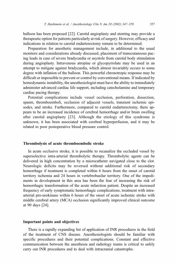
balloon has been proposed [22]. Carotid angioplasty and stenting may provide a
therapeutic option for patients particularly at risk of surgery. However, efficacy and
indications in relation to carotid endarterectomy remain to be determined.
Preparation for anesthetic management include, in additional to the usual
monitors and considerations already discussed, placement of transcutaneous pac-
ing leads in case of severe bradycardia or asystole from carotid body stimulation
during angioplasty. Intravenous atropine or glycopyrrolate may be used in an
attempt to mitigate against bradycardia, which almost invariably occurs to some
degree with inflation of the balloon. This powerful chronotropic response may be
difficult or impossible to prevent or control by conventional means. If indicated by
hemodynamic instability, the anesthesiologist must have the ability to immediately
administer advanced cardiac life support, including catecholamine and temporary
cardiac pacing therapy.
Potential complications include vessel occlusion, perforation, dissection,
spasm, thromboemboli, occlusion of adjacent vessels, transient ischemic epi-
sodes, and stroke. Furthermore, compared to carotid endarterectomy, there ap-
pears to be an increased incidence of cerebral hemorrhage and/or brain swelling
after carotid angioplasty [23]. Although the etiology of this syndrome is
unknown, it has been associated with cerebral hyperperfusion, and it may be
related to poor postoperative blood pressure control.
Thrombolysis of acute thromboembolic stroke
In acute occlusive stroke, it is possible to recanalize the occluded vessel by
superselective intra-arterial thrombolytic therapy. Thrombolytic agents can be
delivered in high concentration by a microcatheter navigated close to the clot.
Neurologic deficits may be reversed without additional risk of secondary
hemorrhage if treatment is completed within 6 hours from the onset of carotid
territory ischemia and 24 hours in vertebrobasilar territory. One of the impedi-
ments in development in this area has been the fear of increasing the risk of
hemorrhagic transformation of the acute infarction patient. Despite an increased
frequency of early symptomatic hemorrhagic complications, treatment with intra-
arterial pro-urokinase within 6 hours of the onset of acute ischemic stroke with
middle cerebral artery (MCA) occlusion significantly improved clinical outcome
at 90 days [24].
Important points and objectives
There is a rapidly expanding list of application of INR procedures in the field
of the treatment of CNS disease. Anesthesiologists should be familiar with
specific procedures and their potential complications. Constant and effective
communication between the anesthesia and radiology teams is critical to safely
carry out INR procedures and to deal with intracranial catastrophe.
T. Hashimoto et al. / Anesthesiology Clin N Am 20 (2002) 347–359 357

Acknowledgments
The authors wish to thank Broderick Belenson, Mark Espinosa, Sabrina Larson,
and Gaurab Basu for assistance in preparation of the manuscript; Van V. Halbach,
MD, and Christopher F. Dowd, MD, John Pile-Spellman, MD, Lawrence Litt, MD,
PhD, and Nancy J. Quinnine, RN, for development of clinical protocols discussed
herein; members of the UCSF Center for Stroke and Cerebrovascular Disease,
UCSF Center for Cerebrovascular Research, and the Columbia University AVM
Study Group for continued support.
References
[1] Pile-Spellman J. Endovascular therapeutic neuroradiology. In: Taveras JM, editor. Neuroradiol-
ogy. 3rd edition. Baltimore: Williams & Wilkins; 1996. p. 1045–179.
[2] Strother CM. Interventional neuroradiology. AJNR Am J Neuroradiol 2000;21:19–24.
[3] YoungWL, Pile-Spellman J. Anesthetic considerations for interventional neuroradiology (review).
Anesthesiology 1994;80:427–56.
[4] Young WL, Pile-Spellman J, Hacein-Bey L, et al. Invasive neuroradiologic procedures for
cerebrovascular abnormalities: anesthetic considerations. Anesth Clinics North Am 1997;15
(3):631–53.
[5] Joint Writing Group of the Technology Assessment Committee American Society of Interven-
tional and Therapeutic Neuroradiology; Joint Section on Cerebrovascular Neurosurgery a
Section of the American Association of Neurological Surgeons and Congress of Neurological
Surgeons; Section of Stroke and the Section of Interventional Neurology of the American
Academy of Neurology. Reporting terminology for brain arteriovenous malformation clinical
and radiographic features for use in clinical trials. Stroke 2001;32:1430–42.
[6] Ravussin P, Tempelhoff R, Modica PA, et al. Propofol vs. thiopental-isoflurane for neurosurgical
anesthesia: comparison of hemodynamics, CSF pressure, and recovery. J Neurosurg Anesth
1991;3:85–95.
[7] Tcheng JE. Platelet integrin glycoprotein IIb/IIIa inhibitors: opportunities and challenges.
J Invasive Cardiol 1996;8:8B–14B.
[8] Rauch RA, Vinuela F, Dion J, et al. Preembolization functional evaluation in brain arteriovenous
malformations: the ability of superselective amytal test to predict neurologic dysfunction before
embolization. AJNR Am J Neuroradiol 1992;13:309–14.
[9] Hashimoto T, Young WL, Aagaard BD, et al. Adenosine-induced ventricular asystole to induce
transient profound systemic hypotension in patients undergoing endovascular therapy. Dose-
response characteristics. Anesthesiology 2000;93:998–1001.
[10] Pile-Spellman J, Young WL, Joshi S, et al. Adenosine-induced cardiac pause for endovascular
embolization of cerebral arteriovenous malformations: technical case report. Neurosurgery
1999;44:881–6; discussion 886–7.
[11] Vinuela F, Dion JE, Duckwiler G, et al. Combined endovascular embolization and surgery in the
management of cerebral arteriovenous malformations: experience with 101 cases. J Neurosurg
1991;75:856–64.
[12] Debrun GM, Vinuela F, Fox AJ, et al. Indications for treatment and classification of 132 carotid-
cavernous fistulas. Neurosurgery 1988;22:285–9.
[13] Setton A, Berenstein A. Interventional neuroradiology. Curr Opin Neurol Neurosurg 1992;
5:870–80.
[14] Marshall RS, Lazar RM, Mohr JP, et al. Higher cerebral function and hemispheral blood flow
during awake carotid artery balloon test occlusions. J Neurol Neurosurg Psychiatry 1999;66:
734–8.
T. Hashimoto et al. / Anesthesiology Clin N Am 20 (2002) 347–359358

[15] Marshall RS, Lazar RM, Pile-Spellman J, et al. Recovery of brain function during induced
cerebral hypoperfusion. Brain 2001;124:1208–17.
[16] Higashida RT, Halbach VV, Dowd CF, et al. Intracranial aneurysms: interventional neurovascular
treatment with detachable balloons—results in 215 cases. Radiology 1991;178:663–70.
[17] Newell DW, Eskridge JM, Mayberg MR, et al. Angioplasty for the treatment of symptomatic
vasospasm following subarachnoid hemorrhage. J Neurosurg 1989;71:654–60.
[18] Fogarty-Mack P, Pile-Spellman J, Hacein-Bey L, et al. Superselective intraarterial papaverine
administration: effect on regional cerebral blood flow in patients with arteriovenous malforma-
tions. J Neurosurg 1996;85:395–402.
[19] Roberts JT, Pile-Spellman J, Joseph M, et al. A patient with massive oral-facial venous malfor-
mation. J Clin Anesth 1991;3:76–9.
[20] Higashida RT, Tsai FY, Halbach VV, et al. Transluminal angioplasty for atherosclerotic disease of
the vertebral and basilar arteries. J Neurosurg 1993;78:192–8.
[21] Tsai FY, Matovich V, Hieshima G, et al. Percutaneous transluminal angioplasty of the carotid
artery. AJNR Am J Neuroradiol 1986;7:349–58.
[22] Theron J, Courtheoux P, Alachkar F, et al. New triple coaxial catheter system for carotid
angioplasty with cerebral protection [followed with Commentary by Ferguson R: Getting it right
the first time, p. 875–7]. AJNR Am J Neuroradiol 1990;11:869–74.
[23] Meyers PM, Higashida RT, Phatouros CC, et al. Cerebral hyperperfusion syndrome after percu-
taneous transluminal stenting of the craniocervical arteries [In Process Citation]. Neurosurgery
2000;47:335–43; discussion 343–5.
[24] Furlan A, Higashida R, Wechsler L, et al. Intra-arterial prourokinase for acute ischemic stroke.
The PROACT II study: a randomized controlled trial. Prolyse in Acute Cerebral Thromboem-
bolism. JAMA 1999;282:2003–11.
T. Hashimoto et al. / Anesthesiology Clin N Am 20 (2002) 347–359 359

CommentaryCommentaire
It is time to clear the confusion about the utility ofsteroids in cases of acute spinal cord injury. A com-mittee of Canadian neurosurgical and orthopedic
spine specialists, emergency physicians and physiatrists(listed at the end of the article) has reviewed the evidenceand concluded that high-dose methylprednisolone infu-sion is not an evidence-based standard of care for patientswith such an injury.1
The consequences of a spinal cord injury are often deva-stating, and any possibility of mitigating neurologic loss is attractive. To this end, management of acute spinal cord in-juries has included the use of steroids for the past 30 years,based in large part on physiological hypotheses with limitedclinical support.2,3 Mechanical injury to the spinal cord initi-ates a cascade of secondary events that include ischemia, in-flammation and calcium-mediated cell injury. Animal ex-periments have shown that methylprednisolone exhibitspotential neuroprotective effects through its inhibition oflipid peroxidation and calcium influx and through its anti-inflammatory effects.4,5 Three well-designed, large, random-ized clinical trials (the National Acute Spinal Cord InjuryStudies [NASCIS I, II and III]) examined the effect of steroidadministration in patients with acute spinal cord injury.6–11
NASCIS I examined the change in motor function inspecific muscles and changes in light touch and pinpricksensation from baseline.6,7 The study detected no benefitfrom methylprednisolone, but the dose was considered tobe below the therapeutic threshold determined from ani-mal experiments. Therefore, NASCIS II used a muchhigher dose, and patients were randomly assigned to re-ceive a 24-hour infusion of methylprednisolone, naloxoneor placebo within 12 hours after acute spinal cord injury.8,9
Again, there was no benefit overall in the methylpred-nisolone group; however, post hoc analyses detected a smallgain in the total motor and sensory score in a subgroup ofpatients who had received the drug within 8 hours aftertheir injury. As a result, this 24-hour, high-dose methyl-prednisolone infusion, if started within 8 hours after injury,quickly became an implied standard of care despite consid-erable criticism of the validity of such a post hoc analysis.
Subsequent clinical trials have provided conflicting evi-dence about steroid treatment in acute spinal cord injury. AJapanese study attempted to replicate the results seen in the8-hour subgroup from NASCIS II and reported improvedfunction at 6 months in a larger number of muscles and
sensory dermatomes among subjects who received high-dose methylprednisolone infusion than among those whoreceived only low doses of the drug or no drug.12 However,the study lacked detail about randomization and outcomemeasures, and it included only 74% of the enrolled subjectsin the outcome analysis. Conversely, an underpoweredprospective randomized trial that used a methylpred-nisolone regimen similar to that used in NASCIS II foundno improvement in motor and sensory scores at 1 year.13,14
NASCIS III compared a 48-hour infusion of methylpred-nisolone with a 24-hour infusion started within 8 hours af-ter injury and found no benefit from extending the infusionbeyond 24 hours. Again, only post hoc analysis showed abenefit from extending the infusion to 48 hours when treat-ment was started between 3 and 8 hours after injury. Noother study has verified the primary outcome of 48 hoursversus 24 hours or the post hoc conclusion of benefit fromstarting treatment between 3 and 8 hours after injury.
A meta-analysis of all of the trials concluded, on the basisof the controversial subgroup post hoc analyses in NASCISII and III and the data from the Japanese study, that a 24-hour high-dose methylprednisolone infusion within 8 hoursafter injury is efficacious.15 Despite this meta-analysis, theefficacy of such a regimen remains uncertain and will re-quire further study. The controversy about the post hocanalyses of NASCIS data continues,16–23 and unfortunatelythe studies that could have clarified the efficacy of such aregimen have lacked the rigour to do so.
Steroid therapy is not without risk. Most patients withacute spinal cord injury are treated in intensive care units,have polytrauma, have impaired lung capacity and are vul-nerable to sepsis. In all 3 NASCIS studies and other,smaller studies, the incidence of sepsis and pneumonia washigher in the high-dose methylprednisolone groups than inthe placebo or other treatment groups;6–11,24–26 the differ-ences were not significant except in NASCIS III. Hyper-glycemia and gastrointestinal complications were alsoreported following high-dose methylprednisolone treat-ment.13,24 Therefore, it has been proposed that, withoutcompelling evidence for its efficacy, methylprednisoloneshould be used with caution and may even be harmful, par-ticularly if infusion goes beyond 24 hours.17
The cost of a 24-hour methylprednisolone infusion is notprohibitive, and a gain of antigravity strength in one ormore muscles below a spinal segment can provide an impor-
Methylprednisolone for acute spinal cord injury: not a standard of care
Herman Hugenholtz
CMAJ • APR. 29, 2003; 168 (9) 1145
© 2003 Canadian Medical Association or its licensors
tant functional gain, especially for patients with cervicalspinal cord injuries. Therefore, even the small improvementobserved in the NASCIS subgroups could be viewed as abenefit in cases of complete or incomplete cervical cord in-jury. Despite the risk of complications and as long as theoutcomes in the NASCIS subgroups remain a possibility,physicians may still opt to administer a high-dose methyl-prednisolone infusion within 8 hours after injury. However,they should no longer feel compelled to do so. Physicianswho conduct the initial triage and resuscitation of patientswith acute spinal cord injury should consult their specialistcolleagues who will be continuing the care of these patientsregarding their preference for methylprednisolone infusion.
The Canadian Neurosurgical Society, the CanadianSpine Society and the Canadian Association of EmergencyPhysicians have adopted the committee’s recommendationthat a high-dose, 24-hour infusion of methylprednisolonestarted within 8 hours after an acute closed spinal cord in-jury is not a standard treatment nor a guideline for treat-ment but, rather, a treatment option, for which there is veryweak level II and III evidence.27
References
1. Hugenholtz H, Cass DE, Dvorak MF, Fewer DH, Fox RJ, Izukawa DMS, etal. High-dose methylprednisolone for acute closed spinal cord injury — onlya treatment option. Can J Neurol Sci 2002;29(3):227-35.
2. Tator CH. Acute spinal cord injury: a review of recent studies of treatmentand pathophysiology. CMAJ 1972;107(2):143-5.
3. Green BA, Kahn T, Klose KJ. A comparative study of steroid therapy in acuteexperimental spinal cord injury. Surg Neurol 1980;13(2):91-7.
4. Braughler JM, Hall ED. Lactate and pyruvate metabolism in injured catspinal cord before and after a single large intravenous dose of methylpred-nisolone. J Neurosurg 1983;59:256-61.
5. Hall ED. The neuroprotective pharmacology of methylprednisolone. J Neu-rosurg 1992;76:13-22.
6. Bracken MB, Collins WF, Freeman DF, Shepard MJ, Wagner FW, SiltenRM, et al. Efficacy of methylprednisolone in acute spinal cord injury. JAMA1984;251(1):45-52.
7. Bracken MB, Shepard MJ, Hellenbrand KG, Collins WF, Leo LS, FreemanDF, et al. Methylprednisolone and neurological function 1 year after spinalcord injury. Results of the National Acute Spinal Cord Injury Study. J Neuro-surg 1985;63(5):704-13.
8. Bracken MB, Shepard MJ, Collins WF, Holford TR, Young W, Baskin DS,et al. A randomized, controlled trial of methylprednisolone or naloxone in thetreatment of acute spinal-cord injury. Results of the Second National AcuteSpinal Cord Injury Study. N Engl J Med 1990;322(20):1405-11.
9. Bracken MB, Shepard MJ, Collins WF Jr, Holford TR, Baskin DS, EisenbergHM, et al. Methylprednisolone or naloxone treatment after acute spinal cordinjury: 1-year follow-up data. Results of the second National Acute SpinalCord Injury Study. J Neurosurg 1992;76(1):23-31.
10. Bracken MB, Shepard MJ, Holford TR, Leo-Summers L, Aldrich EF, FazlM, et al. Administration of methylprednisolone for 24 or 48 hours or tirilazadmesylate for 48 hours in the treatment of acute spinal cord injury. Results ofthe Third National Acute Spinal Cord Injury Randomized Controlled Trial.National Acute Spinal Cord Injury Study. JAMA 1997;277(20):1597-604.
11. Bracken MB, Shepard MJ, Holford TR, Leo-Summers L, Aldrich EF, Fazl M,et al. Methylprednisolone or tirilazad mesylate administration after acutespinal cord injury: 1-year follow up. Results of the third National Acute Spinal
Cord Injury randomized controlled trial. J Neurosurg 1998;89(5):699-706.12. Otani K, Abe H, Kadoya S, Nakagawa H, Ikata T, Tominaga S, et al. Benefi-
cial effect of methylprednisolone sodium succinate in the treatment of acutespinal cord injury. Sekitsui Sekizui J 1996;7:633-47.
13. Petitjean ME, Pointillart V, Dixmerias F, Wiart L, Sztark F, Lassie P, et al.Medical treatment of spinal cord injury in the acute stage. Ann Fr Anesth Re-anim 1998;17(2):114-22.
14. American Spinal Cord Injury Association. Standard for neurological classificationof spinal cord patients. Chicago: The Association; 1992.
15. Bracken MB. Pharmacological intervention for acute spinal cord injury[Cochrane review]. In: The Cochrane Library; Issue 1, 2001. Oxford: UpdateSoftware.
16. Coleman WP, Benzel D, Cahill DW, Ducker T, Geisler F, Green B, et al. Acritical appraisal of the reporting of the National Acute Spinal Cord InjuryStudies (II and III) of methylprednisolone in acute spinal cord injury. J SpinalDisord 2000;13(3):185-99.
17. Hurlbert RJ. Methylprednisolone for acute spinal cord injury: an inappropri-ate standard of care. J Neurosurg 2000;93(Suppl 1):1-7.
18. Nesathurai S. Steroids and spinal cord injury: revisiting the NASCIS 2 andNASCIS 3 trials. J Trauma 1998;45(6):1088-93.
19. Short DJ, El Masry WS, Jones PW. High dose methylprednisolone in themanagement of acute spinal cord injury — a systematic review from a clinicalperspective [review]. Spinal Cord 2000;38(5):273-86.
20. Section on disorders of the spine and peripheral nerves of the American Asso-ciation of Neurological Surgeons and the Congress of Neurological Sur-geons. Guidelines for the management of acute cervical spine and spinal cordinjuries. Neurosurgery 2002;50(Suppl 3):S67-72.
21. Oxman AD, Guyatt GH. A consumer’s guide to subgroup analyses. Ann In-tern Med 1992;116:78-84.
22. Fehlings MG. Summary statement: the use of methylprednisolone in acutespinal cord injury. Spine 2001;26(Suppl 24):S55.
23. Fehlings MG. Recommendations regarding the use of methylprednisolone inacute spinal cord injury: making sense out of the controversy [editorial]. Spine2001;26(Suppl 24):S56-7.
24. Matsumoto T, Tamaki T, Kawakami M, Yoshida M, Ando M, Yamada H. Earlycomplications of high-dose methylprednisolone sodium succinate treatment inthe follow-up of acute cervical spinal cord injury. Spine 2001;26(4):426-30.
25. Galandiuk S, Raque G, Appel S, Polk HC Jr. The two-edged sword of large-dosesteroids for spinal cord trauma. Ann Surg 1993;218(4):419-25; discussion 425-7.
26. Gerndt SJ, Rodriguez JL, Pawlik JW, Taheri PA, Wahl WL, Micheals AJ, etal. Consequences of high-dose steroid therapy for acute spinal cord injury. JTrauma 1997;42(2):279-84.
27. Canadian Task Force on the Periodic Health Examination. The periodichealth examination. 1. Introduction. CMAJ 1986;134(7):721-9.
Commentaire
1146 JAMC • 29 AVR. 2003; 168 (9)
This article has been peer reviewed.
Competing interests: None declared.
Dr. Hugenholtz is with the Division of Neurosurgery, Queen Elizabeth II HealthSciences Centre, Halifax, NS.
Correspondence to: Dr. Herman Hugenholtz, New HalifaxInfirmary, Rm. 3808, 1796 Summer St., Halifax NS B3H 3A7; fax 902 473-8912
Members of the Committee of the Canadian Spine Society andthe Canadian Neurosurgical Society to Review the Role ofMethylprednisolone in Acute Spinal Cord Injury: HermanHugenholtz (chair), Division of Neurosurgery, Queen Elizabeth IIHealth Sciences Centre, Halifax, NS; Nirmala D. Bharatwal,Toronto Rehabilitation Institute, Toronto, Ont.; Dan E. Cass,Director of Emergency Services, St. Michael’s Hospital, Toronto,Ont.; Marcel F. Dvorak, Medical Director, Combined SpineProgram, Vancouver Hospital and Health Sciences Centre,Vancouver, BC; Derek Fewer, Section of Neurosurgery, HealthSciences Centre, Winnipeg, Man.; Richard J. Fox, Department ofNeurosurgery, Walter C. Mackenzie Health Science Centre,University Hospital, Edmonton, Alta.; Dennis M.S. Izukawa,Department of Neurosurgery, Trillium Health Centre,Mississauga, Ont.; Joel Lexchin, Emergency Department,University Health Network, Toronto, Ont.; Christine Short, NovaScotia Rehabilitation Centre, Halifax, NS; and Sagun Tuli,Department of Neurosurgery, Brigham and Women’s Hospital,Boston, Mass.

CC
Neuroprotection in traumatic br
ain injury: a complex struggleagainst the biology of natureJoost W. Schouten
Purpose of review
Translating the efficacy of neuroprotective agents in
experimental traumatic brain injury to clinical benefit has
proven an extremely complex and, to date, unsuccessful
undertaking. The focus of this review is on neuroprotective
agents that have recently been evaluated in clinical trials
and are currently under clinical evaluation, as well as on
those that appear promising and are likely to undergo
clinical evaluation in the near future.
Recent findings
Excitatory neurotransmitter blockage and magnesium have
recently been evaluated in phase III clinical trials, but
showed no neuroprotective efficacy. Cyclosporin A,
erythropoietin, progesterone and bradykinin antagonists are
currently under clinical investigation, and appear promising.
Summary
Traumatic brain injury is a complex disease, and
development of clinically effective neuroprotective agents is
a difficult task. Experimental traumatic brain injury has
provided numerous promising compounds, but to date
these have not been translated into successful clinical trials.
Continued research efforts are required to identify and test
new neuroprotective agents, to develop a better
understanding of the sequential activity of pathophysiologic
mechanisms, and to improve the design and analysis of
clinical trials, thereby optimizing chances for showing
benefit in future clinical trials.
Keywords
clinical trial, head injury, neuroprotection, neurotrauma,
traumatic brain injury
Curr Opin Crit Care 13:134–142. � 2007 Lippincott Williams & Wilkins.
Department of Neurosurgery, Erasmus Medical Center, Rotterdam, TheNetherlands
Correspondence to Joost W. Schouten, MD, Department of Neurosurgery, ErasmusMedical Center, Postbus 2040, 3000 CA, Rotterdam, The NetherlandsE-mail: [email protected]
Current Opinion in Critical Care 2007, 13:134–142
Abbreviations
CSF c
opyrigopyrig
134
erebrospinal fluid
iNOS in ducible nitric oxide synthase NMDA N -methyl-D-aspartic acid NOS n itric oxide synthase TBI tr aumatic brain injury� 2007 Lippincott Williams & Wilkins1070-5295
ht © Lippincott Williams & Wilkins. Unauthoht © Lippincott Williams & Wilkins. Unautho
IntroductionTraumatic brain injury (TBI) is a major cause of death
and disability worldwide, leading to immense personal
suffering to victims and relatives, and high costs to
society. Injuries are the leading cause of death between
the ages of 15 and 44, and head trauma accounts for the
majority of all trauma deaths. Today, at least 11.5 million
people live with TBI-related disability, impairment,
complaint or handicap in Europe (6.2 million) and the
USA (5.3 million) alone [1��,2].
Depending on the severity of injury, the medical
management of brain-injured patients currently
includes specialized prehospital care, clinical (inten-
sive) care, and, for some, long-term rehabilitation, but
lacks clinically proven effective management with
neuroprotective agents to limit pathophysiologic
cascades or enhance repair. The enormous burden of
TBI, however, clearly supports the need for such
neuroprotective agents. Translating promising exper-
imental results into clinical benefit has proven an
extremely complex issue. First, although many patho-
physiologic cascades inducing secondary damage have
been identified, it remains uncertain which of these are
active in individual patients and at what time after
injury. Moreover, many pathways may initially have
detrimental effects, but at later stages can be protective.
Second, clinical trials have suffered from inadequacy
in their design and analysis, not in the least part due
to heterogeneity of the population and variability in
treatment approaches [3].
Rationale for treatment: primary andsecondary injuryBrain trauma results in brain damage and dysfunction
from both primary injury (due to biomechanical effects)
and subsequent secondary damage due to activation of
pathophysiologic cascades. These are further aggravated
by secondary insults. Early detection of such secondary
insults, including intracranial insults (e.g. mass lesions,
increased intracranial pressure) and systemic insults
(e.g. hypoxia, hypotension), followed by appropriate
intervention currently forms the basis of clinical
management [4].
Secondary damage consists of seemingly innumerable
complex biochemical and cellular pathways that influ-
ence progression of the primary injury. The primary goal
of neuroprotection is to prevent and/or reduce secondary
rized reproduction of this article is prohibited.rized reproduction of this article is prohibited.

C
Neuroprotection in traumatic brain injury Schouten 135
damage and to enhance repair [5]. Over the past decades
our understanding of the pathophysiology of TBI has
greatly increased and based on this understanding
numerous pharmacological therapies have been
developed, tested and proven effective in the treatment
of experimental TBI. To date, however, promising
experimental results have not been translated into
successful clinical trials, and hence the cornerstone of
management of TBI patients remains the prevention of
initial injury and the minimization or reversal of second-
ary insults. The excitement about the new knowledge of
the neurobiology and neuropharmacology of TBI should
not detract from the absolute importance of correcting
hypoxia, hypotension, raised intracranial pressure and
other causes of secondary ischemic insult [6,7]. On the
other hand, we should not be discouraged by negative
results and difficulties in previous clinical trials, but
continue our search for effective neuroprotective drugs
for TBI patients in order to further improve outcome of
this devastating disease.
Neuroprotective strategiesThe complex pathobiology of TBI offers numerous tar-
gets for potential neuroprotective agents. Many of these
have been or will be investigated in experimental models
of TBI (Table 1 [8��,9–13]). The few that made it into
opyright © Lippincott Williams & Wilkins. Unauth
Table 1 Neuroprotective strategies evaluated in experimental traum
Pharmacological target Remarks
Excitatory amino acids Numerous compounds have been evadifferent pharmacological profiles (e
Calcium channels Extensively studied, also in clinical TBinjury seems to limit further clinical
Scavenging oxygen radicals Tirilazad Mesylate, PEG-SOD and Lubcompounds are at least promising i
Inflammation A double-edged sword in TBI, both dis a high potential target for neuropand nitroxides.
Caspases Caspases are important enzymes in amatter of debate whether apoptosis
Calpains Calcium-dependent proteases involveTBI reduce damage to fiber tracts,
Hormonal treatment Progesterone is currently being evaluapast. Experimental compounds attrathyrotropin-releasing hormone and t
Neurotransmission Widespread changes in neurotransmiserotonin, histamine, g-aminobutyricpotential interest following TBI. Cogwhich might benefit from this appro
Neurotrophic factors These growth/survival factors effectiveTBI. Many questions about dosage,
Coagulation Recombinant human factor VII has befollowing TBI, relate to outcome, antreatment of microvascular thrombo
Anticonvulsants Seizures occur frequently following TBacute administration can be neurop
Immunophilin ligands Cyclosporin A is currently being evaluinvestigation.
Minocycline Minocycline is a broad-spectrum antibOthers In experimental research additional ho
improvement of axonal outgrowth aclinical trial in pediatric TBI has bee
TBI, traumatic brain injury. Neuroprotective strategies are discussed more e
clinical trials join a growing list of neuroprotective agents
without proven clinical benefit (Table 2 [14–24]). The
focus of this review is on neuroprotective agents that
have recently been evaluated in clinical trials and are
currently under clinical evaluation, as well as on those
that appear promising and are likely to undergo clinical
evaluation in the near future.
Excitatory neurotransmitter antagonismDisturbances in neurotransmitter concentration occur
frequently following TBI. Excitotoxicity refers to an
excessive release of excitatory neurotransmitters
(primarily glutamate) initiating various pathophysiologic
processes including excessive calcium influx in neurons,
resulting in neuronal cell death [10]. High concentrations
of extracellular glutamate have been demonstrated in
both experimental models and clinical patients with TBI.
Experimental research has elucidated many aspects of
excitotoxicity and identified a number of glutamate
antagonists acting either pre- or postsynaptically on
N-methyl-D-aspartic acid (NMDA), a-amino-3-hydroxy-
5-methyl-4-isoxazolyl-propionic acid (AMPA)/kainate or
metabotropic receptors, in a competitive, noncompetitive
or modulating way. However, glutamate receptors are of
utmost importance to normal functioning, so antagonism
of excessive excitotoxic activity must be achieved
orized reproduction of this article is prohibited.
atic brain injury
luated and reviewed elsewhere [8��,10]. New compounds with.g. memantine) require further experimental evaluation.I (Nimodipine and SNX-111); the short time frame followinguse.eluzole have been clinically evaluated; many new
n experimental TBI.etrimental and beneficial. The massive inflammatory responserotection, with special attention for NO inhibitors, nitrones
poptotic cell death known to occur following TBI. It is however still ais a good or bad thing compared to necrosis following TBI.
d in cytoskeletal remodeling. Calpain inhibitors in experimentaland therefore are of major interest in axonal injury.ted in a clinical trial. Steroids have been extensively studied in thecting a lot of attention are dehydroepiandrosterone,heir analogs.tters occur following TBI. All compounds interfering in cathecholamine,
acid (GABA) and acetylcholine metabolism are therefore ofnitive problems and depression frequently present following TBI,ach, although a rationale for more acute administration exists.ly reduce apoptosis and improve functional outcome in experimentaltime-window and route of administration remain to be answered.
en evaluated in a clinical trial. Coagulation disorders are commond will be a hot topic for future research. Controversies regardingsis and progressive hemorrhagic contusions require attention [11].I, and anticonvulsants may reduce early seizures. In addition,
rotective [12,13].ated in a clinical trial, other compounds are under experimental
iotic, shown to be neuroprotective in experimental studies.t topics far from translation into clinical trials are neurogenesis,nd stem-cell transplantation, although for the latter a smalln initiated.
xtensively in [8��,9].

Copyright © Lippincott Williams & Wilkins. Unauthorized reproduction of this article is prohibited.
136 Neuroscience
Ta
ble
2D
isa
pp
oin
tin
gre
su
lts
of
tria
lso
nn
eu
rop
rote
cti
ve
ag
en
tsin
tra
um
ati
cb
rain
inju
ry
Stu
dy
and
agen
tS
tud
yp
op
ulat
ion
No
.o
fp
atie
nts
Sta
rto
ftr
eatm
ent
Yea
ro
fst
udy
Sta
tus
Pub
lishe
dR
esul
t
Bra
dyk
inin
inhi
biti
on
Bra
dyc
or/
CP
-01
27�
GC
S3
–8
13
9�
12
h1
99
6C
om
ple
ted
[14
]1
2%
imp
rove
men
tin
favo
rab
leo
utco
me
(P¼
0.2
6)
Cal
cium
-med
iate
dd
amag
eH
ITI
Nim
od
ipin
e�N
ot
ob
eyin
gco
mm
and
s3
51
24
h1
98
7–
19
89
Co
mp
lete
d[1
5]
No
sig
nific
ant
effe
ct
HIT
IIN
imo
dip
ine�
No
to
bey
ing
com
man
ds
85
21
2h
of
not
ob
eyin
gco
mm
and
sw
ithin
24
ho
fin
jury
19
89
–1
99
1C
om
ple
ted
[16
]N
osi
gni
fican
tef
fect
ove
rall
po
pul
atio
n
HIT
IIIN
imo
dip
ine
Tra
umat
icsu
bar
achn
oid
hem
orr
hag
e1
23
�1
2h
19
94
Co
mp
lete
d[1
7]
Sig
nific
ant
red
uctio
nin
unfa
vora
ble
out
com
eH
ITin
trav
eno
usN
imo
dip
ine
GC
S<
15þ
trau
mat
icsu
bar
achn
oid
hem
orr
hag
e5
92
�1
2h
19
97
–1
99
9C
om
ple
ted
No
No
sig
nific
ant
effe
ct
Par
keD
avis
/SN
X-1
11
GC
S4
–8
23
7�
12
h1
99
7–
19
98
Ter
min
ated
No
Hig
her
mo
rtal
ityG
luta
mat
eex
cito
toxi
city
Elip
rod
ilst
udy
GC
S4
–8
45
2�
12
h1
99
3–
19
95
Co
mp
lete
dN
oN
osi
gni
fican
tef
fect
rep
ort
edS
elfo
tel�
GC
S4
–8
69
3�
8h
and
with
in4
ho
fad
mis
sion
19
94
–1
99
6T
erm
inat
ed[1
8]
No
sig
nific
ant
effe
ct
Cer
esta
t/A
ptig
anely
GC
S4
–8
;G
CS
3if
pup
ilsre
activ
e5
32
�8
h1
99
6–
19
97
Ter
min
ated
No
No
sig
nific
ant
effe
ct
Sap
hir/� D
-CP
P-e
neN
ot
ob
eyin
gco
mm
and
s,�
one
reac
tive
pup
il9
24
�1
2h
19
95
–1
99
7C
om
ple
ted
No
No
sig
nific
ant
effe
ctre
po
rted
Pfiz
er/C
P-1
01
60
6G
CS
4–
84
16
�8
h1
99
7–
20
00
Co
mp
lete
dN
oN
osi
gni
fican
tef
fect
Lip
idp
ero
xid
atio
n/fr
eera
dic
ald
amag
eP
EG
-SO
D�
GC
S�
81
56
2W
ithin
8h
19
93
–1
99
5C
om
ple
ted
[19
],in
par
tN
osi
gni
fican
td
iffer
ence
Tiri
laza
dd
om
estic
tria
l�G
CS�
8,
70
%;
GC
S9
–1
2,
30
%1
15
5�
4h
19
91
–1
99
4T
erm
inat
edN
oN
osi
gni
fican
tef
fect
rep
ort
ed
Tiri
laza
din
tern
atio
naltr
ial�
GC
S�
8,
85
%;
GC
S9
–1
2,
15
%1
12
0�
4h
19
92
–1
99
4C
om
ple
ted
[20
]N
osi
gni
fican
tef
fect
Ste
roid
sT
riam
cini
lone
ster
oid
tria
lS
ever
ehe
adin
jury
,no
tfu
rthe
rd
efine
d3
96
�4
h1
98
5–
19
90
Co
mp
lete
d[2
1]
No
sig
nific
ant
effe
ct
Dex
amet
haso
nem
ega
do
setr
ial
GC
S<
13
30
0�
3h
19
86
-19
89
Co
mp
lete
d[2
2]
No
sig
nific
ant
effe
ct
CR
AS
H/s
tero
idtr
ialy
GC
S<
15
10
00
8�
8h
20
00
–2
00
4T
erm
inat
ed[2
3,2
4]
Hig
her
mo
rtal
ityM
ultip
leac
tions
Pha
rmo
s/d
exan
abin
oly
Mo
tor
sco
re2
–5þ
CT
abno
rmal
ities
86
1�
6h
20
00
–2
00
4C
om
ple
ted
[27� ]
No
sig
nific
ant
effe
ct
Mag
nesi
umG
CS
3–
12
and
/or
intr
acra
nial
surg
ery
49
9�
8h
19
98
–2
00
4C
om
ple
ted
[32� ]
Po
ore
ro
utco
me
atlo
wd
ose
;hi
ghe
rm
ort
ality
athi
gh
do
se
CT
,co
mp
uted
tom
og
rap
hy;
GC
S,
Gla
sgo
wC
om
aS
core
.�
Cur
rent
lyin
clud
edin
the
IMP
AC
Td
atab
ase.
yP
lann
edfo
rin
clus
ion
inth
eIM
PA
CT
dat
abas
e.

C
Neuroprotection in traumatic brain injury Schouten 137
without interference in normal function [25]. Some
highly neuroprotective NMDA antagonists have not
been evaluated in clinical trials because of concerns of
psychotropic side effects whereas for other compounds
trials were terminated prematurely due to excess
mortality in concomitant stroke trials. Recently,
Traxoprodil, a second-generation NMDA antagonist that
selectively targets NMDA receptors containing the
NR2B subunit, has been evaluated in a clinical trial.
Traxoprodil treatment was well tolerated and, although
not statistically significant, resulted in increased
favorable outcome and reduced mortality, which was
more pronounced in the more severe subset of patients
[26]. Dexanabinol is a synthetic cannabinoid devoid of
psychotropic activity, but with strong neuroprotective
potential due to antioxidant, anti-inflammatory and
antiexcitotoxic properties. This compound was recently
evaluated in a phase III trial and found safe, but not
efficacious in the treatment of TBI [27�]. Efficacy of
blocking excitotoxic responses following TBI as well as
other insults to the central nervous system, to date,
remains unproven [28,29]. Termination of trials before
definitive evidence could be obtained, incomplete pub-
lication of data and underpowered studies limit definitive
conclusions for this group of neuroprotective drugs.
New drugs with different pharmacological profiles are
currently under investigation in experimental TBI and
show promising results. Translation into clinical trials
should only occur in well designed trials based on what
we have learned from previous trials.
MagnesiumMagnesium plays an important role in normal cellular
functioning, and has demonstrated neuroprotective
properties in experimental studies in models of cerebral
ischemia as well as TBI. Magnesium treatment results in
reduction of cerebral edema and neuronal cell death, and
attenuated motor impairment and cognitive dysfunction
following experimental TBI [30,31]. One of the see-
mingly great advantages of magnesium, besides being
inexpensive and widely available, is its multidirectional
effect. Where other neuroprotective compounds usually
interfere with just one pathophysiological mechanism,
magnesium exerts its neuroprotective effects among
others by noncompetitive NMDA receptor blockade,
inhibition of presynaptic excitatory neurotransmitter
release, suppression of cortical spreading depression,
and blockade of voltage-gated calcium channels. Despite
the solid amount of experimental evidence concerning
the neuroprotective effects of magnesium, a recently
completed randomized double-blind trial evaluating
the efficacy of a 5-day continuous magnesium adminis-
tration in 499 patients with moderate or severe TBI
was unable to show neuroprotective effects and even
indicated a possibility of harm [32�]. The absence of
efficacy is consistent with a recently reported stroke trial
opyright © Lippincott Williams & Wilkins. Unauth
[33]. Possible statistical and methodological causes
(e.g. lack of power to detect differences) of these negative
results have not been identified. Even though magnes-
ium administration (dose, start time, duration, concen-
tration) in these studies was based on positive preclinical
data, and targeted serum levels of magnesium were
achieved, availability of magnesium in cerebrospinal fluid
(CSF) or cerebral extracellular fluid might be a concern
[34–36]. Further studies to elucidate the relationships
between total and ionized concentrations of magnesium
in serum and CSF at different times following clinical
TBI may hopefully provide an explanation for the nega-
tive results of recent clinical trials.
Mitochondrial dysfunctionMitochondria, as the centers of aerobic metabolism,
show marked dysfunction following experimental and
clinical TBI, contributing to cell death through several
mechanisms. Increased mitochondrial calcium results in
decreased ATP production and generation of reactive
oxygen species as well as increased permeability of
the inner mitochondrial membrane. The opening of the
mitochondrial permeability transition pore is responsible
for mitochondrial swelling and membrane rupture,
resulting in cell death. Mitochondrial dysfunction can
be attenuated by inhibitors of mitochondrial permeability
transition such as cyclosporin A and its derivatives [37].
Based on preclinical data cyclosporin A has been evaluated
in two phase II clinical trials, and was found to improve
cerebral perfusion pressure and cerebral metabolism, as
evaluated with microdialysis. Cyclosporin A is considered
safe in TBI patients, and its CSF pharmacokinetics in the
injured central nervous system have been elucidated,
supporting the initiative for a phase III clinical trial, which
is currently being designed [38�,39,40].
In experimental research, blockage of N-type voltage-
gated calcium channels by ziconotide (SNX-111) has
been shown to induce partial restoration of mitochondrial
function, but a clinical trial was terminated prematurely
because of increased mortality in the treatment group.
Newly developed, more selective N-type voltage-gated
calcium-channel blockers like SNX-185 have better bio-
availability, and appear neuroprotective in experimental
models, but will need additional preclinical evaluation
[41].
ErythropoietinErythropoietin is a kidney-derived cytokine regulating
hematopoiesis, and has recently been recognized as being
neuroprotective. Abundant expression of erythropoietin
and its receptor in most of the cell types in the central
nervous system exists, and in response to hypoxia or
excitotoxicity this expression is increased, suggesting a
central role in endogenous protection from deleterious
stimuli [42]. Erythropoietin has been shown to be
orized reproduction of this article is prohibited.

C
138 Neuroscience
neuroprotective in experimental models of stroke, and
following experimental TBI treatment with erythropoie-
tin leads to decreased lesion volume and improved func-
tional outcome, possibly by limiting the inflammatory
reaction [43]. Based on these experimental data clinical
trials were initiated and the safety of erythropoietin
administration in stroke patients was confirmed. A double-
blind proof-of-concept trial showed no adverse events,
and suggested improved functional outcome in erythro-
poietin-treated patients [44]. These results prompted
further clinical research, which is currently being con-
ducted as a multicenter phase II/III trial in stroke patients,
as well as an additional pharmacokinetic study evaluating
CSF erythropoietin following systemic administration
[45]. In TBI, a randomized phase II clinical trial is
currently ongoing in Wisconsin, USA. This trial focuses
primarily on moderate TBI patients, and instead of using
the Glasgow Outcome Score evaluates neuronal cell-death
markers as a primary outcome measure.
HormonesThere has been considerable debate about sex differences
in outcome following TBI, but a large meta-analysis
suggests that no differences in outcome between men
and women exist in outcome following TBI [46]. This
debate, however, together with gender difference in
treatment response and outcome in experimental TBI,
stimulated research directed at the role of sex steroids
following TBI. Both progesterone and estrogen may exert
neuroprotective effects [47]. Based on experimental
research, progesterone is thought to exert its neuroprotec-
tive effects through a variety of mechanisms including
a decrease of edema formation due to changes in the
blood–brain barrier and modulation of g-aminobutyric
acid (GABA)-ergic neurotransmission resulting in
decreased excitotoxicity. In addition progesterone inhibits
apoptosis and reduces gliosis and the posttraumatic
inflammatory response [48,49]. Allopregnanolone, a
metabolite of progesterone, has also been shown to be
neuroprotective, and might even be more effective than
progesterone [50]. Both progesterone and allopregnano-
lone improve neuronal survival and functional recovery
following experimental TBI [51].
In experimental TBI estrogen has shown to possess
antioxidant and antiapoptotic properties, and improves
cerebral blood flow. However, estrogen supplementation
in females increased mortality following experimental
brain injury [47], and most studies evaluating estrogen
used pretreatment paradigms, raising questions about the
value of estrogen in clinical TBI.
The wealth of experimental data on neuroprotective
effects of progesterone together with adequate pharma-
cokinetic studies [52] resulted in a phase II clinical trial
which concluded that no serious adverse events occurred
opyright © Lippincott Williams & Wilkins. Unautho
due to progesterone administration in TBI patients. Even
more interesting was the observation that, in moderate
TBI survivors treated with progesterone, the outcome
was better than in those treated with placebo [53]. A
large multicenter study to prove what this study suggests
has been initiated by the authors.
Bradykinin antagonistsIncreased production of kinins has been reported following
brain trauma, and their interaction with the constitutive
B2-bradykinin receptor has been shown to be important in
the development of postinjury inflammation-induced sec-
ondary damage. Specific inhibition of the B2-bradykinin
receptor is considered a promising strategy for neurorotec-
tion. Experimental data support this strategy, and recently
a phase I clinical trial to investigate the pharmacokinetics
of Anatibant, a selective potent bradykinin receptor
antagonist, was conducted and published [54]. Currently
a phase II safety study is being conducted on 500 patients
with TBI.
Nitric oxide and inhibitors of nitric oxidesynthasesNitric oxide is a key factor in the development of second-
ary injury, regulating the dilation of blood vessels and
acting as chemotoxin during inflammatory processes.
Nitric oxide is synthesized from L-arginine by the enzyme
nitric oxide synthase (NOS), of which four isoforms have
been identified. Three of these are constitutive and
one inducible [55]. The three constitutive isoforms are
neuronal NOS, endothelial NOS and mitochondrial NOS.
The fourth isoform is the inducible NOS (iNOS), which
is induced under pathological conditions [55–57].
In excess, nitric oxide is potentially neurotoxic because it
contributes to excitotoxic neuronal death, generates cyto-
toxic peroxynitrites, damages DNA directly, inhibits
DNA synthesis, inhibits mitochondrial respiration, and
has been associated with apoptotic cell death [58]. In
addition to producing nitric oxide, NOSs are also known
to produce considerable amounts of the free radicals
superoxide and hydrogen peroxide. This occurs particu-
larly when substrate levels fall below those required to
saturate the enzyme. This mechanism is called uncou-
pling of NOS [59,60]. Following TBI, nitric oxide syn-
thesis is also activated by inflammation, which is initiated
by both primary and secondary injuries. Proinflammatory
cytokines can induce iNOS, thereby promoting persist-
ent iNOS over-activation for several days after injury [61].
iNOS is mainly expressed in macrophages, microglia and
infiltrating neutrophils recruited from the blood and thus
has a substantially greater capacity to synthesize nitric
oxide than endothelial NOS [58,61]. During the course of
the pathophysiological process triggered by TBI, nitric
oxide accumulates in the brain immediately after injury,
as well as several hours or days later.
rized reproduction of this article is prohibited.

C
Neuroprotection in traumatic brain injury Schouten 139
Various studies have shown that nitric oxide and the NOS
pathways are involved, both positively and negatively, in
the secondary injury cascade following injury. These path-
ways, and the recognition of the importance of nitric oxide
in modulating regional cerebral blood flow, indicate a
promising treatment target. Overall, experimental studies
with inhibitors of NOS have shown beneficial effects, in
particular with inhibitors of neuronal NOS and iNOS [8��].
Other studies, however, failed to show benefit and a few
even show deleterious effects (see [8��] for references).
These differences may reflect differential effects of agents
investigated on the different isoforms of NOS as well as
time-dependent influences. A new drug currently under
investigation is the compound VAS203, a structural analog
of 5678-tetrahydro-l-biopterin (BH4), the endogenous
cofactor of NOS, and one of the most potent inhibitors
of NOSs discovered so far. This compound competitively
displaces BH4 from NOS and thus inhibits the formation
of nitric oxide, but does not interact with the binding site of
the substrate (L-arginine) [62]. Further, VAS203 is capable
of reducing uncoupling of NOS, with an additional effect,
the inhibition of increased superoxide production.
Translational neuroprotection:neuroprotection from bench to bedsideThe neuroprotective agents that fail in clinical trials have
all been proven effective in experimental models. One
could question whether the current models of TBI
adequately mimic human TBI. Many aspects of human
TBI, either focal or diffuse, are reflected by the different
experimental models, but experimental models cannot
reproduce the entire heterogeneous spectrum of clinical
TBI [63]. Additional concerns exist about the severity of
experimental injuries as well as ultra-early or even
pretreatment paradigms in experimental TBI. Extrapol-
ation of results obtained in animals to clinical setting will
remain problematic.
In ‘early’ TBI trials patient inclusion was primarily based
on Glasgow Coma Score on admission (Table 2). The
actual presence of the pathophysiological mechanism that
was targeted with the neuroprotective compound that was
studied, however, has hardly or never been confirmed. The
presence of certain pathophysiological mechanisms in
patients should be the basis of inclusion in a clinical trial
evaluating the compound that interferes with this
mechanism in a hopefully beneficial manner. In other
words: diffuse axonal injury patients and subdural hema-
toma patients should not be included in the same trial even
though their Glasgow Coma Scores on admission are
identical. We should select and include patients in trials
that are likely to benefit from the evaluated treatment [64].
Similarly, mild and moderate TBI may be considered
completely different diseases than severe TBI. Unsuc-
cessful clinical trials in which the subpopulation of
opyright © Lippincott Williams & Wilkins. Unauth
moderately injured patients responded to therapy (e.g.
[53]) raise the question of whether trials should not focus
more on moderate or even mild TBI patients whose
brains actually are ‘salvageable’. This population remains
poorly represented in clinical trials, but constitutes the
majority of all TBI worldwide.
To date, both clinical trials and experimental research
have focused on the evaluation of a single potential
neuroprotective compound at a time when it is con-
sidered unlikely that one ‘magic bullet’ will improve
outcome in all subtypes of TBI. Serious consideration
should be given to the possibility of combination thera-
pies in which multiple compounds are administered
sequentially, each in their own appropriate therapeutic
time window.
Another major concern in clinical trials is the uncertainty
about pharmacokinetics in the TBI patient, which are
considerably different from the normal physiological
situation. In recent phase III trials drug administration
was based on specifically designed phase II trials
[32�,40]. More detailed pharmacokinetic studies are
advocated by some, in which, in addition to the target,
serum concentrations in CSF or cerebral extracellular
fluid should be monitored [34,57]. In this development,
cerebral microdialysis is likely to become an increasingly
important monitoring tool for diagnosis (presence of a
pathophysiological mechanism in a patient), drug
monitoring (measurement of drug concentration in the
target organ) and outcome (surrogate outcome markers)
in clinical TBI [65].
OutcomePharmaceutical clinical trials in TBI, to date, have been
largely unsuccessful [66��]. To improve the quality of
future clinical trials it is imperative that all data, including
negative data, are published. Unfortunately, studies with
negative results have not always been pulished (Table 2).
In addition, trials have been prematurely terminated by
sponsors because of interim analysis or (primarily psy-
chomimetic) adverse effects in concurrent stroke trials.
The question remains whether adverse effects leading to
termination of clinical trials in stroke should have the
same consequence in TBI trials because the average TBI
patient, in contrast to the average stroke patient, is
younger and – more importantly – comatose and fully
sedated at the time of drug administration.
The primary end point in most TBI trials to date has been
the dichotomized Glasgow Outcome Score (favorable/
unfavorable). The use of this scale, together with the
hypothesis of most trials that a 10% increase in favorable
outcome is considered a positive result, has led to
negative trial results. Although reported as negative,
many trials showed some improvement in outcome,
orized reproduction of this article is prohibited.

C
140 Neuroscience
but this was not statistically significant, indicating that
neither efficacy nor inefficacy of the tested compound
was proven.
In the recently published Dexanabinol trial, a new stat-
istical method to reduce the effect of differences in initial
prognostic risk factors on outcome analysis has been
introduced. This so-called sliding dichotomy reduces
the effect of differences in initial prognostic risk on
outcome analysis, and thereby improves statistical power
[27�,67]. Somewhat similar, an improved outcome
measure was introduced in the recently published
magnesium trial. A composite outcome measure consist-
ing of mortality, seizures, functional measures and
neuropsychological tests was used as a primary outcome
in this study, and is suggested to be more sensitive for the
detection of a treatment effect [32�]. The value of such
composite outcomes, however, remains to be further
evaluated in additional clinical trials.
In addition to clinical outcome markers there is an
argument that surrogate outcome markers, like intracra-
nial pressure, improved lactate/pyruvate ratio, or other
biochemical markers, can be used to reflect therapeutic
efficacy. Such surrogate outcome markers, however,
would have to correlate with outcome, and this has not
been proven to date, even for the most studied surrogate
outcome marker, intracranial pressure.
A further complicating factor in clinical trials to date has
been the limited standardization of treatment. Analysis of
multicenter trials has shown heterogeneities in the
patient population and treatment approaches, which have
not been corrected for by covariate adjustment [68,69]. In
contrast, generalization of results from single-center trials
is not necessarily valid, as a result of differences in
treatment. Adherence to treatment protocols using evi-
dence-based guidelines is likely to reduce heterogeneity
in multicenter clinical trials [4].
ConclusionTBI is a major central-nervous-system disorder, with
enormous burden to individual patients and society.
Although extensive preclinical research has identified
numerous effective neuroprotective agents, none of
these agents has been proven to be effective in clinical
trials. Before translation into clinical trial, experimental
evidence should be strong, based on multiple exper-
iments, preferably in multiple models, and include
pharmacokinetic analysis. Successful translation of
compounds into clinical trials will probably require a
more mechanistic approach, in which only patients with
the proven presence of a certain pathophysiological
mechanism are included in trials evaluating a compound
that interferes with this particular mechanism. Extensive
pharmacokinetic evaluation of the potential neuropro-
opyright © Lippincott Williams & Wilkins. Unautho
tective agent in the injured brain should be required,
ensuring adequate tissue penetration once the agent is
studied in efficacy trials. A more sensitive analysis of
outcome in new types of clinical trials is advocated,
with an important role for surrogate outcome measures
as well as new types of outcome analysis. Further
standardization in treatment is likely to benefit from
further development of evidence-based treatment
guidelines. Implementation of these suggestions, even
though a complex challenge, is likely to improve the
chance that experimentally effective agents will show
positive results in future clinical trials.
References and recommended readingPapers of particular interest, published within the annual period of review, havebeen highlighted as:� of special interest�� of outstanding interest
Additional references related to this topic can also be found in the CurrentWorld Literature section in this issue (pp. 226–227).
1
��Tagliaferri F, Compagnone C, Korsic M, et al. A systematic review of braininjury epidemiology in Europe. Acta Neurochir (Wien) 2006; 148:255–268.
This paper summarizes the current knowledge on the epidemiology of TBI inEurope and serves to highlight the lack of standardized data.
2 Langlois JA, Rutland-Brown W, Wald MM. The epidemiology and impact oftraumatic brain injury: a brief overview. J Head Trauma Rehabil 2006;21:375–378.
3 Maas AI, Marmarou A, Murray GD, et al. Prognosis and clinical trial design intraumatic brain injury: the IMPACT� study. J Neurotrauma (in press).
4 Brain Trauma Foundation. Guidelines for the management of severe traumaticbrain injury. J Neurotrauma (in press).
5 Graham DI, McIntosh TK, Maxwell WL, Nicoll JA. Recent advances inneurotrauma. J Neuropathol Exp Neurol 2000; 59:641–651.
6 Teasdale GM, Graham DI. Craniocerebral trauma: protection and retrievalof the neuronal population after injury. Neurosurgery 1998; 43:723–737.
7 Narayan RK, Michel ME, Ansell B, et al. Clinical trials in head injury.J Neurotrauma 2002; 19:503–557.
8
��Marklund N, Bakshi A, Castelbuono DJ, et al. Evaluation of pharmacologicaltreatment strategies in traumatic brain injury. Curr Pharm Des 2006;12:1645–1680.
This is an excellent and extensive review of almost all preclinical research in TBIresearch.
9 Maas AI, Schouten JW, Teasdale GM. Neuroprotection. In: Reilly P, BullockMR, editors. Head Injury. London: Hodder Arnold Publishers; 2005. pp. 406–440.
10 Muir KW. Glutamate-based therapeutic approaches: clinical trials with NMDAantagonists. Curr Opin Pharm 2006; 6:53–60.
11 Stein SC, Smith DH. Coagulopathy in traumatic brain injury. Neurocrit Care2004; 1:479–488.
12 Hoover RC, Motta M, Davis J, et al. Differential effects of the anticonvulsanttopiramate on neurobehavioral and histological outcomes following traumaticbrain injury in rats. J Neurotrauma 2004; 21:501–512.
13 Willmore LJ. Antiepileptic drugs and neuroprotection: current status andfuture roles. Epilepsy Behav 2005; 7 (Suppl 3):S25–S28.
14 Marmarou A, Nichols J, Burgess J, et al. Effects of the bradykinin antagonistBradycor (deltibant, CP-1027) in severe traumatic brain injury: results of amulti-center, randomized, placebo-controlled trial. American Brain InjuryConsortium Study Group. J Neurotrauma 1999; 16:431–444.
15 Bailey I, Bell A, Gray J, et al. A trial of the effect of nimodipine on outcome afterhead injury. Acta Neurochir (Wien) 1991; 110:97–105.
16 The European Study Group on Nimodipine in Severe Head Injury. A multi-center trial of the efficacy of nimodipine on outcome after severe head injury.J Neurosurg 1994; 80:797–804.
17 Harders A, Kakarieka A, Braakman R. Traumatic subarachnoid hemorrhageand its treatment with nimodipine. German tSAH Study Group. J Neurosurg1996; 85:82–89.
rized reproduction of this article is prohibited.

C
Neuroprotection in traumatic brain injury Schouten 141
18 Morris GF, Bullock R, Marshall SB, et al. Failure of the competitive N-methyl-D-aspartate antagonist Selfotel (CGS 19755) in the treatment of severe headinjury: results of two phase III clinical trials. The Selfotel Investigators.J Neurosurg 1999; 91:737–743.
19 Young B, Runge JW, Waxman KS, et al. Effects of pegorgotein on neurologicoutcome of patients with severe head injury. A multicenter, randomizedcontrolled trial. JAMA 1996; 276:538–543.
20 Marshall LF, Maas AI, Marshall SB, et al. A multicenter trial on the efficacy ofusing tirilazad mesylate in cases of head injury. J Neurosurg 1998; 89:519–525.
21 Grumme T, Baethmann A, Kolodziejczyk D, et al. Treatment of patients withsevere head injury by triamcinolone: a prospective, controlled multicenterclinical trial of 396 cases. Res Exp Med (Berl) 1995; 195:217–229.
22 Gaab MR, Trost HA, Alcantara A, et al. ‘Ultrahigh’ dexamethasone in acutebrain injury. Results from a prospective randomized double-blind multicentertrial (GUDHIS). German Ultrahigh Dexamethasone Head Injury Study Group.Zentralbl Neurochir 1994; 55:135–143.
23 Roberts I, Yates D, Sandercock P, et al. Effect of intravenous corticosteroidson death within 14 days in 10008 adults with clinically significant head injury(MRC CRASH trial): randomised placebo-controlled trial. Lancet 2004;364:1321–1328.
24 Edwards P, Arango M, Balica L, et al. Final results of MRC CRASH,a randomised placebo-controlled trial of intravenous corticosteroid inadults with head injury-outcomes at 6 months. Lancet 2005; 365:1957–1959.
25 Chen HS, Lipton SA. The chemical biology of clinically tolerated NMDAreceptor antagonists. J Neurochem 2006; 97:1611–1626.
26 Yurkewicz L, Weaver J, Bullock MR, Marshall LF. The effect of the selectiveNMDA receptor antagonist traxoprodil in the treatment of traumatic braininjury. J Neurotrauma 2005; 22:1428–1443.
27
�Maas AI, Murray G, Henney H 3rd, et al. Efficacy and safety of dexanabinol insevere traumatic brain injury: results of a phase III randomised, placebo-controlled, clinical trial. Lancet Neurol 2006; 5:38–45.
This paper reports the results of one of the most recent phase III trials in TBI; this isthe first trial in TBI to use the sliding-dichotomy approach to improve outcomeanalysis.
28 Willis C, Lybrand S, Bellamy N. Excitatory amino acid inhibitors for traumaticbrain injury. Cochrane Database Syst Rev 2004; 1:CD003986.
29 Muir KW. Glutamate-based therapeutic approaches: clinical trials with NMDAantagonists. Curr Opin Pharmacol 2006; 6:53–60.
30 Bareyre FM, Saatman KE, Raghupathi R, McIntosh TK. Postinjury treatmentwith magnesium chloride attenuates cortical damage after traumatic braininjury in rats. J Neurotrauma 2000; 17:1029–1039.
31 van den Heuvel C, Vink R. The role of magnesium in traumatic brain injury. ClinCalcium 2004; 14:9–14.
32
�Temkin NR, Anderson GD, Winn HR, et al. Magnesium sulfate for neuropro-tection after traumatic brain injury: a randomized controlled trial. Lancet Neurol2007; 6:29–38.
This paper reports results of a single-center phase III trial of magnesium in TBI. Incontrast to general belief the results show that magnesium is not effective and mayeven have harmful effects.
33 Muir KW, Lees KR, Ford I, et al. Magnesium for acute stroke (IntravenousMagnesium Efficacy in Stroke trial): randomised controlled trial. Lancet 2004;363:439–445.
34 McKee JA, Brewer RP, Macy GE, et al. Analysis of the brain bioavailability ofperipherally administered magnesium sulfate: a study in humans with acutebrain injury undergoing prolonged induced hypermagnesemia. Crit Care Med2005; 33:661–666.
35 McKee JA, Brewer RP, Macy GE, et al. Magnesium neuroprotection is limitedin humans with acute brain injury. Neurocrit Care 2005; 2:342–351.
36 Sakamoto T, Takasu A, Saitoh D, et al. Ionized magnesium in the cerebrospinalfluid of patients with head injuries. J Trauma 2005; 58:1103–1109.
37 Sullivan PG, Rabchevsky AG, Waldmeier PC, Springer JE. Mitochondrialpermeability transition in CNS trauma: cause or effect of neuronal cell death?J Neurosci Res 2005; 79:231–239.
38
�Merenda A, Bullock R. Clinical treatments for mitochondrial dysfunctions afterbrain injury. Curr Opin Crit Care 2006; 12:90–96.
Recent review on the central role of mitochondria in the pathophysiology of TBI.
39 Mazzeo AT, Kunene NK, Gilman CB, et al. Severe human traumatic braininjury, but not cyclosporin A treatment, depresses activated T lymphocytesearly after injury. J Neurotrauma 2006; 23:962–975.
40 Empey PE, McNamara PJ, Young B, et al. Cyclosporin A disposition followingacute traumatic brain injury. J Neurotrauma 2006; 23:109–116.
opyright © Lippincott Williams & Wilkins. Unauth
41 Lee LL, Galo E, Lyeth BG, et al. Neuroprotection in the rat lateral fluidpercussion model of traumatic brain injury by SNX-185, an N-type voltage-gated calcium channel blocker. Exp Neurol 2004; 190:70–78.
42 Hasselblatt M, Ehrenreich H, Siren AL. The brain erythropoietin system and itspotential therapeutic exploitation in brain disease. J Neurosurg Anesth 2006;18:132–138.
43 Yatsiv I, Grigoriadis N, Simeonidou C, et al. Erythropoietin is neuroprotective,improves functional recovery, and reduces neuronal apoptosis and inflamma-tion in a rodent model of experimental closed head injury. FASEB J 2005;19:1701–1703.
44 Ehrenreich H, Hasselblatt M, Dembowski C, et al. Erythropoietin therapy foracute stroke is both safe and beneficial. Mol Med 2002; 8:495–505.
45 Xenocostas A, Cheung WK, Farrell F, et al. The pharmacokinetics oferythropoietin in the cerebrospinal fluid after intravenous administration ofrecombinant human erythropoietin. Eur J Clin Pharmacol 2005; 61:189–195.
46 Mushkudiani N, Engel DC, Steyerberg EW, et al. The prognostic valueof demographic characteristics in traumatic brain injury: results from theIMPACT� study. J Neurotrauma (in press).
47 Stein DG, Hoffman SW. Estrogen and progesterone as neuroprotectiveagents in the treatment of acute brain injuries. Pediatr Rehabil 2003;6:13–22.
48 Pettus EH, Wright DW, Stein DG, Hoffman SW. Progesterone treatmentinhibits the inflammatory agents that accompany traumatic brain injury. BrainRes 2005; 1049:112–119.
49 Robertson CL, Puskar A, Hoffman GE, et al. Physiologic progesteronereduces mitochondrial dysfunction and hippocampal cell loss after traumaticbrain injury in female rats. Exp Neurol 2006; 197:235–243.
50 He J, Hoffman SW, Stein DG. Allopregnanolone, a progesterone metabolite,enhances behavioral recovery and decreases neuronal loss after traumaticbrain injury. Restor Neurol Neurosci 2004; 22:19–31.
51 Djebaili M, Guo Q, Pettus EH, et al. The neurosteroids progesterone andallopregnanolone reduce cell death, gliosis, and functional deficits aftertraumatic brain injury in rats. J Neurotrauma 2005; 22:106–118.
52 Wright DW, Ritchie JC, Mullins RE, et al. Steady-state serum concentrationsof progesterone following continuous intravenous infusion in patients withacute moderate to severe traumatic brain injury. J Clin Pharmacol 2005;45:640–648.
53 Wright DW, Kellermann AL, Hertzberg VS, et al. ProTECT: A randomizedclinical trial of progesterone for acute traumatic brain injury. Ann Emerg Med2006 [epub ahead of print].
54 Marmarou A, Guy M, Murphey L, et al. A single dose, three-arm, placebo-controlled, phase I study of the bradykinin B2 receptor antagonist Anatibant(LF16-0687Ms) in patients with severe traumatic brain injury. J Neurotrauma2005; 22:1444–1455.
55 Wada K, Chatzipanteli K, Busto R, Dietrich WD. Role of nitric oxide intraumatic brain injury in the rat. J Neurosurg 1998; 89:807–818.
56 Cherian L, Hlatky R, Robertson CS. Nitric oxide in traumatic brain injury. BrainPathol 2004; 14:195–201.
57 Guix FX, Uribesalgo I, Coma M, Munoz FJ. The physiology and pathophysiol-ogy of nitric oxide in the brain. Prog Neurobiol 2005; 76:126–152.
58 Clark RSB, Kochanek PM, Obrist WD, et al. Cerebrospinal fluid and plasmanitrite and nitrate concentrations after head injury in humans. Crit Care Med1996; 24:1243–1251.
59 Pou S, Pou WS, Bredt DS, et al. Generation of superoxide by purified brainnitric oxide synthase. J Biol Chem 1992; 267:24173–24176.
60 Stuehr D, Pou S, Rosen GM. Oxygen reduction by nitric-oxide synthases.J Biol Chem 2001; 276:14533–14536.
61 Gahm C, Holmin S, Wiklund PN, et al. Neuroprotection by selective inhibitionof inducible nitric oxide synthase after experimental brain contusion. J Neuro-trauma 2006; 23:1343–1354.
62 Werner ER, Schmidt HH. Nitric oxide synthase inhibitors: pterin antagonistsand antipterins. In Handbook of experimental pharmacology. Berlin: SpringerVerlag; 2000: pp.137–157.
63 Morales DM, Marklund M, Lebold D, et al. Experimental models of traumaticbrain injury: do we really need to build a better mousetrap? Neuroscience2005; 136:971–989.
64 Doppenberg EM, Choi SC, Bullock R. Clinical trials in traumatic brain injury:lessons for the future. J Neurosurg Anesthesiol 2004; 16:87–94.
65 Hillered L, Persson L, Nilsson P, et al. Continuous monitoring of cerebralmetabolism in traumatic brain injury: a focus on cerebral microdialysis. CurrOpin Crit Care 2006; 12:112–118.
orized reproduction of this article is prohibited.

C
142 Neuroscience
66
��Wang KK, Larner SF, Robinson G, Hayes RL. Neuroprotection targets aftertraumatic brain injury. Curr Opin Neurol 2006; 19:514–519.
This is a brief review with the same topic as the current review. There is someoverlap in the drugs discussed, some points of view are different.
67 Murray GD, Barer D, Choi S, et al. Design and analysis of phase III trials withordered outcome scales: the concept of the sliding dichotomy. J Neurotrauma2005; 22:511–517.
opyright © Lippincott Williams & Wilkins. Unautho
68 Clifton GL, Miller ER, Choi SC, et al. Lack of effect of induction of hypothermiaafter acute brain injury. N Engl J Med 2001; 344:556–563.
69 Hukkelhoven CW, Steyerberg EW, Farace E, et al. Regional differences inpatient characteristics, case management, and outcomes in traumaticbrain injury: experience from the tirilazad trials. J Neurosurg 2002; 97:549–557.
rized reproduction of this article is prohibited.

� REVIEW ARTICLE
David C. Warltier, M.D., Ph.D., Editor
Anesthesiology 2006; 104:1293–318 © 2006 American Society of Anesthesiologists, Inc. Lippincott Williams & Wilkins, Inc.
Airway Management in Adults after Cervical Spine TraumaEdward T. Crosby, M.D., F.R.C.P.C.*
This article has been selected for the AnesthesiologyCME Program. After reading the article, go to http://www.asahq.org/journal-cme to take the test and apply forCategory 1 credit. Complete instructions may be found inthe CME section at the back of this issue.
Cervical spinal injury occurs in 2% of victims of blunt trau-ma; the incidence is increased if the Glasgow Coma Scale scoreis less than 8 or if there is a focal neurologic deficit. Immobili-zation of the spine after trauma is advocated as a standard ofcare. A three-view x-ray series supplemented with computedtomography imaging is an effective imaging strategy to rule outcervical spinal injury. Secondary neurologic injury occurs in2–10% of patients after cervical spinal injury; it seems to be aninevitable consequence of the primary injury in a subpopula-tion of patients. All airway interventions cause spinal move-ment; immobilization may have a modest effect in limitingspinal movement during airway maneuvers. Many anesthesiol-ogists state a preference for the fiberoptic bronchoscope tofacilitate airway management, although there is considerable,favorable experience with the direct laryngoscope in cervicalspinal injury patients. There are no outcome data that wouldsupport a recommendation for a particular practice option forairway management; a number of options seem appropriateand acceptable.
THE provision of acute medical care to patients withcervical spinal injuries (CSIs) is a complex, challenging,and rewarding task. It is also an anxiety-provoking en-deavor because care is provided in a milieu where thereis constant concern about medical interventions result-ing in the conversion of a spinal injury without neuro-logic sequelae to one in which the two are now concur-rent. It is also a topic of continuous debate because careproviders struggle in an environment of limited data andincomplete answers to try to craft clinical care para-digms designed to optimize preservation and return ofneurologic function, while minimizing the risk of creat-ing additional injury and neurologic compromise. Manyquestions regarding the initial care of these patients,particularly as they relate to airway management, remain
unresolved, but there has been great effort, energy, andenthusiasm expended during the past two decadessearching for these answers. This article reviews theliterature that has been generated on the topic of airwaymanagement after CSI, particularly that published in thepast 10 yr, identifying new areas of knowledge andevolving practice patterns. It also attempts to addressand resolve controversy surrounding areas of care thathave proven more contentious, most particularly the useof the direct laryngoscope to facilitate direct trachealintubation in these patients.
The Adult Cervical Spine: Stability, Injury,and Instability
Movement and Stability of the Upper Cervical SpineFlexion–extension occurs in the upper cervical spine
at both the atlanto-occipital and atlantoaxial articula-tions, and a combined 24° of motion may be achieved.1
Flexion is limited by contact between the odontoid pro-cess and the anterior border of the foramen magnum atthe atlanto-occipital articulation and by the tectorialmembrane and posterior elements at the Cl–C2 level.Extension is limited by the contact of the posterior archof the atlas with the occiput superiorly and with the archof the axis inferiorly. The distance from the posteriorarch of the atlas to the occiput is termed the atlanto-occipital gap, and a narrow atlanto-occipital gap hasbeen cited as being a cause of difficult intubation.2 Ni-chol and Zuck2 suggested that attempts to extend thehead in patients with a narrow atlanto-occipital gapresults in anterior bowing of the cervical spine, forwarddisplacement of the larynx, and a poor view duringlaryngoscopy. This concept, although offering an elegantanatomical explanation for the clinical experience ofdifficult laryngoscopy, has yet to be validated, and thetruth may be simpler. Calder et al.3 have reported thatlimited separation of the occiput from the atlas and theatlas from the axis yields an immobile upper spine andreduces both cervical spine extension and mouth open-ing, resulting in difficult direct laryngoscopy.
The ligaments contributing to the stability of the uppercomplex are the transverse, apical, and alar ligamentsas well as the superior terminations of the anterior andposterior longitudinal ligaments (fig. 1). In adults, thetransverse ligament normally allows no more than 3mm of anteroposterior translation between the dens and
* Professor.
Received from the Department of Anesthesiology, University of Ottawa, Ot-tawa, Ontario, Canada. Submitted for publication March 10, 2005. Accepted forpublication October 24, 2005. Support was provided solely from institutionaland/or departmental sources.
Address correspondence to Dr. Crosby: Department of Anesthesiology, TheOttawa Hospital–General Campus, Room 2600, 501 Smyth Road, Ottawa, On-tario, Canada, K1H 8L6. [email protected]. Individual article reprints may bepurchased through the Journal Web site, www.anesthesiology.org.
Anesthesiology, V 104, No 6, Jun 2006 1293

the anterior arch of the atlas. This may be measured onlateral radiographs of the neck and is termed the atlas–dens interval. If the transverse ligament alone is dis-rupted and the alar and apical ligaments remain intact,up to 5 mm of movement may be seen. If all the liga-ments have been disrupted, 10 mm or more of displace-ment may be seen. Destruction of these ligaments is acommon consequence of severe and long-standing rheu-matoid arthritis.4
Significant posterior displacement of the dens reducesthe space available for the spinal cord (SAC) in thevertebral column. The SAC is defined as the diameter ofthe spinal canal measured in the anteroposterior plane,at the Cl level, that is not occupied by the odontoidprocess. The SAC represents the area composed of bothcord and space. The area of the spinal canal at Cl may bedivided into one third odontoid, one third cord, and onethird “space.” The one third space allows for some en-croachment of the spinal lumen without cord compro-mise. However, when this margin of safety has beenexhausted, compression of neural elements will occur;persistent compression will eventually lead to myelopa-thy and neurologic deficit. The cord occupies a greaterproportion of the available SAC in the subaxial spine; atthe C6 level, approximately 75% of the SAC is occupiedby the cord.5
Movement and Stability of the Lower CervicalSpineA further 66° of flexion–extension may be achieved in
the lower cervical spine, with the C5–C7 segments con-tributing the largest component. There is an inverserelation between age and range of motion, i.e., as ageincreases, mobility decreases. However, most of the de-crease occurs at the C5–C7 motion segments, and thisusually does not have a significant impact on the ease ofdirect laryngoscopy. With the head in the standard sniff-ing position, the cervical spine below C5 is relativelystraight; there is increasing flexion from C4 to C2, andthe occipitoatlantoaxial complex is at or near full exten-sion.
In the lower cervical spine, the structures contributing
to stability include, from anterior to posterior, the ante-rior longitudinal ligament, the intervertebral discs, theposterior longitudinal ligament, the facet joints withtheir capsular ligaments and the intertransverse liga-ments, the interspinous ligament, and the supraspi-nous ligaments (fig. 2). The posterior longitudinal lig-ament and the structures anterior to it are grouped asthe anterior elements or anterior column (fig. 3). Theposterior elements or posterior column are thosegrouped behind the posterior ligament. Motion seg-ments are defined as two adjacent vertebrae and theintervening soft tissue elements.
Fig. 1. Ligaments of the atlantoaxial joint. View is from above,with the skull removed.
Fig. 2. The ligaments of the lower cervical spine, sagittal section.
Fig. 3. Schematic representation of the two column concept ofthe spine. From White AA III, Panjabi MM: Clinical biomechan-ics of the spine. Philadelphia, JB Lippincott, 1978; used withpermission.
1294 EDWARD T. CROSBY
Anesthesiology, V 104, No 6, Jun 2006

Cervical Spinal Instability after Injury: Mechanismsand ConsequencesWhite et al.6 have defined stability as “the ability of the
spine to limit its pattern of displacement under physio-logic loads so as not to allow damage or irritation of thespinal cord or nerve roots.” Instability occurs whenphysiologic loading causes patterns of vertebral displace-ment that jeopardize the spinal cord or nerve roots.7
Instability may result from congenital anomalies, ac-quired conditions related to chronic disease, and acutelyafter trauma. The following discussion will primarilyrelate to traumatic instability.
One element in the injured column must be preservedto achieve spinal stability. Clinically, to ensure a marginof safety, preservation of elements in the injured columncannot be assumed, and the spine must be considered tobe potentially unstable until proven otherwise. The an-terior column contributes more to the stability of thespine in extension, and the posterior column exerts itsmajor forces in flexion. Therefore, the anterior elementstend to be disrupted in hyperextension injuries, and theposterior elements tend to be disrupted in hyperflexioninjuries. With extreme flexion or extension or if either acompressive or rotational force is added, both columnsmay be disrupted.
Flexion injuries usually cause compression of the an-terior column and distraction of the posterior column(fig. 4).5 Pure flexion trauma may result in wedge frac-ture of the vertebral body without ligamentous injuries.These injuries are stable and are rarely associated withneurologic injuries. With more extreme trauma, ele-ments of the posterior column are disrupted as well, andfacet joint dislocation may result. These injuries are un-stable and are associated with a high incidence of corddamage. Flexion–rotation injuries also commonly dis-rupt the posterior ligamentous complex and may alsoproduce facet joint dislocation. They tend to be stableand are not usually associated with spinal cord injury,although cervical root injury is common. Hyperexten-sion injuries cause compression of the posterior column
and distraction of the anterior column (fig. 4). Hyperex-tension combined with compressive forces (e.g., divinginjury) may result in injury to the lateral vertebralmasses, pedicles, and laminae. Because both anterior andposterior columns are disrupted, this injury is unstableand is associated with a high incidence of cord injury.Violent hyperextension, with fracture of the pedicles ofC2 and forward movement of C2 on C3, produces atraumatic spondylolisthesis of the axis, or hangman’sfracture. The fracture is unstable, but the degree ofneurologic compromise is highly variable, because thebilateral pedicular fractures serve to decompress thespinal cord at the site of injury.
Burst fractures are caused by compressive loading ofthe vertex of the skull in the neutral position and are notas common as flexion–extension injuries. Compressionforces in the lower cervical spine result in the explosionof intervertebral disc material into the vertebral body.Depending on the magnitude of the compression load-ing and associated angulating forces, the resulting injuryranges from loss of vertebral body height with relativelyintact margins, to complete disruption of the vertebralbody. Posterior displacement (retropulsion) of commi-nuted fragments may result, producing cord injury; thespine is usually stable. Pure distraction injuries areuncommon but, if severe, may result in ligamentousdisruption causing both cord trauma and an unstablespine.
Determining Stability of the Cervical Spine afterInjuryBecause spinal instability usually results in vertebral
displacement, it may be detected in many instances byradiography. White and Panjabi8 identified the upperlimit of vertebral displacement and that which is beyondthe physiologic range. They concluded that a normaladult spine would not permit horizontal motion greaterthan 2.7 mm between vertebrae. Therefore, if horizontaldisplacement exceeding 3.5 mm (corrected for x-raymagnification) or 20% of the vertebral body width was
Fig. 4. Injuring force mechanisms and re-sulting lesions. In A, a compression hy-perextension force has resulted in dis-traction of the elements of the anteriorcolumn and compression of posteriorcolumn elements; an avulsion fracturefrom the anterior-inferior margin of thevertebral body (small arrow 2) and afracture of the articular process (smallarrow 1) have resulted. In B, a flexion(large arrow 2), compression (large ar-row 1) force has produced a wedge frac-ture of the vertebral body (small arrow2) and an incomplete disruption of theinterspinous and supraspinous liga-ments (small arrow 1).
1295AIRWAY MANAGEMENT AFTER CERVICAL SPINE INJURY
Anesthesiology, V 104, No 6, Jun 2006

found on lateral radiographs of the neck (or with flex-ion–extension views or dynamic fluoroscopy), this mo-tion was deemed abnormal and the spine was consid-ered unstable. With respect to angular displacement, theupper limit of physiologic angular displacement of avertebral body compared with adjacent vertebrae was11°. If there is greater angulation of the vertebra inquestion demonstrated on imaging studies, the spine isdeemed unstable at the site of the excessively rotatedvertebra.
The ligamentous structures, intervertebral discs, andosseous articulations have been extensively studied, andtheir major role in determining clinical stability has beendemonstrated.7 Although the muscles in the neck exertsome stabilizing forces, the contribution that they maketoward clinical stability has not been studied. The re-peated observation that secondary neurologic injuriesoccur frequently in spine-injured patients who are notimmobilized suggests that muscle splinting is not highlyprotective after injury.9,10
Not all cervical spine injuries result in clinical instabil-ity. Generally, fractures are considered to be clinicallyinsignificant if failing to identify them would be unlikelyto result in harm to the patient or, alternatively, recog-nizing the injury would prompt no specific treatment.Two groups have categorized, by expert consensus, anumber of injuries as not clinically important.11,12 TheNational Emergency X-Radiography Utilization Study(NEXUS) group identified the following injuries as notclinically significant: spinous process fractures, wedgecompression fractures with loss of 25% or less of bodyheight, isolated avulsion fractures without ligament in-jury, type 1 odontoid fractures, end-plate fractures, iso-lated osteophyte fractures, trabecular fractures, and iso-lated transverse process fractures.11 Similarly, theCanadian CT Head and Cervical Spine Study group iden-tified the following injuries as not significant: simpleosteophyte fractures, transverse process fractures, spi-nous process fractures, and compression fractures withloss of less than 25% of body height.12
Mechanisms of Spinal Cord InjuryThere are a number of mechanisms implicated in pri-
mary spinal cord injuries. Immediate neural damage mayresult from shear, compressive, ballistic, or distractingforces, which primarily avulse and devitalize tissues.Persistent cord compression from fracture–dislocationmay lead to ischemia. The cord may be injured by bonefragment or missile injury with resultant laceration, con-tusion or concussion.13 Secondary and progressive in-jury may also result from local perfusion deficits due tovascular compression by deranged anatomy (e.g., tissuedamage or edema) or from global perfusion compromisecaused by systemic hypotension. In addition, tissue hy-poxemia leading to secondary injury may also occur as aresult of hypoventilation caused by head or cord injury
or by primary lung trauma. Finally, there are multiplemechanisms at the cellular and subcellular level that mayresult in exacerbation of the injury resulting in an exten-sion of the clinical deficit.14
The impact of persistent cord compression and thebenefits of urgent decompression of injured cord havebeen assessed by a number of authors. Carlson et al.15
determined the relation between the duration of sus-tained spinal cord compression and the extent of spinalcord injury and the capacity for functional recovery afterimmediate decompression. Sixteen dogs underwent spi-nal cord compression for 30 or 180 min. Sustained cordcompression was associated with a gradual decline inthe amplitude of evoked potentials. Within 1 h of de-compression, dogs that had experienced 30 min of com-pression had recovery of the evoked potentials, but noanimal that had been subjected to 180 min of compres-sion had similar recovery. Motor tests demonstratedrapid recovery of hind-limb function in the 30-mingroup, but there was considerable impairment in the180-min group, and this impairment was persistent. In asimilar model, Delamarter et al.16 demonstrated thatneurologic recovery after 1 h of cord compression oc-curred after immediate decompression but not whencord compression persisted for 6 h or more.
Despite the basic science support for early decompres-sion after spinal cord injury, two recent reviews haveconcluded that the evidence supports decompression asa practice option only.17,18 The authors of these reviewsconcluded that the data assessing the impact of earlydecompression on neurologic outcomes was limited,consisted of primarily class III (case series, retrospectivereviews, and opinion) and limited class II (prospectivecohort studies or controlled studies with comparisoncohorts) evidence, and demonstrated a possible benefitto patients with incomplete injury only. Both early de-compression and conservative management were asso-ciated with neurologic improvement in some patientsand deterioration in others. Both groups of authors ac-knowledged the need for randomized, controlled trialsto better delineate the role of surgery in the managementof acute spinal cord injury.17,18
Biomechanics of the Spinal Cord and CanalFor proper functioning of the spinal cord, a minimum
canal lumen is required, both at rest and during move-ment. Cord compromise will result if the canal space isless than that required for cord function; neurologicinjury will occur if this reduction in canal space is per-sistent. The neurologic injury results from sustained me-chanical pressure on the cord leading to both anatomicaldeformation and ischemia. A reduction in canal size isoften seen with age-related changes in spinal anatomysuch as disc degeneration, osteophyte formation, hyper-trophy of the ligaments of the spinal column, and thevertebral subluxations common in the chronic arthriti-
1296 EDWARD T. CROSBY
Anesthesiology, V 104, No 6, Jun 2006

des. Canal size may also be reduced acutely with trau-matic injury to the spinal column. Although neurologicdeficits do not directly correlate with the degree ofposttraumatic reduction of the spinal canal, canal im-pingement is more commonly observed in patients withboth spinal injury and neurologic deficit than in patientswho do not have a deficit after spinal injury.19
The functional size of the spinal canal may be furtherreduced with movement. The spinal canal is a column ofrelatively fixed volume.20 As it lengthens, its cross-sec-tional area will be reduced, and as it is shortened, its areawill be increased; this behavior is termed the Poissoneffect. With flexion, the canal length is increased and itsarea is reduced; the cord is stretched. This occurs be-cause the axis of rotation of the spine is centered in thevertebral body.21 As the spine flexes, the rotation pointswill transcribe an arc; posterior spinal elements, includ-ing the canal, will also transcribe an arc, but that of alarger circle and will axially lengthen (fig. 5).22 ThePoisson effect dictates that both the lumen of the canaland the spinal cord will narrow as they lengthen. Thecord will tolerate a degree of elastic deformation whilemaintaining normal neurologic function.20 It may befurther stretched and deformed if there is a local anom-aly such as an osteophyte, prolapsed disc, or subluxedvertebral body projecting into the canal. These deforma-tions may, over time, result in the application of strainand shear forces to the cord and ultimately result inaxonal injury and myelopathy.23
With extension, the canal length is decreased and itsarea is increased; the cord is shortened. Again, this is aneffect of the axis of rotation being centered in the ver-tebral bodies and the posterior spinal elements includingthe canal now transcribing the arc of a smaller circle; thePoisson effect will dictate canal widening. However, theshortening and folding of the cord when the spine is inextension may result in a relative increase in the ratio ofcord size to canal lumen, despite the potential increasein the lumen. As well, there is posterior protrusion of thedisc annulus and buckling of the ligamentum flavum inextension, which may further reduce canal dimensionsand the space available for the cord at any given verte-
bral level. A number of age-related pathologic processes,including osteophyte formation and ossification of theposterior longitudinal ligament, may lead to further im-pingement on the canal lumen; these typically manifesta greater impact during spinal extension.
Ching et al.24 measured the impact of different posi-tioning on canal occlusion in a cervical spine burstfracture model. Extension increased the canal occlusionto levels normally associated with the onset of neuro-logic injury. Flexion did not result in a significant in-crease in canal occlusion. These observations run coun-terintuitive to what might be expected on the basis ofthe Poisson effect and are likely manifestations of boththe soft tissue buckling and bone fragment retropulsionwhich occur during extension. Prone positioning is alsooften associated with modest degrees of extension, andthere is evidence that canal stenosis is increased withpatients with cervical myelopathy who are positionedprone compared with supine positioning.25 Again, this islikely a manifestation of the soft tissue encroachment onthe spinal canal with extension and aggravated by thepreexistent canal compromise. The clinical relevance ofthese findings is that a persistent malposition of an ab-normal neck may result in a degree of cord compression.If the abnormality is modest, it is likely that the malpo-sition will need to be of greater magnitude and moreprolonged to cause harm; as the anatomical derange-ment is increased, the duration of positional stress re-quired to cause harm is shortened.15,26 Prone position-ing is also associated with increases in vena cavalpressures that may further reduce cord blood flow al-ready compromised by cord compression.27
Dominguez et al.28 reported the occurrence of irre-versible tetraplegia in a 21-yr-old woman without cervi-cal pathology whose neck was maintained in extremeflexion after tracheal reconstruction; a magnetic reso-nance imaging (MRI) study was consistent with cordinfarction. Deem et al.29 reported the occurrence ofquadriparesis in a 60-yr-old man with severe cervicalstenosis after thoracolumbar surgery in the prone posi-tion. The patient’s trachea was intubated, and he waspositioned prone while still awake; anesthesia was in-
Fig. 5. The Poisson effect: schematic rep-resentation. The axis of rotation is indi-cated by the small squares superimposedon the vertebral bodies. In the neutralposition (A), the gentle arc of the normallordotic curve is transcribed. In exten-sion (B), the elements posterior to thebodies, including the canal, transcribethe arc of a smaller circle than that of thevertebral bodies, indicated by the smallcircles. In flexion (C), the opposite effectis seen, and the arc of a larger circle istranscribed by the posterior elements.The Poisson effect dictates that as thelength increases (the arc is of a largercircle), the cross-sectional area (lumen)of the column will decrease.
1297AIRWAY MANAGEMENT AFTER CERVICAL SPINE INJURY
Anesthesiology, V 104, No 6, Jun 2006

duced after his cervical spinal positioning was ascer-tained to be near neutral, and neurologic examinationresults were deemed normal. When he awoke from an-esthesia after 6 h of surgery, there was evidence of acentral cord syndrome. The authors acknowledge thepossibility that, even though extreme degrees of flexionand extension were avoided, more subtle degrees ofmalpositioning may have been present. Unfortunately,cord injury may occur when positions detrimental tocanal architecture are persistent; the greater the degreeof underlying spinal pathology is, the lesser the magni-tude of malpositioning required to cause harm is. Theprone position may be especially threatening in theseinstances for the reasons already outlined.
Patients with severe cervical spondylosis may manifestsuch severe positional intolerance that they developsymptoms of cord compromise with degrees of malpo-sition that may be imperceptible to the caregivers. Milleret al.30 described exacerbation of neurologic symptomsin a 74-yr-old women with an osseous bar at C3–C4 whopresented with signs of cord compression and wasbooked for cervical laminectomy. On the first surgicaloccasion, after awake tracheal intubation accomplishedwith sedation, she was considerably weaker than beforeintubation. Surgery was cancelled, and her trachea wasextubated; her neurologic condition returned to baselinewithin 2 h. Four days later, she presented for surgery inhalo traction, and after sedation with intravenous diaze-pam, her neurologic condition deteriorated. A joint de-cision was made to induce anesthesia and proceed withtracheal intubation and surgical laminectomy at theC3–C5 spinal levels. Although she awoke with signs ofneurologic deterioration, she recovered to her baselinecondition by the fourth hour. The authors of this reportpostulated that the increased neurologic symptoms werean effect of the medications administered to facilitateawake intubation. Whether the drugs actually causeddeterioration in the patient’s neurologic status or madethe neurologic assessment less reliable is not certain.Equally unknown is whether, in general, patients mightbe more likely to overlook or underreport neurologicchanges that occur if they were sedated during awakeintubation. The reliability of a neurologic assessment ina sedated patient might be questioned, especially if oneis seeking evidence of subtle changes.
Bejjani et al.31 reported the case of a 54-yr-old womanwith cervical spondylosis and canal stenosis from C4 toC7 who developed signs of cord compression while herhead was restrained in a plastic head-holder for thepurpose of cerebral angiography. Approximately 45 minafter the procedure had begun, she reported neck painand upper extremity weakness; her symptoms were at-tributed to anxiety, and she was sedated. At the termi-nation of the procedure, she was hemiparetic on the leftside; an MRI study revealed a high-signal lesion consis-tent with edema. She recovered completely over the
next 6 weeks. The potential for general anesthesia topermit positioning for MRI in postures not tolerated byawake patients with resultant neurologic injury has alsobeen reported.32
Magnaes33 measured cerebral spinal fluid pressurewith the neck in the extended position for trachealintubation, in eight patients with a compromised spinalcanal due to cervical spondylosis. Pressures up to ap-proximately 140 cm H2O were recorded. Longitudinalskeletal traction with the tong placed frontally signifi-cantly reduced the pressure on the spinal cord in allpatients. This finding would suggest that there is likely abenefit, in terms of decreased intracanal pressures, inmaintaining the compromised cervical spine in as closeto the neutral position as possible at all times after injury.As has already been noted, it may be very difficult todetermine neutral position in some patients.
Persistent severe malpositioning at the extremes of thespinal range of motion has the potential to cause harmeven in the normal spine–cord complex. In patientswith disease processes that result in spinal canal com-promise, minor degrees of malpositioning may also re-sult in severe stress to the cord. If these positions areenforced, especially for prolonged periods, neurologicinjury may result. As well, the use of sedation or anes-thesia to allow patients to be maintained in positions thatare neurologically intolerable to them while awake mayalso result in neurologic injury.
Cervical Spine Trauma: Epidemiology andClinical Characteristics
The Incidence of Cervical Spinal Injury after BluntTraumaThe incidence of CSI in victims of blunt trauma is
estimated to be 0.9–3%, with a weighted average of1.8%.34 Many of these previously published studies eval-uating CSI after blunt trauma involved data from individ-ual institutions or limited populations of trauma victims;there have been few data available regarding injury pat-terns at a national level. A substudy of NEXUS wasdesigned to provide such data regarding the prevalence,spectrum, and distribution of CSI after blunt trauma.35 Atotal of 34,069 patients with blunt trauma undergoingcervical spine radiography at 21 US institutions wereenrolled. Consistent with past reports, 818 (2.4%) oftrauma victims had a total of 1,496 distinct CSIs. Thesecond cervical vertebra (C2) was the most commonlevel of injury (24.0% of all fractures), and 39.3% offractures occurred in the two lowest cervical vertebrae(C6, C7). The vertebral body was the most frequentanatomical site of fracture; nearly one third of all injuries(29.3%) were considered clinically insignificant.
1298 EDWARD T. CROSBY
Anesthesiology, V 104, No 6, Jun 2006

Cervical Spine Injury and AssociatedCraniocerebral TraumaAlthough it has been reported that patients with
craniocerebral trauma had an incidence of CSI similar tothat of the general trauma population, review of thelarge databases evolving at major trauma centers nowdispute this finding. Holly et al. 36 reviewed 447 consec-utive, moderately to severely head-injured patients pre-senting to two level l trauma centers. Twenty-four pa-tients (5.4%) had a CSI; patients with an initial GlasgowComa Scale (GCS) score less than 8 were more likely tosustain both a CSI and a cord injury than those withhigher scores. Demetriades et al.37 conducted a similarreview of all CSI patients admitted over a 5-yr period ata major trauma center. During the study period, therewere 14,755 admissions and 292 patients with CSI, foran overall incidence of 2.0%. Again, the incidence of CSIvaried with the GCS score, being 1.4% if the GCS scorewas 13–15, 6.8% when it was 9–12, and 10.2% when itwas less than 8. Hackl et al.38 used a large computerizeddatabase to assess the association between CSI and facialinjuries in 3,083 patients with facial injuries. Two hun-dred six (6.7%) of these patients had experienced aconcomitant CSI, an incidence substantially higher thanwould be expected after blunt trauma. Blackmore etal.39 reviewed their institutional experience with 472patients with trauma (168 with cervical fractures, 302without fractures) to delineate the clinical characteris-tics of trauma patients with cervical fracture. The clinicalpredictors of cervical spine injury included severe headinjury (odds ratio, 8.5; 95% confidence interval [CI],4–17) and focal neurologic deficit (odds ratio, 58; 95%CI, 12–283). In patients with head injury, those whowere persistently unconscious had an even higher like-lihood of spinal injury (odds ratio, 14; 95% CI, 6–35)than those with head injury who were not unconscious.Therefore, new evidence has emerged that consistentlysuggests a higher incidence of cervical injury in patientswho have experienced craniocerebral trauma, especiallyamong those with increasing severity of craniocerebralinjury as determined by low GCS score and unconscious-ness. The finding of a focal neurologic deficit has beenidentified as a highly important clinical finding predict-ing spinal injury.39
Systemic Injuries Associated with Cervical SpineInjuryThe majority of patients with CSI also have other
injuries; in only 20% of instances are traumatic injuriesrestricted to the cervical spine.40 Although 2–10% ofpatients with craniocerebral trauma have CSI, 25–50% ofpatients with CSI have an associated head injury. Patientswith additional injuries are more likely to experiencehypoxia and hypotension, both of which may not onlyprompt urgent airway intervention, but may also predis-pose to secondary neurologic injury. There is data to
suggest reduced neurologic recovery and increased mor-tality in cord-injured patients who have concurrent in-jury. It is not clear whether these patients experiencedmore severe primary injury or whether they are morelikely to experience secondary injury leading to thepoorer outcome.
Defining the Low-risk Trauma PatientThe National Emergency X-Radiography Utiliza-
tion Study. The majority of patients who have experi-enced a blunt traumatic injury do not have a CSI. Enor-mous resources are currently expended to clear thespine (determine the absence of injury when injury doesnot exist) in these patients. The NEXUS project at-tempted to derive a set of clinical criteria to identifyblunt trauma victims at low risk for CSI.41 The decisioninstrument required patients to meet five criteria to beclassified as having a low probability of injury: (1) nomidline cervical tenderness; (2) no focal neurologic def-icit; (3) normal alertness; (4) no intoxication; and (5) nopainful, distracting injury. Distracting injuries were de-fined as including long bone fractures; visceral injuriesrequiring surgical consultation; large lacerations; burns;degloving or crush injuries; or any injury that mightimpair the patient’s ability to participate in a generalphysical, mental, or neurologic examination. The deci-sion instrument was applied to 34,069 patients and iden-tified as high risk all but 8 of the 818 patients who hada CSI (sensitivity, 99%; 95% CI, 98–99.6%). The negativepredictive value was 99.8% (95% CI, 99.6 –100%),the specificity was 12.9%, and the positive predictivevalue was 2.7%. Only two of the eight patients missedby the screening protocol had a clinically significantinjury. In the NEXUS study, plain radiographs alonerevealed 932 injuries in 498 patients but missed 564injuries in 320 patients.42 The majority of missed in-juries (436 injuries in 237 patients) occurred in casesin which plain radiographs were interpreted as abnor-mal (but not diagnostic of injury) or inadequate. How-ever, 23 patients had 35 injuries (including three po-tentially unstable injuries) that were not visualized onadequate plain film imaging. In the absence of all fiveclinical risk factors identified by the NEXUS study aspredicting an increased risk of CSI, the likelihood of asignificant injury is low. The practice of withholdingimaging for patients who meet these exclusionarycriteria has been endorsed by recent neurosurgicalguidelines.43
The Canadian C-Spine Rule for Radiography afterTrauma. The Canadian CT Head and Cervical SpineStudy Group attempted to derive an optimally sensitiveclinical decision rule to allow for selectivity in the use ofradiography in alert and stable trauma patients.12 A pro-spective cohort study was conducted in 10 large Cana-dian hospitals and included 8,924 consecutive adult pa-tients presenting to emergency departments after
1299AIRWAY MANAGEMENT AFTER CERVICAL SPINE INJURY
Anesthesiology, V 104, No 6, Jun 2006

sustaining acute blunt trauma to the head or neck. Pa-tients were eligible for enrollment if they were alert(GCS of 15), if they had stable vitals signs, and if they hadeither neck pain after injury or had no neck pain butvisible injury above the clavicles after a dangerous mech-anism of injury. The patients were assessed using 20standardized clinical findings from the history, generalphysical examination, and an assessment of neurologicstatus. Patients then underwent diagnostic imaging atthe discretion of the treating physician; this imagingconsisted of a minimum of three views of the cervicalspine.
Among the study sample, 151 patients (1.7%) had animportant cervical injury. The resultant rule that wasderived comprises three questions: (1) Is there any high-risk factor present that mandates radiography? (2) Arethere low-risk factors that would allow a safe assessmentof a range of motion? and (3) Is the patient able toactively rotate the neck 45° to the left and to the right?When applied to the study population, the derived rulehad 100% sensitivity and 42.5% specificity for identifyingpatients with clinically important injuries. The rule alsoidentified 27 of 28 patients with clinically unimportantcervical injuries (primarily avulsion fractures), defined asthose not requiring stabilization or follow-up.
The NEXUS Low-Risk Criteria were compared prospec-tively with the Canadian C-Spine Rule in 8,283 patientspresenting to Canadian hospital emergency departmentsafter trauma.44 Two percent of patients had clinicallyimportant cervical injuries, and the C-Spine Rule wasboth more sensitive than the NEXUS criteria (99.4% vs.90.7%) and more specific (45.1% vs. 36.8%) for injury.The C-Spine Rule would have missed one patient, andthe NEXUS criteria would have missed 16 patients withimportant injuries.
Strategies to define a low-risk clinical population con-tinue to evolve. It must be emphasized that the primaryfocus and utility of these strategies is to allow for selec-tive use of diagnostic imaging in patients who have alow-risk of injury, thus reducing imaging use and patientexposure, conserving resources, and allowing for expe-dited and simplified care for this patient group. A criti-cism leveled at the NEXUS protocol is that applicationwould have a limited impact in reducing imaging be-cause only 12.9% of patients presenting after traumawould be deferred; most would not meet at least onedeferral criteria.45 Application of the C-Spine Rule wouldallow for the exclusion of 42.5% of trauma patients fromradiographic imaging. The original rationale for the der-ivation of the protocols, to provide more efficient careand conserve imaging resources, is satisfied to a verylimited degree by the NEXUS protocol but to a greaterdegree by the C-Spine Rule. Application of either proto-col will still demand imaging in a large portion of thetrauma patient population at low risk for CSI.
There will be a small population of patients presenting
for urgent surgical intervention after minor injury whoare fully evaluable using either the NEXUS criteria(12.9%) or the C-Spine Rule (42.5%); it is likely notnecessary to delay surgery to clear the cervical spine ofthese patients with detailed imaging. Unfortunately,many patients presenting for urgent operative interven-tions after trauma will manifest more severe injuries; itwill not be possible to clinically rule out injury in thispatient cohort, and they will still require diagnostic im-aging. As well, application of these protocols is compli-cated by the fact that there is a lack of agreement on thedefinitions of both distracting injury and intoxication.Failure to appreciate the degree of both distraction andintoxication may reduce the clinical index of suspicionfor injury, resulting in missed diagnosis.
Patterns of Practice in Evaluating and Clearing theCervical Spine after TraumaTwo authors have recently reported descriptions of
patterns of practice in the United States and the UnitedKingdom obtained through postal surveys regardingevaluation and clearance of the cervical spine after trau-ma.46,47 Grossmann et al.46 surveyed 165 US traumacenters and reported that between 26 and 73% hadwritten protocols for cervical spine clearance aftertrauma. It was more common for level I and academiccenters to have protocols. In most instances where aprotocol existed, it also described the radiographic ap-proach to clearance; most centers did not consider thateither computerized tomography (CT) or MRI was thestandard of care in this setting. The use of a five-viewseries was moderately prevalent in response to specificscenarios, and the problem of visualizing the cervicotho-racic junction was dealt with in most centers (68%) usingan axillary/swimmer’s radiographic view. For patientswith a head injury who are comatose or who havealtered mental status and who have normal plain films,21% of level II and 10% of level I centers advocatedremoval of the cervical collar without further testingbeyond a five-view series.
Jones et al.47 surveyed 27 United Kingdom neurosur-gical and spine injury units to determine the methods ofcervical spine clearance used in unconscious, adulttrauma patients and the point at which immobilizationwas discontinued. Most centers did not have either awritten protocol to perform clearance or one regardingdiscontinuing cervical immobilization (78%). All unitsrelied to some degree on plain radiography for clear-ance; 10 units (37%) performed only a single lateral viewas the initial evaluation, and the remainder performedtwo more views. Five units routinely used CT imaging,and 17 units (63%) made no use of CT to screen forcervical injury. If the initial investigations were normal,12 units (44%) would discontinue immobilization, and10 units continued it until the patient could be evaluated
1300 EDWARD T. CROSBY
Anesthesiology, V 104, No 6, Jun 2006

clinically irregardless as to the results of the screeningimaging.
The Eastern Association for the Surgery of Traumarecently reported the results of a survey of 31 largeAmerican and Canadian trauma centers.† Centers wereasked to identify their routine practice for determiningcervical spinal stability in obtunded or comatose patients.Twenty-four centers (77%) reported using three views ofthe cervical spine (lateral, odontoid, and antero-posteriorviews) supplemented by CT through suspicious or poorlyvisualized areas. Three centers (9.7%) relied on three viewsonly, and three centers (9.7%) added a swimmer’s view tovisualize the lower cervical spine and the cervicothoracicjunction.
There is considerable variation in the approach thatdifferent centers take in the performance of radiographicevaluation of at-risk patients, making the determinationthat the spine has been cleared, and reaching the deci-sion that immobilizing devices can be safely removed.The most common pattern of practice in North Ameri-can centers is to rely on multiple (at least three views)plain radiographs; the use of supplementary CT is alsocommon.
Radiographic Assessment after Blunt Trauma:Evolving a Best PracticeAn evaluative approach that would provide timely and
accurate assessment of cervical stability in patients whomay not be reliably examined clinically so that immobi-lizing devices can be safely removed is desirable. Thiswould minimize the potential for sequelae related toprolonged immobilization. The reader is referred tothree excellent reviews on the topic of evaluating andclearing the cervical spine in high-risk patients; thesereviews form the basis of the subsequent discus-sion.45,48,49
The cross-table lateral radiograph, of acceptable qual-ity and interpreted by an expert, will disclose the major-ity of injuries. However, the sensitivity of the cross-tableview is such that up to 20% of patients with cervicalinjury will have a normal study. Half of cross-table viewsare deemed inadequate to properly assess the entirecervical anatomy; injuries at both the craniocervical andthe cervicothoracic junctions are often not well visual-ized in the cross-table view. Too many injuries aremissed when only a cross-table view is used for it to beconsidered an acceptable study to rule out injury in ahigh-risk patient. The sensitivity of three views (cervicalseries) approximates 90%; the cervical series was longregarded as an acceptable radiologic evaluation in pa-tients deemed at risk for CSI. Similar technical concerns
apply to the cervical series as to the cross-table lateralview with respect to both anatomical limitations at thecervical junctions and inadequate studies being issues. Itis estimated that 1% of clinically important injuries willbe missed even with a technically adequate cervicalseries.
A three-view cervical series supplemented by CTthrough areas that are either difficult to visualize orsuspicious on plain radiography will detect most spinalinjuries. The negative predictive value of this combina-tion of studies is reported to be 99–100% in several classII and III evidence studies.45,48,49 In the obtunded pa-tient with a normal cervical series and appropriate sup-plemental CT of the cervical spine, the incidence ofsignificant spine injury is less than 1%. High-resolutionCT scanning with sagittal reconstruction of the entirecervical spine rather than directed scanning of only at-risk areas may be even more effective in capturing vir-tually all injuries.
The use of MRI in addition to plain radiography andsupplemental CT has been advocated to perform spinalclearance; the significance of a positive MRI study in thesetting of negative CT imaging is currently unclear be-cause many false-positive findings are reported withMRI. As well, MRI is less sensitive than CT for injuries inthe upper and posterior cervical spine. Shuster et al.50
studied the role of MRI in assessing the spines of patientswith persistent cervical pain and no motor deficits aftertrauma when the CT imaging was negative for injury.Ninety-three patients (3.4%) had a normal admissionmotor examination, a CT result negative for trauma, andpersistent cervical spine pain; they underwent MRI ex-amination. All MRI examinations were negative for clin-ically significant injury, and no patient subsequently ex-perienced a neurologic deterioration. Hogan51 assessedthe role of magnetic resonance imaging in 366 obtundedor unreliable patients who had normal CT imaging aftertrauma. Magnetic resonance images were negative foracute injury in 354 of 366 patients; the most commoninjury seen was a cervical cord contusion, identified in 7patients. Magnetic resonance images were also negativefor spinal ligament injuries in 362 of 366 patients; 4patients had ligament injuries, but in all cases, the injurywas limited to the ligaments of a single column. CT hadnegative predictive values of 98.9% for ligament injuryand 100% predictive value for unstable cervical injury;MRI identified a small number of patients with ligamentinjuries not diagnosed with CT, but none of these weredeemed to be unstable injuries.
In summary, in a patient at high risk for cervical injury,who cannot be evaluated clinically, a three-view cervicalseries supplemented by high-resolution CT scanningwith sagittal reconstruction will reduce the likelihood ofan occult fracture to less than 1%. After a technicallyadequate imaging series has been reviewed and clearedby a radiologist, it is prudent to remove cervical immo-
† Eastern Association for the Surgery of Trauma: Determination of cervicalspine stability in trauma patients. Winston-Salem, North Carolina, EAST, 2000.Available at: www.east.org/tpg/chap3u.pdf. Accessed October 27, 2005.
1301AIRWAY MANAGEMENT AFTER CERVICAL SPINE INJURY
Anesthesiology, V 104, No 6, Jun 2006

bilization. If there is evidence of a neurologic deficitreferable to the cervical spine despite the finding ofnormal cervical radiography and CT imaging, MRI shouldbe considered.
Spinal Ligament Injuries and Spinal Cord Injurywithout Radiographic AbnormalitySpinal ligament injuries are of particular concern be-
cause of the high incidence of resultant spinal instability,the potential for cord injury, and the hemodynamicinstability common at presentation in this subpopula-tion. In Demetriades’37 review of CSI patients admittedduring 5 yr to a major trauma center, 31 patients (10.6%)had a ligament injury (subluxation without fracture), and11 patients (3.8%) had an isolated spinal cord injurywithout fracture or subluxation (spinal cord injury with-out radiographic abnormality [SCIWORA]). Of the 31patients with ligament injury, one third required trachealintubation before clinical evaluation of the spine wascompleted. Of the 11 patients with spinal cord injurywithout radiographic abnormality, 27.3% required intu-bation before spinal evaluation occurred. The diagnosisof cord injury was made on admission in only 5 patients(45.5%) with spinal cord injury without radiographicabnormality. In 3 patients, the neurologic examinationon admission was normal, and neurologic deficits ap-peared a few hours later. In the remaining 3 patients (2intubated, 1 intoxicated), the diagnosis was missed ini-tially. Patients who required urgent airway interventionwere less likely to have had a complete neurologic eval-uation and were more likely to have neurologic injurythan those who did not require urgent interventions.Chiu et al.52 also investigated the incidence of cervicalspinal ligament injury in 14,577 blunt trauma victims. Sixhundred fourteen patients (4.2%) had CSI, and 87 (14%of CSI) had dislocation without evidence of fracture.There were 2,605 (18%) patients who could not beassessed for symptoms, and 143 (5.5%) of these unreli-able patients had a CSI; 129 (90%) had a fracture, and 14had no fracture.
Trauma patients with greater severity of injury aremore likely to have had a CSI; clinical evaluation is moredifficult in these patients, typically because of depressedconsciousness. Patients with ligament injury of the cer-vical spine without fractures frequently require urgentintubation, and not uncommonly, clinical evaluation iseither not possible or not complete at the time thatintervention is required; delay in the diagnosis of injuryis common in these patients.
Failure to Diagnose Cervical Spine Injury at InitialAssessment: Factors and ConsequencesPatients with decreased mental status from trauma,
alcohol, or drugs and patients with other painful ordistracting injuries have an unreliable history andphysical examination for CSI; patients with these char-
acteristics have spinal injuries that are also more likelyto be missed on initial presentation. The commonestreasons for missed diagnosis are failure to obtainradiographs, poor quality of the imaging study, ormisinterpretation of the radiographs.9,10 Inadequateradiographic studies are more likely in patients withhemodynamic compromise on admission or in thosepatients urgently requiring intervention for operativetreatment of associated injuries. Unfortunately, missedinjuries are often unstable, and secondary neurologiclesions occur in 10 –29% of patients whose injuries arenot diagnosed at initial evaluation.9,10 Failure to im-mobilize the spine in patients whose injuries aremissed at the initial assessment is considered to be aleading cause of secondary injury.
Poonnoose et al.53 conducted a detailed review of theexperience of a specialty spinal cord injury unit to de-termine both the incidence of missed injury and theclinical mismanagement that occurred in the setting ofmissed injury. The medical records of 569 patients withneurologic deficits secondary to traumatic spinal cordinjury were reviewed. In 52 instances (9.1%), the diag-nosis was initially missed, and 26 of these patients (50%)had evidence of neurologic deterioration after admissionto care. The median time to recognition of the injury was4 days. Therapeutic interventions were performed in 34patients that were deemed inappropriate to their condi-tion before the diagnosis was made. In 19 patients, therewere significant neurologic findings present on initialassessment, and in 7, the initial neurologic deficit wasminimal. Nine patients eventually developed paralysis,and 6 died with the deaths attributed to the delay indiagnosis. Again, the major cause for delayed diagnosiswas related to radiographic assessments: In 18 cases, theinitial images were of poor quality; in 11 patients, thearea of concern was not adequately visualized; in 10cases, an obvious fracture was missed; in 11 cases, facetjoint malalignment was not recognized; and in 10 cases,prevertebral hematoma went undetected. It was com-mon for the clinicians to consider the spine clearedwhen the radiographs “failed” to reveal injury and toattribute neurologic findings to either preexistent con-ditions (e.g., ankylosing spondylitis) or peripheral trau-matic injuries. As well, 7 patients with evidence of neu-rologic deficits were initially labeled as “hysterical” andnot managed as at-risk.
It is unfortunately the case that patients with CSI arefrequently not correctly diagnosed at the time of initialpresentation.9,10,53,54 This may occur in a small percent-age of CSI patients because the injury is a ligamentousone and the screening imaging seems on initial review tobe negative.37,54 However, it more commonly occursbecause there is a low index of suspicion for injurydespite high-risk mechanisms, inadequate radiographicstudies are deemed acceptable, and neurologic signs orsymptoms are either attributed to other causes or ig-
1302 EDWARD T. CROSBY
Anesthesiology, V 104, No 6, Jun 2006

nored entirely. Delayed diagnosis is associated with avery high incidence of secondary injury, and the magni-tude of that injury is often considerable.9,10,53,54
Secondary Neurologic Injury after Cervical SpineInjurySecondary injury may be precipitated in CSI victims
when management is suboptimal, and in particular whenthe injured spine is not immobilized. However, there isalso evidence that neurologic deterioration occurs afteracute injury despite appropriate management para-digms; the reported incidence of neurologic deteriora-tion in this setting ranges from 2 to 10%.55 Frankel56
reported the occurrence of an ascending myelopathy2–18 days after spinal cord injury despite appropriateclinical management. Only patients with ascension ofinjury level of at least four levels were included in thisanalysis; despite the high threshold for inclusion, thismagnitude of secondary injury occurred in 1% of 808patients admitted to the center. Frankel attributed thedeterioration to either vascular catastrophes (arterial in-sufficiency or venous thrombosis) or inflammation; thisreport predated MRI, so no imaging is available in thesepatients to support the clinical conjecture. Marshall etal.55 reported a prospective study assessing neurologicdeterioration in cord-injured patients conducted in fiveUS trauma centers. Deterioration occurred in 4.9% ofpatients and was consistent across the five centers. Al-though the deterioration was often associated with aspecific intervention (surgery in 4 patients, traction ap-plication in 3, halo vest application in 2, Stryker framerotation in 2, and rotobed rotation in 1), there was noevidence that these procedures were performed poorlyor that they could have been performed in an alteredfashion to prevent the deterioration. There were 375such interventions recorded among the 283 patients.The authors concluded that deterioration is an inevitableconsequence of providing care to cord-injured patientsand will occur in some patients despite acceptable carepractices.
Farmer et al.57 reported the experience of a US re-gional spinal cord center regarding neurologic deterio-ration after cervical cord injury. Deterioration was evi-dent in 1.84% of 1,031 patients assessed. The averagetime from injury to deterioration was 3.95 days, anddeteriorations were associated with early surgery (� 5days after injury), sepsis, ankylosing spondylitis, andtracheal intubation. Tracheal intubation was associatedwith two minor and two major deteriorations, but nofurther details were offered regarding this cohort; it ispossible that the intubation was necessitated by theneurologic deterioration and not the cause of it. In thepatients who experienced deterioration and survived,92% of patients eventually had improvement in theirneurologic status. Harrop et al.58 analyzed the cases of
12 of 186 patients (6%) with acute traumatic cord inju-ries who demonstrated neurologic ascension within 30days after injury. Three subgroups were defined: an earlydeterioration group who worsened within 24 h, a de-layed deterioration group (1–7 days), and a late (beyond7 days) deterioration group. Two patients in the lategroup had vertebral artery injury; vertebral artery injuryis common after midcervical injury, and its clinical sig-nificance is uncertain.59,60
Yablon et al.61 described 14 cases of ascending my-elopathy (involving 1–4 levels) that occurred in the first4 weeks after injury. These cases were attributed tospinal cord edema; MRI studies demonstrated evidenceof this as well as diffuse intrathecal hemorrhage. Be-langer et al.62 identified a similar occurrence of ascend-ing myelopathy, which they labeled as subacute post-traumatic ascending myelopathy, occurring within thefirst 2 weeks after injury. This syndrome occurred inthree patients who experienced neurologic deteriora-tion with a secondary injury ascending six or more levels(6, 9, and 17 levels) from the initial level after an un-eventful early course. No etiologic factors could be iden-tified. In all three patients, T2 weighted MRI studiesrevealed a high signal intensity located centrally withinthe cord and extending rostrally from the site of injury.T2-weighted images are sensitive to the presence ofedema and effectively distinguish pathologic from nor-mal tissue; the high signal intensity identified indicatesinjury and edema.
The above reports suggest that there is a progressivepostinjury course in some patients leading to a second-ary neurologic injury and ascension of injury level, some-times to a striking degree. In some instances, this dete-rioration has been associated with clinical interventions,including immobilization, traction, surgery, intubation,and sepsis. In other instances, no clear factors are asso-ciated, and in particular, both extrinsic cord compres-sion and vascular interruptions have been excluded. Thissyndrome, when witnessed early in the course afterinjury, has usually been attributed to vascular perturba-tions or cord edema and inflammation; MRI studies havebeen consistent with this attribution. More recent workhas also suggested a role for apoptosis in the causationand progression of ascending myelopathy.63 A diagnosisof ascending myelopathy must be considered when asecondary injury has occurred; there is natural tempta-tion to attribute the deterioration to temporally relatedclinical interventions but, in fact, these interventions arerarely associated with neurologic sequelae. Progressiveneurologic injury after CSI may be inevitable in somepatients because of pathophysiologic processes initiatedat the time of the application of the injuring forces andmay occur despite the provision of appropriate manage-ment paradigms and interventions.
1303AIRWAY MANAGEMENT AFTER CERVICAL SPINE INJURY
Anesthesiology, V 104, No 6, Jun 2006

Clinical Care of the Spine-injured Patient
Spinal Immobilization in Trauma Patients: TheOverviewDuring the past 30 yr, the neurologic status of spinal
cord–injured patients arriving in emergency depart-ments has dramatically improved, and the odds of dyingduring the first year after injury have been significantlyreduced.64,65 The improvement in the neurologic statusof patients has been attributed to improved initial careand retrieval systems, recognition of the importance ofinstituting prehospital spinal immobilization, maintain-ing immobilization until clearance is obtained or defini-tive therapy is applied, and hospital practices designedto prevent secondary injury. The routine use of spineimmobilization for all trauma patients, particularly thosewith a low likelihood of spinal injury, has been chal-lenged on the basis that it is unlikely that all patientsrescued from the scene of an accident or site of trau-matic injury require spine immobilization.66 A Cochranesystematic review also concluded that the impact ofimmobilization on mortality, neurologic injury, and spi-nal stability was uncertain and that direct evidence link-ing immobilization to improved outcomes was lacking.67
The Cochrane review further concluded that the poten-tial for immobilization to actually increase morbidity ormortality could not be excluded based on a review of theliterature. However, the current consensus among ex-perts remains that all patients with the potential for a CSIafter trauma should be treated with spinal column im-mobilization until injury has been excluded or definitivemanagement for CSI has been initiated.64
The benefits, consequences, and sequelae of spinalimmobilization in at-risk patients have been recentlyanalyzed, and the reader is referred to these reviews formore detailed discussions.64,68,69 The chief concern dur-ing the initial management of patients with potential CSIis that neurologic function may be further compromisedby pathologic motion of the injured vertebrae. Manage-ment of the potentially traumatized spine emphasizesthree principles: (1) restoration and maintenance of spi-nal alignment, (2) protection of the cord with preserva-tion of intact pathways, and (3) establishment of spinalstability. To achieve these principles, immobilization ofthe cervical spine before radiographic assessment andclearance is the accepted standard of care. The rationalebehind early immobilization is the prevention of neuro-logic injury in the patient with an unstable spine. Insti-tution of a clinical care paradigm that features immobi-lization as a core element has resulted in improvedneurologic outcomes in spine-injured patients during thepast three decades.64,65 Failure to immobilize in thecontext of missed or delayed diagnosis is also associatedwith an increased incidence of neurologic injury.9,10,53
Lack of immobilization has been cited as a cause ofneurologic deterioration among acutely injured trauma
patients being transported to medical facilities for defin-itive care.70
A number of complications to prolonged immobiliza-tion have been identified.64,68,69 Cutaneous ulcerations(pressure sores) are common, and the incidence in-creases when immobilization is prolonged beyond48–72 h. Airway management, central venous accessand line care, provision of oral care, enteral nutrition,and physiotherapy regimes are all made more difficultwhen immobilization must be maintained. The need formultiple staff to allow for safe positioning and transfer ofimmobilized patients makes barrier nursing more diffi-cult and may result in higher rates of cross-contamina-tion and infection in high-dependency units.
The application of cervical collars has also been asso-ciated with increased intracranial pressure (ICP) in bothinjured patients and healthy volunteers. Davies71 pro-spectively analyzed ICP in a series of injured patientstreated with a rigid collar. The ICP increased a mean of4.5 mmHg when the collar was firmly in place. Kolb72
also examined changes in ICP after the application of arigid Philadelphia collar in 20 adult patients. ICP aver-aged 17.68 cm H2O initially and increased to an averageof 20.15 cm H2O after collar placement. Although thedifference in ICP of 2.47 mm H2O was statistically sig-nificant, it remains uncertain that it has clinical rele-vance. Nonetheless, this modest increase in pressuremay be magnified in patients who already have increasedICP and poor intracranial compliance. The potential forcomplications should not discourage the use of immobi-lization where indicated. Rather, because many of thecomplications are time dependent, they should encour-age attempts to promptly assess the patient for cervicalinjury to expedite the discontinuance of immobilizationin those patients whose spines can be cleared.
Techniques and Devices for Preadmission SpinalImmobilizationThe position in which the injured spine should be
placed and held immobile, the “neutral position,” ispoorly defined. De Lorenzo et al.,73 in an MRI study of 19adults, found that 2 cm of occiput elevation produced afavorable increase in spinal canal/spinal cord ratio at theC5 and C6 levels, a region of frequent unstable cervicalspine injuries. Podolsky et al.74 evaluated the efficacy ofcervical spine immobilization techniques. Hard foam andhard plastic collars were better at limiting cervical spinemotion than soft foam collars, although the use of collarsalone did not effectively restrict spinal motion. The useof sandbag-tape immobilization was more effective atreducing spinal movement than any of the other individ-ual methods tested. Adding a Philadelphia collar to thesandbag–tape construct reduced neck extension but hadno effect on any other motion of the cervical spine.These authors found that sandbags and tape combinedwith a rigid cervical collar was the most effective con-
1304 EDWARD T. CROSBY
Anesthesiology, V 104, No 6, Jun 2006

struct of those evaluated to limit cervical spine motion,restricting movement to approximately 5% of the normalrange. The sandbag–tape–backboard–collar and varia-tions thereof have become the most commonly usedextrication and transport assembly in prehospital traumacare to provide spinal immobilization.
Bednar75 assessed the efficacy of soft, semirigid, andhard collars to immobilize the neck in a destabilizedelderly cadaver model. Bednar’s experiment involvedcreation of unstable motion segments at the C3–C4,C4–C5, or C5–C6 levels; isolated posterior column, com-bined column, and then anterior column injuries weresequentially assessed. Soft, semirigid, and rigid collarswere used in an attempt to restrict neck movements, andthen the spines were subjected to unrestrained gravita-tional forces with flexion, lateral side-bending, and ex-tension. The collars were not effective in reducing spinalmovement; in fact, there was evidence for increasedspinal movement. Bednar hypothesized that the in-creased movement resulted from the levering of themobile head and proximal cadaver neck over the collaredge. The model described allowed for the applicationof forces that would rarely be applied or permitted inclinical settings but did emphasize the very limited rolethat collars would play in limiting spinal movement if thespine were subjected to very hostile forces.
Goutcher and Lochhead76 measured maximal mouthopening (interincisor distance) in 52 volunteers, beforeand after the application of a semirigid cervical collar.Three collars were assessed: the Stifneck (Laerdal Medi-cal Corp., Wappinger’s Falls, NY), the Miami J (JeromeMedical, Moorestown, NJ) and the Philadelphia (Phila-delphia Cervical Collar Co., Thorofare, NJ). Applicationof a collar significantly reduced interincisor distancefrom a mean of 41 � 7 mm in the control state to 26 �8 mm with the Stifneck, 29 � 9 mm with the Miami J,and 29 � 9 mm with the Philadelphia. There was a widevariation between subjects, and a significant proportionhad an interincisor distance reduced to less than 20 mmafter application of the collar (Stifneck, 25%; Miami J,21%; Philadelphia, 21%). Goutcher and Lochhead con-cluded that the presence of a semirigid collar signifi-cantly reduced mouth opening and would likely ofteninterfere with airway management; removal of the ante-rior portion of the collar before attempts at trachealintubation was encouraged by these authors.
Manual In-line ImmobilizationThe goal of manual in-line immobilization (MILI) is to
apply sufficient forces to the head and neck to limit themovement which might result during medical interven-tions, most notably, airway management. MILI is typi-cally provided by an assistant positioned either at thehead of the bed or, alternatively, at the side of thestretcher facing the head of the bed. The patient ispositioned supine with the head and the neck in neutral
position. Assistants either grasp the mastoid processedwith their fingertips and cradle the occiput in the palmsof their hands (head-of-bed assistant) or cradle the mas-toids and grasp the occiput (side-of-bed assistant). WhenMILI is in place, the anterior portion of the cervical collarcan be removed to allow for greater mouth opening,facilitating airway interventions. During laryngoscopy,the assistant ideally applies forces that are equal in forceand opposite in direction to those being generated bythe laryngoscopist to keep the head and neck in theneutral position.
Avoiding traction forces during the application of MILImay be particularly important when there is a seriousligamentous injury resulting in gross spinal instability.Lennarson et al.77 noted excess distraction at the site ofa complete ligamentous injury when traction forceswere applied for the purposes of spinal stabilizationduring direct laryngoscopy. Similarly, Kaufmann et al.78
demonstrated that in-line traction applied for the pur-poses of radiographic evaluation resulted in spinal col-umn lengthening and distraction at the site of injury infour patients with ligamentous disruptions. Bivins etal.79 reported that traction applied during orotrachealintubation in four victims of blunt traumatic arrest withunstable spinal injuries resulted in both distraction andposterior subluxation at the fracture site. It is possiblethat the fracture site distraction that was observed re-sulted from application of traction forces not appropri-ately axially aligned.
Majernick et al.80 demonstrated that MILI reduced totalspinal movement during the process of laryngoscopyand tracheal intubation; movement was not reduced to asimilar degree by collars. Similarly, Watts et al.81 mea-sured a reduction of spinal movement with the applica-tion of MILI during tracheal intubation in patients withnormal spines during general anesthesia. However, Len-narson et al.82 were unable to demonstrate that applica-tion of MILI resulted in any significant reduction inmovement during intubation in a cadaver model with aposterior column injury. In a cadaver model with com-plete ligamentous instability, Lennarson et al.77 reportedthat application of MILI minimized distraction and angu-lation at the injured level but had no effect on subluxa-tion at the site of injury.
Manual in-line immobilization may be effective in re-ducing overall spinal movements recorded during airwaymaneuvers but may have lesser restraining effects at theactual point of injury. This may be because spinal move-ment is restricted by the weight of the torso at the caudalend and the MILI forces at the cephalad end but isunrestricted by any force at its cervical midpoint. It ispossible that application of traction forces during MILIwould also reduce midcervical movement in some pa-tients, but traction forces may also result in distraction atthe site of injury; the use of such forces during applica-
1305AIRWAY MANAGEMENT AFTER CERVICAL SPINE INJURY
Anesthesiology, V 104, No 6, Jun 2006

tion of in-line immobilization continues to be discour-aged.
Impact of MILI on the View Obtained atLaryngoscopyThe application of MILI during airway maneuvers may
result in decreases in overall spinal movement, but theevidence also suggests modest, if any, effect at individualmotion segments.77,82 However, the use of MILI mayhave lesser impact on the view obtained during directlaryngoscopy than relying on other immobilization tech-niques, such as axial traction or a cervical collar, tape,and sandbags. Heath83 examined the effect on laryngos-copy of two different immobilization techniques in 50patients. A grade 3 or 4 laryngoscopic view (partial or noview of the glottic structures) was obtained in 64% ofpatients immobilized with a collar, tape, and sandbagscompared with 22% of patients stabilized with MILI. Thelaryngeal view improved by one grade in 56% of patientsand by two grades in 10% when MILI was substituted forthe collar, tape, and sandbags. The main factor contrib-uting to the increased difficulty of laryngoscopy whenpatients were wearing cervical collars was reducedmouth opening. Gerling et al.84 reported the findings ofan analogous study using a cadaver model with a C5–C6destabilization and arrived at similar findings. MILI al-lowed less spinal movement than did cervical collarimmobilization during laryngoscopy and intubation andwas associated with improved laryngeal visualization.
Hastings and Wood85 measured the degree of headextension required to expose the arytenoid cartilagesand glottis and determined the impact of applied MILI.The subjects were 31 anesthetized patients (24 study, 7control) with normal cervical spines and Mallampati 1views on preoperative airway assessment. Two methodsof immobilization were assessed. Either axial tractionwas applied, wherein the assistant pulled the head in acaudal to cephalad direction as strongly as he or shethought was necessary to immobilize the neck, or forcewas applied to the head in a downward direction to holdthe head onto the table. Without stabilization, the bestview of the glottis was achieved with 10°–15° of headextension. Head immobilization reduced extension an-gles of 4°–5° compared with no stabilization, and it wasmore effective than axial traction immobilization in lim-iting extension. In 4 of the 24 study patients (17%), 2 ineach immobilization group, the laryngoscopic view de-teriorated from grade I or II to grade III with the appli-cation of immobilizing forces. Therefore, the use of MILIreduced the amount of head extension that was neces-sary for laryngoscopy but resulted in a poorer view in aportion of the patients studied.
Although MILI seems to have the least impact of allimmobilization techniques on airway management, itmay make direct laryngoscopy more difficult in somepatients than if no immobilizing forces are being applied.
Nolan and Wilson86 assessed the impact of MILI withcricoid pressure on the view obtained at laryngoscopy in157 normal patients and compared it with the viewobtained in the same patients while in the sniffing posi-tion. With application of MILI and cricoid pressure, theview remained the same in 86 patients (54.8%), wasworse by one grade in 56 (35.6%), and was worse by twogrades in 15 (9.5%). A grade 3 view (partial glottic view)was obtained in 34 restrained patients (21.6%) comparedwith 2 (1.3%) in the sniffing position. Wood et al.87 alsostudied the effect of cervical stabilization maneuvers onthe view obtained at laryngoscopy in 78 uninjured, elec-tive surgical patients and concluded that cervical immo-bilization commonly worsened laryngoscopic view. Theeffects of MILI on laryngeal view were in a similar direc-tion to those reported by Hastings but occurred morecommonly in Wood’s study. Anterior laryngeal or cricoidpressure often improved the view of the larynx whenthe neck was immobilized. Concern has been expressedin the past regarding the use of anterior cervical pressurein patients at risk for CSI, but Donaldson et al.88 reportedthat application of cricoid pressure did not result inmovement in an injured upper cervical spine in a ca-daver model.
Manual in-line immobilization may have lesser impacton airway interventions than do other forms of immobi-lization. The experience supports routinely removing atleast the anterior portion of collars to facilitate airwayinterventions provided that cervical spinal immobiliza-tion is maintained by MILI. Removal of the anteriorportion of the collar improves mouth opening and facil-itates airway management; reapplication of the mechan-ical immobilization should occur promptly when airwayinterventions are complete. MILI may increase laryngo-scopic grade in some patients; this may be counteredwith anterior laryngeal or cricoid pressure.
Spinal Movement during Airway InterventionsThe early biomechanical analyses of spinal motion typ-
ically used static radiography to determine the relationsbetween the vertebral elements of the cervical spine andto quantify spinal movements. Unfortunately, no stan-dardized technique of measurement has been used in theworks since published, which have evaluated spinalmovement during airway interventions. Both static radi-ography and dynamic fluoroscopy have been used; studyfindings have been reported movement as absolute dis-tances, relative distances (typically a percentage of ver-tebral body width), and degrees of motion and havefurther been categorized relative to individual motionsegments or upper and lower cervical spinal divisions orsummated across the entire cervical spine. There is alsolittle guidance available as to the clinical importance ofthe movements recorded, especially as they relate to theinjured spine. Those spinal movements that fall withinphysiologic ranges have usually been considered to be
1306 EDWARD T. CROSBY
Anesthesiology, V 104, No 6, Jun 2006

nonthreatening to the cord; whether they are in fact andremain so in a spine with a canal lumen already compro-mised by an acute, a chronic, or an acute superimposedon chronic anatomical derangement is by no meanscertain. Unfortunately, as we analyze the publishedworks, we typically find ourselves in the position ofcomparing the recorded results with physiologic normsand then drawing an empiric conclusion as to the po-tential risk of such movements.
The Effects of Basic Airway Maneuvers on theInjured Neck. Aprahamian89 studied the effect of bothairway maneuvers on a human cadaver, unstable spinemodel. The anterior and most of the posterior columnwere surgically disrupted; the interspinous and supraspi-nous ligaments were spared. Lateral cervical spine radio-graphs were taken during both basic and advanced air-way maneuvers. Basic maneuvers included chin lift, jawthrust, head tilt, and placement of both oral and esoph-ageal airways. Advanced maneuvers included placementof the following: an esophageal obturator airway; anorotracheal tube placed with both a straight and acurved laryngoscopic blade; and a nasotracheal tube,blindly placed. Chin lift and jaw thrust resulted in ex-pansion of the disc space more than 5 mm at the site ofinjury. When blind nasotracheal intubation was facili-tated by anterior pressure to stabilize the airway, 5 mmof posterior subluxation occurred at the site of injury.The other advanced airway maneuvers produced 3–4mm of disc space enlargement. The study was repeatedafter the application of both soft and semirigid cervicalcollars; collars did not effectively immobilize the neckfor either basic or advanced airway maneuvers.
Hauswald90 also determined the impact of basic airwaymaneuvers on cervical spine movement. Eight humantraumatic arrest victims were studied within 40 min ofdeath. All subjects were ventilated by mask, and theirtracheas were intubated orally with a direct laryngo-scope, over a lighted oral stylet and using a flexiblelaryngoscope, and nasally. Cinefluoroscopic measure-ment of maximum cervical displacement during eachprocedure was made with the subjects supine and im-mobilized by a hard collar, backboard, and tape. Themean maximum cervical spine displacement was foundto be 2.93 mm for mask ventilation, 1.51 mm for oralintubation, 1.85 mm for guided oral intubation, and 1.20mm for nasal intubation. Ventilation by mask causedmore cervical spine displacement than the other proce-dures studied. It was concluded that mask ventilationmoves the cervical spine more than any of the com-monly used methods of tracheal intubation.
Airway maneuvers will result in some degree of neckmovement, both in general and specifically at the sites ofinjury. The amounts of movement are small, typicallywell within physiologic ranges, and their impact onsecondary neurologic injury has not been defined. How-ever, as will be subsequently discussed, airway interven-
tions are frequently performed on at-risk trauma pa-tients, and there seems to be a very low incidence ofsecondary injury in these patients associated with airwayclinical interventions.
Cervical Spinal Movement during Direct Laryn-goscopy in Normal Patients. Sawin et al.91 deter-mined the nature, extent, and distribution of segmentalcervical motion produced by direct laryngoscopy andorotracheal intubation in normal human subjects. Tenpatients underwent laryngoscopy while paralyzed andduring general anesthesia. Minimal displacement of theskull base and cervical vertebral bodies was observedduring laryngoscope blade insertion; elevation of thelaryngoscope blade to achieve laryngeal visualizationcaused superior rotation of the occiput and Cl and mildinferior rotation of C3–C5. The largest magnitude mo-tions were at the atlanto-occipital and atlantoaxial joints,but there was extension at each motion segment as-sessed. Tracheal intubation created slight additional su-perior rotation at the craniocervical junction but causedlittle alteration in the postures of C3–C5. Horton et al.92
conducted a similar experiment in volunteers duringtopical anesthesia only. Subjects in a supine, sniffingposition underwent direct laryngoscopy, and at full glot-tic exposure, a lateral radiograph of the head and neckwas performed. The radiographs indicated that exten-sion at the craniocervical junction was near maximal andthat there was progressively increasing extension fromC4 to the base of the skull, but that the position of thelower cervical spine remained static during laryngos-copy. Both Sawin et al. and Horton et al. agreed that,during laryngoscopy, in both awake and unconscioussubjects, most cervical motion occurs at the craniocer-vical junction; the subaxial cervical segments subjacentto and including C4 are minimally displaced (fig. 6).91,92
Spinal Movement during Laryngoscopy in In-jured Spine Models. Donaldson et al.88 studied themotion that occurred during intubation in a cadavermodel with an unstable C1–C2 segment. The followingwere measured in the intact specimen and then againafter creation of the unstable segment: angulation, dis-traction, and the space available for the cord (SAC). Withmaximum flexion and extension, the SAC was narrowed
Fig. 6. Impact of direct laryngoscopy and tracheal intubation oncervical spine movement.91,92
1307AIRWAY MANAGEMENT AFTER CERVICAL SPINE INJURY
Anesthesiology, V 104, No 6, Jun 2006

1.49 mm in the intact cervical spine but 6.06 mm in theunstable spine. Chin lift and jaw thrust reduced the SACby 1 mm and 2.5 mm, respectively; oral intubation andnasal intubation created a similar (1.6 mm) reduction ofSAC. Distraction at the unstable injured level was similarfor chin lift, jaw thrust, and crash intubation (1–2 mm);distraction during gentle oral intubation and nasal intu-bation was less than 1 mm. Chin lift and jaw thrustcreated similar angulations (4°–5°) to those of the oralintubation techniques, but nasal intubation caused less(2.5°). Cricoid pressure resulted in no significant move-ments when it was applied in either the stable or unsta-ble model. Donaldson et al. concluded that (1) the SACwas narrowed to a greater degree by preintubation ma-neuvers than it was by intubation techniques, (2) nasaland oral intubation techniques resulted in similaramounts of SAC narrowing, and (3) application of cri-coid pressure produced no significant movement at thecraniocervical junction.
Stabilization during Airway Interventions in Ca-daver Models of an Injured Spine. Lennarson et al.82
evaluated the impact of commonly used immobilizationtechniques in limiting spinal motion in an injured-spinemodel; the model involved the creation of a posteriorligamentous injury at the C5 level and compared theeffects of MILI and Gardner-Wells traction. The predom-inant motion measured at all spinal levels during laryn-goscopy and intubation in the intact spine was exten-sion; this was consistent with the findings of Donaldsonet al.,88 Sawin et al.,91 and Horton et al.92 Subluxation inthe anterior–posterior dimension remained less than 1mm in both the intact and the partially destabilizedspine; rotatory or angular movements were the onlysignificant movement recorded. Application of Gardner-Wells traction limited rotatory motion at the craniocer-vical junction after destabilization; MILI did not have asimilar effect.
Lennarson et al.77 conducted a similar experimentassessing the efficacy of immobilization maneuvers in amodel of complete C4–C5 segmental instability. Move-ment was measured at the injured level during the ap-plication of traction, during MILI, and without stabiliza-tion. Traction resulted in distraction at the site of injurywhen instability was complete; the magnitude of thesemovements was not reduced by MILI, although theyremained within physiologic limits. Gerling et al.84 alsoevaluated the effect of both MILI and cervical collarimmobilization on spinal movement during direct laryn-goscopy in an unstable C5–C6 cadaver model. Althoughthere was less displacement (2 mm) measured withapplication of MILI compared with collars, the magni-tude of movement was small overall and within physio-logic ranges.
Brimacombe et al.93 assessed spinal movement in acadaver model with a posterior injury at C3, with MILIapplied as various airway interventions (facemask venti-
lation, direct laryngoscopy and tracheal intubation, fiber-optic nasal intubation, laryngeal mask insertion, intubat-ing laryngeal mask airway insertion followed byfiberoptic intubation, and insertion of a Combitube)were performed. Posterior displacement was less whenintubation was performed nasally with a flexible scope(0.1 � 0.7 mm) than for any other maneuver; mostmaneuvers caused 2–3 mm of displacement.
Influence of Laryngoscope Blade Type on SpinalMovement during Direct Laryngoscopy. Three au-thors have assessed the influence of the type of laryngo-scope blade on the spinal movements generated duringdirect laryngoscopy. MacIntyre et al.94 compared theMacintosh and McCoy blades in patients with normalspines during general anesthesia with cervical collarsapplied. There were no significant differences betweenthe two blades with respect to the amount of spinalmovement generated during intubation. Hastings et al.95
compared head movement occurring during laryngos-copy in patients with normal spines using Macintosh andMiller laryngoscopes, and again, there were no differ-ences in the amount of movement measured. Finally,Gerling et al.84 compared spine movement in a cadavermodel with a C5–C6 transection injury while performinglaryngoscopy with Miller, Macintosh, and McCoy-typeblades. There was no difference in the movements re-corded with the different blades with regard to eitheranteroposterior displacement or angular rotation. Lessaxial distraction was measured with the Miller bladecompared with the other two blade types; in absoluteterms, the differences was 1.7 mm. Overall, there seemsto be little difference in the spinal movement resultingfrom direct laryngoscopy relative to the type of bladeused during laryngoscopy.
Cervical Spinal Movement with Indirect Rigid Fi-beroptic Laryngoscopes. Watts et al.81 compared cer-vical spine extension and time to intubation with theBullard (ACMI Corp., Southborough, MA) and Macintoshlaryngoscopes during a simulated emergency with cervi-cal spine precautions taken. Twenty-nine patients wereplaced on a rigid board, and anesthesia was induced.Laryngoscopy was performed on four occasions, twiceeach with the Bullard and Macintosh laryngoscopes,both with and without MILI applied (MILI was appliedwith cricoid pressure). Cervical spine extension (fromthe occiput to C5) was greatest with the Macintosh andwas reduced both when the Macintosh was used withMILI and when the Bullard was used with or withoutstabilization. Times to intubation were similar for theMacintosh with MILI and for the Bullard without MILI.MILI applied during laryngoscopy with the Bullard re-sulted in further reduction in cervical spine extensionbut a prolonged the time to intubation, although it stillwas achieved in less than a minute. In a study designsimilar to that of Watts et al., Hastings et al.95 found thatcervical spine extension from the occiput to C4 was
1308 EDWARD T. CROSBY
Anesthesiology, V 104, No 6, Jun 2006

decreased when comparing the Bullard with both theMacintosh and the Miller laryngoscope blades.
The times to achieve intubation using the Bullard la-ryngoscope, in the study of Watts et al.,81 are similar toothers reported in the literature. Twenty-two of 29 pa-tients (76%) were intubated in less than 30s when usingthe Bullard under standard conditions.81 In a study usingthe dedicated intubating stylet, Cooper et al.96 found70% of patients were intubated in less than 30 s. Therewas also better exposure of the larynx during laryngos-copy with the Bullard than with the direct laryngoscope.Application of MILI resulted in deterioration in the gradeview of the larynx when using the Macintosh in 19 of 29patients (65%). In contrast, only 2 patients (7%) pre-sented an inferior view of the larynx after application ofMILI and cricoid pressure when using the Bullard laryn-goscope.
Rudolph et al.97 compared movement in the uppercervical spine in 20 patients scheduled to undergo elec-tive surgery, when laryngoscopy was performed withthe Bonfils intubation fiberscope (Karl Storz EndoscopyLtd., Tuttlingen, Germany) and the Macintosh laryngo-scope. With the patient’s head in neutral position on thetable and no pillow used, a baseline lateral radiographwas taken. The head was extended, laryngoscopy wasperformed using the Macintosh, a second radiographwas taken, and the head was returned to the neutralposition. Laryngoscopy was then performed with theBonfils fiberscope, and the trachea was intubated. Withthe Macintosh, views at laryngoscopy were class I in 8patients, II in 5, and III in 7; all views obtained with theBonfils fiberscope were class I. The time between inser-tion of the instrument and achievement of optimumview was similar for both instruments. Laryngoscopywith the Macintosh resulted in spinal movement thatwas greater in magnitude than that measured duringBonfils fiberscopy.
The Glidescope (Saturn Biomedical Systems, Burnaby,British Columbia, Canada) is a new video laryngoscopethat incorporates a high resolution digital camera in theblade tip; the image is transmitted to a liquid crystaldisplay monitor via a dedicated video cable. Agro et al.98
compared the laryngeal view obtained initially with aMacintosh and then with the Glidescope in 15 normalpatients presenting for general anesthesia who werewearing cervical collars. The laryngeal view was reducedby one Cormack grade in 14 of the 15 patients (93%)studied when the Glidescope was used compared withthe Macintosh; the average time to intubation with theGlidescope was 38 s. Turkstra et al.99 compared cervicalspine movement, measured fluoroscopically, during in-tubation with a Macintosh, a light wand, and the Glide-scope. In-line immobilization was achieved by taping thepatients’ heads into a Mayfield-type headrest; movementwas measured at the Oc–C1, C1–C2, C2–C5, and C5–Thlevels. The largest amount of motion measured was at
the Oc–C1 complex with all devices. Cervical spinalmovement was reduced 57% overall (all segments com-bined) comparing the light wand with the Macintosh;reduced movement was apparent at each level. Spinalmovement was reduced only at the C2–C5 segmentwhen the Glidescope was compared with the Macin-tosh; 6.9° � 5.2° of flexion was measured during Macin-tosh laryngoscopy, and this was reduced by 50% usingthe Glidescope. Motion was not significantly altered atthe three other segments studied. The time to intubationwas longest with the Glidescope (27 � 12 s) but similarwith the light wand (14 � 9 s) and the Macintosh (16 �7 s).
Cervical spine movements are generally less whenrigid indirect laryngoscopes are used compared with theML direct laryngoscope. Visualization of the glottis is alsoimproved with the use of the rigid laryngoscopes, butthe time to achieve the best view is somewhat longer;these times tend to be short, and the difference com-pared with the direct laryngoscope is likely to be of littleclinical relevance.
Cervical Spinal Movement and Laryngeal MaskAirways. Kihara et al.100 measured cervical movementproduced by the intubating laryngeal mask airway dur-ing MILI in 20 anesthetized patients with cervical pathol-ogy undergoing cervical spine surgery. During the inser-tion of the intubating laryngeal mask airway, C5 andsuperior segmental levels were flexed by less than 2°.During intubation, C4 and superior segmental levelswere flexed by 3° or less, and C3 and levels above wereflexed by an average of 1° during removal. There wassome posterior displacement at the C2–C5 levels duringinsertion and intubation but not during removal.
Keller et al.101 implanted microchip sensors into thepharyngeal surfaces of C2 and C3 in 20 cadavers todetermine the pressures exerted against the cervicalvertebrae by both the standard laryngeal mask airwayand the intubating laryngeal mask airway during inser-tion and manipulation. The impact of these pressures oncervical spine movement was also determined. Keller etal. concluded that laryngeal mask devices exert highpressures against the upper cervical vertebrae duringinsertion, during inflation, and while in situ; these pres-sures could produce posterior displacement of the up-per cervical C-spine. The clinical relevance of thesefindings as they relate to CSI has yet to be clarified.
Cervical Spinal Movement during Surgical Crico-thyrotomy. Surgical cricothyrotomy was initially advo-cated as a preferred airway intervention in patients atrisk for CSI compared with orotracheal intubation andnow is deemed to be an appropriate alternative if oral ornasal routes cannot be used or are unsuccessful. Al-though long considered safe in the presence of a CSI, itsapplication in this scenario has not been well studiedwith respect to either spinal movements or neurologicoutcomes. Gerling et al.102 used a cadaver model to
1309AIRWAY MANAGEMENT AFTER CERVICAL SPINE INJURY
Anesthesiology, V 104, No 6, Jun 2006

quantify movement during cricothyrotomy. Standardopen cricothyrotomy was performed in 13 cadavers withcomplete C5–C6 transection injuries, and cervical spineimages were recorded fluoroscopically during the pro-cedure. Peak axial distraction was measured at 4.5% ofthe C5 width, amounting to 1–2 mm of axial compres-sion; peak antero-posterior displacement was measuredat 6.3% of the C5 width, equivalent to 1–2 mm of dis-placement. Although these values were statistically sig-nificant, there clinical relevance has yet to be deter-mined.
The Clinical Practice of Airway Management inPatients with Cervical Spine InjurySurveys of Patterns of Clinical Practice Regarding
Airway Management after Cervical Spine Injury.Four authors have surveyed North American anesthesi-ologists as to their preferred methods of airway manage-ment in patients with cervical spine trauma or disease.Lord et al.103 compared practice preferences amongsurgical members of the Eastern Association for the Sur-gery of Trauma with anesthesiologists in US anesthesiol-ogy training programs. In the elective situation (CSI butbreathing spontaneously with stable vital signs), anesthe-siologists stated that they were less likely to use nasotra-cheal intubation (53% vs. 69%), equally likely to useorotracheal intubation, and more likely to use the fiber-optic bronchoscope than were trauma surgeons. In theurgent scenario (patient with unstable vital signs), anes-thesiologists tended to use both nasotracheal and orotra-cheal intubation in a manner similar to that of the sur-geons but more frequently (16%) preferred thebronchoscope. In an emergency situation (apneic pa-tient with unstable vital signs), both anesthesiologistsand surgeons relied extensively on the direct laryngo-scope (78% and 81%); anesthesiologists were more likelyto use the bronchoscope (15%) than were surgeons butused a surgical airway less frequently than did the sur-geons (7% vs. 19%).
Rosenblatt et al.104 received 472 responses from 1,000active members of the American Society of Anesthesiol-ogists who were surveyed as to their preferences formanagement methods for the difficult airway in cooper-ative adult patients. With respect to patients with CSI,78% of respondents expressed a preference for an awakeintubation and the use of bronchoscope; the bulk of theremainder induced general anesthesia and used a directlaryngoscope. Rosenblatt et al. did not request informa-tion regarding the levels of experience attained with thedevices preferred but did ascertain that they were avail-able to the practitioners who stated that they would usethem. Jenkins et al.105 collected 833 responses from1,702 members of the Canadian Anesthesiologists’ Soci-ety surveyed regarding their management choices forthe difficult airway in Canada. When faced with a patientwith a cervical cord compression and neurologic deficit
presenting for discectomy, 67% expressed a preferencefor awake intubation, and most (63%) stated that theywould use a bronchoscope. Thirty-one percent wouldinduce general anesthesia before airway intervention,and slightly more would use a direct laryngoscope thanpreferred a lighted stylet in this setting. Jenkins et al. didnot solicit information regarding the level of experiencewith the methods identified as being preferred by thesurvey respondents.
Ezri et al.106 surveyed 452 American-trained AmericanSociety of Anesthesiologists members attending the 1999Annual Meeting. When faced with a cooperative adultpatient with cervical spine disease (rheumatoid arthritisor ankylosing spondylitis) presenting for elective sur-gery, awake fiberoptic intubation was preferred by most.Although 75% stated that they would use it in some ofthe scenarios outlined, only 59% or respondents re-ported skill in the use of the bronchoscope.
The surveys are consistent in revealing that manyNorth American anesthesiologists express a preferencefor the use of a fiberoptic bronchoscope during airwaymanagement in patients with cervical spine disease orinjury including in apneic trauma scenarios. This prefer-ence persists despite the fact that some who state thispreference also acknowledge that they are not confidentregarding their skill levels with the bronchoscope. De-pending on the setting and the perceived urgency of thesituation, direct laryngoscopy is still commonly used,and use of the lightwand is preferred by a significantminority of anesthesiologists, at least in Canada.
Airway Management of Cervical Spine–injuredPatients: The Experience and Outcomes Reported.Meschino et al.107 reviewed their experience with 454patients with critical cervical cord or spine injuries orboth. One hundred sixty-five patients underwent awaketracheal intubation within 2 months of injury; 289 didnot require intubation during the same period. The di-rect laryngoscope was used in 36 patients (22%), thefiberoptic bronchoscope was used in 76 (46%), and 51patients (32%) underwent blind nasal intubation. Pa-tients undergoing intubation were more severely im-paired than those who did not require intubation. De-spite this, there was no difference in the incidence ofneurologic deterioration over time between the twogroups, and tracheal intubation was not associated withneurologic deterioration in any patient. Holley and Jor-dan108 conducted a retrospective analysis of traumatic,unstable cervical spine fractures requiring operativemanagement to determine both the airway managementtechniques used and the incidence of neurologic com-plications. One hundred thirty-three patients with 140fractures were reviewed. Ninety-four patients under-went nasal intubation in the operating room, and 29were intubated with direct laryngoscopy and in-line sta-bilization. No neurologic complications were recognizedin any patient.
1310 EDWARD T. CROSBY
Anesthesiology, V 104, No 6, Jun 2006

Rhee et al.109 analyzed their experience with 21 pa-tients with cervical cord or spine injury who underwenttracheal intubation in the emergency room. Orotrachealintubation was used in 81% of CSI patients; neuromus-cular blockers were used in 82% of these intubations.The authors concluded that no injury was recognized tobe caused or exacerbated by airway maneuvers. How-ever, one patient with a C7–T1 dislocation and a C7 cordtransection was noted to have absent sensation belowthe nipples before intubation (T4 level) and motor andsensory examination results consistent with a C7 cordtransection after intubation. Whether this disparity re-flect an ascension in the level of injury from T4 to C7 orthe difference in findings between an emergency roomscreening neurologic exam and a more precise examina-tion performed later is not certain; the authors’ conclu-sions seem to prefer the latter explanation. Scanell etal.110 reviewed their experience with 81 patients withCSI, including 58 with unstable fractures, who receivedemergency orotracheal intubations performed by expe-rienced anesthesiologists. Neurologic assessment wasdocumented before and after intubation, and in no in-stance was there a recognized deterioration of neuro-logic functions after tracheal intubation. Shatney et al.111
reviewed their experience with 81 patients with 98fractures who were neurologically intact on initial pre-sentations. Orotracheal intubation was performed in 48patients, and no neurologic deteriorations were recog-nized. In-line immobilization was used during the airwaymaneuvers, and agitated or combative patients weresedated, paralyzed, or both. Talucci et al.112 reviewedtheir experience with 335 patients requiring urgent in-tubations. Seven patients with unstable CSI underwentorotracheal intubation after induction of anesthesia andparalysis; none experienced neurologic compromise as aresult of airway management.
Suderman et al.113 reviewed the experience of 150patients with traumatic CSI and well-preserved neuro-logic function presenting for operative stabilization.General anesthesia before intubation was induced in 83patients, of whom 65 had their tracheas intubated withthe direct laryngoscope; 22 patients were intubatedwhile awake using the direct laryngoscope. The remain-der had tracheal intubation performed with a variety ofalternatives to the direct laryngoscope, most commonlythe bronchoscope; the majority of those latter intuba-tions were performed with the patient awake. Two pa-tients experienced new neurologic deficits; one had awire passed through the cervical cord accidentally dur-ing operative stabilization and was rendered quadriple-gic, the second recovered from a new single level radic-ulopathy attributed to the operative decompression.Both of these patients had their tracheas intubated witha direct laryngoscope while anesthetized. McCrory114
performed a similar retrospective analysis of the recordsof 45 patients who presented for operative stabilization
of cervical injuries resulting from trauma. Tracheal intu-bation was performed after induction of general anesthe-sia with neuromuscular block in 40% of cases; in theremainder, intubation was performed with a broncho-scope while the patient was awake. One awake trachealintubation was abandoned as a result of patient noncom-pliance; this patient’s trachea was intubated after induc-tion of general anesthesia. Weighted traction was used inall cases to immobilize the spine. No patient developedeither a new neurologic finding or worsening of a pre-existent deficit.
Wright et al.115 reviewed the records of 987 blunttrauma patients; 60 of the patients had a cervical frac-ture, and 53 of these were deemed to be unstable.Twenty-six patients’ tracheas were intubated orally, 25were intubated nasally, and two were intubated by cri-cothyrotomy. One patient who underwent nasotrachealintubation experienced a neurologic deterioration. Lordet al.103 reviewed the case records of 102 patients whohad a CSI and were admitted to their center after trauma.Sixty-two patients required airway management. Themost common method used was orotracheal intubationfacilitated by direct laryngoscopy (43%), followed bybronchoscope-assisted intubation (27%), nasotracheal in-tubation (22%), and tracheostomy (2%); in 4%, themethod could not be determined. No patient was recog-nized to have experienced a neurologic deteriorationassociated with airway management. Other authors havereported similar findings in smaller series of trauma pa-tients with CSI.112,116
These studies are limited by both their small samplesize and their retrospective nature. However, they doreveal that neurologic deterioration in CSI patients isuncommon after airway management, even in high-riskpatients undergoing urgent tracheal intubation. They arenot sufficient to rule out the potential that airway man-agement provided in isolation or as part of a more com-plex clinical intervention, even provided with the ut-most care, may rarely result in neurologic injury. To doso would require a study of enormous proportions. Asnoted previously, progressive neurologic deteriorationoccurs in a minority of CSI patients. If this incidence wasset at 2% and a study was designed to prove that anairway intervention did not double this baseline inci-dence, approximately 1,800 patients would need to bestudied. No method of airway intervention has beenevaluated with such a study, or anything close to it, andtherefore, statements comparing the relative safety ofdifferent methods have tenuous evidentiary support.
The Use of the Direct Laryngoscope after CervicalSpine Injury: The Debate. As part of the early effortsaimed at reducing secondary injuries in spine-injuredpatients, a hypothesis was generated that the tracheas ofpatients with unstable cervical spines could not be safelymanaged by direct laryngoscopy and oral intubation.117
Oral intubation was deemed dangerous because it alleg-
1311AIRWAY MANAGEMENT AFTER CERVICAL SPINE INJURY
Anesthesiology, V 104, No 6, Jun 2006

edly caused excessive spinal movement, and this move-ment could lead to secondary injury. Such secondaryinjury could theoretically be avoided by the careful per-formance of nasotracheal intubation or cricothyrotomy.These techniques were advocated as the emergencyairway maneuvers of choice in patients at risk for spinalinjury. There were no data at that time to support thisthesis, and the data collected since seem to suggest thatsecondary neurologic injury associated with any form ofairway management is exceedingly rare. The early Ad-vanced Trauma Life Support protocols for airway man-agement were consistent in their support for the nasalintubation/cricothyrotomy strategy, implying a lack ofsupport for the use of direct laryngoscopy in this clinicalsetting. Not all practitioners agreed that the use of directlaryngoscopy was contraindicated in patients at risk forcervical injury. There was evidence made available soonafter publication of these protocols that some experi-enced trauma centers (including our own) were ignoringthe Advanced Trauma Life Support recommendationsand performing direct laryngoscopy in at-risk popula-tions.107,113,118
McLeod and Calder119 examined the association be-tween the use of the direct laryngoscope in patients andsubsequent spinal injury or pathology. They suggestedthat the following five case features would add credenceto the diagnosis of a laryngoscopy-induced cord injury:(1) a short period of unconsciousness, (2) myelopathypresent on recovery, (3) autonomic disturbances afterlaryngoscopy, (4) difficult laryngoscopy, and (5) cranio-cervical disease or gross instability below C3. Thesecriteria were then used to evaluate the likelihood thatlaryngoscopy was the causative factor for neurologicdeterioration in the reports. Although they do makeintuitive sense, whether these criteria discriminate wellin assigning cause to injury recognized after intubation isnot established. Six reports dealing with 10 patients inwhich it was alleged that direct laryngoscopy contrib-uted to a neurologic injury were reviewed.57,120–124
With the possible exception of one case, they concludedafter review and analysis of the case reports, that thereports did not provide sufficient data to allow them tomake the determination that the use of the direct laryn-goscope was the cause of the neurologic injuries re-ported.
The first report analyzed was that of Farmer et al.,57
who reviewed their institutional experience with cord-injured patients. They reported that four patients hadneurologic deteriorations associated with tracheal intu-bation. Two deteriorations were classified as minor andtwo were classified as major, but no further details wereprovided regarding the cases or the intubations. Thesecond report was that of Muckart et al.,120 who re-ported two cases of neurologic deterioration after clini-cal interventions. The first patient was a 45-yr-old maninvolved in a motor vehicle accident who sustained
bilateral femoral fractures and a closed head injury. De-spite the mechanism of injury, a period of unconscious-ness, and the presence of neck pain, no imaging wasperformed, and his spine was not immobilized. He un-derwent anesthesia for operative repair of the femoralfractures and was quadriplegic on awakening; a C2 frac-ture–dislocation was subsequently diagnosed, and herecovered completely. The second patient was a 22-yr-old man with multiple gunshot wounds to the neck; hearrived in the hospital neurologically intact. Imaging ofthe neck revealed no apparent injury to the cervicalspine to a level of C5; the radiograph showed only theupper five vertebrae. He underwent emergency surgeryduring general anesthesia without neck immobilizationand was quadriplegic after. A CT scan demonstrated aburst fracture of C6 with a retropulsed fragment imping-ing on the canal. He was placed in traction, had opera-tive fixation, and recovered completely. Although directlaryngoscopy and tracheal intubation were componentparts of the care of both patients, they were not the soleinterventions; the lack of immobilization and the poten-tial for malpositioning cannot be excluded as significantrisk factors in both cases. The complete recovery in bothpatients suggests that malpositioning may have been anetiologic factor inducing a transient, compressive neura-praxia-like injury.
The third report analyzed was that of Redl,121 whodescribed the case of an 18-yr-old man with undiagnosedspondyloepiphyseal dysplasia congenita resulting in un-recognized craniocervical instability. He underwent gen-eral anesthesia and direct laryngoscopy with trachealintubation for removal of retained knee hardware. Theintraoperative and early postoperative course was un-eventful, but he developed a spastic quadriparesis theday after surgery. A CT scan demonstrated a congenitallyabnormal craniocervical junction with an os odontoi-deum (congenitally nonfused odontoid process) in theforamen magnum compressing the spinal cord. Al-though he made a full recovery, he awoke quadriplegicafter a subsequent craniocervical stabilization procedurefor which his trachea was intubated using a fiberopticbronchoscope. The precise role of the laryngoscopy inthe development of transient neurologic symptoms in apatient with a congenitally abnormal and unstable spineis uncertain; the development of symptoms on the dayafter laryngoscopy reduces the strength of a causativeassociation. The fourth report reviewed is that of Yanand Diggan,122 who described the occurrence of a cen-tral cord syndrome in a 42-yr-old woman with acquiredimmune deficiency syndrome who underwent urgentlaryngoscopy and intubation for respiratory failure. Be-fore her admission, she was using a walker and wheel-chair to ambulate. Following the recognition of upperextremity weakness after intubation and resuscitation,she underwent imaging and evaluation of her central andperipheral nervous system. There was no evidence of
1312 EDWARD T. CROSBY
Anesthesiology, V 104, No 6, Jun 2006

spinal anomaly or instability; there was imaging evidenceof marked and generalized cerebral atrophy and a spinalcord contusion and electrodiagnostic evidence of bothcentral and peripheral neuropathy. The etiology of injurywas attributed to hyperextension, but there was alsoevidence of advanced neurologic disease likely related toinfection with human immunodeficiency virus. The fifthreport was that by Yaszemski et al.,123 who reported thecase of a 59-yr-old woman with advanced rheumatoidarthritis. She underwent a right wrist fusion during gen-eral anesthesia, and her trachea was intubated with abronchoscope while she was awake. Her trachea wasextubated at the end of the procedure, and the earlypostoperative course was uneventful. She had a cardiacarrest 10 h postoperatively and was intubated with di-rect laryngoscopy, but could not be resuscitated. Atautopsy, she was confirmed to have atlantoaxial instabil-ity (recognized preoperatively), and there was micro-scopic evidence of focal areas of ischemia and infarctionin the upper cord and lower medulla oblongata. Theauthors attributed the damage and the cause of death tothe resuscitation intubation, although she was alreadydead at the time of that intubation. Further, the patho-logic finding of infarction suggests that the injury likelytook place remotely from the time of death, perhapsduring the surgery, and may well have been a position-ing injury that was progressive.
The case that MacLeod and Calder cited as being mostlikely (four of five features present) a laryngoscopy-in-duced cord injury was that reported by Hastings andKelley.124 They reported of the case of a 65-yr-old manadmitted to hospital after a motor vehicle accident. De-spite reporting neck pain and exhibiting left arm weak-ness, CSI was not ruled out, nor was spinal immobiliza-tion enforced. His condition deteriorated some hourssubsequently, and after repeated, failed attempts at di-rect laryngoscopy without spinal immobilization, he un-derwent cricothyrotomy; 3 h later, he was found to beparaplegic. A review of the original cervical spine radio-graph demonstrated a widened disc space at C6–C7suggesting disruption of the anterior longitudinal liga-ment. CT scans confirmed that finding as well as notingcongenital spinal stenosis from C3 to C7, osteophytefragments in the spinal canal at C6, a fracture of theC6–C7 facet joint, a C7 laminar fracture, and a C6 spi-nous fracture. The constellation of symptoms could notbe attributed to a single cord lesion, and he was diag-nosed as having both an anterior cord syndrome affect-ing the T11 and subjacent levels and a central cordsyndrome at the cervical level. No MRI study was per-formed to detail the nature of the cord injuries, and it ispossible that his neurologic deterioration was inevitableand perhaps the cause of his respiratory insufficiency.However, at no time from admission until the occur-rence of his neurologic deterioration was his spine im-mobilized.
Two additional cases of intubation-associated neuro-logic injury not reviewed by MacLeod and Calder havebeen reported.125,126 Liang et al.125 reported a case sim-ilar to that of Hastings and Kelley of a man involved in amotor vehicle accident with a suspected CSI who wasleft quadriplegic after airway management. Despite theevidence of a CSI (nature of injury not reported) and aneurologic deficit (limited movement in both upper ex-tremities), repeated and failed attempts were made atboth nasal (five attempts) and then oral intubation witha direct laryngoscopy (five attempts). The last threeattempts at oral intubation were made after removal ofthe cervical collar, but MILI was not used. The tracheawas eventually intubated via a surgical airway. There isno discussion of the care afforded after intubation withrespect to the spine injury or any description of subse-quent imaging studies performed. The next day, it wasrecognized that he was quadriplegic. Powell andHeath126 reported the case of 59-yr-old man found col-lapsed and unconscious. Paramedics found him to beapneic, cyanosed, and unresponsive and attempted butfailed to intubate his trachea. Tracheal intubation wasperformed in the emergency room, and then the spinewas immobilized. A lateral cervical radiograph revealedan odontoid peg fracture, and the patient’s conditionwas consistent with a complete cord injury at the C2level. Although it was inferred that the cord injury mayhave been caused or aggravated by the airway manage-ment, it was acknowledged by the authors that the injurywas probably sustained at the time of the accident.
A number of reports detailing a relation between air-way management and the occurrence of secondary neu-rologic injury in CSI patients have been reviewed. Thesereports consist typically of observations made in a singlepatient or in a small series of patients admitted to a singleinstitution. Although the deterioration has often beenassociated temporally with airway management, in mostcases, it is impossible to determine with certainty thecause of the deterioration because confounding factorsare typically present and acknowledged by the reportingauthors. As well, it is possible in some instances that theassociation between airway management and a worsen-ing neurologic state arises not because of cause andeffect but because the airway intervention was madenecessary by a progressively deteriorating clinical condi-tion such as an ascending myelopathy. It may well bethat the magnitude of the deterioration does not be-comes apparent until after clinical interventions, atwhich time they, themselves, become suspect culprits.As unsatisfactory as it might be, determining the truenature of the association (causal or otherwise) betweenairway management and adverse neurologic outcomes inCSI patients is not possible at this time, given the currentstate of our knowledge.
1313AIRWAY MANAGEMENT AFTER CERVICAL SPINE INJURY
Anesthesiology, V 104, No 6, Jun 2006

The Use of the Flexible Fiberoptic Bronchoscopein Cervical Spine Injury. There is considerable enthu-siasm, particularly among anesthesiologists, for the useof the fiberoptic bronchoscope in patients at risk forcervical spine disease. The advantages are its potentialfor use in awake patients, the minimal cervical move-ment required to achieve tracheal intubation, and theability to perform postintubation neurologic assessmentsin cooperative and cognitively intact patients. However,there have been have been relatively few reports recog-nized in the literature regarding the use of the broncho-scope in the emergency management of the airway aftertrauma.127 The overall success rate for intubation usingthe bronchoscope in the trauma setting has been cited at83.3% (95% CI, 72–94.6%).127 There is a report detailingthe successful use of the bronchoscope to facilitateawake intubation in 327 consecutive patients presentingfor elective cervical spine surgery; the bulk of the pro-cedures were surgeries for cervical disc prolapse, andthere were no patients with traumatic injuries includedin the review.128 Although the procedure was well tol-erated by the majority of the patients, 38 (12%) devel-oped low oxygen saturations; in this group, the meanoxygen saturation measured by pulse oximetry was 84 �4% (range, 72–89%). The potential for desaturation dur-ing bronchoscope-facilitated intubation seems to be asgreat or greater in CSI patients compromised by trau-matic injury as in these elective surgical patients; theincidence and magnitude of hypoxemia in a series ofCSI-trauma patients undergoing such management hasnot been reported.
There are no published data in the English literaturethat would indicate that the cited advantages of thefiberoptic bronchoscope translate into improved out-comes among CSI patients compared with other intuba-tion techniques. As well, Ezri et al.106 reported, after asurvey of American anesthesiologists, that more than40% of respondents acknowledged that they were notcomfortable using a bronchoscope for airway manage-ment. McGuire and El-Beheiry129 reported two cases ofcomplete airway obstruction during elective awakebronchoscope-assisted intubation in patients with unsta-ble cervical spine fractures; both patients were salvagedwith emergency surgical airways. In patients with braininjury, a common concurrent injury to CSI, the use of thebronchoscope is associated with significant increases inICP that are not prevented by the administration ofmorphine, midazolam, and nebulized lidocaine.130
Comparing Rigid and Flexible Fiberoptic Endo-scopes in At-risk Populations. Cohn and Zornow131
compared the fiberoptic bronchoscope and the Bullardlaryngoscope with respect to rapidity of glottic visualiza-tion and intubation in patients requiring cervical immo-bility during tracheal intubation. Seventeen adult pa-tients scheduled to undergo neurosurgical correction ofa cervical spine problem were studied. Each patient was
considered at risk for neurologic injury during trachealintubation based on a request for awake fiberoptic tra-cheal intubation by the neurosurgical team, or radicularsymptoms initiated or exacerbated by neck extension.Most showed evidence of spinal canal impingement on apreoperative MRI. Patients were allocated randomly toone of two study groups for tracheal intubation with theBullard (n � 8) or the fiberoptic bronchoscope (n � 9);before intubation, glottic visualization was performedusing the alternative technique. All intubations wereperformed with the neck in a comfortable position forthe patient and with any preexisting immobilization de-vice (e.g., collar, traction) in place. Glottic visualizationwas uniformly successful on the first attempt in bothgroups. Tracheal intubation was also uniformly success-ful, although one intubation in the bronchoscope grouptook 183 s because of difficulty passing the endotrachealtube through the glottis after an easy laryngoscopy. Nonew neurologic deficits were observed after trachealintubation in either group.
Practice Options for Airway Management afterCervical Spine Injury. There is discordant opinionexpressed in the literature regarding the optimal meansof securing the airway in patients with CSI. Enthusiasmis expressed by some neuroanesthesia experts for theexclusive use of the fiberoptic bronchoscope to facilitatetracheal intubation in spine-injured patients.132 Thereare a number of theoretical factors that would supportsuch a choice. The head and neck may be left in a neutralposition, and little spinal movement is required toachieve laryngeal visualization and tracheal intubation.The patient’s protective reflexes are largely left intact,and the potential for deleterious movements and posi-tioning is perhaps reduced. A neurologic assessment canbe made after intubation to ensure that there has beenno change in the patient’s status, although the accuracyof this evaluation may be diminished by sedation. Finally,the patient could be positioned awake to increase thelikelihood that potentially injurious position could beavoided. These considerations support the use of thetracheal intubation facilitated by a fiberoptic broncho-scope and performed by an experienced care provider asa practice option in the management of the airway inspine-injured patients. Survey evidence also supports theconclusion that many anesthesiologists are of the opin-ion that it is the preferred option, especially in electivescenarios. This preference persists even among physi-cians who acknowledge limited skills with the device.However, there are no data to suggest that better neu-rologic outcomes are achieved with its use. In fact, theapplication of a technique by practitioners not expert inits use may carry risk. Failed awake intubation has beenidentified as a cause of morbidity and mortality in thelatest analysis of difficult airway claims by the AmericanSociety of Anesthesiologists’ Closed Claims Project.133
The use of a direct laryngoscope after induction of
1314 EDWARD T. CROSBY
Anesthesiology, V 104, No 6, Jun 2006

anesthesia is also deemed an acceptable practice optionby the American College of Surgeons as outlined in thestudent manual of the Advanced Trauma Life SupportProgram for Doctors; by experts in trauma, anesthesiol-ogy, and neurosurgery; and by the Eastern Associationfor the Surgery of Trauma.3,107,109,119,127,134–143 Theprinciple advantages of the direct laryngoscope are thatanesthesiologists are very experienced in its use and thatit is a highly effective tool; many anesthesiologists do notconsider themselves similarly skilled with other practiceoptions.106 Direct laryngoscopy can also be performedmore quickly than some, but not all, alternative tech-niques, and it does not require time to obtain and set upspecialized equipment. However, it has the potential tocause greater spinal movement than indirect techniques.In addition, if laryngoscopy is performed after the induc-tion of general anesthesia, the potential for difficult ven-tilation, a failed intubation, and a cannot-intubate, can-not-ventilate scenario cannot be excluded. Finally, ifthere is underlying severe, chronic cervical spinal pa-thology, difficult laryngoscopy should be anticipated be-cause it is more likely to occur.3 This is particularly trueif the upper cervical spine is severely impacted by thedisease process.
The use of the direct laryngoscope is a practice optionaccepted by expert practitioners; its use is commonlyencouraged in urgent or emergent situations. Otherpractice options, such as light wands, rigid fiberopticlaryngoscopes, and laryngeal mask airways, are alsodeemed appropriate. There is no published evidencethat would indicate that one intubation option is supe-rior to others with respect to outcomes in general and, inparticular, with respect to neurologic outcomes. Anycomparative study that could or would support a singlepractice option would have to be very large to be per-suasive.
Summary of the Literature
There is an incidence of CSI approximating 2% amongvictims of blunt trauma, and this incidence is trebled ifthe patient presents unconscious or with a GCS scorereduced to 8 or below. A finding of a focal neurologicdeficit also significantly increases the likelihood of acervical injury. The need to evaluate all at-risk patientswith a complete and technically adequate imaging seriesseems to be accepted as the standard of care, althoughthere is debate as to what constitutes the at-risk popula-tion and an acceptable imaging series. A three-viewspine series (lateral, antero-posterior, and odontoidviews) supplemented by computerized tomographic im-aging through areas that are difficult to visualize or sus-picious is effective in ruling out injury in both coopera-tive and noncooperative patients. MRI studies may beuseful in patients with neurologic symptoms but nega-
tive radiography and CT imaging; they seem to add littleto the evaluation of patients with persistent pain but anormal neurologic examination and negative imaging. Aswell, although MRI may identify CSI not captured by CT,these injuries are not usually unstable. Failure to diag-nose the injury at time of presentation is associated witha worse neurologic outcome; it occurs most commonlyas a result of either failure to appropriately image thespine or misinterpretation of appropriate imaging.
Immobilization of the spine in at-risk patients at thetime of first system contact and maintenance of theimmobilization until the spine is cleared is accepted byexpert consensus as the standard of care. However,there is some debate about the need for immobilizationin patients at low risk. Prolonged spinal immobilizationis costly in terms of system resources and not withoutrisk to the patient. Strategies that permit efficient andprudent spine clearance are available and their use isencouraged so as to reduce costs, conserve resources,and, most importantly, to prevent harm.
Secondary neurologic injury occurs after CSI and maybe associated with clinical care interventions. There isnow recognized a syndrome of progressive, ascendingmyelopathy that occurs in some patients and that ischaracterized by a widely distributed cord injury. It mayoccur after a period of relative clinical stability and in theabsence of both mechanical instability and canal com-promise at the spinal levels to which the injury hasascended. The use of MRI (especially T2-weighted stud-ies) has been instrumental in documenting both theoccurrence and the nature of this injury. It may alsopresent at a time when clinical interventions are ongoingto treat the original traumatic injuries. Although therehas been a past tendency to attribute many secondaryinjuries to clinical interventions, especially in a medical–legal context, critical examination of these cases, sup-plemented with MRI evaluations, may reveal that some,and perhaps most, are an inevitable consequence of theprimary injury.
The routine use of some form of immobilization duringairway maneuvers in at-risk patients is accepted as thestandard of care. All airway maneuvers will result insome degree of neck movement, both in general andspecifically at the sites of injury. The amounts of move-ment are small and may be restrained by in-line immo-bilization, but they are not eliminated. The available dataand the accumulated clinical experience support a con-clusion at the current time that these movements areunlikely to result in neurologic injury provided that rea-sonable care is taken during airway interventions. How-ever, a study of sufficient size to validate this statementhas not been performed.
The most appropriate technique for performing tra-cheal intubation in patients with cervical spine injurycontinues to be debated. There are no clinical outcomedata that suggest better neurologic outcomes with any
1315AIRWAY MANAGEMENT AFTER CERVICAL SPINE INJURY
Anesthesiology, V 104, No 6, Jun 2006

particular technique, and it is acknowledged that a verylarge study would be required to furnish such data.Surveys indicate that the majority of American anesthe-siologists would prefer to use a fiberoptic bronchoscopeto intubate the trachea of at-risk patients and to do sowith the patient awake. A significant proportion of thosesharing this preference acknowledge limited skills withthe bronchoscope. As well, failed awake intubation hasbeen associated with morbidity and mortality in recentanalyses of closed claims. Surprisingly, there are no re-ports of series of CSI patients treated in this fashion, andso it is not possible to comment on the outcomes of thisstrategy.
There are a large number of reports that confirm thatrapid sequence induction of general anesthesia followedby direct laryngoscopy and tracheal intubation is widelyperformed in patients at risk for and with confirmed CSI;the resulting neurologic outcomes compare favorably tosimilar patient populations undergoing awake trachealintubation and to patients who do not require airwayintervention after traumatic injury. This technique doesnot seem to be associated with a higher incidence ofsecondary injury when compared with any other tech-nique of intubation. Unfortunately, these reports arelimited by small sample size and their retrospective na-ture. The current evidence and opinion expressed in theliterature support the use of a range of practice optionsin the management of the airway in CSI patients.
References
1. Jofe MH, White AA, Panjabi MM: Clinically relevant kinematics of thecervical spine, The Cervical Spine, 2nd edition. Edited by the Cervical SpineResearch Society. Philadelphia, JB Lippincott, 1989, pp 57–69
2. Nichol HC, Zuck D: Difficult laryngoscopy: The “anterior” larynx and theatlanto-occipital gap. Br J Anaesth 1983; 55:141–4
3. Calder I, Calder J, Crockard HA: Difficult direct laryngoscopy in patientswith cervical spine disease. Anaesthesia 1995; 50:756–63
4. Bouchard-Chabot A, Liote F: Cervical spine involvement in rheumatoidarthritis: A review. Jt Bone Spine 2002; 69:141–54
5. Cusick JF, Yoganandan N: Biomechanics of the cervical spine 4: Majorinjuries. Clin Biomech 2002; 17:1–20
6. White AA III, Johnson RM, Panjabi MM, Southwick WO: Biomechanicalanalysis of clinical stability in the cervical spine. Clin Orthop 1975; 109:85–95
7. Johnson RM, Wolf JW: Stability, The Cervical Spine. Edited by the CervicalSpine Research Society. Philadelphia, JB Lippincott, 1983, pp 35–53
8. White AA III, Panjabi MM: Clinical Biomechanics of the Spine, 2nd edition.Philadelphia, JB Lippincott, 1990, pp 314–7
9. Reid DC, Henderson R, Saboe L, Miller JDR: Etiology and clinical course ofmissed spine fractures. J Trauma 1987; 27:980–6
10. Davis JW, Phreaner DL, Hoyt DB, MacKersie RC: The etiology of missedcervical spine injuries. J Trauma 1993; 34:342–6
11. Goldberg W, Mueller C, Panacek E, Tigges S, Hoffman J, Mower WR:Distribution and pattern of blunt traumatic cervical spine injury. Ann Emerg Med2001; 38:17–21
12. Stiell IG, Wells GA, Vandemheen KL, Clement CM, Lesiuk H, De Maio VJ,Laupacis A, Schull M, McKnight RD, Verbeek R, Brison R, Cass D, Dreyer J,Eisenhauer MA, Greenberg GH, MacPhail I, Morrison L, Reardon M, WorthingtonJ: The Canadian C-spine rule for radiography in alert and stable trauma patients.JAMA 2001; 286:1841–8
13. Tator CH: Update on the pathophysiology and pathology of acute spinalcord injury. Brain Pathol 1995; 5:407–13
14. Kwon BK, Tetzlaff W, Grauer JN, Beiner J, Vaccaro AR: Pathophysiologyand pharmacologic treatment of acute spinal cord injury. Spine J 2004; 4:451–64
15. Carlson GD, Gorden CD, Oliff HS, Pillai JJ, Lamanna JC: Sustained spinalcord compression: I. Time-dependent effect long-term pathophysiology. J Bone JtSurg 2003; 85A:86–94
16. Delamarter RB, Sherman J, Carr JB: Pathophysiology of spinal cord injury:Recovery after immediate and delayed decompression. J Bone Jt Surg 1995;77A:1042–9
17. Fehlings MG, Tator CH: An evidence-based review of decompressivesurgery in acute spinal cord injury: Rational, indications, and timings based onexperimental and clinical studies. J Neurosurg (Spine 1) 1999; 91:1–11
18. LaRosa G, Conti A, Cardali S, Cacciola F, Tomasello F: Does early decom-pression improve neurological outcome of spinal cord injured patients? Appraisalof the literature using a meta-analytical approach. Spinal Cord 2004; 42:503–12
19. Keene JS, Fischer SP, Vanderby R, Drummond DS, Turski PA: Significanceof acute posttraumatic bony encroachment of the neural canal. Spine 1989;14:799–802
20. Panjabi M, White AA III: Biomechanics of nonacute cervical spinal cordtrauma, The Cervical Spine, 2nd edition. Edited by the Cervical Spine ResearchSociety Editorial Committee. Philadelphia, JB Lippincott, 1989, pp 91–6
21. Panjabi MM, Yue JJ, Dvorak J, Goel V, Fairchild T, White AA: Cervical spinekinematics and clinical instability, The Cervical Spine, 4th edition. Edited byClark CR. Philadelphia, Lippincott Williams & Wilkins, 2005, pp 55–78
22. Paxinos O, Ghanayem AJ: Biomechanics of nonacute cervical spinetrauma, The Cervical Spine, 4th edition. Edited by Clark CR. Philadelphia, Lip-pincott Williams & Wilkins, 2005, pp 102–9
23. Henderson FC, Geddes JF, Vaccaro AR, Woodard E, Bery KJ, Benzel EC:Stretch-associated injury in cervical spondylotic myelopathy: New concept andreview. Neurosurgery 2005; 56:1101–13
24. Ching RP, Watson NA, Carter JW, Tencer AF: The effect of post-injuryspinal position on canal occlusion in a cervical spine burst fracture model. Spine1997; 22:1710–5
25. Graham CB III, Wippold FJ, Bae KT, Pilgram TK, Shaibaini A, Kido DK:Comparison of CT myelography performed in the prone positions in the detec-tion of cervical spinal stenosis. Clin Radiol 2001; 56:35–9
26. Benner BG: Etiology, pathogenesis, and natural history of discogenic neckpain, radiculopathy, and myelopathy, The Cervical Spine, 3rd edition. Edited bythe Cervical Spine Research Society Editorial Committee. Philadelphia, Lippin-cott–Raven, 1998, pp 735–40
27. Lee TC, Yang LC, Chen HJ: Effect of patient position and hypotensiveanesthesia on inferior vena caval pressure. Spine 1998; 23:941–7
28. Dominguez J, Rivas JJ, Lobato RD, Diaz V, Larru E: Irreversible tetraplegiaafter tracheal resection. Ann Thorac Surg 1996; 62:278–80
29. Deem S, Shapiro HM, Marshall LF: Quadriplegia in a patient with cervicalspondylosis after thoracolumbar surgery in the prone position. ANESTHESIOLOGY
1991; 75:527–830. Miller RA, Crosby G, Sundaram P: Exacerbated spinal neurological deficit
during sedation of a patient with cervical spondylosis. ANESTHESIOLOGY 1987;67:844–6
31. Bejjani GK, Rizkallah RG, Tzortidis F, Mark AS: Cervical spinal cord injuryduring cerebral angiography with MRI confirmation: Case report. Neuroradiology1998; 40:51–3
32. Weglinski MR, Berge KH, Davis DH: New-onset neurological deficits aftergeneral anesthesia for MRI. Mayo Clin Proc 2002; 77:101–3
33. Magnaes B: Clinical recording of pressure on the spinal cord and caudaequina: III. Pressure on the cervical spinal cord during endotracheal intubation inpatients with cervical spondylosis. J Neurosurg 1982; 57:64–6
34. Crosby ET, Lui A: The adult cervical spine: Implications for airway man-agement. Can J Anaesth 1990; 37:77–93
35. Goldberg W, Mueller C, Panacek E, Tigges S, Hoffman JR, Mower WR:Distribution and patterns of blunt traumatic cervical spine injury. Ann EmergMed 2001; 38:17–21
36. Holly LT, Kelly DF, Counelis GJ, Blinman T, McArthur DL, Cryer HG:Cervical spine trauma associated with moderate and severe head injury: Inci-dence, risk factors, and injury characteristics. J Neurosurg (Spine 3) 2002;96:285–291
37. Demetriades D, Charalambides K, Chahwan S, Hanpeter S, Alo K, Velma-hos G, Murray J, Asenio J: Nonskeletal cervical spine injuries: Epidemiology anddiagnostic pitfalls. J Trauma 2000; 48:724–7
38. Hackl W, Hausberger K, Sailer R, Ulmer H, Gassner R: Prevalence ofcervical spine injuries in patients with facial trauma. Oral Surg Oral Med OralPathol Oral Radiol Endod 2001; 92:370–6
39. Blackmore CC, Emerson SS, Mann FA, Koepsell TD: Cervical spine imagingin patients with trauma: Determination of fracture risk to optimize use. Radiology1999; 211:759–65
40. Sekhon LHS, Fehlings MG: Epidemiology, demographics and pathophysi-ology of acute spinal cord injury. Spine 2001; 26:S2–12
41. Hoffman JR, Mower WR, Wolfson AB, Todd KH, Zucker MI: Validity of aset of clinical criteria to rule out injury to the cervical spine in patients with blunttrauma. New Engl J Med 2000; 343:94–9
42. Mower WR, Hoffman JR, Pollack CV Jr, Zucker MI, Browne BJ, WolfsonAB, for the NEXUS Group: Use of plain radiography to screen for cervical spineinjuries. Ann Emerg Med 2001; 38:1–7
43. Hadley MN: Guidelines of the American Association of Neurologic Sur-geons and the Congress of Neurologic Surgeons: Radiographic assessment of thecervical spine in asymptomatic trauma patients. Neurosurgery 2002; 50 (suppl):S30–5
44. Stiell IG, Clement CM, McKnight RD, Brison R, Schull MJ, Rowe BH,
1316 EDWARD T. CROSBY
Anesthesiology, V 104, No 6, Jun 2006
Worthington JR, Eisenhauer MA, Cass D, Greenberg G, MacPhail I, Dreyer J, LeeJS, Bandiera G, Reardon M, Holroyd B, Lesiuk H, Wells GA: The Canadian C-SpineRule versus the NEXUS Low-Risk Criteria in patients with trauma. New EnglJ Med 2003; 349:2510–8
45. Lindsey RW, Gugala Z: Clearing the cervical spine in trauma patients, TheCervical Spine, 4th edition. Edited by Clark CR. Philadelphia, Lippincott Williams& Wilkins, 2005, pp 375–86
46. Grossman MD, Reilly PA, Gillett T, Gillett D: National survey of theincidence of cervical spine injury and approach to cervical spine clearance in UStrauma centers. J Trauma 1999; 47:684–90
47. Jones PS, Wadley J, Healy M: Clearing the cervical spine in unconsciousadult trauma patients: A survey of practice in specialist centres in the UK.Anaesthesia 2004; 59:1095–9
48. Hadley MN: Guidelines of the American Association of Neurologic Sur-geons and the Congress of Neurologic Surgeons: Radiographic spinal assessmentin symptomatic trauma patients. Neurosurgery 2002; 50 (suppl):S36–43
49. Morris CGT, McCoy E: Clearing the cervical spine in unconscious poly-trauma victims, balancing risk and effective screening. Anaesthesia 2004; 59:464–82
50. Shuster R, Waxman K, Sanchez B, Becerra S, Chung R, Conner S, Jones T:Magnetic resonance imaging is not needed to clear cervical spines in blunttrauma patients with normal computed tomographic results and no motor defi-cits. Arch Surg 2005; 140:762–6
51. Hogan GJ, Mirvis SE, Shanmuganathan K, Scalea TM: Exclusion of unstablecervical spine injury in obtunded patients with blunt trauma: Is MR imagingneeded when multi-detector row CT findings are normal? Radiology 2005; 237:106–13
52. Chiu WC, Haan JM, Cushing BM, Kramer ME, Scalea TM: Ligamentousinjuries of the cervical spine in unreliable blunt trauma patients: Incidence,evaluation, and outcome. J Trauma 2001; 50:457–64
53. Poonnoose PM, Ravichandran G, McClelland M: Missed and mismanagedinjuries of the spinal cord. J Trauma 2002; 53:314–20
54. Hendey GW, Wolfson AB, Mower WR, Hoffman JR, National EmergencyX-radiography Study Group: Spinal cord injury without radiographic abnormality:Results of the National Emergency X-radiography Utilization Study in bluntcervical trauma. J Trauma 2002; 53:1–4
55. Marshall LF, Knowlton S, Garfin SR, Klauber MR, Eisenberg HM, KopanikyD, Miner ME, Tabbador K, Clifton GL: Deterioration following spinal cord injury:A multicenter study. J Neurosurg 1987; 66:400–4
56. Frankel HL: Ascending cord lesion in the early stages following spinalinjury. Paraplegia 1969; 7:111–8
57. Farmer J, Vaccaro A, Albert TJ, Malone S, Balderston RA, Cotler JM:Neurologic deterioration after cervical spinal cord injury. J Spinal Disord 1998;11:192–6
58. Harrop JS, Sharan AD, Vaccaro AR, Przybylski GJ: The cause of neurologicdeterioration after acute cervical spinal cord injury. Spine 2001; 26:340–6
59. Friedman D, Flanders A, Thomas C, Millar W: Vertebral artery injury afteracute cervical spine trauma: Rate of occurrence as detected by MR angiographyand assessment of clinical consequences. Am J Neuroradiol 1995; 164:443–7
60. Taneichi H, Suda K, Kajino T, Kaneda K: Traumatically induced vertebralartery occlusion associated with cervical spine injuries: Prospective study usingmagnetic resonance angiography. Spine 2005; 30:1955–62
61. Yablon IG, Ordia J, Mortara R, Reed J, Spatz E: Acute ascending myelop-athy of the spine. Spine 1989; 14:1084–9
62. Belanger E, Picard C, Lacerte D, Lacerte D, Lavallee P, Levi ADO: Subacuteposttraumatic ascending myelopathy alter spinal cord injury: Report of threecases. J Neurosurg (Spine 2) 2000; 93:294–9
63. Al-Ghatany M, Al-Schraim M, Levi ADO, Midha R: Pathological featuresincluding apoptosis in subacute posttraumatic ascending myelopathy. J Neuro-surg: Spine 2005; 1:619–23
64. Hadley M: Guidelines of the American Association of Neurologic Surgeonsand the Congress of Neurologic Surgeons: Cervical spine immobilization beforeadmission to hospital. Neurosurgery 2002; 50 (suppl):S7–17
65. DeVivo MJ, Krause JS, Lammertse DP: Recent trends in mortality andcauses of death among persons with spinal cord injury. Arch Phys Med Rehabil1999; 80:1411–9
66. Hauswald M, Braude D: Spinal immobilization in trauma patients: Is itreally necessary? Curr Opin Crit Care 2002; 8:566–70
67. Kwan I, Bunn F, Roberts I, on behalf of the WHO Prehospital Trauma CareSteering Committee: Spinal immobilisation for trauma patients. Cochrane Data-base Syst Rev 2001; (2):CD002803
68. Morris CG, McCoy EP, Lavery GG: Spinal immobilisation for unconsciouspatients with multiple injuries. BMJ 2004; 329:495–9
69. Morris CGT, McCoy E: Cervical immobilization collars in ICU: Friend orfoe? Anaesthesia 2003; 58:1051–5
70. Toscano J: Prevention of neurological deterioration before admission to aspinal cord injury unit. Paraplegia 1988; 26:143–50
71. Davies G, Dealin C, Wilson A: The effect of a rigid collar on intracranialpressure. Injury 1996; 27:647–9
72. Kolb JC, Summers RL, Galli RL: Cervical collar-induced changes in intra-cranial pressure. Am J Emerg Med 1999; 17:135–7
73. DeLorenzo RA, Olson JE, Boska M, Johnston R, Hamilton GC, Augustine J,
Barton R: Optimal positioning for cervical immobilization. Ann Emerg Med 1996;28:301–8
74. Podolsky S, Baraff LJ, Simon RR, Hoffman JR, Larmon B, Ablon W: Efficacyof cervical spine immobilization methods. J Trauma 1983; 23:461–5
75. Bednar DA: Efficacy of orthotic immobilization of the unstable subaxialcervical spine of the elderly patient: Investigation in a cadaver model. Can J Surg2004; 47:251–6
76. Goutcher CM, Lochhead V: Reduction in mouth opening with semi-rigidcervical collars. Br J Anaesth 2005; 95:344–8
77. Lennarson PJ, Smith DW, Sawin PD, Todd MM, Sato Y, Traynelis VC:Cervical spinal motion during intubation: Efficacy of stabilization maneuvers inthe setting of complete segmental instability. J Neurosurg (Spine 2) 2001; 94:265–70
78. Kaufman HH, Harris JH, Spencer JA, Kopansky SR: Danger of tractionduring radiography for cervical trauma (letter). JAMA 1982; 247:2369
79. Bivins HG, Ford S, Bezmalinovic Z, Price HM, Williams JL: The effect ofaxial traction during orotracheal intubation of the trauma victim with an unstablecervical spine. Ann Emerg Med 1988; 17:25–9
80. Majernick TG, Bienek R, Houston JB, Hughes HG: Cervical spine move-ment during orotracheal intubation. Ann Emerg Med 1986; 15:417–20
81. Watts ADJ, Gelb AW, Bach DB, Pelz DM: Comparison of Bullard andMacintosh laryngoscopes for endotracheal intubation of patients with a potentialcervical spine injury. ANESTHESIOLOGY 1997; 87:1335–42
82. Lennarson PJ, Smith D, Todd MM, Carras D, Sawin PD, Brayton J, Sato Y,Traynelis VC: Segmental cervical spine motion during orotracheal intubation ofthe intact and injured spine with and without external stabilization. J Neurosurg(Spine 2) 2000; 92:201–6
83. Heath KJ: The effect on laryngoscopy of different cervical spine immobi-lization techniques. Anaesthesia 1994; 49:843–5
84. Gerling MC, Davis DP, Hamilton RS, Morris GF, Vilke GM, Garfin SR,Hayden SR: Effects of cervical spine immobilization technique and laryngoscopeblade selection on an unstable cervical spine in a cadaver model of intubation.Ann Emerg Med 2000; 36:293–300
85. Hastings RH, Wood PR: Head extension and laryngeal view during laryn-goscopy with cervical spine stabilization maneuvers. ANESTHESIOLOGY 1994; 80:825–31
86. Nolan JP, Wilson ME: Orotracheal intubation in patients with potentialcervical spine injuries. Anaesthesia 1993; 48:630–3
87. Wood PR, Dresner M, Hayden Smith J, Kumar CM, Lawler PGP: Directlaryngoscopy and cervical spine stabilization. Anaesthesia 1994; 49:77–8
88. Donaldson WF III, Heil BV, Donaldson VP, Silvagglo VJ: The effect ofairway maneuvers on the unstable C1-C2 segment: A cadaver study. Spine 1997;22:1215–18
89. Aprahamian C, Thompson BM, Finger WA, Darin JC: Experimental cervicalspine injury model: Evaluation of airway management and splinting techniques.Ann Emerg Med 1984; 13:584–7
90. Hauswald M, Sklar DP, Tandberg D, Garcia JF: Cervical spine movementduring airway management: Cinefluoroscopic appraisal in human cadavers.Am J Emerg Med 1991; 9:535–8
91. Sawin PD, Todd MM, Traynelis VC, Farrell SB, Nader A, Sato Y, Clausen JD,Goel VK: Cervical spine motion with direct laryngoscopy and orotracheal intu-bation: An in vivo cinefluoroscopic study of subjects without cervical abnormal-ity. ANESTHESIOLOGY 1996; 85:26–36
92. Horton WA, Fahy L, Charters P: Disposition of the cervical vertebrae,atlanto-axial joint, hyoid and mandible during x-ray laryngoscopy. Br J Anaesth1989; 63:435–8
93. Brimacombe J, Keller C, Kunzel KH, Gaber O, Boehler M, Puhringer F:Cervical spine motion during airway management; a cinefluoroscopic study ofthe posteriorly destabilized third cervical vertebrae in human cadavers. AnesthAnalg 2000; 91:1274–8
94. MacIntyre PR, McLeod ADM, Hurley R, Peacock C: Cervical spine move-ments during laryngoscopy: Comparison of the Macintosh and McCoy laryngo-scope blades. Anaesthesia 1999; 54:413–8
95. Hastings RH, Vigil AC, Hanna R, Yang BY, Sartoris DJ: Cervical spinemovement during laryngoscopy with the Bullard, Macintosh and Miller laryngo-scopes. ANESTHESIOLOGY 1995; 82:859–69
96. Cooper SD, Benumof JL, Ozaki GT: Evaluation of the Bullard laryngoscopeusing the new intubating stylet: Comparison with conventional laryngoscopy.Anesth Analg 1994; 79:965–70
97. Rudolph C, Schneider JP, Wallenborn J, Schaffranietz L: Movement of theupper cervical spine during laryngoscopy: A comparison of the Bonfils intubationfiberscope and the Macintosh laryngoscope. Anaesthesia 2005; 60:668–72
98. Agro F, Barzoi G, Montechia F: Tracheal intubation using a Macintoshlaryngoscope or a Glidescope in 15 patients with cervical spine immobilization.Br J Anaesth2003; 90:705–6
99. Turkstra TP, Craen RA, Pelz DM, Gelb AW: Cervical spine motion: Afluoroscopic comparison during intubation with lighted stylet, Glidescope, andMacintosh laryngoscope. Anesth Analg 2005; 101:910–5
100. Kihara S, Watanabe S, Brimacombe J, Taguchi N, Yaghuci Y, Yamaski Y:Segmental cervical spine movement with the intubating laryngeal mask duringmanual in-line stabilization in patients with cervical pathology undergoing cer-vical spine surgery. Anesth Analg 2000; 91:195–200
101. Keller C, Brimacombe J, Keller K: Pressures exerted against the cervical
1317AIRWAY MANAGEMENT AFTER CERVICAL SPINE INJURY
Anesthesiology, V 104, No 6, Jun 2006

vertebrae by the standard and intubating laryngeal mask airways: A randomized,controlled, cross-over study in fresh cadavers. Anesth Analg 1999; 89:1296–300
102. Gerling MC, Davis DP, Hamilton RS, Morris GF, Vilke GM, Garfin SR,Hayden SR: Effect of surgical cricothyrotomy on the unstable cervical spine in acadaver model of intubation. J Emerg Med 2001; 20:1–5
103. Lord SA, Boswell WC, Williams JS, Odon JW, Boyd CR: Airway control intrauma patients with cervical spine fractures. Prehosp Disas Med 1994; 9:44–9
104. Rosenblatt WH, Wagner PJ, Ovassapian A, Zain ZN: Practice patterns inmanaging the difficult airway by anesthesiologists in the United States. AnesthAnalg 1998; 87:153–7
105. Jenkins K, Wong DT, Correa R: Management choices for the difficultairway by anesthesiologists in Canada. Can J Anesth 2002; 49:850–6
106. Ezri T, Szmuk P, Warters RD, Katz J, Hagberg CA: Difficult airwaymanagement practice patterns among anesthesiologists practicing in the UnitedStates: Have we made any progress? J Clin Anesth 2003; 15:418–22
107. Meschino A, Devitt JH, Koch JP, Szalai JP, Schwartz ML: The safety ofawake tracheal intubation in cervical spine injury. Can J Anaesth 1992; 39:114–7
108. Holley J, Jordan R: Airway management in patients with unstable cervicalspine fractures. Ann Emerg Med 1989; 10:1237–9
109. Rhee KJ, Green W, Holcroff JW, Mangili JA: Oral intubation in themultiply injured patient: The risk of exacerbating spinal cord damage. Ann EmergMed 1990; 19:511–4
110. Scanell G, Waxman K, Tommaga G, Barker S, Anna C: Orotrachealintubations in trauma patients with cervical fractures. Arch Surg 1993; 128:903–6
111. Shatney CH, Brunner RD, Nguyen TQ: The safety of orotracheal intuba-tion in patients with unstable cervical spine fracture or high spinal cord injury.Am J Surg 1995; 170:676–80
112. Talucci RC, Shaikh KA, Schwab CW: Rapid sequence induction with oralendotracheal intubation in the multiply injured patient. Am Surg 1988; 54:185–7
113. Suderman VS, Crosby ET, Lui A: Elective oral tracheal intubation incervical spine-injured adults. Can J Anaesth 1991; 38:785–9
114. McCrory C, Blunnie WP, Moriarty DC: Elective tracheal intubation incervical spine injuries. Irish Med J 1999; 90:234–5
115. Wright SW, Robinson GG II, Wright MB: Cervical spine injuries in blunttrauma patients requiring emergent endotracheal intubation. Am J Emerg Med1992; 10:104–9
116. Norwood S, Myers MB, Butler TJ: The safety of emergency neuromuscu-lar blockade and orotracheal intubation in the acutely injured trauma patient. JAm Coll Surg 1994; 179:646–52
117. Walls RM,: Orotracheal intubation and potential cervical spine injury(letter). Ann Emerg Med 1987; 16:373
118. Grande CM, Barton CR, Stene JK: Appropriate techniques for airwaymanagement of emergency patients with suspected spinal cord injury. AnesthAnalg 1988; 67:714–5
119. McLeod ADM, Calder I: Spinal cord injury and direct laryngoscopy: Thelegend lives on. Br J Anaesth 2000; 84:705–8
120. Muckart DJ, Bhagwanjee S, van der Merwe R: Spinal cord injury as a resultof endotracheal intubation in patients with undiagnosed cervical spine fractures.ANESTHESIOLOGY 1997; 87:418–20
121. Redl G: Massive pyramidal tract signs after endotracheal intubation: Acase report of spondyloepiphyseal dysplasia congenital. ANESTHESIOLOGY 1998;89:1262–4
122. Yan K, Diggan MF: A case of central cord syndrome caused by intubation:A case report. J Spin Cord Med 1997; 20:230–2
123. Yaszemski MJ, Shepler TR: Sudden death from cord compression associ-ated with atlanto-axial instability in rheumatoid arthritis. Spine 1990; 15:338–41
124. Hastings RH, Kelley SD: Neurologic deterioration associated with airwaymanagement in a cervical spine–injured patient. ANESTHESIOLOGY 1993; 78:580–3
125. Liang BA, Cheng MA, Tempelhoff R: Efforts at intubation: Cervical injuryin an emergency circumstance? J Clin Anesth 1999; 11:349–52
126. Powell RM, Heath KJ: Quadriplegia in a patient with an undiagnosedodontoid peg fracture: The importance of cervical spine immobilization inpatients with head injuries. J Roy Army Med Corps 1996; 142:79–81
127. Dunham CM, Barraco RD, Clark DE, Daley BJ, Davis FE III, Gibbs MA,Knuth T, Letarte PB, Luchetter FA, Omert L, Weirter LJ, Wiles CE III , EASTPractice Management Guidelines Work Group: Guidelines for emergency tra-cheal intubation immediately after traumatic injury. J Trauma 2003; 55:162–79
128. Fuchs G, Schwarz G, Baumgartner A, Kaltenbock F, Voit-Augustin H,Planinz W: Fiberoptic intubation in 327 patients with lesions of the cervicalspine. J Neurosurg Anesth 1999; 11:11–6
129. McGuire G, El-Beheiry H: Complete upper airway obstruction duringawake fiberoptic intubation in patients with unstable cervical spine fractures.Can J Anesth 1999; 46:176–8
130. Kerwin AJ, Croce MA, Timmons SD, Maxwell RA, Malhotra AK, FabianTC: Effects of fiberoptic bronchoscopy on intracranial pressure in patients withbrain injury: A prospective clinical study. J Trauma 2000; 48:878–83
131. Cohn AI, Zornow MH: Awake endotracheal intubation in patients withcervical spine disease: A comparison of the Bullard laryngoscope and the fiber-optic bronchoscope. Anesth Analg 1995; 81:1283–6
132. Chestnut RM: Management of brain and spine injuries. Crit Care Clin2004; 20:25–55
133. Peterson GN, Domino KB, Caplan RA, Posner KL, Lee LA, Cheney FW:Management of the difficult airway: A closed claims analysis. ANESTHESIOLOGY 2005;103:33–9
134. Urdanetta F, Layon AJL: Respiratory complications in patients with trau-matic cervical spine injuries: Case report and review of the literature. J ClinAnesth 2003; 15:398–405
135. Richards CF, Mayberry JC: Initial management of the trauma patient. CritCare Clin 2004; 20:1–11
136. Advanced Trauma Life Support for Physicians Student Manual, 6th edi-tion. Chicago, American College of Surgeons, 1997
137. Ball PA: Critical care of spinal cord injury. Spine 2001; 26 (suppl):S27–S30
138. Ford P, Nolan J: Cervical spine injury and airway management. Curr OpinAnaesth 2000; 15:193–201
139. Ivy ME, Cohn SM: Addressing the myths of cervical spine injury manage-ment. Am J Emerg Med 1997; 15:591–5
140. Abrams KJ, Grande CM: Airway management of the trauma patient withcervical spine injury. Curr Opin Anaesth 1994; 7:184–90
141. Gajraj NM, Pennant JH, Giesecke AH: Cervical spine trauma and airwaymanagement. Curr Opin Anaesth 1993; 6:369–74
142. Crosby ET: Tracheal intubation in the cervical spine-injured patient. CanJ Anaesth 1992; 39:105–9
143. Hastings RH, Marks JD: Airway management for trauma patients withpotential cervical spine injuries. Anesth Analg 1991; 73:471–82
1318 EDWARD T. CROSBY
Anesthesiology, V 104, No 6, Jun 2006

Anesthesia for neuroradiologyJee Jian See and Pirjo H. Manninen
Purpose of review
The role of anesthesia outside the operating room is rapidly
expanding and evolving alongside with the advances in
interventional neuroradiology. Increasingly complex
diagnostic and therapeutic neuroradiological procedures
are being performed on sicker patients. This review
provides an overview of the principles of anesthetic
management and summarizes recent advances in
interventional neuroradiology.
Recent findings
There are many new areas of development in interventional
neuroradiology, but each also brings with it controversy.
Use of newer agents for anesthesia and for anticoagulation
may change the intraoperative management of patients. The
role of neurophysiological monitoring during endovascular
procedures is still to be validated. The optimal mode of
treating cerebral aneurysms is still being debated. There
has been increasing interest in and evidence of the efficacy
of carotid artery stenting in the treatment of carotid artery
disease. The utility of intraoperative magnetic resonance
imaging in neurosurgery is expanding rapidly.
Summary
Providing anesthesia in the interventional neuroradiology
suite continues to be a challenge to the anesthesiologist.
Understanding the anesthetic constraints and complexities
and keeping abreast of the current developments in
neuroradiology are crucial in ensuring the maximal benefits
to and safety of patients.
Keywords
cerebrovascular disorders, interventional neuroradiology,
intraoperative magnetic resonance imaging
Curr Opin Anaesthesiol 18:437–441. # 2005 Lippincott Williams & Wilkins.
Department of Anesthesia, Toronto Western Hospital, University Health Network,University of Toronto, Toronto, Ontario, Canada
Correspondence to Pirjo H. Manninen, Department of Anesthesia, Toronto WesternHospital, 399 Bathurst Street, Toronto, Ontario, Canada M5T2S8Tel: +1 416 603 5118; fax: +1 416 603 6494; e-mail: [email protected]
Current Opinion in Anaesthesiology 2005, 18:437–441
Abbreviations
AVM arteriovenous malformationMRI magnetic resonance imaging
# 2005 Lippincott Williams & Wilkins0952-7907
IntroductionRecent advances in the field of interventional neurora-
diology have resulted in more patients with diseases of
the central nervous system to be managed in neurora-
diological suites. There is increasing evidence to suggest
that the outcome of the patient may be improved with
endovascular therapy for some cerebrovascular disorders.
The aim of this review is to provide an overview of the
recent developments in the anesthetic management of
patients undergoing procedures in the neuroradiology
suite.
General considerationsThe optimal conduct of anesthesia in the neuroradiology
suite requires forethought and thorough planning for
each procedure [1,2]. Detailed patient evaluation and
an understanding of the underlying neuropathology are
essential components for a successful anesthetic. The
establishment of an open channel of communication
among the neuroradiologist, anesthesiologist, nurses
and radiographer is essential for routine care but crucial
for the management of disasters that may occur. Adher-
ence to the basic principles of neuroanesthesia should
continue in the management of patients in the neuro-
radiology suite. This includes the optimization of cere-
bral blood flow, perfusion pressure, control of intracranial
pressure, and careful monitoring of the blood pressures,
fluid status and temperature of the patient. The question
of cerebral neuroprotection during periods of ischemia
should also be considered. A smooth and rapid recovery
from the procedure is desirable for early neurological
assessment and safe transfer of the patient.
Conduct of anesthesiaThe choice of the anesthetic agents and techniques
remains largely in the hands of the anesthesiologist;
however, the needs of the neuroradiologist and the pro-
cedure should be considered. Most institutions develop
their own protocols for specific procedures, whether the
patient is under general anesthesia or conscious sedation.
There is little evidence favoring one technique over the
other. The superior image quality obtained from a
motionless patient during digital subtraction angiography
favors the use of general anesthesia with apnea over
conscious sedation techniques. The increasingly complex
nature of procedures, the need for precise blood pressure
control and preparation for potential catastrophic com-
plications are considerations for general anesthesia. Con-
versely, when an assessment of neurological function is
desired, conscious sedation techniques are required.
437

During procedures such as stenting of the carotid artery
and balloon occlusion tests, patients should only be
minimally sedated as they need to be awake periodically
so that cerebral function integrity can be determined.
Other factors including pediatric age group, unco-
operative patients and the severity of underlying disease
may play a role in the choice of anesthetic technique.
The ideal anesthetic agents should not impair cerebral
autoregulation, carbon dioxide reactivity or cerebral
metabolism. Sevoflurane, desflurane and propofol are
all widely used. Castagnini et al. [3] compared the speed
of recovery in 103 patients after sevoflurane with propofol
for the maintenance of anesthesia during neuroradiology
procedures. Sevoflurane was associated with a more rapid
recovery. The limitations of the study were that the
intraoperative depth of anesthesia was not controlled
and may not have been the same for the two groups of
patients. The time to discharge was also no different
between the two groups. The superiority of sevoflurane
compared with desflurane in neuroanesthesia is still
debatable. Potential increases in cerebral blood flow
and the loss of autoregulation associated with the use
of higher doses of desflurane have raised questions regard-
ing its use in neuroanesthesia [4]. Holmstrom and Akeson
[5�], in an experimental porcine model of intracranial
hypertension, recently found that desflurane at 0.5 and 1
MAC were associated with more cerebral vasodilation
and higher intracranial pressures at normocapnia com-
pared with isoflurane or sevoflurane. However, the dif-
ference in intracranial pressure was less evident during
hyperventilation. These findings were not consistently
evident in human subjects. Sponheim et al. [6] did not
find any significant increase in intracranial pressure in 36
children between the use of sevoflurane or desflurane.
Dexmedetomidine is a selective alpha2-adrenoceptor
agonist with centrally mediated sympatholytic effects
[7]. It significantly reduces the intraoperative and post-
operative anesthetic requirements. Patients sedated
using dexmedetomidine remain rousable and able to
cooperate when stimulated. A lack of respiratory depres-
sion offers another distinct advantage over other seda-
tives [7–9]. The successful use of dexmedetomidine has
been reported in awake craniotomy procedures in which
neuropsychological tests were required [8,9]. However,
Bustillo et al. [10] reported their experience with the use
of dexmedetomidine for endovascular embolization of
cerebral arteriovenous malformations in five patients, and
found that cognitive and neurological testing were
impaired when dexmedetomidine was used. The
patients were unable to perform complex neuropsycho-
logical tests 45 min after the discontinuation of dexme-
detomidine infusion. The use of dexmedetomidine in the
neuroradiology suite, including the ‘ideal’ dosages,
requires further evaluation.
AnticoagulationOne of the critical roles of the anesthesiologist in the
interventional neuroradiology suite is to provide anti-
coagulation and to assist in the treatment of complications.
A complication of endovascular treatment of cerebrovas-
cular disease is the development of intraoperative throm-
bosis; therefore the management of anticoagulation is
important [11,12]. Heparin is still the most commonly
used anticoagulant. The preferred method of monitoring
for the effect of heparin is the activated clotting time rather
than the activated partial thromboplastin time [13]. The
activated partial thromboplastin time may be inaccurate,
especially when high doses of heparin are used. The
United States Food andDrug Administration has recently
approved theuseof argatroban, adirect thrombin inhibitor,
as an anticoagulant in patients with or at risk of heparin-
induced thrombocytopenia undergoing percutaneous cor-
onary interventions [14]. The potential advantages of
argatroban over heparin include a more predictable anti-
coagulant response and a minimal effect on platelets. Its
usemay expand into theneuroradiology suite in the future.
In some institutions, during the treatment of patients
with acutely ruptured aneurysms heparin administration
is delayed until the first coil has been deployed even
though rupture of the aneurysm during the endovascular
procedure is a rare event [12,15]. The pretreatment of
patients with antiplatelet agents in the treatment of
unruptured aneurysms is another strategy to reduce the
risk of intraprocedural thrombosis [12].
In spite of the use of heparin, the risk of thromboembolic
events related to Guglielmi detachable coil embolization
is still present. Thrombolytic agents are commonly used
to treat intraprocedural thrombosis but results have been
mixed. Hahnel et al. [16] described the use of local intra-
arterial fibrinolysis using recombinant tissue plasminogen
activator, and achieved a recanalization rate of 44%.
Fiorella et al. [12] examined the use of intravenous and
intra-arterial abciximab (ReoPro), a GPIIb/IIIa inhibitor,
and found that the results were promising. The elevation
of blood pressure to increase collateral cerebral blood flow
during a thromboembolic event to maintain cerebral
perfusion as well as the institution of neuroprotective
strategies during this time may be helpful.
Neurophysiological monitoringNeurophysiological monitoring in the neuroradiology
suite is desirable but may be difficult to perform rou-
tinely. The lack of space in the neuroradiology suite,
the need for trained personnel, additional costs and
time are constraints to be considered. Monitoring the
descending corticospinal pathways by using motor-
evoked potentials has been shown to be useful in
preventing permanent neurological deficits during
cranial and spinal procedures [17,18]. Liu et al. [19]
438 Anaesthesia outside the operating room
analysed the utility of intraprocedural neurophysiological
monitoring during endovascular therapy of aneurysms in
35 patients, and found that changes were seen in nine
patients, resulting in altered management in five patients
including the abandonment of coiling in one patient. The
authors concluded that neurophysiological monitoring
may reduce ischemic complications and can be used to
help guide therapeutic decisions [19]. Themodification of
anesthetic techniques, including the use of total intrave-
nous anesthesia, may be needed when monitoring motor-
evoked potentials [17].
Intracranial aneurysmsSince the publication of the International Subarachnoid
Hemorrhage (ISAT) trial, comparing neurosurgical
clipping with endovascular coiling in 2143 patients with
ruptured intracranial aneurysms, debate regarding the
optimal technique in the treatment of intracranial
aneurysms continues [20]. The better results after
endovascular treatment reported in that study implied
that more patients will undergo coiling of their
cerebral aneurysm, increasing the role of the anesthe-
siologist in the neuroradiology suite for the care of these
patients.
Aneurysm rupture during endovascular procedures is
not common but remains a potential risk. The incidence
ranges from 2.3 to 3%, and may be higher in patients
with ruptured aneurysms [15,21]. The rupture may be a
slight leak or a massive subarachnoid hemorrhage. With
increasing experience, the incidence of intraprocedural
rupture should decrease. The occurrence of a rupture
should be quickly communicated to the anesthesiolo-
gist by the radiologist. Treatment, which will depend
on the severity of the bleed, includes maintaining
cerebral perfusion pressure, lowering intracranial pres-
sure and the reversal of anticoagulation. Transfer of the
patient under general anesthesia may be needed for
further scanning or for an immediate ventriculostomy in
the operating room. The mortality rate after intrapro-
cedural rupture has been reported to be as high as 20%
[15].
Cerebral vasospasm continues to be the most common
complication after subarachnoid hemorrhage. The effect
of surgical clipping or endovascular coiling of aneurysms
on the incidence of vasospasm remains unclear [22]. The
theoretical advantages of surgical ligation and the ability
to irrigate blood out of the subarachnoid space were
balanced by ‘less invasiveness’ during endovascular coil-
ing. Balloon angioplasty is widely considered to be the
most effective procedure to treat vasospasm, despite the
risk of vessel dissection and rupture [23]. It can also be
utilized in combination with the delivery of intra-arterial
vasodilators such as papaverine. Complications associated
with the use of intra-arterial papaverine have been
reported, including hypertension, tachycardia, transient
elevation in intracranial pressure, paradoxical worsening
of vasospasm, seizures and brainstem depression. Pre-
liminary experiences with intra-arterial nimodipine and
nicardipine to treat vasospasm in small groups of patients
were recently reported to be favorable [23,24].
Arteriovenous malformationsArteriovenous malformations (AVMs) of the brain are
relatively uncommon and their pathophysiology remains
poorly understood. Recent reviews by Fleetwood and
Steinberg [25] and Soderman et al. [26] summarized the
current management of patients with brain AVM. The
development of newer tissue adhesives or blocking sub-
stances and in the delivery technology may herald a
change in the role of interventional neuroradiology in
the management of AVMs. There have been few new
developments in anesthetic management for the embo-
lization of AVMs. Deliberate hypotension to decrease the
velocity of blood flow during embolization may be crucial
for a successful procedure. At present, the preoperative
embolization of AVMs remains largely an adjunctive
therapy to surgery and radiosurgery [27,28].
Carotid artery stentingThe US Food and Drug Administration recently
approved the use of stents for the treatment of arthero-
sclerotic disease of the carotid bifurcation [29��]. The
Stenting and Angioplasty with Protection in Patients at
High Risk for Endarterectomy (SAPPHIRE) trial
results comparing carotid artery stenting using an
emboli protection device with surgical endarterectomy
in 334 high-risk patients were recently published. The
findings showed that stenting was not inferior to sur-
gery. In the secondary analysis, the cumulative inci-
dence of stroke, death or myocardial infarction, as well
as the cumulative incidence of cranial nerve palsy and
revascularization, and hospital stay were lower in the
patients who received a stent [30]. Currently, the Car-
otid Revascularization Endarterectomy versus Stent
Trial (CREST), which is a large prospective rando-
mized multicenter trial, is still ongoing [29��]. The
results may impact on the future management of
patients with carotid artery disease. These develop-
ments imply that an increasing number of elderly
patients with multiple medical conditions will present
for anesthesia in the neuroradiology suite. Mlekusch etal. [31] retrospectively analysed 471 patients who under-
went elective carotid artery stenting for high-grade ste-
nosis. The authors found that 7% of patients experienced
bradycardia and hypotension during the procedure. The
routine administration of anticholinergic agents did not
eliminate the risk of bradycardia and hypotension [31].
Patients with elevated systolic blood pressuremay also be
at an increased risk of hemodynamic instability and
neurological events during stent application [32].
Anesthesia for neuroradiology See and Manninen 439

Intraoperative magnetic resonance imagingMagnetic resonance imaging (MRI) for intraoperative
neuronavigation has attracted considerable attention.
The ability to determine and assess the brain parenchyma
immediately before, during and after therapy, the ability
to assess brain perfusion and metabolism, as well as the
extent of the surgical removal of tumors are reasons for
rapid development in this area [33�]. The use of intra-
operative MRI has increased significantly, and thus the
anesthesiologist needs to be continually involved and to
update anesthetic management of patients in this area.
Schmitz et al. [34] recently reported their experience with
high-field (1.5 Tesla) intraoperative MRI in 80 patients.
Modifications such as the use of an MRI-compatible
ventilator and infusion pumps were necessary.Monitoring
devices were equipped with visual alarms as acoustic
alarms may not be heard during the loud noise of the
scanner.Havingproperly trainedpersonnel involved in the
care of patients in the MRI suite was also important
[33�,34]. General considerations for anesthesia in the
MRI suite will continue to apply but may need further
developments as more powerful MRImachines are devel-
oped and used [35]. Limitations of patient suitability for
anesthesia in theMRI setting still exist. This includes the
inability to monitor the ST segment on the electrocardio-
gram [34]. Therefore patients at risk of coronary heart
disease may currently need to be excluded from intra-
operativeMRI. Birkholz et al. [36] reported that high-field
MRI scanning could interfere with electrocardiogram
monitoring, resulting in an electrocardiogram pattern that
could imitate malignant arrhythmia or provoke ST seg-
ment changes. It was observed that an electrocardiogram
alteration occurred when the patient’s thorax was enter-
ing the inner bore of the scanner. The observed electro-
cardiogram effects were possibly caused by current
induction by the static magnetic field or the Hall effect.
ConclusionThe field of interventional neuroradiology is rapidly and
continually evolving. This provides opportunities for the
anesthesiologist to be part of this exciting branch of med-
icine. In order to provide safe andeffective care to patients,
an understanding of neuropathology as well as keeping up
to date in our knowledge of interventional neuroradiology
and neuroanesthesia is essential. In spite of the relatively
non-invasive nature of the procedures, serious complica-
tions can occur. An ability to respond and treat these
complicationsrequirespre-planning,anticipationandclose
cooperation among the different disciplines involved.
References and recommended readingPapers of particular interest, published within the annual period of review, havebeen highlighted as:� of special interest�� of outstanding interest
1 Kotob F, Twersky RS. Anesthesia outside the operating room: general over-view and monitoring standards. Int Anesthesiol Clin 2003; 41:1–15.
2 Alspach D, Falleroni M. Monitoring patients during procedures conductedoutside the operating room. Int Anesthesiol Clin 2004; 42:95–111.
3 Castagnini HE, van Eijs F, Salevsky FC, Nathanson MH. Sevoflurane forinterventional neuroradiology procedures is associated with more rapid earlyrecovery than propofol. Can J Anaesth 2004; 51:486–491.
4 Bedforth NM, Girling KJ, Skinner HJ, Mahajan RP. Effects of desflurane oncerebral autoregulation. Br J Anaesth 2001; 87:193–197.
5
�Holmstrom A, Akeson J. Desflurane increases intracranial pressure more andsevoflurane less than isoflurane in pigs subjected to intracranial hypertension.J Neurosurg Anesthesiol 2004; 16:136–143.
One of the many papers describing the effect of volatile agents on cerebralhemodynamics in the year reviewed.
6 Sponheim S, Skraastad Ø, Helseth E, et al. Effects of 0.5 and 1.0 MACisoflurane, sevoflurane and desflurane on intracranial and cerebral perfusionpressures in children. Acta Anaesthesiol Scand 2003; 47:932–938.
7 Hall JE, Uhrich TD, Barney JA, et al. Sedative, amnestic, and analgesicproperties of small-dose dexmedetomidine infusions. Anesth Analg 2000;90:699–705.
8 Bekker AY, Kaufman B, Samir H, Doyle W. The use of dexmedetomidineinfusion for awake craniotomy. Anesth Analg 2001; 92:1251–1253.
9 Ard J, Doyle W, Bekker A. Awake craniotomy with dexmedetomidine inpediatric patients. J Neurosurg Anesthesiol 2003; 15:263–266.
10 Bustillo MA, Lazar RM, Finck AD, et al.Dexmedetomidine may impair cognitivetesting during endovascular embolization of cerebral arteriovenous malforma-tions: a retrospective case report series. J Neurosurg Anesthesiol 2002;14:209–212.
11 Soeda A, Sakai N, Sakai H, et al. Thromboembolic events associated withGuglielmi detachable coil embolization of asymptomatic cerebral aneurysms:evaluation of 66 consecutive cases with use of diffusion-weighted MRimaging. Am J Neuroradiol 2003; 24:127–132.
12 Fiorella D, Albuquerque FC, Han P, McDougall CG. Strategies for themanagement of intraprocedural thromboembolic complications with abcix-imab (ReoPro). Neurosurgery 2004; 54:1089–1097.
13 Kubalek R, Berlis A, Schwab M, et al. Activated clotting time or activatedpartial thromboplastin time as the method of choice for patients undergoingneuroradiological intervention. Neuroradiology 2003; 45:325–327.
14 Cetta F, Graham LC, Wrona LL, et al. Argatroban use during pediatricinterventional cardiac catheterization. Catheter Cardiovasc Intervent 2004;61:147–149.
15 Doerfler A, Wanke I, Egelhof T, et al. Aneurysmal rupture during embolizationwith Guglielmi detachable coils: causes, management, and outcome. Am JNeuroradiol 2001; 22:1825–1832.
16 Hahnel S, Schellinger PD, Gutschalk A, et al. Local intra-arterial fibrinolysis ofthromboemboli occurring during neuroendovascular procedures with recom-binant tissue plasminogen activator. Stroke 2003; 34:1723–1728.
17 Scheufler KM, Zentner J. Total intravenous anesthesia for intraoperativemonitoring of the motor pathways: an integral view combining clinical andexperimental data. J Neurosurg 2002; 96:571–579.
18 Niimi Y, Sala F, Deletis V, et al. Neurophysiologic monitoring and pharma-cologic provocative testing for embolization of spinal cord arteriovenousmalformations. Am J Neuroradiol 2004; 25:1131–1138.
19 Liu AY, Lopez JR, Do HM, et al. Neurophysiological monitoring in theendovascular therapy of aneurysms. Am J Neuroradiol 2003; 24:1520–1527.
20 Molyneux A, Kerr R, Stratton I, et al., the International Subarachnoid AneurysmTrial (ISAT) Collaborative Group. International Subarachnoid Aneurysm Trial(ISAT) of neurosurgical clipping versus endovascular coiling in 2143 patientswith ruptured intracranial aneurysms: a randomised trial. Lancet 2002;360:1267–1274.
21 Kwon BJ, Han MH, Oh CW, et al. Procedure-related haemorrhage in embo-lisation of intracranial aneurysms with Guglielmi detachable coils. Neurora-diology 2003; 45:562–569.
22 Hoh BL, Topcuoglu MA, Singhal AB, et al. Effect of clipping, craniotomy, orintravascular coiling on cerebral vasospasm and patient outcome after aneur-ysmal subarachnoid hemorrhage. Neurosurgery 2004; 55:779–786.
23 Biondi A, Ricciardi GK, Puybasset L, et al. Intra-arterial nimodipine for thetreatment of symptomatic cerebral vasospasm after aneurysmal subarachnoidhemorrhage: preliminary results. Am J Neuroradiol 2004; 25:1067–1076.
24 Badjatia N, Topcuoglu MA, Pryor JC, et al. Preliminary experience with intra-arterial nicardipine as a treatment for cerebral vasospasm. Am J Neuroradiol2004; 25:819–826.
25 Fleetwood IG, Steinberg GK. Arteriovenous malformations. Lancet 2002;359:863–873.
440 Anaesthesia outside the operating room

26 Soderman M, Andersson T, Karlsson B, et al. Management of patients withbrain arteriovenous malformations. Eur J Radiol 2003; 46:195–205.
27 Taylor CL, Dutton K, Rappard G, et al.Complications of preoperative emboliza-tionofcerebralarteriovenousmalformations. JNeurosurg2004;100:810–812.
28 Hartmann A, Pile-Spellman J, Stapf C, et al. Risk of endovascular treatment ofbrain arteriovenous malformations. Stroke 2002; 33:1816–1820.
29
��Pelz D, Andersson T, Lylyk P, et al. Stroke review. Advances in interventionalneuroradiology 2004. Stroke 2005; 36:211–214.
This is an excellent review of the recent advances in the field of interventionalneuroradiology.
30 Yadav JS,WholeyMH, Kuntz RE, et al. Protected carotid-artery stenting versusendarterectomy in high-risk patients. N Engl J Med 2004; 351:1493–1501.
31 MlekuschW, Schillinger M, Sabeti S, et al.Hypotension and bradycardia afterelective carotid stenting: frequency and risk factors. J Endovasc Ther 2003;10:851–859.
32 Howell M, Krajcer Z, Dougherty K, et al. Correlation of periprocedural systolicblood pressure changes with neurological events in high-risk carotid stentpatients. J Endovasc Ther 2002; 9:810–816.
33
�Keles GE. Intracranial neuronavigation with intraoperative magnetic reso-nance imaging. Curr Opin Neurol 2004; 17:497–500.
This is a comprehensive review of fast changing trends in intraoperative MRI.
34 Schmitz B, Nimsky C, Wendel G, et al. Anesthesia during high-field intrao-perative magnetic resonance imaging experience with 80 consecutive cases.J Neurosurg Anesthesiol 2003; 15:255–262.
35 Gooden CK, Dilos B. Anesthesia for magnetic resonance imaging. IntAnesthesiol Clin 2003; 41:29–37.
36 Birkholz T, Schmid M, Nimsky C, et al. ECG artifacts during intra-operative high-field MRI scanning. J Neurosurg Anesthesiol 2004; 16:271–276.
Anesthesia for neuroradiology See and Manninen 441
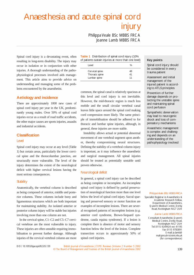
Spinal cord injury is a devastating event, often
resulting in long-term disability. The injury may
occur in isolation or in conjunction with other
injuries. A thorough understanding of the patho-
physiological processes involved aids manage-
ment. This article aims to provide advice on
understanding and managing some of the prob-
lems encountered by the anaesthetist.
Aetiology and incidenceThere are approximately 1000 new cases of
spinal cord injury per year in the UK, predomi-
nantly young males. Over 50% of spinal cord
injuries occur as a result of road traffic accidents,
the other major causes are sports injuries, assaults
and industrial accidents.
ClassificationLevel
Spinal cord injury may occur at any level (Table
1) but certain areas, particularly the lower cervi-
cal spine and the thoracolumbar junction, are
structurally more vulnerable. The level of the
injury determines the extent of the neurological
deficit with higher cervical lesions having the
most serious consequences.
Stability
Anatomically, the vertebral column is described
as being composed of anterior, middle and poste-
rior columns. These columns include bony and
ligamentous structures which are both important
for maintaining stability. An isolated anterior or
posterior column injury will be stable but injuries
involving more than one column are not.
In the cervical spine, C1–C2 and C5–C7 cervi-
cal vertebrae are the most vulnerable to injury.
These injuries are often unstable requiring immo-
bilisation to prevent further damage. Although
injuries of the cervical vertebral column are more
common, the spinal canal is relatively spacious at
this level and cord injury is not inevitable.
However, the mid-thoracic region is much less
mobile and the small circular vertebral canal
leaves little space around the spinal cord making
cord compression more likely. The same princi-
ple of immobilisation should be adhered to for
thoracic and lumbar spine injuries, although, in
general, these injuries are more stable.
Instability allows actual or potential abnormal
movement of one vertebral segment upon anoth-
er, thereby compromising neural structures.
Defining the stability of a vertebral column injury
is important, as it may influence the anaesthetic
and surgical management. All spinal injuries
should be treated as potentially unstable until
proven otherwise.
Neurological deficit
In general, a spinal cord injury can be described
as being complete or incomplete. An incomplete
spinal cord injury is defined by partial preserva-
tion of neurological function more than one level
below the level of spinal cord injury. Sacral spar-
ing and preserved sensory or motor function are
examples of incomplete lesions. There are sever-
al recognised patterns of incomplete lesions (e.g.
anterior cord syndrome, Brown-Sequard syn-
drome, cauda equina syndrome). If a lesion is
complete there is absence of motor and sensory
function below the level of the lesion. Complete
transection occurs in approximately 50% of
spinal cord injuries.
Anaesthesia and acute spinal cordinjury
Philippa Veale BSc MBBS FRCAJoanne Lamb MBBS FRCA
139British Journal of Anaesthesia | CEPD Reviews | Volume 2 Number 5 2002
© The Board of Management and Trustees of the British Journal of Anaesthesia 2002
Key points
Spinal cord injury shouldbe considered in everytrauma patient
Assessment and initialmanagement of theinjured patient is accord-ing to ATLS principles
Prevention of furtherdamage depends on pro-tecting the unstable spineand maintaining spinalcord perfusion
Sympathetic denervationmay lead to neurogenicshock and loss of com-pensatory mechanisms
Anaesthetic managementis complex and challeng-ing and depends on anunderstanding of thepathophysiology involved
Philippa Veale BSc MBBS FRCA
Specialist Registrar in Anaesthetics &Academic Research Fellow,
Department of Anaesthetics,Queen’s Medical Centre, Derby
Road, Nottingham NG7 2UH
Joanne Lamb MBBS FRCA
Consultant Anaesthetist, Queen’sMedical Centre, Derby Road,
Nottingham NG7 2UHTel: 0115 9249924 ext 41195
Fax: 0115 9783891E-mail: jo.lamb@
mail.qmcuh-tr.trent.nhs.uk(for correspondence)
Table 1 Distribution of spinal cord injury (10%of patients sustain injuries at more than one level)
Level %
Cervical spine 48Thoracic spine 41Lumbar spine 11
DOI 10.1093/bjacepd/02.05.139

Anaesthesia and acute spinal cord injury
140 British Journal of Anaesthesia | CEPD Reviews | Volume 2 Number 5 2002
The level of the injury determines the extent of respiratory
involvement (Table 2). Abdominal muscle paralysis contributes to
the respiratory embarrassment and poor cough. Neurogenic cardio-
vascular complications are seen in higher lesions (above T7) due to
the effects of traumatic sympathectomy. Loss of sympathetic vaso-
constrictor tone to blood vessels results in vasodilatation. Cardiac
sympathetic supply (T1–T4) may also be affected; loss of chrono-
tropic and inotropic effects and unopposed vagal reflexes may result
in severe sinus bradycardia. Profound bradycardia and even sinus
arrest may occur during intubation or suction of the airway.
PathophysiologySpinal shock
Spinal shock describes the initial phase after an insult to the spinal
cord and may be defined as a temporary interruption of the physi-
ological function of the spinal cord following injury. It may, in part,
be a vascular phenomenon. All reflex activity is lost and the cord
below the level of the lesion also becomes isolated from the high-
er centres. This accounts for the characteristic picture of flaccid
paralysis. An accurate prognosis is not possible until the stage of
spinal shock has ended (up to 4 weeks). If there is evidence of neu-
rological sparing, i.e. residual sensory, motor or reflex function
below the level of the lesion, full recovery may follow.
Autonomic and reflex activity gradually returns to the injured
cord. Loss of descending inhibitory control leads eventually to spas-
ticity and autonomic hyperreflexia. Respiratory function improves
as spasticity of chest and abdominal wall muscles reduces paradox-
ical movement.
It is important not to confuse spinal shock with spinal neuro-
genic shock. The latter term describes the hypotension seen as a
result of traumatic sympathectomy.
Secondary injury
Trauma to the spinal cord results in an immediate physical injury
(i.e. primary injury). A combination of small intramedullary ves-
sel damage, haemorrhage into grey matter and local vasospasm
causes a critical fall in cord perfusion. The cord becomes
ischaemic leading to the onset within minutes of secondary injury
which may become progressively worse over the ensuing hours.
The release of mediators of postischaemic injury is implicated in
secondary damage as are hypotension, hypoxaemia and hyperther-
mia. After a period of ischaemia, apoptosis or programmed cell
death occurs. This peaks at 8 h and results in irrecoverable damage.
Many interventions have been tried to reduce the severity of
secondary injury, mainly in experimental animal work. Steroids,
free radical scavengers, barbiturates, hypothermia, hyperbaric
oxygen therapy and NMDA and opioid antagonists are some
examples. The results of these interventions are disappointing.
Many spinal centres have a spinal injury protocol that includes
the early administration of high dose methylprednisolone. The
evidence for this intervention comes from the National Acute
Spinal Cord Injury Study (NASCIS) trials which demonstrated
an improvement in long-term neurological outcome following
high dose methylprednisolone. Unfortunately, there is no good
evidence that this equates to an improvement in functional out-
come and the risk of infection is increased.
Measures aimed at ensuring adequate oxygenation and perfu-
sion of the injured cord and avoidance of hypergylcaemia and
hyperthermia are the mainstay of prevention of secondary injury.
Initial assessment and managementPresentation
The patient with an acute spinal cord injury typically presents to
the accident and emergency department having already been
immobilised on a spinal board. Use of pre-hospital spinal immo-
bilisation in trauma patients is now routine but the positioning and
immobilisation of the patient should be scrutinised as part of the
primary survey. The correct technique is placement of a hard cer-
vical collar of the appropriate size, sandbags either side of the head
and adhesive tape across the forehead onto each side of the trolley.
Thoracic and lumbar spine injuries simply require the patient
to be kept supine on a solid surface, avoiding any excessive
movement. If the patient is to be moved, this should be by ‘log-
rolling’, maintaining vertebral column alignment.
Table 2 Respiratory effects of spinal cord injury
Level of injury Effect Clinical signs
T1–T7 Variable degree of intercostal nerve paralysis Impaired chest wall movementPoor cough
C5–C8 Complete intercostal nerve paralysis Ineffective or absent coughFunction of diaphragm intact Paradoxical respiratory pattern
Use of accessory muscles
C3–C5 Partial diaphragm paralysis As above but usually requiring assisted ventilation
C3 or above Denervation of diaphragm Respiratory failure

Spinal cord injury should be considered in all trauma victims.
An appropriately qualified person can rule out cervical spine
injury in the fully conscious patient. Full precautions must be
strictly adhered to in any patient with midline tenderness, neuro-
logical symptoms or signs, a reduced level of consciousness, or a
painful ‘distracting’ injury.
Initial management
The initial management of the trauma victim with a spinal cord
injury is as for any seriously injured patient. The ATLS approach
is proven, standardised and effective and initial management
should be based upon its principles.
Airway
The airway must be examined for patency and, if required, manip-
ulated with a jaw thrust as opposed to a chin lift. This airway-posi-
tioning manoeuvre is associated with less displacement of the cer-
vical spine. A trauma mask with high flow oxygen should be
applied to the patient if the airway is patent. If not, a decision to intu-
bate the trachea should be made earlier rather than later, in order to
maximise oxygen delivery and limit secondary hypoxic damage to
the injured spinal cord. A difficult intubation should be anticipated
because of: (i) suboptimal positioning due to immobilisation of the
cervical spine; (ii) the requirement for a rapid sequence induction
with cricoid pressure; (iii) the potential for pre-vertebral swelling
due to haematoma; and (iv) the potential for poor visibility at laryn-
goscopy due to debris or distorted anatomy in maxillofacial trauma.
There is a great deal of debate in the literature regarding the safest
approach to intubation in the patient with a cervical spine injury and
the likelihood of causing further damage to the spinal cord. One
concern is that unstable bony fragments may be maintained in posi-
tion only by muscular spasm and that muscle relaxation may con-
tribute to the instability. The options for intubation are: (i) direct
laryngoscopy and intubation in the presence of manual in-line
immobilisation; (ii) blind nasal intubation if there is no compromise
to the cribiform plate; (iii) blind oral intubation using the intubating
laryngeal mask airway (ILMA); (iv) awake fibre-optic intubation;
and (iv) surgical airway if intubation is not possible.
Awake fibre-optic intubation with adequate local anaesthesia and
intubation under direct vision has the advantage of avoiding move-
ment of the unstable cervical spine. It may be performed with the
patient immobilised and in halo traction and allows neurological
assessment following intubation. However, this method requires
skill and specialist equipment and is often impractical in the acute
situation, particularly if intubation is required urgently. The choice
depends on the situation and experience of the individual.
Direct laryngoscopy with in-line immobilisation is a safe and
acceptable method. It requires at least three trained personnel and
involves four stages: preparation, manual in-line immobilisation,
rapid sequence induction and intubation. Succinylcholine is the
muscle relaxant of choice. The release of potassium associated with
the use of succinylcholine in spinal cord injury has not been shown
to be a problem until 3 days post-injury at the earliest. Atropine must
be available immediately as should equipment for obtaining a sur-
gical airway. The hard collar is opened at the front to expose fully
the mandible and allow maximum possible mouth opening. In order
to avoid displacement of the injured cervical spine by cricoid pres-
sure, the back of the rigid collar is left in place. Otherwise, a biman-
ual technique should be employed. Asmall amount of movement of
the neck may be inevitable, even with manual in-line immobilisa-
tion. This is unlikely to be significant enough to cause injury to the
cord and must be allowed for in order to secure the airway.
Breathing
Adequate oxygenation is imperative in order to prevent secondary
hypoxic damage. Supplemental oxygen must be administered to all
patients. Ventilation must be assessed both clinically and with oxy-
gen saturation measurements and arterial blood gas analysis.
Inadequate ventilation causing hypoxaemia or hypercapnia should
be rectified by tracheal intubation and ventilation. Hypoxaemia is
found in about 50% of patients with high spinal cord injury, usual-
ly due to the neuromuscular deficit resulting from the injury.
Associated injuries may also be the cause of inadequate ventilation
or oxygenation. Chest injuries are common in polytrauma patients,
pulmonary aspiration and pulmonary oedema are common in head
injury and some patients may have been victims of near-drowning.
Circulation
Maintenance of an adequate circulation is essential in spinal cord
injury in order to minimize secondary ischaemic damage to the
injured cord. Hypotension must be treated promptly with fluid
boluses in the first instance. In the polytrauma patient who is hypo-
tensive, hypovolaemia secondary to haemorrhage from concurrent
injuries must be excluded according to ATLS principles. Remember
that the patient with a high spinal cord injury will not complain of
pain from a fractured pelvis or other injuries. Intra-abdominal bleed-
ing is more difficult to diagnose when the abdominal muscles are
flaccid. This must be ruled out by diagnostic peritoneal lavage,
abdominal ultrasound or computed tomography (CT).
Damage to the spinal cord above T6 may result in spinal neuro-
genic shock. Loss of sympathetic function leading to neurogenic
shock should be actively managed in order to preserve the perfusion
Anaesthesia and acute spinal cord injury
British Journal of Anaesthesia | CEPD Reviews | Volume 2 Number 5 2002 141

of the injured cord. Bradycardia affecting cardiac output should be
treated with intravenous atropine or glycopyrrolate.
Fluid resuscitation is complex in these patients. Judicious volume
loading with crystalloid or colloid solutions guided by central
venous pressure measurement is the first step. Loss of cardiac sym-
pathetic innervation affects myocardial contractility. In the acute
phase, these patients have a limited capacity to respond to volume
stress and are prone to develop pulmonary oedema if volume over-
loaded. If hypotension persists despite fluid loading, low doses of
vasopressors are indicated to counteract the loss of vasoconstriction.
If vasoconstrictors are used, care must be taken to avoid a fall in car-
diac output resulting from a high systemic vascular resistance. Pul-
monary artery flotation catheter or trans-oesophageal Doppler may
be used to guide cardiovascular support in more complex cases.
Assessment of disability
Full neurological examination is important and, whenever possi-
ble, must be carried out before anaesthesia for intubation. It must
be recorded and repeated at regular intervals. Any changes should
be clearly documented and include: (i) sensory level; (ii) motor
level; and (iii) anal tone and reflex activity
The neurological level of injury is designated as the most distal
uninvolved segment of the spinal cord. This differs from the bone
level of injury, which is the level of the spine at which bony dam-
age is actually visualised. There is usually a correlation between the
two levels but there can be some discrepancy, especially in cervi-
cal spine injuries. Ascending spinal cord oedema may result in
deteriorating signs. Improvement in the deficit may be predictive
of some neurological recovery.
RadiologyA detailed discussion of imaging in spinal injury is beyond the
scope of this article. According to ATLS guidelines, all patients
with multiple injuries or significant head injury require a lateral
cervical spine X-ray. If there is a suspicion of an injury in the tho-
racic or lumbar spine, the relevant area should also be X-rayed. Of
patients with cervical spinal injury, 10% will have spinal injury at
another level and this must be excluded.
The lateral cervical spine X-ray must be adequate, i.e. extend-
ing as far as the cervicothoracic junction. All anaesthetists should
have a system for examining cervical spine films but an expert
opinion is often required. Plain radiography of the cervical spine
may also include an anteroposterior and an open mouth (odontoid
peg) view. Upper thoracic spine injuries are difficult to visualize
on plain radiography.
Magnetic resonance imaging (MRI) is particularly useful for
imaging the spinal cord and soft tissues and for identifying cord
compression and oedema. Specialist spinal units will usually per-
form an early CT and often also MRI of the entire spine in any
patient with a spinal injury. However, it is important to avoid send-
ing unstable patients to distant radiology departments and into
inaccessible scanners.
Anaesthetic managementIndications for surgery
Surgical intervention may be indicated in the early or intermediate
phase of spinal cord injury. If the clinical picture at 48 h is of a com-
plete injury, no type of surgery has been shown to improve neuro-
logical function. Despite this, operative spinal fusion may be appro-
priate in order to confer stability thereby aiding rehabilitation and
preventing further complications. Stabilisation procedures may be
best delayed until the patient has recovered from other injuries or is
more cardiovascularly stable.
Early surgery may be indicated if the neurological deficit is
incomplete or due to spinal shock and there is felt to be some poten-
tial for recovery. The surgery is carried out in order to permit: (i)
urgent decompression of the spinal cord, followed by stabilisation;
(ii) restoration of vertebral column alignment if this has not been
achieved by conservative means (halo traction); (iii) exploration for
decompression and stabilisation if the neurological deficit is wors-
ening; and (iv) exploration of open penetrating spinal wounds.
The anaesthetic management of patients with high spinal cord
injury may be challenging and the potential hazards should not be
underestimated. Spinal surgery should be carried out in specialist
centres but an anaesthetist in any hospital receiving trauma patients
may be called upon to manage a spinal injuries patient who requires
urgent surgery for other injuries.
Pre-operative assessment and planning
Early assessment at presentation to hospital has been outlined
above. Before surgery, it is important to re-assess the patient care-
fully and plan accordingly.
Airway
If the patient has not been intubated, the airway must be assessed and
a plan made for airway management. The patient may be in halo
traction and surgeons should be involved if this is to be removed.
Breathing
In cervical and higher thoracic lesions, respiratory function may
have become compromised. Absent or impaired cough leads to the
retention of secretions. Increased work of breathing means patients
relying on diaphragmatic breathing may start to tire. Lung volumes
reach their lowest point at 3–4 days post-injury and spirometry is
Anaesthesia and acute spinal cord injury
142 British Journal of Anaesthesia | CEPD Reviews | Volume 2 Number 5 2002

useful to monitor pulmonary function. If the vital capacity has
fallen to less than 1 litre, the patient is hypoxaemic or has a high
respiratory rate, arrangements must be made for assisted ventila-
tion postoperatively.
Circulation
In the patient with a cervical or high thoracic lesion and sympathet-
ic denervation, the systolic blood pressure will often have stabilised
at 90–100 mmHg. This should be adequate in the supine position
but loss of compensatory mechanisms may jeopardize cord perfu-
sion during positioning for surgery or periods of intra-operative
blood loss. Consequently, it is necessary to make plans for central
venous catheterisation and the availability of infusion pumps and
vasoactive drugs which may be needed to support the circulation.
ECG abnormalities, including signs of subendocardial ischaemia
and arrhythmias, are sometimes seen in high cord injuries.
Major blood loss should be anticipated and blood cross-matched
in advance.
General considerations
Amore general anaesthetic assessment must not be overlooked and
all management plans discussed with the patient. A spinal cord
injury is a devastating event and these patients may be in a state of
considerable psychological distress. Good communication and a
humane and sensitive approach are essential.
Intra-operative management
Monitoring
In addition to standard monitoring, intra-arterial blood pressure
measurement and central venous pressure monitoring are required.
As discussed above, a pulmonary artery flotation catheter or trans-
oesophageal Doppler may be helpful for optimal cardiovascular
management. Urine output should be measured hourly. Thermo-
regulation is impaired in spinal cord injury. Core temperature must
be monitored and patient and fluid warming devices used.
Spinal cord monitoring may be used in specialist centres, partic-
ularly for the patient with an unstable vertebral column injury and
no or partial neurological deficit. It is vital to preserve cord func-
tion in these patients and cord monitoring during, or immediately
after, positioning and during surgery facilitates this. The common-
est method is the use of sensory evoked potentials.
Induction and maintenance
The main goal is to maintain adequate cord perfusion and oxy-
genation during surgery and anaesthesia to prevent any further
damage. Autoregulation of blood flow is lost in the injured cord
and mean arterial blood pressure should be at least 60 mmHg. A
controlled ventilation technique is appropriate due to the prolonged
nature of the surgery and the position required for surgical access.
Mild hypocapnia is of theoretical benefit in decompressing the
spinal cord.
At induction, the anaesthetic agents may be of individual pref-
erence but should be titrated slowly. The usual precautions are
taken if the patient has a potential full stomach but succinyl-
choline should be avoided if the patient is > 3 days post-injury.
Airway management has been discussed earlier and is not a par-
ticular issue if the lesion is below C7 or there is no chance of neu-
rological recovery. Atropine or glycopyrollate should be avail-
able to treat any bradycardia.
Large bore intravenous access is essential. A nasogastric tube
should be placed as high acute cord injury leads to gastric stasis and
gastrointestinal ileus. Spinal injury patients are at particular risk of
venous thrombo-embolism. Compression stockings should be
worn and calf compression devices used intra-operatively.
Most spinal decompression and stabilisation procedures involve
posterior surgery with the patient in the prone position. Halo trac-
tion may be maintained during surgery. For unstable fractures, great
care must be taken to maintain vertebral column alignment during
positioning. For some injuries, access to the anterior spinal column
may be indicated. If this is the case in a thoracic spine injury, a tho-
racotomy will be needed and, if possible, provision should be made
for one lung ventilation. These procedures may take many hours
and the usual precautions must be taken to avoid peripheral nerve
injuries and pressure sores. Major blood loss is not uncommon and
intra-operative blood cell salvage should be used if it is available.
A balanced anaesthetic technique is appropriate but analgesic
requirements postoperatively depend on the nature of surgery and
the extent of the neurological injury. The use of epidural analgesia
may lead to difficulty with neurological assessment postoperatively
and great care should be taken with the use of systemic opioids in
patients with respiratory compromise.
Key referencesCobby TF,Hardman JG,Baxendale BR.Anaesthetic management of the severe-
ly injured patient: spinal injury. Br J Hosp Med 1997; 58: 198–201
Lam AM.Acute spinal cord injury:monitoring and anaesthetic implications.CanJ Anaesth 1991; 38: 60–7
Lu J,Ashwell KWS,Waite P.Advances in secondary spinal cord injury: role ofapoptosis. Spine 2000; 25: 1859–66
Mcleod A, Calder I. Spinal cord injury and direct laryngoscopy – the legendlives on. Br J Anaesth 2000; 84: 705–9
Short DJ, El Masry WS, Jones PW. High dose methylprednisolone in the man-agement of acute spinal cord injury – a systematic review from a clinicalperspective. Spinal Cord 2000; 38: 273–86
See multiple choice questions 94–96.
Anaesthesia and acute spinal cord injury
British Journal of Anaesthesia | CEPD Reviews | Volume 2 Number 5 2002 143

Anaesthesia for spinal surgery in adults
D. A. Raw1*, J. K. Beattie2 and J. M. Hunter1
1University Department Anaesthesia, University Clinical Department, The Duncan Building, Daulby Street,Liverpool, L69 3GA, UK. 2Royal Liverpool and Broadgreen University Hospitals NHS Trust, Prescot Road,
Liverpool L7 8XP, UK
*Corresponding author. E-mail:[email protected]
The spectrum of spinal surgery in adult life is considerable. Anaesthesia for major spinal
surgery, such as spinal stabilization following trauma or neoplastic disease, or for correction of
scoliosis, presents a number of challenges. The type of patients who would have been declined
surgery 20 yr ago for medical reasons, are now being offered extensive procedures. They com-
monly have preoperative co-morbid conditions such as serious cardiovascular and respiratory
impairment. Airway management may be dif®cult. Surgery imposes further stresses of signi®-
cant blood loss, prolonged anaesthesia, and problematical postoperative pain management. The
perioperative management of these patients is discussed. The advent of techniques to monitor
spinal cord function has reduced postoperative neurological morbidity in these patients. The
anaesthetist has an important role in facilitating these methods of monitoring.
Br J Anaesth 2003; 91: 886±904
Keywords: anaesthesia, general; monitoring; surgery, spinal
The scope of spinal surgery is considerable. Both adult
and paediatric patients present for surgery, which may
be elective or urgent. They mainly present with one of
®ve pathologies: trauma, for example an unstable
vertebral fracture; infection, for example vertebral
abscess; malignancy (metastatic or primary disease
with spinal instability, pain, and neurological comprom-
ise); congenital/idiopathic, for example scoliosis; or
degenerative disease. In excess of 25 000 spinal
operations were performed in the UK in 2001±2.112
Surgery may be required at any site in the spine from
cervical to lumbosacral. Procedures range from minim-
ally invasive microdiscectomy, to prolonged operations
involving multiple spinal levels and signi®cant blood
loss. An osteotomy is a decompressive procedure, which
releases compressive forces at a localized site.
Stabilization of the spine involves instrumentation
above and below the unstable spinal level. Distractive
forces may also be applied to the spine, for example in
surgery for scoliosis, with instrumentation placed over
multiple spinal levels. Insertion of such devices may be
through a posterior, anterior, or a combined approach
involving repositioning of the patient part way through
the procedure and major blood loss.
The challenge to the anaesthetist is to provide optimal
surgical conditions whilst ensuring adequate oxygenation to
the brain and spinal cord, and facilitating the use of
intraoperative spinal cord monitoring techniques if appro-
priate.
Pathological conditions requiring spinalsurgery in adult practice
Scoliosis
Scoliosis involves a lateral and rotational deformity of the
spine, which occurs in up to 4% of the population.98 Most
cases are idiopathic (70%) and occur with a male:female
ratio of 1:4 (Table 1). Surgery is usually considered when
the Cobb angle exceeds 50° in the thoracic, or 40° in the
lumbar spine (Fig. 1A and B). Surgery aims to halt
progression of the condition and to at least partially correct
the deformity, preventing further respiratory and cardiovas-
cular deterioration. Left untreated, idiopathic scoliosis
rapidly progresses and is often fatal by the fourth or ®fth
decade of life, as a result of pulmonary hypertension, right
ventricular failure, or respiratory failure.85
Muscle disorders
Muscular dystrophy and cerebral palsy are important causes
of scoliosis. Of the muscular dystrophies, Duchenne
REVIEW ARTICLE
British Journal of Anaesthesia 91 (6): 886±904 (2003)
DOI: 10.1093/bja/aeg253
Ó The Board of Management and Trustees of the British Journal of Anaesthesia 2003
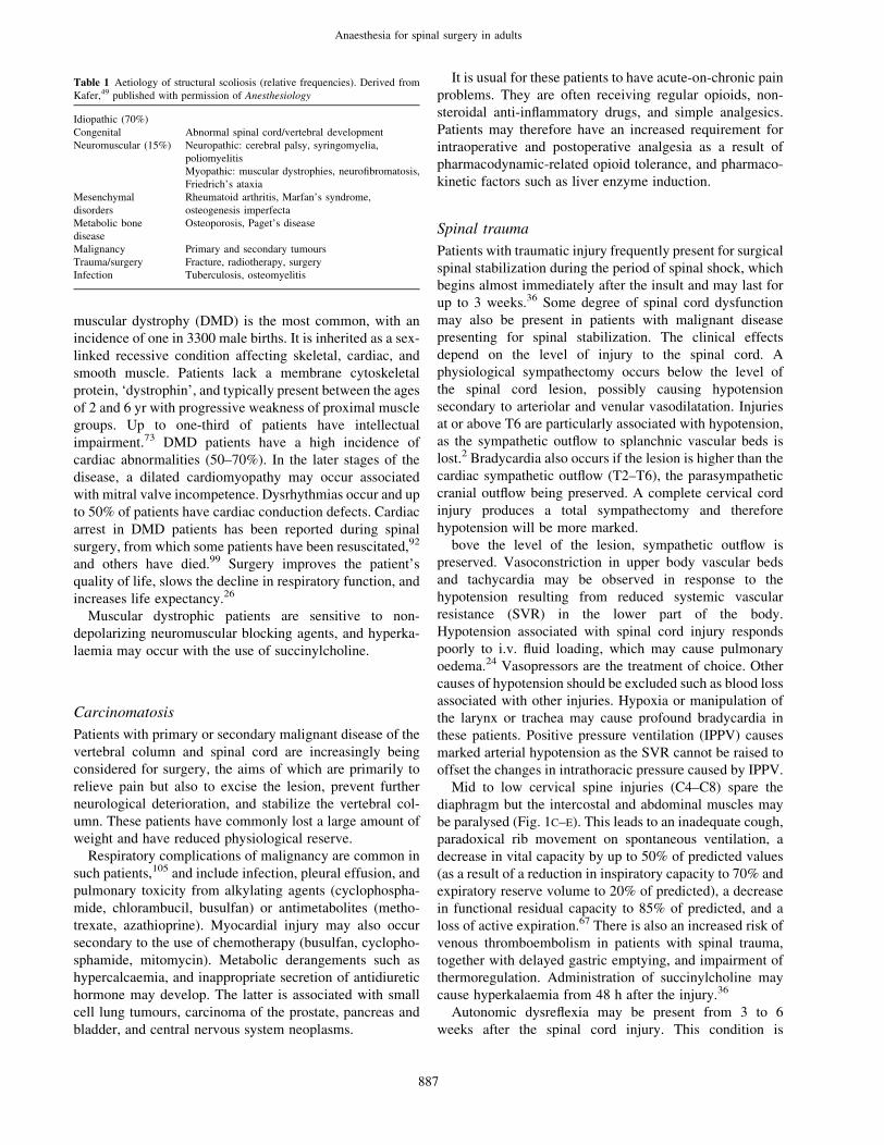
muscular dystrophy (DMD) is the most common, with an
incidence of one in 3300 male births. It is inherited as a sex-
linked recessive condition affecting skeletal, cardiac, and
smooth muscle. Patients lack a membrane cytoskeletal
protein, `dystrophin', and typically present between the ages
of 2 and 6 yr with progressive weakness of proximal muscle
groups. Up to one-third of patients have intellectual
impairment.73 DMD patients have a high incidence of
cardiac abnormalities (50±70%). In the later stages of the
disease, a dilated cardiomyopathy may occur associated
with mitral valve incompetence. Dysrhythmias occur and up
to 50% of patients have cardiac conduction defects. Cardiac
arrest in DMD patients has been reported during spinal
surgery, from which some patients have been resuscitated,92
and others have died.99 Surgery improves the patient's
quality of life, slows the decline in respiratory function, and
increases life expectancy.26
Muscular dystrophic patients are sensitive to non-
depolarizing neuromuscular blocking agents, and hyperka-
laemia may occur with the use of succinylcholine.
Carcinomatosis
Patients with primary or secondary malignant disease of the
vertebral column and spinal cord are increasingly being
considered for surgery, the aims of which are primarily to
relieve pain but also to excise the lesion, prevent further
neurological deterioration, and stabilize the vertebral col-
umn. These patients have commonly lost a large amount of
weight and have reduced physiological reserve.
Respiratory complications of malignancy are common in
such patients,105 and include infection, pleural effusion, and
pulmonary toxicity from alkylating agents (cyclophospha-
mide, chlorambucil, busulfan) or antimetabolites (metho-
trexate, azathioprine). Myocardial injury may also occur
secondary to the use of chemotherapy (busulfan, cyclopho-
sphamide, mitomycin). Metabolic derangements such as
hypercalcaemia, and inappropriate secretion of antidiuretic
hormone may develop. The latter is associated with small
cell lung tumours, carcinoma of the prostate, pancreas and
bladder, and central nervous system neoplasms.
It is usual for these patients to have acute-on-chronic pain
problems. They are often receiving regular opioids, non-
steroidal anti-in¯ammatory drugs, and simple analgesics.
Patients may therefore have an increased requirement for
intraoperative and postoperative analgesia as a result of
pharmacodynamic-related opioid tolerance, and pharmaco-
kinetic factors such as liver enzyme induction.
Spinal trauma
Patients with traumatic injury frequently present for surgical
spinal stabilization during the period of spinal shock, which
begins almost immediately after the insult and may last for
up to 3 weeks.36 Some degree of spinal cord dysfunction
may also be present in patients with malignant disease
presenting for spinal stabilization. The clinical effects
depend on the level of injury to the spinal cord. A
physiological sympathectomy occurs below the level of
the spinal cord lesion, possibly causing hypotension
secondary to arteriolar and venular vasodilatation. Injuries
at or above T6 are particularly associated with hypotension,
as the sympathetic out¯ow to splanchnic vascular beds is
lost.2 Bradycardia also occurs if the lesion is higher than the
cardiac sympathetic out¯ow (T2±T6), the parasympathetic
cranial out¯ow being preserved. A complete cervical cord
injury produces a total sympathectomy and therefore
hypotension will be more marked.
bove the level of the lesion, sympathetic out¯ow is
preserved. Vasoconstriction in upper body vascular beds
and tachycardia may be observed in response to the
hypotension resulting from reduced systemic vascular
resistance (SVR) in the lower part of the body.
Hypotension associated with spinal cord injury responds
poorly to i.v. ¯uid loading, which may cause pulmonary
oedema.24 Vasopressors are the treatment of choice. Other
causes of hypotension should be excluded such as blood loss
associated with other injuries. Hypoxia or manipulation of
the larynx or trachea may cause profound bradycardia in
these patients. Positive pressure ventilation (IPPV) causes
marked arterial hypotension as the SVR cannot be raised to
offset the changes in intrathoracic pressure caused by IPPV.
Mid to low cervical spine injuries (C4±C8) spare the
diaphragm but the intercostal and abdominal muscles may
be paralysed (Fig. 1C±E). This leads to an inadequate cough,
paradoxical rib movement on spontaneous ventilation, a
decrease in vital capacity by up to 50% of predicted values
(as a result of a reduction in inspiratory capacity to 70% and
expiratory reserve volume to 20% of predicted), a decrease
in functional residual capacity to 85% of predicted, and a
loss of active expiration.67 There is also an increased risk of
venous thromboembolism in patients with spinal trauma,
together with delayed gastric emptying, and impairment of
thermoregulation. Administration of succinylcholine may
cause hyperkalaemia from 48 h after the injury.36
Autonomic dysre¯exia may be present from 3 to 6
weeks after the spinal cord injury. This condition is
Table 1 Aetiology of structural scoliosis (relative frequencies). Derived from
Kafer,49 published with permission of Anesthesiology
Idiopathic (70%)
Congenital Abnormal spinal cord/vertebral development
Neuromuscular (15%) Neuropathic: cerebral palsy, syringomyelia,
poliomyelitis
Myopathic: muscular dystrophies, neuro®bromatosis,
Friedrich's ataxia
Mesenchymal
disorders
Rheumatoid arthritis, Marfan's syndrome,
osteogenesis imperfecta
Metabolic bone
disease
Osteoporosis, Paget's disease
Malignancy Primary and secondary tumours
Trauma/surgery Fracture, radiotherapy, surgery
Infection Tuberculosis, osteomyelitis
Anaesthesia for spinal surgery in adults
887

characterized by extreme autonomic responses such as
hypertension and tachycardia after stimulation of nerves
below the level of the spinal cord lesion (for example,
rectal, urological, peritoneal stimulation). Injuries higher
than T7 have an 85% chance of producing serious
cardiovascular derangement.5
Preoperative assessment
When assessing patients before spinal surgery, particular
care should be given to the respiratory, cardiovascular, and
neurological systems; all may be affected by the pathology
for which the spinal surgery is proposed.
Airway assessment
The potential for dif®culty in airway management should
always be considered, particularly in those patients pre-
senting for surgery of the upper thoracic or cervical spine. A
careful assessment should be made for previous dif®culty in
intubation, restriction of neck movement, and the stability
or otherwise of the cervical spine. Stability is de®ned as the
ability of the spine, under physiological loads, to resist
displacement, which causes neurological injury. It is
essential to discuss preoperatively the stability of the
spine with the surgeon. The cervical spine may be assessed
clinically (presence of pain or neurological de®cits), and
Fig 1 (A) Thoracolumbar scoliosis and measurement of the Cobb angle. A perpendicular line is drawn from the end plate of the most caudal vertebrae
involved, whose inferior end plate tilts maximally to the concavity of the curve. A second perpendicular line is drawn from the end plate of the most
cephalad vertebrae, whose superior end plate tilts maximally to the concavity of the curve. The curve value is the number of degrees formed by the
angle of intersection of these two lines. (B) Thoracolumbar scoliosis after surgery, showing long rods and pedicle screws. (C) Dislocation of the 5th
and 6th cervical vertebrae after trauma; (D) same patient's MRI scan, and (E) after surgery to stabilize the cervical spine. SC, spinal cord; C6, sixth
cervical vertebrae.
Raw et al.
888

radiographically (lateral or ¯exion/extension plain ®lms,
computer aided tomography, and magnetic resonance
imaging). The stability of the cervical spine is dependent
on ligamental and vertebral elements. Damage to these
elements may not be detectable by plain x-rays alone. The
adult cervical spine below C2 is unstable or on the brink of
instability when one of the following conditions are met: (i)
all the anterior or all the posterior elements are destroyed;
(ii) there is >3.5 mm horizontal displacement of one
vertebra in relation to an adjacent one on a lateral x-ray; or
(iii) there is more than 11° of rotation of one vertebra to an
adjacent one.119 Above the level of C2, examples of
unstable injuries include: disruption of the transverse
ligament of the atlas (a distance of greater than 3 mm in
adults between the posterior corpus of the anterior arch of
C1 and the anterior border of the odontoid process, when
measured on a lateral plain x-ray ®lm, is diagnostic); and a
Jefferson burst fracture of the atlas following axial loading,
which causes atlantoaxial instability. Disruption of the
tectorial and alar ligaments and some occipital condylar
fractures also cause atlanto-occipital instability.
Some inherited disorders such as DMD may lead to
glossal hypertrophy, and previous radiotherapy to tumours
of the head and neck can cause dif®culty in direct
laryngoscopy. A decision must be made, whether to intubate
the patient awake or asleep.
Respiratory system
Patients presenting for spinal surgery frequently have
impaired respiratory function. Those who have sustained
cervical or high thoracic trauma or who have multiple
injuries may be arti®cially ventilated preoperatively. Others
have recurrent chest infections.
Preoperatively, respiratory function should be assessed
by a thorough history, focusing on functional impairment,
physical examination, and appropriate investigations
(Table 2). Scoliosis causes a restrictive pulmonary de®cit,
with reduced vital capacity and reduced total lung capacity
(TLC). The residual volume is unchanged. The severity of
functional impairment is related to the angle of the scoliosis,
the number of vertebrae involved, a cephalad location of the
curve, and a loss of the normal thoracic kyphosis.53 The
extent of functional impairment cannot, therefore, be
directly inferred from the angle of scoliosis alone. The
most common blood-gas abnormality is a reduced arterial
oxygen tension with a normal arterial carbon dioxide
tension, as a result of the mismatch between ventilation
and perfusion in hypoventilated lung units.48 Respiratory
function should be optimized by treating any reversible
cause of pulmonary dysfunction, including infection, with
physiotherapy and nebulized bronchodilators as indicated.
There is controversy over whether surgery for idiopathic
scoliosis improves,55 59 or worsens14 63 pulmonary function.
However, the type of surgery proposed may have a
signi®cant in¯uence upon postoperative pulmonary func-
tion, and may explain the contradictory ®ndings in studies of
non-homogenous groups of patients. Surgery involving the
thorax (anterior approach, combined approach, or rib
resection) was associated with an initial decline in forced
vital capacity (FVC, 19% of baseline values), forced
expiratory volume in 1 s (FEV1, 13%), and TLC (11%) at
3 months.117 This was followed by subsequent improvement
to preoperative baseline values at 2 yr postoperatively.
Surgery involving an exclusively posterior approach, how-
ever, was associated with an improvement in pulmonary
function tests by 3 months (although not reaching statistical
signi®cance); and an improvement that was statistically
signi®cant at 2-yr follow-up: FVC (14% increase from
baseline), FEV1 (14%), TLC (5%).
Older studies have reported that if preoperative vital
capacity is less than 30±35% of predicted, postoperative
ventilation is likely to be required.45 A history of depend-
ence on continuous nasal positive airways pressure at night
is also a sign of severe functional impairment and of
reduced physiological reserve. These ®ndings should
prompt serious consideration as to whether surgery repre-
Table 2 Suggested preoperative investigations before major spinal surgery
Minimum investigations Optional investigations
Airway Cervical spine lateral x-rays with
¯exion/extension views (for patients with
rheumatoid arthritis)
CT scan
Respiratory
system
Plain chest radiograph Pulmonary function tests (bronchodilator
reversibility)
Arterial blood gas analysis Pulmonary diffusion capacity
Spirometry (FEV1, FVC)
Cardiovascular
system
Electrocardiograph Dobutamine-stress echocardiograph
Echocardiography Dypiridamole/thallium scintigraphy
Blood tests Full blood count Liver function tests
Clotting pro®le
Blood cross match
Urea, electrolytes
Albumin, calcium (neoplastic disease)
Anaesthesia for spinal surgery in adults
889

sents an appropriate balance between its potential bene®ts
and the high risk of long-term postoperative ventilation in
such patients.
Cardiovascular system
Cardiac compromise may be a direct result of the underlying
pathology, for example in patients with muscular dystro-
phies. Cardiac dysfunction may also occur secondary to
scoliosis, which causes distortion of the mediastinum, and
cor pulmonale secondary to chronic hypoxaemia and
pulmonary hypertension. Assessment of functional cardio-
vascular impairment is dif®cult in patients who are wheel-
chair-bound. Minimum investigations should include an
electrocardiograph, and echocardiography to assess left
ventricular function and pulmonary arterial pressures
(Table 2). Dobutamine stress echocardiography may be
used to assess cardiac function in patients with a limited
exercise tolerance.
Thromboembolic prophylaxis
Patients undergoing spinal surgery may be at increased risk
of thromboembolic disease as a result of prolonged surgery,
prone positioning, malignancy, and extended periods of
postoperative recumbency. The use of compression stock-
ings and/or pneumatic boots is recommended.95 Many
surgeons prefer not to administer anticoagulants because
their use may be associated with haemorrhagic complica-
tions, including increased blood loss and epidural haema-
toma.
Neurological system
A full neurological assessment of the patient should be
made preoperatively. This should be documented for three
reasons. First, in patients undergoing cervical spine surgery,
the anaesthetist has a responsibility to avoid further
neurological deterioration during manoeuvres such as
tracheal intubation and patient positioning. Secondly,
muscular dystrophies may involve the bulbar muscles,
increasing the risk of postoperative aspiration. Thirdly, the
level of injury and the time elapsed since the insult are
predictors of the physiological derangements of the
cardiovascular and respiratory systems which occur peri-
operatively. If surgery is contemplated within 3 weeks of the
injury, spinal shock may still be present. After this time,
autonomic dysre¯exia may occur.
Anaesthesia technique
The type of monitoring chosen to assess spinal cord
integrity has a bearing on the anaesthetic technique used.
Premedication
The use of bronchodilating agents may be of value in
optimizing respiratory function preoperatively. In patients
with a high spinal cord lesion, or those in whom ®bre-optic
intubation is to be undertaken, administration of anti-
cholinergic agents such as atropine or glycopyrrolate
(200±400 mg by i.v. or i.m. injection) should be considered.
Many patients will have factors which increase the risk of
regurgitation and aspiration of gastric contents, such as
recent opioid administration, high spinal cord injury, or
recent traumatic injury.20 In these circumstances, it may be
prudent to premedicate patients with a histamine-2 receptor
antagonist such as ranitidine, or a proton pump inhibitor
such as omeprazole, and with sodium citrate. Some patients
may have nasogastric tubes in situ, which decrease the
competence of the upper oesophageal sphincter.
Induction
Choice of induction technique, i.v. or inhalation, is guided
primarily by the patient's condition and by consideration of
the ease with which the trachea may be intubated.
Preoxygenation is advisable in all patients. Unless there
are concerns over the stability of the cervical spine or
airway maintenance (see below), i.v. induction is suitable
for all but the sickest patients.
The use of succinylcholine in patients with muscular
dystrophies has long been known to cause cardiac arrest
secondary to hyperkalaemia, and should be avoided.28 103 In
patients with denervation as a result of spinal cord lesions,
the increased number of perijunctional nicotinic acetylcho-
line receptors on skeletal muscle can cause hyperkalaemia
after administration of succinylcholine. The time between
denervation and the risk of a potentially fatal hyperkalaemic
response is not known in humans. In animal studies, the
peak increase in serum K+ was 14 days after injury, with a
half-peak increase at 8.4 days.46 Changes in potassium
levels began 4 days after injury. It is probably safe to use
succinylcholine in the ®rst 48 h after injury to the spinal
cord. Thereafter, hyperkalaemia may occur for an indeter-
minate period, but most authors agree it is safe to use it
again 9 months after the injury.36
The use of bolus doses of i.v. induction agents reduces the
amplitude of evoked potential responses, and in particular,
cortical responses, but these effects do not prevent useful
intraoperative recording of cortical somatosensory poten-
tials,61 and transcranial electrical motor evoked potentials
(MEP).87 Inhalation agents reduce the amplitude of evoked
potential responses to a greater degree than do i.v. induction
agents, but no studies have compared an inhalation induc-
tion technique with an i.v. technique in this respect.
Intubation
A decision must be made at preoperative assessment
whether to intubate the patient awake or asleep, and whether
®bre-optic laryngoscopy will be required. The patient must
be counselled fully about the decision at this time (Fig. 2).
Raw et al.
890

Awake or asleep?
Indications for awake intubation include the risk of delayed
gastric emptying, the need to assess neurology after
intubation is complete (in cases such as an unstable cervical
spine), or the presence of a neck stabilization device (such
as halo traction), which prevents adequate airway mainten-
ance in an unconscious patient. Otherwise, i.v. induction of
anaesthesia followed by a non-depolarizing neuromuscular
blocking drug is the technique of choice.
Direct or ®bre-optic laryngoscopy?
There is controversy as to whether direct laryngoscopy is a
major factor contributing to cord injury in patients with
cervical spine instability. Other factors such as hypotension
and patient positioning may be equally important.68 Direct
laryngoscopy with manual in-line stabilization or a hard
collar, is an accepted means of intubation for many patients
provided this can be achieved without any neck movement.
Fixed ¯exion deformities, which involve the upper thoracic
and cervical spine may make direct laryngoscopy impos-
sible. These patients require the use of ®bre-optic laryngo-
scopy to facilitate tracheal intubation. The intubating
laryngeal mask airway may be a useful alternative, with
or without ®bre-optic guidance, for anaesthetists familiar
with its use.
Awake ®bre-optic intubation will be required in patients
wearing stabilization devices such as halo vests, which
make conventional airway access impossible, and in those
where dif®culty is anticipated because of anatomical
reasons, for example micrognathia, limited mouth opening.
In patients with an unstable cervical spine, instillation of
local anaesthesia into the airway to facilitate awake
intubation may cause vigorous coughing. In such cases, it
is preferable to use nebulized lidocaine rather than a
cricothyroid injection or administration of local anaesthetic
through the ®bre-optic scope.
Anterior approaches to the thoracic spine may necessitate
the use of a double-lumen endobronchial tube. Alterna-
tively, the surgeon and anaesthetist may agree that a single
lumen tracheal tube will suf®ce, allowing more limited
intraoperative lung retraction.
Maintenance
A stable anaesthetic depth is required in order that changes
to somatosensory or MEPs can be interpreted reliably. A
technique involving nitrous oxide 60% and iso¯urane less
than 0.5 MAC is compatible with somatosensory evoked
potential (SSEP) monitoring,89 but in nitrous oxide 60%,
end-tidal iso¯urane concentrations greater than 0.87% make
MEP monitoring uninterpretable.87 An i.v. technique using
propofol is therefore recommended. Neurophysiologists
monitoring evoked potentials should be made aware of any
sudden decrease in arterial pressure, or the need to
Fig 2 Algorithm for decision making when intubating a patient for proposed surgery involving the upper thoracic or cervical spine. ILMA, intubating
laryngeal mask airway; FO, ®bre-optic; RSI, rapid sequence induction; NDNMB, non-depolarizing neuromuscular blocking agent.
Anaesthesia for spinal surgery in adults
891
administer a bolus of opioid or change the anaesthetic depth.
Sudden cardiovascular instability during anaesthesia may
result from spinal cord and brain stem re¯exes, from
mediastinal distortion as a result of surgical manipulation,
or more commonly from blood loss.
Induced hypotension
Hypotensive anaesthesia may be used to improve the
surgical ®eld and to reduce blood loss during major spinal
surgery. A number of hypotensive agents have been studied
during surgery to correct scoliosis. They include ganglion
blocking agents, volatile agents,7 108 calcium channel
antagonists,38 sodium nitroprusside, nitroglycerin,121 and,
in children, the dopamine-1 receptor agonist, fenoldo-
pam.107 Mean arterial pressure (MAP) is typically main-
tained at 60 mm Hg. There is little evidence that any
particular agent is superior, but the avoidance of tachycardia
is an essential part of a good anaesthetic technique.
Caudal epidural anaesthesia has also been shown to
reduce surgical bleeding by 50% in patients undergoing
lumbar spinal surgery. This was thought to be as a result of a
reduction in sympathetic tone causing a measurable
decrease in lumbar vertebral intraosseus pressure.50 This
technique, however, is not as controllable as continuous i.v.
titration of a short-acting hypotensive agent, nor suitable for
operations involving the thoracic and cervical spine. It may
also hinder early postoperative neurological assessment.
Muscle relaxation
When myogenic motor evoked responses are to be recorded,
neuromuscular block must be carefully monitored and a
constant depth of block maintained. It is advisable to
administer a non-depolarizing neuromuscular blocking
agent such as atracurium using a continuous i.v. infusion
device during major spinal surgery.
Intraoperative monitoring and positioning ofthe patient
Cardiovascular monitoring
Prolonged anaesthesia in unusual positions, combined with
signi®cant blood loss, the haemodynamic effects of thoracic
surgery, and where appropriate controlled hypotension,
necessitates detailed monitoring of the cardiovascular
system. Invasive arterial pressure monitoring is mandatory.
In the prone position, central venous pressure (CVP) may
be a misleading indicator of right and left ventricular end-
diastolic volume. A study of 12 paediatric patients under-
going surgery for scoliosis in the prone position, compared
CVP and transoesophageal echocardiography (TOE) in
assessment of ventricular ®lling.104 Measurements were
made before and after positioning. CVP rose from 8.7 (1.3)
mm Hg (mean (SEM)) supine to 17.7 (2.5) mm Hg prone, but
left ventricular end diastolic diameter measured by TOE,
fell from 37.1 (2.9) to 33.2 (3.0) mm. In three patients in
whom pulmonary artery occlusion pressure (PAOP) was
also measured, there was an increase in PAOP on being
turned from the supine (mean 12.7 mm Hg) to the prone
position (mean 28.0 mm Hg), although there were insuf®-
cient data for statistical analysis. These results demonstrate
that there is no correlation between the measurement of
cardiac volume indicators by TOE and CVP or PAOP in
such conditions. High CVP values may be misleading
indicators of adequate cardiac ®lling in the prone pos-
ition.110 The changes are probably a result of raised
intrathoracic pressure causing reduced ventricular compli-
ance and compression of the inferior vena cava. Dependent
lower limbs cause a reduced venous return to the heart.
Respiratory monitoring
Respiratory system monitoring should always include end-
tidal carbon dioxide concentration and peak airway pres-
sure. In major surgery, serial measurements of arterial
oxygen tension are recommended. Patients with severe
respiratory dysfunction as a result of scoliosis may have an
increased alveolar-arterial oxygen gradient,48 which may be
further increased during prolonged anaesthesia because of
regional hypoventilation.
Temperature monitoring
Thermoregulation may already be impaired in patients who
have spinal cord lesions before surgery. Prolonged anaes-
thesia causes signi®cant heat loss. The use of temperature
monitoring, warming of all i.v. ¯uids, and a warm air
mattress device is recommended.
Positioning
Patient position for spinal surgery varies depending on the
level of the spine to be operated upon and the nature of the
proposed surgery. Patients may be repositioned intraopera-
tively. It is important that venous pressures at the surgical
site are kept low to reduce bleeding (reverse Trendelenburg
tilt and a free abdomen), and peripheral nerves, bony
prominences, and the eyes are protected. It is also important
to avoid displacement of unstable fractures during patient
positioning. Intraoperative x-ray imaging is frequently
required. The relevant spinal level must, therefore, be
placed away from the central support of a radiolucent
operating table.
Lumbar surgery
Anterior approaches require a laparotomy, and general
surgical input may be required in dif®cult cases. Posterior
surgery requires a prone patient with a free abdomen to keep
epidural venous pressure low (the patient supported on a
Wilson frame, for example, or a raised mattress with a hole
for the abdomen).84 For disc surgery, patients are placed in
Raw et al.
892

the knee-chest position with a well padded, secure support
behind the upper thigh to support the patient's weight. This
gives a horizontal lumbar spine with vertical intervertebral
discs.
Thoracic surgery
Anterior approaches to the thoracic spine are via a
thoracotomy with the patient supported in a lateral position.
If a double lumen endobronchial tube is used to allow
de¯ation of one lung for surgical access, a ®bre-optic
laryngoscope should always be used to check tube place-
ment after the patient is ®nally positioned.88 Posterior
approaches to the thoracic spine require a prone patient with
an uncompressed abdomen.
Cervical surgery
Patients are usually positioned with their feet close to the
anaesthetic machine. This allows surgical access to the head
and neck. Extensions are needed to breathing circuits and
i.v. lines, and it may be useful to place an i.v. cannula in the
patient's foot. Tracheal tubes must be carefully secured
without impinging on the surgical ®eld.
For anterior surgery, a reinforced tracheal tube will
reduce the risk of airway obstruction as tracheal retraction
occurs during surgery. The head is supported on a padded
head ring, or the `horseshoe' of a May®eld neurosurgical
operating table attachment. Traction may be required by
tongs and weights placed into the outer bone plate of the
skull for some or all of the procedure. Reverse
Trendelenburg positioning minimizes venous bleeding and
provides counter traction for the weight attached to the
head. Venous pooling in the lower limbs and intraoperative
retraction of the carotid artery make an arterial line
advisable for most patients.
For posterior approaches to the cervical spine, the head of
the prone patient can be supported on the gel-padded
horseshoe of the May®eld table attachment, or placed in a
skull clamp. The orbits, the superior orbital nerve and the
skin over the maxilla are at particular risk of ischaemic
injury if positioning is incorrect. These problems are
avoided by using a skull clamp. The head height and degree
of neck ¯exion may be adjusted intraoperatively, and
pressure areas must be rechecked after such manoeuvres.
Support for the tracheal tube and breathing circuitry is
dif®cult; care must be taken if equipment has been taped to
the operating table, that tubes are not dislodged as the head
position is altered.
Venous air embolism is a risk for these patients because
veins at the operative site are above the level of the
heart.1 106 Unfortunately, methods to reduce blood loss such
as reduced venous pressure increase the risk of this
complication.
Blood conservation
Blood loss during single level procedures, especially for
intervertebral disc-related disease, should not be excessive,
but during more extensive spinal surgery, it can be
considerable; typically losses are 10±30 ml kg±1, a result
of loss from decorticated bone and disruption of rich
vascular networks.113 124 The degree of blood loss is
associated with: the number of spinal levels fused; body
weight;124 surgery for tumours;77 raised intra-abdominal
pressure in the prone position;84 and the presence of
DMD.76
Blood loss is associated with increased operative time,
delayed wound healing, wound infections,120 and increased
requirement for blood transfusion. The risks of allogeneic
blood transfusion include: hypothermia, impairment of
coagulation, hyperkalaemia, hypocalcaemia, transfusion
reactions, acute lung injury, immunomodulation, and viral
and bacterial infection. Allogeneic blood transfusion should
therefore be reduced to a minimum. This can be accom-
plished by techniques to reduce blood loss and by
autologous blood transfusion.
Reducing blood loss
Blood loss can be minimized by careful patient positioning,
good surgical technique, controlled hypotensive anaesthe-
sia, and by the use of agents such as anti®brinolytics. When
patients are placed in the prone or knee-chest positions, care
must be taken to minimize intra-abdominal pressure. It
has been shown that positioning devices, such as the
Relton-Hall frame, which allow the abdominal viscera to
hang freely, reduce inferior vena caval (IVC) pressure by
one-third compared with conventional pads.62 Raised IVC
pressure is associated with lumbar venous engorgement and
increased blood loss. One study has shown minor changes to
patient positioning on the Wilson frame can reduce blood
loss per vertebral level by approximately 50%.84
Hypotensive anaesthesia
Hypotensive anaesthesia has long been established as a safe
and effective method for reducing blood loss by up to 58%
during spinal surgery.66 111
Anti®brinolytic agents
Drugs such as the synthetic lysine analogues, tranexamic
acid, and aminocaproic acid, and the protease inhibitor,
aprotinin,23 74 113 have been used to reduce blood loss
during spinal surgery. Only aprotinin, however, has been
shown to cause a statistically signi®cant reduction in
intraoperative blood loss. Aprotinin is a polypeptide derived
from bovine lung, which is an inhibitor of plasmin and
kallikrein, forming a reversible complex with the serine
binding site on the enzyme. It also preserves platelet
function.34
Urban and colleagues113 compared the use of aprotinin,
aminocaproic acid, or neither in 60 adult patients under-
going anterior-posterior thoracolumbar fusion under hypo-
tensive anaesthesia using sodium nitroprusside and esmolol.
Aprotinin (1 million KIU load over 30 min followed by
0.25 million KIU h±1) and aminocaproic acid (5 g load over
Anaesthesia for spinal surgery in adults
893

30 min followed by 15 mg kg±1 h±1) were both shown to
reduce blood loss when compared with control subjects, but
this reduction only reached statistical signi®cance in the
aprotinin group (mean intraoperative loss in control group
3556 ml, in aminocaproic acid group 2929 ml, and in
aprotinin group 2685 ml). Desmopressin has been shown to
reduce bleeding times in patients with platelet disorders. In
spinal surgery, it may have a tendency to reduce blood loss
but this was not found to reach statistical signi®cance.64
In major spinal surgery, it is the authors' practice to
ensure good patient positioning, use a controlled hypoten-
sive anaesthesia technique, and to use an infusion of
aprotinin.
Provision of autologous blood
Autologous blood can be made available to the patient
by three methods; pre-deposit autologous transfusion,
intraoperative acute normovolaemic haemodilution, and
intraoperative red blood cell salvage.115
Pre-deposit autologous transfusion
The patient donates blood 3±5 weeks before surgery for use
intraoperatively. This technique has been used widely in
orthopaedic, general, and cardiothoracic surgery. It has been
shown to reduce the requirement for allogeneic blood by up
to 75% in lumbar fusion surgery.13 Disadvantages of this
method include repeated visits for preoperative phlebotomy,
wastage of unused blood, high cost, and possible risks of
incompatibility because of clerical errors. Furthermore,
dif®culties arise in using the technique in children less than
30 kg and in adults with pre-existing anaemia or
cardiovascular disease. Recombinant erythropoietin has
been used before major surgery to raise preoperative
haemoglobin levels in patients, such as Jehovah's
Witnesses, who object for ethical reasons to the use of
blood products.102 It reduces allogeneic blood requirements
in children undergoing scoliosis surgery,118 and it may also
be used to facilitate autologous collection of blood, and
intraoperative normovolaemic haemodilution.
Intraoperative normovolaemic haemodilution
This is performed immediately before surgery. Up to 1 litre
of whole blood is removed, and stored in bags with
anticoagulant according to the formula:31
Volume to be removed=EBV3(initial Hct ± target Hct)/
mean Hct
where EBV=estimated blood volume; Hct=haematocrit;
mean Hct=arithmetic mean of initial Hct and the target Hct.
The removed blood is replaced by i.v. infusion of colloid
or crystalloid to achieve normovolaemia with a reduced
haematocrit. During surgery, less red cell mass is lost for a
given volume of blood. The donated whole blood may be
re-transfused once haemostasis is achieved, or a critical
value of haemoglobin reached. The technique has been
shown to reduce homologous blood requirements in spinal
surgery.35 42
Intraoperative cell salvage
Blood lost during surgery is collected using commercially
available equipment and is then anticoagulated, ®ltered for
clots and debris, centrifuged, and resuspended in saline
before re-infusion to the patient. Many litres of blood can be
salvaged. It is the authors' practice to use cell-salvaged
blood where blood loss is anticipated to be greater than 15
ml kg±1. Disadvantages are that re-infused red cells may
contain residual anticoagulant, and also that coagulation
factors and platelets are consumed at the wound site.
Clotting factors may therefore need to be replaced using
donor fresh frozen plasma. Only approximately half of the
blood lost during surgery can be salvaged, and the technique
is unsuitable if the operative ®eld contains malignant cells
or infection, as these may be disseminated through the body.
The use of intraoperative cell salvage has been recom-
mended by a consensus conference,3 concluding that despite
the initial capital investment in equipment and disposables,
`¼ it appears to be relatively inexpensive and may be cost
saving¼'. Nevertheless, a recent survey of UK surgeons
found that logistical considerations were the main obstacles
to using autologous blood transfusion, and that more
surgeons were keen to use it than were actually doing so.109
Spinal cord monitoring
During surgery, when corrective forces are applied to the
spine, while the spinal canal is surgically invaded, or when
an osteotomy is to be undertaken, the spinal cord is at risk of
injury. The incidence of motor de®cit or paraplegia after
surgery to correct scoliosis in the absence of spinal cord
monitoring techniques has been quoted as between 3.7 and
6.9%.21 70 This ®gure may be reduced by intraoperative
monitoring (IOM) to 0.5%.78 The American Academy of
Neurology has published guidelines on IOM concluding
`considerable evidence favours the use of monitoring as a
safe and ef®cacious tool in clinical situations where there is
a signi®cant nervous system risk, provided its limitations
are appreciated'.6 It is now considered mandatory to
monitor spinal cord function for these types of procedures.
IOM ideally detects perturbations in spinal cord function
early in order that the surgeon can take appropriate steps to
correct them before irreversible damage occurs. The time,
however, between a change in the electrophysiological
recordings from the cord after over-distraction, and the
onset of irreversible ischaemic damage is in the order of
only 5±6 min in animal studies.80
A motor de®cit is functionally more devastating to the
patient than a sensory de®cit. This is important to consider
when evaluating the relative merits of each method of
monitoring, some of which assess motor tracts, and some the
sensory tracts of the cord (Fig. 3).
Raw et al.
894

A knowledge of the methods of intraoperative spinal cord
monitoring is important to the anaesthetist, as the anaes-
thetic technique can have profound effects on the ability to
monitor spinal cord function accurately. There are four main
methods of IOM: the ankle clonus test, the Stagnara wake-
up test, SSEP, and MEP. These will be considered brie¯y, as
there have been recent reviews on this subject.32 82
Ankle clonus test
Historically, this was the ®rst test to be used. Clonus is
repeated rhythmic movement elicited by the stretch re¯ex.
The clonus test is usually performed during emergence,
either at the end of surgery or during a wake-up test. All
muscle paralysis must be antagonized. There is only a brief
period between anaesthesia and wakefulness when it is
possible to elicit clonus. The foot is sharply dorsi¯exed at
the ankle joint. Spinal cord injury is indicated by complete
absence of repeated movements at the ankle joint.
In the neurologically intact, awake individual, higher
cortical centres have a descending inhibitory in¯uence on
the re¯ex, and clonus is not observed after ankle stretch. In
healthy individuals during anaesthesia, cortical centres are
inhibited and there is a loss of descending inhibition via the
spinal cord pathways on the ankle joint re¯ex. Clonus may,
therefore, be elicited on ankle stretch, especially during
emergence from anaesthesia. If the spinal cord is injured,
however, the cord undergoes a period of spinal shock, and
there is a loss of re¯ex activity accompanied by ¯accid
paralysis. During emergence from anaesthesia in these
individuals, the ankle clonus re¯ex will not be present.
The test is easy to administer. Proponents of its use point
to a high level of sensitivity (100%) and speci®city
(99.7%).40 However, the test can only be performed
intermittently, and the absence of clonus could be a result
of not only spinal cord damage, but also to an inadequate or
too great a depth of anaesthesia. Furthermore, the presence
of clonus does not exclude spinal cord damage; other parts
of the spinal cord may be damaged leaving the ankle stretch
re¯ex intact.
Stagnara wake-up test
This was ®rst described in 1973.116 Preoperatively, the need
for the test is explained; it will involve the patient making a
speci®ed motor response, usually in the lower limbs, to
verbal command part way through the surgery. The test
evaluates the gross functional integrity of the motor
pathways (lower and upper motor neurons and muscles)
involved in performance of this motor task (Fig. 3). The test
does not assess the integrity of any part of the peripheral
sensory system.
The surgeon must give the anaesthetist adequate warning
of the need to perform a wake-up test as neuromuscular
block must be antagonized and the plane of anaesthesia
lightened. As the patient becomes more conscious, they are
instructed ®rst to perform an action involving muscle groups
above the level of any potential cord damage, usually
involving the upper limbs (for example, to grip the
anaesthetist's ®ngers). When a positive response is
obtained, the patient is then instructed to move their legs,
Fig 3 Diagrammatic representation of motor and sensory pathways of the
spinal cord. The lateral and anterior corticospinal tracts subserve
voluntary movement. The upper motor neurone axon, with its cell body
in the primary motor cortex, descends via the internal capsule to the
medulla oblongata. Most motor ®bres (85%) cross the midline at
the pyramidal decussation to descend the contralateral spinal cord as the
lateral corticospinal tract. The remainder (15%) do not cross the midline
and descend the ipsilateral spinal cord as the anterior spinal tract. The
corticospinal tracts must be functionally intact in order for MEP to be
recorded, and the Stagnara wake-up test to be performed. The
dorsomedial sensory tracts subserve discriminatory touch, proprioception,
and vibration senses. The primary sensory neurone, with its cell body in
the dorsal root ganglion of the spinal cord, sends ®bres in the dorsal
aspect of the ipsilateral spinal cord to the medulla oblongata where they
synapse. The second order sensory neurone projects ®bres to the
thalamus after crossing the midline. After synapsing with the tertiary
sensory neurone in the thalamus, ®bres are projected to the primary
somatosensory cortex. This pathway must be functionally intact in order
for SSEPs to be recorded.
Anaesthesia for spinal surgery in adults
895

and the response to this command noted. If the patient can
move their legs, anaesthesia is deepened and surgery
recommenced. If the patient is unable to move their legs,
corrective measures are instituted immediately.
A wake-up test should be as easy and as rapid to institute
as possible. This necessitates an anaesthetic technique that
is reliable, but which may be quickly antagonized as many
times as the surgeon requires. Wakening should also be
smooth to minimize the risk of tracheal extubation.
Furthermore, the patient should not experience any pain
during the test and have no subsequent recall of intraopera-
tive events.
A number of different anaesthetic techniques for the
Stagnara wake-up test have been advocated, including
volatile-based anaesthesia. A Danish group,58 in a
randomized trial involving 40 patients, described the
successful use of a midazolam-based anaesthetic,
antagonized by ¯umazenil at the time of the wake-up
test, compared with a propofol infusion technique. The
midazolam/¯umazenil group was found to have a shorter
intraoperative wake-up time (mean 2.9 vs 16 min in the
propofol group), shorter postoperative wake-up times
(1.8 vs 13.9 min, respectively), and a better quality of
intraoperative arousal. Five patients in the midazolam
group, however, became resedated in the recovery room
and required further doses of ¯umazenil. Remifentanil is
a potent m-receptor agonist. Its ester linkage renders it
susceptible to hydrolysis by tissue esterases, producing a
half-life at its site of action of less than 10 min. It
therefore has a pharmacokinetic pro®le suitable for use
when a wake-up test must be performed. Preliminary
reports using remifentanil suggest a delay between the
surgeon's request for a wake-up test and adequate
conditions for neurological assessment of only 5 min.94
Despite the use of such techniques, the test has a
number of disadvantages. First, it requires the patient's
co-operation. Secondly, it poses risks to the patient of
moving on or falling from the operating table and of
tracheal extubation, often in the prone position. Thirdly,
it requires not inconsiderable operator skill on the part
of the anaesthetist. Fourthly, it is a valid measure of
motor function at only the precise moment in time the
test is instituted; it does not allow continuous IOM of
motor pathways. The onset of a change in electro-
physiological recordings and permanent neurological
injury can occur more than 20 min after the last
corrective force is applied to the spine.80 It is, therefore,
conceivable that a wake-up test could be normal after
the last corrective manoeuvre has been applied but
before the onset of the resultant neurological de®cit.
The place of the Stagnara wake-up test in spinal cord
monitoring during spinal surgery should therefore be
con®ned to situations in which electrophysiological moni-
toring techniques are not available, fail, or produce
equivocal results.
Somatosensory evoked potentials
SSEPs are elicited by stimulating electrically a mixed
peripheral nerve (usually the posterior tibial, peroneal, or
sural nerves), and recording the response from electrodes at
distant sites cephalad to the level at which surgery is
performed (Fig. 4). Guidelines on stimulation and recording
methods have been published.4 81 Typically, the stimulus is
applied to the peripheral nerve on the left and the right limb
alternately as a square wave for 0.1±0.3 ms, at a rate of 3±
7 Hz. The intensity of the stimulus varies depending upon
the electrodes and quality of skin contact, but is in the 25±40
mA range. Recording electrodes are placed in the cervical
region over the spinous processes or over the somatosensory
cortex on the scalp, or are sited during surgery in the
epidural space. Baseline data are obtained after skin
incision. This allows a stable plane of anaesthesia to be
established during baseline recordings as anaesthetic agents
affect SSEPs. During surgery, responses are recorded
repeatedly. The functional integrity of the somatosensory
pathways is determined by comparing the amplitude change
and the latency change of the responses obtained during
surgery to baseline values. A reduction in the amplitude of
the response by 50% and an increase in the latency by 10%
are considered by most workers as signi®cant.17 78 The
amplitude response is considered the primary criterion.80
The pathways involved in the recorded responses include
a peripheral nerve, the dorsomedial tracts of the spinal cord
and, depending on the electrode placement, the cerebral
cortex (Fig. 3). The physiological role of these tracts is to
Fig 4 Diagrammatic representation of typical recordings of
somatosensory and MEP. (A) Cortical SSEP recordings after stimulation
of the tibial nerve at the ankle. In accordance with international
convention, positive waves are represented by downward de¯ections and
labelled P1, P2, etc. Negative waves are represented by upward
de¯ections, labelled N1, N2, etc. (B) Cortical MEP recordings. After
magnetic stimulation of the motor cortex, compound muscle action
potentials are recorded from electrodes placed in biceps brachii under
partial neuromuscular block.
Raw et al.
896
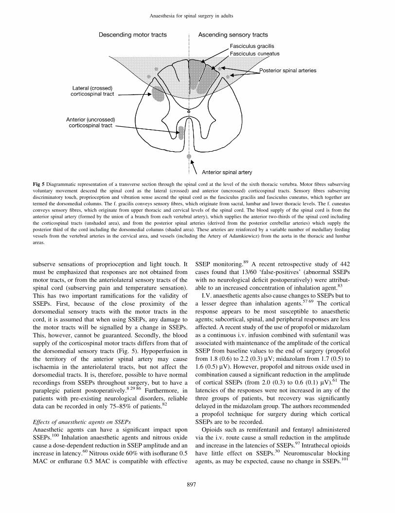
subserve sensations of proprioception and light touch. It
must be emphasized that responses are not obtained from
motor tracts, or from the anteriolateral sensory tracts of the
spinal cord (subserving pain and temperature sensation).
This has two important rami®cations for the validity of
SSEPs. First, because of the close proximity of the
dorsomedial sensory tracts with the motor tracts in the
cord, it is assumed that when using SSEPs, any damage to
the motor tracts will be signalled by a change in SSEPs.
This, however, cannot be guaranteed. Secondly, the blood
supply of the corticospinal motor tracts differs from that of
the dorsomedial sensory tracts (Fig. 5). Hypoperfusion in
the territory of the anterior spinal artery may cause
ischaemia in the anteriolateral tracts, but not affect the
dorsomedial tracts. It is, therefore, possible to have normal
recordings from SSEPs throughout surgery, but to have a
paraplegic patient postoperatively.8 29 86 Furthermore, in
patients with pre-existing neurological disorders, reliable
data can be recorded in only 75±85% of patients.82
Effects of anaesthetic agents on SSEPs
Anaesthetic agents can have a signi®cant impact upon
SSEPs.100 Inhalation anaesthetic agents and nitrous oxide
cause a dose-dependent reduction in SSEP amplitude and an
increase in latency.60 Nitrous oxide 60% with iso¯urane 0.5
MAC or en¯urane 0.5 MAC is compatible with effective
SSEP monitoring.89 A recent retrospective study of 442
cases found that 13/60 `false-positives' (abnormal SSEPs
with no neurological de®cit postoperatively) were attribut-
able to an increased concentration of inhalation agent.83
I.V. anaesthetic agents also cause changes to SSEPs but to
a lesser degree than inhalation agents.57 69 The cortical
response appears to be most susceptible to anaesthetic
agents; subcortical, spinal, and peripheral responses are less
affected. A recent study of the use of propofol or midazolam
as a continuous i.v. infusion combined with sufentanil was
associated with maintenance of the amplitude of the cortical
SSEP from baseline values to the end of surgery (propofol
from 1.8 (0.6) to 2.2 (0.3) mV; midazolam from 1.7 (0.5) to
1.6 (0.5) mV). However, propofol and nitrous oxide used in
combination caused a signi®cant reduction in the amplitude
of cortical SSEPs (from 2.0 (0.3) to 0.6 (0.1) mV).61 The
latencies of the responses were not increased in any of the
three groups of patients, but recovery was signi®cantly
delayed in the midazolam group. The authors recommended
a propofol technique for surgery during which cortical
SSEPs are to be recorded.
Opioids such as remifentanil and fentanyl administered
via the i.v. route cause a small reduction in the amplitude
and increase in the latencies of SSEPs.97 Intrathecal opioids
have little effect on SSEPs.30 Neuromuscular blocking
agents, as may be expected, cause no change in SSEPs.101
Fig 5 Diagrammatic representation of a transverse section through the spinal cord at the level of the sixth thoracic vertebra. Motor ®bres subserving
voluntary movement descend the spinal cord as the lateral (crossed) and anterior (uncrossed) corticospinal tracts. Sensory ®bres subserving
discriminatory touch, proprioception and vibration sense ascend the spinal cord as the fasciculus gracilis and fasciculus cuneatus, which together are
termed the dorsomedial columns. The f. gracilis conveys sensory ®bres, which originate from sacral, lumbar and lower thoracic levels. The f. cuneatus
conveys sensory ®bres, which originate from upper thoracic and cervical levels of the spinal cord. The blood supply of the spinal cord is from the
anterior spinal artery (formed by the union of a branch from each vertebral artery), which supplies the anterior two-thirds of the spinal cord including
the corticospinal tracts (unshaded area), and from the posterior spinal arteries (derived from the posterior cerebellar arteries) which supply the
posterior third of the cord including the dorsomedial columns (shaded area). These arteries are reinforced by a variable number of medullary feeding
vessels from the vertebral arteries in the cervical area, and vessels (including the Artery of Adamkiewicz) from the aorta in the thoracic and lumbar
areas.
Anaesthesia for spinal surgery in adults
897

Effect of controlled hypotension on SSEP
MAP during spinal surgery is usually maintained at lower
than pre-induction values in order to minimize blood
loss.111 Typically, controlled normovolaemic hypotension
to a MAP of 60 mm Hg is used. MAP should not be allowed
to decrease to less than 60 mm Hg as, at this point, SSEPs
are lost and neurological ischaemic injury can occur.90
Maintaining MAP greater than 60 mm Hg is, however, no
guarantee of safety. SSEPs demonstrate that the feline
spinal cord is more vulnerable to distractive injury during
pharmacologically induced hypotensive anaesthesia than
during normotensive anaesthesia.122 Earlier animal work
has shown that peripheral nerves, which do not have an
autoregulated blood ¯ow, are more sensitive to the effects of
hypotension than the spinal cord.39 SSEPs may, therefore,
be reduced by even moderate hypotension. Papastefanou, in
a retrospective series of 442 cases, found 17/60 `false-
positive' SSEP changes attributable to hypotension.83
Changes in SSEPs, whether a result of hypotension, or
mechanical distractive forces, or a combination of both,
must not be ignored.
Other factors
A decrease in core body temperature in animals causes a
reduction in the amplitude of SSEPs by approximately 7%
and an increase in latency of 3% for each 1°C reduction.
There does, however, appear to be a protective effect of
hypothermia on spinal cord function.43
It is clear from a large multicentre study that the
experience of monitoring teams in spinal surgery has a
signi®cant effect on outcome.78 Teams with experience of
less than 100 cases had more than twice the postoperative
neurological complication rate of teams with greater
experience.
Effectiveness of SSEP monitoring
In a large retrospective multicentre study of over 51 000
procedures, SSEP monitoring was found to have a sensitiv-
ity of 92% and speci®city of 98.9%.78 The false negative
rate was 0.127% (normal SSEPs throughout the case but a
neurological de®cit postoperatively), or 1 in 787 proced-
ures. The false positive rate was 1.51% (SSEP had changed,
but no new neurological de®cit postoperatively), or one in
67 procedures. Other studies have found a higher false
positive rate (14.7%),83 and that SSEP monitoring has a
lower speci®city, 85.33%, but a sensitivity of 100%,
probably explained by the smaller number of cases.
SSEP monitoring is currently the mainstay of spinal cord
monitoring techniques. It is, overall, a reliable technique
with a high sensitivity and speci®city for early detection of
intraoperative neurological compromise, and has a proven
record over the last decade.
Motor-evoked potentials
As a result of the inherent problems using SSEPs as a
monitoring tool during spinal surgery, and reports of
postoperative paralysis despite apparently normal intra-
operative SSEPs, efforts have been made to monitor motor
tracts of the spinal cord (Fig. 3) as a more sensitive indicator
of motor function.
MEP monitoring was ®rst used clinically over 10 yr
ago.19 Monitoring techniques are subdivided according to:
the site of stimulation (motor cortex, spinal cord); the
method of stimulation (electrical potential, magnetic ®eld);
and the site of recording (spinal cord, peripheral mixed
nerve, muscle). Each variation of the technique has
advantages and disadvantages. The principle is the same;
stimulation by whatever means cranial to the site of surgery
causes prodromic stimulation of motor tracts in the spinal
cord, and of peripheral nerve and muscle caudal to the site
of surgery. Perturbation of motor pathway function by
surgery leads to a reduction in amplitude and an increase in
latency of the recorded responses.
The motor cortex can be stimulated by electrical,11 52 87
or magnetic37 56 means. Magnetic equipment is bulky and
cumbersome but is not affected by the quality of electrode
contact. Recorded responses are classi®ed as myogenic or
neurogenic. Myogenic responses result from the summed
EMG activity in a muscle, such as tibialis anterior, in
response to stimulation (Fig. 4). Neurogenic recordings
arise from the summed electrical activity in a peripheral
nerve or the spinal cord. The advantage of recording EMG
responses is their large amplitude. The main disadvantage is
their variable morphology. When recording EMG re-
sponses, the depth of muscle relaxation is of critical
importance: if it is too deep, responses are unobtainable;
if only residual block is present, there is a risk of injury to
the patient, if violent movements occur in response to
stimulation. Neuromuscular blocking agents should be
administered by continuous infusion, and the depth of
neuromuscular block monitored. The ®rst twitch of the
train-of-four response should be maintained at 10±20% of
control.37 Neurogenic responses, however, can be recorded
under complete neuromuscular block to avoid patient injury,
and are more reliable in terms of amplitude, latency, and
morphology.
Effects of anaesthetic agents on MEPs
Cortical evoked responses are more prone to the effects of
anaesthetic agents than spinal-evoked responses. Propofol is
a powerful suppressant of cortical evoked responses,
causing a dose-dependent reduction in the amplitude of
the response. A bolus dose of propofol 2 mg kg±1 abolishes
cortical MEPs.51 Volatile agents are also powerful suppres-
sants of cortical evoked MEPs; MEPs are abolished or are
too inconsistent to interpret at end-tidal iso¯urane concen-
trations of 0.87 (0.08)%.87 Midazolam and etomidate cause
a signi®cant but smaller reduction in the amplitude of the
response. Opioids such as fentanyl have been variously
reported as reducing the amplitude of the response,56 or
causing no effect.51 Multiple pulse transcranial electrical
Raw et al.
898

stimulation techniques can improve the reliability of MEP
recording further.
These ®ndings have led to the need for signi®cant
modi®cations to the anaesthetic technique when cortical-
evoked MEPs are measured. In the past, ketamine-based
techniques have been used, but not without complications
such as unpleasant hallucinations.52 An i.v. propofol
infusion with fentanyl or remifentanil has been advocated
and provides for adequate MEP recording in 97% of
neurologically intact patients, when used with a multiple
pulse stimulation technique.87
Effectiveness of MEP monitoring
MEP monitoring is less reliable in patients with a pre-
existing neurological de®cit.37 Furthermore, early hopes of
greater sensitivity of MEPs over other monitoring tech-
niques may have been premature. There are recent reports of
preserved neurogenic MEPs associated with postoperative
motor de®cit,72 which suggest the sensitivity of MEPs is less
than 100%.
SSEP monitoring has become an accepted standard of
care during spinal surgery. It is less affected by the technical
dif®culties associated with MEP monitoring. MEP moni-
toring is, however, becoming more widely used and the two
methods should be regarded as complementary, with the use
of a wake-up test reserved for situations where neuro-
physiological monitoring is not possible or responses are
signi®cantly perturbed during surgery.
Postoperative care
Patients undergoing spinal surgery frequently have signi®-
cant co-morbidity. Surgery imposes the further stresses of
signi®cant blood loss, prolonged anaesthesia, and dif®cul-
ties in acute postoperative pain management. Surgeons
prefer patients to be conscious and able to respond to
command immediately after anaesthesia, for early neuro-
logical assessment. It is also important that patients are able
to expectorate, and to comply with physiotherapy as early as
possible in the postoperative period.
Indications for postoperative ventilation
The decision to provide a period of postoperative arti®cial
ventilation should have been made before surgery com-
mences, and explained to the patient. The need for
postoperative ventilation is suggested by patient and
surgical factors. Patient factors include the presence of a
pre-existing neuromuscular disorder, severe restrictive
pulmonary dysfunction with a preoperative vital capacity
of less than 35% of predicted, a congenital cardiac
abnormality, right ventricular failure, and obesity.
Surgical factors include a prolonged procedure, surgical
invasion of the thoracic cavity,117 and blood loss greater
than 30 ml kg±1. Frequently, it is necessary only to provide
arti®cial ventilation for a few hours in the postoperative care
unit, until hypothermia and metabolic derangements have
been corrected. Chest drains, if present, should be checked
regularly to ensure patency; obstruction may lead to a
pneumo- or haemothorax.
Postoperative analgesia
Pain management can be a considerable challenge. Patients
undergoing spinal surgery, particularly through a thoracic
approach, may have a large incision extending over several
dermatomes. Many patients have pre-existing chronic pain
conditions, may be cognitively impaired (some neuromus-
cular disorders), or be very young (children). A multimodal
approach to analgesia is recommended, using a combination
of simple primary analgesics, opioids, and regional anaes-
thesia techniques where appropriate. For initial post-
operative analgesia, it is useful to restart, if possible, all
the analgesics the patient was receiving preoperatively.
Undoubtedly, the patient's requirements will be increased
postoperatively and additional therapy will be required.
Parenteral opioids
The use of parenteral opioids has been the mainstay of
analgesia for all patients undergoing spinal surgery. Opioids
can be administered via i.m., i.v. (continuous infusion and
patient-controlled analgesia devices with or without back-
ground infusions), intrapleural, epidural, and intrathecal
routes. Their use, via the i.v. route in particular, is associated
with side-effects such as respiratory depression, nausea and
vomiting, sedation, and gastrointestinal ileus. The latter
may be especially disadvantageous after major spinal
surgery, when some degree of paralytic ileus is common.
Patients with cancer may not be naõÈve to opioid drugs and
such individuals must be assumed to have acquired a degree
of opioid tolerance. For patients who have received long-
term opioids preoperatively by other routes (e.g. enteral,
transdermal), these should also be restarted as early as
possible postoperatively, and gradually reduced over sub-
sequent days or weeks.
Non-steroidal anti-in¯ammatory drugs
Simple analgesics alone afford inadequate analgesia even
for relatively minor spinal surgery. Non-steroidal anti-
in¯ammatory drugs (NSAIDs), both non-selective cyclo-
oxygenase inhibitors, and selective cyclo-oxygenase 2
(COX 2) inhibitors, have however been used successfully
after spinal surgery.75 93 But the use of a non-selective
cyclo-oxygenase inhibitor NSAID cannot be recommended
for intraoperative or early postoperative analgesia in such
cases. Guidelines on the use of NSAIDs published by The
Royal College of Anaesthetists96 do not specify their role
for this purpose. The use of NSAIDs may increase bleeding
time by 30±35%, cause gastritis, and be associated with
acute renal failure, particularly in the presence of hypovo-
laemia and hypotension. The safety pro®le of selective
COX 2 inhibitors in major spinal surgery is yet to be fully
evaluated. If, however, patients have been taking NSAIDs to
Anaesthesia for spinal surgery in adults
899
help relieve their pain without complication preoperatively,
it is useful to restart this medication in the immediate
postoperative period for its opioid-sparing effects.
Epidural analgesia
The use of local anaesthetic agents, alone or in combination
with opioids,65 by the epidural route after spinal surgery has
been described, the epidural catheter being placed intra-
operatively by the surgeon. Two recent studies compared
epidural analgesia with parenteral morphine administered
via patient controlled analgesia (PCA) devices after major
spinal surgery.12 114 The retrospective study114 found time
to full diet and hospital stay to be reduced in the epidural
group, but the randomized study was unable to ®nd any
signi®cant differences in pain scores, side-effects, or
resumption of oral intake between the epidural and PCA
groups.12
Epidural anaesthesia with local anaesthetic agents can
make neurological assessment dif®cult. Concerns over the
rare but serious risks of epidural haematoma and infection
associated with indwelling catheters have hindered its
widespread use. The incidence of local super®cial infection
after routine epidural catheter placement has been quoted to
be as high as 12%.16 However, the incidence of epidural
abscess related to epidural catheter placement, although
dif®cult to determine precisely, is rare. A recent review of
the literature produced 42 case reports of epidural abscesses
from 1974 to 1996, only 13 of which were sited for
perioperative analgesia.54 The authors of this study identi-
®ed in their own institution two catheter-related epidural
abscesses in 13 000 epidural blocks (both of which were in
obstetric patients). A higher incidence of epidural catheter-
related abscesses may be anticipated for catheters inserted
via a surgical wound.
The incidence of spinal haematoma in patients receiving
spinal or epidural analgesia and low-molecular weight
heparin is estimated to be between 1:1000 and 1:10 000.41
The clinical signs of epidural haematoma or abscess may be
masked by postoperative pain, and pre-existing abnormal
neurology. The infrequency of these complications further
increases the risk of failing to detect them.
Intrathecal analgesia
The thecal sac is readily accessible during spinal surgical
procedures, and intrathecal medication can be injected with
technical ease before wound closure. Early reports of the use
of intrathecal opioids for analgesia in children after spinal
surgery,15 and other major surgeries,47 suggested that the
use of morphine (20±30 mg kg±1) was associated with
excellent analgesia for up to 24 h, but was complicated by
respiratory depression in 16% of patients.47 More recent
studies suggest the optimum dose of morphine to be 2±5 mg
kg±1, which provides comparable analgesia for 24 h but with
fewer side-effects such as respiratory depression, nausea,
and pruritus.9 27 Doses of this order, in a large retrospective
study of 5969 patients, produced respiratory depression in
3% of patients.33 There were no cases of neurological injury
or spinal haematoma.
Signi®cant pain can be expected for up to 4 days
postoperatively after major spinal surgery. Intrathecal
opioids alone, therefore, will probably be insuf®cient over
this period. Parenteral opioids made available in the
immediate postoperative period (as patient-controlled anal-
gesia regimen, for example), in patients who have received
intrathecal opioids, provide for a smooth transition from
predominately intrathecal opioid-based analgesia to
parenteral opioid-based analgesia.27 The use of such a
regimen is advisable in the setting of a postoperative critical
care facility; animal data suggest systemic and intrathecal
opioids may act synergistically.123
Other techniques
Intrapleural infusions of local anaesthetic and/or opioids
may be considered after a thoracotomy. Intrapleural infu-
sions of local anaesthetic agents have been reported to
reduce systemic opioid requirements in such circum-
stances.44 A number of studies, however, have found
intrapleural analgesia with local anaesthetic agents to
offer inferior analgesia to epidural opioids,25 71 and there
remains concern over the blood levels of local anaesthetic
agents achieved, and the risk of local anaesthetic toxicity.91
Late complications
A retrospective study of 1223 anterior thoracic and lumbar
spinal procedures found a respiratory complication rate of
7% (adult respiratory distress syndrome, pneumonitis,
atelectasis, infection), pulmonary embolism 0.8%, cerebral
vascular accident 0.25%, and death 0.33% (four patients:
one died of ARDS on day 31, one myocardial infarction on
day 1, and two died of fatal pulmonary emboli on days 3 and
8).22
Screening studies for thromboembolic complications
after spinal surgery have quoted varied incidences between
0.395 and 15.5%.79 One partially randomized study found
an incidence of 0.3% when Doppler ultrasonography was
used postoperatively to detect venous thrombosis in patients
treated with either mechanical (compression stockings and
pneumatic boots), or pharmacological (warfarin) methods
of prophylaxis.95 If, however, no methods of thrombo-
prophylaxis are used and venography is the screening
method,79 incidences of venous thrombosis of 15.5% have
been quoted. In one prospective study, symptomatic
pulmonary emboli occurred in 2.2% of all spinal fusions,
and these were more common after combined anterior/
posterior approach procedures (6%), than posterior ap-
proach procedures alone (0.5%).18
There has been a paucity of well-conducted randomized
controlled trials comparing methods of thromboprophy-
laxis. Few would argue against the use of compression
stockings and pneumatic boots, the use of which is relatively
free of complications. However, the use of pharmacological
Raw et al.
900

methods of thromboprophylaxis (using heparin, for exam-
ple) is contentious. Their use may be associated with
haemorrhagic complications such as increased intra-
operative and postoperative blood loss,95 and epidural
haematoma. Further studies are required to adequately
ascertain the most appropriate methods of prophylaxis
against thromboembolism.
Treatment of thromboembolism produces the risk of
major heparin-related complications (44%) such as epidural
haematoma, gastrointestinal bleeding, and wound infec-
tion.10 Insertion of an IVC ®lter may be appropriate in these
circumstances.
Conclusions
Spinal surgery presents a number of challenges to the
anaesthetist. Patients are now undergoing major spinal
surgery for conditions such as malignancy, scoliosis, and
trauma, which would not have been contemplated 20 yr ago.
Despite this, postoperative neurological morbidity has been
reduced by advances in spinal cord monitoring techniques.
The anaesthetist has an important role to play in facilitating
the use of these new techniques. They must also manage the
relief of postoperative pain in these patients, who have
frequently been receiving several analgesics preoperatively.
References1 Albin MS, Ritter RR, Pruett CE, Kalff K. Venous air embolism
during lumbar laminectomy in the prone position: report ofthree cases. Anesth Analg 1991; 73: 346±9
2 Alderson JD. Spinal cord injuries. Care Crit Ill 1999; 15: 48±52
3 Allain J, Akehurst RL, Hunter JM. Autologous transfusion, 3 yronÐwhat is new? What has happened? Second ConsensusConference on autologous transfusion held at the Royal Collegeof Physicians, Edinburgh, November 11, 1998. Br J Anaesth 1999;82: 783±4
4 American EEG Society. Guidelines for intraoperative monitoringof sensory-evoked potentials. J Clin Neurophysiol 1987; 4: 397±416
5 Amzallag M. Autonomic hyperre¯exia. Int Anesthesiol Clin 1993;31: 87±102
6 Anonymous. Assessment: intraoperative neurophysiology.Report of Therapeutics and Technology AssessmentSubcommittee of the American Academy of Neurology.Neurology 1990; 40: 1644±6
7 Beaussier M, Paugam C, Deriaz H, et al. Haemodynamic stabilityduring moderate hypotensive anaesthesia for spinal surgery. Acomparison between des¯urane and iso¯urane. Acta AnaesthesiolScand 1800; 44: 1154±9
8 Ben-David B, Haller G, Taylor P. Anterior spinal fusioncomplicated by paraplegia. A case report of a false-negativesomatosensory-evoked potential. Spine 1987; 12: 536±9
9 Boezaart AP, Eksteen JA, Spuy GV, et al. Intrathecal morphine.Double-blind evaluation of optimal dosage for analgesia aftermajor lumbar spinal surgery. Spine 1999; 24: 1131±7
10 Cain JE, Major MR, Lauerman WC, et al. The morbidity ofheparin therapy after development of pulmonary embolus inpatients undergoing thoracolumbar or lumbar spinal fusion. Spine1995; 18: 1600±3
11 Calancie B, Harris W, Broton JG, et al. `Threshold-level' multi-pulse transcranial electrical stimulation of motor cortex for
intraoperative monitoring of spinal motor tracts: description of
method and comparison to somatosensory evoked potential
monitoring. J Neurosurg 1998; 88: 457±70
12 Cassady JF jr, Lederhaas G, Cancel DD, et al. A randomised
comparison of the effects of continuous thoracic epidural
analgesia and intravenous patient-controlled analgesia afterposterior spinal fusion in adolescents. Reg Anesth Pain Med
2000; 25: 246±53
13 Cha CW, Deible C, Muzzonigro T, et al. Allogeneic transfusion
requirements after autologous donations in posterior lumbar
surgeries. Spine 2002; 27: 99±104
14 Chen SH, Huang TJ, Lee YY, Hsu RW. Pulmonary function after
thoracoplasty in adolescent idiopathic scoliosis. Clin Orthop 2002;
399: 152±61
15 Dalens B, Tanguy A. Intrathecal morphine for spinal fusion inchildren. Spine 1988; 13: 494±8
16 Darchy B, Forceville X, Bavoux E, et al. Clinical and bacteriologic
survey of epidural analgesia in patients in the intensive care unit.
Anesthesiology 1996; 85: 988±98
17 Dawson EG, Sherman JE, Kanim LEA, Nuwer MR. Spinal cord
monitoring. Results of the Scoliosis Research Society and the
European Spinal Deformity Society Survey. Spine 1991; 16
[Suppl.]: 361±4
18 Dearbon JT, Hu SS, Tribus CB, Bradford DS. Thromboembolic
complications after major thoracolumbar spine surgery. Spine
1999; 24: 1471±6
19 Edmonds HJ, Paloheimo MP, Backman MH, et al. Transcranial
magnetic motor evoked potentials (tcMMEP) for functional
monitoring of motor pathways during scoliosis surgery. Spine
1989; 14: 683±6
20 Engelhardt T, Webster NR. Pulmonary aspiration of gastriccontents in anaesthesia. Br J Anaesth 1999; 83: 453±60
21 Epstein NE, Danto J, Nardi D. Evaluation of intraoperative
somatosensory-evoked potential monitoring during 100 cervical
operations. Spine 1993; 18: 737±47
22 Faciszewski T, Winter RB, Lonstein JE, et al. The surgical and
medical perioperative complications of anterior spinal fusion
surgery in the thoracic and lumbar spine in adults: A review of
1223 procedures. Spine 1995; 20: 1592±9
23 Florentino-Pineda I, Blakemore LC, Thompson GH, et al. Theeffect of epsilon-aminocaproic acid on perioperative blood loss
in patients with idiopathic scoliosis undergoing posterior spinal
fusion: a preliminary prospective study. Spine 2001; 26: 1147±51
24 Frazer A, Edmonds-Seal J. Spinal cord injuries. Anaesthesia 1982;
37: 1084±98
25 Gaeta RR, Macario A, Brodsky JB, et al. Pain outcomes after
thoracotomy: lumbar epidural hydromorphone versus
intrapleural bupivacaine. J Cardiothorac Vasc Anesth 1995; 9:534±7
26 Galasko CSB, Delaney C, Morris P. Spinal stabilisation in
Duchenne dystrophy. J Bone Joint Surg 1992; 74B: 210±4
27 Gall O, Aubineau JV, Berniere J, et al. Analgesic effect of low-
dose intrathecal morphine after spinal fusion in children.
Anesthesiology 2001; 94: 447±52
28 Genever EE. Suxamethonium-induced cardiac arrest in
unsuspected pseudohypertrophic muscular dystrophy. Br JAnaesth 1971; 43: 984±6
29 Ginsburg HH, Shetter AG, Raudzens PA. Postoperative
paraplegia with preserved intraoperative somatosensory
evoked potentials. J Neurosurg 1985; 63: 296±300
30 Goodarzi M, Shier N-H, Grogan DP. Effect of intrathecal opioids
Anaesthesia for spinal surgery in adults
901

on somatosensory-evoked potentials during spinal fusion inchildren. Spine 1996; 21: 1565±8
31 Gross JB. Estimating allowable blood loss: corrected for dilution.Anesthesiology 1983; 58: 277±80
32 Guerit JM. Neuromonitoring in the operating room: why, when,and how to monitor? Electroenceph Clin Neurophysiol 1998; 106:1±21
33 Gwirtz KH, Young JV, Byers RS, et al. The safety and ef®cacy ofintrathecal opioids for acute postoperative pain: seven years'experience with 5969 surgical patients at Indiana UniversityHospital. Anesth Analg 1999; 88: 599±604
34 Haas S, Ketterl R, Stemberger A, et al. The effect of aprotinin onplatelet function, blood coagulation and blood lactate level intotal hip replacement: a double blind clinical trial. Adv Exp MedBiol 1984; 167: 287±97
35 Haberkern M, Dangel P. Normovolaemic haemodilution andintraoperative auto-transfusion in children: experience with 30cases of spinal fusion. Eur J Pediatr Surg 1991; 1: 30±5
36 Hambly PR, Martin B. Anaesthesia for chronic spinal cord lesions.Anaesthesia 1998; 53: 273±89
37 Herdmann J, Lumenta CB, Huse KOW. Magnetic stimulation formonitoring of motor pathways in spinal procedures. Spine 1993;18: 551±9
38 Hersey SL, O'Dell NE, Lowe S, et al. Nicardipine versusnitroprusside for controlled hypotension during spinal surgeryin adolescents. Anesth Analg 1997; 84: 1239±44
39 Hitchon PW, Lobosky JM, Yamada T, et al. Effect ofhaemorrhagic shock upon spinal cord blood ¯ow and evokedpotentials. Neurosurgery 1987; 21: 849±57
40 Hoppenfeld S, Gross A, Andrews C, Lonner B. The ankle clonustest for assessment of the integrity of the spinal cord duringoperations for scoliosis. J Bone Joint Surg Am 1997; 79: 208±12
41 Horlocker TT, Wedel DJ. Spinal and epidural blockade andperioperative low molecular weight heparin: smooth sailing onthe Titanic. Anesth Analg 1998; 86: 1153±6
42 Hur SR, Huizenga BA, Major M. Acute normovolemichemodilution combined with hypotensive anesthesia and othertechniques to avoid homologous transfusion in spinal fusionsurgery. Spine 1992; 17: 867±73
43 Jou IM. Effects of core temperature on changes in spinalsomatosensory-evoked potential in acute spinal cordcompression injury. Spine 2000; 25: 1878±85
44 Inderbitzi R, Flueckiger K, Ris HB. Pain relief and respiratorymechanics during continuous intrapleural bupivacaineadministration after thoracotomy. Thorac Cardiovasc Surg 1992;40: 87±9
45 Jenkins JG, Bohn D, Edmonds JF, et al. Evaluation of pulmonaryfunction in muscular dystrophy patients requiring spinal surgery.Crit Care Med 1982; 9: 645±9
46 John DA, Tobey RE, Homer LD, Rice CL. Onset ofsuccinylcholine-induced hyperkalemia following denervation.Anesthesiology 1976; 45: 294±9
47 Jones SEF, Beasley JM, Macfarlane DWR, et al. Intrathecalmorphine for post-operative pain relief in children. Br J Anaesth1984; 56: 137±40
48 Kafer ER. Idiopathic scoliosis: gas exchange and the agedependence of arterial blood gases. J Clin Invest 1976; 58: 825±33
49 Kafer ER. Respiratory and cardiovascular functions in scoliosisand the principles of anesthetic management. Anesthesiology1980; 52: 339±51
50 Kakiuchi M. Reduction of blood loss during spinal surgery byepidural blockade under normotensive general anaesthesia. Spine1997; 22: 889±94
51 Kalkman CJ, Drummond JC, Ribbernick AA, et al. Effects of
propofol, etomidate, midazolam, and fentanyl on motor evokedresponses to transcranial electrical or magnetic stimulation inhumans. Anesthesiology 1992; 76: 502±9
52 Kawaguchi M, Sakamoto T, Inoue S, et al. Low dose propofol as asupplement to ketamine-based anesthesia during intraoperativemonitoring of motor-evoked potentials. Spine 2000; 25: 974±9
53 Kearon C, Viviani GR, Kirkley A, Killian KJ. Factors determiningpulmonary function in adolescent idiopathic thoracic scoliosis.Am Rev Respir Dis 1993; 148: 288±94
54 Kindler CH, Seeberger MD, Staender SE. Epidural abscesscomplicating epidural anaesthesia and analgesia. Acta AnaesthesiolScand 1998; 42: 614±20
55 Kinnear WJ, Johnston ID. Does Harrington instrumentationimprove pulmonary function in adolescents with idiopathicscoliosis? A meta-analysis. Spine 1993; 18: 1556±9
56 Kitagawa H, Nakamura H, Kawaguchi Y, et al. Magnetic-evokedcompound muscle action potential neuromonitoring in spinesurgery. Spine 1995; 20: 2233±9
57 Koht A, Schutz W, Schmidt G, et al. Effects of etomidate,midazolam and thiopental on median nerve somatosensoryevoked potentials and the additive effects of fentanyl and nitrousoxide. Anesth Analg 1988; 67: 435±41
58 Koscielniak-Neilsen ZJ, Stens-Pedersen HL, Hesselbjerg L.Midazolam-¯umazenil versus propofol anaesthesia for scoliosissurgery with wake-up tests. Acta Anaesthesiol Scand 1998; 42:111±6
59 Kumano K, Tsuyama N. Pulmonary function before and aftersurgical correction of scoliosis. J Bone Joint Surg 1982; 64: 242±8
60 Lam AM, Sharar SR, Mayberg TS, et al. Iso¯urane compared withnitrous oxide anaesthesia for intraoperative monitoring ofsomatosensory evoked potentials. Can J Anaesth 1994; 41:295±300
61 Langeron O, Vivien B, Paqueron X, et al. Effects of propofol,propofol-nitrous oxide and midazolam on corticalsomatosensory evoked potentials during sufentanil anaesthesiafor major spinal surgery. Br J Anaesth 1999; 82: 340±5
62 Lee TC, Yang LC, Chen HJ. Effect of patient position andhypotensive anesthesia on inferior vena caval pressure. Spine1998; 23: 941±7
63 Lenke LG, Bridwell KH, Blanke K, Baldus C. Analysis ofpulmonary function and chest cage dimension changes afterthoracoplasty in idiopathic scoliosis. Spine 1995; 20: 1343±50
64 Letts M, Pang E, D'Astous J, et al. The in¯uence of desmopressinon blood loss during spinal fusion surgery in neuromuscularpatients. Spine 1998; 23: 475±8
65 Lowry KJ, Tobias J, Kittle D, et al. Postoperative pain controlusing epidural catheters after anterior spinal fusion foradolescent scoliosis. Spine 2001; 26: 1290±3
66 Malcolm-Smith NA, MacMaster MJ. The use of inducedhypotension to control bleeding during posterior fusion forscoliosis. J Bone Joint Surg 1983; 65: 255±8
67 Mansel JK, Norman JR. Respiratory complications andmanagement of spinal cord injuries. Chest 97: 1446±52
68 McLeod AD, Calder I. Spinal cord injury and directlaryngoscopyÐthe legend lives on. Br J Anaesth 2000; 84: 705±9
69 McPherson RW, Sell B, Traystman RJ. Effects of thiopental,fentanyl, and etomidate on upper extremity somatosensoryevoked potentials in humans. Anesthesiology 1986; 65: 584±9
70 Meyer PR jr, Cotler HB, Gireesan GT. Operative neurologicalcomplications resulting from thoracic and lumbar spine internal®xation. Clin Orthop 1988; 237: 125±31
71 Miguel R, Hubbell D. Pain management and spirometry followingthoracotomy: a prospective, randomised study of fourtechniques. J Cardiothorac Vasc Anesth 1993; 7: 529±34
Raw et al.
902

72 Minahan RE, Sepkuty JP, Lesser RP, et al. Anterior spinal cordinjury with preserved neurogenic `motor' evoked potentials. Clin
Neurophysiol 2001; 112: 1442±50
73 Morris P. Duchenne muscular dystrophy: a challenge for the
anaesthetist. Pediatric Anaesth 1997; 7: 1±4
74 Neilipovitz DT, Murto K, Hall L, et al. A randomised trial oftranexamic acid to reduce blood transfusion for scoliosis
surgery. Anesth Analg 2001; 93: 82±7
75 Nissen I, Jensen KA, OÈ hrstroÈm JK. Indomethacin in the
management of postoperative pain. Br J Anaesth 1992; 69: 304±6
76 Noordeen MH, Haddad FS, Muntoni F, et al. Blood loss in
Duchenne muscular dystrophy: vascular smooth muscledysfunction? J Pediatr Orthop B 1999; 8: 212±5
77 Nuttall GA, Horlocker TT, Santrach PJ, et al. Predictors of blood
transfusions in spinal instrumentation and fusion surgery. Spine
2000; 25: 596±601
78 Nuwer MR, Dawson EG, Carlson LG, et al. Somatosensoryevoked potential monitoring reduces neurological de®cits after
scoliosis surgery: results of a large multicenter study.
Electroenceph Clin Neurophysiol 1995; 96: 6±11
79 Oda T, Fuji T, Kato Y et al. Deep vein thrombosis after posterior
spinal surgery. Spine 2000; 25: 2962±7
80 Owen JH, Naito M, Bridwell KH, Oakley DM. Relationshipbetween duration of spinal cord ischaemia and postoperative
neurologic de®cits in animals. Spine 1990; 15: 846±51
81 Owen JH. Monitoring during surgery for spinal deformities. In:
Birdwell KH, DeWald RL, eds. The Textbook of Spinal Surgery, 2ndEdn. Philadelphia: Lippincott-Raven Publishers, 1997; 39±60
82 Owen JH. The application of intraoperative monitoring during
surgery for spinal deformity. Spine 1999; 24: 2649±62
83 Papastefanou SL, Henderson LM, Smith NJ, et al. Surface
electrode somatosensory-evoked potentials in spinal surgery.
Spine 2000; 25: 2467±72
84 Park CK. The effect of patient positioning on intraabdominalpressure and blood loss in spinal surgery. Anesth Analg 2000; 91:
552±7
85 Pehrsson K, Larsson S, Oden A, Nachemson A. Long-term
follow-up of patients with untreated scoliosis. A study ofmortality, causes of death, and symptoms. Spine 1992; 17: 1091±
6
86 Pelosi L, Jardine A, Webb JK. Neurological complications of
anterior spinal surgery for kyphosis with normal somatosensory
evoked potentials. J Neurol Neurosurg Psychiatry 1999; 66: 662±4
87 Pelosi L, Stevenson M, Hobbs GJ, et al. Intraoperative motorevoked potentials to transcranial electrical stimulation during
two anaesthetic regimens. Clin Neurophysiol 2001; 112: 1076±87
88 Pennefather SH, Russell GN. Placement of double-lumen
tubesÐtime to shed light on an old problem. Br J Anaesth
2000; 84: 308±9
89 Peterson DO, Drummond DC, Todd MM. Effects of halothane,
en¯urane, iso¯urane and nitrous oxide on somatosensory
evoked potentials in humans. Anesthesiology 1986; 65: 35±40
90 Polo A, Tercedor A, Paniagua-Soto J, et al. [Neurophysiological
monitoring during scoliosis surgery using controlledhypotension] (Spanish). Rev Esp Anesthesiol Reanim 2000; 47:
367±70
91 Raf®n L, Fletcher D, Sperandio M, et al. Interpleural infusion of
2% lidocaine with 1:200,000 epinephrine for post-thoracotomy
analgesia. Anesth Analg 1994; 79: 328±34
92 Reid JM, Appleton PJ. A case of ventricular ®brillation in theprone position during back stabilisation surgery in a boy with
Duchenne's muscular dystrophy. Anaesthesia 1999; 54: 364±7
93 Reuben SS, Connelly NR. Postoperative analgesic effects of
celecoxib or rofecoxib after spinal fusion surgery. Anesth Analg2000; 91: 1221±5
94 Rodola F, D'Avolio S, Chierichini A, et al. Wake-up test duringmajor spinal surgery under remifentanil balanced anaesthesia. EurRev Med Pharmacol Sci 2000; 4: 67±70
95 Rotiko SE, Schwartz MC, Neuwirth MG. Deep vein thrombosisafter major reconstructive spinal surgery. Spine 1996; 21: 853±9
96 Rowbotham DJ, Alexander J, Arden C, et al. Guidelines for theuse of non-steroidal anti-in¯ammatory drugs in the perioperativeperiod. The Royal College of Anaesthesists, UK 1998
97 Samra SK, Dy EA, Welch KB, et al. Remifentanil- and fentanyl-based anesthesia for intraoperative monitoring ofsomatosensory evoked potentials. Anesth Analg 2001; 92:1510±5
98 Shands AR, Eisberg HB. The incidence of scoliosis in the state ofDelaware. J Bone Joint Surg 1955; 37: 1243±9
99 Shapiro F, Sethna N, Colan S, et al. Spinal fusion in Duchennemuscular dystrophy: a multidisciplinary approach. Muscle Nerve1992; 15: 604±14
100 Sloan TB. Anaesthetic effects on electrophysiological recordings.J Clin Neurophysiol 1998; 15: 217±26
101 Sloan TB. Non-depolarising neuromuscular blockade does notalter sensory evoked potentials. J Clin Monit 1994; 9: 4±9
102 Snook NJ, O'Beirne HA, Enright S, et al. Use of recombinanterythropoietin to facilitate liver transplantation in a Jehovah'sWitness. Br J Anaesth 1996; 76: 740±3
103 Solares G, Herranz JL, Sanz MD. Suxamethonium-inducedcardiac arrest as an initial manifestation of Duchenne musculardystrophy. Br J Anaesth 1986; 58: 576
104 Soliman DE, Maslow AD, Bokesch PM, et al. Transesophagealechocardiography during scoliosis repair: comparison with CVPmonitoring. Can J Anaesth 1998; 45: 925±32
105 Supkis DE jr, Varon J. Uncommon problems related to cancer.In: Benumof J, ed. Anesthesia and Uncommon Diseases, 4th Edn.Philadelphia: WB Saunders Co., 1998; 545±60
106 Sutherland RW, Winter RJ. Two cases of fatal air embolism inchildren undergoing scoliosis surgery. Acta Anaesthesiol Scand1997; 41: 1073±6
107 Tobias JD. Fenoldopam for controlled hypotension during spinalfusion in children and adolescents. Paediatr Anaesth 2000; 9: 261±6
108 Tobias JD. Sevo¯urane for controlled hypotension during spinalsurgery: preliminary experience in ®ve adolescents. PaediatrAnaesth 1998; 8: 167±70
109 Torella F, Haynes SL, Lardi A, et al. Unchanging attitudes toautologous transfusion in the UK. Transfus Med 2001; 11: 15±9
110 Toyota S, Amaki Y. Hemodynamic evaluation of the proneposition by transesophageal echocardiography. J Clin Anesth 1998;9: 32±5
111 Ulrich PF jr, Keene JS, Hogan KJ, Roecker EB. Results ofhypotensive anesthesia in operative treatment of thoracolumbarfractures. J Spinal Disord 1990; 3: 329±33
112 United Kingdom Department Of Health: http://www.doh.gov.uk/hes/free_data/index.html
113 Urban MK, Beckman J, Gordon M, et al. The ef®cacy ofanti®brinolytics in the reduction of blood loss during complexadult reconstructive spine surgery. Spine 2001; 26: 1152±6
114 VanBoerum DH, Smith JT, Curtlin MJ. A comparison of theeffects of patient-controlled analgesia with intravenous opioidsversus epidural analgesia on recovery after surgery for idiopathicscoliosis. Spine 2000; 25: 2355±7
115 Vanderlinde ES, Heal JM, Blumberg N. Clinical review:autologous transfusion. Br Med J 2002; 324: 772±5
116 Vauzelle C, Stagnara P, Jouvinoux P. Functional monitoring of
Anaesthesia for spinal surgery in adults
903
spinal cord activity during spinal surgery. Clin Orthop 1973; 93:173±8
117 Vedantam R, Lenke LG, Bridwell KH, et al. A prospectiveevaluation of pulmonary function in patients with adolescentidiopathic scoliosis relative to the surgical approach used forspinal arthrodesis. Spine 2000; 25: 82±90
118 Vitale MG, Stazzone EJ, Gelijns AC, et al. The effectiveness ofpreoperative erythropoietin in averting allogenic bloodtransfusion among children undergoing scoliosis surgery. JPediatr Orthop B 1998; 7: 203±9
119 White AA 3rd, Johnson RM, Panjabi MM, et al. Biomechanicalanalysis of clinical stability in the cervical spine. Clin Orthop 1975;109: 85±96
120 Wimmer C, Gluch H, Franzreb M, Ogon M. Predisposing factorsfor infection in spine surgery: a survey of 850 spinal procedures. JSpinal Disord 1998; 11: 124±8
121 Yaster M, Simmons RS, Tolo VT, et al. A comparison ofnitroglycerin and nitroprusside for inducing hypotension inchildren: a double blind study. Anesthesiology 1986; 65: 175±9
122 Yeoman PM, Gibson MJ, Hutchison A, et al. In¯uence of inducedhypotension and spinal distraction on feline spinalsomatosensory evoked potentials. Br J Anaesth 1989; 63: 315±20
123 Yeung JC, Rudy TA. Multiplicative interaction between narcoticagonisms expressed at spinal and supraspinal sites ofantinociceptive action as revealed by concurrent intrathecaland intracerebroventricular injections of morphine. J PharmacolExp Ther 1980; 215: 633±42
124 Zheng F, Cammisa FP jr, Sandhu HS, et al. Factors predictinghospital stay, operative time, blood loss, and transfusion inpatients undergoing revision posterior lumbar spinedecompression, fusion, and segmental instrumentation. Spine2002; 27: 818±24
Raw et al.
904

Cerebral protection
S. Fukuda1 and D. S. Warner1–3*
1Department of Anesthesiology, 2Department of Neurobiology and 3Department of Surgery, Duke University
Medical Center, Box 3094, Durham, NC 27710, USA
*Corresponding author: Department of Anesthesiology, Duke University Medical Center, Box 3094,
Durham, NC 27710, USA. E-mail: [email protected]
Ischaemic/hypoxic insults to the brain during surgery and anaesthesia can result in long-term
disability or death. Advances in resuscitation science encourage progress in clinical manage-
ment of these problems. However, current practice remains largely founded on extrapolation
from animal studies and limited clinical investigation. A major step was made with demon-
stration that rapid induction of mild sustained hypothermia in comatose survivors of out-
of-hospital ventricular fibrillation cardiac arrest reduces death and neurological morbidity with
negligible adverse events. This provides the first irrefutable evidence that outcome can be
favourably altered in humans with widely applicable neuroprotection protocols. How far
hypothermic protection can be extended to global ischaemia of other aetiologies remains to
be determined. All available evidence suggests an adverse response to hyperthermia in ischae-
mic or post-ischaemic brain. Management of other physiological values can have dramatic
effects in experimental injury models and this is largely supported by available clinical data.
Hyperoxaemia may be beneficial in transient focal ischaemia but deleterious in global ischae-
mia. Hyperglycaemia causes exacerbation of most forms of cerebral ischaemia and this can be
abated by restoration of normoglycaemia. Studies indicate little, if any, role for hyperventilation.
There is little evidence in humans that pharmacological intervention is advantageous.
Anaesthetics consistently and meaningfully improve outcome from experimental cerebral
ischaemia, but only if present during the ischaemic insult. Emerging experimental data portend
clinical breakthroughs in neuroprotection. In the interim, organized large-scale clinical trials
could serve to better define limitations and efficacy of already available methods of interven-
tion, aimed primarily at regulation of physiological homeostasis.
Br J Anaesth 2007; 99: 10–17
Keywords: brain, ischaemia; complications, cerebral ischaemia; recovery, neurological
Cerebral ischaemia/hypoxia can occur in a variety of peri-
operative circumstances. Outcomes from such events range
from sub-clinical neurocognitive deficits to catastrophic
neurological morbidity or death. Although certain surgical
procedures present greater risk for ischaemic/hypoxic
brain injury, most insults are not presaged but instead arise
as unintended complications of either the surgical pro-
cedure or the anaesthetic.
It has been the investigative interest of surgeons and
anaesthesiologists to reduce perioperative brain injury
for more than 60 yr.12 Classically, such intervention has
been categorized as either neuroprotection or neuro-
resuscitation. Neuroprotection was defined as treatment
initiated before onset of ischaemia, intended to modify
intra-ischaemic cellular and vascular biological responses
to deprivation of energy supply so as to increase tolerance
of tissue to ischaemia resulting in improved outcome.
Neuroresuscitation, in contrast, implied treatment begun
after the ischaemic insult had occurred with the intent of
optimizing reperfusion.
However, it has become increasingly clear that an
ischaemic/hypoxic insult does not simply constitute energy
failure with consequent interruption of ongoing metabolic
events. Indeed this does occur. In addition, though, ischae-
mia and hypoxia stimulate active responses in the brain,
which persist long after substrate delivery has been
restored. These responses include activation of transcrip-
tion factors which up-regulate expression of genes contri-
buting to apoptosis and inflammation, inhibition of protein
synthesis, sustained oxidative stress, and neurogenesis.
Although some of these responses may have a teleological
advantage [e.g. elimination of dead or dysfunctional
tissue or increased tolerance to a subsequent insult (pre-
conditioning)], most responses aggravate damage caused
by the primary insult. Consequently, the concept that neu-
roprotection can be extended well into the reperfusion
phase seems appropriate, albeit with different targets other
than preservation of energy stores. This possibility may, in
part, explain the efficacy of various experimental post-
ischaemic interventions, which have manifested either as
# The Board of Management and Trustees of the British Journal of Anaesthesia 2007. All rights reserved. For Permissions, please e-mail: [email protected]
British Journal of Anaesthesia 99 (1): 10–17 (2007)
doi:10.1093/bja/aem140

clinically available therapies (e.g. mild hypothermia) or
instead as promising candidates for future clinical use tar-
geting events, such as oxidative stress, apoptosis, and
neurogenesis.
The above logic is presented as a taste of where we are
going with investigations aimed at ameliorating long-term
improvement from an ischaemic/hypoxic insult that may
occur in the perioperative period. However, the rest of this
article will focus on the opportunities and limitations of
currently available interventions (Table 1).
Anaesthetics
Barbiturates
It has been postulated for more than 50 yr that anaesthetics
increase the tolerance of brain to an ischaemic insult.28 The
logic is simple. Most drugs selected to be anaesthetics sup-
press neurotransmission. This suppression reduces energy
requirement, and reduction in energy requirement should
allow tissue better to preserve energy balance during a tran-
sient interruption of substrate delivery. Since adenosine tri-
phosphate (ATP) synthesis recovers rapidly after restoration
of substrate delivery, anaesthetics would be expected to be
protective if present during ischaemia but not if given after
restoration of substrate delivery. It would also follow that
efficacy of an anaesthetic is dependent upon the severity of
the ischaemic insult. If the insult were sufficiently severe to
cause loss of all electrical activity, there would be no
activity for anaesthetics to suppress and thus no mechanism
for such drugs to increase tolerance to ischaemia. In
contrast, in less severe insults, suppression of activity by
the anaesthetic before onset of ischaemia should delay
decay of ATP concentrations and thus also delay loss of
ionic gradients and calcium influx.
Many studies have supported this logic. Indeed, during
abrupt onset of hypoxaemia, barbiturates and isoflurane
slow deterioration of ATP concentrations.43 48 Furthermore,
post-ischaemic treatment with either barbiturates or volatile
anaesthetics has no effect on outcome.1 59 Surprisingly,
irrefutable data supporting efficacy of pre-treatment with
anaesthetics have proved difficult to acquire.
Early work testing intra-ischaemic anaesthetic efficacy
was confounded by poor physiological control of exper-
imental subjects. It was recognized later in the evolution of
anaesthetic efficacy studies that factors such as blood
glucose, brain temperature, and perfusion pressure were
important determinants of ischaemic outcome and that
anaesthetics independently modulated these factors. In
addition, early studies typically compared one anaesthetic
against another. The assumption was that the ‘control’
anaesthetic was not protective and thus failure to improve
outcome by the ‘test’ anaesthetic indicated lack of a pro-
tective state. However, little work was done to confirm that
the ‘control’ anaesthetic was not protective. Subsequent
studies, which became feasible as experimental models
evolved, often found considerable protection from the
‘control’ anaesthetic when compared with an awake state.
Thus, the field remained confused for more than a
decade and insufficient data were generated to warrant
human trials of anaesthetic efficacy when employed intra-
operatively. Even then, the early results were mixed. One
Table 1 Evidence-based status of plausible interventions to reduce perioperative ischaemic brain injury. þþ, Repeated physiologically controlled studies in
animals/randomized, prospective, adequately powered clinical trials; þ, consistent suggestion by case series/retrospective or prospective small sample size trials,
or data extrapolated from other paradigms; 2/þ, inconsistent findings in clinical trials; may be dependent on characteristics of insult; 2, well-defined absence of
benefit; 22, absence of evidence in physiologically controlled studies in animals/randomized, prospective, adequately powered clinical trials; 222, evidence
of potential harm; *, out-of-hospital ventricular fibrillation cardiac arrest
Intervention Pre-ischaemic
efficacy in
experimentalanimals
Post-ischaemic
efficacy in
experimentalanimals
Pre-ischaemic
efficacy in
humans
Post-ischaemic
efficacy in
humans
Sustained
protection in
experimentalanimals
Sustained
protection
in humans
Moderate
hypothermia
þþ þþ 2/þ þþ* þþ þþ
Mild
hyperthermia
222 222 22 22 222 22
Hyperventilation 22 22 22 22 22 22
Normoglycaemia þþ 22 þ þ þþ 22
Hyperbaric
oxygen
þþ 22 22 2/þ 22 22
Barbiturates þþ 2 þ 2 22 22
Propofol þþ þ 2 22 22 2
Etomidate 222 22 22 22 22 22
Nitrous oxide 2 22 22 22 22 22
Isoflurane þþ 22 22 22 þþ 22
Sevoflurane 22 22 22 þþ 22
Desflurane þþ 22 22 22 22 22
Lidocaine þþ 22 þ 22 22 22
Ketamine þþ 22 22 22 22 22
Glucocorticoids 222 22 22 22 22 22
Cerebral protection
11

study found efficacy from thiopental when given in
cardiac surgical patients, whereas another did not.50 67
However, only short-term outcomes were assessed, which
prevented assessment of the full evolution of the ischae-
mic injury. Furthermore, surgical procedures and cardio-
pulmonary bypass conditions were markedly different
between the two trials. Numerous other explanations have
been offered, but perhaps the overall potency of barbitu-
rates as neuroprotective agents is weak in the face of
severe ischaemic insults.65
One problem with barbiturates is their prolonged dur-
ation of action. It was believed that optimal protection
would be present only when massive doses were adminis-
tered to abolish electroencephalographic (EEG) activity,
thereby eliciting maximal suppression of cerebral meta-
bolic rate (CMR) before onset of the insult. Some
practitioners still adhere to this principle when using bar-
biturates to protect the brain but such large doses can
markedly delay anaesthesia emergence, which has limited
their clinical application. Although it is unlikely that these
massive doses are necessary to obtain maximal efficacy,65
recognition that volatile anaesthetics can also produce
EEG isoelectricity at doses which still allow rapid anaes-
thesia emergence was greeted with optimism because such
compounds could be more widely applied in clinical
settings.
Volatile anaesthetics
The efficacy of volatile anaesthetics as neuroprotective
agents has undergone more than 30 yr of scrutiny and still
no human outcome trials have been conducted to guide
clinical practice. We know the following facts from the
laboratory. Volatile anaesthetics provide major improve-
ment in ischaemic outcome. The dose required to obtain
this protection is within a clinically relevant range, with
higher doses potentially worsening outcome.46 Volatile
anaesthetics protect against both focal (e.g. obstruction of
flow distal to the circle of Willis) and global (e.g. com-
plete cessation of blood flow to the brain or forebrain)
ischaemia. However, the improvement in outcome is tran-
sient in global ischaemia,23 whereas it is persistent in
focal ischaemia.58 Sevoflurane has also been shown to
provide long-term protection in one experimental model.51
The mechanism by which volatile anaesthetics protect is,
in part, attributable to suppression of energy require-
ments.47 Both inhibition of excitatory neurotransmission
and potentiation of inhibitory receptors are likely to be
involved.15 22 30 It is also likely that volatile anaesthetics
have other important effects that include regulation of
intracellular calcium responses during ischaemia,29 and
activation of TREK-1 two-pore-domain Kþ channels.25
Although a great deal has been learned from the labora-
tory, in the absence of human outcome data, it cannot be
stated that volatile anaesthetics improve outcome from
perioperative ischaemic insults. However, if an anaesthetic
is required for a surgical procedure, inclusion of volatile
anaesthetics can be considered. Isoflurane and sevoflurane
carry the largest data set to this decision. Desflurane also
offers promise,33 38 but has been insufficiently studied to
determine whether it should be equally considered in this
class of potential neuroprotective compounds.
Other anaesthetics
Other anaesthetics possess properties that suggest potential
for intra-ischaemic neuroprotection. These include propo-
fol, etomidate, and lidocaine. Study of these drugs has not
been as extensive as for either barbiturates or volatile
anaesthetics. The principle feature of propofol and etomi-
date is suppression of CMR by inhibition of synaptic
activity.19 35 Propofol may also have free radical scaven-
ging and anti-inflammatory properties.57 Propofol appears
unique among anaesthetics in the laboratory setting
because it offers efficacy with post-ischaemic therapy
onset, although such treatment provides only transient pro-
tection.9 Propofol appears to offer efficacy similar to bar-
biturates but a dose-dependent study of its efficacy has not
been completed, leaving little guidance for potential clini-
cal use. Furthermore, propofol infused to induce EEG
burst suppression failed to improve outcome in cardiac
valve surgery patients.56 Etomidate, although initially her-
alded as a substitute for barbiturates,8 has never met rigor-
ous evaluation for neuroprotective properties. In fact,
some work has indicated that etomidate may paradoxically
exacerbate ischaemic injury by inhibiting nitric oxide
synthase, thereby intensifying the ischaemic insult.21 As a
result of this and other studies, the use of etomidate for
neuroprotection has fallen out of favour in clinical
settings.
Lidocaine also suppresses CMR, but this effect is only
meaningful at doses beyond those typically employed in
clinical environments. Numerous laboratory studies have
found efficacy for lidocaine, with perhaps its principle
mechanism of action relating to inhibition of apoptosis.39
The efficacy of lidocaine appears dependent on dose, with
doses in the range used to manage cardiac dysrhythmias
having greatest efficacy.61 There have been no long-term
outcome studies of lidocaine efficacy in experimental
stroke. One small human trial found benefit from low-dose
lidocaine infusion during cardiac surgery on long-term
neuropsychological impairment.44 Lidocaine should be
further evaluated for neuroprotective properties since its
use is supported by a litany of laboratory successes such
as short-duration of action and ease of use. However,
because it has not been evaluated in a large-scale clinical
trial, efficacy in clinical environments remains speculative.
Ketamine offers potent inhibition of glutamatergic
neurotransmission at the N-methyl-D-aspartate (NMDA)
receptor. There is a long history of NMDA receptor antag-
onists as potential neuroprotective agents but, overall, such
compounds offer little or no protection against global
Fukuda and Warner
12

insults. Protection against focal insults is substantial, but
only if the drug is given before ischaemia onset. Because
ketamine is clinically available, it is tempting to argue that
it should be considered when a focal ischaemic insult is
anticipated. To date, however, there are no human data
supporting this practice. Little is also known about dose–
response properties, even in animals. Thus, it is difficult to
recommend ketamine for the purposes of neuroprotection
in the clinical environment at this time.
Physiological management
Temperature
Hypothermia has been proposed to offer therapeutic
benefit for more than 60 yr.24 Early investigators examined
its effects in both neurosurgery and cardiac surgery
patients. In the same era, it was also considered to offer
benefit in survivors of cardiac arrest and hypoxic insults.10
It remains unclear why hypothermia fell out of favour
in subsequent decades. One factor may have been its
apparent lack of efficacy, which reduced enthusiasm for
the logistical issues necessary routinely to cool and
re-warm a large patient population. Another factor may
have been the influence of mechanistic studies conducted
in the laboratory.42 That work examined effects of
hypothermia on brain energy metabolism and found
hypothermia to reduce CMR in a temperature-dependent
fashion, which became the presumed mechanism of
action. The most impressive effects on CMR were at very
low temperatures, and those temperatures required use of
cardiopulmonary bypass. The effects of mild (32–358C)
hypothermia on CMR were negligible. In contrast, barbitu-
rates can reduce CMR by 50–60% without the use of car-
diopulmonary bypass and were therefore viewed as having
a greater potential benefit. Perhaps for those reasons, the
use of perioperative hypothermia persisted only in the
context of caring for some cardiac surgical patients.
There is no doubt that deep hypothermia (e.g. 18–
228C) is highly neuroprotective. We know that only a few
minutes of complete global ischaemia will cause neuronal
death in normothermic brain. This has been best examined
in the laboratory, but human evidence is consistent with
those findings.53 In contrast, it is widely observed that
induction of deep hypothermia before circulatory arrest
routinely allows the brain to tolerate intervals of no-flow
exceeding 40 min, and substantially greater intervals of
arrest with complete or near-complete neurological recov-
ery are frequently reported. As a result of this prima facie
evidence, the efficacy of deep hypothermia has not been
subjected to randomized controlled trials. However, there
is still much to be learned with respect to optimizing
cooling and re-warming methods, optimal magnitude of
hypothermia, determination of brain temperature using sur-
rogate sites, and defining within individual patients when
the duration of circulatory arrest approaches the limits of
deep hypothermic neuroprotection.
The story might have ended there had it not been for
several laboratory studies that ignored the CMR hypoth-
esis. Those studies re-visited the possibility that mild
hypothermia could protect the brain against ischaemia
insults.14 40 To most people’s surprise, reduction in brain
temperature by only a few degree Celsius provided major
protection. These findings stimulated numerous clinical
trials in both adults and newborns, which have since pro-
vided a scientific basis defining the opportunities and
limitations of using off-bypass hypothermia to provide
meaningful neuroprotection.
The first reported work related to traumatic brain injury
(TBI). Three pilot studies provided suggestive evidence
that mild hypothermia improved either brain physiology or
outcome. However, those studies employed small sample
sizes and more definitive evidence was needed. Thus, a
large-scale prospective human trial was conducted, but
disappointing results were obtained.18 Cooling TBI
patients within the first several hours after injury failed to
improve outcome. The design and conduct of this trial
have been vigorously debated but what is clear is that
induced hypothermia is not a panacea for TBI. If it is
proven effective in later trials, it will probably be shown
to have efficacy only in certain patient populations and
only when conducted with specific protocols. Such work
is ongoing.
If the TBI study had been performed in isolation,
perhaps off-bypass hypothermia would have been aban-
doned in the clinic again. However, other studies were
already underway, two of which markedly altered the
mood of the investigative community. Both studies were
reported simultaneously and used similar experimental
designs wherein comatose survivors of out-of-hospital
cardiac arrest were randomized to normothermia or mild
hypothermia, which involved rapid surface cooling as
soon as spontaneous circulation was restored.2 11 Both
studies found significantly more patients with good
outcome in the hypothermia group and negligible adverse
events. Finally, convincing evidence is available that off-
bypass hypothermia can appreciably improve outcome
from at least cardiac arrest in humans.
These findings have prompted publication of guidelines
recommending that comatose survivors of out-of-hospital
cardiac arrest undergo cooling after restoration of spon-
taneous circulation.3 49 The extent to which the efficacy of
induced hypothermia can be extrapolated to other con-
ditions of cardiac arrest (loss of airway, asphyxia, and
drowning) may never be known given the sporadic and
relatively rare nature of those events. However, such inter-
vention may be considered.41
In addition, there is an increasing evidence that peripar-
tum neonatal asphyxial brain injury favourably responds to
treatment with hypothermia. Two trials have been
reported. The first employed selective head cooling and
Cerebral protection
13

could only find a beneficial effect of hypothermia in a
subset of the study population.27 The second employed
total body cooling.60 In this study, the benefit of induced
mild hypothermia was clear. Despite this, some feel
additional trials are required before such intervention can
be widely advocated.32
In the course of defining hypothermia efficacy, it has
also become apparent that hyperthermia has adverse effects
on post-ischaemic brain. Spontaneous post-ischaemic
hyperthermia is common4 and, in animals, intra-ischaemic
or even delayed post-ischaemic hyperthermia dramatically
worsens outcome. Spontaneous hyperthermia has also been
associated with poor outcome in humans.36 These facts
provide sufficient evidence to advocate frequent tempera-
ture monitoring in patients with cerebral injury (and those
at risk for cerebral injury). Aggressive treatment of
hyperthermia should be considered.
Glucose
Glucose is a fundamental substrate for brain energy metab-
olism. Deprivation of glucose in the presence of oxygen
can result in neuronal necrosis, but the presence of
glucose in the absence of oxygen carries a worse fate. The
mechanistic basis for this dichotomy remains unclear. The
most persistent hypothesis is that glucose, in the absence
of oxygen, undergoes anaerobic glycolysis resulting in
intracellular acidosis, which amplifies the severity of other
deleterious cascades initiated by the ischaemic insult.
Many animal studies have demonstrated adverse effects of
hyperglycaemia from a wide variety of brain insults.
Human studies remain principally correlative in nature,
that is, patients having worse outcomes from stroke, TBI,
etc. also tend to have higher blood glucose concentrations
on hospital admission. For some time, it was unclear
whether admission hyperglycaemia simply represented a
stress response to the brain insult, or instead was contribut-
ing to a worsened injury. The animal data clearly favour
the latter interpretation. More importantly, human research
has demonstrated more rapid expansion of ischaemic
lesions in hyperglycaemic, compared with normoglycaemic
patients.6 52 In addition, there is accumulating evidence that
regulation of blood glucose yields a higher incidence of
good outcome in stroke patients.26 For all of these reasons,
it is rational to maintain normoglycaemia in all patients at
risk for, or recovering from acute brain injury.
Arterial carbon dioxide partial pressure (PaCO2)
Because cerebral blood flow and PaCO2are linearly related
within physiologically relevant ranges, hyperventilation
had become an entrenched practice in cerebral resuscita-
tion. Reduction in PaCO2was presumed to augment cer-
ebral perfusion pressure favourably by reducing the
cross-sectional diameter of the arterial circulation and thus
cerebral blood volume. This would offset increases in
intracranial pressure. Although the logic behind this
practice can be appreciated, in fact, it is contradicted by
direct examination of cerebral well being. The most salient
evidence is derived from TBI investigations. These studies
support a different concept, that being worsening of per-
fusion by hyperventilation-induced vasoconstriction in
ischaemic tissue. Indeed, the volume of ischaemic tissue,
elegantly assessed with positron emission tomography in
TBI patients, was markedly increased when moderate hypo-
capnia was induced.20 This is consistent with the only pro-
spective trial of hyperventilation on TBI outcome, which
observed a decreased number of patients with good or mod-
erate disability outcomes when chronic hyperventilation
was employed.45 It remains unevaluated whether acute
hyperventilation improves outcome from pending transten-
torial herniation or when rapid surgical decompression of a
haematoma (e.g. epidural) is anticipated. Within the
context of focal ischaemic stroke, clinical trials have found
no benefit from induced hypocapnia,17 62 although hyper-
ventilation is sometimes employed in cases of refractory
brain oedema. Use of hyperventilation during cardiopul-
monary resuscitation may serve to increase mean intrathor-
acic pressure thereby decreasing perfusion pressure and is
not advocated.5 Consequently, there are few data to support
use of hyperventilation in the context of cerebral
resuscitation.
Arterial oxygen partial pressure
It makes sense that optimization of oxygen delivery to
ischaemic tissue should improve outcome. Indeed, oxygen
deprivation is the fundamental fault leading to tissue
demise. However, reperfusion presents deranged oxygen
metabolism with the opportunity to increase formation of
reactive oxygen species that plausibly induce secondary
insults, thereby worsening outcome. There are few human
data regarding the effects of normobaric hyperoxaemia in
human resuscitation. One retrospective perinatal resuscita-
tion analysis found worse long-term outcome in children
when either hyperoxaemia or hypocapnia was present
during resuscitation or early recovery.37 Others found more
rapid normalization of Apgar scores when 40% oxygen
compared with 100% oxygen was used for resuscitation.31
In animal models, it is becoming evident that the effect
of hyperoxaemia is dependent on the nature of the ischae-
mic insult. Rats subjected to middle cerebral artery occlu-
sion had smaller infarcts when normobaric hyperoxaemia
was present during both ischaemia and reperfusion. This is
consistent with the demonstrated efficacy of hyperbaric
oxygen (HBO) in rats undergoing a similar focal ischaemic
insult.63 Evidence for HBO efficacy in humans is weak.16
In contrast, in dogs subjected to cardiac arrest, it has been
repeatedly observed that outcome is worsened by normoba-
ric hyperoxaemia present during early recirculation.64 This
has been attributed to oxidation and decreased pyruvate
dehydrogenase activity, the enzymatic link between anaero-
bic and aerobic glycolysis.55 Management of oxygen
Fukuda and Warner
14

delivery after restoration of spontaneous circulation, so as
to maintain pulse oximeter values within the range of
94–96, optimized short-term neurological outcome.7 These
compelling data should serve as a stimulus for a random-
ized clinical trial and stimulates re-consideration of the
necessity for hyperoxaemia in the early post-resuscitation
interval.
Steroids
Steroids such as dexamethasone reduce oedema surround-
ing brain tumours. Beyond that, evidence for benefit from
the use of steroids is weak. Evidence that methylpredniso-
lone improves outcome from acute spinal cord trauma is
controversial,13 but some surgeons have extended this
observation to intraoperative use in spinal cord surgery.
There is insufficient evidence to define the role of gluco-
corticoids in focal ischaemic stroke.54 A large retrospec-
tive analysis found no benefit from glucocorticoid
treatment in patients with cardiac arrest.34 In fact, there is
animal evidence that such glucocorticoids exacerbate
injury from global ischaemia by increasing plasma glucose
concentration.66 Given the potential adverse effects of
steroids and lack of demonstrable efficacy in ischaemic
brain, their use cannot be advocated.
Conclusion
Ischaemic brain injury remains a potentially devastating
disorder, although progress is being made in resuscitation
science. Two key advances occurred in the past decade.
The first was repeated demonstration that induced mild
hypothermia reduces neurological morbidity and mortality
associated with out-of-hospital ventricular fibrillation
cardiac arrest. Beyond the immediate potential to apply
this intervention is the larger message that post-ischaemic
intervention can favourably influence outcome in humans.
The second advance was recognition that efficacy of mild
hypothermia depends at least in part upon the type of
ischaemic lesion being treated. Trauma and focal ischae-
mia could not be shown to be amenable to hypothermic
intervention, at least within the bounds of the clinical trial
protocols employed.
Other than the use of mild hypothermia for ventricular
fibrillation cardiac arrest, practice of clinical neuroprotection
rests on extrapolation from animal studies and weak
clinical trials. Review of these data allows some recommen-
dations to be made (Table 2). Such recommendations are
likely to be advanced with increased understanding of
cellular responses to ischaemia and appropriately conducted
clinical trials.
References1 Randomized clinical study of thiopental loading in comatose sur-
vivors of cardiac arrest. Brain Resuscitation Clinical Trial I StudyGroup. N Engl J Med 1986; 314: 397–403
2 Hypothermia after Cardia Arrest Study Group. Mild therapeutichypothermia to improve the neurologic outcome after cardiacarrest. N Engl J Med 2002; 346: 549–56
3 Part 7.5: Postresuscitation support. Circulation 2005; 112: IV84–84 Albrecht RF 2nd, Wass CT, Lanier WL. Occurrence of potentially
detrimental temperature alterations in hospitalized patients atrisk for brain injury. Mayo Clin Proc 1998; 73: 629–35
5 Aufderheide TP, Sigurdsson G, Pirrallo RG, et al.Hyperventilation-induced hypotension during cardiopulmonaryresuscitation. Circulation 2004; 109: 1960–5
6 Baird TA, Parsons MW, Phanh T, et al. Persistent poststrokehyperglycemia is independently associated with infarct expansionand worse clinical outcome. Stroke 2003; 34: 2208–14
7 Balan IS, Fiskum G, Hazelton J, Cotto-Cumba C, Rosenthal RE.
Oximetry-guided reoxygenation improves neurological outcomeafter experimental cardiac arrest. Stroke 2006; 37: 3008–13
8 Batjer HH, Frankfurt AI, Purdy PD, Smith SS, Samson DS. Use ofetomidate, temporary arterial occlusion, and intraoperativeangiography in surgical treatment of large and giant cerebral
aneurysms. J Neurosurg 1988; 68: 234–409 Bayona NA, Gelb AW, Jiang Z, Wilson JX, Urquhart BL,
Cechetto DF. Propofol neuroprotection in cerebral ischemia andits effects on low-molecular-weight antioxidants and skilledmotor tasks. Anesthesiology 2004; 100: 1151–9
10 Benson DW, Williams GR Jr, Spencer FC, Yates AJ. The use ofhypothermia after cardiac arrest. Anesth Analg 1959; 38: 423–8
11 Bernard SA, Gray TW, Buist MD, et al. Treatment of comatosesurvivors of out-of-hospital cardiac arrest with induced hypother-mia. N Engl J Med 2002; 346: 557–63
12 Bigelow WG, Lindsay WK, Greenwood WF. Hypothermia; itspossible role in cardiac surgery: an investigation of factors gov-erning survival in dogs at low body temperatures. Ann Surg 1950;132: 849–66
13 Bracken MB, Shepard MJ, Holford TR, et al. Administration ofmethylprednisolone for 24 or 48 hours or tirilazad mesylate for48 hours in the treatment of acute spinal cord injury. Results ofthe Third National Acute Spinal Cord Injury RandomizedControlled Trial. National Acute Spinal Cord Injury Study. JAMA
1997; 277: 1597–60414 Busto R, Dietrich WD, Globus M-T, Ginsberg MD. The import-
ance of brain temperature in cerebral ischemic injury. Stroke1989; 20: 1113–14
15 Canas PT, Velly LJ, Labrande CN, et al. Sevoflurane protects rat
mixed cerebrocortical neuronal-glial cell cultures against transientoxygen-glucose deprivation: involvement of glutamate uptake andreactive oxygen species. Anesthesiology 2006; 105: 990–8
Table 2 Considerations when anticipating or managing a perioperative
ischaemic insult
Assure absence of hyperthermia
Manage blood glucose with insulin to induce normoglycaemia
Optimize haemoglobin-oxygen saturation (increasing concern that
hyperoxaemia may be adverse in global ischaemia)
Establish normocapnia
Consider the use of volatile anaesthetics if surgery ongoing (consistent
sustained benefit in experimental animal studies, reversible allowing
neurological examination, human trials not performed)
Resist the use of glucorticoids (no evidence of efficacy, preclinical evidence
of adverse effect in global ischaemia)
Consider the use of postoperative sustained induced moderate hypothermia if
global ischaemia (not tested by clinical trials in perioperative environment,
but supported by consistent evidence of efficacy when used in out-of-hospital
ventricular fibrillation cardiac arrest)
Cerebral protection
15

16 Carson S, McDonagh M, Russman B, Helfand M. Hyperbaricoxygen therapy for stroke: a systematic review of the evidence.Clin Rehabil 2005; 19: 819–33
17 Christensen MS, Paulson OB, Olesen J, et al. Cerebral apoplexy
(stroke) treated with or without prolonged artificialhyperventilation. 1. Cerebral circulation, clinical course, andcause of death. Stroke 1973; 4: 568–631
18 Clifton GL, Miller ER, Choi SC, et al. Lack of effect of induction
of hypothermia after acute brain injury. N Engl J Med 2001; 344:556–63
19 Cold GE, Eskesen V, Eriksen H, Blatt Lyon B. Changes in CMRO2,EEG and concentration of etomidate in serum and brain tissueduring craniotomy with continuous etomidate supplemented with
N2O and fentanyl. Acta Anaesthesiol Scand 1986; 30: 159–6320 Coles JP, Fryer TD, Coleman MR, et al. Hyperventilation following
head injury: effect on ischemic burden and cerebral oxidativemetabolism. Crit Care Med 2007; 35: 568–78
21 Drummond JC, McKay LD, Cole DJ, Patel PM. The role of nitric
oxide synthase inhibition in the adverse effects of etomidate inthe setting of focal cerebral ischemia in rats. Anesth Analg 2005;100: 841–6
22 Elsersy H, Mixco J, Sheng H, Pearlstein RD, Warner DS. Selectivegamma-aminobutyric acid type A receptor antagonism reverses iso-
flurane ischemic neuroprotection. Anesthesiology 2006; 105: 81–9023 Elsersy H, Sheng H, Lynch JR, Moldovan M, Pearlstein RD,
Warner DS. Effects of isoflurane versus fentanyl-nitrous oxideanesthesia on long-term outcome from severe forebrain ischemia
in the rat. Anesthesiology 2004; 100: 1160–624 Fay T. Observations on generalized refrigeration in cases of severe
cerebral trauma. Assoc Res Nerv Ment Dis Proc 1943; 24: 611–1925 Franks NP, Honore E. The TREK K2P channels and their role in
general anaesthesia and neuroprotection. Trends Pharmacol Sci
2004; 25: 601–826 Gentile NT, Seftchick MW, Huynh T, Kruus LK, Gaughan J.
Decreased mortality by normalizing blood glucose after acuteischemic stroke. Acad Emerg Med 2006; 13: 174–80
27 Gluckman PD, Wyatt JS, Azzopardi D, et al. Selective head
cooling with mild systemic hypothermia after neonatal encephalo-pathy: multicentre randomised trial. Lancet 2005; 365: 663–70
28 Goldstein A, Jr, Wells BA, Keats AS. Increased tolerance tocerebral anoxia by pentobarbital. Arch Int Pharmacodyn Ther 1966;161: 138–43
29 Gray JJ, Bickler PE, Fahlman CS, Zhan X, Schuyler JA. Isofluraneneuroprotection in hypoxic hippocampal slice cultures involvesincreases in intracellular Ca2þ and mitogen-activated proteinkinases. Anesthesiology 2005; 102: 606–15
30 Harada H, Kelly PJ, Cole DJ, Drummond JC, Patel PM. Isofluranereduces N-methyl-D-aspartate toxicity in vivo in the rat cerebralcortex. Anesth Analg 1999; 89: 1442–7
31 Hellstrom-Westas L, Forsblad K, Sjors G, et al. Earlier Apgarscore increase in severely depressed term infants cared for in
Swedish level III units with 40% oxygen versus 100% oxygenresuscitation strategies: a population-based register study.Pediatrics 2006; 118: e1798–804
32 Higgins RD, Raju TN, Perlman J, et al. Hypothermia and perinatalasphyxia: executive summary of the National Institute of Child
Health and Human Development workshop. J Pediatr 2006; 148:170–5
33 Hoffman WE, Charbel FT, Edelman G, Ausman JI. Thiopental anddesflurane treatment for brain protection. Neurosurgery 1998; 43:1050–3
34 Jastremski M, Sutton-Tyrrell K, Vaagenes P, Abramson N,Heiselman D, Safar P. Glucocorticoid treatment does not
improve neurological recovery following cardiac arrest. BrainResuscitation Clinical Trial I Study Group. JAMA 1989; 262:3427–30
35 Kaisti KK, Langsjo JW, Aalto S, et al. Effects of sevoflurane, pro-
pofol, and adjunct nitrous oxide on regional cerebral blood flow,oxygen consumption, and blood volume in humans. Anesthesiology2003; 99: 603–13
36 Kammersgaard LP, Jorgensen HS, Rungby JA, et al. Admission
body temperature predicts long-term mortality after acutestroke: the Copenhagen Stroke Study. Stroke 2002; 33: 1759–62
37 Klinger G, Beyene J, Shah P, Perlman M. Do hyperoxaemia andhypocapnia add to the risk of brain injury after intrapartumasphyxia? Arch Dis Child Fetal Neonatal Ed 2005; 90: F49–52
38 Kurth CD, Priestley M, Watzman HM, McCann J, Golden J.Desflurane confers neurologic protection for deep hypothermiccirculatory arrest in newborn pigs. Anesthesiology 2001; 95: 959–64
39 Lei B, Popp S, Capuano-Waters C, Cottrell JE, Kass IS. Lidocaineattenuates apoptosis in the ischemic penumbra and reduces
infarct size after transient focal cerebral ischemia in rats.Neuroscience 2004; 125: 691–701
40 Leonov Y, Sterz F, Safar P, et al. Mild cerebral hypothermia duringand after cardiac arrest improves neurologic outcome in dogs.J Cereb Blood Flow Metab 1990; 10: 57–70
41 McDonagh DL, Allen IN, Keifer JC, Warner DS. Induction ofhypothermia after intraoperative hypoxic brain insult. AnesthAnalg 2006; 103: 180–1
42 Michenfelder J, Terry HJ, Daw E, Uihlein A. Induced hypothermia:
physiologic effects, indications, and techniques. Surg Clin NorthAm 1965; 45: 889
43 Michenfelder JD, Theye RA. Cerebral protection by thiopentalduring hypoxia. Anesthesiology 1973; 39: 510–7
44 Mitchell SJ, Pellett O, Gorman DF. Cerebral protection by lido-
caine during cardiac operations. Ann Thorac Surg 1999; 67:1117–24
45 Muizelaar JP, Marmarou A, Ward JD, et al. Adverse effects of pro-longed hyperventilation in patients with severe head injury: a ran-domized clinical trial. J Neurosurg 1991; 75: 731–9
46 Nasu I, Yokoo N, Takaoka S, et al. The dose-dependent effects ofisoflurane on outcome from severe forebrain ischemia in the rat.Anesth Analg 2006; 103: 413–8
47 Nellgard B, Mackensen GB, Pineda J, Wellons JC 3rd, Pearlstein RD,Warner DS. Anesthetic effects on cerebral metabolic rate predict
histologic outcome from near-complete forebrain ischemia in therat. Anesthesiology 2000; 93: 431–6
48 Newberg LA, Michenfelder JD. Cerebral protection by isofluraneduring hypoxemia or ischemia. Anesthesiology 1983; 59: 29–35
49 Nolan JP, Morley PT, Vanden Hoek TL, et al. Therapeutichypothermia after cardiac arrest: an advisory statement by theadvanced life support task force of the International LiaisonCommittee on Resuscitation. Circulation 2003; 108: 118–21
50 Nussmeier NA, Arlund C, Slogoff S. Neuropsychiatric compli-
cations after cardiopulmonary bypass: cerebral protection by abarbiturate. Anesthesiology 1986; 64: 165–70
51 Pape M, Engelhard K, Eberspacher E, et al. The long-term effectof sevoflurane on neuronal cell damage and expression of apop-totic factors after cerebral ischemia and reperfusion in rats.
Anesth Analg 2006; 103: 173–952 Parsons MW, Barber PA, Desmond PM, et al. Acute hyperglyce-
mia adversely affects stroke outcome: a magnetic resonanceimaging and spectroscopy study. Ann Neurol 2002; 52: 20–8
53 Petito CK, Feldmann E, Pulsinelli WA, Plum F. Delayed hippocam-
pal damage in humans following cardiorespiratory arrest.Neurology 1987; 37: 1281–6
Fukuda and Warner
16

54 Qizilbash N, Lewington SL, Lopez-Arrieta JM. Corticosteroidsfor acute ischaemic stroke. Cochrane Database Syst Rev 2002:CD000064
55 Richards EM, Rosenthal RE, Kristian T, Fiskum G. Postischemic
hyperoxia reduces hippocampal pyruvate dehydrogenase activity.Free Radic Biol Med 2006; 40: 1960–70
56 Roach GW, Newman MF, Murkin JM, et al. Ineffectiveness ofburst suppression therapy in mitigating perioperative cerebrovas-
cular dysfunction. Multicenter Study of Perioperative Ischemia(McSPI) Research Group. Anesthesiology 1999; 90: 1255–64
57 Rodriguez-Lopez JM, Sanchez-Conde P, Lozano FS, et al.Laboratory investigation: effects of propofol on the systemicinflammatory response during aortic surgery. Can J Anaesth 2006;
53: 701–1058 Sakai H, Sheng H, Yates RB, Ishida K, Pearlstein RD, Warner DS.
Isoflurane provides long-term protection against focal cerebralischemia in the rat. Anesthesiology 2007; 106: 92–99
59 Sarraf-Yazdi S, Sheng H, Brinkhous AD, Pearlstein RD, Warner DS.
Effects of postischemic halothane administration on outcome fromtransient focal cerebral ischemia in the rat. J Neurosurg Anesthesiol1999; 11: 31–6
60 Shankaran S, Laptook AR, Ehrenkranz RA, et al. Whole-bodyhypothermia for neonates with hypoxic-ischemic encephalopathy.
N Engl J Med 2005; 353: 1574–84
61 Shokunbi MT, Gelb AW, Wu XM, Miller DJ. Continuous lidocaineinfusion and focal feline cerebral ischemia. Stroke 1990; 21:107–11
62 Simard D, Paulson OB. Artifical hyperventilation in stroke. Trans
Am Neurol Assoc 1973; 98: 309–1063 Veltkamp R, Sun L, Herrmann O, et al. Oxygen therapy in perma-
nent brain ischemia: potential and limitations. Brain Res 2006;1107: 185–91
64 Vereczki V, Martin E, Rosenthal RE, Hof PR, Hoffman GE, Fiskum G.Normoxic resuscitation after cardiac arrest protects against hippo-campal oxidative stress, metabolic dysfunction, and neuronal death.J Cereb Blood Flow Metab 2006; 26: 821–35
65 Warner DS, Takaoka S, Wu B, et al. Electroencephalographic
burst suppression is not required to elicit maximal neuroprotec-tion from pentobarbital in a rat model of focal cerebral ischemia.Anesthesiology 1996; 84: 1475–84
66 Wass CT, Scheithauer BW, Bronk JT, Wilson RM, Lanier WL.Insulin treatment of corticosteroid-associated hyperglycemia and
its effect on outcome after forebrain ischemia in rats.Anesthesiology 1996; 84: 644–51
67 Zaidan JR, Klochany A, Martin WM, Ziegler JS, Harless DM,Andrews RB. Effect of thiopental on neurologic outcome follow-ing coronary artery bypass grafting. Anesthesiology 1991; 74:
406–11
Cerebral protection
17

New Age Neurosurgery: Avoiding Complications in Interventional Neuroradiology
William L. Young, M.D. San Francisco, California
311 Page 1
This talk will outline the roles of the Anesthesiologist in the Interventional Neuroradiology (INR) suite with an emphasis on management strategies to prevent complications and minimize their effects if they occur. We will discuss fundamental management principles of affording "protection," of which direct pharmacological protection is perhaps the least important. Planning the anesthetic and perioperative management is predicated on understanding the goals of the therapeutic intervention and anticipating potential problems. Endovascular neurosurgery / INR is firmly established in the management of cerebrovascular disease, most notably in the management of intracranial aneurysms.1 For the overall management approach to the patient with cerebrovascular disease, there is accelerating interest and discussion in appropriate management of asymptomatic or unruptured lesions.2,3 There are several anesthetic concerns that are particularly important for INR procedures, including: (1) maintaining immobility during the procedure to facilitate imaging; (2) rapid recovery from anesthesia at the end of the case to facilitate neurological examination and monitoring, or provide for intermittent evaluation of neurological function during the procedure; (3) managing anticoagulation; (4) treating and managing sudden unexpected procedure-specific complications during the procedure, i.e., hemorrhage or vascular occlusion, which may involve manipulating systemic or regional blood pressures; (5) guiding the medical management of critical care patients during transport to and from the radiology suites; (6) self-protection issues related to radiation safety.4,5 PRE-OPERATIVE PLANNING AND PATIENT PREPARATION Baseline blood pressure and cardiovascular reserve should be assessed carefully. This almost axiomatic statement is particularly important for several reasons. Blood pressure manipulation is commonly required and treatment-related perturbations should be anticipated. Therefore, a clear sense of “where the patient lives” needs to be established. One must keep in mind that “autoregulation” as presented in the textbooks is a description of a population; individual patients are likely to vary considerably, a concept based on the historical observations that underlie our modern notions of autoregulatory behavior.6,7 In those cases where intra-arterial catheters are used, the concordance between blood pressure cuff and intra-arterial readings needs to be considered; pre-operative blood pressure range is likely to be known through blood pressure cuff values. Pre-operative calcium channel blockers for prophylaxis for cerebral ischemia may be used and can affect hemodynamic management. In addition, these agents or trans-dermal nitroglycerin are sometimes used to lessen the incidence of catheter-induced vasospasm. For cases managed with an unsecured airway, routine evaluation of the potential ease of laryngoscopy in an emergent situation should take into account that direct access to the airway may be limited by table or room logistics. Recent pterional craniotomy can sometimes result in impaired tempomandicular joint mobility. For i.v. sedation cases, careful padding of pressure points and working with the patient to obtain final comfortable positioning may assist in the patient’s ability to tolerate a long period of lying supine and motionless, decreasing the requirement for sedation, anxiolysis, and analgesia. The possibility of pregnancy in female patients and a history of adverse reactions to radiographic contrast agents should be explored. Secure intravenous (iv) access should be available with adequate extension tubing to allow drug and fluid administration at maximal distance from the image intensifier during fluoroscopy. Access to intravenous or arterial catheters can be difficult when the patient is draped and the arms are restrained at the sides; connections should be secure. Infusions of anticoagulant, primary anesthetics or vasoactive agents should be through proximal ports with minimal dead space. In addition to standard monitors, capnography sampling via the sampling port of nasal cannula is useful for i.v. sedation cases. A pulse oximeter probe can be placed on the great toe of the leg that will receive the femoral introducer sheath to provide an early warning of femoral artery obstruction or distal thromboembolism. For intracranial procedures and post-operative care, beat-to-beat arterial pressure monitoring and blood sampling can be facilitated by an arterial line. A side port of the femoral artery introducer sheath can be used, but the sheath is usually removed immediately after the procedure. In a patient who requires continuous blood pressure monitoring post-operatively or frequent blood sampling, it is convenient to have a separate radial arterial blood pressure catheter. Using a co-axial or tri-axial catheter system, arterial pressure at the carotid artery, vertebral artery, and the

311 Page 2 distal cerebral circulation can be measured. Pressures in these distal cathethers usually underestimate systolic and overestimates diastolic pressure; however, mean pressures are reliable. Bladder catheters assist in fluid management as well as patient comfort. A significant volume of heparinized flush solution and radiographic contrast is may be used. Radiation Safety is a critical part of pre-operative planning. It is probably reasonable to assume that the x-ray machine is always on. There are three sources of radiation in the INR suite: direct radiation from the X-ray tube, leakage (through the collimators' protective shielding), and scattered (reflected from the patients and the area surrounding the body part to be imaged). A fundamental knowledge of radiation safety is essential for all staff members working in an INR suite. The amount of exposure decreases proportionally to the inverse of the square of the distance from the source of radiation (inverse square law). Digital subtraction angiography (DSA) delivers considerably more radiation than fluoroscopy. Optimal protection would dictate that all personnel should wear lead aprons, thyroid shields, and radiation exposure badges. The lead aprons should be periodically evaluated for any cracks in the lead lining that may allow accidental radiation exposure. Movable lead glass screens may provide additional protection for the anesthesia team. Clear communication between the INR and anesthesia teams is also crucial for limiting radiation exposure. With proper precautions the anesthesia team should be exposed to far less than the annual recommended limit for health care workers (see URL http://pdg.lbl.gov/). ANESTHETIC TECHNIQUE Choice of Anesthetic Technique Most centers routinely involved use general endotracheal anesthesia for aneurysm coiling and endovascular treatment of vasospasm. Choice of anesthetic technique varies between centers with no clear superior method. General Anesthesia A primary reason for employing general anesthesia is to minimize motion artifacts and to improve the quality of image. Relative normocapnia or modest hypocapnia consistent with the safe conduct of positive pressure ventilation should be maintained unless intracranial pressure is a concern. The specific choice of anesthesia may be guided primarily by other cardio- and cerebrovascular considerations. Total intravenous anesthetic techniques, or combinations of inhalational and intravenous methods, may optimize rapid emergence. To date, pharmacological protection against ischemic injury during neurosurgical procedures has not been proven. A theoretical argument could be made for eschewing the use of N2O because of the possibility of introducing air emboli into the cerebral circulation and reports that it worsens outcome after experimental brain injury. Intravenous Sedation Intravenous sedation in aneurysm management is used most often for patients coming for interim follow-up angiography to assess the necessity for retreatment after primary coiling. If further treatment is indicated, the technique can be converted to a general anesthetic. Goals of anesthetic choice for intravenous sedation are to alleviate pain, anxiety, and discomfort, provide patient immobility and allow rapid recovery. There may be a discomfort associated with injection of contrast into the cerebral arteries (burning) and with distention or traction on them (headache). A long period of lying can cause significant discomfort. A variety of sedation regimens are available, and specific choices are based on the experience of the practitioner and the goals of anesthetic management. Common to all intravenous sedation techniques is the potential for upper airway obstruction. Placement of nasopharyngeal airways may cause troublesome bleeding in anticoagulated patients and is generally avoided. Dexmetetomidine is a new agent that may have applicability in the setting of INR. It is a potent, selective alpha2-agonist with sedative, anxiolytic, and analgesic properties, with recent regulatory approval for sedation. Dexmedetomidine is especially noteworthy for its ability to produce a state of patient tranquility without depressing respiration. However, there are two caveats to consider. First, there are still unclear effects on cerebral perfusion.8 More importantly, there is a tendency for patients managed with dexmedetomidine to have relatively low blood pressure in the post-anesthesia recovery period.9 Because patients with aneurismal subarachnoid hemorrhage may be critically dependent on adequate collateral perfusion pressure, use of regimens that may result in blood pressure decreases should be used with great caution.

311 Page 3 ANTICOAGULATION Heparin: Careful management of coagulation is required to prevent thromboembolic complications during and after the procedure. Generally, after a baseline activated clotting time (ACT) is obtained, intravenous heparin (70 units/kg) is given to a target prolongation of 2 ~ 3 times of baseline. Then heparin can be given continuously or as an intermittent bolus with hourly monitoring of ACT. Occasionally, a patient may be refractory to attempts to obtain adequate anticoagulation. Switching from bovine to porcine heparin or vice versa should be considered. If antithrombin III deficiency is suspected, administration of fresh frozen plasma may be necessary. Direct Thrombin Inhibitors: Heparin-induced thrombocytopenia (HIT) is a potentially devastating prothrombotic syndrome caused by heparin dependent antibodies after exposure. Direct thrombin inhibitors may be used in patients with or at risk of HIT, although they entail their own risks, including a small risk of anaphylaxis. They inhibit thrombin both in the free form or bound to the clot. Monitoring of action is done by measuring the aPTT, or ACT. Lepirudin is FDA-approved for anticoagulation in patients with HIT. The half-life of lepirudin is 40 to 120 minutes, and it undergoes renal elimination. For HIT patients with renal impairment, Argatroban, predominantly metabolized in the liver, may be preferrable. Bivalirudin, a synthetic derivative of lepirudin, has a short half-life of about 25 minutes. Since bivalirudin is partially renally eliminated, dose adjustments may be needed in patients with renal dysfunction. A recent report described bivalirudin as a potential alternative during INR procedures to heparin for intravenous anticoagulation and intra-arterial thrombolysis.10 Antiplatelet agents: Antiplatelet agents (aspirin, the glycoprotein IIb/IIIa receptor antagonists and the thienopyridine derivatives) are increasingly being used for cerebrovascular disease management, as well as rescue from thromboembolic complications.11,12 Activation of the platelet membrane glycoprotein (GP) IIb/IIIa leads to fibrinogen binding and is a final common pathway for platelet aggregation. Abciximab, eptifibatide and tirofiban are glycoprotein IIb/IIIa receptor antagonists. The long duration and potent effect of Abciximab also increase the likelihood of major bleeding. The smaller molecule agents, eptifibatide and tirofiban, are competitive blockers and have a shorter half-life (about 2 hours). Thienopyridine derivatives (ticlopidine and clopidogrel) bind to the platelet’s ADP receptors and permanently alter the receptor; therefore, the duration of action is the life span of the platelet. The addition of clopidogrel to the antiplatelet regimen is used when stent-assisted coiling is anticipated, and also for management of unruptured aneurysms. Reversal of Anticoagulation: At the end of the procedure or at occurrence of hemorrhagic complication, heparin may be reversed with protamine. Since there is no specific antidote for the direct thrombin inhibitors or the antiplatelet agents, the biological half-life is one of the major considerations in drug choice and platelet transfusion is a non-specific therapy, should reversal be indicated. There is no currently available accurate test to measure platelet function in patients taking the newer antiplatelet drugs. Desmopressin (DDAVP) has been reported to shorten the prolonged bleeding time of individuals taking antiplatelet agents such as aspirin and ticlopidine. There are also increasing recent reports on using specific clotting factors, including recombinant factor VIIa and factor IX complex, to rescue severe life-threatening bleeding, including intracranial hemorrhage uncontrolled by standard transfusion therapy. The safety and efficacy of these coagulation factors remain to be investigated. DELIBERATE HYPERTENSION During acute arterial occlusion or vasospasm, the only practical way to increase collateral blood flow may be an augmentation of the collateral perfusion pressure by raising the systemic blood pressure. The Circle of Willis is a primary collateral pathway in cerebral circulation. However, in as many as 21% of otherwise normal subjects, the circle may not be complete. There are also secondary collateral channels that bridge adjacent major vascular territories, most importantly for the long circumferential arteries that supply the hemispheric convexities. These pathways are known as the pial-to-pial collateral or leptomeningeal pathways. The extent to which the blood pressure has to be raised depends on the condition of the patient and the nature of the disease. Typically, during deliberate hypertension the systemic blood pressure is raised by 30-40% above the baseline, in the absence of some direct outcome measure such as resolution of ischemic symptoms or imaging evidence of improved perfusion. Phenylephrine is usually the first line agent for deliberate hypertension and is

311 Page 4 titrated to achieve the desired level of blood pressure. The risk of causing hemorrhage into the ischemic area must be weighed against the benefits of improving perfusion, but augmentation of blood pressure in the face of acute cerebral ischemia is probably protective in most settings. DELIBERATE HYPOTENSION The two primary indications for induced hypotension are: (1) to test cerebrovascular reserve in patients undergoing carotid occlusion, and (2) to slow flow in a feeding artery of BAVMs before glue injection. The most important factor in choosing a hypotensive agent is the ability to safely and expeditiously achieve the desired reduction in blood pressure while maintaining the patient physiologically stable. The choice of agent should be determined by the experience of the practitioner, the patient's medical condition, and the goals of the blood pressure reduction in a particular clinical setting. Intravenous adenosine has been used to induce transient cardiac pause and may be a viable method of partial flow arrest.13 MANAGEMENT OF NEUROLOGICAL AND PROCEDURAL CRISES A well thought-out plan, coupled with rapid and effective communication between the anesthesia and radiology teams, is critical for good outcomes. The primary responsibility of the anesthesia team is to preserve gas exchange and, if indicated, secure the airway. Simultaneous with airway management, the first branch in the decision-making algorithm is for the anesthesiologist to communicate with the INR team and determine whether the problem is hemorrhagic or occlusive. In the setting of vascular occlusion, the goal is to increase distal perfusion by blood pressure augmentation with or without direct thrombolysis. If the problem is hemorrhagic, immediate cessation of heparin and reversal with protamine is indicated. As an emergency reversal dose, 1 mg protamine can be given for each 100 units of initial heparin dosage that resulted in therapeutic anticoagulation. The ACT can then be used to fine-tune the final protamine dose. Complications of protamine administration include hypotension, true analphylaxis and pulmonary hypertension. With the advent of new long-acting direct thrombin inhibitors such as bivalirudin, new strategies for emergent reversal of anticoagulation will need to be developed. Bleeding catastrophes are usually heralded by headache, nausea, vomiting and vascular pain related to the area of perforation. Sudden loss of consciousness is not always due to intracranial hemorrhage. Seizures, as a result of contrast reaction or transient ischemia, and the resulting post-ictal state can also result in an obtunded patient. In the anesthetized or comatose patient, the sudden onset of bradycardia and hypertension (Cushing response) or the endovascular therapist’s diagnosis of extravasation of contrast may be the only clues to a developing hemorrhage. Most cases of vascular rupture can be managed in the angiography suite. The INR team can attempt to seal the rupture site endovascularly and abort the procedure; a ventriculostomy catheter may be placed emergently in the angiography suite. Patients with suspected rupture will require emergent CT scan, but emergent craniotomy is usually not indicated. SPECIFIC PROCEDURES Intracranial Aneurysm Ablation The two basic approaches for INR therapy of cerebral aneurysms are occlusion of proximal parent arteries and obliteration of the aneurysmal sac. With the publication of the ISAT trial,14 coil embolization of intracranial aneurysms has become a routine first choice therapy for many lesions. The anesthesiologist should be prepared for aneurysmal rupture and acute SAH at all times, either from spontaneous rupture of a leaky sac or direct injury of the aneurysm wall by the vascular manipulation. There is great interest in the development of stent-assisted coiling methods. The stent can provide protection of the parent vessel. Stent placement requires a greater degree of instrumentation and manipulation, probably increasing the ever-present intra-procedural risk of parent vessel occlusion, thromboembolism or vascular rupture. Angioplasty of Cerebral Vasospasm from Aneurysmal SAH Roughly 1 out of 4 patients with SAH will develop symptomatic vasospasm. Angioplasty, either mechanical (balloon) or pharmacological (intraarterial vasodilators), may be used as a treatment.15 Angioplasty is ideally done in patients that have already had the symptomatic lesion surgically clipped and for patients in the early course of symptomatic ischemia in order to prevent hemorrhagic transformation of an ischemia region. A balloon catheter is guided under fluoroscopy into the spastic segment and inflated to mechanically distend the constricted area. It is also possible to perform a “pharmacologic” angioplasty by direct intra-arterial infusion. There

311 Page 5 is the greatest experience with papaverine, but there are potential CNS toxic effects.16 Other agents such as calcium channel blockers (nicardipine and verapamil) are being used.17 Intraarterial vasodilators may have systemic effects (bradycardia and hypotension). Patients who come for angioplasty are often critically ill with a variety of challenging co-morbidities, including neurocardiac injury, volume overload from “triple-H” therapy, hydrocephalus, brain injury from recent craniotomy, and residual effects of the presenting hemorrhage. Procedural complications include arterial rupture, reperfusion hemorrhage, thromboembolism, and arterial dissection. Carotid Test Occlusion and Therapeutic Carotid Occlusion Large or otherwise unclippable aneurysms may be partly or completely treated by proximal vessel occlusion. In order to assess the consequences of carotid occlusion in anticipation of surgery, the patient may be scheduled for a test occlusion in which cerebrovascular reserve is evaluated in several ways. A multimodal combination of angiographic, clinical, and physiologic tests can be used to arrive at the safest course of action for a given patient’s clinical circumstances. The judicious use of deliberate hypotension can increase the sensitivity of the test.18 The most important factor in choosing a hypotensive agent is the ability to safely and expeditiously achieve the desired reduction in blood pressure while maintaining the patient physiologically stable. The choice of agent should be determined by the experience of the practitioner, the patient's medical condition and the goals of the blood pressure reduction in a particular clinical setting. Brain Arteriovenous Malformations (BAVMs) Also called cerebral or pial AVMs, these are typically large, complex lesions made up of a tangle of abnormal vessels (called the nidus) frequently containing several discrete fistulae served by multiple feeding arteries and draining veins. The goal of the therapeutic embolization is to obliterate as many of the fistulae and their respective feeding arteries as possible. BAVM embolization is usually an adjunct for surgery or radiotherapy. The cyanoacrylate glues offer relatively “permanent” closure of abnormal vessels. Passage of glue into a draining vein can result in acute hemorrhage; in smaller patients, pulmonary embolism of glue can be symptomatic. For these reasons, deliberate hypotension may increase safety of glue delivery. Although less durable, polyvinyl alcohol microsphere embolization is also commonly used. If surgery is planned within days after PVA embolization, the rate of recanalization is low. Dural AVMs Dural AVM is considered an acquired lesion resulting from venous dural sinus stenosis or occlusion, opening of potential AV shunts, and subsequent recanalization. Symptoms are variable according to which sinus is involved. Venous hypertension of pial veins is a risk factor for intracranial hemorrhage. Dural AVMs may be fed by multiple meningeal vessels, and therefore, multi-staged embolization is often necessary. Dural AV fistulas can induce markedly increased venous pressure and decrease net cerebral perfusion pressure. Therefore, presence of venous hypertension should be factored into management of systemic arterial and cerebral perfusion pressure. Angioplasty and Stenting for Atherosclerotic Lesion Angioplasty and stenting for atherosclerosis for treatment of atherosclerotic disease involving the cervical and intracranial arteries continue to supplant open surgical management.19,20 Risk of distal thromboembolism is a major issue in this procedure. Catheter systems employing some kind of trapping system distal to the angioplasty balloon are being developed. There are multiple ongoing trials to compare the utility of stenting to carotid endarterctomy for extracranial carotid disease. It is likely that use of stenting will continue to increase as favorable data supporting its safety and efficacy emerge. Preparation for anesthetic management may include placement of transcutaneous pacing leads, in case of severe bradycardia or asystole from carotid body stimulation during angioplasty. Intravenous atropine or glycopyrrolate may be also used in an attempt to mitigate against bradycardia, which almost invariably occurs to some degree with inflation of the balloon. This powerful chronotropic response may be difficult or impossible to prevent or control by conventional means. Adverse effects of increasing myocardial oxygen demand need to be considered in anti-bradycardia interventions.

311 Page 6 Potential complications include vessel occlusion, perforation, dissection, spasm, thrombo-emboli, occlusion of adjacent vessels, transient ischemic episodes, and stroke. Similar to carotid endarterectomy, there is about a 5% risk of symptomatic cerebral hemorrhage and/or brain swelling after carotid angioplasty.21 Although the etiology of this syndrome is unknown, it has been associated with cerebral hyperperfusion, and it may be related to poor post-operative blood pressure control. Thrombolysis of Acute Thromboembolic Stroke In acute occlusive stroke, it is possible to recanalize the occluded vessel by superselective intra-arterial thrombolytic therapy. Thrombolytic agents can be delivered in high concentration by a microcatheter navigated close to the clot. Neurological deficits may be reversed without additional risk of secondary hemorrhage if treatment is completed within 4-6 hours from the onset of carotid territory ischemia and 24 hours in vertebrobasilar territory. One of the impediments in development in this area has been the fear of increasing the risk of hemorrhagic transformation of the acute infarction patient. Despite an increased frequency of early symptomatic hemorrhagic complications, treatment with intra-arterial pro-urokinase within 6 hours of the onset of acute ischemic stroke with MCA occlusion significantly improved clinical outcome at 90 days.22 Details of anesthetic management are reviewed elsewhere.23 POST-OPERATIVE MANAGEMENT Endovascular surgery patients pass the immediate post-operative period in a monitored setting, to watch for signs of hemodynamic instability or neurologic deterioration. Control of blood pressure may be necssary during transport and post-operative recovery, e.g., induced hypertension, if indicated. Abrupt restoration of normal systemic pressure to a chronically hypotensive (ischemic) vascular bed may overwhelm autoregulatory capacity and result in hemorrhage or swelling (normal perfusion pressure breakthrough, NPPB). In the absence of collateral perfusion pressure inadequacy, fastidious attention to preventing hypertension is warranted. Complicated cases may go first to CT or some other kind of tomographic imaging; critical care management may need to be extended during transport and imaging. References: 1. A. J. Molyneux, et al., Lancet 366, 809-17 (2005) 2. C. Stapf, et al., Curr Opin Neurol 19, 63-68 (2006) 3. D. O. Wiebers, et al., Lancet 362, 103-10 (2003) 4. W. L. Young, et al., Anesthesiology 80, 427-456 (1994) 5. W. L. Young, et al., Anesthesiology Clinics of North America 15(3), 631-653 (1997) 6. S. Strandgaard, et al., Br Med J 1, 507-510 (1973) 7. J. C. Drummond, et al., Anesthesiology 86, 1431-1433 (1997) 8. R. C. Prielipp, et al., Anesth Analg 95, 1052-9, table of contents (2002) 9. S. R. Arain, et al., Anesth Analg 95, 461-6, table of contents (2002) 10. M. R. Harrigan, et al., Neurosurgery 54, 218-22; discussion 222-3 (2004) 11. T. Hashimoto, et al., Anesthesiol Clin North America 20, 347-59, vi (2002) 12. D. Fiorella, et al., Neurosurgery 54, 1089-98 (2004) 13. T. Hashimoto, et al., Anesthesiology 93, 998-1001 (2000) 14. A. Molyneux, et al., Lancet 360, 1267-74 (2002) 15. D. W. Newell, et al., J Neurosurg 71, 654-660 (1989) 16. W. S. Smith, et al., Stroke 35, 2518-22 (2004) 17. L. Feng, et al., AJNR Am J Neuroradiol 23, 1284-1290 (2002) 18. R. S. Marshall, et al., Brain 124, 1208-17 (2001) 19. R. T. Higashida, et al., AJNR Am J Neuroradiol 26, 2323-7 (2005) 20. P. P. Goodney, et al., J Vasc Surg 43, 406-11 (2006) 21. P. M. Meyers, et al., Neurosurgery 47, 335-43; discussion 343-5 (2000) 22. A. Furlan, et al., Jama 282, 2003-11 (1999) 23. C. Z. Lee, et al., J Vasc Interv Radiol 15 (1), S13-S19 (2004)

Interventional neuroradiology—anesthetic
considerations
Tomoki Hashimoto, MDa,d, Dhanesh K. Gupta, MDa,d,William L. Young, MDa,b,c,d,*
aDepartment of Anesthesia and Perioperative Care, University of California,
San Francisco, CA 94110, USAbDepartment of Neurological Surgery, University of California, San Francisco, CA 94110, USA
cDepartment of Neurology, University of California, San Francisco, CA 94110, USAdCenter for Cerebrovascular Research, University of California, San Francisco,
San Francisco General Hospital, 1001 Potrero Avenue, Room 3C-38,
San Francisco, CA 94110, USA
Interventional neuroradiology (INR) is a hybrid of traditional neurosurgery
and neuroradiology, with certain overlaps with aspects of head-and-neck
surgery. It can be broadly defined as treatment of central nervous system (CNS)
disease by endovascular access for the purpose of delivering therapeutic
agents, including both drugs and devices [1]. Because of a recent advance-
ment in the field of INR [2], more anesthesiologists are involved in care of
patients undergoing INR procedures. Anesthesiologists have several important
concerns when providing care to patients who undergo INR procedures,
including (1) maintenance of patient immobility and physiologic stability; (2)
manipulating systemic or regional blood flow; (3) managing anticoagulation;
(4) treating and managing sudden unexpected complications during the pro-
cedure; (5) guiding the medical management of critical care patients during
transport to and from the radiology suites; and (6) rapid recovery from
anesthesia and sedation during or immediately after the procedure to facilitate
neurologic examination and monitoring [3,4]. To achieve these goals, anes-
thesiologists should be familiar with specific radiological procedures and their
potential complications.
0889-8537/02/$ – see front matter D 2002, Elsevier Science (USA). All rights reserved.
PII: S0889 -8537 (01 )00005 -0
This work is supported in part by National Institutes of Health, Grants K24-NS02091 (W.L.Y.).
* Corresponding author. Center for Cerebrovascular Research, University of California, San
Francisco General Hospital, 1001 Potrero Ave., Room 3C-38, San Francisco, CA 94110, USA.
E-mail address: [email protected] (W.L. Young).
Anesthesiology Clin N Am
20 (2002) 347–359

Preanesthetic considerations
The preanesthetic evaluation of a patient undergoing a potentially long
diagnostic and therapeutic procedure in the neuroradiology suite expands on
the routine preanesthetic examination of the neurosurgical patient. Airway eval-
uation should include routine evaluation of the potential ease of laryngoscopy in
an emergent situation, and also take into the account the fact that, with the head
and neck kept in a neutral position, sedation may compromise airway patency.
Further, this patient population often includes head-and-neck tumor patients with
their associated airway considerations.
Baseline blood pressure and cardiovascular reserve should be assessed care-
fully, especially when blood pressure manipulation and perturbations are antici-
pated. A careful neurologic examination should be performed to characterize any
deficits that may be present prior to the procedure, and special note should be
made of the patient’s sensorium. Furthermore, careful padding of pressure points
may assist in the patient’s ability to tolerate a long period of lying supine and
motionless and decrease the requirement for sedation, anxiolysis, and analgesia.
In addition to the issues normally considered during the preanesthetic evaluation
of the neurosurgical patient, the anesthesiologist should review the patient’s
previous experiences with angiography, noting if there were adverse reactions to
radiographic contrast agents, such as allergy or excessive dehydration. Because
of the possibility of significant radiation exposure, the possibility of pregnancy in
female patients should be explored.
Prophylaxis for cerebral ischemia is in a state of development. Some centers
use a variety of agents such as oral nimodipine for this purpose. The use of
calcium channel blockers has been suggested to decrease catheter-induced vaso-
spasm as well; transdermal nitroglycerin has also been used for this purpose.
Monitoring and vascular access
Secure intravenous (i.v.) access should be available with adequate extension
tubing to allow drug and fluid administration at maximal distance from the image
intensifier during fluoroscopy. Access to i.v. or arterial catheters can be difficult
when the patient is draped and the arms are restrained at the sides. Stopcocks and
nonlocking tubing connections under the drapes should be minimized. Prior to
covering the patient, the tightness of connections between segments of tubing
should be verified. Infusions of anticoagulant or potent medications, such as
nitroprusside and remifentanil, should be through minimal dead space, into ports
that are as proximal to the patient as possible (e.g., into a T-connector at an i.v.
catheter). This allows the infusion of medications to be relatively independent of
the rate of the i.v. carrier fluid.
Standard monitors should be applied, regardless of anesthetic technique. For
i.v. sedation, capnography sampling via the sampling port of special nasal
cannula is especially useful. A pulse oximeter probe can be placed on the great
T. Hashimoto et al. / Anesthesiology Clin N Am 20 (2002) 347–359348

toe of the leg that will receive the femoral introducer sheath. This may give an
early warning of femoral artery obstruction or distal thromboembolism.
For intracranial procedures and postoperative care, beat-to-beat arterial pres-
sure monitoring and blood sampling can be facilitated by an arterial line. A side
port of the femoral artery introducer sheath can be used, but most radiologists will
remove the sheath immediately after the procedure. Using a coaxial or triaxial
catheter system, arterial pressure at the carotid artery, vertebral artery, and the
distal cerebral circulation can be measured [5]. The presence of a coaxial catheter
frequently underestimates the systolic and overestimates the diastolic pressure;
however, mean pressures are reliable, and may be used to safely monitor the
induction of either hyper- or hypotension. In a patient who requires continuous
blood pressure monitoring postoperatively, it is convenient to have a separate
radial arterial blood pressure catheter. Bladder catheters are required for most of
the procedures; they assist in fluid management as well as patient comfort. A
significant volume of heparinized flush solution and radiographic contrast is
often used.
Radiation safety
There are three sources of radiation in the INR suite: direct radiation from the
x-ray tube, leakage (through the collimators’ protective shielding), and scattered
(reflected from the patients and the area surrounding the body part to be imaged).
A fundamental knowledge of radiation safety is essential for all staff members
working in an INR suite. It must be realized that the amount of exposure de-
creases proportionally to the square of the distance from the source of radiation
(inverse square law). It should also be realized that digital subtraction angiog-
raphy delivers considerably more radiation than fluoroscopy.
Optimal protection would dictate that all personnel should wear lead aprons,
thyroid shields, and radiation exposure badges. The lead aprons should be
periodically evaluated for any cracks in the lead lining that may allow accidental
radiation exposure. Movable lead glass screens may provide additional protection
for the anesthesia team. Clear communication between the INR and anesthesia
teams is crucial for limiting radiation exposure. With proper precautions, the
anesthesia team should be exposed to less than the annual recommended limit for
health care workers (see http://pdg.lbl.gov/).
Anesthetic technique
Choice of anesthetic technique is a controversial area, and varies between
centers. There are no data that support improved outcome with one technique or
another. There appears to be a trend to move more towards general endotracheal
anesthesia, but it is highly dependent on local practice and training.
T. Hashimoto et al. / Anesthesiology Clin N Am 20 (2002) 347–359 349

Intravenous sedation
Primary goals of anesthetic choice for i.v. sedation are to alleviate pain,
anxiety and discomfort, and to provide patient immobility. A rapid recovery from
sedation is often required for neurologic testing.
Many neuroangiographic procedures, while not painful per se, can be psycho-
logically stressful. This is especially true when there is a risk of serious stroke or
death, particularly patients who have already suffered a preoperative hemorrhage
or stroke. There may be an element of pain associated with injection of contrast
into the cerebral arteries (burning) and with distention or traction on them (head-
ache). A long period of lying can cause significant pain and discomfort.
A variety of sedation regimens are available, and specific choices are based on
the experience of the practitioner and the aforementioned goals of anesthetic
management. Common to all i.v. sedation techniques is the potential for upper
airway obstruction. Placement of nasopharyngeal airways may cause troublesome
bleeding in anticoagulated patients, and is generally avoided. Laryngeal Mask
Airways may be useful in rare emergencies in patients with difficult airway.
Endotracheal intubation, however, remains a mainstay for securing the airway
during neurological crises.
General anesthesia
The primary reason for employing general anesthesia is to reduce motion
artifacts and to improve the quality of images, especially in small children and
uncooperative adult patients. This is especially pertinent to INR treatment of
spinal pathology, in which extensive multilevel angiography may be performed.
The specific choice of anesthesia may be guided primarily by other cardio- and
cerebrovascular considerations. Total i.v. anesthetic techniques, or combinations
of inhalational and i.v. methods, may optimize rapid emergence [6]. To date,
pharmacologic protection against ischemic injury during neurosurgical proce-
dures has not been proven. A theoretical argument could be made for eschewing
the use of N2O because of the possibility of introducing air emboli into the
cerebral circulation, but there are no data to support this.
Anticoagulation
Careful management of coagulation is required to prevent thromboembolic
complications during and after the procedures. Whether heparinization should be
used for every case of intracranial catheterization is not clear to date. Generally,
after a baseline activated clotting time (ACT) is obtained, i.v. heparin (70 units/kg)
is given to a target prolongation of two to three times baseline. Heparin can then be
given continuously or as an intermittent bolus with hourly monitoring of ACT.
Occasionally, a patient may be refractory to attempts to obtain adequate anti-
coagulation. Switching from bovine to porcine heparin or vice versa should be
T. Hashimoto et al. / Anesthesiology Clin N Am 20 (2002) 347–359350

considered. If antithrombin III deficiency is suspected, administration of fresh-
frozen plasma may be necessary. At the end of the procedure, heparin may need to
be reversed with protamine.
Antiplatelet agents (aspirin, ticlopidine, and the glycoprotein IIb/IIIa receptor
antagonists) are used quite extensively in patients with coronary stents, and may
have great relevance for patients undergoing INR procedures. Activation of the
glycoprotein IIb/IIIa receptor is a final common pathway for platelet aggregation.
Abciximab (ReoPro), a chimeric murine–human monoclonal antibody that di-
rectly binds to the receptor, has been shown to decrease mortality and morbidity
after coronary stenting [7]. Other agents in this class include the peptide receptor
antagonists, Eptifibatide (Integrilin) and Tirofiban (Aggrastat).
These agents have various pharmacokinetic and pharmacodynamic properties.
Based on experiences in coronary stenting, several basic observations on their use
become clear. First, the effects of these agents on platelet aggregation are difficult
to monitor clinically because there is no accurate bedside test of platelet aggre-
gation. Second, the duration of the effects is approximately 12–24 hours. Rapid
reversal of antiplatelet activity can only be achieved by platelet transfusion. Final-
ly, use of these agents along with heparin may result in unexpected hemorrhage.
Therefore, reducing procedural heparin dosage and early removal of vascular
access sheaths should be carefully considered to decrease bleeding complications.
The sustained long-term reduction in morbidity and mortality of coronary throm-
bosis patients (undergoing angioplasty/stenting or thrombolysis) by an antiplatelet
agent has led to great interest for use in endovascular procedures of the CNS, but
their use is not clearly defined in the setting of cerebrovascular disease.
Superselective anesthesia functional examination (SAFE)
SAFE is carried out to determine, prior to therapeutic embolization, if the tip
of the catheter has been inadvertently placed proximal to the origin of nutritive
vessels to eloquent regions, either in the brain or spinal cord [8]. Such testing is
an extension of the Wada and Rasmussen test in which amobarbital is injected
into the internal carotid artery to determine hemispheric dominance and language
function. Its primary application is in the setting of brain arteriovenous mal-
formation (BAVM) treatment, but it may also be used for tumor or other vascular
malformation work. Prior to the testing, the patient should be fully awake from
sedation or general anesthesia. Careful selection of motivated patients and
preoperative teaching may decrease the anxiolytic requirements of these patients
and ensure ideal testing conditions. This topic is reviewed elsewhere [4].
Deliberate hypotension
The two primary indications for induced hypotension are (1) to test cerebro-
vascular reserve in patients undergoing carotid occlusion, and (2) to slow flow in
a feeding artery of BAVMs before glue injection.
T. Hashimoto et al. / Anesthesiology Clin N Am 20 (2002) 347–359 351

Themost important factor in choosing a hypotensive agent is the ability to safely
and expeditiously achieve the desired reduction in blood pressure while maintain-
ing the physiological stability of the patients. The choice of agent should be
determined by the experience of the practitioner, the patient’s medical condition,
and the goals of the blood pressure reduction in a particular clinical setting.
Intravenous adenosine has been used to induce transient cardiac pause, and
may be a viable method of partial flow arrest [9,10]. Further study for its safety
and efficacy is needed.
Deliberate hypertension
During acute arterial occlusion or vasospasm, the only practical way to in-
crease collateral blood flow may be an augmentation of the collateral perfusion
pressure by raising the systemic blood pressure. The Circle of Willis is a primary
collateral pathway in cerebral circulation. However, in as many as 21% of other-
wise normal subjects, the circle may not be complete. There are also secondary
collateral channels that bridge adjacent major vascular territories, most importantly
for the long circumferential arteries that supply the hemispheric convexities. These
pathways are known as the pial-to-pial collateral or leptomeningeal pathways.
The extent to which the blood pressure has to be raised depends on the condition
of the patient and the nature of the disease. Typically, during deliberate hyperten-
sion the systemic blood pressure is raised by 30–40% above the baseline or until
ischemic symptoms resolve. Phenylephrine is usually the first line agent for delib-
erate hypertension, and is titrated to achieve the desired level of blood pressure.
Management of neurologic and procedural crises
Complications during endovascular instrumentation of the cerebral vascu-
lature can be rapid and life threatening, and require a multidisciplinary collab-
oration. Having a well thought-out plan for dealing with intracranial catastrophe
may make the difference between an uneventful outcome and death. Rapid and
effective communication between the anesthesia and radiology teams is critical.
The primary responsibility of the anesthesia team is to preserve gas exchange
and, if indicated, secure the airway. Simultaneous with airway management, the
first branch in the decision-making algorithm is for the anesthesiologist to
communicate with the INR team and determine whether the problem is hemor-
rhagic or occlusive. In the setting of vascular occlusion, the goal is to increase
distal perfusion by blood pressure augmentation with or without direct throm-
bolysis. If the problem is hemorrhagic, immediate cessation of heparin and re-
versal with protamine is indicated. As an emergency reversal dose, 1 mg protamine
can be given for each 100 units heparin total dosage during the case. The ACT
can then be used to fine tune the final protamine dose.
Bleeding catastrophes are usually heralded by headache, nausea, vomiting,
and vascular pain related to the area of perforation. Sudden loss of consciousness
T. Hashimoto et al. / Anesthesiology Clin N Am 20 (2002) 347–359352

is not always due to intracranial hemorrhage. Seizures, as a result of contrast
reaction or transient ischemia, and the resulting post-ictal state can also result in
an obtunded patient. In the anesthetized patient, the sudden onset of bradycardia
or the radiologist’s diagnosis of extravasation of contrast may be the only clues to
a developing hemorrhage.
Postoperative management
After INR procedures, patients spend the immediate postoperative period in a
monitored setting to watch for signs of hemodynamic instability or neurologic
deterioration. Blood pressure control, either induced hypotension or induced
hypertension, may be continued during the postoperative period. Complicated
cases may go first to CT or some kind of physiologic imaging such as single
photon emission computed tomography (SPECT) scanning; only rarely is an
emergent craniotomy indicated.
Specific procedures
Brain arteriovenous malformations (BAVMs).
BAVMs are typically large, complex lesions made up of a table of abnormal
vessels (called the nidus) frequently containing several discrete fistulae [5]. They
are often called cerebral or pial arterio-venous malformations. There are usually
multiple feeding arteries and draining veins. The goal of the therapeutic embo-
lization is to obliterate as many of the fistulae and their respective feeding arteries
as possible. BAVM embolization is usually an adjunct for surgery or radiotherapy
[11]. In rare cases, embolization treatment is aimed for total obliteration. SAFE is
frequently used during BAVM embolization.
There are generally two schools of thought on how to manage anesthesia in the
patient undergoing endovascular therapy, especially with permanent agents such as
cyanoacrylate glues. One must rely on the knowledge of neuroanatomy and
vascular architecture to ascertain the likelihood of neurologic damage after
deposition of the embolic agents. The ‘‘anatomy’’ school, therefore, will prefer
to embolize under general anesthesia. Arguments for this approach include
improved visualization of structures with the absence of patient movement,
especially if temporary apnea is used. Further, it is argued that if the glue is placed
‘‘intranidal,’’ then, by definition, no normal brain is threatened. There are two
major concerns for this approach. A considerable variation in the normal local-
ization of function exists, and cerebral pathology may cause neurologic function to
shift from its native location to another one. The other school, which we might call
the ‘‘physiologic’’ school, trades off the potential for patient movement for the
increased knowledge of the true functional anatomy of a given patient. Localization
of cerebral function may not always follow textbook descriptions, as described in
T. Hashimoto et al. / Anesthesiology Clin N Am 20 (2002) 347–359 353

the section on SAFE. Furthermore, the BAVMnidus or a previous hemorrhage may
result in a shift or relocalization of function. The ‘‘physiologic’’ approach demands,
at the present, careful titration of sedation to wake the patient for SAFE before
injection of embolic material.
The cyanoacrylate glues offer relatively ‘‘permanent’’ closure of abnormal
vessels. Although less durable, polyvinyl alcohol microsphere embolization is
also commonly used. If surgery is planned within days after PVA embolization, the
rate of recanalization is low and PVA is felt to be easier and safer to work with.
Advances in polymer development may obviate some of the risks of glue therapy.
Dural arterio-venous malformations
Dural AVM is currently considered an acquired lesion resulting from venous
dural sinus stenosis or occlusion, opening of potential arterio-venous shunts, and
subsequent recanalization. Symptoms are variable according to which sinus is
involved. Dural AVMs may be fed by multiple meningeal vessels, and therefore,
multistaged embolization is usually performed. SAFE is performed in certain
vessels such as the middle meningeal artery and the ascending pharyngeal artery
to evaluate the blood supply to peripheral cranial nerves and the possible
existence of dangerous extra- to intracranial anastomosis. Complete obliteration
is not always necessary considering the purpose of treatment, which is to reduce
risk of bleeding or to alleviate symptoms. Subsequent spontaneous thrombosis
can be expected in view of pathogenesis of this disease.
It is important to bear in mind that dural AV fistulas can induce increased venous
pressure. Venous hypertension of pial veins is a risk factor for intracranial
hemorrhage. Additionally, the venous hypertension should be factored into
estimating safe levels of reductions in systemic arterial, and therefore, cerebral
perfusion pressure.
Carotid cavernous and vertebral fistulae
Carotid cavernous fistulae (CCF) are direct fistulae usually caused by trauma
to the cavernous carotid artery leading to communication with the cavernous
sinus, usually associated with basal skull fracture. Treatment of CCF, a challeng-
ing surgical procedure, has become relatively easier with the development of
detachable balloons [12]. Vertebral artery fistulae are connections to surrounding
paravertebral veins, usually as a result of penetrating trauma, but may be
congenital, associated with neurofibromatosis, or result from blunt trauma. In
addition to cerebral involvement, spinal cord function may also be impaired.
Vein of Galen malformations
These are relatively uncommon but complicated lesions that present in infants
and require a multidisciplinary approach including an anesthesiologist skilled in
T. Hashimoto et al. / Anesthesiology Clin N Am 20 (2002) 347–359354

the care of critically ill neonates. The patients may have intractable congestive
heart failure, myocardial lesions, intractable seizures, hydrocephalus, and mental
retardation [13].
Spinal cord lesions
Embolization may be used for intramedullary spinal AVMs, dural fistulae, or
tumors invading the spinal canal. Often, general endotracheal anesthesia with
controlled ventilation is used to provide temporary apnea that may increase the
ability to see small spinal cord arteries at the limits of angiography imaging re-
solution and exquisitely sensitive to motion artifact. For selected lesions, intra-
operative somatosensory and motor-evoked potentials may be helpful in both
anesthetized and sedated patients. Intraoperative wake-up tests may be requested
to test neurologic function during embolization.
In cases where wake-up tests might be needed, preoperative discussion of the
logistics of the wake-up procedure and the testing process may facilitate the
intraoperative management of this part of the procedure.
Carotid test occlusion and therapeutic carotid occlusion
Carotid occlusion, both permanent and temporary, may be used in several
circumstances. Skull base tumors frequently involve the intracranial or petrous
portion of the carotid artery or its proximal Willisian branches. Large or other-
wise unclippable aneurysms may be partly or completely treated by proximal
vessel occlusion. To assess the consequences of carotid occlusion in anticipation
of surgery, the patient may be scheduled for a test occlusion in which ce-
rebrovascular reserve is evaluated in several ways. A multimodal combination of
angiographic, clinical, and physiologic tests can be used to arrive at the safest
course of action for a given patient’s clinical circumstances. The judicious use of
deliberate hypotension can increase the sensitivity of the test [14,15].
Intracranial aneurysm ablation
The two basic approaches for INR therapy of cerebral aneurysms are occlusion
of proximal parent arteries and obliteration of the aneurysmal sac. The aneu-
rysmal sac may be obliterated by use of coils and balloons. However, obliterating
the aneurysmal sac while sparing the parent vessel is still challenging [16].
Manipulation of the sac may cause distal thromboembolism and rupture. In-
complete obliteration may result in recurrence and hemorrhage. The anesthesi-
ologist should be prepared for aneurysmal rupture and acute SAH at all times,
either from spontaneous rupture of a leaky sac or direct injury of the aneurysm
wall by the vascular manipulation. It should be noted after coil ablation of
aneurysms, that at the present time, there is not the same degree of certainty that
T. Hashimoto et al. / Anesthesiology Clin N Am 20 (2002) 347–359 355

the lesion has been completely removed from the circulation as with application
of a surgical clip. There may be areas of the aneurysmal wall that are still in
contact with the arterial blood flow and pressure. Therefore, attention to post-
operative blood pressure control is warranted.
Balloon angioplasty of cerebral vasospasm from aneurysmal SAH
Angioplasty may be used to treat symptomatic vasospasm with correlating
angiographic stenosis refractory to maximal medical therapy [17]. Angioplasty is
usually reserved for patients that have already had the symptomatic lesion sur-
gically clipped (for fear of rerupture), or for patients in the early course of symp-
tomatic ischemia to prevent transformation of a bland infarct into a hemorrhagic
one. A balloon catheter is guided under fluoroscopy into the spastic segment and
inflated to mechanically distend the constricted area.
It is also possible to perform a ‘‘pharmacologic’’ angioplasty. There is the
greatest experience with papaverine, but there are potential CNS toxic effects (see
ref. [18] for a review), but other agents such as calcium channel blockers may
find a place for this purpose.
Sclerotherapy of venous angiomas
Craniofacial venous malformations are congenital disorders causing significant
cosmetic deformities, that may impinge on the upper airway and interfere with
swallowing. Absolute alcohol (95% ethanol) opacified with contrast is injected
percutaneously into the lesion, resulting in a chemical burn to the lesion and
eventually shrinking it. The procedures are short (30–60 minutes) but painful, and
general endotracheal anesthesia is used. Complex airway involvement may require
endotracheal intubation with fiberoptic techniques [19]. Because marked swelling
often occurs immediately after alcohol injection, the ability of the patient to
maintain a patent airway must be carefully assessed in discussion with the
radiologist before extubation. Alcohol has several noteworthy side effects. First,
upon injection it can cause changes in the pulmonary vasculature and create a short-
lived shunt or a ventilation-perfusion mismatch. Desaturation on the pulse
oximeter is frequently noted after injection. Absolute alcohol may also cause
hypoglycemia, especially in younger children. Finally, the predictable intoxication
and other side effects of ethanol may be evident after emergence from anesthesia.
Angioplasty and stenting for atherosclerotic lesion
Angioplasty with or without stenting for atherosclerosis has been tried in
cervical and intracranial arteries with favorable results [20,21]. Risk of distal
thromboembolism is the major issue to be resolved in this procedure and methods.
A catheter system that employs an occluding balloon distal to the angioplasty
T. Hashimoto et al. / Anesthesiology Clin N Am 20 (2002) 347–359356

balloon has been proposed [22]. Carotid angioplasty and stenting may provide a
therapeutic option for patients particularly at risk of surgery. However, efficacy and
indications in relation to carotid endarterectomy remain to be determined.
Preparation for anesthetic management include, in additional to the usual
monitors and considerations already discussed, placement of transcutaneous pac-
ing leads in case of severe bradycardia or asystole from carotid body stimulation
during angioplasty. Intravenous atropine or glycopyrrolate may be used in an
attempt to mitigate against bradycardia, which almost invariably occurs to some
degree with inflation of the balloon. This powerful chronotropic response may be
difficult or impossible to prevent or control by conventional means. If indicated by
hemodynamic instability, the anesthesiologist must have the ability to immediately
administer advanced cardiac life support, including catecholamine and temporary
cardiac pacing therapy.
Potential complications include vessel occlusion, perforation, dissection,
spasm, thromboemboli, occlusion of adjacent vessels, transient ischemic epi-
sodes, and stroke. Furthermore, compared to carotid endarterectomy, there ap-
pears to be an increased incidence of cerebral hemorrhage and/or brain swelling
after carotid angioplasty [23]. Although the etiology of this syndrome is
unknown, it has been associated with cerebral hyperperfusion, and it may be
related to poor postoperative blood pressure control.
Thrombolysis of acute thromboembolic stroke
In acute occlusive stroke, it is possible to recanalize the occluded vessel by
superselective intra-arterial thrombolytic therapy. Thrombolytic agents can be
delivered in high concentration by a microcatheter navigated close to the clot.
Neurologic deficits may be reversed without additional risk of secondary
hemorrhage if treatment is completed within 6 hours from the onset of carotid
territory ischemia and 24 hours in vertebrobasilar territory. One of the impedi-
ments in development in this area has been the fear of increasing the risk of
hemorrhagic transformation of the acute infarction patient. Despite an increased
frequency of early symptomatic hemorrhagic complications, treatment with intra-
arterial pro-urokinase within 6 hours of the onset of acute ischemic stroke with
middle cerebral artery (MCA) occlusion significantly improved clinical outcome
at 90 days [24].
Important points and objectives
There is a rapidly expanding list of application of INR procedures in the field
of the treatment of CNS disease. Anesthesiologists should be familiar with
specific procedures and their potential complications. Constant and effective
communication between the anesthesia and radiology teams is critical to safely
carry out INR procedures and to deal with intracranial catastrophe.
T. Hashimoto et al. / Anesthesiology Clin N Am 20 (2002) 347–359 357

Acknowledgments
The authors wish to thank Broderick Belenson, Mark Espinosa, Sabrina Larson,
and Gaurab Basu for assistance in preparation of the manuscript; Van V. Halbach,
MD, and Christopher F. Dowd, MD, John Pile-Spellman, MD, Lawrence Litt, MD,
PhD, and Nancy J. Quinnine, RN, for development of clinical protocols discussed
herein; members of the UCSF Center for Stroke and Cerebrovascular Disease,
UCSF Center for Cerebrovascular Research, and the Columbia University AVM
Study Group for continued support.
References
[1] Pile-Spellman J. Endovascular therapeutic neuroradiology. In: Taveras JM, editor. Neuroradiol-
ogy. 3rd edition. Baltimore: Williams & Wilkins; 1996. p. 1045–179.
[2] Strother CM. Interventional neuroradiology. AJNR Am J Neuroradiol 2000;21:19–24.
[3] YoungWL, Pile-Spellman J. Anesthetic considerations for interventional neuroradiology (review).
Anesthesiology 1994;80:427–56.
[4] Young WL, Pile-Spellman J, Hacein-Bey L, et al. Invasive neuroradiologic procedures for
cerebrovascular abnormalities: anesthetic considerations. Anesth Clinics North Am 1997;15
(3):631–53.
[5] Joint Writing Group of the Technology Assessment Committee American Society of Interven-
tional and Therapeutic Neuroradiology; Joint Section on Cerebrovascular Neurosurgery a
Section of the American Association of Neurological Surgeons and Congress of Neurological
Surgeons; Section of Stroke and the Section of Interventional Neurology of the American
Academy of Neurology. Reporting terminology for brain arteriovenous malformation clinical
and radiographic features for use in clinical trials. Stroke 2001;32:1430–42.
[6] Ravussin P, Tempelhoff R, Modica PA, et al. Propofol vs. thiopental-isoflurane for neurosurgical
anesthesia: comparison of hemodynamics, CSF pressure, and recovery. J Neurosurg Anesth
1991;3:85–95.
[7] Tcheng JE. Platelet integrin glycoprotein IIb/IIIa inhibitors: opportunities and challenges.
J Invasive Cardiol 1996;8:8B–14B.
[8] Rauch RA, Vinuela F, Dion J, et al. Preembolization functional evaluation in brain arteriovenous
malformations: the ability of superselective amytal test to predict neurologic dysfunction before
embolization. AJNR Am J Neuroradiol 1992;13:309–14.
[9] Hashimoto T, Young WL, Aagaard BD, et al. Adenosine-induced ventricular asystole to induce
transient profound systemic hypotension in patients undergoing endovascular therapy. Dose-
response characteristics. Anesthesiology 2000;93:998–1001.
[10] Pile-Spellman J, Young WL, Joshi S, et al. Adenosine-induced cardiac pause for endovascular
embolization of cerebral arteriovenous malformations: technical case report. Neurosurgery
1999;44:881–6; discussion 886–7.
[11] Vinuela F, Dion JE, Duckwiler G, et al. Combined endovascular embolization and surgery in the
management of cerebral arteriovenous malformations: experience with 101 cases. J Neurosurg
1991;75:856–64.
[12] Debrun GM, Vinuela F, Fox AJ, et al. Indications for treatment and classification of 132 carotid-
cavernous fistulas. Neurosurgery 1988;22:285–9.
[13] Setton A, Berenstein A. Interventional neuroradiology. Curr Opin Neurol Neurosurg 1992;
5:870–80.
[14] Marshall RS, Lazar RM, Mohr JP, et al. Higher cerebral function and hemispheral blood flow
during awake carotid artery balloon test occlusions. J Neurol Neurosurg Psychiatry 1999;66:
734–8.
T. Hashimoto et al. / Anesthesiology Clin N Am 20 (2002) 347–359358

[15] Marshall RS, Lazar RM, Pile-Spellman J, et al. Recovery of brain function during induced
cerebral hypoperfusion. Brain 2001;124:1208–17.
[16] Higashida RT, Halbach VV, Dowd CF, et al. Intracranial aneurysms: interventional neurovascular
treatment with detachable balloons—results in 215 cases. Radiology 1991;178:663–70.
[17] Newell DW, Eskridge JM, Mayberg MR, et al. Angioplasty for the treatment of symptomatic
vasospasm following subarachnoid hemorrhage. J Neurosurg 1989;71:654–60.
[18] Fogarty-Mack P, Pile-Spellman J, Hacein-Bey L, et al. Superselective intraarterial papaverine
administration: effect on regional cerebral blood flow in patients with arteriovenous malforma-
tions. J Neurosurg 1996;85:395–402.
[19] Roberts JT, Pile-Spellman J, Joseph M, et al. A patient with massive oral-facial venous malfor-
mation. J Clin Anesth 1991;3:76–9.
[20] Higashida RT, Tsai FY, Halbach VV, et al. Transluminal angioplasty for atherosclerotic disease of
the vertebral and basilar arteries. J Neurosurg 1993;78:192–8.
[21] Tsai FY, Matovich V, Hieshima G, et al. Percutaneous transluminal angioplasty of the carotid
artery. AJNR Am J Neuroradiol 1986;7:349–58.
[22] Theron J, Courtheoux P, Alachkar F, et al. New triple coaxial catheter system for carotid
angioplasty with cerebral protection [followed with Commentary by Ferguson R: Getting it right
the first time, p. 875–7]. AJNR Am J Neuroradiol 1990;11:869–74.
[23] Meyers PM, Higashida RT, Phatouros CC, et al. Cerebral hyperperfusion syndrome after percu-
taneous transluminal stenting of the craniocervical arteries [In Process Citation]. Neurosurgery
2000;47:335–43; discussion 343–5.
[24] Furlan A, Higashida R, Wechsler L, et al. Intra-arterial prourokinase for acute ischemic stroke.
The PROACT II study: a randomized controlled trial. Prolyse in Acute Cerebral Thromboem-
bolism. JAMA 1999;282:2003–11.
T. Hashimoto et al. / Anesthesiology Clin N Am 20 (2002) 347–359 359

CommentaryCommentaire
It is time to clear the confusion about the utility ofsteroids in cases of acute spinal cord injury. A com-mittee of Canadian neurosurgical and orthopedic
spine specialists, emergency physicians and physiatrists(listed at the end of the article) has reviewed the evidenceand concluded that high-dose methylprednisolone infu-sion is not an evidence-based standard of care for patientswith such an injury.1
The consequences of a spinal cord injury are often deva-stating, and any possibility of mitigating neurologic loss is attractive. To this end, management of acute spinal cord in-juries has included the use of steroids for the past 30 years,based in large part on physiological hypotheses with limitedclinical support.2,3 Mechanical injury to the spinal cord initi-ates a cascade of secondary events that include ischemia, in-flammation and calcium-mediated cell injury. Animal ex-periments have shown that methylprednisolone exhibitspotential neuroprotective effects through its inhibition oflipid peroxidation and calcium influx and through its anti-inflammatory effects.4,5 Three well-designed, large, random-ized clinical trials (the National Acute Spinal Cord InjuryStudies [NASCIS I, II and III]) examined the effect of steroidadministration in patients with acute spinal cord injury.6–11
NASCIS I examined the change in motor function inspecific muscles and changes in light touch and pinpricksensation from baseline.6,7 The study detected no benefitfrom methylprednisolone, but the dose was considered tobe below the therapeutic threshold determined from ani-mal experiments. Therefore, NASCIS II used a muchhigher dose, and patients were randomly assigned to re-ceive a 24-hour infusion of methylprednisolone, naloxoneor placebo within 12 hours after acute spinal cord injury.8,9
Again, there was no benefit overall in the methylpred-nisolone group; however, post hoc analyses detected a smallgain in the total motor and sensory score in a subgroup ofpatients who had received the drug within 8 hours aftertheir injury. As a result, this 24-hour, high-dose methyl-prednisolone infusion, if started within 8 hours after injury,quickly became an implied standard of care despite consid-erable criticism of the validity of such a post hoc analysis.
Subsequent clinical trials have provided conflicting evi-dence about steroid treatment in acute spinal cord injury. AJapanese study attempted to replicate the results seen in the8-hour subgroup from NASCIS II and reported improvedfunction at 6 months in a larger number of muscles and
sensory dermatomes among subjects who received high-dose methylprednisolone infusion than among those whoreceived only low doses of the drug or no drug.12 However,the study lacked detail about randomization and outcomemeasures, and it included only 74% of the enrolled subjectsin the outcome analysis. Conversely, an underpoweredprospective randomized trial that used a methylpred-nisolone regimen similar to that used in NASCIS II foundno improvement in motor and sensory scores at 1 year.13,14
NASCIS III compared a 48-hour infusion of methylpred-nisolone with a 24-hour infusion started within 8 hours af-ter injury and found no benefit from extending the infusionbeyond 24 hours. Again, only post hoc analysis showed abenefit from extending the infusion to 48 hours when treat-ment was started between 3 and 8 hours after injury. Noother study has verified the primary outcome of 48 hoursversus 24 hours or the post hoc conclusion of benefit fromstarting treatment between 3 and 8 hours after injury.
A meta-analysis of all of the trials concluded, on the basisof the controversial subgroup post hoc analyses in NASCISII and III and the data from the Japanese study, that a 24-hour high-dose methylprednisolone infusion within 8 hoursafter injury is efficacious.15 Despite this meta-analysis, theefficacy of such a regimen remains uncertain and will re-quire further study. The controversy about the post hocanalyses of NASCIS data continues,16–23 and unfortunatelythe studies that could have clarified the efficacy of such aregimen have lacked the rigour to do so.
Steroid therapy is not without risk. Most patients withacute spinal cord injury are treated in intensive care units,have polytrauma, have impaired lung capacity and are vul-nerable to sepsis. In all 3 NASCIS studies and other,smaller studies, the incidence of sepsis and pneumonia washigher in the high-dose methylprednisolone groups than inthe placebo or other treatment groups;6–11,24–26 the differ-ences were not significant except in NASCIS III. Hyper-glycemia and gastrointestinal complications were alsoreported following high-dose methylprednisolone treat-ment.13,24 Therefore, it has been proposed that, withoutcompelling evidence for its efficacy, methylprednisoloneshould be used with caution and may even be harmful, par-ticularly if infusion goes beyond 24 hours.17
The cost of a 24-hour methylprednisolone infusion is notprohibitive, and a gain of antigravity strength in one ormore muscles below a spinal segment can provide an impor-
Methylprednisolone for acute spinal cord injury: not a standard of care
Herman Hugenholtz
CMAJ • APR. 29, 2003; 168 (9) 1145
© 2003 Canadian Medical Association or its licensors

tant functional gain, especially for patients with cervicalspinal cord injuries. Therefore, even the small improvementobserved in the NASCIS subgroups could be viewed as abenefit in cases of complete or incomplete cervical cord in-jury. Despite the risk of complications and as long as theoutcomes in the NASCIS subgroups remain a possibility,physicians may still opt to administer a high-dose methyl-prednisolone infusion within 8 hours after injury. However,they should no longer feel compelled to do so. Physicianswho conduct the initial triage and resuscitation of patientswith acute spinal cord injury should consult their specialistcolleagues who will be continuing the care of these patientsregarding their preference for methylprednisolone infusion.
The Canadian Neurosurgical Society, the CanadianSpine Society and the Canadian Association of EmergencyPhysicians have adopted the committee’s recommendationthat a high-dose, 24-hour infusion of methylprednisolonestarted within 8 hours after an acute closed spinal cord in-jury is not a standard treatment nor a guideline for treat-ment but, rather, a treatment option, for which there is veryweak level II and III evidence.27
References
1. Hugenholtz H, Cass DE, Dvorak MF, Fewer DH, Fox RJ, Izukawa DMS, etal. High-dose methylprednisolone for acute closed spinal cord injury — onlya treatment option. Can J Neurol Sci 2002;29(3):227-35.
2. Tator CH. Acute spinal cord injury: a review of recent studies of treatmentand pathophysiology. CMAJ 1972;107(2):143-5.
3. Green BA, Kahn T, Klose KJ. A comparative study of steroid therapy in acuteexperimental spinal cord injury. Surg Neurol 1980;13(2):91-7.
4. Braughler JM, Hall ED. Lactate and pyruvate metabolism in injured catspinal cord before and after a single large intravenous dose of methylpred-nisolone. J Neurosurg 1983;59:256-61.
5. Hall ED. The neuroprotective pharmacology of methylprednisolone. J Neu-rosurg 1992;76:13-22.
6. Bracken MB, Collins WF, Freeman DF, Shepard MJ, Wagner FW, SiltenRM, et al. Efficacy of methylprednisolone in acute spinal cord injury. JAMA1984;251(1):45-52.
7. Bracken MB, Shepard MJ, Hellenbrand KG, Collins WF, Leo LS, FreemanDF, et al. Methylprednisolone and neurological function 1 year after spinalcord injury. Results of the National Acute Spinal Cord Injury Study. J Neuro-surg 1985;63(5):704-13.
8. Bracken MB, Shepard MJ, Collins WF, Holford TR, Young W, Baskin DS,et al. A randomized, controlled trial of methylprednisolone or naloxone in thetreatment of acute spinal-cord injury. Results of the Second National AcuteSpinal Cord Injury Study. N Engl J Med 1990;322(20):1405-11.
9. Bracken MB, Shepard MJ, Collins WF Jr, Holford TR, Baskin DS, EisenbergHM, et al. Methylprednisolone or naloxone treatment after acute spinal cordinjury: 1-year follow-up data. Results of the second National Acute SpinalCord Injury Study. J Neurosurg 1992;76(1):23-31.
10. Bracken MB, Shepard MJ, Holford TR, Leo-Summers L, Aldrich EF, FazlM, et al. Administration of methylprednisolone for 24 or 48 hours or tirilazadmesylate for 48 hours in the treatment of acute spinal cord injury. Results ofthe Third National Acute Spinal Cord Injury Randomized Controlled Trial.National Acute Spinal Cord Injury Study. JAMA 1997;277(20):1597-604.
11. Bracken MB, Shepard MJ, Holford TR, Leo-Summers L, Aldrich EF, Fazl M,et al. Methylprednisolone or tirilazad mesylate administration after acutespinal cord injury: 1-year follow up. Results of the third National Acute Spinal
Cord Injury randomized controlled trial. J Neurosurg 1998;89(5):699-706.12. Otani K, Abe H, Kadoya S, Nakagawa H, Ikata T, Tominaga S, et al. Benefi-
cial effect of methylprednisolone sodium succinate in the treatment of acutespinal cord injury. Sekitsui Sekizui J 1996;7:633-47.
13. Petitjean ME, Pointillart V, Dixmerias F, Wiart L, Sztark F, Lassie P, et al.Medical treatment of spinal cord injury in the acute stage. Ann Fr Anesth Re-anim 1998;17(2):114-22.
14. American Spinal Cord Injury Association. Standard for neurological classificationof spinal cord patients. Chicago: The Association; 1992.
15. Bracken MB. Pharmacological intervention for acute spinal cord injury[Cochrane review]. In: The Cochrane Library; Issue 1, 2001. Oxford: UpdateSoftware.
16. Coleman WP, Benzel D, Cahill DW, Ducker T, Geisler F, Green B, et al. Acritical appraisal of the reporting of the National Acute Spinal Cord InjuryStudies (II and III) of methylprednisolone in acute spinal cord injury. J SpinalDisord 2000;13(3):185-99.
17. Hurlbert RJ. Methylprednisolone for acute spinal cord injury: an inappropri-ate standard of care. J Neurosurg 2000;93(Suppl 1):1-7.
18. Nesathurai S. Steroids and spinal cord injury: revisiting the NASCIS 2 andNASCIS 3 trials. J Trauma 1998;45(6):1088-93.
19. Short DJ, El Masry WS, Jones PW. High dose methylprednisolone in themanagement of acute spinal cord injury — a systematic review from a clinicalperspective [review]. Spinal Cord 2000;38(5):273-86.
20. Section on disorders of the spine and peripheral nerves of the American Asso-ciation of Neurological Surgeons and the Congress of Neurological Sur-geons. Guidelines for the management of acute cervical spine and spinal cordinjuries. Neurosurgery 2002;50(Suppl 3):S67-72.
21. Oxman AD, Guyatt GH. A consumer’s guide to subgroup analyses. Ann In-tern Med 1992;116:78-84.
22. Fehlings MG. Summary statement: the use of methylprednisolone in acutespinal cord injury. Spine 2001;26(Suppl 24):S55.
23. Fehlings MG. Recommendations regarding the use of methylprednisolone inacute spinal cord injury: making sense out of the controversy [editorial]. Spine2001;26(Suppl 24):S56-7.
24. Matsumoto T, Tamaki T, Kawakami M, Yoshida M, Ando M, Yamada H. Earlycomplications of high-dose methylprednisolone sodium succinate treatment inthe follow-up of acute cervical spinal cord injury. Spine 2001;26(4):426-30.
25. Galandiuk S, Raque G, Appel S, Polk HC Jr. The two-edged sword of large-dosesteroids for spinal cord trauma. Ann Surg 1993;218(4):419-25; discussion 425-7.
26. Gerndt SJ, Rodriguez JL, Pawlik JW, Taheri PA, Wahl WL, Micheals AJ, etal. Consequences of high-dose steroid therapy for acute spinal cord injury. JTrauma 1997;42(2):279-84.
27. Canadian Task Force on the Periodic Health Examination. The periodichealth examination. 1. Introduction. CMAJ 1986;134(7):721-9.
Commentaire
1146 JAMC • 29 AVR. 2003; 168 (9)
This article has been peer reviewed.
Competing interests: None declared.
Dr. Hugenholtz is with the Division of Neurosurgery, Queen Elizabeth II HealthSciences Centre, Halifax, NS.
Correspondence to: Dr. Herman Hugenholtz, New HalifaxInfirmary, Rm. 3808, 1796 Summer St., Halifax NS B3H 3A7; fax 902 473-8912
Members of the Committee of the Canadian Spine Society andthe Canadian Neurosurgical Society to Review the Role ofMethylprednisolone in Acute Spinal Cord Injury: HermanHugenholtz (chair), Division of Neurosurgery, Queen Elizabeth IIHealth Sciences Centre, Halifax, NS; Nirmala D. Bharatwal,Toronto Rehabilitation Institute, Toronto, Ont.; Dan E. Cass,Director of Emergency Services, St. Michael’s Hospital, Toronto,Ont.; Marcel F. Dvorak, Medical Director, Combined SpineProgram, Vancouver Hospital and Health Sciences Centre,Vancouver, BC; Derek Fewer, Section of Neurosurgery, HealthSciences Centre, Winnipeg, Man.; Richard J. Fox, Department ofNeurosurgery, Walter C. Mackenzie Health Science Centre,University Hospital, Edmonton, Alta.; Dennis M.S. Izukawa,Department of Neurosurgery, Trillium Health Centre,Mississauga, Ont.; Joel Lexchin, Emergency Department,University Health Network, Toronto, Ont.; Christine Short, NovaScotia Rehabilitation Centre, Halifax, NS; and Sagun Tuli,Department of Neurosurgery, Brigham and Women’s Hospital,Boston, Mass.

SPINAL CORD MONITORING AND PROTECTION DURING SPINAL SURGERY
A. Travers Pathogenesis of spinal cord injury during spinal surgery Patients at risk for persistent neurological injury following spinal surgery are those with preoperative neurological deficit (tumour, spinal stenosis, disc herniation). Operative procedures associated with greater risk for spinal injury include the passage of sub laminar wires, thoracic pedicle screws, distraction derotation manoeuvres, osteotomies, segmental vessel ligation, and combined anterior-posterior fusioni, ii, iii, iv For example, an inappropriately sized laminar hook or the osteotome may cause direct contusion to the spinal cord. Sub laminar wires can cause direct cord trauma. The risk of neurological damage with pedicle screw placement increases the higher up in the spinal column surgery is performed. Furthermore, rotational deformities make placement of pedicle screws more difficult. During scoliosis surgery the application of corrective forces is another potential cause of cord damage. Furthermore, neurological complications are more likely in longer cases and those with large blood losses. Large time-temperature integrals are linked to complications, suggesting that it is the quantity of hypothermia rather than the nadir temperature that is a risk factor.v In summary, mechanisms of spinal cord injury include ischaemia, compression and over distraction / rotation.vi Nerve root injury (C5 injury has an incidence as high as 12.9% after laminectomy) is attributed to posterior migration and expansion of the spinal cord and oedema of the affected root.vii Anatomy relevant to spinal cord monitoring Figure 1: Motor and sensory pathways of the spinal cord.

The lateral and anterior corticospinal tracts subserve voluntary movement. From the primary motor cortex the upper motor neuron axon descends via the internal capsule to the medulla oblongata. 85% of the motor fibres cross the midline at the pyramidal decussation to descend contralaterally as the lateral corticospinal tract. 15% do not cross the midline and descend ipsilaterally as the anterior spinal tract. A functional corticospinal tract is necessary for recording motor evoked potentials (MEPs) and the Stagnara wake-up test. The dorso-medial sensory tracts subserve touch, proprioception and vibration. From the dorsal root ganglion the primary sensory neuron runs ipsilaterally in the dorsal aspect of the spinal cord to the medulla oblongata. After synapsing the second order neuron crosses the midline and projects to the thalamus. From here the third order neurons project to the primary somatosensory cortex. This path must be intact to record somatosensory evoked potentials (SSEPs). Figure 2: Transverse section through spinal cord at T6. The blood supply of the spinal cord is from the anterior spinal artery (formed by the union of a branch from each vertebral artery), which supplies the anterior two thirds of the cord including the corticospinal tracts (unshaded area fig 2). The posterior spinal arteries (derived from the posterior cerebellar arteries) supply the posterior third of the cord including the dorsomedial columns (shaded area fig 2). These arteries are reinforced by a variable number of medullary feeding arteries from the vertebral arteries in the cervical region, and vessels from the aorta in the thoracic and lumbar regions (including the Artery of Adamkiewicz). Spinal cord monitoring The incidence of motor deficit or paraplegia after scoliosis surgery without spinal cord monitoring is between 3.7 and 6.9% I, ii iii, viii, ix This figure may be reduced to 0.5% when intraoperative monitoring (IOM) is used.x Ideally, IOM should detect changes in spinal cord function early, allowing the surgeon and anaesthesiologist to take action to prevent irreversible damage. The disturbing fact is that the time between onset of changes in electrophysiological recordings and permanent ischaemic damage of the cord with over distension is only 5 – 6 minutes in animal studies. xi

A. Clinical tests
1. Ankle clonus test
This test is performed during emergence at the end of surgery or during the wake-up test. The foot is sharply dorsiflexed at the ankle joint. Spinal cord injury is indicated by complete absence of clonus. The test is based on a number of physiological principles: In the awake individual, higher cortical centres have a descending inhibitory influence on the reflex, and clonus is not elicited on ankle joint dorsiflexion. During anaesthesia there is a loss of this descending inhibition, and clonus can be elicited during emergence in the neurologically intact individual. Spinal cord injury results in a period of spinal shock with a loss of reflex activity and consequently a flaccid paralysis – no ankle clonus can be elicited. Obviously neuromuscular paralysis must be completely reversed before performing this test. Although this test has both a high sensitivity (100%) and specificity (99.7%) xii, there are a number of problems. The test can only be performed intermittently. Advances in surgical methods have changed the period of neural risk from one identifiable event (distraction) to multiple, potentially deleterious events (sub laminar wires, multiple hooks, pedicle screws). A more continuous form of monitoring has become mandatory. Furthermore, the absence of clonus has other causes besides cord damage: inadequate or too great a depth of anaesthesia. xiii
2. Stagnara wake-up test In 1973 this test was described by Vauzelle and Stagnaraxiv and evaluates the functional integrity of the motor pathways (lower + upper motor neurons and muscles). Integrity of peripheral sensory function is not assessed. Preoperatively the patient is told that he will be woken and asked to perform a motor function. Usually they are instructed to first perform an action involving muscle groups above the level of potential cord damage e.g. grip the anaesthesiologist’s fingers. Following a positive response, the patient is subsequently instructed to move his legs. If the patient is unable to respond corrective measures are instituted immediately. The technique has a number of disadvantages. It requires the patient’s co-operation and poses risks to the patient of falling from the operating table and of tracheal extubation. It requires skill from the anaesthesiologist. As with the ankle clonus test, it does not allow continuous IOM of motor pathways. The onset of a change in electrophysiological recordings and permanent neurological injury can occur more than 20 minutes after the last corrective force is applied to the spine.xv Because of the limitations of the wake-up test, neurophysiologic monitoring of the spinal cord has become a preferred method of optimising safety during spinal surgery. If reliable monitoring is in place, the wake-up test can be omitted.xv

B. Neurophysiologic monitoring Neurophysiologic monitoring methods are based on evoking electrical potentials and quantifying these potentials as an assessment of an otherwise silent neural tract. These evoked electrical potentials are extremely small, necessitating digital signal averaging to resolve them from the much longer EEG and ECG activity. This method entails repeatedly stimulating the nervous system and measuring the response for a set window of time (needed to resolve the evoked neural activity from other electrical activity). The evoked response becomes apparent because this unwanted background activity is unrelated to the stimulus and thus averaged out. The peaks and valleys of the evoked response are thought to arise from specific neural generators (often more than one neural structure per peak) and therefore can be used to follow the response at various points along the stimulated tract. xiii Several studies have confirmed the efficacy and cost effectiveness of intraoperative monitoring. The American Academy of Neurology has concluded that “considerable evidence favours the use of monitoring as a safe and efficacious tool in clinical situations where there is a significant nervous system risk, provided its limitations are appreciated.” xvi
1. Somatosensory evoked potentials (SSEP) This is the electrophysiological technique that is probably used most extensively for spinal cord monitoring. SSEP are elicited by stimulating electrically a mixed peripheral nerve (usually post. tibial, peroneal, ulnar or median), and recording the response from electrodes at sites cephalad to the level at which the surgery is performed. Dermatomal, rather than mixed nerve stimulation is less frequently used. In these cases, electrodes are applied that stimulate the sensory fibres innervating the skin surface, or alternatively the underlying muscle groups. In theory, proper placement of electrodes will monitor responses mediated by a single nerve root.
With mixed nerve SSEP stimulation sites are chosen because of easily identifiable anatomical landmarks. To stimulate the median nerve, the cathode of the stimulating pair of electrodes is placed 2 – 4 cm proximal to the wrist crease between the tendons of flexor carpi radialis and palmaris longus. The anode is placed 2 – 3 cm distal to cathode to avoid anodal block. For ulnar nerve stimulation the cathode is placed 2 – 4 cm proximal to the wrist crease on either side of the tendon of flexor carpi ulnaris. Stimulation of the posterior tibial nerve is done at the ankle between the medial malleolus and the Achilles tendon. To stimulate the peroneal nerve the cathode is placed distal to the lateral aspect of the knee and slightly medial to the head of the fibula.xvii
Typically the stimulus is applied to the peripheral nerve on the left and the right limb alternately as a square wave for 0.1 – 0.3 ms, at a rate of 3 – 5 Hz. The intensity varies, depending on the electrodes and quality of skin contact, a current of 25 – 40 mA is used.

Recording electrodes are placed in the cervical region over the spinous processes or over the somatosensory cortex on the scalp, or are sited during surgery in the epidural space. Baseline values are obtained after skin incision to ensure a stable plane of anaesthesia as anaesthetic agents affect SSEPs.
Responses are recorded repeatedly during surgery and the functional integrity of the somatosensory pathways is determined by comparing amplitude and latency changes of the responses obtained during surgery to baseline values. A significant response is considered one in which a 50% reduction in amplitude and 10% increase in latency is recorded. x
The pathways involved in the recorded responses include a peripheral nerve, the dorsomedial tracts of the spinal cord, and the cerebral cortex (when cortex electrodes are used). It is currently thought that upper limb SSEP represents primarily the activity in the spinal pathways of proprioception and vibration (posterior column) The response from the lower limbs probably includes a contribution from the antero-lateral spinal cord (pain + temperature) dorsal spino cerebellar pathways in addition to posterior column activity. Because of the close proximity of the dorsomedial sensory tracts with the motor tracts in the cord, it is assumed that when using SSEPs, any damage to the motor tracts will be signalled by a change in SSEPs. THIS CANNOT BE GUARANTEED. Furthermore, the blood supply of the anteriorly situated corticospinal tracts comes from the anterior spinal artery, while the posteriorly situated dorsomedial tracts are supplied by the posterior spinal arteries. It is, therefore, possible to have normal SSEP recordings throughout surgery, but have a paraplegic patient postoperatively.xviii Another limitation is that in patients with pre-existing neurological disorders reliable data can only be collected in 75 – 85% of patients.xix
For monitoring purposes TIVA is the best choice. When SSEP’s alone and no motor evoked potentials are monitored, an alternative is inhalational agent at <0.5 MAC without N2O. Effectiveness of SSEP A large retrospective study including over 51 000 cases, showed SSEP to have a sensitivity of 92% and specificity of 98.9% x The false negative rate was 0.127% (normal SSEP throughout the procedure but neurological deficit post op) The false positive rate was 1.5% (SSEP changed, but no new neurological deficit post op). SSEP is currently the “gold-standard” of spinal cord monitoring with a proven record. It is a reliable technique with a high sensitivity and specificity for early detection of neurological deficit.
2. Motor evoked potentials (MEP) MEPs are neuroelectric events elicited from descending motor pathways:
corticospinal tracts → spinal cord interneurons → anterior horn cells →

peripheral nerves → skeletal muscle innervated by alpha motor neurons. In contrast to the white matter-mediated sensory neurons monitored with SSEP, the corticospinal axons that originate in the motor cortex enter the spinal cord grey matter. In the grey matter corticospinal tract axons interact with spinal interneurons that synapse with alpha motor neurons that innervate skeletal muscles. MEP monitoring techniques are subdivided according to: Site of stimulation:
Motor cortex or spinal cord Method of stimulation: Electrical potential
Magnetic fields Site of recording: Spinal cord
Peripheral mixed nerve Muscle
The principles of all the above techniques are similar: Stimulation cranial to the site of surgery causes prodromic stimulation of motor tracts in the spinal cord, and of peripheral nerve and muscle caudal to the site of surgery.xx A compromised motor pathway results in a reduction in amplitude and an increase in latency of the recorded response.
The motor cortex can be stimulated by electrical or magnetic means.xxi, xxii Magnetic stimulation equipment is bulky but has the advantage of not being affected by the quality of electrode contact.
Recorded responses may be either myogenic or neurogenic. Myogenic responses result from the summed EMG activity in a muscle in response to stimulation. Neurogenic recordings result from the summed electrical activity in a peripheral nerve or the spinal cord. Responses recorded from the spine consist of a direct or D wave representing the direct activation of the corticospinal tract cells by the transcranial stimulus. In awake or lightly anaesthetised patients, the D wave is followed by a series of I waves (indirect waves) generated by cortical synapses.
The advantage of recording EMG responses is their large amplitude, but the main disadvantage is the variable morphology of EMG waves. When EMG recording is done, the depth of neuromuscular relaxation is critical: if too deep no response is obtained; if residual block only, violent movement in response to stimulation could cause injury. It is therefore recommended that neuromuscular blocking agents be administered by continuous infusion. The depth of blockade should be maintained at first twitch of train-of-four at 10 – 20% of control.
Neurogenic responses are not affected by neuromuscular blockade, and are more reliable in terms of morphology.

Spontaneous EMG (spEMG) can be used to monitor for injury to spinal nerve roots. In this case the stimulus is involuntary: decompression, hook / screw insertion, removal of bony fragments, tumour resection or traction on a spinal root will provoke ion depolarisation, and the resultant motor unit potential can be recorded from the muscle innervated by that specific nerve root. These events are observed as discrete EMG “bursts”.
Stimulus evoked electromyography (stEMG) is another useful technique during pedicular screw placement. The technique involves applying an electrical stimulus to the pedicle screw and recording EMG activity from muscles innervated by the nerve root adjacent to the pedicle. If the pedicle wall is fractured, the nerve root will depolarise at a much lower current (< 7mA) compared with an intact pedicle.
A TIVA technique using propofol and an opioid (fentanyl or remifentanil) has been advocated for providing adequate MEP recording in 7% of cases when multiple pulse stimulation is used.xxx
Effectiveness of MEPs MEP monitoring is less reliable in patients with preoperative neurological deficit. Sensitivity of MEP is not 100%, especially when neurogenic MEPs are used. Neurogenic spinal evoked potentials are mediated through pathways that are responsible for SSEPs, thus measuring sensory rather than motor function, and offering an explanation for the false negative cases. xxxii MEP is complementary to SSEP, rather than a replacement. The wake-up test is reserved for situations where neurophysiological monitoring is not available / possible, or responses are significantly compromised. Conclusion The detection of emerging injury through intraoperative neurological monitoring is the best way to prevent neurologic injury during spinal surgery. Intraoperative care in the ideal setting should include neurological monitoring and interpretation by a team comprising the anaesthesiologist, neurophysiologist and surgeon. i Mooney III J.F, Bernstein R., Hennrikus W.L. Neurologic risk management in scoliosis surgery. J Pediatr Orthop 2002;22:683-
9.
ii Drummond D.S., Schwartz D.M., Johnson D.R. et al. Neurological injury complicating surgery. In: De Wald R.L, Arlet V., Allen C.L. et al, editors. Spinal deformities: the comprehensive text. New York. Thieme; 2004. pp. 615 – 25.
iii Brown C.A., Lenke L.G., Bridwell K.H. et al. Complications of pediatric throacolumbar and lumbar pedicle screws. Spine 1998;23:1566 – 71.
iv Wilber R.G., Thompson G.H., Shaffer J.W. et al. Postoperative neurological deficits in segmental spinal instrumentation. A study using spinal cord monitoring. J Bone Joint Surg Am 1984 Oct;66(8):1178 – 87.
v Guest J.D., Vanri S., Silbert L. Mild hypothermia, blood loss and complications in elective spinal surgery. The Spine Journal
2004;4:130 – 137.

vi Bridwell K.H., Lenker L.G., Baldus C. et al. Major intraoperative neurologic deficits in pediatric and adult spinal deformity
patients: Incidence and etiology at one institution. Spine 1998;23:324 – 31. vii Fan D., Schwartz D.M., Vaccano A.R. et al. intraoperative neurophysiologic detection of iatrogenic C5 nerve root injury
during laminectomy for cervical compression myelopathy. Spine 2002;27(22):2499 – 2502.
viii Epstein N.E., Danto J., Nardi D. Evaluation of intraoperative somatosensory-evoked potential monitoring during 100 cervical
operations. Spine 1993;18:737-47.
ix Meyer P.R. Cotter H.B., Gireeson G.T. Operative neurological complications resulting from thoracic and lumbar spine internal
fixation. Clin Otrhop 1988;237;125-31. x Nuwer M.R., Dawson E.G., Carlson L.G. et al. Somatosensory evoked potential monitoring reduces neurological deficits after
scoliosis surgery: results of a large multicenter study.j Electroenceph. Clin Neurophysiol. 1995;96:6 – 11. xi Owen J.H, Naito M., Bridwell K.H. Relationship between duration of spinal cord ischaemia and postoperative neruologic
deficits in animals. Spine 1990;15:846-51. xii Hopperfield S., Gross A., Andrews C. The ankle clonus test for assessment of the integrity of the spinal cord during operations
for scoliosis. J Bone Joint Surg Am 1997;79:208-12.
xiii Sloan T.B. Clinical monitoring of the brain and spinal cord. In: ASA, Annual Meeting Refresher Course Lectures. 2004:402.
xiv Vauzelle L., Stagnara P. Functional monitoring of spinal cord activity during spinal surgery. Clin Orthop 1973;93:173-8.
xv Padberg A.M., Wilson-Holden T.J., Lenke L.G. et al. somatosensory and motor evoked potential monitoring without a wake-
up test during idiopathic scoliosis surgery: an accepted standard of care. Spine 1998;23:1392 – 400.
xvi Assessment: Intraoperative neurophysiology. Report of Therapeutic and Tehcnology assessment subcommittee of the
American Academy of Neurology. Neurology 1990;40:1644 – 6.
xvii Toleikis J.R. Intraoperative monitoring using somatosensory evoked potentials. A position statement by the American
Society of Neurophysiological Monitoring. J Clin Monit and Comp 2005;19:241 – 258.
xviii Pelosi L., Jardine A., Webb J.K. Neurological complications of anterior spinal surgery for kyphosis with normal
somatosensory evoked potentials. J Neurol Neurosurg Psychiatry 1999;66:662 – 4.
xix Owen J.M. The application of intraoperative monitoring during surgery for spinal deformity. Spine 1999;24:2649 – 62.
xx Raw D.A., Beattie J.K, Hunger J.M. Anaesthesia for spinal surgery in adults. BJA 2003;91(6):886 – 904.
xxi Calancie B., Harris W., Broton J.G. et al. “Threshold-level” multipulse transcranial electrical stimulation of motor cortex for
intraooperative monitoring of spinal motor tracts: description of methods and comparison to somatosensory evoked potential
monitoring. J Neurosurg 1998;88:457 – 70.
xxii Kitagawa H., Nakamura H., Kawagucki Y et al. Magnetic-evoked compound muscle action potential neuro monitoring in spine surgery. Spine 1995;20:2233 – 9.
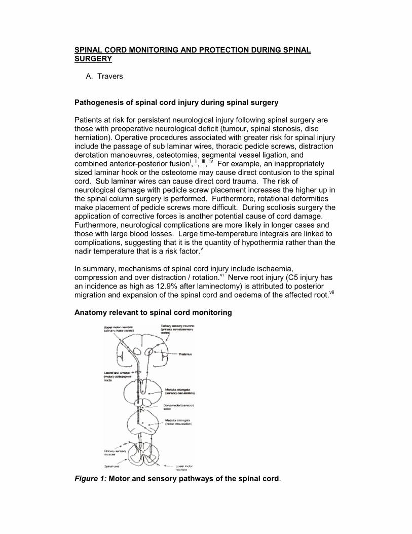