vpac2 receptors mediate vip-induced neuroprotection against
TRANSCRIPT
JPET #86405
1
VPAC2 receptors mediate VIP-induced neuroprotection against
neonatal excitotoxic brain lesions in mice
Claire-Marie Rangon, Stéphanie Goursaud,1 Fadia Medja, Vincent Lelièvre, Lourdes Mounien,2
Isabelle Husson, Philippe Brabet,3 Sylvie Jégou,2 Thierry Janet,1 and Pierre Gressens
INSERM U 676 & Service de Neurologie Pédiatrique, Hôpital Robert Debré, Paris, France
(CMR, FM, VL, IH, PG).
JPET Fast Forward. Published on May 4, 2005 as DOI:10.1124/jpet.105.086405
Copyright 2005 by the American Society for Pharmacology and Experimental Therapeutics.
This article has not been copyedited and formatted. The final version may differ from this version.JPET Fast Forward. Published on May 4, 2005 as DOI: 10.1124/jpet.105.086405
at ASPE
T Journals on February 20, 2018
jpet.aspetjournals.orgD
ownloaded from
JPET #86405
2
Running title : VPAC2 receptors mediate VIP-induced neuroprotection
Corresponding author : Dr Pierre Gressens, INSERM U 676, Hôpital Robert Debré,
48 Blvd Sérurier, 75019 Paris, France. Phone 33 1 40 03 47 83; fax 33 1 40 03 47 74;
Number of text pages : 30
Number of tables : 1
Number of figures : 6
Number of references : 40
Number of words in Abstract : 250
Introduction : 754
Discussion : 715
List of abbreviations : ANP, atrial natriuretic peptide; GPCR, G protein-coupled receptor; GRF,
growth hormone-releasing factor; MAPK, mitogen-associated protein kinase; NPR-C, natriuretic
peptide clearance receptor; PACAP, pituitary adenylate cyclase-activating peptide; PBS,
phosphate buffer saline; PHI, peptide histidine-isoleucine ; PKC, protein kinase C ; VIP,
vasoactive intestinal peptide.
Recommended section assignment: Neuropharmacology
This article has not been copyedited and formatted. The final version may differ from this version.JPET Fast Forward. Published on May 4, 2005 as DOI: 10.1124/jpet.105.086405
at ASPE
T Journals on February 20, 2018
jpet.aspetjournals.orgD
ownloaded from
JPET #86405
3
ABSTRACT
Prepro-vasoactive intestinal peptide (VIP) mRNA codes for two neuropeptides: VIP and
peptide histidine isoleucine (PHI). Two VIP receptors, shared with a similar affinity by pituitary
adenylate cyclase-activating polypeptide (PACAP), have been cloned: VPAC1 and VPAC2. PHI
binds to these receptors with a lower affinity. VPAC receptors are classically associated with a
cAMP-dependent pathway although other pathways including calcium mobilization and protein
kinase C activation have been described. We previously showed that intracerebral administration
of the glutamate agonist ibotenate to postnatal day 5 mice induces white matter lesions
mimicking human periventricular leukomalacia. In this model, co-injection of VIP protects
against white matter lesions. This neuroprotection is independent from cAMP and is mediated by
protein kinase C. This study aimed, using this model, to determine the receptor involved in VIP-
induced neuroprotection. VIP effects were mimicked with a similar potency by VPAC2 agonists
and PHI but not by VPAC1 agonists, PACAP 27 or PACAP 38. VIP neuroprotective effects were
lost in mice lacking VPAC2 receptor. In situ hybridization confirmed the presence of VPAC2
mRNA in the postnatal day 5 white matter. When analyzed between embryonic life and
adulthood, VIP specific binding site density peaked at postnatal day 5. These data suggest that, in
this model, VIP-induced neuroprotection is mediated by VPAC2 receptors. The pharmacology of
this VPAC2 receptor seems unconventional as i) PACAP does not mimic VIP effects, ii) PHI acts
with a comparable potency and iii) PACAP 27 modestly inhibited the VIP specific binding while
for PHI or VIP, inhibition was complete.
.
This article has not been copyedited and formatted. The final version may differ from this version.JPET Fast Forward. Published on May 4, 2005 as DOI: 10.1124/jpet.105.086405
at ASPE
T Journals on February 20, 2018
jpet.aspetjournals.orgD
ownloaded from
JPET #86405
4
INTRODUCTION
Vasoactive intestinal peptide (VIP) is a central nervous system neurotransmitter and
neuromodulator with neurotrophic properties (Rosselin et al., 1982; Moody et al., 2003).
The mouse VIP precursor can produce two peptides: VIP, a 28-amino acid peptide, and
peptide histidine-isoleucine (PHI). However, PHI shares moderate amino acid identity with VIP
(37 %) and its biological significance still remains unclear. VIP is a member of a superfamily of
peptides including pituitary adenylate cyclase-activating polypeptide (PACAP), a 27-
(PACAP27) or 38-amino acid (PACAP38) peptide. The biologically active region of PACAP,
corresponding to the PACAP27 sequence, shows 68 % identity with VIP (for a review, Vaudry et
al., 2000).
In transfected cells, VIP, PACAP27 and PACAP38 bind with similar affinities to two
receptors common for VIP and PACAP called VPAC1 and VPAC2 receptors (Ishihara et al.,
1992; Lutz et al., 1993). Furthermore, PACAP27 and PACAP38, but not VIP, bind with high
affinity to a specific PACAP receptor called the PAC1 receptor (Vaudry et al., 2000). [125I]-VIP
and [125I]-PHI binding experiments suggested that a 48 kDa component expressed in chicken
liver membranes display the properties of a GTP-insensitive VIP/PHI receptor that can be
pharmacologically discriminated from the GTP-sensitive 60 kDa form, through its much higher
affinity for PHI. Therefore, GTP-insensitive VIP receptors may correspond to a subclass of high-
affinity PHI receptors (Pineau et al., 2001).
VPAC receptors are preferentially coupled to Gαs protein that stimulates adenylate
cyclase activity and induces cAMP increase (Harmar et al., 1998). VPAC receptors can also be
coupled to Gαq and Gαi proteins that stimulate the inositol phosphate / calcium / protein kinase
This article has not been copyedited and formatted. The final version may differ from this version.JPET Fast Forward. Published on May 4, 2005 as DOI: 10.1124/jpet.105.086405
at ASPE
T Journals on February 20, 2018
jpet.aspetjournals.orgD
ownloaded from
JPET #86405
5
C (PKC) pathways (Langer et al., 2001; Olah et al., 1994; Rawlings et al., 1995; Ransjo et al.,
2000). Interestingly, the natriuretic peptide clearance receptor (NPR-C), which binds to atrial
natriuretic peptide (ANP), has been proposed to be involved in VIP-induced calcium entry
response in gastric smooth muscle (Murthy et al., 1998). Indeed, in binding studies using [125I]-
labelled atrial natriuretic peptide ([125I]-ANP) and [125I]-VIP as radioligands, VIP, ANP, and the
selective NPR-C ligand cANP-(4-23) bound with high affinity to NPR-C. ANP, cANP-(4-23),
and VIP initiated identical signalling cascades consisting of Ca++ influx, stimulation of cGMP
formation, and muscle relaxation.
In line with its previously reported neurotrophic properties (Gressens et al. 1993, 1994),
VIP potently protects the developing brain against an excitotoxic insult in newborn mice
(Gressens et al., 1997). In this in vivo model, VIP, co-injected with the glutamatergic agonist
ibotenate in the brain of five day-old (P5) pups, reduces ibotenate-induced white matter lesions
by up to 85 % when compared to controls. Surprisingly, VIP-induced neuroprotection is not
mimicked by PACAP 38 but by stearyl norleucine VIP, a specific VIP agonist that does not
activate adenylate cyclase. Moreover, treatment with forskolin, an adenylate cyclase activator,
fails to provide a VIP-like protection. In contrast, VIP protective effects are abolished by a PKC
inhibitor and a mitogen-associated protein kinase (MAPK) inhibitor in a dose dependent manner
(Gressens et al., 1997, 1998).
This atypical pharmacology of VIP-induced neuroprotection in newborn mice raises
several hypotheses: i) activation of PAC1 receptors could have a toxic effect on the excitotoxic
lesions while activation of VPAC receptors could be neuroprotective, leading to a lack of
detectable effect for PACAP38. Interestingly, VIP and PACAP have been shown to have
opposite effects on synaptic transmission (Ciranna and Cavallaro, 2003). ii) During some stages
of brain development, the binding of VIP or PACAP to VPAC receptors leads to activation of
This article has not been copyedited and formatted. The final version may differ from this version.JPET Fast Forward. Published on May 4, 2005 as DOI: 10.1124/jpet.105.086405
at ASPE
T Journals on February 20, 2018
jpet.aspetjournals.orgD
ownloaded from
JPET #86405
6
separate transduction pathways. This differential coupling could be secondary to VPAC receptor
dimerization (homo- or heterodimers) or to their interaction with larger oligomeric complexes, as
demonstrated for other types of G protein-coupled receptors (GPCRs) (for a review see Milligan,
2004). iii) VIP acts through a yet to be identified specific VIP receptor which is not recognised
by PACAP. Indeed, Ekblad et al. (2000) characterized a PACAP 27 preferring receptor and a VIP
specific receptor, distinct from those that have been cloned (VPAC1, VPAC2, and PAC1
receptors), in intestine of rat and PAC1-/- mice.
Accordingly, in the present study, the neuroprotective properties of several agonists of
VIP and PACAP receptors were tested in the above-described neonatal excitotoxic model.
Neuroprotective effects of VIP and/or PACAP were evaluated in mutant mice lacking PAC1 or
VPAC2 receptors, further allowing the characterization of the receptor involved in VIP-induced
neuroprotection. In addition, in situ hybridization for VPAC2 receptor mRNA was performed on
P5 mouse brains. Finally, binding studies using radiolabelled VIP, PHI and PACAP27 were
performed on brain membranes from embryonic day 17 (E17), P5 and adult animals.
This article has not been copyedited and formatted. The final version may differ from this version.JPET Fast Forward. Published on May 4, 2005 as DOI: 10.1124/jpet.105.086405
at ASPE
T Journals on February 20, 2018
jpet.aspetjournals.orgD
ownloaded from
JPET #86405
7
MATERIALS AND METHODS
Animals
Different types of mice of both sexes were used in this study: Swiss mice (Janvier,
France), C57Bl/6 control PAC1+/+ mice, C57Bl/6 PAC1
-/- mice (Jamen et al., 2000), C57Bl/6
control VPAC2+/+ mice, C57Bl/6 control VPAC2
+/- mice and C57Bl/6 VPAC2-/- mice (Goetzl et
al., 2001). C57Bl/6 VPAC2 mice were kindly provided by Dr Anthony J. Harmar (Division of
Neuroscience, University of Neuroscience, Edinburgh, Scotland).
PAC1-/- and VPAC2
-/- mice were generated as previously described (Jamen et al., 2000;
Goetzl et al., 2001; Harmar et al., 2002). Briefly, for PAC1-/- knockout mice, a 2.3-kb NcoI
fragment containing PAC1 receptor exons 8-11 was replaced by a pPolII-neoRcassette. The
targeting vector contained 4.2 kb and 2.8 kb of homologous sequences up- and downstream from
the pPolII-neomycin sequence, respectively (Jamen et al., 2000). The VPAC2 receptor null mice
were generated by replacing 132 bp of exon 1 including the translation start site with a lac Z-neoR
cassette. The total construct was 10 kb in size (Goetzl et al., 2001).
Experimental protocols meet the guidelines of the “Institut National de la Santé et de la
Recherche Médicale” and were carried out in accordance with the Guide for the Care and use of
Laboratory Animals as adopted and promulgated by the U.S. National Institutes of Health.
Drugs
Ibotenate (Sigma, St-Quentin Fallavier, France) and VIP (Peninsula, Belmont, CA, USA) were
diluted in phosphate buffer saline (PBS) containing 0.01% acetic acid. PACAP27 (Bachem, Weil
am Rhein, Germany), PACAP38 (Peninsula), the selective VPAC1 receptor agonist derived from
VIP and growth hormone-releasing factor (GRF) [Lys15, Arg16, Leu27] VIP (1-7) / GRF (8-27)
This article has not been copyedited and formatted. The final version may differ from this version.JPET Fast Forward. Published on May 4, 2005 as DOI: 10.1124/jpet.105.086405
at ASPE
T Journals on February 20, 2018
jpet.aspetjournals.orgD
ownloaded from
JPET #86405
8
(VIP-GRF) (generous gift from Dr Patrick Robberecht, Laboratoire de Chimie Biologique et de
la Nutrition, University of Brussels Medical School, Brussels, Belgium) (Gourlet et al., 1997a),
the selective VPAC1 receptor agonist [R16]chicken secretin (generous gift from Dr Patrick
Robberecht) (Gourlet et al., 1997a), the selective VPAC2 receptor agonist RO25-1553 Ac-[Glu8,
Lys12, Nle17, Ala19, Asp25, Leu26, Lys27,28, Gly29,30, Thr31]-VIP cyclo21–25 (generous gift from Dr
Patrick Robberecht) (Gourlet et al., 1997b), PHI (Bachem), the selective NPR-C receptor agonist
des-[Gln18, Ser19, Gly20, Leu21, Gly22] ANP (4-23) amide (des-ANP; Sigma) (Brown et al., 1990)
and ANP (Sigma) were diluted in PBS.
Administration of the excitotoxic drug
Excitotoxic brain lesions were induced by injecting ibotenate into developing mouse
brains, as previously described (Marret et al., 1995; Gressens et al., 1997, 1998). Briefly, mouse
pups anesthetized with isoflurane were kept under a warming lamp and were injected
intracerebrally (into the neopallial parenchyma) at P5. Intraparenchymal injections were
performed with a 25-gauge needle on a 50 µl Hamilton syringe mounted on a calibrated
microdispenser. The needle was inserted 2 mm under the external surface of the scalp skin in the
frontoparietal area of the right hemisphere, 2 mm from the midline in the lateral-medial plane,
and 3 mm anterior to bregma in the rostro-caudal plane. Histopathology confirmed that the tip of
the needle always reached the periventricular white matter. Two 1µl boluses of ibotenate were
injected at 20-seconds intervals. The needle was left in place for an additional 20 seconds. 10 µg
ibotenate were administered to each pup.
This article has not been copyedited and formatted. The final version may differ from this version.JPET Fast Forward. Published on May 4, 2005 as DOI: 10.1124/jpet.105.086405
at ASPE
T Journals on February 20, 2018
jpet.aspetjournals.orgD
ownloaded from
JPET #86405
9
Experimental groups
Pups from at least two different litters were used in each experimental group, and data
were obtained from two or more successive experiments.
Mice were injected with ibotenate alone (controls) or in combination with different peptides :
0.01, 0.1 or 1 µg VIP; 0.0001, 0.001, 0.01, 0.1 or 1 µg PACAP38; 0.001, 0.01, 0.1 or 1 µg
PACAP27; 0.01, 0.1 or 1 µg RO25-1553; 1 µg [R16] chicken secretin; 1 µg VIP-GRF; 0.01, 0.1
or 1 µg PHI ; 0.01, 0.1 or 1 µg des-ANP; 0.01, 0.1 or 1 µg ANP.
Determination of lesion size
Mouse pups were sacrificed by decapitation five days following the excitotoxic challenge.
Brains were immediately fixed in 4% formalin for seven days. Following embedding in paraffin, we
cut 15 µm thick coronal or sagittal sections. Every third section was stained with cresyl-violet.
Previous studies (Marret et al., 1995; Gressens et al., 1997; Husson et al., 2002) have shown an
excellent correlation between the maximal size of the lesion in the lateral-medial and fronto-occipital
axes of the excitotoxic lesions. To further confirm these observations, we cut serial sections of the
entire brain in the coronal plane or in the sagittal plane. This permitted an accurate and reproducible
determination of the maximal fronto-occipital (on coronal sections) or lateral-medial (on sagittal
sections) diameters of the lesions (which is equal to the number of sections where the lesion was
present multiplied by 15 µm). We used these linear measures as an index of the volume of the
lesion.
In situ hybridization
P5 Swiss mice were perfused intracardiacally with a 4% paraformaldehyde solution. The
brains were put in 4% paraformaldehyde for 2 hours at 4°C, then in a 10% saccharose 0.12 M
This article has not been copyedited and formatted. The final version may differ from this version.JPET Fast Forward. Published on May 4, 2005 as DOI: 10.1124/jpet.105.086405
at ASPE
T Journals on February 20, 2018
jpet.aspetjournals.orgD
ownloaded from

JPET #86405
10
phosphate buffer for 24 hours. Brains were frozen at –80°C and 10-µm thick coronal sections
were cut.
VPAC2 receptor riboprobes were prepared by in vitro transcription of rat VPAC2 receptor
cDNA fragments generated by RT-PCR, and subcloned into the pGEM-T cloning vector
(Promega, Charbonnières, France). The primers used for PCR reaction were designed as follows:
forward primer, 5’-GTCAACTTTGCCCTCTCCATCA-3’; reverse primer, 5’-
GCCTCTCCACCTTCTTTTCAGT-3’ (accession no. Z25885). Antisense and sense riboprobes
were generated with T7 or SP6 polymerase in the presence of [35S]-UTP (Amersham Biosciences
Europe, Les Ulis, France).
In situ hybridization was performed as previously described (Bellemere et al., 2004).
Briefly, sections were acetylated, treated with Triton X-100 (0.2%) and covered with
prehybridization buffer (50% formamide, 0.6 M NaCl, 10 mM Tris-HCl pH 7.5, 0.02% Ficoll,
0.02% polyvinylpyrollidone, 0.02 % bovine serum albumin (BSA), 1 mM EDTA pH 8.0, 550
µg/ml denatured salmon sperm DNA, 50 µg/ml yeast tRNA). Hybridization was performed
overnight at 55°C in the same buffer (except for salmon sperm DNA, the concentration of which
was lowered to 60 µg/ml) supplemented with 10 mM dithiothreitol, 10% dextran sulfate and
1.5×10⁷ cpm/ml heat-denatured riboprobes. Sections were then washed in 2 X standard saline
citrate (SSC) at 60°C and treated with RNase A (50 µg/ml) for 1 hour at 37°C. Five final high-
stringency washes were performed in 0.01 X SSC containing 14 mM β-mercaptoethanol and
0.05% sodium pyrophosphate. Brain slices were dehydrated in graded alcohols containing
ammonium acetate and apposed onto Hyperfilm β-max (Amersham Biosciences Europe) for 2
weeks. Slices were subsequently dipped into Kodak NTB2 liquid emulsion at 40°C, exposed for
30 days and developed. To identify anatomic structures, sections were stained with hematoxylin.
Autoradiograms were analysed by means of a computer-assisted image analysis system (SAMBA
This article has not been copyedited and formatted. The final version may differ from this version.JPET Fast Forward. Published on May 4, 2005 as DOI: 10.1124/jpet.105.086405
at ASPE
T Journals on February 20, 2018
jpet.aspetjournals.orgD
ownloaded from
JPET #86405
11
Autoradio 4.10; SAMBA Technologies, Meylan, France). Photomicrographs were imported into
MERCATOR software (Explora Nova, La Rochelle, France) interfaced with a Nikon Eclipse
E600 microscope (Les Ulis, France).
Receptor binding studies on mouse forebrain membrane preparations
Forebrains were freshly removed from embryonic day (E) 17, P5 and P60 Swiss mice.
They were immediately frozen in liquid nitrogen and stored at -80°C until membrane
purification. Membranes were prepared as previously described (Pineau et al., 2001). Protein
content was determined with a Bradford assay (BioRad Protein assay dye reagent concentrate,
(BioRad, Ivry-sur-Seine, France) and BSA fatty acid free fraction V (Sigma) as a standard.
Membranes aliquots were stored at -80°C until use.
VIP, PHI and PACAP27 (Neosystem, Strasbourg, France) were radioiodinated with the
chloramine-T method as previously reported (Martin et al., 1986) using a modified protocol. Free
[125I]-iodine was removed onto C18 Sep-Pak Cartridges (Waters Corp., St-Quentin-en-Yvelines,
France) according to the manufacturer’s instructions. Radioiodinated peptidic forms were
separated by reverse-phase high-performance liquid chromatography (Spectraphysics, Waters
Corp.) using a 5 µm VYDAC C18 column (Interchim, Montluçon, France). Elution was
conducted for 37 min at 1 ml/min with a 0-85% acetonitrile linear gradient in 0.1% trifluoroacetic
acid-H2O. Fractions containing mono [125I]-VIP, [125I]-PHI or [125I]-PACAP27 were pooled,
evaporated under nitrogen and stored at -20°C until use.
Membrane preparations were homogenized in cold binding buffer pH 7.4 (Dulbecco’s
modified Eagle’s medium supplemented with 15 mM HEPES, 150 µM phenylmethylsulfonyl
fluoride, 0.1% bacitracin and 1% BSA) containing 50 pM of [125I]-VIP or [125I]-PHI or [125I]-
PACAP27 and unlabelled peptides at specified concentrations. Binding experiments were
This article has not been copyedited and formatted. The final version may differ from this version.JPET Fast Forward. Published on May 4, 2005 as DOI: 10.1124/jpet.105.086405
at ASPE
T Journals on February 20, 2018
jpet.aspetjournals.orgD
ownloaded from
JPET #86405
12
performed with shaking for 50 min at 20 °C and stopped at 4 °C. Samples were filtered through
Wathman GF/C glass microfiber filters (Fisher Scientific LABOSI), pretreated for 30 min with
a 0.5% polyethyleneimine and 0.5% BSA solution, using a samples collector (Millipore, Bedford,
MA, USA). Filters were washed with 1 ml and 3 ml of a cold 150 mM Tris with 5 mM MgCl2,
6H2O pH 7.5 solution, dried under vacuum. The retained radioactivity was measured in a Cobra
II Packard γ-counter.
Preliminary dose-dependent experiments (data not shown) were performed on the mouse
brain membrane preparations to determine protein quantities (350 µg) necessary to maximize
specific binding. Specific binding represents the difference between values obtained in the
absence or in presence of 10-6 M unlabelled neuropeptides.
Statistical analysis
Quantitative data were expressed as the means ± SEMs for each treatment group. Means
were compared using Student t-test or ANOVA with Dunnett's or Newman-Keul’s multiple
comparison of means test (GraphPad Prism version 3.03 for Windows, GraphPad Software).
This article has not been copyedited and formatted. The final version may differ from this version.JPET Fast Forward. Published on May 4, 2005 as DOI: 10.1124/jpet.105.086405
at ASPE
T Journals on February 20, 2018
jpet.aspetjournals.orgD
ownloaded from
JPET #86405
13
RESULTS
Excitotoxic brain lesions
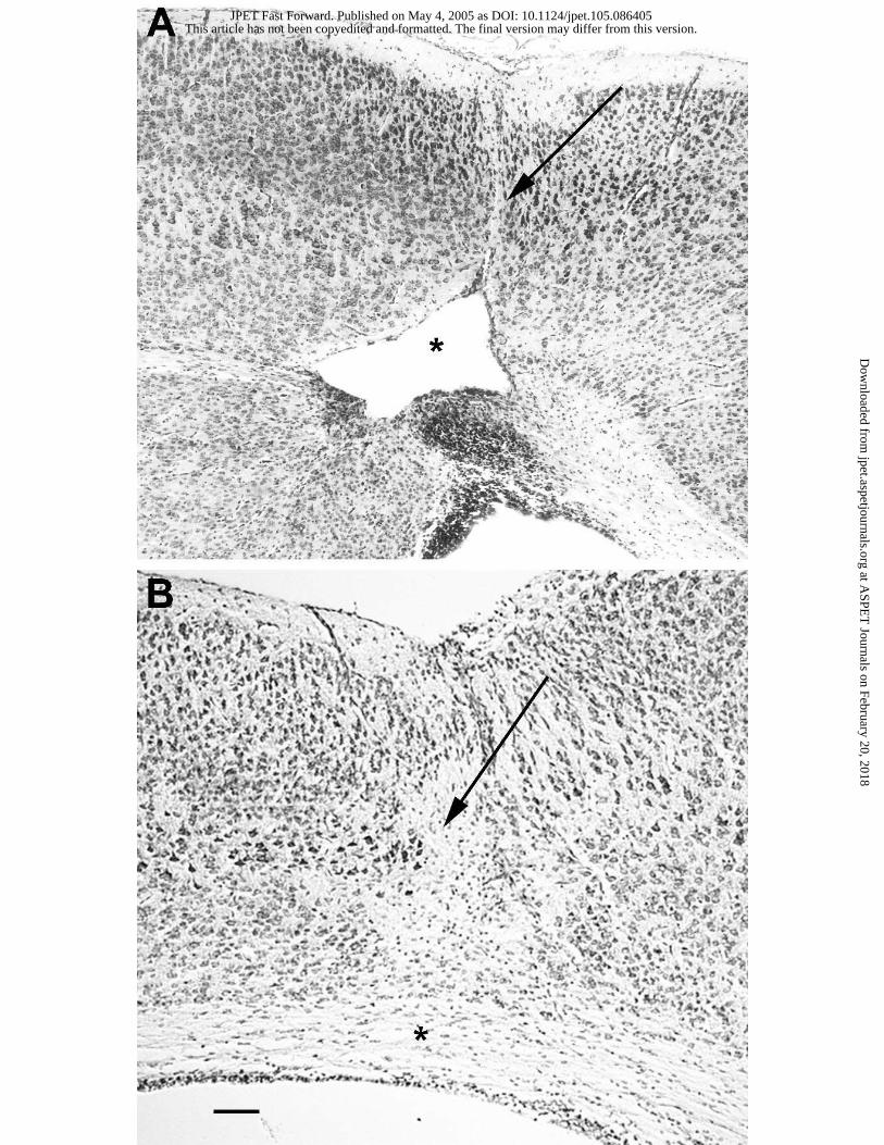
All animals injected with ibotenate at P5 and sacrificed five days later displayed large
periventricular white matter cysts in the injected right hemisphere (Fig. 1A). Cortical lesions
characterized by neuronal loss affecting all cortical layers (Fig. 1A) were also observed in the
injected right hemisphere these animals.
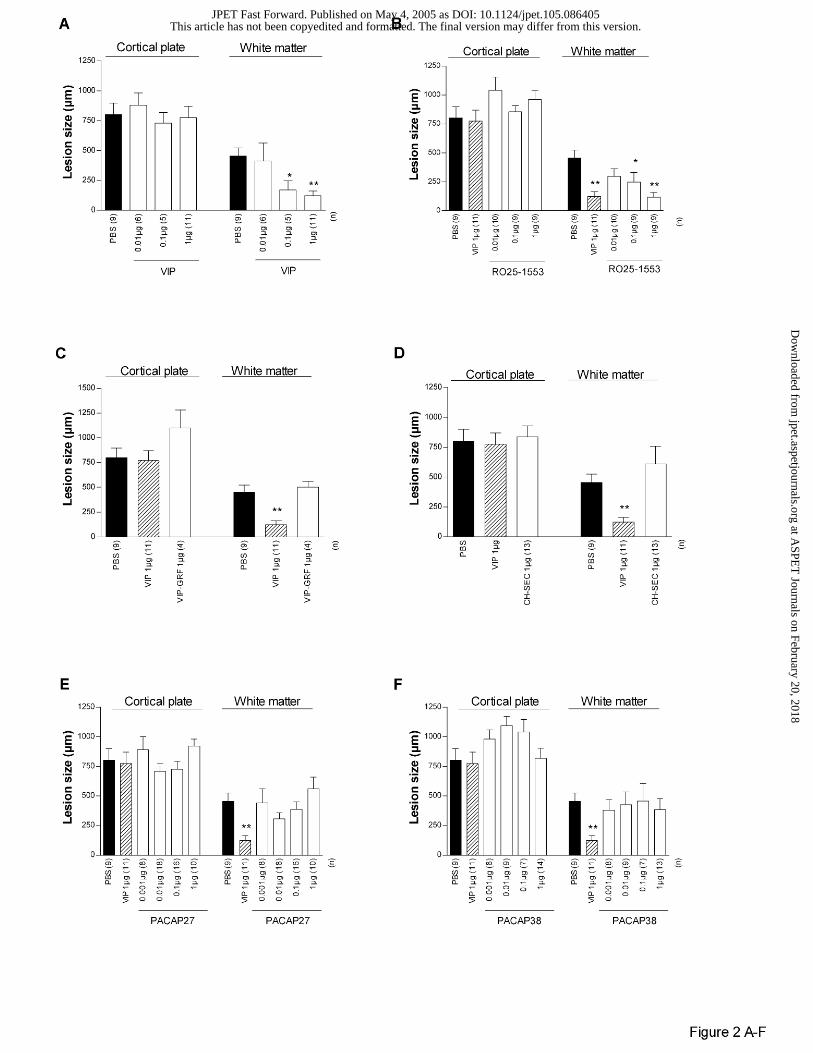
In Swiss mice, as previously described (Gressens et al., 1997, 1998), VIP significantly
protected the white matter against ibotenate but had no significant effect on cortical plate lesion
(Figs 1B and 2A). RO25-1553, a VPAC2 agonist, mimicked the neuroprotective effects of VIP
(Fig. 2B) while VIP-GRF and [R16] chicken secretin, two VPAC1 agonists, had no detectable
effects of white matter and cortical plate lesions (Fig. 2C-D). A large range of doses (0.001 to 1
µg) of PACAP27 or PACAP38 did not protect or significantly exacerbate the white matter or the
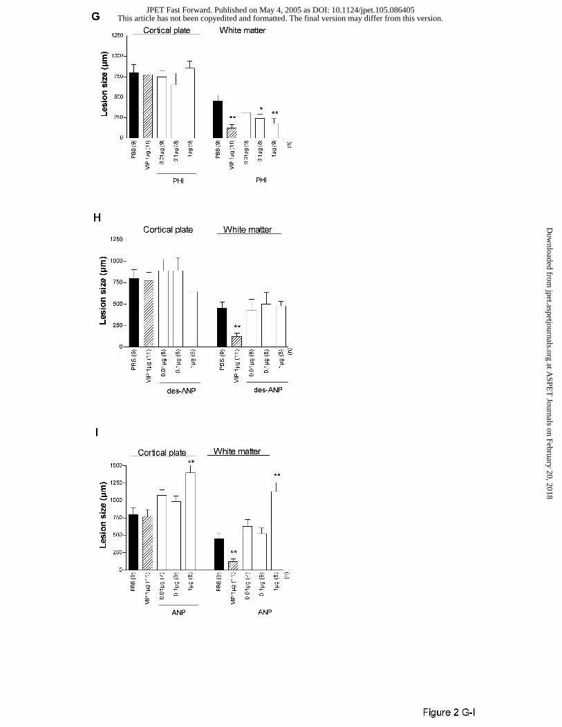
grey matter against excitotoxic lesions (Fig. 2E-F). In contrast, PHI mimicked VIP-induced
neuroprotection of white matter with a potency similar to VIP (Fig. 2G). As for VIP, PHI did not
protect the cortical plate against ibotenate (Fig. 2G). Finally, des-ANP or ANP did not mimic
neuroprotective effects of VIP on excitotoxic white matter damage (Fig. 2H-I) and the highest
dose of ANP even exacerbated excitotoxic cortical plate and white matter (Fig. 2I).
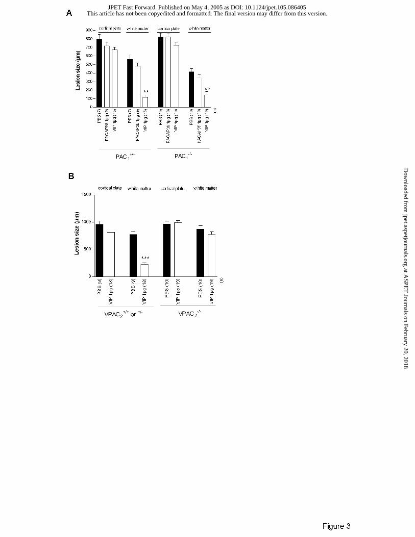
In control C57Bl/6 control PAC1+/+ mice, VIP significantly protected the white matter
against ibotenate (Fig. 3A). As in Swiss mice, PACAP38 failed to mimic VIP neuroprotective
effects (Fig. 3A). In C57Bl/6 PAC1-/- mice, VIP-induced neuroprotection was maintained while
PACAP38 had no detectable effect on ibotenate-induced lesions (Fig. 3A).
VIP-induced neuroprotection of the white matter was observed in C57Bl/6 control
VPAC2+/+ and VPAC2
+/- mice but was totally abrogated in C57Bl/6 VPAC2-/- mice (Fig. 3B).
This article has not been copyedited and formatted. The final version may differ from this version.JPET Fast Forward. Published on May 4, 2005 as DOI: 10.1124/jpet.105.086405
at ASPE
T Journals on February 20, 2018
jpet.aspetjournals.orgD
ownloaded from
JPET #86405
14
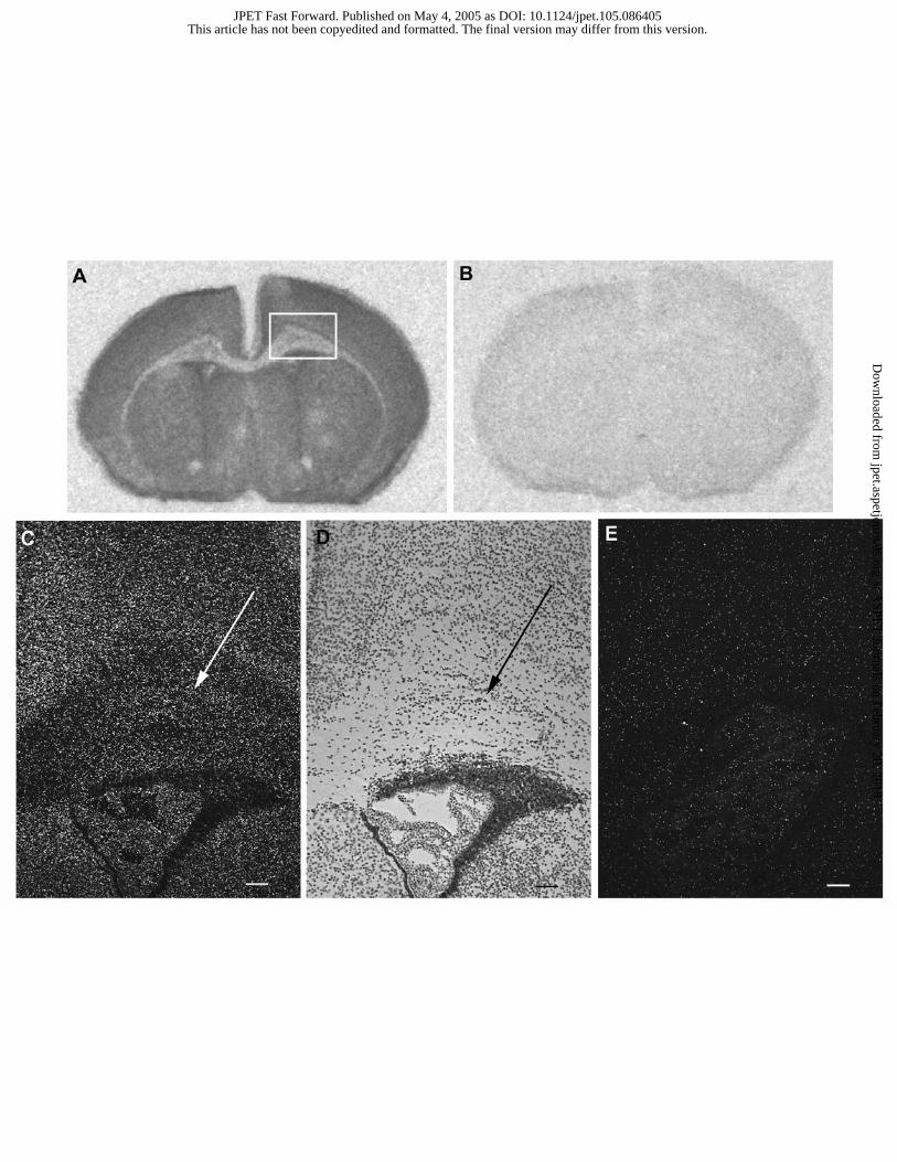
In situ hybridization for VPAC2 mRNA in untreated Swiss mice
VPAC2 mRNA was detected by in situ hybridization in P5 mouse brain (Fig. 4). Specific
signal was present in the neocortex and also in the underlying periventricular white matter. This
VPAC2 mRNA expression was sustained along the rostro-caudal axis, including the fronto-
parietal area where ibotenate was injected in experimental groups described above. When the
sense probe was used, no hybridization was observed (Fig. 4).
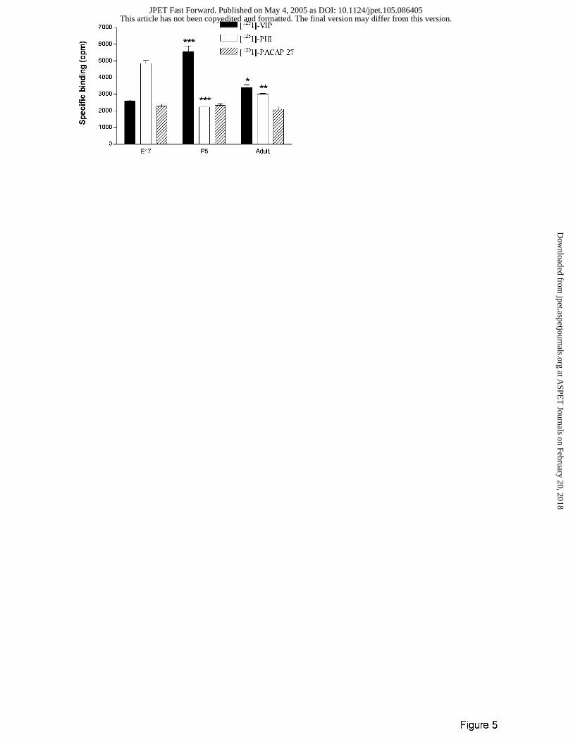
Neuropeptide binding site studies in untreated Swiss mice
[125I]-VIP and -PHI specific binding site densities changed over time (between E17 and
P60) while [125I]-PACAP27 specific binding sites density remained stable (Fig. 5 and Table 1).
Of particular interest, the density of [125I]-VIP specific binding sites peaked at P5.
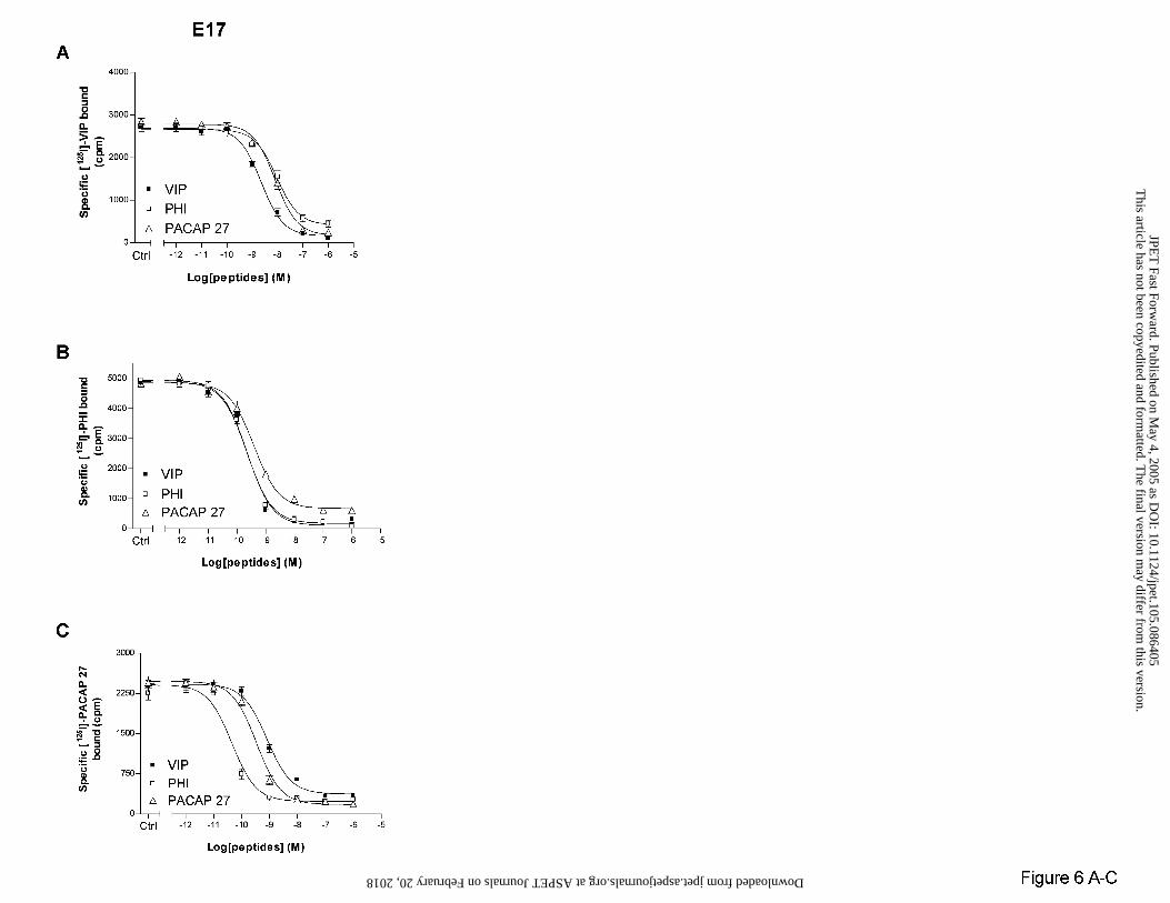
In E17 brains, [125I]-VIP (Fig. 6A), [125I]-PHI (Fig. 6B) or [125I]-PACAP 27 (Fig. 6C)
binding were similarly inhibited by unlabelled VIP, PHI or PACAP 27 with kinetics consistent
with the presence of a single binding site (Hill slopes varied from 0.96 ± 0.35 to 0.98 ± 0.28).
Specific [125I]-PHI or [125I]-PACAP 27 binding were inhibited with a high affinity (IC50 values
were measured between 0.04 ± 0.009 and 0.82 ± 0.16 nM), whereas neuropeptide affinities for
[125I]-VIP binding sites were at least 10-fold lower (IC50 from 2.25 ± 0.67 to 9.10 ± 3.04 nM).
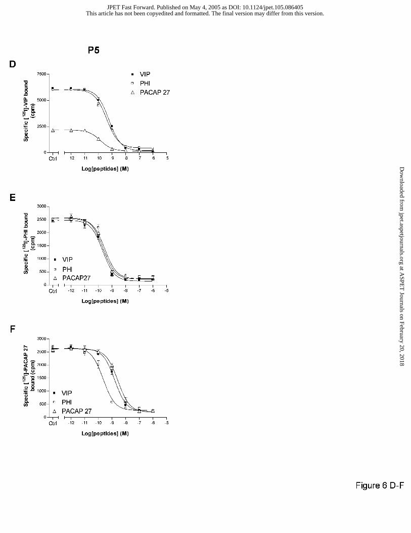
In P5 brains, unlabelled VIP, PHI or PACAP 27 inhibited in the same manner the [125I]-
PHI (Fig. 6E) or [125I]-PACAP 27 (Fig. 6F) binding, on one site (Hill slopes from 0.93 ± 0.13 to
0.98 ± 0.06) with a high affinity (IC50 from 0.20 ± 0.06 to 0.66 ± 0.06 nM). PACAP27 only
modestly inhibited the specific high affinity [125I]-VIP binding (33%) whereas VIP and PHI
totally abolished the specific radiotracer interaction (Fig. 6D).
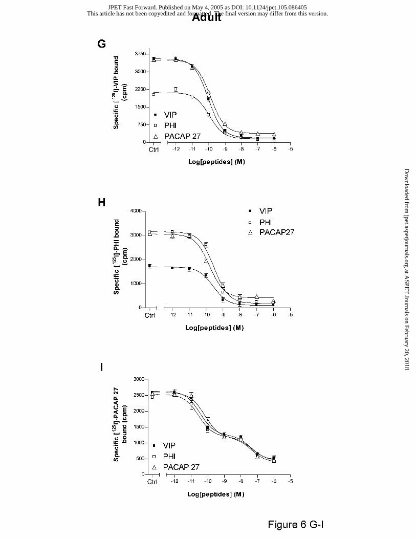
In adult brain, the specific high affinity [125I]-VIP (Fig. 6G) and -PHI (Fig. 6H) binding,
totally abolished by unlabelled PACAP27, were partially inhibited by respectively unlabelled
This article has not been copyedited and formatted. The final version may differ from this version.JPET Fast Forward. Published on May 4, 2005 as DOI: 10.1124/jpet.105.086405
at ASPE
T Journals on February 20, 2018
jpet.aspetjournals.orgD
ownloaded from
JPET #86405
15
PHI (57%) and VIP (53%). When [125I]-PACAP27 was used, two binding sites subsets (Hill
slopes from 0.74 ± 0.07 to 0.77 ± 0.01) were distinguished by the neuropeptides (Fig. 6I), one
with a high affinity (IC50 from 0.03 ± 0.01 to 0.23 ± 0.02 nM) and the other with a low affinity
(IC50 from 41.19 ± 5.28 to 55.47 ± 2.09 nM).
This article has not been copyedited and formatted. The final version may differ from this version.JPET Fast Forward. Published on May 4, 2005 as DOI: 10.1124/jpet.105.086405
at ASPE
T Journals on February 20, 2018
jpet.aspetjournals.orgD
ownloaded from
JPET #86405
16
DISCUSSION
The present study extends the evidence on neuroprotective properties of VIP against
excitotoxic white matter lesions in the developing mouse brain (Gressens et al., 1997, 1998,
1999) and identifies VPAC2 receptors as critical mediators of VIP effects. The role of VPAC2
receptors is supported by the neuroprotective effects of VPAC2 receptor agonists, the loss of VIP
neuroprotective effects in C57Bl/6 VPAC2-/- mice and the presence of VPAC2 receptor mRNA in
the P5 white matter.
Our results are in accordance with an in vitro study suggesting VPAC2 involvement in
neuroprotection (Zusev et al., 2004). They studied Activity-Dependent Neuroprotective protein
(ADNF) expression in astrocytes cell cultures. Indeed i) ADNF was previously shown to be a
VIP responsive gene in astrocytes derived from the cerebral cortex of newborn rats (Bassan et al.,
1999) ; and ii) VIP neuroprotective properties are mediated through interaction with glial cells
(Brenneman et al., 1987 ; Gozes et al., 1991 ; Gressens et al., 1998). They showed that VIP
induced changes in ADNF expression in astrocyte cell cultures via the VPAC2 receptor (Zusev et
al., 2004).
However, in vivo, the pharmacology of the involved VPAC2 receptors is atypical when
compared to data obtained in transfected cultured cells (Ishihara et al., 1992; Lutz et al., 1993;
Vaudry et al., 2000 ; Laburthe et al., 2002) as i) using the same mouse model of neonatal
excitotoxic brain lesions, we previously showed that VIP neuroprotective effects against
excitotoxic white matter damage are not mediated by cAMP-PKA pathway but by a PLC-PKC
transduction pathway (Gressens et al., 1997, 1998), ii) neither PACAP 27 nor PACAP 38 mimic
VIP effects, iii) PHI mimics VIP effects with a similar potency, and iv) in P5 pups, PACAP 27
modestly inhibited the specific high [125I]-VIP binding while PHI or VIP totally inhibited it.
This article has not been copyedited and formatted. The final version may differ from this version.JPET Fast Forward. Published on May 4, 2005 as DOI: 10.1124/jpet.105.086405
at ASPE
T Journals on February 20, 2018
jpet.aspetjournals.orgD
ownloaded from
JPET #86405
17
. The previously stated hypothesis that activation of PAC1 receptors could have a toxic
effect on the excitotoxic lesions while activation of VPAC receptors could be neuroprotective,
leading to a lack of detectable effect for PACAP38, can be ruled out by the lack of protective
effects of PACAP 38 in PAC1-/- mice.
As mentioned above, the NPR-C, which binds to ANP, has been proposed to be involved
in VIP-induced calcium entry response in gastric smooth muscle (Murthy et al., 1998).
However, in the present study, ANP and des-ANP that classically bind NPR-C did not reproduce
VIP neuroprotective effects and ANP was even toxic at high dose.
Therefore, in order to explain the observed characteristics of VPAC2 receptors in the
present study, some hypotheses can be formulated: i) During some stages of brain development,
the binding of VIP or PACAP to VPAC2 receptors leads to activation of separate transduction
pathways. This differential coupling could be secondary to VPAC2 receptors dimerization (homo-
or heterodimers) or to their interaction with larger oligomeric complexes, as demonstrated for
other types of GPCRs (for a review Milligan, 2004). A variant of this hypothesis would be a
developmental change in the G proteins available for the receptor to couple to in the relevant cells.
ii) An alternative hypothesis has been suggested by recent studies. A first study identified a
deletion variant (receptor lacking amino acids 367-380 at the carboxyl-terminal end of the seventh
transmembrane domain) of the mouse VPAC2 receptor in immune cells (Grinninger et al., 2004).
This natural deletion abrogates VIP-induced cAMP production without apparent alterations of
expression or ligand binding. Secondly, Langer and Robberecht (2005) showed that mutations in
the proximal domain of the third intracellular loop of the VPAC1 receptor reduced the capability of
VIP to increase adenylate cyclase activity without any change in the calcium response, whereas
mutations in the distal part of the loop markedly reduced the calcium increase and Gαi coupling
This article has not been copyedited and formatted. The final version may differ from this version.JPET Fast Forward. Published on May 4, 2005 as DOI: 10.1124/jpet.105.086405
at ASPE
T Journals on February 20, 2018
jpet.aspetjournals.orgD
ownloaded from
JPET #86405
18
but only weakly reduced the adenylate cyclase activity. Based on these studies, we can hypothesize
that a yet-to-be-identified substitution or deletion in the newborn mouse VPAC2 receptor
transcript, through RNA editing for instance, might be able to induce VIP specificity and modulate
the coupling with different G proteins.
Altogether, these data strongly support the hypothesis that, in newborn mice, VIP
neuroprotective effects against an excitotoxic insult are mediated by VPAC2 receptors showing
atypical pharmacological properties. Further studies will be required to further characterize these
VPAC2 receptors in order to unravel the molecular basis of their intriguing pharmacology.
This article has not been copyedited and formatted. The final version may differ from this version.JPET Fast Forward. Published on May 4, 2005 as DOI: 10.1124/jpet.105.086405
at ASPE
T Journals on February 20, 2018
jpet.aspetjournals.orgD
ownloaded from
JPET #86405
19
ACKNOWLEDGMENTS
We are grateful to Marc Laburthe for helpful discussions and Leslie Schwendimann for
excellent technical assistance. We deeply thank Patrick Robberecht for providing VIP agonists
and Anthony J. Harmar for providing VPAC2 mutant mice.
This article has not been copyedited and formatted. The final version may differ from this version.JPET Fast Forward. Published on May 4, 2005 as DOI: 10.1124/jpet.105.086405
at ASPE
T Journals on February 20, 2018
jpet.aspetjournals.orgD
ownloaded from
JPET #86405
20
REFERENCES
Bassan M, Zamostiano R, Davidson A, Pinhasov A, Giladi E, Perl O, Bassan H, Blat C, Gibney
G, Glazner G, Brenneman DE and Gozes I (1999) Complete sequence of a novel protein
containing a femtomolar-activity-dependent neuroprotective peptide. J Neurochem 72:1283-
1293.
Bellemère G, Vaudry H, Mounien L, Boutelet I and Jégou S (2004) Localization of the mRNA
encoding prolyl endopeptidase in the rat brain and pituitary. J Comp Neurol 471:128-143.
Brenneman DE, Neale EA, Foster GA, d'Autremont SW and Westbrook GL (1987) Nonneuronal
cells mediate neurotrophic action of vasoactive intestinal peptide. J Cell Biol 104:1603-1610.
Brown J and Czarnecki A (1990) Distribution of atrial natriuretic peptide receptor subtypes in rat
brain. Am J Physiol 258:R1078-1083.
Ciranna L and Cavallaro S (2003) Opposing effects by pituitary adenylate cyclase-activating
polypeptide and vasoactive intestinal peptide on hippocampal synaptic transmission. Exp Neurol
184:778-784.
Ekblad E, Jongsma H, Brabet P, Bockaert J and Sundler F (2000) Characterization of intestinal
receptors for VIP and PACAP in rat and in PAC1 receptor knockout mouse. Ann N Y Acad Sci
921:137-147.
This article has not been copyedited and formatted. The final version may differ from this version.JPET Fast Forward. Published on May 4, 2005 as DOI: 10.1124/jpet.105.086405
at ASPE
T Journals on February 20, 2018
jpet.aspetjournals.orgD
ownloaded from
JPET #86405
21
Goetzl EJ, Voice JK, Shen S, Dorsam G, Kong Y, West KM, Morrison CF and Harmar AJ (2001)
Enhanced delayed-type hypersensitivity and diminished immediate-type hypersensitivity in mice
lacking the inducible VPAC(2) receptor for vasoactive intestinal peptide. Proc Natl Acad Sci
USA 98:13854-13859.
Gourlet P, Vandermeers A, Vertongen P, Rathe J, De Neef P, Cnudde J, Waelbroeck M,
Robberecht P (1997a) Development of high affinity selective VIP1 receptor agonists. Peptides
10:1539-1545.
Gourlet P, Vertongen P, Vandermeers A, Vandermeers-Piret MC, Rathe J, De Neef P,
Waelbroeck M and Robberecht P (1997b) The long-acting vasoactive intestinal polypeptide
agonist RO 25-1553 is highly selective of the VIP2 receptor subclass. Peptides 18:403-408.
Gozes I, McCune SK, Jacobson L, Warren D, Moody TW, Fridkin M and Brenneman DE (1991)
An antagonist to vasoactive intestinal peptide affects cellular functions in the central nervous
system. J Pharmacol Exp Ther 257:959-966.
Gressens P (1999) VIP neuroprotection against excitotoxic lesions of the developing mouse
brain. Ann N Y Acad Sci 897:109-124.
Gressens P, Hill JM, Gozes I, Fridkin M and Brenneman DE (1993) Growth factor function of
vasoactive intestinal peptide in whole cultured mouse embryos. Nature 362:155-158.
This article has not been copyedited and formatted. The final version may differ from this version.JPET Fast Forward. Published on May 4, 2005 as DOI: 10.1124/jpet.105.086405
at ASPE
T Journals on February 20, 2018
jpet.aspetjournals.orgD
ownloaded from
JPET #86405
22
Gressens P, Hill JM, Paindaveine B, Gozes I, Fridkin M and Brenneman DE (1994) Severe
microcephaly induced by blockade of vasoactive intestinal peptide function in the primitive
neuroepithelium of the mouse. J Clin Invest 94:2020-2027.
Gressens P, Marret S, Hill JM, Brenneman DE, Gozes I, Fridkin M and Evrard P (1997)
Vasoactive intestinal peptide prevents excitotoxic cell death in the murine developing brain. J
Clin Invest 100:390-397.
Gressens P, Marret S, Martin JL, Laquerriere A, Lombet A and Evrard P (1998) Regulation of
neuroprotective action of vasoactive intestinal peptide in the murine developing brain by protein
kinase C and mitogen-activated protein kinase cascades: in vivo and in vitro studies. J
Neurochem 70:2574-2584.
Grinninger C, Wang W, Oskoui KB, Voice JK and Goetzl EJ (2004) A natural variant type II G
protein-coupled receptor for vasoactive intestinal peptide with altered function. J Biol Chem
279:40259-40262.
Harmar AJ, Arimura A, Gozes I, Journot L, Laburthe M, Pisegna JR, Rawlings SR, Robberecht
P, Said SI, Sreedharan SP, Wank SA and Waschek JA (1998) International Union of
Pharmacology. XVIII. Nomenclature of receptors for vasoactive intestinal peptide and pituitary
adenylate cyclase-activating polypeptide. Pharmacol Rev 50:265-270.
This article has not been copyedited and formatted. The final version may differ from this version.JPET Fast Forward. Published on May 4, 2005 as DOI: 10.1124/jpet.105.086405
at ASPE
T Journals on February 20, 2018
jpet.aspetjournals.orgD
ownloaded from
JPET #86405
23
Harmar AJ, Marston H, Shen S, Spratt C, West K, Sheward W, Morrison C, Dorin J, Piggins H
and Reubi J (2002) The VPAC2 receptor is essential for circadian function in the mouse
suprachiasmatic nuclei. Cell 109:497-508.
Husson I, Mesples B, Bac P, Vamecq J, Evrard P and Gressens P (2002) Melatoninergic
neuroprotection of the murine periventricular white matter against neonatal excitotoxic challenge.
Ann Neurol 51:82-92.
Ishihara T, Shigemoto R, Mori K, Takahashi K and Nagata S (1992) Functional expression and
tissue distribution of a novel receptor for vasoactive intestinal polypeptide. Neuron 8:811-819.
Jamen F, Persson K, Bertrand G, Rodriguez-Henche N, Puech R, Bockaert J, Ahren B and Brabet
P (2000) PAC1 receptor-deficient mice display impaired insulinotropic response to glucose and
reduced glucose tolerance. J Clin Invest 105:1307-1315.
Laburthe M, Couvineau A and Marie JC (2002) VPAC receptors for VIP and PACAP. Receptors
Channels 8:137-153.
Langer I, Perret J, Vertongen P, Waelbroeck M and Robberecht P (2001) Vasoactive intestinal
peptide (VIP) stimulates [Ca2+]i and cyclic AMPin CHO cells expressing Galpha16. Cell
Calcium 30:229-234.
This article has not been copyedited and formatted. The final version may differ from this version.JPET Fast Forward. Published on May 4, 2005 as DOI: 10.1124/jpet.105.086405
at ASPE
T Journals on February 20, 2018
jpet.aspetjournals.orgD
ownloaded from
JPET #86405
24
Langer I and Robberecht P (2005) Mutations in the carboxy-terminus of the third intracellular
loop of the human recombinant VPAC1 receptor impair VIP-stimulated [Ca2+]i increase but not
adenylate cyclase stimulation. Cell Signal 17:17-24.
Lutz EM SW, West KM, Morrow JA, Fink G and Harmar AJ. (1993) The VIP2 receptor:
molecular characterisation of a cDNA encoding a novel receptor for vasoactive intestinal peptide.
FEBS Lett 1:3-8.
Magistretti PJ and Schorderet M (1984) VIP and noradrenaline act synergistically to increase
cyclic AMP in cerebral cortex. Nature 308:280-282.
Marret S, Mukendi R, Gadisseux JF, Gressens P and Evrard P (1995) Effect of ibotenate on brain
development: an excitotoxic mouse model of microgyria and posthypoxic-like lesions. J
Neuropathol Exp Neurol 54:358-370.
Martin JL, Feinstein DL, Yu N, Sorg O, Rossier C and Magistretti PJ (1992) VIP receptor
subtypes in mouse cerebral cortex: evidence for a differential localization in astrocytes,
microvessels and synaptosomal membranes. Brain Res 587:1-12.
Martin JL, Rose K, Hughes GJ and Magistretti PJ (1986) [mono[125I]iodo-Tyr10,MetO17]-
vasoactive intestinal polypeptide. Preparation, characterization, and use for radioimmunoassay
and receptor binding. J Biol Chem 261:5320-5327.
This article has not been copyedited and formatted. The final version may differ from this version.JPET Fast Forward. Published on May 4, 2005 as DOI: 10.1124/jpet.105.086405
at ASPE
T Journals on February 20, 2018
jpet.aspetjournals.orgD
ownloaded from
JPET #86405
25
Martin SC and Shuttleworth TJ (1994) Vasoactive intestinal peptide stimulates a cAMP-mediated
Cl- current in avian salt gland cells. Regul Pept 52:205-214.
Milligan G (2004) G protein-coupled receptor dimerization: function and ligand pharmacology.
Mol Pharmacol 66:1-7.
Moody TW HJ and Jensen RT. (2003) VIP as a trophic factor in the CNS and cancer cells.
Peptides 1:163-177.
Murthy KS, Teng B, Jin J and Makhlouf GM (1998) G protein-dependent activation of smooth
muscle eNOS via natriuretic peptide clearance receptor. Am J Physiol 275:C1409-1416.
Olah Z, Lehel C, Anderson WB, Brenneman DE and van Agoston D (1994) Subnanomolar
concentration of VIP induces the nuclear translocation of protein kinase C in neonatal rat cortical
astrocytes. J Neurosci Res 39:355-363.
Pineau N, Lelievre V, Goursaud S, Hilairet S, Waschek JA, Janet T and Muller JM (2001) The
polypeptide PHI discriminates a GTP-insensitive form of VIP receptor in liver membranes.
Neuropeptides 35:117-126.
Ransjo M, Lie A, Mukohyama H, Lundberg P and Lerner UH (2000) Microisolated mouse
osteoclasts express VIP-1 and PACAP receptors. Biochem Biophys Res Commun 274:400-404.
This article has not been copyedited and formatted. The final version may differ from this version.JPET Fast Forward. Published on May 4, 2005 as DOI: 10.1124/jpet.105.086405
at ASPE
T Journals on February 20, 2018
jpet.aspetjournals.orgD
ownloaded from
JPET #86405
26
Rawlings SR, Piuz I, Schlegel W, Bockaert J and Journot L (1995) Differential expression of
pituitary adenylate cyclase-activating polypeptide/vasoactive intestinal polypeptide receptor
subtypes in clonal pituitary somatotrophs and gonadotrophs. Endocrinology 136:2088-2098.
Rosselin G MM, Besson J and Rostene W (1982) A new neuroregulator: the vasoactive intestinal
peptide or VIP. Mol Cell Endocrinol. 3:243-262.
Vaudry D, Basille M, Yon L, Fournier A and Vaudry H. (2000) Pituitary adenylate cyclase-
activating polypeptide and its receptors: from structure to functions. Pharmacol Rev 2:269-324.
Zusev M and Gozes I (2004) Differential regulation of activity-dependent neuroprotective protein
in rat astrocytes by VIP and PACAP. Regul Pept 123:33-41.
This article has not been copyedited and formatted. The final version may differ from this version.JPET Fast Forward. Published on May 4, 2005 as DOI: 10.1124/jpet.105.086405
at ASPE
T Journals on February 20, 2018
jpet.aspetjournals.orgD
ownloaded from
JPET #86405
27
FOOTNOTES
This work was supported by the INSERM, Assistance Publique - Hôpitaux de Paris, Académie de
Médecine, and Fondation Grace de Monaco.
Reprint requests should be addressed to Dr Pierre Gressens, INSERM U 676, Hôpital Robert
Debré, 48 Blvd Sérurier, 75019 Paris, France. Phone 33 1 40 03 47 83; fax 33 1 40 03 47 74;
1 IPBC CNRS-UMR 6187 Pôle Biologie Santé, Poitiers, France.
2 INSERM U 413, Laboratory of Cellular and Molecular Neuroendocrinology, European Institute
for Peptide Research (IFRMP23), University of Rouen, 76821 Mont St Aignan, France.
3 Institut des Neurosciences de Montpellier - INSERM U583, CHU Hôpital St. Eloi , Montpellier,
France
This article has not been copyedited and formatted. The final version may differ from this version.JPET Fast Forward. Published on May 4, 2005 as DOI: 10.1124/jpet.105.086405
at ASPE
T Journals on February 20, 2018
jpet.aspetjournals.orgD
ownloaded from
JPET #86405
28
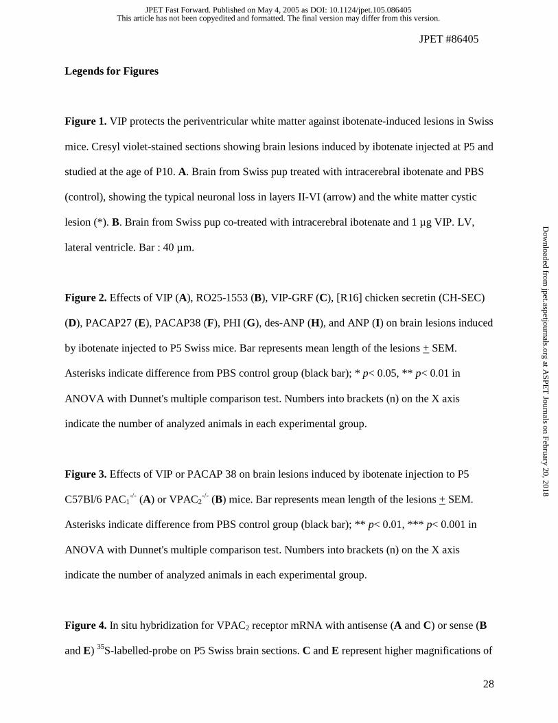
Legends for Figures
Figure 1. VIP protects the periventricular white matter against ibotenate-induced lesions in Swiss
mice. Cresyl violet-stained sections showing brain lesions induced by ibotenate injected at P5 and
studied at the age of P10. A. Brain from Swiss pup treated with intracerebral ibotenate and PBS
(control), showing the typical neuronal loss in layers II-VI (arrow) and the white matter cystic
lesion (*). B. Brain from Swiss pup co-treated with intracerebral ibotenate and 1 µg VIP. LV,
lateral ventricle. Bar : 40 µm.
Figure 2. Effects of VIP (A), RO25-1553 (B), VIP-GRF (C), [R16] chicken secretin (CH-SEC)
(D), PACAP27 (E), PACAP38 (F), PHI (G), des-ANP (H), and ANP (I) on brain lesions induced
by ibotenate injected to P5 Swiss mice. Bar represents mean length of the lesions + SEM.
Asterisks indicate difference from PBS control group (black bar); * p< 0.05, ** p< 0.01 in
ANOVA with Dunnet's multiple comparison test. Numbers into brackets (n) on the X axis
indicate the number of analyzed animals in each experimental group.
Figure 3. Effects of VIP or PACAP 38 on brain lesions induced by ibotenate injection to P5
C57Bl/6 PAC1-/- (A) or VPAC2
-/- (B) mice. Bar represents mean length of the lesions + SEM.
Asterisks indicate difference from PBS control group (black bar); ** p< 0.01, *** p< 0.001 in
ANOVA with Dunnet's multiple comparison test. Numbers into brackets (n) on the X axis
indicate the number of analyzed animals in each experimental group.
Figure 4. In situ hybridization for VPAC2 receptor mRNA with antisense (A and C) or sense (B
and E) 35S-labelled-probe on P5 Swiss brain sections. C and E represent higher magnifications of
This article has not been copyedited and formatted. The final version may differ from this version.JPET Fast Forward. Published on May 4, 2005 as DOI: 10.1124/jpet.105.086405
at ASPE
T Journals on February 20, 2018
jpet.aspetjournals.orgD
ownloaded from
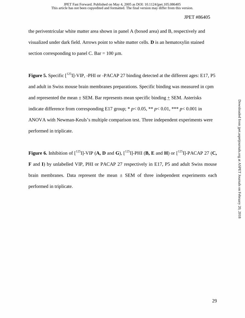
JPET #86405
29
the periventricular white matter area shown in panel A (boxed area) and B, respectively and
visualized under dark field. Arrows point to white matter cells. D is an hematoxylin stained
section corresponding to panel C. Bar = 100 µm.
Figure 5. Specific [125I]-VIP, -PHI or -PACAP 27 binding detected at the different ages: E17, P5
and adult in Swiss mouse brain membranes preparations. Specific binding was measured in cpm
and represented the mean ± SEM. Bar represents mean specific binding + SEM. Asterisks
indicate difference from corresponding E17 group; * p< 0.05, ** p< 0.01, *** p< 0.001 in
ANOVA with Newman-Keuls’s multiple comparison test. Three independent experiments were
performed in triplicate.
Figure 6. Inhibition of [125I]-VIP (A, D and G), [125I]-PHI (B, E and H) or [125I]-PACAP 27 (C,
F and I) by unlabelled VIP, PHI or PACAP 27 respectively in E17, P5 and adult Swiss mouse
brain membranes. Data represent the mean ± SEM of three independent experiments each
performed in triplicate.
This article has not been copyedited and formatted. The final version may differ from this version.JPET Fast Forward. Published on May 4, 2005 as DOI: 10.1124/jpet.105.086405
at ASPE
T Journals on February 20, 2018
jpet.aspetjournals.orgD
ownloaded from
JPET #86405
30
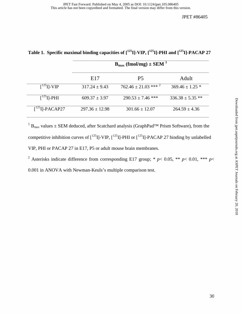
Table 1. Specific maximal binding capacities of [125I]-VIP, [125I]-PHI and [125I]-PACAP 27
Bmax (fmol/mg) ± SEM 1
E17 P5 Adult
[125I]-VIP 317.24 ± 9.43 762.46 ± 21.03 *** 2 369.46 ± 1.25 *
[125I]-PHI 609.37 ± 3.97 290.53 ± 7.46 *** 336.38 ± 5.35 **
[125I]-PACAP27 297.36 ± 12.98 301.66 ± 12.07 264.59 ± 4.36
1 Bmax values ± SEM deduced, after Scatchard analysis (GraphPad™ Prism Software), from the
competitive inhibition curves of [125I]-VIP, [125I]-PHI or [125I]-PACAP 27 binding by unlabelled
VIP, PHI or PACAP 27 in E17, P5 or adult mouse brain membranes.
2 Asterisks indicate difference from corresponding E17 group; * p< 0.05, ** p< 0.01, *** p<
0.001 in ANOVA with Newman-Keuls’s multiple comparison test.
This article has not been copyedited and formatted. The final version may differ from this version.JPET Fast Forward. Published on May 4, 2005 as DOI: 10.1124/jpet.105.086405
at ASPE
T Journals on February 20, 2018
jpet.aspetjournals.orgD
ownloaded from
This article has not been copyedited and formatted. The final version may differ from this version.JPET Fast Forward. Published on May 4, 2005 as DOI: 10.1124/jpet.105.086405
at ASPE
T Journals on February 20, 2018
jpet.aspetjournals.orgD
ownloaded from
This article has not been copyedited and formatted. The final version may differ from this version.JPET Fast Forward. Published on May 4, 2005 as DOI: 10.1124/jpet.105.086405
at ASPE
T Journals on February 20, 2018
jpet.aspetjournals.orgD
ownloaded from
This article has not been copyedited and formatted. The final version may differ from this version.JPET Fast Forward. Published on May 4, 2005 as DOI: 10.1124/jpet.105.086405
at ASPE
T Journals on February 20, 2018
jpet.aspetjournals.orgD
ownloaded from
This article has not been copyedited and formatted. The final version may differ from this version.JPET Fast Forward. Published on May 4, 2005 as DOI: 10.1124/jpet.105.086405
at ASPE
T Journals on February 20, 2018
jpet.aspetjournals.orgD
ownloaded from
This article has not been copyedited and formatted. The final version may differ from this version.JPET Fast Forward. Published on May 4, 2005 as DOI: 10.1124/jpet.105.086405
at ASPE
T Journals on February 20, 2018
jpet.aspetjournals.orgD
ownloaded from
This article has not been copyedited and formatted. The final version may differ from this version.JPET Fast Forward. Published on May 4, 2005 as DOI: 10.1124/jpet.105.086405
at ASPE
T Journals on February 20, 2018
jpet.aspetjournals.orgD
ownloaded from
This article has not been copyedited and form
atted. The final version m
ay differ from this version.
JPET
Fast Forward. Published on M
ay 4, 2005 as DO
I: 10.1124/jpet.105.086405 at ASPET Journals on February 20, 2018 jpet.aspetjournals.org Downloaded from
This article has not been copyedited and formatted. The final version may differ from this version.JPET Fast Forward. Published on May 4, 2005 as DOI: 10.1124/jpet.105.086405
at ASPE
T Journals on February 20, 2018
jpet.aspetjournals.orgD
ownloaded from
This article has not been copyedited and formatted. The final version may differ from this version.JPET Fast Forward. Published on May 4, 2005 as DOI: 10.1124/jpet.105.086405
at ASPE
T Journals on February 20, 2018
jpet.aspetjournals.orgD
ownloaded from