n angiotensin ii- and vasopeptidase- inhibitory...
TRANSCRIPT

ANGIOTENSIN II- AND VASOPEPTIDASE-
INHIBITORY ACTIVITIES OF
GYNURA PROCUMBENS
POH TING FUNG
FACULTY OF MEDICINE
UNIVERSITY OF MALAYA
KUALA LUMPUR
2014
PO
H T
ING
FU
NG
A
NG
IOT
EN
SIN
II- AN
D V
AS
OP
EP
TID
AS
E- IN
HIB
ITO
RY
AC
TIV
ITIE
S O
F G
YN
UR
A P
RO
CU
MB
EN
S
MM
edS
c
2014

ANGIOTENSIN II- AND VASOPEPTIDASE-
INHIBITORY ACTIVITIES OF
GYNURA PROCUMBENS
POH TING FUNG
DISSERTATION SUBMITTED IN FULFILMENT
OF THE REQUIREMENT
FOR THE DEGREE OF MASTER OF MEDICAL SCIENCE
FACULTY OF MEDICINE
UNIVERSITY OF MALAYA
KUALA LUMPUR
2014


ii

iii
ABSTRACT
Blood pressure (BP) is partly determined by vasoconstriction or vasodilation. The
most important vasoconstrictor is angiotensin II (Ang II) whilst nitric oxide (NO),
prostacyclin (PGI2) and bradykinin (BK) are main vasodilators. The vasopeptidase,
angiotensin-converting enzyme (ACE), is able to hydrolyse BK and convert angiotensin I
(Ang I) to Ang II. Bradykinin is also inactivated by another vasopeptidase, neutral
endopeptidase (NEP). Increases in ACE and NEP activities would elevate Ang II but
decrease BK levels that could cause increases in BP which in the long-term could result in
hypertension.
Many antihypertensive drugs are available but with side-effects. Alternative forms
of treatments, especially those from the plant kingdom, are frequently sought after. Gynura
procumbens (G. procumbens) is widely used in folk medicine to treat hypertension.
Previous findings show that leaves of this plant seem to decrease BP by inhibiting ACE
activity and blocking calcium channels. The aims of this project are to further study the
BP-lowering properties of this plant by exploring the Ang II and ACE/NEP inhibitory
activities of a partially purified fraction (FA-I) and to characterise the bioactive
subfractions.
Endothelium-intact and -denuded rat aortic rings suspended in organ chambers were
used to study the effects of FA-I on Ang I- and Ang II-induced contractions. The role of
endothelium on the effects of FA-I was tested further by adding Nω-nitro-L-arginine methyl
ester (L-NAME) or indomethacin to ascertain the involvement of NO and PGI2 respectively.
Effects of FA-I on the vasodilatory response due to BK were studied by in vitro and in vivo
methods. Rats were also given oral doses of FA-I to examine the stability of the fraction
against digestive enzyme degradation. The FA-I fraction was subjected to further
separation by thin layer chromatography (TLC) to obtain the FA-I subfractions, the

iv
activities of which on ACE/NEP were analysed by spectrophotometric and
spectrofluorometric assays. After 1H-nuclear magnetic resonance analysis, one of the
bioactive subfractions was sent for further structural characterisations.
Results show that FA-I significantly (p<0.05) decreased the contraction evoked by
Ang I and Ang II in endothelium-intact and -denuded rings with the degree of relaxation
being significantly (p<0.05) higher in intact ones. However, in the presence of L-NAME
or indomethacin, the inhibitory effect of FA-I on Ang II-induced contraction was
significantly (p<0.05) reduced. In contrast, FA-I enhanced the vasorelaxant effects of BK,
which may contribute to the greater BP-lowering effects of BK. Activities of ACE and
NEP was inhibited by FA-I. Early-eluted subfractions obtained from TLC exhibited
specific inhibition on ACE alone. Study also shows that the ACE inhibitory activity of
FA-I was still preserved after the actions of proteases and spectral analyses showed that
FA-I subfractions may contain 16-carbon compounds.
In conclusion, FA-I was found to be able to inhibit Ang II-induced contractions,
probably via endothelium-dependent pathways by activating NO or PGI2 release, and also
of being able to inhibit ACE/NEP activities. Nuclear magnetic resonance and mass
spectroscopic analyses indicate that one of the subfractions (FA-Ia) could be a 16-carbon
compound but the precise structure remains to be elucidated.

v
ABSTRAK
Tekanan darah (BP) ditentukan secara separa oleh aktiviti vasokonstriksi atau
vasodilatasi. Angiotensin II merupakan vasokonstriksi yang terpenting manakala nitrit
oksida (NO), prostaglandin I2 (PGI2) dan bradikinin (BK) adalah vasodilatasi utama.
Vasopeptidase, enzim pengubah angiotensin (ACE), mampu menyahaktif BK serta
menukar angiotensin I (Ang I) kepada Ang II. Bradikinin juga dinyahaktif oleh satu lagi
vasopeptidase, neutral endopeptidase (NEP). Peningkatan dalam aktiviti ACE dan NEP
yang meningkatkan Ang II tetapi mengurangkan tahap BK boleh menyebabkan
peningkatan BP yang dalam jangka masa panjang mengakibatkan hipertensi.
Beberapa jenis ubat kini boleh didapati untuk mengawal hipertensi tetapi adalah
dengan kesan sampingan. Alternatif rawatan, terutamanya daripada alam tumbuhan sering
dicari. Gynura procumbens (G. procumbens) merupakan salah satu tumbuhan yang
digunakan secara tradisi untuk merawat hipertensi. Penemuan sebelum ini telah
menunjukkan bahawa daun daripada tumbuhan ini mengenakan kesan antihipertensi
seolah-olahnya melalui aktiviti perencatan ACE dan penyekatan saluran kalsium. Jadi,
projek ini bertujuan untuk terus mengkaji sifat penurunan tekanan darah tumbuhan ini
dengan menerokai aktiviti perencatan Ang II dan ACE/NEP oleh fraksi akueus
G. procumbens yang separa tulen (FA-I) dan mencirikan subfraksi FA-I yang mempunyai
bioaktiviti.
Cincin aorta tikus dengan endotelium-tergantung dan -tidak tergantung yang
dipasang dalam kamar organ telah digunakan untuk mengkaji kesan FA-I terhadap
vasokonstriksi yang disebabkan oleh Ang I dan Ang II. Peranan endotelium dalam kesan
FA-I seterusnya diuji dengan menambah Nω-nitro-L-arginina methyl ester (L-NAME) atau
indomethacin untuk memerhatikan penglibatan NO dan PGI2 masing-masing. Kesan FA-I
terhadap vasodilatasi yang dibawa oleh BK dikaji dengan kaedah in vitro dan in vivo.

vi
Tikus juga disuap dengan FA-I untuk memeriksa kestabilan fraksi ini terhadap kemusnahan
yang dibawa oleh enzim pencernaan. Fraksi FA-I ditakluk kepada pemisahan lagi oleh
lapisan nipis kromatografi (TLC) untuk mendapatkan subfraksi FA-I, di mana aktiviti-
aktiviti perencatan ACE/NEP dianalisis oleh spektrofotometer dan spektrofluorometer.
Selepas analisis dengan 1H-resonans magnetik nuklear, salah satu subfraksi dihantar
selanjutnya untuk pencirian struktur molekulnya.
Keputusan menunjukkan bahawa FA-I dengan ketaranya (p<0.05) menurunkan
vasokonstriksi oleh Ang I dan Ang II dalam cincin endotelium-tergantung dan -tidak
tergantung di mana vasodilatasi lebih tinggi di endotelium-tergantung. Walau
bagaimanapun, dengan penambahan L-NAME atau indomethacin, kesan FA-I yang
melarang vasokontraksi oleh Ang II telah berkurang dengan ketara (p<0.05). Sementara itu,
FA-I meningkatkan kesan vasodilatasi BK yang mungkin mengakibatkan penurunan BP
yang lebih kuat. Kesan penurunan tekanan darah oleh FA-I masih jelas walaupun selepas
suapan menunjukkan bahawa aktiviti FA-I dapat menahani kesan enzim pencernaan.
Tambahan pula, FA-I merencat aktiviti ACE dan NEP. Dalam subfraksi-subfraksi yang
didapati, hanya subfraksi yang didapati lebih awal oleh TLC memamerkan aktiviti
perencatan yang spesifik pada ACE. Baccan juga menunjukkan FA-I masih mengekalkan
activiti perencatan ACE selepas tindakan oleh protease dan analisis spektra menunjukkan
bahawa subfraksi FA-I mungkin mengandungi kompaun 16-karbon.
Sebagai kesimpulannya, FA-I didapati berupaya mengurangkan vasokonstriksi yang
dibawa oleh Ang II dengan endotelium mungkin memainkan peranan untuk mengaktifkan
rembesan NO dan PGI2 di samping merencatkan aktiviti ACE/NEP. Selain itu, analisis
spektroskopi resonans magnetik nuklear dan spektrometri jisim menunjukkan bahawa salah
satu daripada subfraksi (FA-Ia) mungkin adalah kompaun 16-karbon tetapi struktur yang
tepat masih belum dijelaskan.

vii
ACKNOWLEDGEMENTS
I would like to express my sincere thanks to all those who gave me a chance to
complete this thesis. I want to thank the University of Malaya for offering me a
scholarship under the University of Malaya Fellowship Scheme.
My greatest thanks go to both my supervisors, Prof. Lam Sau Kuen and Dr. Hoe
See Ziau for their professional knowledge sharing, guidance, suggestions and invaluable
problem solving methods throughout the whole research and the completion of this thesis.
Through their supervision for the past few years, their creative ideas, patience and
enthusiasm have impressed me deeply and their encouragement on me to overcome the
obstacles is greatly appreciated. I owe Prof. Lam immense gratitude for guiding me
through the trials and tribulations of thesis writing.
My heartiest thanks to Dr. Chee Chin Fei from Department of Chemistry, Faculty of
Science, University of Malaya for his kindness in helping me in the characterisation of my
plant extracts. Thanks are also due to Mr. Johgalingam, a former senior technician from
the department for his technical guidance in handling the rats. His cheerful attitude has
helped me enjoy doing research. I also wish to thank Mr. Nazari and Mr. Mohan, both
technicians from the department for their technical advice. I would also like to thank Prof.
Ruby Husain, Head of the Department of Physiology, for her kind advice.
I would like to thank Ng Hien Kun, Tee Bae Huey, Dr. Hong Yet Hoi, Eng Sue
Ping and all other postgraduate students in the department who helped me tremendously to
keep me on the right track.
This thesis is dedicated to my family who is always there to love, support and
encourage me to complete my research. Many more people were involved to assist for the

viii
completion of this research than the above mentioned, and I am greatly thankful to all of
them.
This research was partly supported by the University of Malaya Research Grant
(RG 028/09BIO) and the post-graduate research funds (PS 108/2009A and PS 207/2010B).

ix
TABLE OF CONTENTS
Page
ABSTRACT/ ABSTRAK iii
ACKNOWLEDGEMENTS vii
LIST OF FIGURES xiii
LIST OF TABLES xvi
LIST OF ABBREVIATIONS xvii
LIST OF APPENDICES xxii
CHAPTER 1 - INTRODUCTION
1.1 Blood pressure
1.1.1 Regulation of blood pressure 1
1.1.2 Endothelium 2
1.1.2.1 Nitric oxide 3
1.1.2.2 Prostacyclin and thromboxane A2 4
1.1.2.3 Endothelin-1 5
1.2 Renin-angiotensin system
1.2.1 Overview 6
1.2.2 Renin 8
1.2.3 Angiotensin-converting enzyme 8
1.2.4 Angiotensin-converting enzyme 2 9
1.2.5 Neutral endopeptidase 10
1.2.6 Angiotensin II 11
1.2.7 Non angiotensin-converting enzyme dependent angiotensin II 13
formation
1.3 Kallikrein-kinin system
1.3.1 Overview 13
1.3.2 Bradykinin 15
1.4 Hypertension
1.4.1 Overview 17
1.4.2 Global burden of hypertension 18
1.4.3 Risk factors for hypertension 19
1.4.4 Treatments for hypertension 20
1.4.4.1 Angiotensin-converting enzyme inhibitor 24
1.4.4.2 Vasopeptidase inhibitor 24
1.4.4.3 Combination therapy 25
1.4.4.4 Vaccine for hypertension 26

x
1.5 Complementary and alternative medicine
1.5.1 Overview 27
1.5.2 Herb-drug interactions 28
1.6 Methods and techniques
1.6.1 Fractionation, purification and isolation of natural products 30
1.6.2 Blood pressure measurement methods 32
1.6.3 In vitro vascular tension studies 33
1.6.4 Laboratory animals 34
1.6.5 Nuclear magnetic resonance spectroscopy and mass spectrometer 35
1.6.6 In vitro assay of angiotensin-converting enzyme activity 36
1.6.7 In vitro assay of neutral endopeptidase activity 38
1.6.8 Effects of proteases on plant extract 39
1.7 Plant material
1.7.1 Family Asteraceae/ Compositae 40
1.7.2 Gynura procumbens 41
1.8 Research objectives 43
CHAPTER 2 - MATERIALS AND METHODS
2.1 Materials
2.1.1 Plant material 44
2.1.2 Animals 44
2.1.3 Chemicals and reagents 45
2.1.4 Equipment 46
2.2 Extraction and fractionation of Gynura procumbens leaves 47
2.3 In vitro vascular tension studies
2.3.1 Preparation of rat aortic rings 49
2.3.2 Effects of FA-I pretreatment on angiotensin I-induced contraction 51
2.3.3 Effects of FA-I pretreatment on angiotensin II-induced contraction 51
2.3.4 Effects of FA-I pretreatment on angiotensin II-induced contraction 51
of aortic rings in the presence of Nω-nitro-L-arginine methyl ester
or indomethacin
2.3.5 Effects of FA-I pretreatment on bradykinin-induced relaxation of 51
phenylephrine-precontracted aortic rings
2.4 Effects of FA-I on the action of bradykinin on mean arterial pressure and 52
heart rate of rats
2.5 In vitro assays
2.5.1 In vitro effect of FA-I and its subfractions on angiotensin- 54
converting enzyme activity
2.5.2 In vitro effect of FA-I and its subfractions on neutral endopeptidase 56
activity

xi
2.6 Effect of orally administered FA-I on blood pressure of spontaneously 58
hypertensive and Wistar-Kyoto rats
2.7 Effect of proteases on angiotensin-converting enzyme inhibitory 59
activity of FA-I
2.8 Data analyses 60
2.9 Statistical analyses 60
CHAPTER 3 - RESULTS
3.1 Extraction and fractionation of Gynura procumbens leaves 61
3.2 In vitro vascular tension studies
3.2.1 Effects of FA-I pretreatment on angiotensin I-induced contraction 63
3.2.2 Effects of FA-I pretreatment on angiotensin II-induced contraction 63
3.2.3 Effects of FA-I pretreatment on angiotensin II-induced contraction 65
of aortic rings in the presence of Nω-nitro-L-arginine methyl ester
or indomethacin
3.2.4 Effects of FA-I pretreatment on bradykinin-induced relaxation of 67
phenylephrine-precontracted aortic rings
3.3 Effects of FA-I on the action of bradykinin on mean arterial pressure and 68
heart rate of rats
3.4 In vitro assays
3.4.1 In vitro effect of FA-I and its subfractions on angiotensin- 70
converting enzyme activity
3.4.2 In vitro effect of FA-I and its subfractions on neutral endopeptidase 72
activity
3.5 Effect of orally administered FA-I on blood pressure of spontaneously 74
hypertensive and Wistar-Kyoto rats
3.6 Characterisation of FA-I subfractons 75
3.7 Effect of proteases on angiotensin-converting enzyme inhibitory 76
activity of FA-I

xii
CHAPTER 4 - DISCUSSION
4.1 In vitro vascular tension studies 78
4.2 Effects of FA-I on bradykinin by in vitro and in vivo studies 80
4.3 Vasopeptidase inhibitory activities of FA-I and FA-I subfractions 82
4.4 Effect of orally administered FA-I on blood pressure of rats 84
4.5 Effect of proteases on angiotensin-converting enzyme inhibitory 85
activity of FA-I and nuclear magnetic resonance studies on FA-Ia
4.6 General discussions and future studies 86
CHAPTER 5 - CONCLUSIONS 88
~ PUBLICATIONS 90
~ REFERENCES 100
~ APPENDICES 119

xiii
LIST OF FIGURES
Page
Figure 1.1 Schematic representation of the activation of endothelial nitric
oxide synthase that finally produces nitric oxide to cause
vasodilation.
4
Figure 1.2 Activation of renin-angiotensin system and the effects on
related receptors as well as the actions of neutral
endopeptidase.
7
Figure 1.3 Activation of kallikrein-kinin system that finally leads to
activation of bradykinin receptors (B1R and B2R).
15
Figure 1.4 Degradation of bradykinin that occurs at amino (A) and
carboxy (C) terminals.
16
Figure 1.5 The algorithm for the treatment of hypertension according to
The Seventh Report of the Joint National Committee on
Prevention, Detection, Evaluation, and Treatment of High
Blood Pressure (JNC 7).
22
Figure 1.6 The principle for the measurement of angiotensin-converting
enzyme activity by a colorimetric assay.
37
Figure 1.7 The two-step reaction that finally produces fluorescence 7-
amino-4-methylcoumarin.
38
Figure 1.8 Gynura procumbens (Lour.) Merr.
42
Figure 2.1 Extraction and fractionation of Gynura procumbens that
finally produce FA-I subfractions.
48
Figure 2.2 The aortic ring was mounted in an organ bath containing 10 ml
Krebs-Henseleit solution with the lower hook being fixed to
the bottom of the bath whereas the upper hook is connected to
a force-displacement transducer connected to a digital
physiographic setup.
50
Figure 2.3 Setup of the experimental procedure for the measurement of
blood pressure and heart rate in a rat.
53
Figure 2.4 Measurement of rat systolic blood pressure of rat by tail-cuff
method.
58

xiv
Figure 3.1 Chromatogram of a partially purified aqueous fraction (FA-I)
on thin layer chromatography (TLC) plates after being sprayed
with ninhydrin reagent. The dark circle reveals FA-Ia.
61
Figure 3.2 Flow chart of extraction and fractionation of the leaves of
Gynura procumbens (All % values in parenthesis indicate %
yield of each fraction obtained from the original dried leaves
weight except for FA-I which was calculated from 1 g of FA).
62
Figure 3.3 Effects of a partially purified aqueous fraction (FA-I) on
angiotensin I-induced contraction in (a) endothelium-intact
(Endo +) and (b) endothelium-denuded (Endo -) aortic rings.
The effects were determined in the absence or presence of FA-
I (1.0 x 10⁻⁴
or 1.0 x 10⁻³
g/ml). Values are mean ± S.E.M. (n
= 6). **p<0.01; ***p<0.001 compared with controls (without
FA-I) and +p<0.05;
+++p<0.001 compared with Endo +.
64
Figure 3.4 Effect of a partially purified aqueous fraction (FA-I) on
angiotensin II-induced contraction in (a) endothelium-intact
(Endo +) and (b) endothelium-denuded (Endo -) aortic rings.
The effect was determined in the absence or presence of FA-I
(1.0 x 10⁻⁴
or 1.0 x 10⁻³
g/ml). Values are mean ± S.E.M.
(Endo +, n = 7; Endo –, n = 6). ***p < 0.001; compared with
controls (without FA-I).
64
Figure 3.5
Figure 3.6
Effect of a partially purified aqueous fraction (FA-I) on
angiotensin II-induced contraction in endothelium-intact aortic
rings preincubated with (a) Nω-nitro-L-arginine methyl ester
(L-NAME) or (b) indomethacin. The effect was determined in
the absence or presence of FA-I (1.0 x 10⁻⁴
or 1.0 x 10⁻³
g/ml).
Values are mean ± S.E.M. (n = 6).
Effect of a partially purified aqueous fraction (FA-I) on
bradykinin-induced relaxation of aortic rings precontracted
with phenylephrine. The effect was determined in the absence
or presence of FA-I (1.0 x 10⁻⁴
or 1.0 x 10⁻³
g/ml). Values are
mean ± S.E.M. (n = 8). *p < 0.05 **p < 0.01 compared with
controls (without FA-I).
66
67

xv
Figure 3.7
Figure 3.8
Effect of intravenous bradykinin on the (a) mean arterial
pressure (MAP) and (b) heart rate (HR) of anaesthetised rats
before and after 10mg/kg FA-I administration. Values are
mean ± S.E.M. (n = 8). *p < 0.05; **p < 0.01; ***p < 0.001
compared with 0 ng/kg bradykinin and +p < 0.05;
++p < 0.01
compared with controls (before FA-I administration). Effect of a partially purified aqueous fraction (FA-I) on
angiotensin-converting enzyme (ACE) activity. Data are
means (duplicate assays).
69
70
Figure 3.9 Inhibition of angiotensin-converting enzyme (ACE) by (a)
FA-Ia, (b) FA-Ib, (c) FA-Ic, (d) FA-Id and (e) FA-Ie. Data
are means (duplicate assays).
71
Figure 3.10 Effect of a partially purified aqueous fraction (FA-I) on
neutral endopeptidase (NEP) activity. Data are means
(triplicate assays).
72
Figure 3.11 Inhibition of neutral endopeptidase (NEP) by (a) FA-Ia, (b)
FA-Ib, (c) FA-Ic, (d) FA-Id and (e) FA-Ie. Data are means
(triplicate assays).
73
Figure 3.12 Changes in systolic blood pressure after a single orally
administered of partially purified aquwous fraction (FA-I) in
(a) Wistar-Kyoto (WKY) and (b) spontaneously hypertensive
(SHR) rats. Values are mean ± S.E.M. (WKY, n = 6; SHR, n
= 7). *p < 0.05, **p < 0.01 and ***p < 0.001 compared with
vehicle (distilled water).
74
Figure 3.13 Changes in systolic blood pressure in Wistar-Kyoto (WKY)
and spontaneously hypertensive (SHR) rats at 6 hours after
orally administered of partially purified aqueous fraction
(FA-I). Values are mean ± S.E.M. (WKY, n = 6; SHR, n =
7). *p < 0.05, **p < 0.01 and ***p < 0.001 compared with
vehicle (distilled water).
75
Figure 3.14 The angiotensin-converting enzyme (ACE) inhibitory activity
of FA-I (a) without and after (b) pepsin, (c) chymotrypsin and
(d) trypsin. Data are means (duplicate assays).
77
Figure 5.1 Summary of extraction and fractionation of Gynura
procumbens, effects and mechanisms of actions.
89

xvi
LIST OF TABLES
Page
Table 1.1 Effects of angiotensin II type 1 receptor activation.
12
Table 1.2 Classification of blood pressure for adults ≥18 years old
according to The Seventh Report of the Joint National
Committee on Prevention, Detection, Evaluation, and
Treatment of High Blood Pressure (JNC 7, 2003).
18
Table 1.3 Identifiable risk factors for hypertension.
19
Table 1.4 Classes of antihypertensive drugs, examples and possible side-
effects.
23
Table 1.5 Examples of antihypertensive medicinal herbs with different
types of mechanisms of action.
29
Table 1.6 Taxonomy of Gynura procumbens.
41
Table 3.1 Effect of a partially purified aqueous fraction (FA-I) on the
pEC50 values for angiotensin II-induced contraction of aortic
rings pretreated with Nω-nitro-L-arginine methyl ester (L-
NAME) or indomethacin.
66
Table 3.2 IC50 values of FA-I subfractions.
70
Table 3.3 The effect of digestive enzymes on the IC50 values of FA-I. 77

xvii
LIST OF ABBREVIATIONS
ACE Angiotensin-converting enzyme
ACEI Angiotensin-converting enzyme inhibitor
ACh Acetylchlorine
AMC 7-amino-4-methylcoumarin
APN Leucine aminopeptidase
Ang I Angiotensin I
Ang II Angiotensin II
Ang III Angiotensin III
Ang IV Angiotensin IV
Ang (1-7) Angiotensin (1-7)
Ang (1-9) Angiotensin (1-9)
AngQb CYT006-AngQb
ANOVA Analysis of variance
ANP Atrial natriuretic peptide
ANS Autonomic nervous system
APN Leucine aminopeptidase
ATP Adenosine triphosphate
AT1R Angiotensin II type 1 receptor
AT2R Angiotensin II type 2 receptor
AT4R Angiotensin type 4 receptor
B : A : W n-butanol : acetic acid : distilled water
BB Beta blocker
BK Bradykinin
BP Blood pressure

xviii
B1 Bradykinin type 1
B2 Bradykinin type 2
Ca2+
Calcium ion
CaCl2 Calcium chloride
CAM Complementary and alternative medicine
cAMP Cyclic adenosine-3‘, 5-monophosphate
CCB Calcium channel blocker
cGMP Cyclic guanosine-3‘, 5-monophosphate
CO Cardiac output
COX Cyclooxygenase
CVD Cardiovascular disease
D Dimensional
DAGNPG N-dansyl-Ala-Gly-D-nitro-Phe-Gly
DASH Dietary approaches to stop hypertension
DBP Diastolic blood pressure
DEPT 2D distortionless enhancement by polarisation transfer
EDH Endothelium-dependent hyperpolarisation
eNOS Endothelial nitric oxide synthase
ET Endothelin
ET-1 Endothelin-1
FA Final aqueous fraction
FA-I Purer final aqueous fraction
FDA Food and Drug Administration
FT Fourier transform
gACE Germinal ACE

xix
GC Guanylate cyclase
G. procumbens Gynura procumbens
GTP Guanosine triphosphate
H3BO3 Boric acid
HA Hippuric acid
HCl Hydrochloric acid
HEPES 1,4-dioxan, 4-(2-hydroxyethyl)-1-piperazineethanesulfonic
acid
HHL Hippuryl-L-histidyl-L-leucine
HL Histidyl-leucine
HMWK High molecular weight kininogen
HPLC High performance liquid chromatography
HR Heart rate
JG Juxtaglomerular
JNC 7 The seventh report of the joint national committee on
prevention, detection, evaluation, and treatment of high
blood pressure
KCl Potassium chloride
K-H Krebs-Henseleit
KH2PO4 Potassium dihydrogen phosphate
L-NAME Nω-nitro-L-arginine methyl ester
LMWK Low molecular weight kininogen
MAP Mean arterial pressure
MgSO4 Magnesium sulphate
MS Mass spectrometry
MW Molecular weight

xx
NaCl Sodium chloride
NaHCO3 Sodium bicarbonate
NaOH Sodium hydroxide
NEP Neutral endopeptidase
NEPI Neutral endopeptidase inhibitor
NMR Nuclear magnetic resonance
NO Nitric oxide
NOS Nitric oxide synthase
NP Natriuretic peptide
P-AMC Phe-7-amino-4-methylcoumarin
PE Phenylephrine
PGI2 Prostacyclin
RAS Renin-angiotensin system
Rf Retention factor
ROS Reactive oxygen species
SAAP-AMC Suc-L-Ala-L-Ala-Phe-7-amino-4-methylcoumarin
sACE Somatic ACE
SARS Severe acute respiratory syndrome
SBP Systolic blood pressure
SHR Spontaneously hypertensive
SD Sprague-Dawley
SV Stroke volume
TCM Traditional Chinese medicine
TLC Thin layer chromatography

xxi
TPR Total peripheral resistance
TXA2 Thromboxane A2
UV Ultraviolet
VPI Vasopeptidase inhibitor
VSM Vascular smooth muscle
v/v Volume/ volume
WHO World Health Organisation
WKY Wistar-Kyoto
w/v
Zn2+
[Ca2+
]i
Weight/ volume
Zinc ion
Intracellular calcium ion

xxii
LIST OF APPENDICES
Appendix Page
A Sephadex LH-20 gel filtration chromatography
119
B Preparative thin layer chromatography
120
C Ninhydrin reagent
121
D Compositions of Krebs-Henseleit solution
121
E 1H nuclear magnetic resonance spectrum (NMR) of FA-Ia
and the tabulated chemical shifts
122
F 1H nuclear magnetic resonance spectrum (NMR) of FA-Ib
123
G 1H nuclear magnetic resonance spectrum (NMR) of FA-Ic
124
H 13
C nuclear magnetic resonance spectrum (NMR) of FA-Ia
and the tabulated chemical shifts
125
I
2D distortionless enhancement by polarisation transfer
(DEPT) spectrum of FA-Ia
126
J Liquid chromatography-mass spectrometry (LC-MS)
spectrum of FA-Ia
127
K Infrared spectrum of FA-Ia
127
L Suggested random coil chemical shifts (in ppm) for the
common amino acids
128

1
CHAPTER 1 - INTRODUCTION
1.1 Blood pressure
1.1.1 Regulation of blood pressure
Blood pressure (BP) is the force exerted by the blood against any unit area of the
blood vessel wall (Guyton and Hall, 2006). The BP in humans is usually measured by the
indirect auscultatory method and is expressed as systolic BP (SBP) over diastolic BP (DBP)
(SBP/DBP) mmHg. In physiology, BP can be calculated from the formula: BP = cardiac
output (CO) x total peripheral resistance (TPR) (Kirkman and Sawdon, 2010). Cardiac
output is the volume of blood that is pumped out of the ventricle per minute and is
expressed as CO = stroke volume (SV) x heart rate (HR) (Kirkman and Sawdon, 2010),
whereas TPR refers to the total peripheral resistance of the entire systemic circulation. In
CO, SV is the volume of blood that is pumped out of the ventricles during one heart beat
whereas HR is the number of beats per minute. The SV is mainly controlled by venous
return (preload), outflow resistance (afterload) and force of ventricular contractility
(Ackermann, 2004; Wilcken, 2010). Increases in preload and ventricular contractility, and
a decrease in afterload will increase the SV. As for the HR, it is mainly controlled by
cardiac pacemaker cells that are increased by positive chronotropic agents (e.g. cardiac
sympathetic nervous activity) and decreased by negative chronotropic agents (e.g. cardiac
parasympathetic nervous activity) (Ackermann, 2004).
Resistance in blood vessels largely depends on smooth muscle activities that change
the radius of the lumen, either by vasocontraction or vasodilation. Vascular smooth muscle
(VSM) activities are affected by the endothelium, autonomic nervous system and blood
bornes substances such as adrenaline, acetylcholine, adenosine triphosphate (ATP) and
substance P (Kirkman and Sawdon, 2010). Overall, any factor that influences SV, HR and
TPR will change the BP (Kirkman and Sawdon, 2010).

2
In order to provide and maintain sufficient blood supply to organs and tissues, BP is
regulated all the times, either by short-term or long-term mechanisms. Under resting
conditions, arterial BP is controlled mainly by baroreceptors (Kirkman and Sawdon, 2010)
via altering the cardiovascular parameters of SV, HR or TPR (Ackermann, 2004). In this
short-term regulation of BP, the change in the stretch of arterial walls is rapidly sensed by
baroreceptors located in the walls of aortic arch and carotid sinuses (Kirkman and Sawdon,
2010) that reflexedly activate or inhibit the sympathetic or parasympathetic division of the
ANS and renin-angiotensin system (RAS). In the long-term regulation of BP, however, the
kidney plays the most important role (Ackermann, 2004) by regulating the total body
content of sodium, water and other electrolyte balance.
In addition, circadian rhythm also plays a key role in BP regulation. In a healthy
person, the BP rises in the morning, followed by night time drop in a cycle that occurs once
every 24 hours (Rudic and Fulton, 2009). This circadian rhythm may become abnormal
during hypertension in which the person may become nondippers (absence of nocturnal BP
drop), extreme dippers (marked nocturnal BP drop) or reverse dippers (rise in nocturnal BP)
(Rudic and Fulton, 2009).
1.1.2 Endothelium
The endothelium is a monolayer of cells lining the entire inner layer of blood
vessels. This layer represents an important component in inflammation, platelet
aggregation, angiogenesis, endocrine functions (Feletou, 2011) and the regulation of
vascular contractility by releasing vasoactive substances that change the degree of
contraction of the underlying VSM (Vanhoutte and Mombouli, 1996). The vasoactive
substances produced by the endothelium are vasodilators such as nitric oxide (NO) and
prostacyclin (PGI2) that cause endothelium-dependent hyperpolarisation (EDH) (Feletou

3
and Vanhoutte, 2013) or vasoconstrictors such as endothelin-1 (ET-1) and thromboxane A2
(TXA2) that presumably result in membrane depolarisation (Sandoo et al, 2010). Another
powerful vasoconstrictor is that of angiotensin II (Ang II) and it is discussed in the RAS
section (page 11).
1.1.2.1 Nitric oxide
Nitric oxide, first identified by Furchgott and Zawadzki in 1980 (Furchgott and
Zawadzki, 1980) is an important component in the control of basal vasodilator tone in
blood vessels (Vallance et al, 1989). This compound is formed from L-arginine by the
action of NO synthase (NOS), an enzyme that exists in three isoforms: neuronal NOS,
macrophage or inducible NOS and endothelial NOS (eNOS) (Vanhoutte and Mombouli,
1996). Of these, eNOS appears to play a main role in dilation of blood vessels (Sandoo et
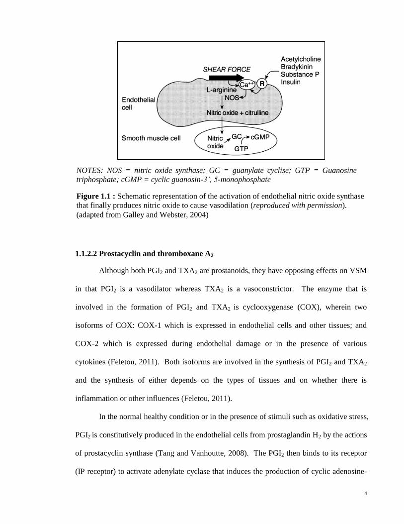
al, 2010). Figure 1.1 summarises the formation of NO by activation of eNOS. Stimuli
such as shear stress and the binding of NO agonists such as bradykinin (BK), acetylcholine
(ACh), insulin or substance P to respective receptors increase intracellular calcium ion
(Ca2+
) ([Ca
2+]i) and cause calmodulin in the endothelial cells to bind to eNOS that then
converts L-arginine to NO. The released NO then diffuses into smooth muscle cells and
activates soluble guanylate cyclise (GC), which converts guanosine triphosphate (GTP) to
cyclic guanosin-3‘, 5-monophosphate (cGMP) (Sandoo et al, 2010). Subsequently, a
protein kinase is activated by cGMP to inhibit calcium influx into the smooth muscle cells
which then decreases the contraction of smooth muscle cells to cause vasodilation (Galley
and Webster, 2004).
4
1.1.2.2 Prostacyclin and thromboxane A2
Although both PGI2 and TXA2 are prostanoids, they have opposing effects on VSM
in that PGI2 is a vasodilator whereas TXA2 is a vasoconstrictor. The enzyme that is
involved in the formation of PGI2 and TXA2 is cyclooxygenase (COX), wherein two
isoforms of COX: COX-1 which is expressed in endothelial cells and other tissues; and
COX-2 which is expressed during endothelial damage or in the presence of various
cytokines (Feletou, 2011). Both isoforms are involved in the synthesis of PGI2 and TXA2
and the synthesis of either depends on the types of tissues and on whether there is
inflammation or other influences (Feletou, 2011).
In the normal healthy condition or in the presence of stimuli such as oxidative stress,
PGI2 is constitutively produced in the endothelial cells from prostaglandin H2 by the actions
of prostacyclin synthase (Tang and Vanhoutte, 2008). The PGI2 then binds to its receptor
(IP receptor) to activate adenylate cyclase that induces the production of cyclic adenosine-
Figure 1.1 : Schematic representation of the activation of endothelial nitric oxide synthase
that finally produces nitric oxide to cause vasodilation (reproduced with permission).
(adapted from Galley and Webster, 2004)
NOTES: NOS = nitric oxide synthase; GC = guanylate cyclise; GTP = Guanosine
triphosphate; cGMP = cyclic guanosin-3’, 5-monophosphate

5
3‘, 5-monophosphate (cAMP) (Vanhoutte and Mombouli, 1996) to cause vasodilation
(Fetalvero et al, 2007).
Thromboxane A2 is also produced from prostaglandin H2, by the actions of
thromboxane A synthase (Yokoyama et al, 1991). In healthy VSM or presence of physical
stimuli such as stretch, this unstable compound with a short half-life (about 30 seconds)
binds to its receptor (TP receptor) to cause platelet aggregation, angiogenesis, and
endothelium-dependent contractions by increasing [Ca2+
]i (Nakahata, 2008).
1.1.2.3 Endothelin-1
Endothelin (ET) is a strong vasoconstrictor and exists in three isoforms: ET-1, ET-2
and ET-3 (Inoue et al, 1989). Endothelin-1, the only isoform produced in vascular
endothelium (Masaki, 2004), is formed from big ET-1 by the action of endothelin-
converting enzyme, a zinc-metallopeptidase that is anchored to the plasma membrane
(Yanagisawa et al, 1988).
In tissues, two types of ET-1 receptors are found: ETA and ETB (Luscher, 1994).
Endothelin-1 binds to these receptors, predominantly ETA (Luscher, 1994) to increase
[Ca2+
]i and cause vasocontraction (Zhang1, 2000). However, under certain conditions,
ET-1 can cause vasodilation via the activation of ETB receptor, by stimulating the release of
NO (Cardillo et al, 2000).

6
1.2 Renin-angiotensin system
1.2.1 Overview
The main homeostatic mechanism in the body that is involved in the regulation of
BP, as well as extracellular fluid volume and sodium content balance is RAS (Lote, 2006).
In the RAS, the key enzyme renin, is synthesised in an inactive form as ‗prorenin‘ in the
granular cells of the afferent arterioles in the juxtaglomerular (JG) cells of the kidneys
(Guyton and Hall, 2006). Renin splits 10-amino acid fragments from angiotensinogen that
is produced in the liver to form the decapeptide angiotensin I (Ang I) which has no
physiological effect (Hayashi and Kimoto, 2010) and within seconds to minutes, it is
cleaved of two amino acids to form octapeptide Ang II by an enzyme called angiotensin-
converting enzyme (ACE) (Guyton and Hall, 2006; Lote, 2006). Although the Ang II
persists in the blood for 1-2 minutes only, it accounts for the main function of RAS to
restore the BP to the normal level (Guyton and Hall, 2006).
The RAS is involved in both physiological and pathological processes. In the
physiological processes, RAS takes part in development, learning and memory as well as
tissue growth. However, in pathological process, RAS plays a role in the progression of
disorders such as diabetes, hypertension, cardiovascular disease (CVD) and also tumors
(Nguyen, 2006; Atlas, 2007). The roles and characteristics of renin, Ang II, ACE and the
roles of receptors for angiotensin II are shown in Figure 1.2.

7
Inactive metabolites
Stimulation of renin release
SNS
Wall tension in renal afferent arteriole
Delivery of NaCl to macula densa
AT2R Vasodilation
Antiproliferation
Apoptosis
AT1R
(predominates) Vasoconstriction
Proliferation
Inflammation
Oxidative stress
Na+ and fluid retention
ACE
Figure 1.2 : Activation of renin-angiotensin system and the effects on related receptors as
well as the actions of neutral endopeptidase (adapted from Ferrario and Iyer, 1998; Turner
and Hooper, 2002; Carey and Park, 2006; Hunyady and Catt, 2006; Atlas, 2007; Daull et
al, 2007; Lambert et al, 2008; Harrison-Bernard, 2009; Benigni et al, 2010).
NOTES: SNS = Sympathetic nervous system; Ang = Angiotensin; ACE = Angiotensin converting enzyme;
BK = Bradykinin; NPs = Natriuretic peptides; NEP = Neutral endopeptidase; AT1R, AT2R = Angiotensin
type 1, 2 receptor;
(+)
Angiotensinogen
(liver)
Ang I
Ang II
Renin Prorenin
NEP
BK NPs

8
1.2.2 Renin
Renin is the key regulatory enzyme in the RAS (Sequeira Lopez and Gomez, 2010)
and it exists in plasma in two forms: inactive prorenin (about 90%) and mature renin
(Nguyen, 2006). The inactive prorenin becomes mature renin in the JG cells of the kidneys,
which is then released into the plasma when triggered by the three main mechanisms that
are increased sympathetic activity to the granular cells of the afferent arterioles, decreased
tension in the walls of the afferent arterioles and decreased delivery of sodium chloride to
the macula densa (Lote, 2006). In addition, renin expressing cells are restricted to JG cells
only (Sequeira Lopex et al, 2004). However, in conditions such as hemorrhage that
triggers the homeostasis response, there is an increase in renin expressing cells outside the
JG cells such as in renal vascular smooth muscle cells and glomerular cells (Sequeira
Lopex et al, 2004). This is useful in order to produce enough renin to reestablish
homeostasis (Sequeira Lopex et al, 2004).
1.2.3 Angiotensin-converting enzyme
Angiotensin-converting enzyme, a zinc ion (Zn2+
)-dependent dipeptidyl
carboxypeptidase and an ectoenzyme, is anchored to cell membranes by C-terminal (Shen
et al, 2008). It exists as two isoforms: somatic ACE (sACE) (Bernstein et al, 1988) which
is found in many tissues such as vascular endothelia (Ryan et al, 1976), endothelial lining
of lungs (Turner and Hooper, 2002), brush borders of kidney and intestine (Cushman and
Cheung, 1971); and the testicular or germinal ACE (gACE) isoform which is located only
in testes (Bernstein et al, 1989). Somatic ACE has two active sites with two domains (C-
and N-domains) whereas gACE has only one active site (C-domain), which is identical to
the C-domain of sACE (Ehlers et al, 1989).

9
In general, ACE functions by hydrolysing Ang I, BK, substance P and
cholecystokinin (Turner and Hooper, 2002) as well as cleaving Ang (1-7) (Chappell et al,
1998). Both the domains of sACE are believed to have distinct functions wherein the
C-domain is involved in RAS regulation and BK hydrolysis, whereas the N-domain is
connected with the processing of bioactive peptides and regulation of renal structure and
functions (Coates, 2003). In contrast, the C-domain in gACE is involved in the sperm
maturation process (Metayer et al, 2002; Coates, 2003). Nevertheless, these two isoforms
of ACE cannot act as a substitute for each other in their respective physiological functions
(Kessler et al, 2007).
1.2.4 Angiotensin-converting enzyme 2
A homologue for ACE that shares 42% of amino acid sequence with ACE is ACE 2.
It is less distributed and restricted to the heart, kidney and testes (Donoghue et al, 2000).
There are differences in the structures and actions of ACE homologues in which ACE is a
dipeptidyl carboxypeptidase that cleaves dipeptides from substrate whereas ACE 2 is a
carboxypeptidase that cleaves only one single amino acid from its substrate (Guy et al,
2005; Lambert et al, 2008).
In the RAS, ACE 2 cleaves Ang I to form angiotensin (1-9) [Ang (1-9)] and also
splits Ang II to angiotensin (1-7) [Ang (1-7)], a compound which is becoming popular for
their protective role in RAS. The Ang (1-9) is also converted by another ectoenzyme, the
neutral endopeptidase 24.11 (NEP), and also by ACE to form Ang (1-7) (Welches et al,
1993). Studies have shown that Ang (1-7) potentiates the effect of BK (Greco et al, 2006),
stimulates NO and prostaglandin release (Rajendran, Chirkov et al. 2005) but antagonises
the effect of Ang II (Grobe et al, 2007). The affinity of ACE 2 for Ang I is weaker when
compared to ACE and the imbalance in the level of ACE and ACE 2 in the body is believed

10
to be responsible for CVD and also hypertension (Der Sarkissian et al, 2006). Other than
that, a report shows that ACE 2 is a receptor for severe acute respiratory syndrome (SARS)
coronavirus (Li et al, 2003).
1.2.5 Neutral endopeptidase
Another Zn2+
-dependent metalloendopeptidase that is also involved in BK
metabolisms (Figure 1.2), is neutral endopeptidase (NEP), also known as enkephalinase,
neprilysin, neutral metalloendopeptidase or common acute lymphoblastic leukemia antigen.
Similar to ACE, NEP also exists in the membrane-bound form and it is widely distributed
in kidneys, the central nervous system, lungs, male genital tract, intestines, and in
neutrophils, fibroblasts and epithelial cells (Erdos and Skidgel, 1989).
Neutral endopeptidase metabolises vasoconstrictor peptides such as enkephalins and
Ang II as well as the vasodilator peptides, BK, substance P and natriuretic peptide (NP)
(Ruschitzka et al, 2001). The main effect of NEP is similar to ACE that is to increase BP.
Although ACE is the main enzyme in the metabolism of BK, in human cardiac tissues,
NEP is more important in metabolising BK (Kokkonen et al, 1999). The most important
role of NEP is in the metabolism of NPs which present in three isoforms: the well-studied
atrial-NP (ANP), brain-NP and C type-NP (Ruschitzka et al, 2001). The ANP reduces BP
by increasing natriuresis as well as inhibiting RAS, endothelin and angiogenesis
(Ruschitzka et al, 2001; Sagnella, 2002).

11
1.2.6 Angiotensin II
Angiotensin II binds to two types of G protein-coupled receptors, known as Ang II
type 1 receptor (AT1R) and type 2 receptor (AT2R) (Lote, 2006; Benigni et al, 2010).
Angiotensin II type 1 receptor is widely distributed especially in VSM cells (Hunyady and
Catt, 2006) and it mediates most of the actions of Ang II (Lote, 2006) which vary at
different effector tissues (Peach and Dostal, 1990). For instance, Ang II is a
vasoconstrictor in systemic circulation (Peach and Dostal, 1990) but has both vasodilator
and vasoconstrictor effects in cerebral circulation (Maktabi et al, 1990). Nevertheless, the
overall main effect of Ang II is to increase BP by causing vasocontraction and this
contraction is a fast reaction and occurs rapidly after Ang II binding to AT1R (Touyz and
Schiffrin, 2000). Increased production of Ang II due to increased RAS activity is
considered one of the many contributing factors for the onset of hypertension (Navar et al,
2011). Table 1.1 summarises the effects brought about by activation of AT1R by Ang II.
In contrast, the level of AT2R is low in healthy adults but only exists at high level in
fetal tissues (Ichiki et al, 1995) and pathological states such as heart failure (Liu et al, 1997)
and myocardial infarction (Searles and Harrison, 1999). This receptor may take part in
brain development as well (Gendron et al, 2003). Stimulation of AT2R by Ang II
antagonises the effects brought by AT1R activation (Benigni et al, 2010) and also activates
the BK type 2 (B2) receptor to cause vasodilation which is likely to be mediated by the NO
pathway and cGMP cell signalling cascade (Searles and Harrison, 1999; Carey and Park,
2006).

12
Vasoconstriction
Stimulation of thirst
Release of antidiuretic hormone and aldosterone
Increased sodium reabsorption by direct effects on the proximal tubule of the
nephrone, effect of aldosterone on distal tubules and also increase in renal
sympathetic nerve activity
Angiogenesis, cellular growth and hypertrophy
Induction of production of reactive oxygen species (ROS)
Increase in the production of vascular endothelial growth factor in the inflammatory
process
(Adapted from Searles and Harrison, 1999; Hunyady and Catt, 2006; Lote, 2006; Benigni
et al, 2010)
Angiotensin II has a short half-life of about one minute (Lote, 2006). It is cleaved
by aminopeptidase A to angiotensin III (Ang III) which has the same affinity for AT1R and
AT2R. Angiotensin III plays a main role in the regulation of brain RAS as well as
stimulating the release of aldosterone. It is then cleaved by aminopeptidase N to
angiotensin IV (Ang IV) which has low affinity for AT1R and AT2R (Hunyady and Catt,
2006) but a higher affinity for angiotensin type 4 receptor (AT4R) to increase natriuresis
and renal blood flow. Angiotensin IV is also involved in memory and learning process
(Turner and Hooper, 2002).
Table 1.1 : Effects of angiotensin II type 1 receptor activation.

13
1.2.7 Non angiotensin-converting enzyme dependent angiotensin II formation
Angiotensin II can also be formed from non-ACE dependent pathway and the
enzyme responsible for this is chymase (Huang et al, 2003). Chymase, a chymotrypsin-like
serine protease found in the mast cells (Takai et al, 1999), is thought to be the main enzyme
for Ang II formation in human hearts (Urata et al, 1990), arteries (Huang et al, 2003) and
the kidneys (Huang et al, 2003). This pathway provides a need to suppress the non ACE-
dependent Ang II formation in order to fully suppress the RAS (Petrie et al, 2001).
1.3 Kallikrein-kinin system
1.3.1 Overview
The kallikrein-kinin system is a complex system that has a close relationship with
the RAS in the regulation of BP. There are three main constituents in the kallikrein-kinin
system: kallikreins, kininogens and kinins (Campbell, 2000). Kallikreins are serine
proteases and can be classified into tissue kallikrein and plasma kallikrein. Kininogens are
proteins of two types: low molecular weight kininogen (LMWK) and high molecular
weight kininogen (HMWK). The kallikrein-kinin system produces two main kinins which
are BK and kallidin.
The kallikrein-kinin system is predominantly found in tissues rather than in the
circulation (Golias et al, 2007). As shown in Figure 1.3, the precursor for plasma kallikrein
is prekallikrein that forms a complex with Factor XII (Hageman Factor). When there is
tissue damage, prekallikrein is changed to plasma kallikrein that then converts HMWK to
BK while tissue kallikrein hydrolysess LMWK to kallidin that will eventually be converted
to BK by aminopeptidase (Campbell, 2000).
There are two types of G-coupled protein receptors for BK, known as BK type 1 (B1)
and B2 receptors. The B2 receptor predominates under healthy conditions whereas the B1

14
receptor is induced by tissues injuries such as myocardial ischemia and inflammation and
also by endogenous factors such as growth factors, endotoxins and cytokines (Campbell,
2000; Sharma and Al-Sherif, 2011). When the B1 receptor is activated, it causes
vasodilation and initiates the inflammatory response (Sharma and Al-Sherif, 2011). As for
B2 receptor, it has higher affinity for BK (Leeb-Lundberg et al, 2005) and activation of B2
receptor causes vasodilation by releasing EDH mediators and cGMP from NO (Vanhoutte,
2001). Figure 1.3 shows the activation of kallikrein-kinin system that leads to the
production of kallidin and BK as well as the actions of BK receptors.
Kinin receptors are also linked to many types of diseases such as CVD, renal
disease, airway disease especially asthma (Barnes 1992), neurological disease, cancer as
well as arthritis, hereditary angioedema and gastrointestinal disease (Leeb-Lundberg et al,
2005). Thus, kinin receptors are also a pharmacological target in treating these diseases.

15
NOTES:
LMWK = Low molecular weight kininogen;
HMWK = High molecular weight kininogen;
B1R, B2R = Bradykinin type 1, type 2 receptor
Aminopeptidase
1.3.2 Bradykinin
Bradykinin is important in the regulation of BP and pain and in the inflammatory
process, and it is also said to have cardioprotective (Sharma and Al-Sherif, 2011), diuretic
and natriuretic effect (Willis et al, 1969). Binding of BK to B2 receptor increases local
production of BK to induce endothelium dependent relaxation and the main action of BK is
at the heart, kidney and blood vessels (Vanhoutte, 2001). In the inflammatory process, BK
increases vascular permeability to fluid and plasma proteins that results in oedema (Sharma
and Al-Sherif, 2011) while in the management of pain, BK is both algesic and hyperalgesic
(Sharma and Al-Sherif, 2011).
Degradation of BK occurs either at its amino or carboxy terminal (Figure 1.4).
Enzymes that cleave at amino terminal are aminopeptidase M and P whereas enzymes that
are responsible for the degradation of BK at carboxy terminal are kininase I and kininase II
Kininogens
LMWK HMWK
Kallidin Bradykinin
B2 receptor
(predominates) -vasodilation
-inflammation
-diuresis and natriuresis
B1 receptor -vasodilation
-inflammation
Prekallikrein +
Hageman factor
Plasma
kallikrein
Tissue
kallikrein
Figure 1.3 : Activation of kallikrein-kinin system that finally leads to activation of
bradykinin receptors (B1 and B2) (adapted from Campbell, 2000 and Sharma and Al-
Sherif, 2011).

16
BRADYKININ
(Sharma and Al-Sherif, 2011). Kininase I includes carboxypeptidase M and N whereas
kininase II are ACE and NEP (Dorer et al, 1974; Kokkonen et al, 1999; Sharma and Al-
Sherif, 2011). Figure 1.4 shows the enzymes that degrades BK at its amino or carboxy
terminal.
A C
Kininase I Carboxypeptidase M
Carboxypeptidase N
Kininase II ACE
NEP
Figure 1.4 : Degradation of bradykinin that occurs at amino (A) and carboxy (C)
terminals.
NOTES:
ACE = Angiotensin converting enzyme;
NEP = Neutral endopeptidase
Aminopeptidase M
Aminopeptidase P

17
1.4 Hypertension
1.4.1 Overview
Hypertension or high BP occurs when the body fails to bring the BP back to the
normal set-point. Hypertension is generally regarded as chronic elevation of BP ≥140/90
mmHg. It is called ‗silent killer‘ because it is asymptomatic and can cause severe health
problems and even death (Rudic and Fulton, 2009). Hypertension is classified into primary
or secondary hypertension. Primary, also well-known as essential or idiopathic
hypertension accounts for 95% of all cases of hypertension and it is referred to the
hypertensive condition in which secondary causes such as renal failure, pheochromocytoma
and aldosteronism are absent (Carretero and Oparil, 2000). Secondary hypertension is the
hypertension in which the underlying causes such as kidney diseases, sleep apnea,
hormonal diseases and vascular diseases are known. According to The Seventh Report of
the Joint National Committee on Prevention, Detection, Evaluation, and Treatment of High
Blood Pressure (JNC 7) (Chobanian et al, 2003), for adults 18 years old and older, their BP
can be classified into normal, prehypertension and hypertension (stage 1 and stage 2)
(Chobanian et al, 2003), as shown in Table 1.2.
However, the definition for hypertension by JNC 7 which is according to the BP
threshold method lacks the ability to identify underlying CVDs in people with normal BP
(Giles et al, 2005). Thus, a writing group within the American Society of Hypertension has
produced a new definition of hypertension which takes into account different physiological
abnormalities in the cardiovascular system as well as other organs caused by hypertension.
They define hypertension as ―a progressive cardiovascular syndrome arising from complex
and interrelated etiologies. Early markers of the syndrome are often present before blood
pressure elevation is observed; therefore, hypertension cannot be classified solely by
discrete blood pressure thresholds. Progression is strongly associated with functional and

18
structural cardiac and vascular abnormalities that damage the heart, kidneys, brain,
vasculature, and other organs, and lead to premature morbidity and death.‖ (Giles et al,
2005; Giles et al, 2009)
Notes: BP = Blood pressure; SBP = Systolic BP; DBP = Diastolic BP
1.4.2 Global burden of hypertension
In 2000, more than 25% of the adult population in the world was estimated to have
hypertension and it is predicted that 1.56 billion adult population will have hypertension by
2025 (Kearney et al, 2005; Kim et al, 2010). The overall prevalence of hypertension for
subjects aged ≥18 years in the United States in 2003-2004 was 29.3% (Ong et al, 2007). In
Malaysia, the prevalence of hypertension for subjects aged ≥15 years was 40.5% in 2004
(Rampal et al, 2008).
Hypertension is the most common risk factor for CVD and end organ damage
which are the main cause for mortality and morbidity worldwide (Foex and Sear, 2004;
Schmieder, 2010). According to JNC 7, individuals with BP between 130-139/85-89
mmHg have more than twice the risk of getting CVD than individuals with BP below
120/80 mmHg (Chobanian et al, 2003). Although the morbidity and mortality that linked
BP Classification SBP (mmHg) DBP (mmHg)
Normal <120 and <80
Prehypertension 120-139 or 80-89
Stage 1 hypertension 140-159 or 90-99
Stage 2 hypertension ≥160 or ≥100
Table 1.2 : Classification of blood pressure for adults ≥18 years old according to The
Seventh Report of the Joint National Committee on Prevention, Detection, Evaluation, and
Treatment of High Blood Pressure (Chobanian et al, 2003).

19
to hypertension is high, the control of BP to optimal level among hypertensive patients is
less than 50% (Foex and Sear, 2004; Jackson et al, 2008; Nahas, 2008).
1.4.3 Risk factors for hypertension
Blood pressure is the product of CO and TPR which means that hypertensive
patients may have increased CO, increased TPR or increased in both of these parameters
(Foex and Sear, 2004). Increase in any of these parameters rise the BP. Table 1.3 shows
some of the identifiable risk factors for hypertension.
Unchangable factors:
- Genetics (Lifton, 1995; Agarwal et al, 2005)
- Aging (Chobanian et al, 2003; Lee and Oh, 2010)
- Gender (Dubey et al, 2002)
Behavioral factors:
- High alcohol and salt intake (Carretero and Oparil, 2000)
- Stress (Dogru et al, 2010; Joyner et al, 2010)
- Sleep deprivation (Gangwisch et al, 2006)
Other factors:
- Obesity (Strazzullo et al, 2001; Beltowski, 2010)
- Diabetes (Esler et al, 2006; Kotsis et al, 2010)
Table 1.3 : Identifiable risk factors for hypertension.

20
1.4.4 Treatments for hypertension
In general, the treatment goal in the control of BP for hypertensive patients is
<140/90mmHg while the goal for hypertensive patients with diabetes or renal diseases is
<130/80mmHg (Chobanian et al, 2003). The algorithm for the treatment of hypertension
by JNC 7 is shown in Figure 1.5. According to JNC 7, the first line of treatment for
hypertension is lifestyle modification which includes weight reduction, adoption of the
Dietary Approaches to Stop Hypertension (DASH) eating plan, dietary sodium reduction,
regular physical activity and moderation of alcohol consumption (Chobanian et al, 2003).
The DASH eating plan includes diet high in vegetables, fruits, low-fat dairy products with
smaller amount of red meat and sugar intake, and with decreased amount of total and
saturated fat and cholesterol (Sacks et al, 2001). Combination of this eating plan and
dietary sodium reduction has been proven to be effective in reducing BP (Sacks et al, 2001).
If lifestyle modification fails to control the BP at normal levels, pharmacological
treatment will be considered by physicians. Choices of drugs rely on their BP lowering
effects and also consideration of potential CVD risk (Gradman et al, 2010). Generally,
there are five main classes of drugs that are popularly used in treating hypertension, and
they are low-dose thiazide diuretic, beta blocker (BB), calcium channel blocker (CCB),
ACE inhibitor (ACEI) and angiotensin receptor blocker (Hill and Smith, 2005). Among
them, low dose thiazide-diuretics are the first line of drug treatment for most hypertensive
patient without complications (Hill and Smith, 2005).
In selecting the classes of drugs for treatment, circadian rhythms have to be taken
into consideration whenever possible due to the efficacies of many antihypertensive drugs
vary at different times of administration (Lemmer, 2006). For instance, it is advisable that
antihypertensive drugs given to dippers (nightly drop in BP) should be in the early morning,

21
whereas in non-dippers it should be at evening; or add an extra evening dose in addition to
morning dose (Lemmer, 2006).
Hypertension becomes more difficult to be treated if resistant hypertension develops.
Resistant hypertension means that BP of the patient is consistently above the goal level
despite being treated with a combination therapy of at least three different classes of
antihypertensive agents (Viera and Hinderliter, 2009). Non-compliance of patient is
considered the main reason for resistant hypertension (Thrall et al, 2004) and this can be
due to misunderstanding of patient about the medication regimen, adverse side-effects of
drugs or poor patient-doctor relationship (Thrall et al, 2004). An interesting study
conducted by (Ross et al, 2004) shows that hypertensive patients who believed that the
treatment is necessary for them or who are more confident that the treatment actually works
are likely to be more compliant to the said treatment. In addition to the five major classes
of drugs, other types of drugs are direct vasodilators, alpha-1 adrenoceptor blocker and
central alpha-2 agonist. Table 1.4 shows the examples for the classes antihypertensive
drugs and their possible side-effects.

22
NOTES: ACEI, angiotensin converting enzyme inhibitor; ARB, angiotensin receptor blocker; BB, beta
blocker; CCB, calcium channel blocker; AldoAnt, aldosterone antagonist
Lifestyle modifications
Not at target BP (<140/90 mmHg for all hypertensive and <130/80 mmHg for
hypertensive patients with diabetes or renal disease)
With compelling indications
Heart failure Diuretic, BB, ACEI, ARB or AldoAnt
Postmyocardial infarction BB, ACEI or AldoAnt
High coronary disease risk Diuretic, BB, ACEI or CCB
Diabetes Diuretic, BB, ACEI, ARB or CCB
Chronic kidney disease AECI or ARB
Recurrent stroke prevention
Diuretic or ACEI
Without compelling indications
Stage 1 hypertension Thiazide-type diuretics for most
May consider ACEI, ARB, BB, CCB,
or combination
Stage 2 hypertension Two-drug combination for most
(usually thiazide-type diuretic and
ACEI, or ARB, or BB, or CCB)
Initial drug choices
Not at target BP
Optimise dosages or add additional drugs until goal BP is achieved.
Figure 1.5 : The algorithm for the treatment of hypertension according to The Seventh
Report of the Joint National Committee on Prevention, Detection, Evaluation, and
Treatment of High Blood Pressure (adapted from Chobanian et al, 2003).

23
Table 1.4 : Classes of antihypertensive drugs, examples and possible side-effects
Class Drug Possible side-effects
Diuretics Chlorothiazide
Hydrochlorothiazide
Furosemide
Torsemide
Amiloride
Triamterene
Hypokalaemia
Aggravation of gout
Hyperglycaemia
Impotence
Beta blockers Atenolol
Bisoprolol
Propranolol
Acebutolol
Pendutolol
Pindolol
Aggravation of bronchial asthma
Precipitation of cardiac failure
Mask the impending hypoglycaemia symptoms of diabetic
patient
Calcium channel
blockers
Diltiazem
Verapamil
Amlodipine
Felodipine
Nicardipine
Nisoldipine
Flushing of the skin
Headaches
Dizziness
Gravitational oedema
Bradycardia
Precipitation of cardiac failure
Angiotensin
receptor
blockers
Candesartan
Irbesartan
Losartan
Olmesartan
telmisartan
Valsartan
Prolong hypovolaemia caused by diuretics
Precipitate renal failure
Angiotensin-
converting
enzyme
inhibitors
Captopril
Enalapril
Lisinopril
Perindopril
Ramipril
trandolapril
Prolong hypovolaemia caused by diuretics
Angioedema
Dry cough
Direct
vasodilators
Hydralazine
Minoxidil
Reflex tachycardia
Fluid retention
Induce autoimmune condition
Alpha-1
adrenergic
blockers
Prazosin
Doxazosin
Terazosin
Postural hypotension
Retrograde ejaculation of seminal fluid into bladder
Central alpha-2
agonist and
centrally acting
drugs
Clonidine
Methyldopa
Reserpine
Drowsiness
Depression
Salt and water retention
(Adapted from Chobanian et al, 2003 and Roger et al, 2011)

24
1.4.4.1 Angiotensin-converting enzyme inhibitor
An ACEI acts by inhibiting the activity of ACE to decrease the level of Ang II, thus
causing vasodilation that lowers the TPR and hence the BP. Other effects of ACEI include
increasing BK levels, reducing sympathetic nervous system activities, and lowering
aldosterone levels. The ACEIs are popular among antihypertensive patient as well as CVD
patient because in addition to the inhibition of ACE activity, they have other beneficial
effects especially on the hearts and kidneys (Corti et al, 2001; Comini et al, 2007). With
the exception of captopril and lisinopril, all ACEIs used in the clinics are prodrugs that
need to be bioactivated by the liver and each of them has different potency, plasma half-life,
efficacy and affinity for tissue ACE (Roger et al, 2011).
1.4.4.2 Vasopeptidase inhibitor
Two of the most important vasodilators in the body are BK and NPs. Bradykinin is
mainly degraded by ACE (Turner and Hooper, 2002) whereas NPs are degraded primarily
by NEP (Ruschitzka et al, 2001). Infusion of NPs to hypertensive patient has been proven
to be able to decrease BP by increasing natriuresis, but this method is not practical because
NP is orally inactive (Xu et al, 2004) and the production cost is high (Corti et al, 2001).
Thus, inhibition of ACE or NEP may become a powerful target for combating hypertension.
However, both ACEI and NEP inhibitor (NEPI) have their drawbacks. One of the
weaknesses for ACEI is that ACEI is less responsive in some patients which may be due to
when ACE is inhibited by ACEI, RAS is being activated continuously or kinins such as BK
may be continuously metabolised by NEP (Fielitz et al, 2002; Xu et al, 2004). Similarly,
NEPI is less efficient in reducing BP due to its nonspecificity (Sagnella, 2002) and it also
increases the production of a vasoconstrictor, ET-1 (Daull et al, 2007). Although NEPI has

25
weak antihypertensive effect, it was found to be more effective than ACEI in the treatment
of salt- and volume- dependent hypertensive rats (Pham et al, 1993).
Since ACE and NEP have the same active site, vasopeptidase inhibitor (VPI) which
inhibits both enzymes, may overcome their respective disadvantages (Xu et al, 2004).
Vasopeptidase inhibitors refer to agents that can simultaneously block at least two of the
three vasopeptidases, ACE, NEP and endothelin-converting enzyme (Ruschitzka et al,
2001). Among the VPIs, simultaneous inhibition of both ACE and NEP is the most
popular treatment form and the VPI discussed here refers to ACE/NEP inhibition. The
overall effect of VPI is to inhibit the activity of vasopeptidases, decrease the production of
Ang II and to potentiate the effects of NPs and BK, which leads to increased diuresis and
vasodilation that produce BP lowering and cardioprotective effects (Xu et al, 2004). It is
said to have better BP reducing effect and cardioprotective effect than either ACEI or NEPI
alone, which may due to more complete protection of kinins (Xu et al, 2004). However,
the main concern arises from VPI is the often fatal side-effect of angioedema which occurs
at a higher rate than its counterparts (Sagnella, 2002). The safety issue regarding
angioedema is the main reason that led to the decision of Food and Drug Administration
(FDA) to disapprove the popular VPI omapatrilat (Song and White, 2001). Since then,
development of potential VPIs is limited.
1.4.4.3 Combination therapy
When a single drug fails to achieve its goal, usually combination therapy will be
considered. Combination therapy for hypertension involves combined use of two or more
different classes of antihypertensive drugs (Gradman et al, 2010). Combination therapy
uses the synergistic effect of each component and is considered to be more effective in
lowering BP than monotherapy, and with less dose-dependent side-effects (Gradman et al,

26
2010). Hypertensive patients who are receiving combination therapy take either two or
more different medications separately or take them in fixed-drug combination form
(Rosenthal and Gavras, 2006). Fixed-drug combination involves fewer pills, better
tolerability, lower cost, increased convenience and it is also faster to achieve the BP
reduction target but the main disadvantages are loss of flexibility and unclear causes of
side-effects (Rosenthal and Gavras, 2006). Examples of combination therapies that are
available in market include diuretic + BB, ACEI + diuretic, ACEI + CCB and CCB + BB
(Rosenthal and Gavras, 2006).
1.4.4.4 Vaccine for hypertension
Since the success rate for managing hypertension is low and the main regulatory
mechanism of BP is the RAS, an active immunisation strategy against Ang II has been
mooted and it is now in phase II clinical trial (Ambuhl et al, 2007). Immunisation has
obvious benefits in that it does not require daily dosing because of the longer-lasting effect.
This vaccine, CYT006-AngQb (AngQb), is a conjugate vaccine which uses Ang II linked
to a recombinant virus-like particle (Ambuhl et al, 2007). Several studies have shown that
this vaccine is safe with no severe side-effect, has 100% immune response with half-life
about 4 months after the third booster dose, is reversible and the BP lowering effect is
similar with that of low-dose direct renin inhibitor (Ambuhl et al, 2007; Tissot et al, 2008).

27
1.5 Complementary and alternative medicine
1.5.1 Overview
Complementary and alternative medicine (CAM) represents groups of medical
practice that fall outside of conventional therapies, and their efficacies and safeties may not
have been scientifically proven (Mainardi et al, 2009). Examples of CAM that are being
commonly practised are yoga, meditation, acupuncture, herbal and supplementary medicine,
massage, chiropractic and spiritual healing (Miller et al, 2004). In the United States of
America, there appears to be a tendency for patients to seek CAM practitioners for
treatment than to look for conventional physicians (Frishman et al, 2009). In addition,
according to the World Health Organisation (WHO), 80% of the world population use
plant-derived medicine (Gurib-Fakim, 2006) and among the traditional medicine from
various countries, traditional Chinese medicine (TCM) seems to be growing rapidly
(Davidson et al, 2003). The TCM is used either as an adjunct or an alternative to Western
medicine (Davidson et al, 2003).
Among CAM, herbal and supplemental therapy is growing at the fastest speed
(Buck and Michel, 2000). In fact many of the commercially available drugs are derived
from herbs (Frishman et al, 2009) such as ephedrine from Ephedra sinica (Ma Huang) (Lee,
2011), digitoxin from Digitalis purpurea (foxglove) (Warren, 1986), salicin (source of
aspirin) from Salix alba (willow bark)(Meier et al, 1988) and reserpine from Rauwolfia
serpentine (snakeroot) (Cieri, 1998). The routes of administration of herbs include oral,
nasal, topical, rectal, bathe and subcutaneous or intramuscular injection (Gurib-Fakim,
2006). However, several problems are encountered especially when using herbs that are
without the approval of FDA including drug safety and standardisation of preparation
(Valli and Giardina, 2002).

28
1.5.2 Herb-drug interactions
Herbs, although being natural, are not without side-effects after consuming. There
are always misconceptions that herbs are safe without side-effects and herbs, are panacea
and the efficacies of herbs can be obtained over a wide-range of doses (Chang, 2000).
Indeed, concurrent use of medicinal herbs with drugs may produce the same effect,
magnify or even counteract the effect of drugs and drugs that are effective at one dose can
become toxic at another dose (Fugh-Berman, 2000). Many reports regarding the side-
effects of concurrent use of herbs with drugs are well documented which include increased
risk of bleeding between anticoagulants (e.g. warfarin) and garlic, ginkgo, ginseng or dong
quai (Evans, 2000); increased BP between ACEIs and St John‘s wort (Buck and Michel,
2000); decreased antidepressant effect between antidepressants and yohimbine (Kearney et
al, 2010); increased risk of toxicity between BBs, decongestants and Ma Huang (White et
al, 1997) and also increased sedation between central nervous system depressants and
valerian (Buck and Michel, 2000).
Extensive studies on herbs of different pharmacological effects are being explored
and carried out. These comprise of antidiabetic, anticancer, antiulcer, lipids lowering as
well as the antihypertensive effects. Examples of medicinal herbs that are scientifically
shown to be effective in treating hypertension are Rauwolfia serpentina, Stephania
tetrandra, Lingusticum wallichii and Uncaria rhynchophylla (Frishman et al, 2009).
Different modes of antihypertensive actions have also been found from medicinal herbs and
some of the findings are listed in Table 1.5.

29
Modes of
action
Medicinal plants
(Common name)
Parts of
plants
Solvents References
Diuretics Orthosiphon
stamineus
(Cat‘s whiskers)
Coriandrum sativum
(Yuen sai)
Bidens odorata
(Mozote blanco)
Tropaeolum majus
(Chaguinha)
Spilanthes acmella
(Akkirakaran)
Lepidium sativum
(Hab arachad)
Leaves
Seeds
Aerial
parts
Leaves
Flowers
Seeds
Aqueous
Aqueous
Aqueous
Hydroethanol
Aqueous
Aqueous
(Adam et al, 2009)
(Aissaoui et al, 2008)
(Camargo et al, 2004)
(Gasparotto et al, 2009)
(Ratnasooriya et al, 2004)
(Maghrani et al, 2005)
Calcium
channel
blockers
Laelia autumnalis
(Orquidea)
Laelia anceps
Valeriana officinalis
(Valerian)
Hibiscus sabdariffa
(Sour tea)
Flowers
Roots
Roots
Calyces
Methanol
Methanol
Ethanol, aqueous
Methanol
(Vergara-Galicia et al,
2008)
(Vergara-Galicia et al,
2010)
(Circosta et al, 2007)
(Ajay et al, 2007)
Angiotensin-
converting
enzyme
inhibitors
Clerodendron
trichotomum
Rabdosia coetsa
Cuscuta japonica
Tribulus terrestris
Musanga
cecropioides
(Umbrella tree)
Stems
Whole
plant
Seeds
Fruits
Stems
Ethyl acetate
Ethyl acetate
Ethyl acetate
Aqueous
Aqueous
(Kang et al, 2003)
(Li et al, 2008)
(Oh et al, 2002)
(Sharifi et al, 2003)
(Adeneye et al, 2006)
Angiotensin
receptor
blockers
Salvia elegans
(Mirto)
Bocconia frutescens
Citrus limetta
Astragalus
complanatus
Hippophae
rhamnoides
(Shaji)
Aerial
parts
Roots
Leaves
Seeds
Seeds
Hydroethanol
Methanol/
dichloromethane
Aqueous
Total flavonoid fraction
in distilled water
Total flavones in distilled
water
(Jimenez-Ferrer et al,
2010)
(Caballero-George et al,
2003)
(Perez et al, 2010)
(Xue et al, 2008)
(Pang et al, 2008)
Table 1.5 : Examples of antihypertensive medicinal herbs with different types of
mechanisms of action.
30
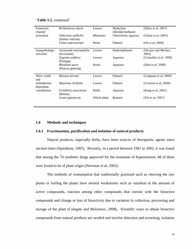
‗Table 1.5, continued‘
Potassium
channel
activation
Brillantaisia nitens
Valeriana wallichii
(Indian valerian)
Ulmus macrocarpa
Leaves
Rhizomes
Roots
Methylene
chloride/methanol
Chloroform, aqueous
Ethanol
(Dimo et al, 2007)
(Gilani et al, 2005)
(Oh et al, 2008)
Sympatholegic
activities
Jacaranda minosaefolia
(Jacaranda)
Eugenia uniflora
(Pitanga)
Rhodiola sacra
(Plateau ginseng)
Leaves
Leaves
Roots
Hydromethanol
Aqueous
Aqueous
(Nicasio and Meckes,
2005)
(Consolini et al, 1999)
(Shih et al, 2008)
Nitric oxide
and
endothelium
dependant
vasodilation
Mansoa hirsute
Maytenus ilicifolia
Fritillaria ussuriensis
(Beimu)
Geum japonicum
Leaves
Leaves
Bulbs
Whole plant
Ethanol
Ethanol
Aqueous
Butanol
(Campana et al, 2009)
(Crestani et al, 2009)
(Kang et al, 2002)
(Xie et al, 2007)
1.6 Methods and techniques
1.6.1 Fractionation, purification and isolation of natural products
Natural products, especially herbs, have been sources of therapeutic agents since
ancient times (Spainhour, 2005). Recently, in a period between 1981 to 2002, it was found
that among the 74 synthetic drugs approved for the treatment of hypertension, 48 of them
were found to be of plant origin (Newman et al, 2003).
The methods of consumption that traditionally practised such as chewing the raw
plants or boiling the plants have several weaknesses such as variation in the amount of
active compounds, reaction among other compounds that coexist with the bioactive
compounds and change or loss of bioactivity due to variation in collection, processing and
storage of the plant (Colegate and Molyneux, 2008). Scientific ways to obtain bioactive
compounds from natural products are needed and involve detection and screening, isolation
31
and purification, and structure identification of bioactive compounds (Spainhour, 2005;
Colegate and Molyneux, 2008).
Detection and screening of potential bioactive plants are done by various in vitro as
well as in vivo assays. After identifying certain bioactive plants, the next step is isolation
and purification which involves various techniques and technologies. The first step is
usually extraction of the plant by using solvents with different polarities. The next step
involves further solvent partition and extensive chromatography methods by considering
acidity, polarity and molecular size of the compounds (Colegate and Molyneux, 2008).
These involve techniques such as solid-phase extraction, high performance liquid
chromatography (HPLC), thin layer chromatography (TLC), counter current
chromatography, desalting and ion exchange chromatography (Spainhour, 2005). Today,
the last step structural elucidation of bioactive compounds uses advanced technologies and
techniques such as nuclear magnetic resonance (NMR) spectroscopy, mass spectrometry
(MS), ultraviolet (UV)/visible and infrared absorption spectroscopy and also ‗hyphenated‘
technologies such as HPLC-MS, HPLC-NMR and MS-MS (Spainhour, 2005; Colegate and
Molyneux, 2008).

32
1.6.2 Blood pressure measurement methods
Measurement of BP in laboratory rats can be divided into direct and indirect
methods. Both of these methods have their advantages and disadvantages and the choice of
method depends on the objectives of the study (Kurtz et al, 2005).
In indirect method, it is non-invasive which means no surgical procedure is required.
The most common indirect method is the use of a cuff in which changes in the blood flow
in a tail or limb is detected by the occlusion or release of the cuff (Kurtz et al, 2005). The
advantages of this method are: (1) non-invasive; (2) less expensive compared to other
method such as telemetry; and (3) can be used to study changes in BP in large groups of
animals over a short or long period of time (Van Vliet et al, 2000; Kurtz et al, 2005). In
contrast, the main disadvantage is that this method requires physical restraint and heating
that poses stress to the animal (Irvine et al, 1997). This method is thus advised to be used
in study in which large BP difference is expected (Gross and Luft, 2003).
In direct method, it is invasive and involves surgical procedure. A sensor device is
implanted into the arterial system in rat to measure the arterial BP. There are three
common types of sensing devices: fluid-filled catheters, transducer-tipped catheters and
radiotelemetries (Van Vliet et al, 2000). Fluid-filled catheter is the oldest and the most
widely used direct method. It uses a heparin-filled catheter to be inserted into a major
artery of rat while the other end of the catheter is connected to a BP transducer. This
method can be used in acute as well as chronic measurement of BP in animals. The main
advantage of this fluid-filled catheter method is that it produces accurate and reliable
readings and permits infusion of various experimental agents (Kurtz et al, 2005). However,
this method imposes some stress to animals because the animals are not free-moving and
the presence of clotting around the catheter tips can affect the BP readings (Kurtz et al,
2005). Moreover, there is also a risk of infection because it involves surgical procedure.
33
Although transducer-tipped catheter is more sensitive, it is more expensive and only
suitable for short term study (Van Vliet et al, 2000). The method of radiotelemetry can be
used in wider areas of studies. Radiotelemetry uses a BP sensor (radiotelemetric probe)
which is implanted in the animal and adopts an electrical device to receive and process the
signal. The advantages include: (1) measures BP of free moving conscious animals; (2)
causes less stress as no restraint of animal is required; (3) less labor intensive; (4) more
efficient, convenience and reliable; (5) measures BP for a longer duration compared to the
catheterised method; (6) no anesthesia is involved during BP measurement; (7) can
measure BP around the clock (Mills et al, 2000; Van Vliet et al, 2000). The main problem
arises from radiotelemetry is its high cost, requires more technical skills and also has the
risk of infection (Kurtz et al, 2005).
1.6.3 In vitro vascular tension studies
To study the pharmacological effect of vasoactive agents on smooth muscles and
their drug-receptor interaction, strips or ring segments of arteries isolated from laboratory
animals such as rats and rabbits are used (Furchgott and Bhadrakom, 1953). In this in vitro
method, the strips are removed from laboratory animals and mounted with stainless steel S-
hooks in physiological solutions (eg. Krebs-bicarbonate solution) bubbled with 95%
oxygen and 5% carbon dioxide in organ chambers (Furchgott and Bhadrakom, 1953). The
strips or ring segments of arteries are attached to force transducer to record the changes in
tension in arteries to various vasoactive drugs (Gonzales et al, 2000). One of the most
popular findings using this in vitro vascular tension method was the discovery of
endothelial-derived hyperpolarising factor which involves the release of NO that causes the
relaxation of VSM (Furchgott, 1999). Earlier studies using this method have shown that
epinephrine, norepinephrine and ACh caused vasocontraction whereas sodium nitrite

34
caused vasorelaxation (Furchgott and Bhadrakom, 1953), but later studies proved that ACh
is a vasorelaxant agent rather than a vasoconstrictor agent (Furchgott, 1999).
1.6.4 Laboratory animals
Spontaneously hypertensive (SHR) rat is the most widely used animal model for
hypertension. It belongs to the phenotype-driven genetic model of hypertension (Lerman et
al, 2005). The SHR strain was obtained by selective breeding of spontaneously
hypertensive Wistar-Kyoto (WKY) male rat with relatively higher BP female rat (Okamoto
and Aoki, 1963). This strain of rat is widely used in hypertension studies because the
incidence of hypertension is 100%, it is sensitive to various antihypertensive agents which
are also effective in human (Yamori and Okamoto, 1973) and it presents hypertensive
complications (Yamori and Okamoto, 1973). Other than that, stroke prone SHR and
arteriolipidosis-prone rats were developed from SHR as a model for stoke and
atherosclerosis (Yamori, 1978).
The most commonly used control strain for SHR rat is WKY rat (Johnson et al,
1992). Initially it was thought that all WKY rats constituted a single inbred strain. Later
studies showed that there was a great biological variability among WKY rats themselves
and also between SHR and WKY rats (Kurtz et al, 1989; Johnson et al, 1992). Although
these findings were discovered, WKY rat is still extensively used as a control for SHR
because SHR was derived from WKY rat (Kurtz and Morris, 1987).
Another strain of rats that are widely studies and extensively used in various
research fields are Sprague-Dawley (SD) rats. This strain was produced by the Sprague-
Dawley Company, Madison, Wisconsin, in 1925. The main advantages of SD rat are its
docile character that makes it easier to be handled and also its excellent reproductive
performance.

35
1.6.5 Nuclear magnetic resonance spectroscopy and mass spectrometer
One of the most powerful instruments to obtain the structure of an organic
compound is NMR spectroscopy. This instrument uses a principle in which when a
compound containing nuclei such as 1H
or
13C is put in a strong magnetic field and
simultaneously irradiated with certain electromagnetic energy; the absorption of energy is
quantized by the NMR spectroscopy and displayed as NMR spectra which can be either 1
dimensional (D) or 2D as well. The most common nuclei used in NMR spectroscopy are
1H and
13C.
Nuclear magnetic resonance spectroscopy usually works together with MS for a
better understanding of the molecule. The MS provides the molecular weight (MW) of a
certain molecule whereas NMR spectroscopy tells the connectivity between atoms in a
molecule. In comparison, NMR spectroscopy has an advantage over MS that it is not
destructive (the sample can be recovered) although more amount of sample is needed for
NMR spectroscopy.

36
1.6.6 In vitro assay of angiotensin-converting enzyme activity
Inhibition of ACE has been an effective therapeutic approach for hypertension and
ACE inhibitory compounds can be found in some plants. There are researches carrying out
to search for potential ACE inhibitory compounds from plants (Hansen et al, 1995; Duncan
et al, 1999; Lacaille et al, 2001). Many methods have been used to study the potential
ACE inhibitory activities from compounds isolated from plants or crude extracts from
plants. These methods use spectrophotometry, fluorometry, HPLC or radiochemistry (Lam
le et al, 2007).
In 1991, a new method, based on the cleavage of chromophore- and fluorophore-
labeled substrate, dansyltriglycine by ACE into dansylglycine, which was measured by
HPLC, was introduced (Elbl and Wagner, 1991). From the past times until now, many
methods, either modified or with new principles are introduced and adopted in many
studies. For example, in 2005, a cost-effective and high throughput method with the
absorbance of final product measured by using microtitre plate reader was introduced
(Serra et al, 2005). Then, a method which used a new substrate known as
3-hydroxybutyrylglycyl-glycyl-glycine, was claimed to be more sensitive, rapid and
accurate than conventional method (Lam le et al, 2007). Later that, a rapid, sensitive, high-
throughput and advanced colorimetric assay to measure hippuric acid (HA) has also been
created (Jimsheena and Gowda, 2009).
Although there are plenty of methods to measure ACE activity, the colorimetric
assay introduced by Cushman and Cheong by using synthetic peptide, hippuryl-L-histidyl-
L-leucine (HHL) as a substrate is the most popular (Jimsheena and Gowda, 2009). In this
method, HHL is hydrolysed by ACE to form HA and histidyl-leucine (HL). The amount of
HA produced is proportional to the ACE activity which are then extracted and measured by
spectrophotometer at 228 nm (Cushman and Cheung, 1971). This method has some
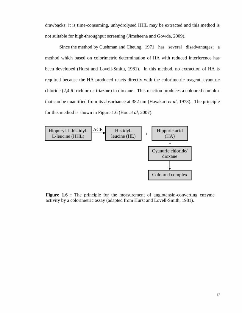
37
ACE
drawbacks: it is time-consuming, unhydrolysed HHL may be extracted and this method is
not suitable for high-throughput screening (Jimsheena and Gowda, 2009).
Since the method by Cushman and Cheung, 1971 has several disadvantages; a
method which based on colorimetric determination of HA with reduced interference has
been developed (Hurst and Lovell-Smith, 1981). In this method, no extraction of HA is
required because the HA produced reacts directly with the colorimetric reagent, cyanuric
chloride (2,4,6-trichloro-s-triazine) in dioxane. This reaction produces a coloured complex
that can be quantified from its absorbance at 382 nm (Hayakari et al, 1978). The principle
for this method is shown in Figure 1.6 (Hoe et al, 2007).
Hippuryl-L-histidyl-
L-leucine (HHL)
Histidyl-
leucine (HL)
Hippuric acid
(HA)
+
+
Cyanuric chloride/
dioxane
Coloured complex
Figure 1.6 : The principle for the measurement of angiotensin-converting enzyme
activity by a colorimetric assay (adapted from Hurst and Lovell-Smith, 1981).

38
1.6.7 In vitro assay of neutral endopeptidase activity
In search of potential ACE inhibitory compounds from plants, NEP inhibitory
compounds, although less popular, can be found in some plants as well (Kiss et al, 2005).
Methods with the principles of radiochemistry, colorimetic, enzyme-linked immunosorbent
assay and the most popular fluorometric method have been used to study the NEP activities
in tissues or sera (Yandle et al, 1992; Tudoric et al, 2000). The synthetic substrates that are
being used include glutaryl-Ala-Ala-Phe-amidomethylcoumarin(Yandle et al, 1992), N-
dansyl-Ala-Gly-D-nitro-Phe-Gly (DAGNPG) (Jiang et al, 2004) and Suc-L-Ala-L-Ala-Phe-
7-amino-4-methylcoumarin (SAAP-AMC) (Kiss et al, 2005).
Until now, methods to measure the NEP activities have been modified and
transferred to screen for potential NEP inhibitory compounds from plants. For example, a
two-step assay based on Bormann and Melzig (Bormann and Melzig, 2000) has been
widely used by Kiss A (Kiss et al, 2005; Kiss et al, 2006; Kiss et al, 2008). In this two-
step assay, the first reaction is initiated by the addition of NEP to the substrate SAAP-AMC
to form Phe-7-amino-4-methylcoumarin (P-AMC). The second reaction is the action of
leucine aminopeptidase (APN) on P-AMC to form 7-amino-4-methylcoumarin (AMC)
which is fluorescence and measured by spectrofluorometer at λexcitation = 355 nm and λemission
= 460 nm. The two-step reaction is shown in Figure 1.7.
SAAP-AMC P-AMC AMC
(fluoresce
nce)
NEP APN
Figure 1.7 : The two-step reaction that finally produces fluorescence 7-amino-4-
methylcoumarin (adapted from Bormann and Melzig, 2000).

39
1.6.8 Effects of proteases on plant extract
Proteases are enzymes that hydrolyse proteins and they are involved in many
physiological reactions in the human body. One of the reactions is the digestion process
wherein some of the important proteases are pepsin, trypsin and chymotrypsin (Guyton and
Hall2, 2006). These enzymes may change the actions or functions of the compounds that
taken into the body.
Study by Hoe et al, 2007 postulated that the putative hypotensive agents in this
plant extract seem to be peptidal substances in nature. This aspect is further studied by
using NMR spectroscopy and HPLC-MS and also by the effects of these three proteases on
ACE inhibitory activities of the plant extract. Loss of ACE inhibitory activity of plant
extract may show that the extract is degraded by proteases and it is most probably a kind of
protein.

40
1.7 Plant material
1.7.1 Family Asteraceae/ Compositae
Family Asteraceae or Compositae comprises of herbs or shrubs, but are rarely trees
or vines (Mbagwu et al, 2009). Their leaves are simple, opposite or alternate but rarely
compound (Mbagwu et al, 2009). There are involucres to support the flower heads and the
flowers have ray florets at the margin and disc florets at the central (Mbagwu et al, 2009).
Most members of Asteraceae or Compositae undergo asexual reproduction by seed (Noyes,
2007). Family Asteraceae comprises of 1,600 genera and 23,000 species is the largest
family of flowering plants (Gao et al, 2010). This family is widely distributed on all
continents except Antarctica.
Asteraceae has a wide range of uses such as in medicine, ornament and economy as
well (Gao et al, 2010). In medical application, Artemisia annua which contains
antimalarial compound, artemisinin, has been listed in the Chinese Pharmacopoeia to treat
fevers such as malaria (Mueller et al, 2000). It is also found that Saussurea involucrate,
listed in the Chinese Pharmacopoeia, has anticancer, anti-inflammation and free radical
scavenging activities (Wu et al, 2009). Gynura procumbens (G. procumbens), which will
be discussed later is also popular among traditional medicine.

41
1.7.2 Gynura procumbens
Kingdom Plantae
Division Magnoliophyta
Class Magnoliopsida
Order Asterales
Family Asteraceae or Compositae
Genus Gynura
Species procumbens
(adapted from Botanica Sistematica, 2009)
Gynura procumbens (Lour.) Merr. is a weak climbing or scrambling perennial herb.
In Malaysia, its common names are akar sebiak, sambung nyawa, daun dewa, kelemai
merah, acham akar and dewa raja. Its stems can grow up to 6 meters long, with smooth or
sparsely pubescent. Young stems are green and become light purple with green spots when
mature. The leaves are almost oval-shaped with size of 3-10cm X 0.5-3cm. The lower
surfaces of the leaves are purplish. Gynura procumbens can be found in Central and
Western Africa, Southeast Asia, China and Papua New Guinea. This plant is fast growing
and easy to breed by using stem cuttings (Globinmed, 2010). Figure 1.8 shows the photo
of G. procumbens planted in Kuala Kangsar, Perak, Malaysia.
In folk medicine, the plant is used for treating kidney diseases, fever and rashes
(Perry, 1980) and for hypertension. Scientifically, G. procumbens is found to have the
following properties:
1. Antihypertensive (Lam et al, 1998; Hoe and Lam, 2005)
2. Anti-ulcer (Mahmood et al, 2010)
3. Anti-oxidant (Puangpronpitag et al, 2010)
4. Antidiabetic and antihyperlipidemic (Zhang and Tan2, 2000)
Table 1.6 : Taxonomy of Gynura procumbens.

42
Figure 1.8 : Gynura procumbens (Lour.) Merr.

43
1.8 Research objectives
Hypertension is one of the major health problems worldwide and most of the
conventional treatments have side-effects. Searching of potential antihypertensive plants
has been an alternative in belief that the plant, being natural may have lesser side-effects.
Previous studies from our laboratory have shown that a partially purified fraction (FA-I)
from final aqueous fraction (FA), from G. procumbens possesses antihypertensive effect by
inhibiting ACE (Hoe et al, 2007) and blocking calcium channels (Hoe et al, 2011). The
present study was designed with the following objectives:
1. To explore the effects and mechanism of FA-I on Ang II-induced contraction of
aortic rings as well as hypotensive and vasodilatory effects of BK
2. To test the dual ACE/NEP inhibitory activities of FA-I and its subfractions
3. To study the acute oral antihypertensive effects of FA-I
4. To characterise bioactive FA-I subfractions

44
CHAPTER 2 - MATERIALS AND METHODS
2.1 Materials
2.1.1 Plant material
Whole plants of G. procumbens were collected from Peninsular Malaysia. Three
voucher specimens (KLU 047690, KLU 047691 and KLU 047692) have been deposited in
the Herbarium at Rimba Ilmu, University of Malaya. The photo below shows one of the
voucher specimens with herbarium number KLU 047690.
2.1.2 Animals
Adult male SD, SHR and WKY rats, weighing between 250-320 g, were obtained
from the Experimental Animal Care unit, University of Malaya and kept under standard
conditions. Normal commercial rat chow (Gold Coin Animal Feed, Malaysia) and tap
water were provided ad libitum. All studies on experimental animals have been conducted
under protocols reviewed and approved by the Animal Ethics Committee, University of
Malaya Medical Center (Ethics Reference Number: FIS 14/ 10/ 2009/ 0112/ PTF (R)).

45
2.1.3 Chemicals and reagents
All the chemicals and reagents used were of analytical grade unless otherwise stated:
Ang I (AnaSpec, INC, USA), heparin sodium 5,000 I.U. (LEO Pharmaceutical Products
Ltd., Ballerup, Denmark), SephadexTM
LH-20 (Amersham Bioscience AB, Uppsala,
Sweden), sodium hydroxide (NaOH) (Fisher Scientific, U.K.) and Nembutal sodium
pentobarbitone (CEVA Sante Animale, Libourne, France).
Chemicals and reagents from Merck KGaA, Darmstadt, Germany are Ang II, BK,
boric acid (H3BO3), calcium chloride (CaCl2), glucose, indomethacin, magnesium sulphate
(MgSO4), NEP from porcine kidney, ninhydrin, phosphoramidon, potassium dihydrogen
phosphate (KH2PO4), potassium chloride (KCl), silica gel 60 F254 TLC aluminium sheets,
sodium bicarbonate (NaHCO3), sodium chloride (NaCl) and 1,4-dioxan, 4-(2-
hydroxyethyl)-1-piperazineethanesulfonic acid (HEPES).
Apart from that, ACh chloride, APN, bovine chymotrypsin, bovine trypsin,
cyanuric chloride, HA, HHL acetate, hydrochloric acid (HCl), Nω-nitro-L-arginine methyl
ester (L-NAME), pepsin from porcine gastric mucous membrane, phenylephrine (PE)
hydrochloride, rabbit ACE and SAAP-AMC were produced by Sigma-Aldrich Co., St
Louis, MO, USA. The solvents acetic acid, acetone, ethanol, ethyl acetate, hexane,
methanol and n-butanol are from R & M Marketing, Essex, U.K.
Rat chows were purchase from Gold Coin Feedmills (M) Sdn Bhd, Selangor,
Malaysia.

46
2.1.4 Equipment
The main equipments used in this study were:
Vacuum evaporation system consisting of a rotary evaporator, an oil bath (model
OSB-2000) and an aspirator (model A-1000S ) from Eyela Tokyo Rikakikai Co., Ltd.,
Japan, with a low temperature circulator (model WCR-P8) from Daihan Scientific Co., Ltd.,
Seoul, Korea.
Digital physiographic recording system (PowerLab, ADInstruments Pty. Ltd.,
Australia) consisting of a PowerLab Data Acquisition System (model ML866), a
BridgeAmp (model ML224), a BioAmp (model ML136), a NIBP controller (model
ML125), a force transducer (model MLT050/D) and a BP transducer (model MLT0380/D)
that is controlled by a HP personal computer (model HPL1908w) loaded with the
PowerLab Chart Version 5.0 Software.
Other equipments include a chromatography column (2.5 cm, i.d. x 75 cm, height),
fraction collector (Bio-Rad 2100, Bio-Rad Laboratories, U.S.A.), reagent spray (Camag,
Switzerland), viewing cabinet (Chromato-vue CC-10, U.K.) combined with multi-band
254/365nm lamp (UVGL-58), micro tube pump (Eyela MP-3, Japan), NMR spectrometer
400 MHz (JEOL, JEOL Ltd, Japan), microprocessor-based bench-top pH meter (Hanna®
pH 211, Hanna® Instruments, U.S.A.), centrifuge (Jouan CR 312, American Laboratory
Trading, Inc, U.S.A.), UV-VIS spectrophotometer (Model UV mini 1240, Shimadzu,
Germany), spectrofluorometer (Model Fluoroskan Ascent FL, Thermo Scientific, U.K.).

47
2.2 Extraction and fractionation of Gynura procumbens leaves
The protocols for extraction and fractionation follow the method used by Hoe, 2007
(Figure 2.1). Briefly, leaves from fresh G. procumbens were cleaned and dried in an oven
at 40oC and then ground to powder. They were then processed through the following steps
in a sequential manner ie extract in 96% ethanol and 80% aqueous ethanol, defatted in
hexane and fractionated by water-saturated butanol to obtain the final aqueous fraction
(FA), which was then passed through a column containing Sephadex LH-20 gel (Appendix
A) to obtain FA-I fraction (Figure 2.1). The FA-I fraction was subjected to preparative
TLC (Appendix B) resulted in several FA-I subfractions (FA-Ia – FA-Ie) that were
revealed by ninhydrin reagent (Appendix C). Using a bioassay guided protocol, the FA-I
subfractions that were tested for positive bioactivities were subsequently sent for
characterisation by 1H NMR 400 MHz spectrometer (JEOL, Japan). The FA-Ia subfraction
was analysed more extensively by 13
C NMR 100 MHz, 2D distortionless enhancement by
polarisation transfer (DEPT) NMR 100 MHz, infrared spectroscopy and LCMS.

48
1. Reconstituted in 80% aqueous
ethanol
2. Defatted with hexane
Figure 2.1 : Extraction and fractionation of Gynura procumbens that finally produce the
FA-I subfractions.
1. Cleaned, dried and ground
2. Extracted with 96% ethanol
3. Evaporated in vacuo
Butanol extract
FA-I
Sephadex LH-20 column chromatography
FA-I subfractions
Preparative thin layer chromatography
G. procumbens leaves
Crude ethanol extract
Aqueous ethanol extract Hexane extract
1. Evaporated in vacuo to remove ethanol
2. Aqueous solution was filtered out
Fractionated with water-saturated n-butanol
Final aqueous extract (FA)
Aqueous extract Ethanol extract

49
2.3 In vitro vascular tension studies
The effects of FA-I on blood vessels were studied using in vitro techniques on
isolated rat thoracic aorta.
2.3.1 Preparation of rat aortic rings
The descending thoracic aorta was removed from male SD rats and mounted in ice-
cold oxygenated Krebs-Henseleit (K-H) solution (Appendix D). The isometric contractile
force of the aorta was recorded by the PowerLab computer system. The procedures for
preparation of rat aortic rings are presented below while Figure 2.2 shows a photograph of
an aortic ring in an organ bath chamber.
For some preparations, the endothelial layer of aortic ring was mechanically
removed by inserting a pair of fine forceps into the lumen and gently rotating the aorta
around the forceps. These endothelium-denuded rings were similarly equilibrated in organ
bath for 60 min. The success of the endothelial denudation was tested by precontracting
the rings with 1.0 x 10⁻⁶
M PE and observing the response to 1.0 x 10⁻⁵
M ACh. A lack of
any relaxation of the PE-precontracted rings indicated that the endothelium was
satisfactorily removed whilst relaxation of at least 70% of the PE-induced contraction
showed that the endothelial layer was still present. The instructions for the preparation of
aortic rings are shown below:

50
Lower hook
Upper hook connected
to a force transducer Organ bath with
10 ml K-H solution
Aortic ring
The rat sacrificed by decapitation and the descending thoracic aorta taken and placed
in a petri dish containing ice-cold oxygenated K-H solution.
The connective tissues and fats from aorta removed and cut into rings (1 – 3 mm in
length).
The aortic ring mounted in an organ bath containing 10 ml K-H solution with 2
stainless-steel hooks; the lower hook fixed to the bottom of the bath whereas the
upper hook connected to a force-displacement transducer.
The bath solution maintained at 37 oC and bubbled continuously with 95% oxygen
and 5% carbon dioxide gas mixture.
Each ring subjected to a resting tension of 1 g and allowed to equilibrate for 60 min
while being rinsed every 15 min. During the equilibration period, the rings
stimulated at least three times with 6 x 10⁻²
M KCl until a reproducible contractile
response obtained.
Figure 2.2 : The aortic ring was mounted in an organ bath containing 10 ml Krebs-
Henseleit solution with the lower hook being fixed to the bottom of the bath whereas the
upper hook is connected to a force-displacement transducer connected to a digital
physiographic setup.

51
2.3.2 Effects of FA-I pretreatment on angiotensin I-induced contraction
Endothelium-intact (n = 6) and -denuded aortic rings (n = 6) were preincubated for
20 min in the absence or presence of FA-I (final concentration: 1.0 x 10⁻⁴
and 1.0 x 10⁻³
g/ml) (with reference to Hoe, 2007). After the incubation period, contraction of the rings
was evoked by adding cumulative concentrations of Ang I (1 x 10-8.5
M - 1 x 10⁻⁶
M) and
their responses in the absence or presence of FA-I were recorded.
2.3.3 Effects of FA-I pretreatment on angiotensin II-induced contraction
Endothelium-intact (n = 6) and -denuded aortic rings (n = 6) were preincubated for
20 min in the absence or presence of FA-I (final concentration: 1.0 x 10⁻⁴
and 1.0 x 10⁻³
g/ml). After the incubation period, contraction of the rings was evoked by adding
cumulative concentrations of Ang II (1 x 10-8.5
M – 1 x 10⁻⁶
M) and their responses in the
absence or presence of FA-I were recorded.
2.3.4 Effects of FA-I pretreatment on angiotensin II-induced contraction of aortic
rings in the presence of Nω-nitro-L-arginine methyl ester or indomethacin
To study the involvement of endothelium in the response to Ang II brought by FA-I,
endothelium-intact rings (n = 6) were preincubated for 20 min with or without FA-I (final
concentration: 1.0 x 10⁻⁴
and 1.0 x 10⁻³
g/ml) and indomethacin (final concentration: 10
µM), a non-selective COX inhibitor or L-NAME (final concentration: 0.1 µM), an eNOS
inhibitor, and followed by the cumulative additions of Ang II.
2.3.5 Effects of FA-I pretreatment on bradykinin-induced relaxation of
phenylephrine-precontracted aortic rings
After the equilibration period, intact aortic rings (n = 8) were precontracted again
with PE (1.0 x 10⁻⁶
M). The contractions of aortic rings to PE were allowed to reach a

52
plateau before the cumulative addition of BK (1.0 x 10⁻¹¹
M – 1.0 x 10⁻⁶
M). The relaxation
responses of PE-precontracted aortic rings to the cumulative additions of BK were
determined in the presence or absence of pretreatment with FA-I (final concentration: 1.0 x
10-4
and 1.0 x 10⁻³
g/ml) 20 min before the sequential additions of PE and BK.
2.4 Effects of FA-I on the action of bradykinin on mean arterial pressure and
heart rate of rats
The BP was recorded by a pressure transducer connected to a PowerLab setup. The
procedures for direct BP measurement in rat are shown as follows:
In this method, heparinised saline (10 U/ml) was used to prevent blood coagulation
and the FA-I was dissolved in normal saline. After the equilibration period, increasing
doses of BK (0 – 625 ng/kg) were injected into the rat to be followed by a single
Each rat (n = 8) anaesthetised with sodium pentobarbitone (50 mg/kg) by
intraperitoneal injection and placed on a surgical board.
The trachea, left carotid artery and right jugular vein exposed surgically.
A heparinised polyethylene tube connected to a pressure transducer inserted into the
left carotid artery for continuous BP measurement.
The trachea cannulated to facilitate respiration.
A polyethylene tube inserted into the right jugular vein for intravenous injection of
test solutions.
Small animal electrographic leads attached to the limbs of the rat to record the HR.
The set up equilibrated for at least 30 min before injection of test solutions. The rat
kept warm (35 ± 2 oC) with a heating lamp throughout the experiment.

53
administration of FA-I (10 mg/kg) (with reference to Hoe, 2007) and then the same
increasing doses of BK again. A minimum interval of 3 min was allowed between each
injection of BK whilst a minimum period of at least 10 min was given between the
administrations of BK and FA-I to allow the BP to return to resting state. Each injection
was set at a fixed volume of 0.1 ml per 300 g rat body weight. Changes in the BP due to
the administration of BK and FA-I were obtained by calculating the difference between the
BP before and the lowest BP after the injections. The mean arterial pressure (MAP) was
calculated using the formula: MAP = DBP + 1/3(SBP - DBP). Figure 2.3 shows the
photography of the set up for the measurement of BP and HR of rat.
Cannulated
jugular vein
Cannulated carotid
artery
Cannulated
trachea
Electrocardiographic leads
Heparinised saline
Pressure
transducer
Anaesthetised
rat
Data acquisition
system
BridgeAmp
BioAmp
Figure 2.3 : Setup of the experimental procedure for the measurement of blood pressure
and heart rate in a rat.

54
2.5 In vitro assays
2.5.1 In vitro effect of FA-I and its subfractions on angiotensin-converting enzyme
activity
The ACE inhibitory activity of G. procumbens was studied by using an in vitro
ACE colourimetric assay of Hurst and Lovell-Smith, 1981 with some modifications. The
procedures for the in vitro ACE inhibitory activity assay are shown as follows:
0.1 ml of HHL (20 mM) added into 0.4 ml of assay mixture (extracts + 20 mU ACE
+ 0.8 M NaCl + 0.08 M H3BO3 buffer (pH 8.3).
The mixture incubated in water bath at 37 oC for 15 min.
0.5 ml of 1 M HCl added to stop the reaction.
After 30 s, 0.5 ml of 1 M NaOH added to neutralise the reaction mixture.
2.0 ml of diluent buffer (20 mM KH2PO4, pH 8.3) followed by 1.5 ml of colour
reagent (3% cyanuric chloride in 1,4-dioxan) then added.
The mixture vortex-mixed vigorously for 30 s and leaved for 5 min, then vortex-
mixed again.
The mixture centrifuged at 3,000 rpm for 10 min.
The absorbance of the supernatant measured by spectrophotometer at 382 nm
against the reagent blank.

55
In order to prepare the enzyme blank, the terminating and neutralising solutions
were added before the HHL; to prepare the reagent blank, the HHL solution was replaced
with distilled water whereas for hippuric acid standards preparation, the HHL solution was
replaced with standard HA solutions.
For calculations, one unit of ACE activity is defined as the amount of enzyme
required to produce one µmole of HA per minute at 37 oC. The effect of FA-I and its
subfractions on ACE activity was calculated by the following formula:
Where: A = ACE activity in the absence of inhibitor
B = ACE activity in the presence of inhibitor (plant extract)
The IC50 value, which is the concentration of the fraction required to inhibit 50% of
the ACE activity, was determined using linear regression analysis.
B
A
X 100%

56
2.5.2 In vitro effect of FA-I and its subfractions on neutral endopeptidase activity
A two-steps assay widely used by Kiss et al., 2008, with some modifications was
used in this study, as shown in the following:
50 µl of SAAP-AMC (400 µM) added to 350 µl of HEPES-buffer with or without
50 µl of extracts in eppendorf tubes.
150 µl of NEP (5 mU) added to start the first reaction.
The mixture incubated at 37 oC for 1 h.
50 µl of phosphoramidon solution (50 µM) added to stop the reaction.
20 µl of APN (1 : 235, v/v) added to start the second reaction.
The mixture incubated at 56 oC for 1 h.
800 µl acetone added to terminate the reaction.
The fluorescence of the released NEP measured at
λexcitation = 355 nm and λemission = 460 nm

57
The effect of FA-I and its subfractions on NEP activity was calculated by the
following formula:
INEP= [(Itotal - IAPN)/(100 - IAPN)]x100
Where: Itotal = 100 - (F1 / K0) x 100
IAPN = 100 - (F2 / K0) x 100
All the measurements are fluorescent and represented as P1, P2, K0 and K:
F1= P1 - K and F2 = P2 – K
Where: P1 = Fluorescence of sample with extract
P2 = Fluorescence of sample with extract added in second reaction
(effect of extract on APN activity)
K0 = Fluorescence of sample without extract
K = Fluorescence of sample with extract without enzyme

58
2.6 Effect of orally administered FA-I on blood pressure of spontaneously
hypertensive and Wistar-Kyoto rats
In this study, SHR (n = 7) and WKY (n = 8) rats were used. In at least 7 days prior
to the experiment, the rats were trained to enter the restraint cage and their SBPs were
measured. On the experimental day, FA-I was dissolved in distilled water and
administered orally to rats at doses of 50, 100 and 200 mg/kg. Control rats were
administered with same amount of distilled water. The SBP was measured by tail-cuff
method with a device connected to a PowerLab setup before and also 2, 4, 6, 8, 24 h after
administration. Figure 2.4 shows the setup for a rat kept in a restraint cage and its SBP was
measured by a cuff connected to a PowerLab setup.
Restraint cage
Cuff
PowerLab computer system
Figure 2.4 : Measurement of rat systolic blood pressure of rat by tail-cuff method.

59
2.7 Effect of proteases on angiotensin-converting enzyme inhibitory activity of
FA-I
The stability of FA-I to maintain the ACE inhibitory activity against proteases
pepsin, trypsin and chymotrypsin was assessed individually by the method of Kuba et al,
2003 with slight modifications. In this in vitro assay, FA-I (0.25 – 2 mg/ml) was mixed
with 0.2 ml of 0.05% (w/v) pepsin, chymotrypsin or trypsin solution. Pepsin was dissolved
in 0.1 M HCl at pH 2.0 whereas chymotrypsin and trypsin were both dissolved in 0.1 M
potassium phosphate buffer at pH 8.0. The mixture was incubated at 37 oC for 6 h, after
which it was boiled for 10 min to stop the digestion. Each sample was then centrifuged at
3,000 rpm for 20 min and the supernatant was collected and freeze-dried before it was sent
for the measurement of ACE inhibitory activity (refer section 2.5.1).

60
2.8 Data analyses
When the cumulative concentration-response curves of Ang I or Ang II were
examined, the contractions in the presence of FA-I were calculated as a percentage of the
highest contractile response obtained in the absence of FA-I. This contractile response
evoked by the highest concentration of Ang I or Ang II was designated as Emax. The pEC50
value (-log EC50 ), determined from the EC50 value (the concentration required to produce a
half-effect in the concentration-response curves) was calculated from individual log
concentration-response curves of the Ang I or Ang II by sigmoidal non-linear regression
analysis within the 95% confidence intervals using GraphPad Prism® v.4.00 (GraphPad
Software Inc., La Jolla, CA, USA). The vasorelaxant response to BK was measured from
the plateau of the PE-induced contraction and expressed as a percentage (%) relaxation of
the PE-induced contraction. The IC50 value, which is the concentration of extracts to
inhibit 50% of ACE or NEP activity, was obtained from dose-response curves by linear
regression.
2.9 Statistical analyses
All values are expressed as mean ± SEM for n number of rats or separate
experiments. Statistical differences for in vitro studies were evaluated by one-way analysis
of variance (ANOVA) while student‘s t-test was used for in vivo studies. A probability
level of less than 0.05 (p<0.05) was considered to be significantly different.

61
CHAPTER 3 - RESULTS
3.1 Extraction and fractionation of Gynura procumbens leaves
Gynura procumbens leaves were ground to powder form and extracted sequentially
with several solvents to obtain FA-I. The FA-I fraction on TLC plates sprayed with
ninhydrin reagent was shown in Figure 3.1. The yield of each solvent extract is shown in
Figure 3.2. Heating on FA-I at TLC plates sprayed with ninhydrin reagent revealed five
different fractions (FA-Ia – FA-Ie) with retention factor (Rf) values of 0.21, 0.31, 0.45, 0.53
and 0.66 respectively (Figure 3.2).
Figure 3.1 : Chromatogram of a partially purified aqueous fraction (FA-I) on thin layer
chromatography (TLC) plates after being sprayed with ninhydrin reagent. The dark
circle reveals FA-Ia.

62
Fractionated with
water-saturated n-butanol
Butanolic extract
(10.0 g; 2.0%)
FA-I
(0.76 g; 76.0% of 1.0 g FA)
Dried G. procumbens leaves
(500 g)
Crude ethanolic extract
(60.0 g; 12.0%)
Aqueous ethanolic extract
Hexanic extract
(18.0 g; 3.6%)
Final aqueous extract (FA)
(15.0 g; 3.0%)
Figure 3.2 : Flow chart of extraction and fractionation of the leaves of Gynura
procumbens (All % values in parenthesis indicate % yield of each fraction obtained
from the original dried leaves weight except for FA-I which was calculated from 1 g of
FA).
FA-Ie (Rf : 0.66)
FA-Id (Rf : 0.53)
FA-Ic (Rf : 0.45)
FA-Ib (Rf : 0.31)
FA-Ia (Rf : 0.21)
Ethanolic extract
(13.4 g; 2.7%)
Aqueous extract
Extracted with 96% ethanol
Sephadex LH-20 gel
filtration chromatography
Silica gel 60 F254 TLC plate
B : A : W (4 : 1: 5, v/v,
upper layer)
Defatted with hexane
Evaporated in vacuo

63
3.2 In vitro vascular tension studies
3.2.1 Effects of FA-I pretreatment on angiotensin I-induced contraction
Pretreatment with FA-I at 1.0 x 10⁻⁴
and 1.0 x 10⁻³
g/ml inhibited the Ang I-induced
contraction of both endothelium-intact (Figure 3.3a) and -denuded (Figure 3.3b) aortic
rings in a concentration-dependent manner with significant (p < 0.01) reduction in the Emax
values. Furthermore, rings that were kept intact (Figure 3.3a) relaxed significantly (p <
0.05) more than those that were denuded of endothelium (Figure 3.3b).
3.2.2 Effects of FA-I pretreatment on angiotensin II-induced contraction
Experiments were then carried out with endothelium-intact and -denuded aortic
rings to study the involvement of endothelium in the response of FA-I to the contraction
evoked by Ang II. Results show that pretreatment of endothelium-intact aortic rings with
FA-I attenuated the contraction caused by Ang II (Figure 3.4a), with the Emax values being
significantly (p < 0.001) decreased when compared with controls. However, exposure of
endothelium-denuded aortic rings to FA-I for 20 min before the addition of increasing
concentrations of Ang II did not blunt the contractions induced by Ang II (Figure 3.4b).

64
Figure 3.3 : Effects of a partially purified aqueous fraction (FA-I) on angiotensin I-
induced contraction in (a) endothelium-intact (Endo +) and (b) endothelium-denuded
(Endo -) aortic rings. The effects were determined in the absence or presence of FA-I (1.0
x 10-4
or 1.0 x 10⁻³
g/ml). Values are mean ± S.E.M. (n = 6). **p < 0.01; ***p < 0.001
compared with controls (without FA-I) and +p < 0.05;
+++p < 0.001 compared with Endo +.
Figure 3.4 : Effect of a partially purified aqueous fraction (FA-I) on angiotensin II-induced
contraction in (a) endothelium-intact (Endo +) and (b) endothelium-denuded (Endo -) aortic
rings. The effect was determined in the absence (without FA-I) or presence of FA-I (1.0 x
10-4
or 1.0 x 10⁻³
g/ml). Values are mean ± S.E.M. (Endo +, n = 7; Endo –, n = 6) ***p <
0.001; compared with controls (without FA-I).

65
3.2.3 Effects of FA-I pretreatment on Ang II-induced contraction of aortic rings in
the presence of Nω-nitro-L-arginine methyl ester or indomethacin
To further study the role of endothelial factors in the action of FA-I, Ang II-induced
contraction in endothelium-intact rings were performed in the presence of both FA-I and
L-NAME or indomethacin. Results show that pretreatment of FA-I at 1.0 x 10⁻⁴
and
1.0 x 10⁻³
g/ml in the presence of L-NAME (Figure 3.5a) or indomethacin (Figure 3.5b) did
not cause any significant changes in the Emax values for Ang II as compared to controls.
In aortic rings pretreated with L-NAME, although there were no significant changes
in Emax values, the concentration-response curves of Ang II are shifted to the right (Figure
3.5a) with a significant (p < 0.05) decrease in pEC50 values (Table 3.1). Similarly,
preincubation of FA-I and indomethacin also shifted the concentration-response curve to
the right (Figure 3.5b), with a significant (p < 0.01) reduction of pEC50 values for Ang II in
the presence of FA-I at 1.0 x 10⁻³
g/ml (Table 3.1). These results show that the eNOS and
COX pathways may be involved in the response of FA-I on Ang II-induced contraction of
aortic rings.

66
FA-I
(x 10-3
g/ml)
pEC50
L-NAME Indomethacin
0 7.84 ± 0.06 7.89 ± 0.08
0.1 7.61 ± 0.12* 7.75 ± 0.16
1.0 7.54 ± 0.11** 7.53 ± 0.14**
Table 3.1 : Effect of a partially purified aqueous fraction (FA-I) on the pEC50 values for
Ang II-induced contraction of aortic rings pretreatment with L-NAME or indomethacin.
Values are mean ± S.E.M. (n = 6)
*p < 0.05; **p < 0.01 compared with controls (without FA-I).
Figure 3.5 : Effect of a partially purified aqueous fraction (FA-I) on angiotensin II-induced
contraction in endothelium-intact aortic rings preincubated with (a) Nω-nitro-L-arginine
methyl ester (L-NAME) or (b) indomethacin. The effect was determined in the absence or
presence of FA-I (1.0 x 10⁻⁴
or 1.0 x 10⁻³
g/ml). Values are mean ± S.E.M. (n = 6).

67
3.2.4 Effects of FA-I pretreatment on bradykinin-induced relaxation of
phenylephrine-precontracted aortic rings
Bradykinin (1 x 10-11
– 1 x 10⁻⁶
M) dose-dependently relaxed PE-induced
contraction of aortic rings. This observation was significantly (p < 0.05) enhanced by
pretreatment of aortic rings with FA-I (Figure 3.6).
Figure 3.6 : Effect of a partially purified aqueous fraction (FA-I) on bradykinin-induced
relaxation of aortic rings precontracted with phenylephrine. The effect was determined in
the absence (without FA-I) or presence of FA-I (1.0 x 10⁻⁴
or 1.0 x 10⁻³
g/ml). Values are
mean ± S.E.M. (n = 8). *p < 0.05 **p < 0.01 compared with controls (without FA-I).

68
3.3 Effects of FA-I on the action of bradykinin on mean arterial pressure and
heart rate of rats
Intravenous administrations of BK at different doses induced immediate and
significant (p < 0.05) decrease in the MAP (Figure 3.7a) and HR (Figure 3.7b) of rats.
After pretreatment with 10 mg/kg FA-I, the BP lowering effect produced by BK was
markedly (p < 0.05) greater. The difference in the hypotensive effect of BK before and
after FA-I pretreatment became significant (p < 0.05) at doses of 78, 156, 312 and 625
ng/kg. Bradykinin also decreased the HR but there was no significant difference in the
bradycardic effect between before and after FA-I administration. Pre-dose baseline levels
were recovered within 10 min after BK administration.
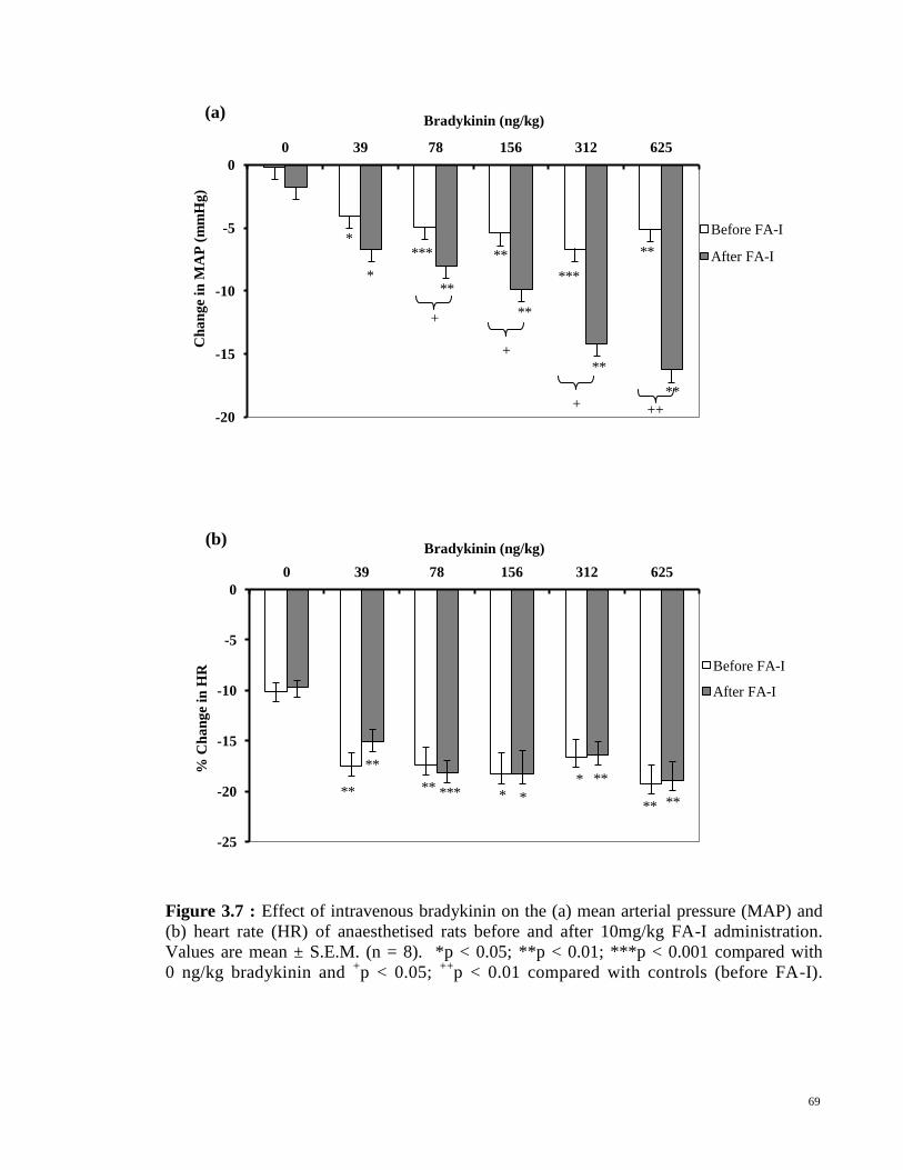
69
-25
-20
-15
-10
-5
0
0 39 78 156 312 625
% C
ha
ng
e in
HR
Bradykinin (ng/kg)
Before FA-I
After FA-I
** ** *** * *
** *
** **
(b)
**
-20
-15
-10
-5
0
0 39 78 156 312 625
Ch
an
ge
in M
AP
(m
mH
g)
Bradykinin (ng/kg)
Before FA-I
After FA-I
*
*
***
**
**
**
***
**
**
**
(a)
Figure 3.7 : Effect of intravenous bradykinin on the (a) mean arterial pressure (MAP) and
(b) heart rate (HR) of anaesthetised rats before and after 10mg/kg FA-I administration.
Values are mean ± S.E.M. (n = 8). *p < 0.05; **p < 0.01; ***p < 0.001 compared with
0 ng/kg bradykinin and +p < 0.05;
++p < 0.01 compared with controls (before FA-I).
++ +
+
+
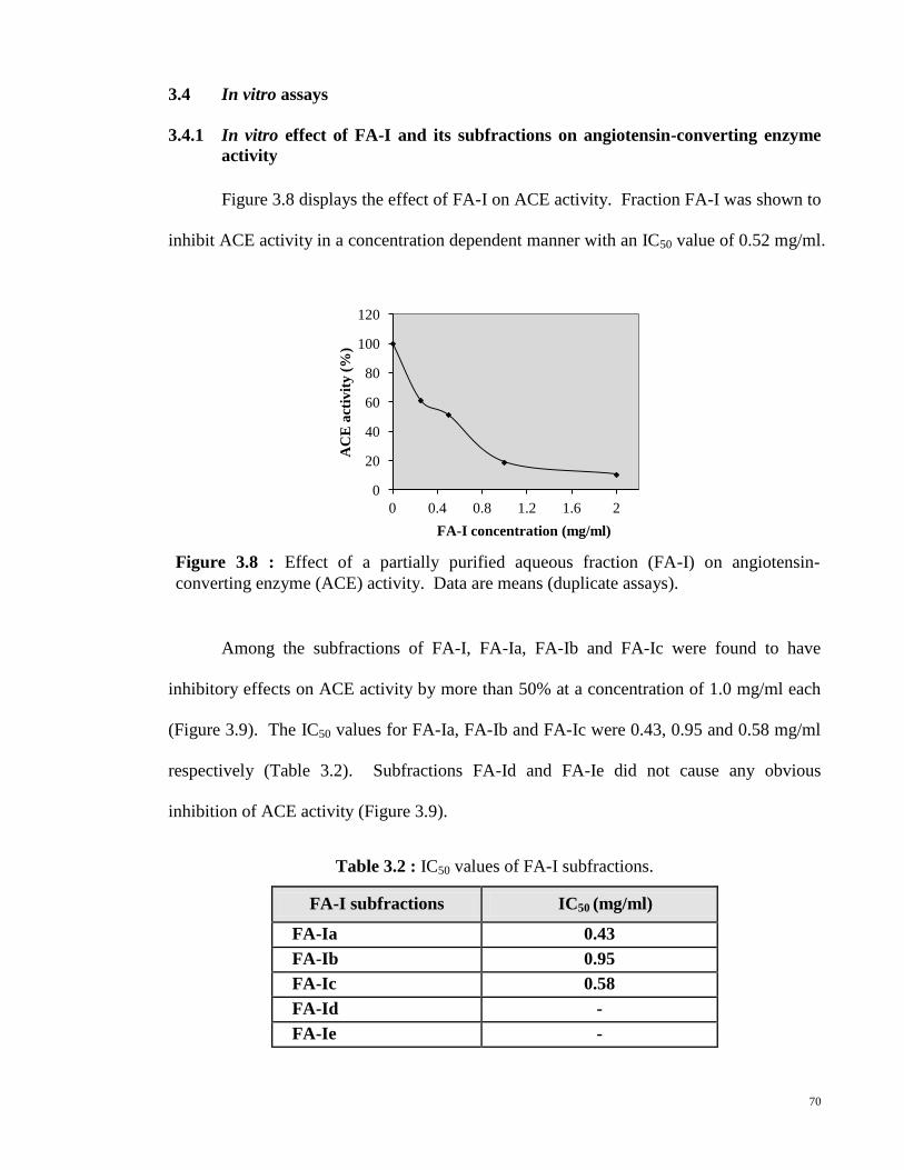
70
3.4 In vitro assays
3.4.1 In vitro effect of FA-I and its subfractions on angiotensin-converting enzyme
activity
Figure 3.8 displays the effect of FA-I on ACE activity. Fraction FA-I was shown to
inhibit ACE activity in a concentration dependent manner with an IC50 value of 0.52 mg/ml.
Among the subfractions of FA-I, FA-Ia, FA-Ib and FA-Ic were found to have
inhibitory effects on ACE activity by more than 50% at a concentration of 1.0 mg/ml each
(Figure 3.9). The IC50 values for FA-Ia, FA-Ib and FA-Ic were 0.43, 0.95 and 0.58 mg/ml
respectively (Table 3.2). Subfractions FA-Id and FA-Ie did not cause any obvious
inhibition of ACE activity (Figure 3.9).
FA-I subfractions IC50 (mg/ml)
FA-Ia 0.43
FA-Ib 0.95
FA-Ic 0.58
FA-Id -
FA-Ie -
Table 3.2 : IC50 values of FA-I subfractions.
0
20
40
60
80
100
120
0 0.4 0.8 1.2 1.6 2
AC
E a
ctiv
ity
(%
)
FA-I concentration (mg/ml)
Figure 3.8 : Effect of a partially purified aqueous fraction (FA-I) on angiotensin-
converting enzyme (ACE) activity. Data are means (duplicate assays).

71
0
20
40
60
80
100
120
0 0.2 0.4 0.6 0.8 1 1.2
AC
E a
ctiv
ity
(%
)
Concentration (mg/ml)
0
20
40
60
80
100
120
0 0.2 0.4 0.6 0.8 1 1.2
AC
E a
ctiv
ity
(%
)
Concentration (mg/ml)
0
20
40
60
80
100
120
0 0.2 0.4 0.6 0.8 1 1.2
AC
E a
ctiv
ity
(%
)
Concentration (mg/ml)
0
20
40
60
80
100
120
0 0.2 0.4 0.6 0.8 1 1.2
AC
E a
ctiv
ity
(%
)
Concentration (mg/ml)
0
20
40
60
80
100
120
0 0.2 0.4 0.6 0.8 1 1.2
AC
E a
ctiv
ity
(%
)
Concentration (mg/ml)
(a) FA-Ia (b) FA-Ib
(c) FA-Ic (d) FA-Id
(e) FA-Ie
Figure 3.9 : Angiotensin-converting enzyme (ACE) inhibition by (a) FA-Ia, (b) FA-Ib, (c)
FA-Ic, (d) FA-Id and (e) FA-Ie. Data are means (duplicate assays).

72
0
20
40
60
80
100
120
0 50 100 150 200 250 300 350 400 450
NE
P a
ctiv
ity (
%)
FA-I (µg/ml)
3.4.2 In vitro effect of FA-I and its subfractions on neutral endopeptidase activity
Figure 3.10 displays the effect of FA-I on NEP activity. Fraction FA-I was shown
to inhibit NEP activity in a concentration dependent manner with an IC50 value of
225 µg/ml. Among the subfractions of FA-I, none of them shows obvious NEP inhibitory
activity (Figure 3.11).
Figure 3.10 : Effect of a partially purified aqueous fraction (FA-I) on neutral endopeptidae
(NEP) activity. Data are means (triplicate assays).

73
0
20
40
60
80
100
120
0 100 200 300 400
NE
P a
ctiv
ity
(%
)
Concentration (x100µg/ml)
0
20
40
60
80
100
120
0 100 200 300 400
NE
P a
ctiv
ity
(%
)
Concentration (µg/ml)
0
20
40
60
80
100
120
0 100 200 300 400
NE
P a
ctiv
ity
(%
)
Concentration (µg/ml)
0
20
40
60
80
100
120
0 100 200 300 400
NE
P a
ctiv
ity
(%
)
Concentration (µg/ml)
0
20
40
60
80
100
120
0 100 200 300 400
NE
P a
ctiv
ity
(%
)
Concentration (µg/ml)
(a) FA-Ia
(d) FA-Id (c) FA-Ic
(b) FA-Ib
(e) FA-Ie
Figure 3.11 : Neutral endopeptidase (NEP) inhibition by (a) FA-Ia, (b) FA-Ib, (c) FA-Ic,
(d) FA-Id and (e) FA-Ie. Data are means (triplicate assays).
*

74
3.5 Effect of orally administered FA-I on blood pressure of spontaneously
hypertensive and Wistar-Kyoto rats
Figure 3.12 shows the time-course changes in the SBP of SHR and control WKY
rats after a single oral administration of FA-I (50, 100 or 200 mg/kg) or vehicle (distilled
water). An oral dose of FA-I was able to cause a significant (p < 0.05) dose-dependent
decrease in the SBP especially at the higher doses when compared to controls (Figure 3.12).
In the SHR rats, there is a tendency for the SBP to return to the initial level within 24 h
after oral administration and the highest antihypertensive effect of FA-I occurred at 6 h
after the oral administration (Figure 3.13).
-35
-30
-25
-20
-15
-10
-5
0
5
10
2 4 6 8 24
Ch
an
ge in
sy
sto
lic b
loo
d p
ress
ure (
mm
Hg
)
Time after administration (h)
Vehicle 50 mg/kg FA-I
100 mg/kg FA-I 200 mg/kg FA-I
Figure 3.12 : Change in systolic blood pressure after a single orally administered of
partially purified aqueous fraction (FA-I) in (a) Wistar-Kyoto (WKY) and (b)
spontaneously hypertensive (SHR) rats. Values are mean ± S.E.M. (WKY, n = 6; SHR, n
= 7). *p < 0.05, **p < 0.01 and ***p < 0.001 compared with vehicles (distilled water).
(a) WKY rats (b) SHR rats
-35.0
-30.0
-25.0
-20.0
-15.0
-10.0
-5.0
0.0
5.0
2 4 6 8 24
Ch
an
ge in
sy
sto
lic b
loo
d p
ress
ure (
mm
Hg
)
Time after administration (h)
Vehicle 50 mg/kg FA-I
100 mg/kg FA-I 200 mg/kg FA-I
* *
*
**
***
***
**
**
** *
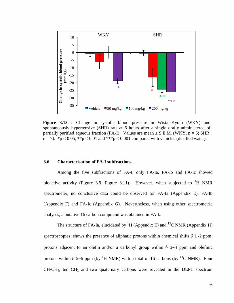
75
3.6 Characterisation of FA-I subfractions
Among the five subfractions of FA-I, only FA-Ia, FA-Ib and FA-Ic showed
bioactive activity (Figure 3.9, Figure 3.11). However, when subjected to 1H NMR
spectrometer, no conclusive data could be observed for FA-Ia (Appendix E), FA-Ib
(Appendix F) and FA-Ic (Appendix G). Nevertheless, when using other spectrometric
analyses, a putative 16 carbon compound was obtained in FA-Ia.
The structure of FA-Ia, elucidated by 1H (Appendix E) and
13C NMR (Appendix H)
spectroscopies, shows the presence of aliphatic protons within chemical shifts 12 ppm,
protons adjacent to an olefin and/or a carbonyl group within 34 ppm and olefinic
protons within 56 ppm (by 1H NMR) with a total of 16 carbons (by
13C NMR). Four
CH/CH3, ten CH2 and two quaternary carbons were revealed in the DEPT spectrum
Figure 3.13 : Change in systolic blood pressure in Wistar-Kyoto (WKY) and
spontaneously hypertensive (SHR) rats at 6 hours after a single orally administered of
partially purified aqueous fraction (FA-I). Values are mean ± S.E.M. (WKY, n = 6; SHR,
n = 7). *p < 0.05, **p < 0.01 and ***p < 0.001 compared with vehicles (distilled water).
-35
-30
-25
-20
-15
-10
-5
0
5
10
Ch
an
ge
in s
yst
oli
c b
loo
d p
ress
ure
(mm
Hg
)
Vehicle 50 mg/kg 100 mg/kg 200 mg/kg
WKY SHR
*
*** ***
*
76
(Appendix I). The mass spectrum data obtained from LC-MS showed a molecular ion at
m/z 279 [M + H]+ (Appendix J) suspected for C16H22O4. Strong infrared absorptions for
functional groups hydroxyl (OH), 3297 cm-1
; aliphatic (C-H), 2923 and 2854 cm-1
;
carbonyl (C=O), 1740 cm-1
and ether (C-O), 1047 cm-1
stretching frequencies were found
in the molecule (Appendix K). However, the definitive structure of FA-Ia remains
unresolved.
3.7 Effect of proteases on angiotensin-converting enzyme inhibitory activity of
FA-I
Figure 3.14 shows the ACE inhibitory activity of FA-I without and after the
treatment of proteases. Overally, the ACE inhibitory activity of FA-I was still preserved
after the treatment of proteases. There was a slight decrease in ACE inhibitory activity of
FA-I, as indicated by increase in their IC50 values, after pepsin, trypsin or chymotrypsin
treatment (Table 3.3).

77
0
20
40
60
80
100
120
0 0.4 0.8 1.2 1.6 2
AC
E a
ctiv
ity
(%
)
FA-I concentration (mg/ml)
0
20
40
60
80
100
120
0 0.4 0.8 1.2 1.6 2
AC
E a
ctiv
ity
(%
)
FA-I concentration (mg/ml)
0
20
40
60
80
100
120
0 0.4 0.8 1.2 1.6 2
AC
E a
ctiv
ity
(%
)
FA-I concentration (mg/ml)
0
20
40
60
80
100
120
0 0.4 0.8 1.2 1.6 2
AC
E a
ctiv
ity
(%
)
FA-I concentration (mg/ml)
Digestion IC50 of FA-I (mg/ml)
None 0.52
Pepsin 0.70
Trypsin 0.60
Chymotrypsin 0.64
(a) Without treatment
(c) Chymotrypsin treatment
(b) Pepsin treatment
treatment
Figure 3.14 : The angiotensin-converting enzyme (ACE) inhibitory activity of FA-I (a)
without and after (b) pepsin, (c) chymotrypsin and (d) trypsin. Data are means (duplicate
assays).
Table 3.3 : The effect of digestive enzymes on the IC50 values of FA-I.
(d) Trypsin treatment

78
CHAPTER 4 - DISCUSSION
4.1 In vitro vascular tension studies
In vitro vascular tension study was performed to extend previous studies that clearly
demonstrated the BP-lowering effects of G. procumbens by inhibition of ACE activity
(Hoe et al, 2007). Aortic rings were used to study the effect of FA-I on the contraction
caused by Ang I and Ang II because ACE is abundantly present in the aortic rings (Andre et
al, 1990; Egleme et al, 1990). As shown in Figure 3.3, preincubation with FA-I
significantly inhibited the Ang I-induced vasocontraction in both endothelium-intact and
-denuded aortic rings, with the degree of relaxation produced in endothelium-intact rings
being significantly (p < 0.05) greater than that obtained in the denuded ones. These
observations indicate that the actions of FA-I are more enhanced in the presence of the
endothelium, probably because of the presence of NO (Sandoo et al, 2010) and PGI2
(DalBo et al, 2008). The effects are likely to be mediated through the newly converted
Ang II from Ang I by ACE that are abundantly found in aortic rings (Andre et al, 1990;
Egleme et al, 1990) as Ang I per se appears to have no physiological function (Peach, 1977;
Hayashi and Kimoto, 2010). This finding also further confirms the previous studies (Hoe
et al, 2007) that FA-I inhibits ACE which then reduces the production of Ang II and causes
a drop in the contractile response of Ang II.
Angiotensin II is a potent vasoconstrictor and acts mainly through the widely
distributed AT1R to increase BP (Lote, 2006). Increased RAS activity which leads to the
increase in Ang II has been implicated as a major contributing factor in the development of
hypertension (Navar et al, 2011). The present study shows that FA-I is able to inhibit the
Ang II-induced contraction of aortic rings with endothelium (Figure 3.4a). Experiments
were then carried out on endothelium-denuded aortic rings to investigate the involvement
of endothelium on the effect of FA-I on Ang II-induced contraction of aortic rings. In

79
endothelium-denuded aortic rings, FA-I did not appear to have a significant effect on the
contraction evoked by Ang II (Figure 3.4b). This may exclude the notion that FA-I could
be an AT1R blocker as AT1R blockers are known to inhibit Ang II-induced contractions in
endothelium-denuded aortic rings as well (d'Uscio et al, 1998; Watts et al, 1998). Hence,
this strongly suggests that the inhibitory effect of FA-I on the vascular contraction is
entirely endothelium dependent.
It is well known that the endothelium plays an important role in the regulation of
vascular tone by releasing both relaxant and contractile factors in response to chemical and
physical stimulation (Villar et al, 2006; DalBo et al, 2008; Sandoo et al, 2010). The
vasorelaxant substances released by endothelial cells include NO mainly produced by
eNOS (Sandoo et al, 2010) and PGI2 produced by the COX pathway (DalBo et al, 2008).
In the present study, pEC50 value was used to study the influence of L-NAME and
indomethacin because there is no significant difference in the Emax values among the aortic
rings preincubated with L-NAME (Figure 3.5a) or indomethacin (Figure 3.5b). The
inhibitory effect of FA-I on the Ang II-induced contraction of aortic rings was reduced in
the presence of L-NAME (Figure 3.5a) or indomethacin (Figure 3.5b), with significant
(p < 0.01) reduction in pEC50 values (Table 3.1). A reduced pEC50 value shows that the
graph is shifted to the right, indicating that in the presence of FA-I, the contractile effect of
Ang II is reduced and more Ang II is needed to cause the same level of contraction. These
findings support the notion that FA-I is able to counteract the contractile response of Ang II
by stimulating the release of vasorelaxing factors via both the eNOS and COX pathways.
In the presence of L-NAME in which eNOS is inhibited, FA-I is able to decrease Ang II-
induced contraction probably by triggering the release of PGI2. Similarly, in the presence
of indomethacin, FA-I may be able to stimulate the release of NO, despite COX being
inhibited.
80
4.2 Effects of FA-I on bradykinin by in vitro and in vivo studies
Bradykinin is a vasodilator produced in the kallikrein-kinin system (Campbell, 2000)
in which most of the effects of BK are being mediated by B2 receptor under normal
conditions
(Nishiyama and Kim-Mitsuyama, 2010). Inhibition of ACE may partly
potentiate the actions of BK, by either an indirect action on B2 receptor (Deddish et al,
2002) or direct activation of B1 receptor (Erdos, 1999) to confer additional benefits to the
CVS. In order to study the effects of FA-I on hypotensive and vasodilatory effects of BK,
both in vitro and in vivo methods with reference to the methods performed by Anozie et al,
2007 and Maia et al, 2004, with slight modifications.
In the in vitro study, BK evoked a dose-dependent relaxation of PE-induced
contraction of aortic rings, similar to the study of that reported by Anozie et al, 2007. As in
the in vivo study, BK produced a dose-dependent hypotensive effect, similar to the report
by Maia et al, 2004. At the highest dose of BK (625 ng/kg), the drop in MAP is smaller,
but not significant when compared to the previous dose (312 ng/kg). This may be due to
the baroreceptor reflex (Guyton and Hall3, 2006) in the rats to prevent further drop in MAP.
In this in vivo study, the MAP, which is the pressure that drives the blood into tissues
during the cardiac cycle, was used as the parameter for BP comparison because it minimise
the inaccuracies of SBP and DBP (Smulyan and Safar, 2011). The formula of MAP that
used in this study was MAP = DBP + 1/3(SBP - DBP) because it is the most common
formula that is being used worldwide (Smulyan and Safar, 2011).
Studies shown here demonstrated that in the presence of FA-I, the vasorelaxant (in
vitro study) and BP-lowering (in vivo study) effects of BK are greater. In the study on
aortic rings in the presence of FA-I, the relaxation effect brought about by BK on PE-
precontracted endothelium-intact aortic rings is increased (Figure 3.6). However, parallel
experiments on endothelium-denuded rings were not performed as it is well-established

81
that vasodilation caused by BK is entirely endothelium dependent (Vanhoutte, 2001). In
the in vivo studies, the BP-lowering effect of BK was also greater after administration of
FA-I (Figure 3.7a). Although BK is known to cause bradycardia (Wang et al, 2003), the
infusion of FA-I did not alter the bradycardic effect of BK (Figure 3.7b). This could be due
to the baroreceptor reflex (Wang et al, 2003) being involved in preventing further reduction
in the HR brought about by FA-I. Hence, FA-I is able to potentiate the hypotensive effect
of BK probably by enhancing the vasodilator activities of BK through inhibition of ACE or
NEP, but without alteration in HR.
Other than that, it is speculated that FA-I may potentiate the hypotensive effect by
BK through the vasodilatory effect of Ang (1-7) which is increased after inhibition of ACE
(Fernandes et al, 2001). The FA-I may also possess antioxidant effect that preserves the
production of NO which is reduced due to oxidative stress produced by PE (Hao et al,
2006). Nevertheless, the exact mechanism responsible for the BK potentiation effects of
FA-I remains to be elucidated.

82
4.3 Vasopeptidase inhibitory activities of FA-I and FA-I subfractions
One of the most popular VPIs, omapatrilat, had high incidence of life-threatening
side-effect angioedema and was thus withdrawn by FDA (Song and White, 2001).
Searching for potential VPIs from natural products may alleviate the side-effects, especially
that of angioedema. Although VPI is deemed to be more superior to ACEI or NEPI alone,
studies on potential dual ACE/NEP inhibitory activities from natural products have less
popular attention. Examples of plants with dual ACE/NEP inhibitory activities are
Ligustrum vulgare (Kiss et al, 2008) and Epilobium angustifolium (Kiss et al, 2006). Since
FA-I was shown to have strong ACE inhibitory activity (Hoe et al, 2007), it would be
interesting to investigate whether this plant has NEP inhibitory activity as well.
In studying the potential dual ACE/NEP inhibitory activities, spectrophotometric
method (for measurement of ACE activity) and spectrofluorometric method (for
measurement of NEP activity) were used. Both of these methods are volume sensitive and
the enzymes are sensitive to the environment and need to be handled with care. The
amount of FA-I and its subfractions used were referred to the previous study with little
adjustment (Hoe et al, 2007). Micrograms of extracts were used in NEP activity study as
reference to Kiss et al, 2008 (Kiss et al, 2008).
Result shows that FA-I has strong ACE inhibitory activities (Figure 3.8), which is
similar to that reported by Hoe et al, 2007. Among the FA-I subfractions, FA-Ia, FA-Ib
and FA-Ic inhibited ACE activities at 1.0 mg/ml (Figure 3.9). In this study, an ACEI drug,
captopril (2.6 mg/ml) is able to inhibit 98.44% of ACE activity. The ACE inhibitory effect
of FA-I and its bioactive subfractions seem to be comparable to captopril but no further
conclusion can be made because only a single dose of captopril was used. Among the FA-I
subfractions, FA-Ia may have the strongest ACE inhibitory activity because it has the
lowest IC50 value (Table 3.2). These findings show that some FA-I subfractions retain the

83
ACE inhibitory activity and the ACE inhibitory compounds that are present in FA-I may
also present in these subfractions.
Besides the ACE inhibitory activity, our study successfully shows that FA-I
possesses some NEP inhibitory activity, with IC50 value of 225 µg/ml (Figure 3.10). The
NEP inhibitory effect is far weaker when comparing to a single dose of phosphoramidon
(100 µg/ml), a reference drug used in this study, which decreases 97.9% of NEP activity.
This may be because FA-I is only a partially purified fraction but the phosphoramidon is a
popular NEP inhibitor drug. However, this NEP inhibitory activity diminishes in FA-I
subfractions (Figure 3.11). It is speculated that there may exist more than one NEP
inhibitory compounds present in FA-I. In order to exhibit strong NEP inhibitory effect,
these compounds need to act synergistically and the amount of NEP inhibitory compounds
that are present in each FA-I subfraction may not be enough to react to give strong
inhibitory effects (Colegate and Molyneux, 2008). Another postulation is the loss of
bioactive compounds during separation step (Colegate and Molyneux, 2008). The presence
of NEP inhibitory activity in FA-I but not in its subfractions could also be due to the
existence of unknown compounds in FA-I that are not revealed at the TLC plate by
ninhydrin reagent. It is also possible that FA-I subfractions may also act at the allosteric
sites of the enzyme that give a conformational change in the enzyme to enable the binding
of other subfractions.
The dual ACE/NEP inhibitory effect of FA-I may also be responsible for the
potentiation of the hypotensive effect of BK. This is because the main enzymes
responsible for the hydrolysis of BK are ACE and NEP and the inhibition of these enzymes
would result in an increase in the level of BK to enhance vasodilation to cause a drop in BP
(Sharma and Al-Sherif, 2011).

84
4.4 Effect of orally administered FA-I on blood pressure of rats
At present, there are many methods being used to measure the BP of rats. Among
these methods, radiotelemetry is claimed to be the most efficient and accurate method in
measuring the BP of rats (Mills et al, 2000; Van Vliet et al, 2000). Tail-cuff method was
adopted in this study because this method has the main advantage of no surgical and
anesthesia procedures which minimise the risk of infecton.
In order to minimise false positive readings brought by changes in BP due to
heating and restraining (Irvine et al, 1997), rats were subjected to same sized restrainers,
same preheating time and heating temperature; and similar environment. Training of rats
prior to the experimental procedures made the rats better at adapting to the restraining
conditions and they remained calmer in the restrainers.
Some studies have shown that antihypertensive agents may lose their effects after
being treated orally (Li et al, 2004). Thus, oral feeding of FA-I on SHR and WKY rats
were performed in this study. The SHR rat was chosen because it is the most widely used
hypertensive animal model and the incidence of developing hypertension is 100% (Yamori
and Okamoto, 1973) whereas the normotensive WKY rats serve as controls for SHR rats
(Kurtz and Morris, 1987).
A single oral administration of FA-I exerted antihypertensive effect on SBP in SHR
rats, with the highest effect occurring at 6 h after the administration (Figure 3.12b).
However, no significant suppression in SBP was found in normotensive WKY rats except
for the highest concentration, 200mg/kg of FA-I (Figure 3.12a). This shows that FA-I has
antihypertensive effect and a lower dose of FA-I may be safe to normotensive subjects. In
order to cause antihypertensive effect after oral administration, the agents need to reach the
blood circulation (Vermeirssen et al, 2004). Therefore, active substances in FA-I are
believed to be able to reach the blood circulation in an active form after oral treatment.

85
4.5 Effect of proteases on angiotensin-converting enzyme inhibitory activity of
FA-I and nuclear magnetic resonance studies on FA-Ia
The results show that FA-I is able to retain most of its ACE inhibitory activity after
pepsin, trypsin and chymotrypsin treatment with little increase in IC50 values (Table 3.3).
This protease study indicates that the active compounds in FA-I may not be amino acid and
they can withstand the effects by proteases.
Ninhydrin (2,2-dihydroxyindane-1,3-dione) is a chemical used to detect ammonia,
primary and secondary amines. When reacting with these amines, a deep blue or purple
colour stain is produced on the TLC. Hence, ninhydrin is commonly used to detect amines
in peptides or amino acids (Carey et al, 2010). However, this ninhydrin staining test can
only be used as a prepliminary phytochemical screening method because not all the amine-
containing compounds give positive results with ninhydrin and false positive
results were also reported in non-amine-containing compounds (Carey et al, 2010). A
more reliable structural characterisation method is NMR spectroscopy (Colegate and
Molyneux, 2008). An amine-containing compound, i.e. amino acid could be detected
explicitly by using 1H-NMR and
13C-NMR. The
13C-NMR chemical shifts for an amino
acid for example are in the range of 53.1 – 63.3 ppm (C-NH2) and 174.1 – 177.6 ppm
(COOH) (Appendix L) (Wishart and Nip, 1998). In the case of fraction FA-Ia, no amine or
amino acid functional group was detected in the 1H-NMR and
13C-NMR. Hence, FA-Ia is
not an amino acid. This is contrary to the study by Hoe et al, 2007 who postulated that FA-
I may be peptides.

86
4.6 General discussion and future studies
Hypertension is the most important risk factor for cardiovascular mortality and
morbidity worldwide. Although many types of antihypertensive treatments are available
either clinically or traditionally, the prevalence of hypertension remains high and keeps on
increasing (Kim et al, 2010). Many studies have been carried out in search of novel
antihypertensive treatments.
Natural products with great structural diversities have made them valuable sources for
the discovery of novel antihypertensive drugs. The present study has shown some advance
findings of the plant G. procumbens. It is found to be being able to inhibit significantly
(p < 0.05) the contraction brought by Ang II, probably via activation of the release of NO and
PGI2 or it may act on an unknown receptor which is responsible for the contraction of Ang II.
Future studies may be carried out to identify this receptor which may be present in the
vasculars.
Bradykinin has been known to contribute to the cardioprotective effects of ACEIs
(Murphey et al, 2003). Previous findings have shown that G. procumbens has strong ACE
inhibitory activities (Hoe et al, 2007) and study was carried out to test whether this plant has
effects on BK. Results presented here have shown that the BP-lowering effect of BK is
enhanced which may be caused by the greater vasodilatory effect of BK in the presence of plant
extract. One of the possible pathways other than the ACE and NEP inhibition, is activation of
B1 or B2 receptors which can be explored in future.
Acute oral feeding studies showed that the antihypertensive effect of G. procumbens is
still preserved after going through the gastrointestinal system. This is a great finding which
gives clue that it may be effective when applied orally to the human. The possible adverse
side-effects that may be arising have not studied and this remains to be explored.

87
Finally, in order to contribute to the antihypertensive treatments, the compounds are
needed to be isolated, identified and produced industrially. It is necessary to identify the
bioactive compounds that are responsible for the effects of G. procumbens, although one needs
to take into account the heterogeneity of hypertension in order to evaluate the potential impact
of treatment for the management of this disorder. The systems in the human body react
differently to the hypertension and hypertensive patient react differently to the antihypertensive
treatments. Side-effects of the drugs that are arising should not be ignored. Natural products
represent the major source of chemical diversity for antihypertensive screening purposes and
communication among the experts such as botanists, chemists, clinicians and natural products
researchers should be well established so that the biodiversity that exists in nature is not lost for
antihypertensive drug discovery.

88
CHAPTER 5 - CONCLUSIONS
This study conclusively demonstrates and extends previous investigations that FA-I,
fractionated from the leaves of G. procumbens, has potent BP-lowering effect acting via
dual ACE/NEP inhibitory activity as well as BK enhancement. Finding of FA-I being able
to antagonise the vasoconstrictive effect of Ang II is reported. The effect of FA-I on Ang
II-induced contraction of aortic rings appears to be mediated by an endothelium-dependent
pathway that involves the activation of NO and COX mechanisms. In addition, FA-I is
able to enhance the BK effect, most probably by stimulation of the release of NO and PGI2.
These findings are summarised in Figure 5.1.
As part of our search for potential VPI from plant, this report indicates that FA-I has
dual ACE/NEP inhibitory activity. As far as can be ascertained, this is the first study that
demonstrates the dual effect of G. procumbens. Although only FA-I and not its
subfractions show NEP inhibitory activity, ACE inhibitory activities are shown by FA-Ia,
Fa-Ib and FA-Ic (Figure 5.1).
This study also shows that FA-I has antihypertensive effect on SHR and little effect
on WKY rats after it is being treated orally. It is also able to withstand proteases to
maintain most of its ACE inhibitory activity. The NMR and HPLC-MS studies indicate
that FA-Ia is a 16 carbon molecule suspected for C16H22O4 that has OH, aliphatic C-H, C=O
and C-O functional groups.

89
Defatted with hexane
Figure 5.1 : Extraction and fractionation of Gynura procumbens, the effects and
mechanisms of actions.
SUMMARY
Butanol extract
FA-I
Sephadex LH-20 column chromatography
Preparative thin layer chromatography
Dried, powdered G. procumbens leaves
Crude ethanol extract
Aqueous ethanol extract Hexane extract
Evaporated in vacuo
Fractionated with water-saturated n-butanol
Final aqueous extract (FA)
Aqueous extract Ethanol extract
Ang II inhibitory activity appears
to be mediated by endothelium-
dependent pathway
BK-enhancing effects
NEP inhibitory activitiy in
addition to ACE inhibitory activity
Oral antihypertensive effect
FA-Ic Possible
ACE
inhibitory
activity
FA-Ia Possible
ACE
inhibitory
activity
FA-Id FA-Ie
FA-Ib Possible
ACE
inhibitory
activiy

90
PUBLICATIONS
Papers
1. Poh T.F., Ng, H.K., Hoe, S.Z. and Lam, S.K. (2013) Gynura procumbens causes
vasodilation by inhibiting angiotensin II and enhancing bradykinin actions. Journal of
Cardiovascular Pharmacology. 61 (5): 378-384.
2. Poh T.F., Ng, H.K., Hoe, S.Z. and Lam, S.K. First report of dual inhibition of ACE and
NEP by a partially purified fraction of Gynura procumbens. (In preparation).
3. Poh T.F., Chin, C.F., Hoe, S.Z. and Lam, S.K. Chemical analysis of an active compound
of Gynura procumbens using LCMS and HPLC and NMR spectroscopy.
(In preparation).
Abstract
1. Poh T.F., Ng, H.K., Hoe, S.Z. and Lam, S.K. Hypotensive effect of oral administration
of putative ACE inhibitors from Gynura procumbens. In proceeding: 24th
Malaysian
Society of Pharmacology and Physiology (MSPP) Scientific Meeting. 2nd
– 3rd
June 2010.
SACC Convention Centre, Shah Alam. (p. 125)

91

92

93

94

95

96

97

98

99
100
REFERENCES
Ackermann, U. (2004). Regulation of arterial blood pressure. Surgery (Oxford) 22 (5):
120a-120f.
Adam, Y., Somchit, M.N., Somchit, M.N., Sulaiman, M.R., Nasaruddin, A. A., Zuraini, A.,
Bustamam, A.A. and Zakaria, Z.A. (2009). Diuretic properties of Orthosiphon
stamineus Benth. Journal of Ethnopharmacology 124 (1): 154-158.
Adeneye, A.A., Ajagbonna, O.P., Mojiminiyi, F.B., Odigie, I.P., Ojobor, P.D., Etarrh, R.R.
and Adeneye, A.K. (2006). The hypotensive mechanisms for the aqueous stem bark
extract of Musanga cecropioides in Sprague-Dawley rats. Journal of
Ethnopharmacology 106 (2): 203-207.
Agarwal, A., Williams, G.H. and Fisher, N.D. (2005). Genetics of human hypertension.
Trends in Endocrinology and Metabolism 16 (3): 127-133.
Aissaoui, A., El-Hilaly, J., Israili, Z.H. and Lyoussi, B. (2008). Acute diuretic effect of
continuous intravenous infusion of an aqueous extract of Coriandrum sativum L. in
anesthetized rats. Journal of Ethnopharmacology 115 (1): 89-95.
Ajay, M., Chai, H.J., Mustafa, A.M., Gilani, A.H. and Mustafa, M.R. (2007). Mechanisms
of the anti-hypertensive effect of Hibiscus sabdariffa L. calyces. Journal of
Ethnopharmacology 109 (3): 388-393.
Ambuhl, P.M., Tissot, A.C., Fulurija, A., Maurer, P., Nussberger, J., Sabat, R., Nief, V.,
Schellekens, C., Sladko, K., Roubicek, K., Pfister, T., Rettenbacher, M., Volk, H.D.,
Wagner, F., Muller, P., Jennings, G.T. and Bachmann, M.F. (2007). A vaccine for
hypertension based on virus-like particles: preclinical efficacy and phase I safety
and immunogenicity. Journal of Hypertension 25 (1): 63-72.
Andre, P., Schott, C., Nehlig, H. and Stoclet, J.C. (1990). Aortic smooth muscle cells are
able to convert angiotensin I to angiotensin II. Biochemical and Biophysical
Research Communications 173 (3): 1137-1142.
Anozie, O., Ross, R., Oyekan, A.O. and Yakubu, M.A. (2007). Differential modulation of
bradykinin-induced relaxation of endothelin-1 and phenylephrine contractions of rat
aorta by antioxidants. Acta Pharmacologica Sinica 28 (10): 1566-1572.
Atlas, S. (2007). The renin-angiotensin aldosterone system: pathophysiological role and
pharmacologic inhibition. Journal of Managed Care Pharmacy 13 (8): S9-S20.
Barnes, P.J. (1992). Bradykinin and asthma. Thorax 47 (11): 979-983.
Beltowski, J. (2010). Leptin and the regulation of renal sodium handling and renal Na-
transporting ATPases: Role in the pathogenesis of arterial hypertension. Current
Cardiology Reviews 6 (1): 31-40.
Benigni, A., Cassis, P. and Remuzzi, G. (2010). Angiotensin II revisited: new roles in
inflammation, immunology and aging. EMBO Molecular Medicine 2 (7): 247-257.

101
Bernstein, K.E., Martin, B.M., Bernstein, E.A., Linton, J., Striker, L. and Striker, G. (1988).
The isolation of angiotensin-converting enzyme cDNA. Journal of Biological
Chemistry 263 (23): 11021-11024.
Bernstein, K.E., Martin, B.M., Edwards, A.S. and Bernstein, E.A. (1989). Mouse
angiotensin-converting enzyme is a protein composed of two homologous domains.
Journal of Biological Chemistry 264 (20): 11945-11951.
Bormann, H. and Melzig, M.F. (2000). Inhibition of metallopeptidases by flavonoids and
related compounds. Pharmazie 55 (2): 129-132.
Botanica Sistematica (2009). Retrieved August 21, 2013, from http://angr.altervista.org/
botanica/hypertext/0791.htm
Buck, M.L. and Michel, R.S. (2000). Talking with families about herbal therapies. Journal
of Pediatrics 136 (5): 673-678.
Caballero-George, C., Vanderheyden, P.M., Solis, P.N., Gupta, M.P., Pieters, L., Vauquelin,
G. and Vlietinck, A. (2003). In vitro effect of sanguinarine alkaloid on binding of
[3H]candesartan to the human angiotensin AT1 receptor. European Journal of
Pharmacology 458 (3): 257-262.
Camargo, M.E., Berdeja, B. and Miranda, G. (2004). Diuretic effect of the aqueous extract
of Bidens odorata in the rat. Journal of Ethnopharmacology 95 (2-3): 363-366.
Campana, P.R., Braga, F.C. and Cortes, F.S. (2009). Endothelium-dependent
vasorelaxation in rat thoracic aorta by Mansoa hirsuta D.C. Phytomedicine 16 (5):
456-461.
Campbell, D.J. (2000). Towards understanding the kallikrein-kinin system: insights from
measurement of kinin peptides. Brazilian Journal of Medical and Biological
Research 33 (6): 665-677.
Cardillo, C., Kilcoyne, C.M., Cannon, R.O. and Panza, J.A. (2000). Interactions between
nitric oxide and endothelin in the regulation of vascular tone of human resistance
vessels in vivo. Hypertension 35 (6): 1237-1241.
Carey, R.M. and Park, J. (2006). Role of angiotensin type 2 receptors in vasodilation of
resistance and capacitance vessels. Hypertension 48 (5): 824-825.
Carey, B.B., Samir, S.H. and Donald, J.S. (2010). Mechanism of the ninhydrin reaction.
Biochemical Education 6 (1): 4-5.
Carretero, O.A. and Oparil, S. (2000). Essential hypertension. Part I: definition and
etiology. Circulation 101 (3): 329-335.
Chang, J. (2000). Medicinal herbs: drugs or dietary supplements? Biochemical
Pharmacology 59 (3): 211-219.

102
Chappell, M.C. and Pirro, N.T. (1998). Metabolism of angiotensin-(1-7) by angiotensin-
converting enzyme. Hypertension 31 (1 Pt 2): 362-367.
Chiang, W.D. (2008). Production of angiotensin I-converting enzyme inhibitor derived
from egg white protein hydrolysates using a membrane reactor. Journal of Food
and Drug Analysis 16 (2): 54-60.
Chobanian, A.V., Bakris, G.L., Black, H.R., Cushman, W.C., Green, L.A., Izzo, J.L.Jr.,
Jones, D.W., Materson, B.J., Oparil, S., Wright, J.T.Jr. and Roccella, E.J. (2003).
Seventh report of the Joint National Committee on Prevention, Detection,
Evaluation, and Treatment of High Blood Pressure. Hypertension 42 (6): 1206-1252.
Cieri, U.R. (1998). Determination of reserpine and rescinnamine in Rauwolfia serpentina
powders and tablets: collaborative study. Journal of AOAC International 81 (2):
373-380.
Circosta, C., De Pasquale, R., Samperi, S., Pino, A. and Occhiuto, F. (2007). Biological and
analytical characterization of two extracts from Valeriana officinalis. Journal of
Ethnopharmacology 112 (2): 361-367.
Coates, D. (2003). The angiotensin converting enzyme (ACE). The International Journal of
Biochemistry & Cell Biology 35 (6): 769-773.
Colegate, S.M. and Molyneux, R.J. (2008). An introduction and overview. Colegate, S.M.
and Molyneux, R.J. (ed), Bioactive natural products: detection, isolation, and
structural determination. London: Taylor & Francis Group.
Comini, L., Bachetti, T., Cargnoni, A., Bastianon, D., Gitti, G.L., Ceconi, C. and Ferrari, R.
(2007). Therapeutic modulation of the nitric oxide: all ace inhibitors are not
equivalent. Pharmacological Research 56 (1): 42-48.
Consolini, A.E., Baldini, O.A. and Amat, A.G. (1999). Pharmacological basis for the
empirical use of Eugenia uniflora L. (Myrtaceae) as antihypertensive. Journal of
Ethnopharmacology 66 (1): 33-39.
Corti, R., Burnett, J.C.Jr., Rouleau, J.L., Ruschitzka, F. and Luscher, T.F. (2001).
Vasopeptidase inhibitors: a new therapeutic concept in cardiovascular disease?
Circulation 104 (15): 1856-1862.
Crestani, S., Rattmann, Y.D., Cipriani, T.R., de Souza, L.M., Iacomini, M., Kassuya, C.A.,
Marques, M.C. and da Silva-Santos, J.E. (2009). A potent and nitric oxide-
dependent hypotensive effect induced in rats by semi-purified fractions from
Maytenus ilicifolia. Vascular Pharmacology 51 (1): 57-63.
Cushman, D.W. and Cheung, H.S. (1971). Concentrations of angiotensin-converting
enzyme in tissues of the rat. Biochimica et Biophysica Acta 250 (1): 261-265.
Cushman, D.W. and Cheung, H.S. (1971). Spectrophotometric assay and properties of the
angiotensin-converting enzyme of rabbit lung. Biochemical Pharmacology 20 (7):
1637-1648.

103
DalBo, S., Moreira, E.G., Brandao, F.C., Horst, H., Pizzolatti, M.G., Micke, G.A. and
Ribeiro-do-Valle, R.M. (2008). Mechanisms underlying the vasorelaxant effect
induced by proanthocyanidin-rich fraction from Croton celtidifolius in rat small
resistance arteries. Journal of Pharmacological Sciences 106 (2): 234-241.
Daull, P., Jeng, A.Y. and Battistini, B. (2007). Towards triple vasopeptidase inhibitors for
the treatment of cardiovascular diseases. Journal of Cardiovascular Pharmacology
50 (3): 247-256.
Davidson, P., Hancock, K., Leung, D., Ang, E., Chang, E., Thompson, D.R. and Daly, J.
(2003). Traditional Chinese Medicine and heart disease: what does Western
medicine and nursing science know about it? European Journal of Cardiovascular
Nursing 2 (3): 171-181.
Deddish, P.A., Marcic, B.M., Tan, F., Jackman, H.L., Chen, Z. and Erdos, E.G. (2002).
Neprilysin inhibitors potentiate effects of bradykinin on b2 receptor. Hypertension
39 (2 Pt 2): 619-623.
Der Sarkissian, S., Huentelman, M.J., Stewart, J., Katovich, M.J. and Raizada, M.K. (2006).
ACE2: A novel therapeutic target for cardiovascular diseases. Progress in
Biophysics and Molecular Biology 91 (1-2): 163-198.
Dimo, T., Mtopi, O.S., Nguelefack, T.B., Kamtchouing, P., Zapfack, L., Asongalem, E.A.
and Dongo, E. (2007). Vasorelaxant effects of Brillantaisia nitens Lindau
(Acanthaceae) extracts on isolated rat vascular smooth muscle. Journal of
Ethnopharmacology 111 (1): 104-109.
Dogru, M.T., Simsek, V., Sahin, O. and Ozer, N. (2010). Differences in autonomic activity
in individuals with optimal, normal, and high-normal blood pressure levels. Turkish
Society of Cardiology 38 (3): 182-188.
Donoghue, M., Hsieh, F., Baronas, E., Godbout, K., Gosselin, M., Stagliano, N., Donovan,
M., Woolf, B., Robison, K., Jeyaseelan, R., Breitbart, R.E. and Acton, S. (2000). A
novel angiotensin-converting enzyme-related carboxypeptidase (ACE2) converts
angiotensin I to angiotensin 1-9. Circulation Research 87 (5): E1-E9.
Dorer, F.E., Kahn, J.R., Lentz, K.E., Levine, M. and Skeggs, L.T. (1974). Hydrolysis of
bradykinin by angiotensin-converting enzyme. Circulation Research 34 (6): 824-
827.
Dubey, R.K., Oparil, S., Imthurn, B. and Jackson, E.K. (2002). Sex hormones and
hypertension. Cardiovascular Research 53 (3): 688-708.
Duncan, A.C., Jager, A.K. and van Staden, J. (1999). Screening of Zulu medicinal plants
for angiotensin converting enzyme (ACE) inhibitors. Journal of
Ethnopharmacology 68 (1-3): 63-70.
d'Uscio, L.V., Shaw, S., Barton, M. and Luscher, T.F. (1998). Losartan but not verapamil
inhibits angiotensin II-induced tissue endothelin-1 increase: role of blood pressure
and endothelial function. Hypertension 31 (6): 1305-1310.

104
Egleme, C., Cressier, F. and Wood, J.M. (1990). Local formation of angiotensin II in the rat
aorta: effect of endothelium. British Journal of Pharmacology 100 (2): 237-240.
Ehlers, M.R., Fox, E.A., Strydom, D.J. and Riordan, J.F. (1989). Molecular cloning of
human testicular angiotensin-converting enzyme: the testis isozyme is identical to
the C-terminal half of endothelial angiotensin-converting enzyme. Proceedings of
the National Academy of Sciences of the United States of America 86 (20): 7741-
7745.
Elbl, G. and Wagner H. (1991). A new method for the in vitro screening of inhibitors of
angiotensin-converting enzyme (ACE), using the chromophore- and fluorophore-
labelled substrate, dansyltriglycine. Planta Medica 57 (2): 137-141.
Erdos, E.G. and Skidgel, R.A. (1989). Neutral endopeptidase 24.11 (enkephalinase) and
related regulators of peptide hormones. FASEB Journal 3 (2): 145-151.
Erdos, E.G., Deddish, P.A. and Marcic, B.M. (1999). Potentiation of Bradykinin Actions
by ACE Inhibitors. Trends in Endocrinology & Metabolism 10 (6): 223-229.
Esler, M., Straznicky, N., Eikelis, N., Masuo, K., Lambert, G. and Lambert, E. (2006).
Mechanisms of sympathetic activation in obesity-related hypertension.
Hypertension 48 (5): 787-796.
Evans, V. (2000). Herbs and the brain: friend or foe? The effects of ginkgo and garlic on
warfarin use. Journal of Neuroscience Nursing 32 (4): 229-232.
Feletou, M. (2011). The Endothelium: Part 1: Multiple Functions of the Endothelial
Cells—Focus on Endothelium-Derived Vasoactive Mediators. Granger, D.N. and
Granger, J.P. (ed). California: Morgan & Claypool Life Sciences Publishers.
Feletou, M. and Vanhoutte, P.M. (2013). Endothelium-dependent hyperpolarization: no
longer an f-word! Journal of Cardiovascular Pharmacology 61 (2): 91-92.
Fernandes, L., Fortes, Z.B., Nigro, D., Tostes, R.C., Santos, R.A. and Catelli De Carvalho,
M.H. (2001). Potentiation of bradykinin by angiotensin-(1-7) on arterioles of
spontaneously hypertensive rats studied in vivo. Hypertension 37 (2 Part 2): 703-
709.
Ferrario, C.M. and Iyer S.N. (1998). Angiotensin-(1-7): a bioactive fragment of the renin-
angiotensin system. Regulatory Peptides 78 (1-3): 13-18.
Fetalvero, K.M., Martin, K.A. and Hwa, J. (2007). Cardioprotective prostacyclin signaling
in vascular smooth muscle. Prostaglandins & Other Lipid Mediators 82 (1-4): 109-
118.
Fielitz, J., Dendorfer, A., Pregla, R., Ehler, E., Zurbrugg, H.R., Bartunek, J., Hetzer, R. and
Regitz-Zagrosek, V. (2002). Neutral endopeptidase is activated in cardiomyocytes
in human aortic valve stenosis and heart failure. Circulation 105 (3): 286-289.

105
Foex, P. and Sear, J. (2004). Hypertension: pathophysiology and treatment The Board of
Management and Trustees of the British Journal of Anaesthesia 4 (3): 71-75.
Frishman, W.H., Beravol, P. and Carosella, C. (2009). Alternative and complementary
medicine for preventing and treating cardiovascular disease. Dis Mon 55 (3): 121-
192.
Fugh-Berman, A. (2000). Herb-drug interactions. Lancet 355 (9198): 134-138.
Furchgott, R.F. (1999). Endothelium-derived relaxing factor: discovery, early studies, and
identification as nitric oxide. Bioscience Reports 19 (4): 235-251.
Furchgott, R.F. and Bhadrakom, S. (1953). Reactions of strips of rabbit aorta to
epinephrine, isopropylarterenol, sodium nitrite and other drugs. Journal of
Pharmacology and Experimental Therapeutics 108 (2): 129-143.
Furchgott, R.F. and Zawadzki, J.V. (1980). The obligatory role of endothelial cells in the
relaxation of arterial smooth muscle by acetylcholine. Nature 288 (5789): 373-376.
Galley, H.F. and Webster, N.R. (2004). Physiology of the endothelium. British Journal of
Anaesthesia 93 (1): 105-113.
Gangwisch, J.E., Heymsfield, S.B., Boden-Albala, B., Buijs, R.M., Kreier, F., Pickering,
T.G., Rundle, A.G., Zammit, G.K. and Malaspina, D. (2006). Short sleep duration
as a risk factor for hypertension: analyses of the first National Health and Nutrition
Examination Survey. Hypertension 47 (5): 833-839.
Gao, T., Yao, H., Song, J., Zhu, Y., Liu, C. and Chen, S. (2010). Evaluating the feasibility
of using candidate DNA barcodes in discriminating species of the large Asteraceae
family. BMC Evolutionary Biology 10: 324.
Gasparotto, A.Jr., Boffo, M.A., Lourenco, E.L., Stefanello, M.E., Kassuya, C.A. and
Marques, M.C. (2009). Natriuretic and diuretic effects of Tropaeolum majus
(Tropaeolaceae) in rats. Journal of Ethnopharmacology 122 (3): 517-522.
Gendron, L., Payet, M.D. and Gallo-Payet, N. (2003). The angiotensin type 2 receptor of
angiotensin II and neuronal differentiation: from observations to mechanisms.
Journal of Molecular Endocrinology 31 (3): 359-372.
Gilani, A.H., Khan, A.U., Jabeen, Q., Subhan, F. and Ghafar, R. (2005). Antispasmodic
and blood pressure lowering effects of Valeriana wallichii are mediated through K+
channel activation. Journal of Ethnopharmacology 100 (3): 347-352.
Giles, T.D., Berk, B.C., Black, H.R., Cohn, J.N., Kostis, J.B., Izzo, J.L.Jr. and Weber, M.A.
(2005). Expanding the definition and classification of hypertension. The Journal of
Clinical Hypertension (Greenwich) 7 (9): 505-512.
Giles, T.D., Materson, B.J., Cohn, J.N. and kostis, J.B. (2009). Definition and classification
of hypertension: an update. The Journal of Clinical Hypertension (Greenwich)
11(11): 611-614.

106
Globinmed. (2010). Retrieved August 21, 2013, from http://www.globinmed.com/index.
php?option=com_content&view=article&id=62750:gynura-procumbens-lour-
merr&Itemid=113
Golias, C., Charalabopoulos, A., Stagikas, D., Charalabopoulos, K. and Batistatou, A.
(2007). The kinin system--bradykinin: biological effects and clinical implications.
Multiple role of the kinin system--bradykinin. Hippokratia 11 (3): 124-128.
Gonzales, R.J., Carter, R.W. and Kanagy, N.L.(2000). Laboratory demonstration of
vascular smooth muscle function using rat aortic ring segments. Advances in
Physiology Education 24 (1): 13-21.
Gradman, A.H., Basile, J.N., Carter, B.L., Bakris, G.L., Materson, B.J., Black, H.R., Izzo,
J.L.Jr., Oparil, S. and Weber, M.A. (2010). Combination therapy in hypertension.
Journal of the American Society of Hypertension 4 (2): 90-98.
Greco, A.J., Master, R.G., Fokin, A.Jr., Baber, S.R. and Kadowitz, P.J. (2006).
Angiotensin-(1-7) potentiates responses to bradykinin but does not change
responses to angiotensin I. Canadian Journal of Physiology and Pharmacology 84
(11): 1163-1175.
Grobe, J.L., Mecca, A.P., Lingis, M., Shenoy, V., Bolton, T.A., Machado, J.M., Speth, R.C.,
Raizada, M.K. and Katovich, M.J. (2007). Prevention of angiotensin II-induced
cardiac remodeling by angiotensin-(1-7). American Journal of Physiology: Heart
and Circulatory Physiology 292 (2): H736-H742.
Gross, V. and Luft F.C. (2003). Exercising restraint in measuring blood pressure in
conscious mice. Hypertension 41 (4): 879-881.
Gurib-Fakim, A. (2006). Medicinal plants: traditions of yesterday and drugs of tomorrow.
Molecular Aspects of Medicine 27 (1): 1-93.
Guy, J.L., Lambert, D.W., Warner, F.J., Hooper, N.M. and Turner, A.J. (2005). Membrane-
associated zinc peptidase families: comparing ACE and ACE2. Biochimica et
Biophysica Acta 1751 (1): 2-8.
Guyton, A.1 and Hall, J. (2006). Dominant role of the kidney in long-term regulation of
arterial pressure and in hypertension: the integrated system for pressure control.
Schmitt, W. and Gruliow, R. (ed), Textbook of Medical Physiology. Philadelphia:
Elsevier Saunders. (p. 216-231).
Guyton, A.2 and Hall, J. (2006). Secretary functions of the alimentary tract. Schmitt, W.
and Gruliow, R. (ed), Textbook of Medical Physiology. Philadelphia: Elsevier
Saunders. (p. 791-807).
Guyton, A.3 and Hall, J. (2006). Nervous regulation of the circulation, and rapid control of
arterial pressure. Schmitt, W. and Gruliow, R. (ed), Textbook of Medical Physiology.
Philadelphia: Elsevier Saunders. (p. 791-807).

107
Hansen, K., Nyman, U., Smitt, U.W., Adsersen, A., Gudiksen, L., Rajasekharan, S. and
Pushpangadan, P. (1995). In vitro screening of traditional medicines for anti-
hypertensive effect based on inhibition of the angiotensin converting enzyme (ACE).
Journal of Ethnopharmacology 48 (1): 43-51.
Hao, L., Nishimura, T., Wo, H. and Fernandez-Patron, C. (2006). Vascular responses to
alpha1-adrenergic receptors in small rat mesenteric arteries depend on
mitochondrial reactive oxygen species. Arteriosclerosis, Thrombosis and Vascular
Biology 26 (4): 819-825.
Harrison-Bernard, L.M. (2009). The renal renin-angiotensin system. Advances in
Physiology Education 33 (4): 270-274.
Hayakari, M., Kondo, Y. and Izumi, H. (1978). A rapid and simple spectrophotometric
assay of angiotensin-converting enzyme. Analyical Biochemistry 84 (2): 361-369.
Hayashi, A. and Kimoto, K. (2010). Studies on the mechanism of antihypertensive action
by nicotianamine. Journal of Nutritional Science and Vitaminology (Tokyo) 56 (4):
242-246.
Hill, S. and Smith A. (2005). First-line medicine in the treatment of hypertension.
Australian Prescriber 28: 34-37.
Hoe, S.Z. (2007). Extraction, purification and effects of gynura procumbens on blood
pressure and mechanisms of action. (Unpublished doctoral dissertation). University
of Malaya, Kuala Lumpur.
Hoe, S.Z., Kamaruddin, M.Y. and Lam, S.K. (2007). Inhibition of angiotensin-converting
enzyme activity by a partially purified fraction of Gynura procumbens in
spontaneously hypertensive rats. Medical Principles and Practice 16 (3): 203-208.
Hoe, S.Z. and Lam, S.K. (2005). Hypotensive activity of aqueous fraction of Gynura
procumbens and its possible mechanism of action. Circulation Journal 69 (S1).
Hoe, S.Z., Lee, C.N., Mok, S.L., Kamaruddin, M.Y. and Lam, S.K. (2011). Gynura
procumbens Merr. decreases blood pressure in rats by vasodilatation via inhibition
of calcium channels. Clinics (Sao Paulo) 66 (1): 143-150.
Huang, X.R., W. Y. Chen, Truong, L.D. and Lan, H.Y. (2003). Chymase is upregulated in
diabetic nephropathy: implications for an alternative pathway of angiotensin II-
mediated diabetic renal and vascular disease. Journal of the America Society of
Nephrology 14 (7): 1738-1747.
Hunyady, L. and Catt, K.J. (2006). Pleiotropic AT1 receptor signaling pathways mediating
physiological and pathogenic actions of angiotensin II. Molecular Endocrinology 20
(5): 953-970.
Hurst, P.L. and Lovell-Smith, C.J. (1981). Optimized assay for serum angiotensin-
converting enzyme activity. Clinical Chemistry 27 (12): 2048-2052.

108
Ichiki, T., Labosky, P.A., Shiota, C., Okuyama, S., Imagawa, Y., Fogo, A., Niimura, F.,
Ichikawa, I., Hogan, B.L. and Inagami, T. (1995). Effects on blood pressure and
exploratory behaviour of mice lacking angiotensin II type-2 receptor. Nature 377
(6551): 748-750.
Inoue, A., Yanagisawa, M., Kimura, S., Kasuya, Y., Miyauchi, T., Goto, K. and Masaki, T.
(1989). The human endothelin family: three structurally and pharmacologically
distinct isopeptides predicted by three separate genes. Proceedings of the National
Academy of Sciences of the United States of America 86 (8): 2863-2867.
Irvine, R.J., White, J. and Chan, R. (1997). The influence of restraint on blood pressure in
the rat. Journal of Pharmacological and Toxicological Methods 38 (3): 157-162.
Jackson, J.H., Sobolski, J., Krienke, R., Wong, K.S., Frech-Tamas, F. and Nightengale, B.
(2008). Blood pressure control and pharmacotherapy patterns in the United States
before and after the release of the Joint National Committee on the Prevention,
Detection, Evaluation, and Treatment of High Blood Pressure (JNC 7) guidelines.
The Journal of the American Board of Family Medicine 21 (6): 512-521.
Jiang, W., Jiang, H.F., Cai, D.Y., Pan, C.S., Qi, Y.F., Pang, Y.Z. and Tang, C.S. (2004).
Relationship between contents of adrenomedullin and distributions of neutral
endopeptidase in blood and tissues of rats in septic shock. Regulatory Peptides 18
(3): 199-208.
Jimenez-Ferrer, E., Badillo, F.H., Gonzalez-Cortazar, M., Tortoriello, J. and Herrera-Ruiz,
M. (2010). Antihypertensive activity of Salvia elegans Vahl. (Lamiaceae): ACE
inhibition and angiotensin II antagonism. Journal of Ethnopharmacology 130 (2):
340-346.
Jimsheena, V.K. and Gowda, L.R. (2009). Colorimetric, high-throughput assay for
screening Angiotensin I-converting enzyme inhibitors. Analytical Chemistry 81 (22):
9388-9394.
Johnson, M.L., Ely, D.L. and Turner, M.E. (1992). Genetic divergence between the Wistar-
Kyoto rat and the spontaneously hypertensive rat. Hypertension 19 (5): 425-427.
Joyner, M.J., Charkoudian, N. and Wallin, B.G. (2010). Sympathetic nervous system and
blood pressure in humans: individualized patterns of regulation and their
implications. Hypertension 56 (1): 10-16.
Kang, D.G., Lee, Y.S., Kim, H.J., Lee, Y.M. and Lee, H.S. (2003). Angiotensin converting
enzyme inhibitory phenylpropanoid glycosides from Clerodendron trichotomum.
Journal of Ethnopharmacology 89 (1): 151-154.
Kang, D.G., Oh, H., Cho, D.K., Kwon, E.K., Han, J.H. and Lee, H.S. (2002). Effects of
bulb of Fritillaria ussuriensis maxim. on angiotensin converting enzyme and
vascular release of NO/cGMP in rats. Journal of Ethnopharmacology 81 (1): 49-55.

109
Kearney, P.M., Whelton, M., Reynolds, K., Muntner, P., Whelton, P.K. and He, J. (2005).
Global burden of hypertension: analysis of worldwide data. Lancet 365 (9455): 217-
223.
Kearney, T., Tu, N. and Haller, C. (2010). Adverse drug events associated with yohimbine-
containing products: a retrospective review of the California Poison Control System
reported cases. The Annals of Pharmacotherapy 44 (6): 1022-1029.
Kessler, S.P., Senanayake, P., Gaughan, C. and Sen, G.C. (2007). Vascular expression of
germinal ACE fails to maintain normal blood pressure in ACE-/- mice. The FASEB
Journal 21 (1): 156-166.
Kim, J.I., Choi, J.Y., Lee, H., Lee, M.S. and Ernst, E. (2010). Moxibustion for hypertension:
a systematic review. BMC Cardiovascular Disorders 10: 33.
Kirkman, E. and Sawdon, M. (2010). Neurological and humoral control of blood pressure.
Anaesthesia and Intensive Care Medicine 11(5): 159-164.
Kiss, A., Kowalski, J. and Melzig, M.F. (2006). Induction of neutral endopeptidase activity
in PC-3 cells by an aqueous extract of Epilobium angustifolium L. and oenothein B.
Phytomedicine 13 (4): 284-289.
Kiss, A., Polcyn, A. and Kowalski, J. (2005). Metallopeptidases ACE, NEP and APN
inhibition by plant extracts. Acta Poloniae Pharmaceutica - Drug Research 62 (4):
299-302.
Kiss, A.K., Mank, M. and Melzig, M.F. (2008). Dual inhibition of metallopeptidases ACE
and NEP by extracts, and iridoids from Ligustrum vulgare L. Journal of
Ethnopharmacology 120 (2): 220-225.
Kokkonen, J.O., Kuoppala, A., Saarinen, J., Lindstedt, K.A. and Kovanen, P.T. (1999).
Kallidin- and bradykinin-degrading pathways in human heart: degradation of
kallidin by aminopeptidase M-like activity and bradykinin by neutral endopeptidase.
Circulation 99 (15): 1984-1990.
Kotsis, V., Stabouli, S., Papakatsika, S., Rizos, Z. and Parati, G. (2010). Mechanisms of
obesity-induced hypertension. Hypertension Research 33 (5): 386-393.
Kuba, M., Tanaka, K., Tawata, S., Takeda, Y. and Yasuda, M. (2003). Angiotensin I-
converting enzyme inhibitory peptides isolated from tofuyo fermented soybean food.
Bioscience, Biotechnology and Biochemistry 67:1278-83.
Kurtz, T.W., Griffin, K.A., Bidani, A.K., Davisson, R.L. and Hall, J.E. (2005).
Recommendations for blood pressure measurement in humans and experimental
animals. Part 2: Blood pressure measurement in experimental animals: a statement
for professionals from the subcommittee of professional and public education of the
American Heart Association council on high blood pressure research. Hypertension
45 (2): 299-310.

110
Kurtz, T.W., Montano, M., Chan, L. and Kabra, P. (1989). Molecular evidence of genetic
heterogeneity in Wistar-Kyoto rats: implications for research with the
spontaneously hypertensive rat. Hypertension 13 (2): 188-192.
Kurtz, T.W. and Morris, R.C.Jr. (1987). Biological variability in Wistar-Kyoto rats.
Implications for research with the spontaneously hypertensive rat. Hypertension 10
(1): 127-131.
Lacaille, D., Franck, U. and Wagner, H. (2001). Search for potential angiotensin converting
enzyme (ACE)-inhibitors from plants. Phytomedicine 8 (1): 47-52.
Lam le, H., Shimamura, T., Sakaguchi, K., Noguchi, K., Ishiyama, M., Fujimura, Y. and
Ukeda, H. (2007). Assay of angiotensin I-converting enzyme-inhibiting activity
based on the detection of 3-hydroxybutyric acid. Analytical Biochemistry 364 (2):
104-111.
Lam, S.K., Idris, A. Abu Bakar, Z.A. and Ismail, R. (1998). Gynura procumbens and blood
pressure in the rat: preliminary study. Asia Pacific Jouranl of Pharmacology 13
(S1): S15-S15.
Lambert, D.W., Hooper, N.M. and Turner, A.J. (2008). Angiotensin-converting enzyme 2
and new insights into the renin-angiotensin system. Biochemical Pharmacology 75
(4): 781-786.
Lee, H.Y. and Oh, B.H. (2010). Aging and arterial stiffness. Circulation Journal 74 (11):
2257-2262.
Lee, M.R. (2011). The history of Ephedra (ma-huang). The Journal of the Royal College of
Physicians of Edinburgh 41 (1): 78-84.
Leeb-Lundberg, L.M., Marceau, F., Muller-Esterl, W., Pettibone, D.J. and Zuraw, B.L.
(2005). International union of pharmacology. XLV. Classification of the kinin
receptor family: from molecular mechanisms to pathophysiological consequences.
Pharmacological Reviews 57 (1): 27-77.
Lemmer, B. (2006). The importance of circadian rhythms on drug response in hypertension
and coronary heart disease--from mice and man. Pharmacology & Therapeutics 111
(3): 629-651.
Lerman, L.O., Chade, A.R., Sica, V. and Napoli, C. (2005). Animal models of hypertension:
an overview. Journal of Laboratory and Clinical Medicine 146 (3): 160-173.
Li, G.H., Le, G.W., Shi, Y.H. and Shrestha, S. (2004). Angiotensin I-converting enzyme
inhibitory peptides derived from food proteins and their physiological and
pharmacological effects. Nutrition Research 24: 469-486.
Li, Q.L., Li, B.G., Zhang, Y., Gao, X.P., Li, C.Q. and Zhang, G.L. (2008). Three
angiotensin-converting enzyme inhibitors from Rabdosia coetsa. Phytomedicine 15
(5): 386-388.

111
Li, W., Moore, J.M., Vasilieva, N., Sui, J., Wong, S.K., Berne, M.A., Somasundaran, M.,
Sullivan, J.L., Luzuriaga, K., Greenough, T.C., Choe, H. and Farzan, M. (2003).
Angiotensin-converting enzyme 2 is a functional receptor for the SARS coronavirus.
Nature 426 (6965): 450-454.
Lifton, R.P. (1995). Genetic determinants of human hypertension. Proceedings of the
National Academy of Sciences of the United States of America 92 (19): 8545-8551.
Liu, Y.H., Yang, X.P., Sharov, V.G., Nass, O., Sabbah, H.N., Peterson, E. and Carretero,
O.A. (1997). Effects of angiotensin-converting enzyme inhibitors and angiotensin II
type 1 receptor antagonists in rats with heart failure. Role of kinins and angiotensin
II type 2 receptors. The Journal of Clinical Investigation 99 (8): 1926-1935.
Lote, C. (2006). The renin-angiotensin system and regulation of fluid volume. Surgery 24
(5): 154-159.
Luscher, T.F. (1994). Endothelin, endothelin receptors, and endothelin antagonists. Current
Opinion in Nephrology and Hypertension 3 (1): 92-98.
Maghrani, M., Zeggwagh, N.A., Michel, J.B. and Eddouks, M. (2005). Antihypertensive
effect of Lepidium sativum L. in spontaneously hypertensive rats. Journal of
Ethnopharmacology 100 (1-2): 193-197.
Mahmood, A.A., Mariod, N.A., Al-Bayaty, F. and Abdel-Wahab, S.I. (2010). Anti-
ulcerogenic activity of Gynura procumbens leaf extract against experimentally-
induced gastric lesions in rats. Journal of Medicinal Plants Research 4 (8): 685-691.
Maia, L.G., Ramos, M.C., Fernandes, L., de Carvalho, M.H., Campagnole-Santos, M.J. and
Souza dos Santos, R.A. (2004). Angiotensin-(1-7) antagonist A-779 attenuates the
potentiation of bradykinin by captopril in rats. Journal of Cardiovascular
Pharmacology 43 (5): 685-691.
Mainardi, T., Kapoor, S. and Bielory, L. (2009). Complementary and alternative medicine:
herbs, phytochemicals and vitamins and their immunologic effects. Journal of
Allergy and Clinical Immunology 123 (2): 283-294
Maktabi, M.A., Heistad, D.D. and Faraci, F.M. (1990). Effects of angiotensin II on blood
flow to choroid plexus. American Journal of Physiology 258 (2 Pt 2): H414-H418.
Masaki, T. (2004). Historical review: Endothelin. Trends in Pharmacological Sciences 25
(4): 219-224.
Mbagwu, F., Chime, E. and Unamba, C.I.N. (2009). Palynological studies on five species
of Asteraceae. African Journal of Biotechnology 8 (7): 1222-1225.
Meier, B., Sticher, O. and Julkunen-Tiitto, R. (1988). Pharmaceutical aspects of the use of
willows in herbal remedies. Planta Medica 54 (6): 559-560.

112
Metayer, S., Dacheux, F., Dacheux, J.L. and Gatti, J.L. (2002). Germinal angiotensin I-
converting enzyme is totally shed from the rodent sperm membrane during
epididymal maturation. Biology of Reproduction 67 (6): 1763-1767.
Miller, K.L., Liebowitz, R.S. and Newby, L.K. (2004). Complementary and alternative
medicine in cardiovascular disease: a review of biologically based approaches.
American Heart Journal 147 (3): 401-411.
Mills, P.A., Huetteman, D.A., Brockway, B.P., Zwiers, L.M., Gelsema, A.J., Schwartz, R.S.
and Kramer, K. (2000). A new method for measurement of blood pressure, heart
rate, and activity in the mouse by radiotelemetry. Journal of Applied Physiology 88
(5): 1537-1544.
Mueller, M.S., Karhagomba, I.B., Hirt, H.M. and Wemakor, E. (2000). The potential of
Artemisia annua L. as a locally produced remedy for malaria in the tropics:
agricultural, chemical and clinical aspects. Journal of Ethnopharmacology 73 (3):
487-493.
Murphey, L., Vaughan, D., Brown, N (2003). Contribution of bradykinin to the
cardioprotective effects of ACE inhibitors European Heart Journal Supplements 5
(Supplement A): A37-A41.
Nahas, R. (2008). Complementary and alternative medicine approaches to blood pressure
reduction: An evidence-based review. Canadian Family Physician 54 (11): 1529-
1533.
Nakahata, N. (2008). Thromboxane A2: physiology/pathophysiology, cellular signal
transduction and pharmacology. Pharmacology & Therapeutics 118 (1): 18-35.
Navar, L.G., Prieto, M.C., Satou, R. and Kobori, H. (2011). Intrarenal angiotensin II and
its contribution to the genesis of chronic hypertension. Current Opinion in
Pharmacology 11 (2): 180-186.
Newman, D.J., Cragg, G.M. and Snader, K.M. (2003). Natural products as sources of new
drugs over the period 1981-2002. Journal of Natural Products 66 (7): 1022-1037.
Nguyen, G. (2006). Renin/prorenin receptors. Kidney International 69 (9): 1503-1506.
Nicasio, P. and Meckes, M. (2005). Hypotensive effect of the hydroalcoholic extract from
Jacaranda mimosaefolia leaves in rats. Journal of Ethnopharmacology 97 (2): 301-
304.
Nishiyama, A. and Kim-Mitsuyama, S. (2010). New approaches to blockade of the renin-
angiotensin-aldosterone system: overview of regulation of the renin-angiotensin-
aldosterone system. Journal of Pharmacological Sciences 113 (4): 289-291.
Noyes, R. (2007). Apomixis in the Asteraceae: Diamonds in the Rough. Functional Plant
Science and Biotechnology 1 (2): 207-222.

113
Oh, H., Kang, D.G., Lee, S. and Lee, H.S. (2002). Angiotensin converting enzyme
inhibitors from Cuscuta japonica Choisy. Journal of Ethnopharmacology 83 (1-2):
105-108.
Oh, K.S., Ryu, S.Y., Oh, B. K., Seo, H.W., Kim, Y.S. and Lee, B.H. (2008).
Antihypertensive, vasorelaxant, and antioxidant effect of root bark of Ulmus
macrocarpa. Biology & Pharmaceutical Bulletin 31 (11): 2090-2096.
Okamoto, K. and Aoki, K. (1963). Development of a strain of spontaneously hypertensive
rats. Japanese Circulation Journal 27: 282-293.
Ong, K.L., Cheung, B.M., Man, Y.B., Lau, C.P. and Lam, K.S. (2007). Prevalence,
awareness, treatment, and control of hypertension among United States adults 1999-
2004. Hypertension 49 (1): 69-75.
Pang, X., Zhao, J., Zhang, W., Zhuang, X., Wang, J., Xu, R., Xu, Z. and Qu, W. (2008).
Antihypertensive effect of total flavones extracted from seed residues of Hippophae
rhamnoides L. in sucrose-fed rats. Journal of Ethnopharmacology 117 (2): 325-331.
Peach, M.J. (1977). Renin-angiotensin system: biochemistry and mechanisms of action.
Physiological Reviews 57 (2): 313-370.
Peach, M.J. and Dostal, D.E. (1990). The angiotensin II receptor and the actions of
angiotensin II. Journal of Cardiovascular Pharmacology 16 Suppl 4: S25-S30.
Perez, Y.Y., Jimenez-Ferrer, E., Alonso, D., Botello-Amaro, C.A. and Zamilpa, A. (2010).
Citrus limetta leaves extract antagonizes the hypertensive effect of angiotensin II.
Journal of Ethnopharmacology 128 (3): 611-614.
Perry, L.M. (1980). Medicinal plants of East and Southeast Asia: attributed properties and
uses. The MIT Press, England: 94-95.
Petrie, M.C., Padmanabhan, N., McDonald, J.E., Hillier, C., Connell, J.M. and McMurray,
J.J. (2001). Angiotensin converting enzyme (ACE) and non-ACE dependent
angiotensin II generation in resistance arteries from patients with heart failure and
coronary heart disease. Journal of the American College of Cardiology 37 (4):
1056-1061.
Pham, I., Gonzalez, W., el Amrani, A.I., Fournie-Zaluski, M.C., Philippe, M.,
Laboulandine, I., Roques, B.P. and Michel, J.B. (1993). Effects of converting
enzyme inhibitor and neutral endopeptidase inhibitor on blood pressure and renal
function in experimental hypertension. Journal of Pharmacology and Experimental
Therapeutics 265 (3): 1339-1347.
Puangpronpitag, D., Chaichanadec, S., Naowaratwattana, W., Sittiwet, C., Thammasarn, K.,
Luerang, A. and Kaewseejan, N. (2010). Evaluation of nutritional value and
antioxidative properties of the medicinal plant Gynura procumbens extract. Asian
Journal of Plant Sciences 9 (3): 146-151.

114
Rajendran, S., Chirkov, Y.Y. Campbell, D.J. and Horowitz, J.D. (2005). Angiotensin-(1-7)
enhances anti-aggregatory effects of the nitric oxide donor sodium nitroprusside.
Journal of Cardiovascular Pharmacology 46 (4): 459-463.
Rampal, L., Rampal, S., Azhar, M.Z. and Rahman, A.R. (2008). Prevalence, awareness,
treatment and control of hypertension in Malaysia: a national study of 16,440
subjects. Public Health 122 (1): 11-18.
Ratnasooriya, W.D., Pieris, K.P., Samaratunga, U. and Jayakody, J.R. (2004). Diuretic
activity of Spilanthes acmella flowers in rats. Journal of Ethnopharmacology 91 (2-
3): 317-320.
Roger, V.L., Go, A.S., Lloyd-Jones, D.M., Adams, R.J., Berry, J.D., Brown, T.M.,
Carnethon, M.R., Dai, S., de Simone, G., Ford, E.S., Fox, C.S., Fullerton, H.J.,
Gillespie, C., Greenlund, K.J., Hailpern, S.M., Heit, J.A., Ho, P.M., Howard, V.J.,
Kissela, B.M., Kittner, S.J., Lackland, D.T., Lichtman, J.H., Lisabeth, L.D., Makuc,
D.M., Marcus, G.M., Marelli, A., Matchar, D.B., McDermott, M.M., Meigs, J.B.,
Moy, C.S., Mozaffarian, D., Mussolino, M.E., Nichol, G., Paynter, N.P., Rosamond,
W.D., Sorlie, P.D., Stafford, R.S., Turan, T.N., Turner, M.B., Wong, N.D. and
Wylie-Rosett, J. (2011) Heart disease and stroke statistics-2011 update: a report
from the American Heart Association. Circulation 123 (4): e18-e209.
Rosenthal, T. and Gavras, I. (2006). Fixed-drug combinations as first-line treatment for
hypertension. Progress in Cardiovascular Diseases 48 (6): 416-425.
Ross, S., Walker, A. and MacLeod, M.J. (2004). Patient compliance in hypertension: role
of illness perceptions and treatment beliefs. Journal of Human Hypertension 18 (9):
607-613.
Rudic, R.D. and Fulton, D.J. (2009). Pressed for time: the circadian clock and hypertension.
Journal of Applied Physiology 107 (4): 1328-1338.
Ruschitzka, F., Corti, R., Quaschning, T., Hermann, M. and Luscher, T.F. (2001).
Vasopeptidase inhibitors--concepts and evidence. Nephrology Dialysis
Transplantation 16 (8): 1532-1535.
Ryan, U.S., Ryan, J.W., Whitaker, C. and Chiu, A. (1976). Localization of angiotensin
converting enzyme (kininase II). II. Immunocytochemistry and
immunofluorescence. Tissue Cell 8 (1): 125-145.
Sacks, F.M., Svetkey, L.P., Vollmer, W.M., Appel, L.J., Bray, G.A., Harsha, D.,
Obarzanek, E., Conlin, P.R., Miller, E.R., Simons-Morton, D.G., Karanja, N. and
Lin, P.H. (2001). Effects on blood pressure of reduced dietary sodium and the
Dietary Approaches to Stop Hypertension (DASH) diet. DASH-Sodium
Collaborative Research Group. The New England Journal of Medicine 344 (1): 3-10.
Sagnella, G.A. (2002). Vasopeptidase inhibitors. Journal of the Renin-Angiotensin-
Aldosterone-System 3 (2): 90-95.

115
Sandoo, A., van Zanten, J.J., Metsios, G.S., Carroll, D. and Kitas, G.D. (2010). The
endothelium and its role in regulating vascular tone. The Open Cardiovascular
Medicine Journal 4: 302-312.
Schmieder, R.E. (2010). End organ damage in hypertension. Deutsches Arzteblatt
International 107 (49): 866-873.
Searles, C.D. and Harrison, D.G. (1999). The interaction of nitric oxide, bradykinin, and
the angiotensin II type 2 receptor: lessons learned from transgenic mice. Journal of
Clinical Investigation 104 (8): 1013-1014.
Sequeira Lopex, M., Pentz, E., Nomasa, T., Smithies, O. and Gomez, R.A. (2004). Renin
cells are precursors for multiple cell types that switch to the renin phenotype when
homeostasis is threatened. Developmental Cell 6: 719-728.
Sequeira Lopez, M. and Gomez, R. (2010). Novel mechanisms for the control of renin
synthesis and release. Current Hypertension Reports 12: 26-32.
Serra, C.P., Cortes, S.F., Lombardi, J.A., Braga de Oliveira, A. and Braga, F.C. (2005).
Validation of a colorimetric assay for the in vitro screening of inhibitors of
angiotensin-converting enzyme (ACE) from plant extracts. Phytomedicine 12 (6-7):
424-432.
Sharifi, A.M., Darabi, R. and Akbarloo, N. (2003). Study of antihypertensive mechanism of
Tribulus terrestris in 2K1C hypertensive rats: role of tissue ACE activity. Life
Sciences 73 (23): 2963-2971.
Sharma, J. and Al-Sherif, G. (2011). The kinin system: present and future pharmacological
targets. American Journal of Biomedical Sciences 3 (2): 156-169.
Shen, X.Z., Xiao, H.D., Li, P., Lin, C.X., Billet, S., Okwan-Duodu, D., Adams, J.W.,
Bernstein, E.A., Xu, Y., Fuchs, S. and Bernstein, K.E. (2008). New insights into the
role of angiotensin-converting enzyme obtained from the analysis of genetically
modified mice. Journal of Molecular Medicine 86 (6): 679-684.
Shih, C.D., Kuo, D.H., Huang, C.W., Gu, Y.H. and Chen, F.A. (2008). Autonomic nervous
system mediates the cardiovascular effects of Rhodiola sacra radix in rats. Journal
of Ethnopharmacology 119 (2): 284-290.
Smulyan, H. and Safar, M.E. (2011). Blood pressure measurement: retrospective and
prospective views. American Journal of Hypertension 24 (6): 628-634.
Song, J. and White, C. (2001). Focus on omapatrilat: a novel neutral endopeptidase/ACE
inhibitor for treatment of hypertension and heart failure. Formulary 36: 698-707.
Spainhour, C. (2005). Natural Products. Gad, S.C. (ed), Drug Discovery Handbook. New
York: John Wiley & Sons. (p. 11-72).

116
Strazzullo, P., Barba, G., Cappuccio, F.P., Siani, A., Trevisan, M., Farinaro, E., Pagano, E.,
Barbato, A., Iacone, R. and Galletti, F. (2001). Altered renal sodium handling in
men with abdominal adiposity: a link to hypertension. Journal of Hypertension 19
(12): 2157-2164.
Takai, S., Jin, D. Sakaguchi, M. and Miyazaki, M. (1999). Chymase-dependent angiotensin
II formation in human vascular tissue. Circulation 100 (6): 654-658.
Tang, E.H. and Vanhoutte, P.M. (2008). Gene expression changes of prostanoid synthases
in endothelial cells and prostanoid receptors in vascular smooth muscle cells caused
by aging and hypertension. Physiological Genomics 32 (3): 409-418.
Thrall, G., Lip, G.Y. and Lane, D. (2004). Compliance with pharmacological therapy in
hypertension: can we do better, and how? Journal of Human Hypertension 18 (9):
595-597.
Tissot, A.C., Maurer, P., Nussberger, J., Sabat, R., Pfister, T., Ignatenko, S., Volk, H.D.,
Stocker, H., Muller, P., Jennings, G.T., Wagner, F. and Bachmann, M.F. (2008).
Effect of immunisation against angiotensin II with CYT006-AngQb on ambulatory
blood pressure: a double-blind, randomised, placebo-controlled phase IIa study.
Lancet 371 (9615): 821-827.
Touyz, R.M. and Schiffrin, E.L. (2000). Signal transduction mechanisms mediating the
physiological and pathophysiological actions of angiotensin II in vascular smooth
muscle cells. Pharmacological Reviews 52 (4): 639-672.
Tudoric, N., Zhang, M., Kljajic-Turkalj, M., Niehus, J., Cvoriscec, B., Jurgovsky, K. and
Kunkel, G. (2000). Allergen inhalation challenge induces decrease of serum neutral
endopeptidase (NEP) in asthmatics. Peptides 21(3): 359-364.
Turner, A.J. and Hooper, N.M. (2002). The angiotensin-converting enzyme gene family:
genomics and pharmacology. Trends in Pharmacological Sciences 23 (4): 177-183.
Urata, H., Kinoshita, A., Misono, K.S., Bumpus, F.M. and Husain, A. (1990). Identification
of a highly specific chymase as the major angiotensin II-forming enzyme in the
human heart. The Journal of Biological Chemistry 265 (36): 22348-22357.
Vallance, P., Collier, J. and Moncada, S. (1989). Effects of endothelium-derived nitric
oxide on peripheral arteriolar tone in man. Lancet 2 (8670): 997-1000.
Valli, G. and Giardina, E.G. (2002). Benefits, adverse effects and drug interactions of
herbal therapies with cardiovascular effects. Journal of the American College of
Cardiology 39 (7): 1083-1095.
Van Vliet, B.N., Chafe, L.L., Antic, V., Schnyder-Candrian, S. and Montani, J.P. (2000).
Direct and indirect methods used to study arterial blood pressure. Journal of
Pharmacological and Toxicological Methods 44 (2): 361-373.
Vanhoutte, P. (2001). B2 kinin receptors: a major player in the release of nitric oxide and
EDHF. Dialogues in Cardiovascular Medicine 6 (4): 223-232.

117
Vanhoutte, P.M. and Mombouli, J.V. (1996). Vascular endothelium: vasoactive mediators.
Progress in Cardiovascular Diseases 39 (3): 229-238.
Vergara-Galicia, J., Ortiz-Andrade, R., Castillo-Espana, P., Ibarra-Barajas, M., Gallardo-
Ortiz, I., Villalobos-Molina, R. and Estrada-Soto, S. (2008). Antihypertensive and
vasorelaxant activities of Laelia autumnalis are mainly through calcium channel
blockade. Vascular Pharmacology 49 (1): 26-31.
Vergara-Galicia, J., Ortiz-Andrade, R., Rivera-Leyva, J., Castillo-Espana, P., Villalobos-
Molina, R., Ibarra-Barajas, M., Gallardo-Ortiz, I. and Estrada-Soto, S. (2010).
Vasorelaxant and antihypertensive effects of methanolic extract from roots of Laelia
anceps are mediated by calcium-channel antagonism. Fitoterapia 81 (5): 350-357.
Vermeirssen, V., Van Camp, J. and Verstraete, W. (2004). Bioavailability of angiotensin I
converting enzyme inhibitory peptides. British Journal of Nutrition 92 (3): 357-366.
Viera, A.J. and Hinderliter, A.L. (2009). Evaluation and management of the patient with
difficult-to-control or resistant hypertension. American Family Physician 79 (10):
863-869.
Villar, I.C., Francis, S., Webb, A., Hobbs, A.J. and Ahluwalia, A. (2006). Novel aspects of
endothelium-dependent regulation of vascular tone. Kidney International 70 (5):
840-853.
Wang, Y., Soukhova, G., Proctor, M., Walker, J. and Yu, J. (2003). Bradykinin causes
hypotension by activating pulmonary sympathetic afferents in the rabbit. Journal of
Applied Physiology 95 (1): 233-240.
Warren, J.V. (1986). William Withering revisited: 200 years of the foxglove. American
Journal of Cardiology 58 (1): 189-190.
Watts, S.W., Florian, J.A. and Monroe, K.M. (1998). Dissociation of angiotensin II-
stimulated activation of mitogen-activated protein kinase kinase from vascular
contraction. Journal of Pharmacology and Experimental Therapeutics 286 (3):
1431-1438.
Welches, W.R., Brosnihan, K.B. and Ferrario, C.M. (1993). A comparison of the properties
and enzymatic activities of three angiotensin processing enzymes: angiotensin
converting enzyme, prolyl endopeptidase and neutral endopeptidase 24.11. Life Sci
52(18): 1461-1480.
White, L.M., Gardner, S.F., Gurley, B.J., Marx, M.A., Wang, P.L. and Estes, M. (1997).
Pharmacokinetics and cardiovascular effects of ma-huang (Ephedra sinica) in
normotensive adults. Journal of Clinical Pharmacology 37 (2): 116-122.
Wilcken, D. (2010). Physiology of the normal heart. Medicine 38 (7): 336-339.
Willis, L.R., Ludens, J.H., Hook, J. B. and Williamson, H.E. (1969). Mechanism of
natriuretic action of bradykinin. American Journal of Physiology 217 (1): 1-5.

118
Wishart, D.S. and Nip, A.M. (1998). Protein chemical shift analysis: a practical guide.
Biochemical Cell Biology 76: 153-163.
Wu, W., Qu, Y., Gao, H., Yang, J., Xu, J. and Wu, L. (2009). Novel ceramides from aerial
parts of Saussurea involucrata Kar. et. Kir. Archives of Pharmacal Research 32 (9):
1221-1225.
Xie, Y.W., Xu, H.X., Dong, H., Fiscus, R.R. and But, P.P. (2007). Role of nitric oxide in
the vasorelaxant and hypotensive effects of extracts and purified tannins from Geum
japonicum. Journal of Ethnopharmacology 109 (1): 128-133.
Xu, J., Carretero, O.A., Liu, Y.H., Yang, F., Shesely, E.G., Oja-Tebbe, N. and Yang, X.P.
(2004). Dual inhibition of ACE and NEP provides greater cardioprotection in mice
with heart failure. Journal of Cardiac Failure 10 (1): 83-89.
Xue, B., Li, J., Chai, Q., Liu, Z. and Chen, L. (2008). Effect of total flavonoid fraction of
Astragalus complanatus R. Brown on angiotensin II-induced portal-vein contraction
in hypertensive rats. Phytomedicine 15 (9): 759-762.
Yamori, Y. (1978). Pathogenesis of hypertensive diseases--recent progress in studies on
animal models for hypertension and stroke (author's transl). Nippon Naika Gakkai
Zasshi 67 (8): 793-802.
Yamori, Y. and Okamoto, K. (1973). Spontaneous hypertension in rats versus essential
hypertension in man. Singapore Medical Journal 14 (3): 393-394.
Yanagisawa, M., Kurihara, H., Kimura, S., Tomobe, Y., Kobayashi, M., Mitsui, Y., Yazaki,
Y., Goto, K. and Masaki, T. (1988). A novel potent vasoconstrictor peptide
produced by vascular endothelial cells. Nature 332 (6163): 411-415.
Yandle, T., Richards, M., Smith, M., Charles, C., Livesey, J. and Espiner, E. (1992). Assay
of endopeptidase-24.11 activity in plasma applied to in vivo studies of
endopeptidase inhibitors. Clinical Chemistry 38 (9): 1785-1791.
Yokoyama, C., Miyata, A., Ihara, H., Ullrich, V. and Tanabe, T. (1991). Molecular cloning
of human platelet thromboxane A synthase. Biochemical and Biophysical Research
Communications 178 (3): 1479-1484.
Zhang, X.F.1, Iwamuro, Y., Okamoto, Y., Kawanabe, Y., Masaki, T. and Miwa, S. (2000).
Endothelin-1-induced contraction of rat thoracic aorta depends on calcium entry
through three types of calcium channel. Journal of Cardiovascular Pharmacology
36 (5 Suppl 1): S105-106.
Zhang, X.F.2 and Tan, B.K. (2000). Effects of an ethanolic extract of Gynura procumbens
on serum glucose, cholesterol and triglyceride levels in normal and streptozotocin-
induced diabetic rats. Singapore Medical Journal 41 (1): 9-13.

119
APPENDIX A - SEPHADEX LH-20 GEL FILTRATION CHROMATOGRAPHY
About 80 g of dry Sephadex LH-20 gel were swelled in distilled water and then
packed into a glass column with stopcock (2.5 cm, i.d. x 75 cm, height). Five grams of
lyophilised FA dissolved in 5.0 ml of distilled water were applied to the column. The
column was eluted with distilled water continuously at a flow rate of 0.5 ml/min by using a
peristaltic pump. Caution was needed to avoid drying of the column. A fraction collector
was used and each tube collected about 6.0 ml of fractions. The obtained fractions were
then analysed by plotting on TLC aluminium plates coated with silica gel 60 F254 (20 cm x
10 cm x 0.25 mm) with mobile phase being a combination of n-butanol : acetic acid :
distilled water (B : A : W) (4 : 1 : 5 v/v, upper layer). The TLC plates were developed
twice and viewed under UV light at 365 and 254 nm. The fractions that showed the same
profile with the first fraction eluted were further confirmed by spraying with ninhydrin
reagent before combining them together to obtain the FA-I fraction. The FA-I was then
lyophilised and stored at -20oC for future use.
120
APPENDIX B - PREPARATIVE THIN LAYER CHROMATOGRAPHY
According to the study by Hoe et al, 2007, FA-I showed significant
antihypertensive activity and yielded the highest amount, thus it was subjected to
preparative TLC for further purification. Thin layer chromatography aluminium plates
coated with silica gel 60 F254 were used. The FA-I solution was applied to several spots
arranged in a straight line with each spot separated 1 cm. The mobile phase was B : A : W
(4 : 1 : 5, v/v, upper layer) and the TLC plates were developed twice. After development,
strips of 1.0 cm width were cut from the center and edges of the TLC plates. The strips that
cut out were sprayed with ninhydrin reagent and then compared with the unsprayed TLC
plates to locate the positions of the FA-I subfractions. Each region that was corresponding
to each compound on the plate was scrapped off and collected in separate vials. The
compounds that were absorbed to the silica gel were desorbed by mixing them with
methanol. The vials were then centrifugated at 3,000 x g for 20 min. This desorption
process was repeated three times for maximum recovery of the compounds. The methanol
solvent was then evaporated to obtain the compounds from each region in TLC plates.

121
APPENDIX C - NINHYDRIN REAGENT
Ninhydrin reagent is commonly used to detect amino acids, amines and peptides. It
reacts with amino acids and peptides to give a purple/red colour. In this study, it was
prepared by dissolving 0.2 g of ninhydrin in 100 ml of acetone. After spraying the TLC
plates with ninhydrin reagent, the plates were heated with hair dryer until colouration was
seen. Care must be taken not to leave any fingerprint on TLC plates as ninhydrin is
sensitive to fingerprint.
APPENDIX D - COMPOSITIONS OF KREBS-HENSELEIT SOLUTION
Materials Composition (mM)
NaCl 118.0
KCl 4.70
CaCl2 2.5
MgSO4 1.2
KH2PO4 1.2
NaHCO3 24.9
Glucose 11.1

122
APPENDIX E - 1H NUCLEAR MAGNETIC RESONANCE SPECTRUM (NMR) OF
FA-IA AND THE TABULATED CHEMICAL SHIFTS
Hydrogen* Chemical shift
δ (ppm)
H1 5.38
H2 5.37
H3 3.73
H4 3.71
H5 3.6
H6 3.33
H7 3.31
H8 3.3
H9 3.29
H10 3.29
H11 3.28
H12 1.87
H13 1.26
H14 1.23
*Unassigned

123
APPENDIX F - 1H NUCLEAR MAGNETIC RESONANCE SPECTRUM (NMR) OF
FA-IB

124
APPENDIX G - 1H NUCLEAR MAGNETIC RESONANCE SPECTRUM (NMR) OF
FA-IC

125
APPENDIX H - 13
C NUCLEAR MAGNETIC RESONANCE SPECTRUM (NMR)
OF FA-IA AND THE TABULATED CHEMICAL SHIFTS
Carbon* Chemical shift
δ (ppm)
C1 104.45
C2 92.81
C3 82.9
C4 78.48
C5 74.83
C6 73.73
C7 73.53
C8 73.42
C9 72.55
C10 72.33
C11 70.47
C12 63.14
C13 62.54
C14 61.32
C15 29.89
C16 23.35
*Unassigned

126
APPENDIX I - 2D DISTORTIONLESS ENHANCEMENT BY POLARISATION
TRANSFER (DEPT) SPECTRUM OF FA-IA

127
APPENDIX J - LIQUID CHROMATOGRAPHY-MASS SPECTROMETRY (LC-MS)
SPECTRUM OF FA-IA
APPENDIX K - INFRARED SPECTRUM OF FA-IA
MS Spectrum

128
APPENDIX L - SUGGESTED RANDOM COIL CHEMICAL SHIFTS (IN PPM)
FOR THE COMMON AMINO ACIDS
13
C
Amino acid 1HN
1Hα α
13Cβ
13CO
Cys (red) 8.32 4.55 58.2 28.0 174.6
Cys (oxd) 8.43 4.71 55.4 41.1 174.6
Asp 8.34 4.64 54.2 41.1 176.3
Glu 8.42 4.35 56.6 29.9 176.6
Phe 8.30 4.62 57.7 39.6 175.8
Gly 8.33 3.96 45.1 - 174.9
His 8.42 4.73 55.0 29.0 174.1
Ile 8.00 4.17 61.1 38.8 176.4
Lys 8.29 4.32 56.2 33.1 176.6
Leu 8.16 4.34 55.1 42.4 177.6
Met 8.28 4.48 55.4 32.9 176.3
Asn 8.40 4.74 53.1 38.9 175.2
Pro − 4.42 63.3 32.1 177.3
Gln 8.32 4.34 55.7 29.4 176.0
Arg 8.23 4.34 56.0 30.9 176.3
Ser 8.31 4.47 58.3 63.8 174.6
Thr 8.15 4.35 61.8 69.8 174.7
Val 8.03 4.12 62.2 32.9 176.3
Trp 8.25 4.66 57.5 29.6 176.1
(Wishart and Nip, 1998, with slight modifications)