myositis ossificans traumatica of the masticatory muscles
TRANSCRIPT

RESEARCH Open Access
Myositis ossificans traumatica of themasticatory muscles: etiology, diagnosisand treatmentMarcel Hanisch1* , Lale Hanisch2, Leopold F. Fröhlich3, Richard Werkmeister4, Lauren Bohner1
and Johannes Kleinheinz1
Abstract
Background: Myositis ossificans describes a heterotopic bone formation within a muscle. Thereby myositisossificans is classified in two different groups: myositis ossificans progressiva (MOP) which describes a geneticautosomal dominant rare disease and myositis ossificans traumatica (MOT). The exact pathogenesis of MOT isunclear. The aim of this article was to analyse and interpret the existing literature reporting MOT of masticatorymuscles and compare the results with our own clinical experience with MOT. Risk-factors, etiology, clinical features,diagnostic imaging, as well as different treatment options were evaluated and recommendations for theprevention, diagnosis, and therapy of MOT of the masticatory muscles were given.
Methods: Following the PRISMA-Guidelines, a systematic search within the PubMed/Medline database with a viewto record literature of MOT of the masticatory muscles was performed. Furthermore, the database of our own clinicwas screened for cases of MOT.
Results: In total, 63 cases of MOT of the masticatory muscles which were reported in English-based literature wereincluded in this study. Overall, 25 female and 37 male patients could be analysed whereas one patient’s gender wasunknown. Complication of wisdom-tooth infection (n = 3) as well as the results of dental procedures like dentalextraction (n = 7), mandibular nerve block (n = 4), periodontitis therapy (n = 1) were reported as MOT cases. Fromthe 15 reported cases that appeared after dental treatment like extraction or local anesthesia the medial pterygoid(n = 10) was the most affected muscle. Hereof, females were more affected (n = 9) than males (n = 6). The mostreported clinical symptom of MOT was trismus (n = 54), followed by swelling (n = 17) and pain (n = 13). One clinicalcase provided by the authors was detected.
Conclusions: Dental procedures, such as local anesthesia or extractions, may cause MOT of the masticatorymusculature. Demographical analyses demonstrate that females have a higher risk of developing MOT with respectto dental treatment. The most important treatment option is surgical excision. Subsequent physical therapy canhave beneficial effects. Nevertheless, a benefit of interpositional materials and drugs as therapy of MOT of themasticatory muscles has not yet been proven. Myositis ossificans progressiva has to be excluded.
Keywords: Myositis ossificans, Myositis ossificans traumatica, Myositis ossificans circumscripta, Heterotropicossification, Masticatory muscles
* Correspondence: [email protected] of Cranio-Maxillofacial Surgery, Research Unit Rare Diseaseswith Orofacial Manifestations (RDOM), University Hospital Münster,Albert-Schweitzer-Campus 1, Gebäude W 30, D-48149 Münster, GermanyFull list of author information is available at the end of the article
© The Author(s). 2018 Open Access This article is distributed under the terms of the Creative Commons Attribution 4.0International License (http://creativecommons.org/licenses/by/4.0/), which permits unrestricted use, distribution, andreproduction in any medium, provided you give appropriate credit to the original author(s) and the source, provide a link tothe Creative Commons license, and indicate if changes were made. The Creative Commons Public Domain Dedication waiver(http://creativecommons.org/publicdomain/zero/1.0/) applies to the data made available in this article, unless otherwise stated.
Hanisch et al. Head & Face Medicine (2018) 14:23 https://doi.org/10.1186/s13005-018-0180-6

BackgroundMyositis ossificans describes a heterotopic bone forma-tion within a muscle. Depending on its cause, the syn-drome was classified into two different groups: myositisossificans progressiva (MOP), also known as fibrodypla-sia ossificans progressiva which describes a genetic auto-somal dominant genetic disease, and myositis ossificanstraumatica (MOT). According to its name MOP de-velops systemically in muscles, ligaments, fascia, andtendons [1]. The prognosis for MOP is generally poor[2, 3]. However, MOT, which is also called myositis ossi-ficans circumscripta, is characterized by ectopic boneformation within muscles and other soft tissues as a re-sult of a preceded trauma [4]. Recent literature also de-fines further types of myositis ossificans likepost-infectous myositis ossificans [5] or idiopathic myo-sitis ossificans [6]. MOT is mostly reported in the ortho-pedic literature as a result of repeated trauma in muscleslike quadriceps femoris. In masticatory muscles, how-ever, MOT is a rare condition which was first reportedby Ivy and Eby in 1924 affecting the masseter muscle[7]. In this sense, trismus is the most frequent symptomin the masticatory muscles [8]. The diagnosis MOT canbe made if trauma, characteristic clinical and radio-logical signs, as well as histopathological confirmationare presented [9]. Differential diagnosis must be per-formed to exclude malignancies like sarcomas, or chon-drosarcomas, as well as other neoplasias like osteoma,haemangioma, osteochondroma, or nodular fascitis [10].Also the anchored disc phenomen and myofibrotic con-tracture of muscle should be considered [1]. The exactmechanism of the pathogenesis of MOT is unclear.Nevertheless, traumatic, iatrogenic lessions caused bythe dentist such as extractions, mandibular block, orperiodontal therapy are suspected to be a triggering fac-tor similary to infections like pericoronitis [2, 5, 10–21] .Therefore, the aim of this article was to analyse and in-terpret the existing literature reporting MOT of mastica-tory muscles and compare the results with the authorsown clinical experience with MOT. The focused ques-tion to be answered in this review was: what etiologicalfactors, clinical symptoms, diagnostic imaging and treat-ments options are reported in current literature to theprevention, diagnosis and therapy of MOT of the masti-catory muscles?
MethodsLiterature reviewProtocolThe literature search was conducted in accordance tothe guidelines available at the “Preferred ReportingItems for Systematic Reviews and Meta-Analyses”(PRISMA) [22].
Eligibity criteriaThe inclusion criteria consisted of studies describingclinical data reporting on myositis ossificans of the mas-ticatory muscles since the year of the first report (1924)up to date. Due to the lack of clinical trials regardingthis issue, no restriction was applied to the study design.Conversely, literature review, books or abstracts or thosewritten in other language than english were excludedfrom this study.
Search strategyA search strategy was constructed based on PICOS (P =patients; I = Intervention; C = Comparison; O =Out-come, S = Study design), as described in Table 1. Thesearch was conducted in PubMed/Medline databasefrom July to October 2016. Additionally, a manualsearch was performed based on the references of thescreened articles.
Study selectionThe study selection was independently performed bytwo reviewers (MH and LH) and, in case of disagree-ment, a third reviewer (JK) was consulted. First, the arti-cles were screened based on the review of titles andabstracts. Thus, the screened articles were selected forfull-text reading and only those considered relevant forthis review were included for analysis.
Data collection process and itemsThe first reviewer (MH) extracted the relevant data fromthe eligible articles and organized them in tables, whichwere then crosschecked by the second reviewer (LH).The extracted data comprised information regardinggender and age of the affected patient, chief-compliant,
Table 1 Search strategy constructed based on PICOSICOS Search terms
P = Patients with MOT • “myositis ossificans traumaticaAND masticatory muscle”
• “myositis ossificans traumaticaAND masseter”
• “myositis ossificans traumaticaAND pterygoid”
• “myositis ossificans traumaticaAND temporalis”
• “myositis ossificans circumscriptaAND masticatory muscle”
• “myositis ossificans circumscriptaAND masseter”
• “myositis ossificans circumscriptaAND pterygoid”
• “myositis ossificans circumscriptaAND temporalis”
• “fibrodysplasia ossificans circumscriptaAND masticatory muscle”
• “fibrodysplasia ossificans circumscriptaAND masseter”
• “fibrodysplasia ossificans circumscriptaAND pterygoid”
• “fibrodysplasia ossificans circumscriptaAND temporalis”
I = Ossification of masticatory muscles
C = −
O = Diagnosis, prevention and treatment
S = clinical studies, case reports
Hanisch et al. Head & Face Medicine (2018) 14:23 Page 2 of 15
affected muscle, history of trauma, treatment protocol,surgical intervention, and follow-up assessment.
Risk of bias within studiesThe qualitative assessment of the studies was performedusing a critical appraisal checklist for case reports [23].The original check-list consisted of 8 items assessing thequality of case reports. For this study, one item of theoriginal check-list was excluded (“Were adverse eventsor unanticipated events identified and described?”), asthis was not applicable for the most part of the selectedstudies. All items were marked as yes, no, or unclear.Further, the percentage of positive response (yes) wascalculated for each study (Additional file 1).
Clinical case reported by the authorsThe ethical approval for this study was obtained fromthe ethical review committee (Ref. no. 2017–052-f-N),Ethikkommission der Ärztekammer Westfalen-Lippeund der Westfälischen Wilhelms-Universität, Münster,Germany.The electronic documentation system, which was
maintained in our Dental-Clinic (University HospitalMünster) since 2010, was screened for cases of MOT.The following (german) search terms were used:
� Myositis ossificans� MOT� Heterotrope Ossifikation� Fibrodyplasia ossificans
ResultsLiterature reviewStudy selectionA first literature search in PubMed database with thekeywords indicated in Methods displayed 97 entries.After removing duplicates, 46 articles remained whichunderwent preselection by screening their abstracts.During the preselection round, two articles were ex-cluded since they were not published in English lan-guage (Italian, Turkish) and further 12 articles wereeliminated since they did not describe MOT. From these12 excluded reports, 11 represented MOP cases and onereported about the Carey-Fineman-Ziter syndrome. Sub-sequently, 32 full-length articles were selected of whichone was further excluded because of not detailing MOT.Screening of the references from these selected 31 arti-cles led to further inclusion of 38 articles from whichfour were rejected again due to publication in nationallanguage (German: 2, Japanese: 1, Russian: 1), not de-scribing MOT (n = 4), or unavailability (n = 2). The modeof literature search was summarized in Fig. 1.As a final result, it was possible to provide 59 articles
reporting about 63 cases of MOT of the masticatory
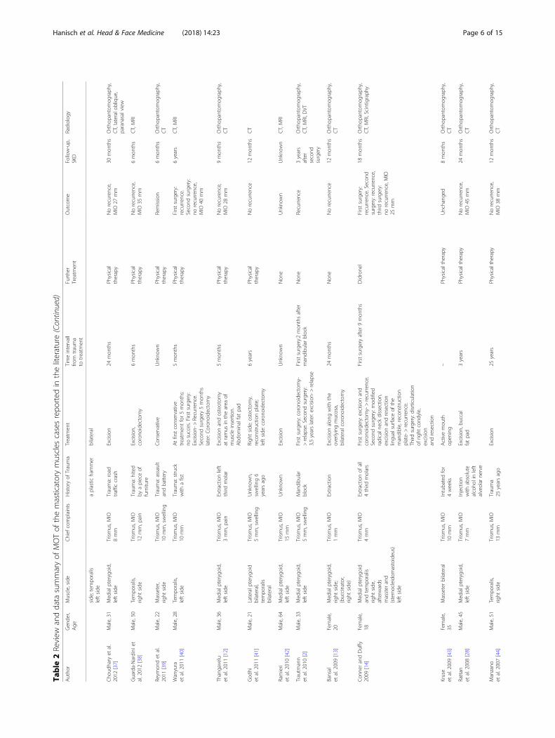
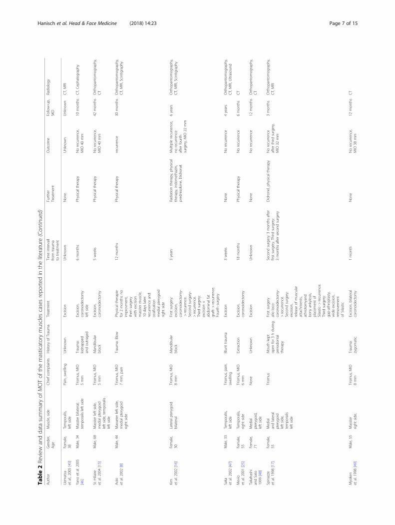
muscles in this study. The study characteristics of the in-cluded articles are described in Table 2.
Results of individual studiesGender prevalence and ageOverall, 63 patients were reported involving 25 femaleand 37 male patients that were analysed. One patient’sgender was not indicated. Therefore, approximately twoout of three patients were male. The age ranged from 10to 73 years in the female group (mean: 38.6 years). Inthe male group the age ranged from 21 to 68 years(mean: 37.4 years).
Affected muscleThe most frequent affected muscle was the massetermuscle, which was hit 35 times (left side: 23-fold, rightside: 11-fold, side unknown: 1-fold). The temporalismuscle was concerned 22 times (left side: 14-fold, rightside: 8-fold) followed by the medial pterygoid musclewith 21 cases (left side: 12-fold, right side: 9-fold). Thelateral pterygoid muscle was affected 12 times (left side:8-fold, right side: 4). In18 cases more than a singlemuscle was hit by MOT.
Clinical symptomsThe most reported clinical symptoms of MOT were tris-mus (n = 54), followed by swelling (n = 17), and pain (n= 13). Facial paralysis was outlined in one case, whilethree cases were reported to be devoid of any clinicalsymptoms. Trismus ranged from 0 to 15 mm (mean:7.3 mm).
Kind of traumaAs triggering event, strokes or falls were reported mostfrequently (n = 21), while in 12 cases a triggering eventwas unknown. Car accidents seemed to be the reasonfor five cases of MOT but MOT development due todental procedures like dental extraction (n = 7), man-dibular nerve block (n = 4), periodontitis therapy (n = 1),or as a result of alcohol injection into the alveolar nerve(n = 2) were also described. MOT as a complication ofwisdom-tooth infection was reported in three cases. Fur-thermore, occurrence of MOT was published as a conse-quence of post-fracture (n = 3), gunshot injury (n = 2),perforating wound (n = 1), injury caused by a shell (n =1), and after intubating a patient for 4 weeks (n = 1).
Time interval from trauma to treatmentTime intervals from trauma to treatment were not ad-dressed in 13 cases, while in two reports no treatmentwas initiated. In 48 cases, time intervals were reported,which ranged from 3 weeks to 25 years, whith an aver-age time of 31 months.
Hanisch et al. Head & Face Medicine (2018) 14:23 Page 3 of 15

TreatmentThe most frequent described treatment for MOT was sur-gical excision (n = 23) followed by surgery and physicaltherapy (n = 22). In addition to surgery, interposition graftsand physical therapy were performed by five authors, inter-ponate with silastic and physical therapy was reported inone case, while another author described interponate withsilastic, physical therapy, and drug administration usingdiodronel. Didronel was administered in addition to surgeryaccording to one report. The use of dermalgraft in combin-ation with surgical excision was also reported in one case.The use of radiation and surgery in combination with phys-ical therapy and drug administration with indomethacineand etidronate was furthermore published in one case. Ex-clusive physical therapy was done in four cases, while treat-ment in two reports was not indicated. Multiple surgeries
were necessary in 9 patients. Two patients were nottreated at all.
Clinical outcome: No recurrenceIn 41 cases, no recurrence was reported after the firstsurgery. Nineteen out of these 41 cases were treatedwith a combination of surgery and physical therapywhile 20 of 41 cases underwent exclusively surgery.One patient was treated with surgery in combinationwith physical and pharmacological therapy, while an-other patient was handled with surgery in combin-ation with interponate and physical therapy. Incontrast, recurrence took place in 11 cases whereasno treatment was performed or the outcome was notoutlined in 11 cases.
Fig. 1 Data analyses of recorded literature for MOT of the masticatory muscles according to PRISMA-Guidelines
Hanisch et al. Head & Face Medicine (2018) 14:23 Page 4 of 15

Table
2Review
anddata
summaryof
MOTof
themasticatorymuscles
casesrepo
rted
intheliterature
Autho
rGen
der,
Age
Muscle,side
Chief
complaints
History
ofTrauma
Treatm
ent
Timeintervall
from
trauma
totreatm
ent
Furthe
rTreatm
ent
Outcome
Follow-up,
SKD
Radiolog
y
Fité-Trepat
etal.2016[10]
Female,
49Masseter,
leftside
Trismus,p
ain,
swelling
Repe
titive
wisdo
mteethinfection
Excision
with
1cm
oftumor-free
margins
3mon
ths
Non
eNorecurren
ce3mon
ths,
Ortho
pantom
ograph
y,CT
Torres
etal.2015[11]
Female,
36Med
ial
pterygoid,
right
side
Trismus,p
ain,
swelling
Extractio
nup
perrig
htwisdo
mteeth,
4mon
thslater
excision
ofMOalio
loco
with
recurren
ce
Excision
,abdo
minal
fatgraft
>5mon
thsafter
firstsurgery
Physical
therapy
for1mon
th
Recurren
ce2mon
ths
Ortho
pantom
ograph
y,CT,MRI
Mashiko
etal.2015[31]
Male,36
Masster
bilateral
Trismus,M
IO10
mm
Freq
uently
abused
abou
ttheface
15yearsago
Osteo
tomies
bilateral,
corono
idectomy
bilateral
15years
Physical
therapyfor
2mon
ths
Norecurren
ce,
MIO
36mm
12mon
ths
CT,PET-CT
Jiang
etal.2015[5]
Female,
42Med
ial
andlateral
pterygoid
right
side
Trismus,M
IO2mm
Wisdo
mteethinfection
Exzcision,
corono
idectomy;
pedicled
buccal
fatpad
36mon
ths
Physical
therapy,
Celecoxib
200mg
2xdfor
1week
Norecurren
ce,
MIO
25mm
36mon
ths
Ortho
pantom
ograph
y,CT,MRI
Kumar
etal.2014[32]
Male,26
Masseter,
leftside
Painless
swelling,
MIO
38mm
Epileptic
with
multip
lefalls
Excision
30mon
ths
Non
eUnkno
wn
Unkno
wn
Ortho
pantom
ograph
y,CT,MRI
Alm
eida
etal.2014[30]
Female,
12Lateral
pterygoid,
leftside
Trismus,M
IO10
mm
Unkno
wn
Excision
,fat
pad
Unkno
wn
Physical
therapy,
corticosteroids
Recurren
ce1mon
thCT
Boffano
etal.2014[26]
Female,
37Med
ial
pterygoid,
leftside
Trismus,M
IO5mm
Trauma:blow
ofthelefside
ofhe
rface
Excision
toge
ther
with
left
corono
idand
cond
yle,TM
J
24mon
ths
Physicaltherapy
Norecurren
ce,
MIO
31mm
36mon
ths
Ortho
pantom
ograph
y,CT
Redd
yet
al.2014[33]
Male,21
Med
ial
pterygoid
andtempo
ralis,
leftside
Trismus,M
IO15
mm,swelling
Trauma:hitby
ahe
avyvehicle
jack
rod
Firstsurgery:
suspected
haem
atom
aelim
inated
->MIO
2mm
after6weeks.
Second
surgery:
Excision
andcorono
idectomy
6weeks
Physical
therapy
Norecurren
ce,
MIO
30mm
6mon
ths
CT/MRI
Spinizia
etal.2014[17]
Male,30
Lateral
pterygoid,
leftside
Trismus,M
IO10
mm
Trauma:
motorcycle
cciden
t
Con
servative
1mon
thPh
ysical
therapy
Norecurren
ce,
MIO
30mm
12mon
ths
CT
Schiff
etal.2013[29]
Female,
41Tempo
ralis,
leftside
Trismus,M
IO2mm,swelling
Unkno
wn
Excision
,corono
idectomy
Unkno
wn
Physical
therapy
Norecurren
ce,
MIO
518mon
ths
Ortho
pantom
ograph
y,CT
Jayade
etal.2013[34]
Female,
25Lateraland
med
ialp
terygo
idrig
htside
;tempo
ralis
leftside
Trismus,p
ain,
swelling
Unkno
wn
Excision
,corono
idectomy
leftside
Unkno
wn
Physical
therapy
Norecurren
ce,
MIO
39mm
3mon
ths
Ortho
pantom
ograph
y,po
steroanterior,CT,
MRI
Piom
bino
etal.2013[35]
Female,
62Masseter,
right
side
Trismus
Unkno
wn
Excision
Unkno
wn
Non
eNorecurren
ce24
mon
ths
Ortho
pantom
ograph
y,CT
Nem
oto
etal.2012[36]
Male,39
Masseter
bilateral;lateral
pterygoidleft
Trismus,M
IO5mm
Trauma:
repe
ated
lystruck
with
Excision
masseterbilateral,
corono
idectomy
12mon
ths
Physical
therapy
Norecurren
ce,
MIO
37mm
12mon
ths
CT,po
steroanterior
Hanisch et al. Head & Face Medicine (2018) 14:23 Page 5 of 15
Table
2Review
anddata
summaryof
MOTof
themasticatorymuscles
casesrepo
rted
intheliterature(Con
tinued)
Autho
rGen
der,
Age
Muscle,side
Chief
complaints
History
ofTrauma
Treatm
ent
Timeintervall
from
trauma
totreatm
ent
Furthe
rTreatm
ent
Outcome
Follow-up,
SKD
Radiolog
y
side
;tem
poralis
leftside
aplastic
hammer
bilateral
Cho
udhary
etal.
2012
[37]
Male,31
Med
ialp
terygo
id,
leftside
Trismus,M
IO8mm
Trauma:road
traffic
crash
Excision
24mon
ths
Physical
therapy
Norecurence,
MIO
27mm
30mon
ths
Ortho
pantom
ograph
y,CT,lateralo
blique,
paranasalview
Guarda-Nardini
etal.2012[38]
Male,50
Tempo
ralis,
right
side
Trismus,M
IO12
mm,p
ain
Trauma:hited
byapieceof
furnitu
re
Excision
,corono
idectomy
6mon
ths
Physical
therapy
Norecurren
ce,
MIO
35mm
6mon
ths
CT,MRI
Reym
ondet
al.
2011
[39]
Male,22
Masseter,
right
side
Trismus,M
IO10
mm,swelling
Trauma:assault
andbattery
Con
servative
Unkno
wn
Physical
therapy
Remission
6mon
ths
Ortho
pantom
ograph
y,CT
Wanyura
etal.2011[40]
Male,28
Tempo
ralis,
leftside
Trismus,M
IO10
mm
Trauma:struck
with
afist
Atfirstconservative
treatm
entfor5mon
ths:
nosucces.Firstsurgery:
Excision
->Recurren
ce.
Second
surgery5mon
ths
later:Coron
oide
ctom
y
5mon
ths
Physical
therapy
Firstsurgery:
recurren
ce.
Second
surgery:
norecurren
ce,
MIO
40mm
6years
CT,MRI
Thangavelu
etal.2011[12]
Male,36
Med
ialp
terygo
id,
leftside
Trismus,M
IO3mm,p
ain
Extractio
nleft
third
molar
Excision
andosteotom
yat
ramus
inthearea
ofmuscleinsertion.
Abd
ominalfatpad
5mon
ths
Physical
therapy
Norecurence,
MIO
28mm
9mon
ths
Ortho
pantom
ograph
y,CT
God
hiet
al.2011[41]
Male,21
Lateralp
terygo
idbilateral,
tempo
ralis
bilateral
Trismus,M
IO5mm,swelling
Unkno
wn,
swelling6
yearsago
Righ
tside
:ostectomy,
reconstructio
nplate;
leftside
:coron
oide
ctom
y
6years
Physical
therapy
Norecurren
ce12
mon
ths
CT
Ramieri
etal.2010[42]
Male,64
Med
ialp
terygo
id,
leftside
Trismus,M
IO15
mm
Unkno
wn
Excision
Unkno
wn
Non
eUnkno
wn
Unkno
wn
CT,MRI
Trautm
ann
etal.2010[2]
Male,33
Med
ialp
terygo
id,
leftside
Trismus,M
IO5mm,swelling
Mandibu
lar
block
Firstsurgery:corono
idectomy-
>relapse.Second
surgery:
3,5yearslater:excision
->relapse
Firstsurgery:2mon
thsafter
mandibu
larblock
Non
eRecurren
ce3years
after
second
surgery
Ortho
pantom
ograph
y,CT,MRI,D
VT
Bansal
etal.2009[13]
Female,
20Med
ialp
terygo
id,
right
side
;(buccinator,
right
side
)
Trismus,M
IO1mm
Extractio
nExcision
alon
gwith
the
overlyingmucosa,
bilateralcoron
oide
ctom
y
24mon
ths
Non
eNorecurren
ce12
mon
ths
Ortho
pantom
ograph
y,CT
Con
nerandDuffy
2009
[14]
Female,
18Med
ialp
terygo
idandtempo
ralis
right
side
,afterw
ards
masster
and
(sternocleidom
astoideus)
leftside
Trismus,M
IO4mm
Extractio
nof
all
4third
molars
Firstsurgery:excision
and
corono
idectomy->recurren
ce.
Second
surgery:mod
ified
radicaln
eckdissectio
n,excision
andresection
lingu
alsurface
ofthe
mandible,reconstructio
nplate->recurren
ce.
Third
surgery:disrticulation
ofrig
htcond
yle,
excision
andresection
Firstsurgeryafter9mon
ths
Didrone
lFirstsurgery:
recurren
ce.Secon
dsurgery:recurren
ce,
third
surgery:
norecurren
ce,M
IO25
mm
18mon
ths
Ortho
pantom
ograph
y,CT,MRI,Scintigraph
y
Kruse
etal.2009[43]
Female,
35Masseterbilateral
Trismus,M
IO10
mm
Intubatedfor
4weeks
Activemou
thop
ening
–Ph
ysicaltherapy
Unchang
ed8mon
ths
Ortho
pantom
ograph
y,CT
Rattan
etal.2008[28]
Male,45
Med
ialp
terygo
id,
leftside
Trismus,M
IO7mm
Injection
with
absolute
alcoho
linleft
alveolar
nerve
Excision
,buccal
fatpad
3years
Physicaltherapy
Norecurren
ce,
MIO
45mm
24mon
ths
Ortho
pantom
ograph
y,CT
Manzano
etal.2007[44]
Male,51
Tempo
ralis,
right
side
Trismus,M
IO13
mm
Trauma
25yearsago
Excision
25years
Physicaltherapy
Norecurren
ce,
MIO
38mm
12mon
ths
Ortho
pantom
ograph
y,CT
Hanisch et al. Head & Face Medicine (2018) 14:23 Page 6 of 15
Table
2Review
anddata
summaryof
MOTof
themasticatorymuscles
casesrepo
rted
intheliterature(Con
tinued)
Autho
rGen
der,
Age
Muscle,side
Chief
complaints
History
ofTrauma
Treatm
ent
Timeintervall
from
trauma
totreatm
ent
Furthe
rTreatm
ent
Outcome
Follow-up,
SKD
Radiolog
y
Uem
atsu
etal.,2005
[45]
Female,
38Tempo
ralis,
leftside
Pain,swelling
Unkno
wn
Excision
Unkno
wn
Non
eUnkno
wn
Unkno
wn
CT,MRI
Yano
etal.2005
[46]
Male,34
Masster
bilateral;
tempo
ralis
leftside
Trismus,M
IO5mm
Trauma:
kidn
appe
dandou
traged
Excision
,corono
idectomy
leftside
6mon
ths
Physicaltherapy
Norecurren
ce,
MIO
40mm
10mon
ths
CT,Cep
halography
St.-H
ilaire
etal.2004[15]
Male,68
Masster
leftside
,med
ialp
terygo
idleftside
,tem
poralis
leftside
Trismus,M
IO5mm
Mandibu
lar
block
Excision
,corono
idectomy
5weeks
Physicaltherapy
Norecurren
ce,
MIO
40mm
42mon
ths
Ortho
pantom
ograph
y,CT
Aoki
etal.2002[8]
Male,44
Masseterleftside
,med
ialp
terygo
idrig
htside
Trismus,M
IO7mm,p
ain
Trauma:Blow
Physicaltherapie
for2mon
ths:no
improvmen
t,then
surgery
with
excision
masster
muscle,
10days
later:
recurren
ceand
ossification
med
ialp
terygo
idrig
htside
12mon
ths
Physicaltherapy
recurren
ce30
mon
ths
Ortho
pantom
ograph
y,CT,MRI,Scintigraph
y
Kim
etal.2002[16]
Female,
30Lateralp
terygo
idbilateral
Trismus,M
IO8mm
Mandibu
lar
block
Firstsurgery:
excision
,corono
idectomy-
>recurence.
Second
surgery-
>recurren
ce.
Third
surgery:
excision
+abdo
minalfat
graft->recurren
ce.
Fourth
surgery
3years
Radiationtherapy,ph
ysical
therapy,indo
methazin,
pred
nisolone
,Etid
ronat
Multip
lerecurren
ce,
norecurren
ceafterfourth
surgery,MIO
22mm
6years
Ortho
pantom
ograph
y,CT,MRI,Scintigraph
y
Saka
etal.2002[47]
Male,33
Tempo
ralis,
leftside
Trismus,p
ain,
swelling
Blun
ttrauma
Excision
3weeks
Non
eNorecurren
ce4years
Ortho
pantom
ograph
y,CT,MRI,U
ltrasou
nd
Mevio
etal.2001[25]
Female,
55Tempo
ralis,
right
side
Trismus,M
IO6mm
Extractio
nExcision
,corono
idectomy
18mon
ths
Physicaltherapy
Norecurren
ce6mon
ths
CT
Takahashi
andSato
1999
[48]
Female,
71Med
ial
pterygoid,
leftside
Non
eUnkno
wn
Excision
Unkno
wn
Non
eNorecurren
ce12
mon
ths
Ortho
pantom
ograph
y,CT
Spinazze
etal.1998[17]
Female,
55Med
ial
andlateral
pterygoid
leftside
,tempo
ralis
leftside
Trismus
Mou
thkept
open
for3hdu
ring
perio
dontal
therapy
Firstsurgery
alio
loco:
corono
idectomy-
>recurren
ce.
Second
surgery:
excision
,releaseof
muscular
attachmen
ts,
athrotom
yand
bony
ankylosis,
placem
entof
Silastic->
recurren
ce.
Third
surgery:
gap-athrop
lasty,
wideexcision
,removem
ent
ofSilastic
Second
surgery:3mon
thsafter
firstsurgery.Third
surgery:
3mon
thsaftersecond
surgery
Didrone
l,ph
ysicaltherapy
Norecurren
ceafterthird
surgery,
MIO
32mm
3mon
ths
Ortho
pantom
ograph
y,CT,MRI
Myoken
etal.1998[49]
Male,55
Masster
right
side
,Trismus,M
IO8mm
Trauma:
zygo
matic
Excision
,bilateral
corono
idectomy
1mon
thNon
eNorecurren
ce,
MIO
38mm
12mon
ths
CT
Hanisch et al. Head & Face Medicine (2018) 14:23 Page 7 of 15

Table
2Review
anddata
summaryof
MOTof
themasticatorymuscles
casesrepo
rted
intheliterature(Con
tinued)
Autho
rGen
der,
Age
Muscle,side
Chief
complaints
History
ofTrauma
Treatm
ent
Timeintervall
from
trauma
totreatm
ent
Furthe
rTreatm
ent
Outcome
Follow-up,
SKD
Radiolog
y
tempo
ralis
bilateral
arch
fracture
Geistet
al.1998[50]
Male,44
Masseter
leftside
Trismus,M
IO5mm,p
ain
Trauma:
fractureof
theleftmandible
Excision
12mon
ths
Non
eUnkno
wn
Unkno
wn
Ortho
pantom
ograph
y,half-axial,CT
Steine
ret
al.1997[51]
Male,40
Masseter
leftside
Trismus,M
IO5mm
Trauma:
fractureof
themandible
Excision
12mon
ths
Physicaltherapy
Norecurren
ce,
MIO
30mm
3mon
ths
Ortho
pantom
ograph
y,CT
Steine
ret
al.,1997
[51]
Female,
15Masseter
leftside
Trismus,8
mm
Shotgu
nwou
ndto
the
face
7yearsago
Excision
7years
Physicaltherapy
Norecurren
ce,
MIO
26mm
Unkno
wn
CT
Tong
etal.1994[52]
Female,
73Med
ial
pterygoid
bilateral
Non
eUnkno
wn
Non
eUnkno
wn
Non
eUnkno
wn
Unkno
wn
CT
El-Labban
etal.1993[53]
Male,42
Masster,
side
unknow
nTrismus
Trauma:blow
totheside
6mon
ths
before
Unkno
wn
Unkno
wn
Unkno
wn
Unkno
wn
Unkno
wn
Unkno
wn
ParkashandGoyal
1992
[18]
Male,28
Med
ial
pterygoid,
leftside
Trismus,M
IO0mm
Pericoron
itis
leftthird
molar
Firstsurgery:
cond
ylectomy
andcorono
idectomy->
recurren
ceSecond
surgey:
excision
6½
years
Physicaltherapy
Recurren
ce,
aftersecond
surgery:MIO
20mm
3mon
ths
Ortho
pantom
ograph
y,CT
Nilner
and
And
ersson
1989
[54 ]
Male,57
Med
ial
pterygoid,
right
side
Trismus
Injectionwith
alcoho
linrig
htalveolar
nerve
Non
e–
Non
eUnkno
wn
8years
Ortho
pantom
ograph
y,CT,TM
Jradiog
raph
Lello
andMakek
1986
[19]
Female,
31Masster
leftside
Trismus,M
IO10
mm,p
ain,
swelling
Mandibu
lar
block
Excision
5weeks
Non
eNorecurren
ce,
MIO
40mm
4years
Ortho
pantom
ograph
y,po
steroanterior
Scintig
raph
y
Lello
andMakek
1986
[19]
Male,32
Masster,
leftside
Trismus,M
IO10
mm,swelling
Trauma:blow
totheleftmandible
Excision
2mon
ths
Non
eNorecurren
ce5years
Unkno
wn
Lello
andMakek
1986
[19]
Male,34
Tempo
ralis
leftside
Non
eTrauma:motor
vehicleaccide
ntExcision
9mon
ths
Non
eNorecurren
ce4years
CT
Wiesenfeldet
al.
1985
[55]
Female,
10Tempo
ralis
right
side
Painless
swelling
Unkno
wn
Excision
Unkno
wn
Non
eNorecurren
ce6mon
ths
Ortho
pantom
ograph
y,CT
Arim
aet
al.1984
[56]
Male,25
Masseter,left
Trismus,M
IO11
mm,p
ain
Trauma:
contusionin
afig
htExcision
6mon
ths
Non
eNorecurren
ce,
MIO
47mm
11mon
ths
Posterioanterio
r
Abd
inandPrabhu
1984
[57]
Female,
43Lateral
pterygoid
leftside
Totaltrismus,
painless
swelling
Hug
epainful
swellingat
the
ageof
19
Excision
24years
Physicaltherapy
Norecurren
ce,
MIO
30mm
6mon
ths
Ortho
pantom
ograph
y
Christm
asand
Ferguson
1982
[58]
Male,51
Masseter,
leftside
Trismus,M
IO10
mm,swelling
Trauma:falling
from
horseand
strikingagainst
afencepo
st
Excision
18mon
ths
Non
eNorecurren
ce,
MIO
40mm
4mon
ths
Posterioanterio
r
Pleziaet
al.1977
[59]
Female,
47Masseter,
leftside
Trismus,
MIO
8mm
Trauma:blow
Excision
2mon
ths
Non
eNorecurren
ce,
MIO
44mm
unknow
nPo
sterioanterio
r
NarangandDixon
1974
[20]
Male,50
Med
ial
pterygoid,
right
side
Trismus,
MIO
12mm
Extractio
nFirstsurgery:
excision
->recurren
ceSecond
surgery:
excision
,
Firstsurgery:15
mon
ths
Second
surgery:1mon
thPh
ysicaltherapy
Norecurren
ce,
MIO
49mm
unknow
nCep
halography,
posterioanterio
r
Hanisch et al. Head & Face Medicine (2018) 14:23 Page 8 of 15

Table
2Review
anddata
summaryof
MOTof
themasticatorymuscles
casesrepo
rted
intheliterature(Con
tinued)
Autho
rGen
der,
Age
Muscle,side
Chief
complaints
History
ofTrauma
Treatm
ent
Timeintervall
from
trauma
totreatm
ent
Furthe
rTreatm
ent
Outcome
Follow-up,
SKD
Radiolog
y
corono
idectomy,
insetio
nof
silastic
Hatzifotiadis
1970
[60]
Male,50
Masseter,
leftside
Trismus,
MIO
5mm,
swelling
Trauma:fallen
oniro
npe
gFirst:conservative
treatm
entwith
out
succes
for2mon
ths.
Surgery:Excision
4mon
ths
Physicaltherapy,acrylic
appliancefor2days
Norecurren
ce12
mon
ths
Radiog
raph
Trester
etal.1969[61]
Female,
29Masseter,
leftside
Trismus,
MIO
3–4mm,
swelling
Trauma:
epileptic
seizure->blow
Excision
->recurren
ce,
than
physicaltherapy
1mon
thPh
ysicaltherapy
Recurren
ceafter
surgery->with
physicaltherapy:
MIO
25mm
3mon
ths
Posterioanterio
r
Vernale1968
[62]
Male,31
Masseter,
right
side
Trismus,
pain,swelling
Trauma:
caraccide
ntExcision
1mon
thNon
eNorecurren
ce2mon
ths
Posterioanterio
r
Vernale1968
[62]
Male,29
Masseter,
leftside
Trismus,
MIO
4mm
Trauma:blow
Excision
4mon
ths
Non
eNorecurren
ce6years
Posterioanterio
r,rig
htandleftlateralo
blique
Shaw
kat1967
[21]
Male,24
Masseter,
tempo
ralis,
(myloh
yoid),
leftside
Facial
paralysis
Extractio
nleft
maxillarymolar
region
Unkno
wn
Unkno
wn
Non
eUnkno
wn
Unkno
wn
Cep
halography
Parnes
andHinds
1965
[63]
Female,
27Masster,
leftside
Trismus,
MIO
10mm,p
ain
Trauma:be
aten
with
afist
Excision
1mon
thNon
eNorecurren
ce,
MIO
25mm
Unkno
wn
Posterioanterio
r,rig
htandleftlateralo
blique
Hellinge
r1965
[64]
Female,
21Masster,
tempo
ralis,
(buccinator)
pterygoid,
leftside
Trismus,
MIO
3–4mm
Unkno
wn
Excision
12years
Non
eNorecurren
ce6mon
ths
Posterioanterio
r,lateral
oblique
Goo
dsell1962[65]
Male,39
Masseter,
right
side
Trismus,
pain,swelling
Trauma:blow
Excision
5weeks
Non
eNorecurren
ceUnkno
wn
Unkno
wn
Kostrubalaand
Tailbot
1948
[66]
Male,21
Masseter,
right
side
Trismus
Trauma:struck
byan
enem
ybu
llet
Firstsurgery:Excision
->recurren
ceSecond
surgery:
excision
+de
rmalgraft
6mon
ths,second
surgeryafter
4mon
ths
Before
surgery:ph
ysical
therapy->no
succes
After
second
surgery:no
recurren
ce
9mon
ths
Laminog
raph
NizelandPrigge
1946
[4]
Male,21
Masseter,
right
side
Trismus,
MIO
4mm
Trauma:
perfo
ratin
gwou
ndCon
servative
treatm
ent
4mon
ths
Cou
nter-trismus
appliance
MIO
21mm
Posterioanterio
r
IvyandEby1924
[7]
Unkno
wn
Masseter,
leftside
Trismus
Trauma:wou
nded
byasm
all
shellfragm
ent
Excision
Unkno
wn
Trismus
apparatus
Fullextent
ofop
eningachieved
immed
iaely
postop
erative
Unkno
wn
Radiog
raph
Hanisch et al. Head & Face Medicine (2018) 14:23 Page 9 of 15

Clinical outcome: RecurrenceRecurrence was reported in a total of 11 cases. In 7 outof these cases multiple surgeries were performed whichstopped any further recurrence. In four reports, unsuc-cessful treatment of MOT hampered recurrenceanalysis.
Clinical outcome: Recurrence in correlation with time oftreatmentTo evaluate the clinical outcome “recurrence” in correl-ation with time of treatment, two groups were defined.In the first group, surgery was performed less than6 months after trauma (n = 21). In this group five caseswith recurrence were stated. In the second group, theinterval from trauma to treatment was longer than6 months (n = 27). In that herein also five cases withclinical recurrence occurred. In one case undergoing re-currence no interval from trauma to treatment was indi-cated. In a total of 13 reports, the interval from traumato treatment was not noted and in two cases no treat-ment was initiated.
Clinical outcome: Recurrence in correlation with the type oftreatmentRecurrence after the first treatment was found in 3 casesin which only surgery took place. Surgery in combin-ation with physical therapy led to 3 cases of recurrence.Surgery in combination with fat pad and physical ther-apy led to recurrence in two reports, while recurrencealso occurred to a patient who was treated with surgeryin combination with diodronel. Recurrence was also re-ported during treatment with surgery in combinationwith radiation, indomethacine, diodronel, and physicaltherapy, as well as surgery with silastic interponate, dio-dronel, and physical therapy.
Clinical outcome: Maximal incisal opening (MIO)developmentIn the group of successful treated patients, 20 authorsreported about the development of MIO before and aftertherapy. MIO ranged from 15 to 49 mm in length with amean of 29.6 mm. Only physical therapy (n = 1) yieldeda 20 mm long MIO. Surgery in combination with fat pad(n = 3) resulted in a MIO of 28.6 mm length (range:23 mm–38 mm), while the MIO of patients with surgeryin combination with physical therapy (n = 12) exhibiteda MIO of 27.2 mm length (range: 15 mm–49 mm). Sur-gery alone (n = 4) yielded a MIO of 31.3 mm length(range: 30 mm–35 mm).
Risk of bias within studiesIn general, the risk of bias was considered low, sincemost part of the case reports were described in
accordance to the check-list. Only 2 studies showed apercentage of positive response lower than 60% (Supple-ment 1).
Clinical documentation system screeningAfter searching the clinical documentation system of theUniversity Hospital Münster only one self-generatedentry for MOT could be recovered.
Clinical case reported by the authorsA 28 year-old male was referred to our Clinic ofCranio-Maxillofacial Surgery with trismus in March2016. The patient was not able to open or to close hismouth and, moreover, he was unable to protrude or toproduce a lateral excursion. So he possessed an interin-cisal mouth opening of 5 mm. The patient indicated thathe underwent a filling therapy on the right mandiblemolar by his dentist 7 months ago. As according therapya right mandibular nerve block was performed. Fourweeks later the patient developed trismus. His dentistdescribed oral antibiosis and physical examination. How-ever, no clinical improvement was observed. Therefore,the patient was referred to a Clinic ofCranio-Maxillofacial Surgery where the diagnosis ofpericoronitis of the lower right third molar was stated.Extraction of the right upper and lower third molar anda forced mouth-opening was performed under generalanesthesia. Subsequently, the trismus disappeared butreappeared 2 weeks later. Because of this relapse, coro-noidectomy was performed on the right side. Conse-quently, the trismus disappeared, but a relapsereoccurred a few weeks later. A multislice computertomography (CT) of the head was performed and theCT revealed a calcification of the right medial pterygoidmuscle (Fig. 2). Due to the given diagnosis of MOT ofthe right medial pterygoid, the patient was finally re-ferred to the Clinic of Cranio-Maxillofacial Surgery atthe University of Münster. For excluding MOP, we re-ferred the patient to the department of human genetics.Indeed, MOP could be excluded and also all laboratorytest results ranged within normal limits, including theresulting values for calcium, phosphate, alkaline phos-phatase and parathyroid hormone measurements. Thus,we decided to perform renewed surgery 6 months afterthe last surgical intervention. Pre-operative radiationwas performed with 6 Gy as single-dose radiation. Surgi-cal excision of the ossified right medial pterygoid musclewas performed through combined intra- and extraoralaccess under general anesthesia. During this interven-tion, solid bone mass could be excised (Fig. 3). Histo-pathological analysis confirmed the diagnosis of MOT(Fig. 4). Physical therapy was started 2 days after surgeryand 1 week after surgical intervention the patient couldbe released. Post-operative long-term application of
Hanisch et al. Head & Face Medicine (2018) 14:23 Page 10 of 15

ibuprofen 400 mg was performed for 2 weeks. At thistime point, the MIO reached 23 mm in length. The pa-tient was instructed to perform intensive physical ther-apy with an functional orthodontic gadget, the so-called“Jeckel-spreader”, for exercising mouth opening. Thisdevice serves for mobilisation of the masticatory mus-cles. Two weeks later, the MIO still yielded 25 mm inlength. Thereafter, the patient stopped physical therapyusing the “Jeckel-spreader” against our recommendation.Consequently, the MIO decreased to 10 mm in length.Thus, we advised the patient strongly to restart physicaltherapy but he declined. Digital volume tomography(DVT) was performed which revealed renewed calcifica-tion (Fig. 5). Six months after surgery, MIO exhibited a
length of about 8 mm. This enabled the patient to eat,to perform and to do a small lateral excursion.We have derived a decision tree for diagnosis and
treatement of MOT (Fig. 6).
Discussion The pathogenesis of MOT has not been fi-nally clarified. In 1924, Carey [24] already listed fourmajor theories for the development of MOT: 1) Dis-placement of bony fragments into soft tissue andhematoma with subsequent proliferation; 2) detachmentof periosteal fragments into surrounding tissue with pro-liferation of osteoprogenitor cells; 3) migration of sub-periostal osteoprogenitor cells into surrounding softtissue through periosteal perforations induced by
Fig. 2 Cone beam scan showing calcification of the rightmedial pterygoid
Fig. 3 Piece of the excised solid bone mass
Fig. 4 Microscopic image of lesion demonstrating sclerotic, solidand cancellous bone with fatty bone marrow. (HE,magnification: 10-fold)
Fig. 5 Digital volume tomography showing recurrenceof calcification
Hanisch et al. Head & Face Medicine (2018) 14:23 Page 11 of 15

trauma; 4) differentiation of extraosseous cells exposedto bone morphogenic proteins. The results of thepresent study confirm the assumption,that multiple pro-cesses lead to the development of MOT. If a triggeringevent is present at all, its nature seems to be tooheterogenous from case to case to support the theory ofa single initiating cause. In 12 of the cases summarizedhere, no specific triggering traumatic event was identi-fied (idiopathic myositis ossificans). Nevertheless, itseems that minor traumatic lesions unnoticed by thesepatients could be a possible cause. According to Torres[11] the intensity of the trauma may not be related tothe occurrence of MOT. This statement could explainwhy no cases of MOT occurring in individuals that pur-sue the sport of boxing have been reported in the litera-ture so far. These cases would be expected because ofregularly occurring blows to the face and masticatorymuscles (especially the masseter and temporal muscles)of boxers. On the other hand, a relation between dentalsurgery and the onset of MOT seems obvious. There are7 case reports of MOT with previous tooth extraction[11–14, 20, 21, 25] though it is not possible to fully dif-ferentiate whether the extraction or the dentalanesthesia in the context with the extraction representsthe triggering event. The latter as a cause of MOT wasreported in four cases [2, 15, 16, 19]. Mandibular block
as reported by Trautmann [2] as well as in our reportedcase, could be a more possible triggering factor forMOT. Therefore local anesthesia cannot be excluded asa cause of MOT occurring after periodontal treatment,either [17]. Furthermore, three cases of MOT followingrepetitive wisdom tooth infection have been published[5, 10, 18]. This would represent an additional indicationrequiring surgical removal of wisdom teeth if normalplacement in the row of teeth is not expected.Trismus is the most frequently observed symptom of
MOT in the masticatory muscles which was also pre-sented in our case. In this respect, MOT should be con-sidered in the differential diagnosis in case of persistingtrismus without a clinically manifesting cause. In suchcases, radiographic findings are being expected only 3–6 weeks after the appearance of clinical symptoms [2].So far, male patients have been considered as the main
group at risk of developing MOT of the masticatorymuscles with a male/female ratio of 2.4/1 [26]. However,our data analysis demonstrated a gender-specific differ-ence to a lesser extent with a male/female ratio of ap-proximately 1.5/1. Since however MOT has beenfrequently related to traumas (e. g. fracture, blow) a pos-sible explanation could be: males might have experi-enced traumas more often than females and thus alsosuffer more often from MOT. Of particular interest is
Fig. 6 Decision tree for diagnosis and treatement of myositis ossificans traumatica
Hanisch et al. Head & Face Medicine (2018) 14:23 Page 12 of 15

the view at the cases of MOT occurring after dentaltreatment where more women (n = 9) were concernedthan men (n = 6). This means prevalence for female pa-tients of MOT of the masticatory musculature in contextof dental treatment with a 1.5/1 ratio.In most cases of MOT of the masticatory muscles the
masseter muscle is the most affected one [10]. However,this is not true for those cases of MOT occurring afterdental treatment. Of these cases (n = 10), 66% involvedthe medial pterygoid muscle. Given the potential risk ofdamaging this muscle in the context of a mandibularnerve block, local dental anesthesia seems to be thecause of MOT here, as potentially in our case. Whetherthe patient has to be informed about this extremely rarecomplication remains questionable in view of the largenumbers of local dental anesthesia administered daily.On the other hand the consequences represent a severeimpairment for the patient. Nevertheless, MOT shouldbe considered in the differential diagnosis in cases oftherapy-resistant trismus developing in the weeks afterlocal anesthesia.Generally, excision of the affected muscle is recom-
mended as treatment of choice [10]. However, there aredifferent opinions about the time when the excision hasto be done and about possible additional measures, suchas the use of interpositional materials, treatment withdrugs, or physical therapy. Some authors recommended[12, 14, 27] that the excision as well as the use of inter-positional material should be performed after completematuration, about 6 to 12 months after initial symptoms.In contrast, other authors prefered excision at an earlystage [11]. There were five relapses, both, in the groupof early excision (treatment less than six months afterfirst symptoms), and in the group of excision at a laterstage (treatment more than six months after first symp-toms). However, the group with intervention at a latertime point included 27 cases that was somewhat biggerthan the early-intervention group (n = 21). Nonetheless,it is not possible to make any clear recommendation forthe ideal time point of surgical intervention based onthese data.While some authors suggested aggressive physical
therapy after surgical excision [17], others advisedagainst this procedure [14]. They feared that physicaltherapy stimulates bone formation with the conse-quence of exacerbation of MOT. Of the 22 reportedcases undergoing excision combined with physicaltherapy, 3 cases relapsed. In the group of 23 patientswho only underwent excision without physical therapythere were also 3 relapses. In consequence, no differ-ence in the rate of recurrence was found dependingon physical therapy.In addition to excision, − with or without physical
therapy, the use of interpositional materials [12, 16, 17,
20, 28] or pharmaceuticals, such as etidronate or ibupro-fen [29] have been proposed. Often, these additionalmeasures were applied in clinical cases with multiple re-currences [11, 14, 16, 17, 30] so that the benefit of add-itional treatment cannot be assessed conclusively.The major limitation of this review is the rarity of the
evaluated condition, resulting in a lack of researchsources which could offer reliable evidence-based infor-mation. With this regard, all studies selected for this re-view were case reports, which hampered a deeperanalysis of risk of bias of each study. Nonetheless, thepresent study aimed to offer a guide decision for themanagement and diagnosis of MOT. Additionally, thecase reported described the authors clinical experienceregarding this condition and shows a treatment optionfor patients with MOT.
ConclusionsDental procedures, such as local anesthesia or extrac-tions, may cause MOT of the masticatory musculature.Women have a higher risk of developing MOT with re-spect to dental treatment. The most important treatmentoption for MOT is surgical excision and subsequentphysical therapy can have beneficial effects. A benefit ofinterpositional materials and drugs as therapy of MOTof the masticatory muscles has not yet been proven.MOP has to be excluded.
Additional file
Additional file 1. Quality assessment of the included literature. (DOC139 kb)
AbbreviationsCT: Computer tomography; DVT: Digital volume tomography; MIO: Maximalincisal opening; MOP: Myositis ossificans progressiva; MOT: Myositis ossificanstraumatica
AcknowledgementsWe acknowledge support by Open Access Publication Fund of University ofMuenster.
FundingThis research did not receive any specific grant from funding agencies in thepublic, commercial, or not-for-profit sectors.
Availability of data and materialsThe datasets supporting the conclusions of this article are available at theDepartment of Cranio-Maxillofacial Surgery, University Hospital MünsterGermany.
Authors’ contributionsMH, LH reviewed the literature. JK, RW, LB and LFF helped in theinterpretation of data. MH analysed the dabase of the clinic. MH, LH, JK, RW,LB and LFF participated in design, and drafting of the manuscript. All authorsread and approved the final manuscript.
Ethics approval and consent to participateThe ethical approval for this study was obtained from the ethical reviewcommittee (Ref. no. 2017–052-f-N), Ethikkommission der Ärztekammer
Hanisch et al. Head & Face Medicine (2018) 14:23 Page 13 of 15

Westfalen-Lippe und der Westfälischen Wilhelms-Universität, Münster, Germa-ny.Written informed consent was obtained from the patient.
Consent for publicationWritten informed consent was obtained from the patient for publication ofthe case report and any accompanying images. A copy of the writtenconsent is available for review by the Editor-in-Chief of this journal.
Competing interestsThe authors declare that they have no competing interests.
Publisher’s NoteSpringer Nature remains neutral with regard to jurisdictional claims inpublished maps and institutional affiliations.
Author details1Department of Cranio-Maxillofacial Surgery, Research Unit Rare Diseaseswith Orofacial Manifestations (RDOM), University Hospital Münster,Albert-Schweitzer-Campus 1, Gebäude W 30, D-48149 Münster, Germany.2Department of Orthodontics, Faculty of Health, School of Dentistry, Witten/Herdecke University, Alfred-Herrhausen-Strasse 44, 58455 Witten, Germany.3Department of Cranio-Maxillofacial Surgery, AG VABOS, University HospitalMünster, Albert-Schweitzer-Campus 1, Gebäude W 30, D-48149 Münster,Germany. 4Department of Oral and Maxillofacial Surgery, Central GermanArmed Forces Hospital, Rübenacher Strasse 170, 56072 Koblenz, Germany.
Received: 9 April 2018 Accepted: 15 October 2018
References1. Kaplan FS, Seemann P, Haupt J, Xu M, Lounev VY, Mullins M, Shore EM.
Investigations of activated ACVR1/ALK2, a bone morphogenetic proteintype I receptor, that causes fibrodysplasia ossificans progressiva. MethodsEnzymol. 2010;484:357–73.
2. Trautmann F, Pd M, Fernandes TL, Gondak RO, Castilho JC, Filho EM.Myositis ossificans traumatica of the medial pterygoid muscle: a case report.J Oral Sci. 2010 Sep;52(3):485–9.
3. Wiggins RL, Thurber D, Abramovitch K, Bouquot J, Vigneswaran N. Myositisossificans circumscripta of the buccinator muscle: first report of a rarecomplication of mandibular third molar extraction. J Oral Maxillofac Surg.2008 Sep;66(9):1959–63.
4. Nizel AE, Prigge EK. Trismus due to myositis ossificans traumatica; report ofa case. J Oral Surg. 1946;4:93–101.
5. Jiang Q, Chen MJ, Yang C, Qiu YT, Tian Z, Zhang ZY, Qiu WL. Post-infectiousmyositis ossificans in medial, lateral pterygoid muscles: a case report andreview of the literature. Oncol Lett. 2015;9(2):920–6.
6. Jayade B, Adirajaiah S, Vadera H, Kundalaswamy G, Sattur AP, Kalkur C.Myositis ossificans in medial, lateral pterygoid, and contralateral temporalismuscles: a rare case report. Oral Surg Oral Med Oral Pathol Oral Radiol.2013;116(4):261–6.
7. Ivy and Eby: The medical Dept. of the United States Army, U.S governmentprinting office, 1924, Vol. II, Sec 2; P. 457.
8. Aoki T, Naito H, Ota Y, Shiiki K. Myositis ossificans traumatica of themasticatory muscles: review of the literature and report of a case. J OralMaxillofac Surg. 2002;60(9):1083–8.
9. Ackermann LV. Extra-osseous localized non-neoplastic bone and cartilageformation (so-called myositis ossificans): clinical and pathological confusionwith malignant neoplasms. J Bone Joint Surg Am 1958;40-A(2):279–298.
10. Fité-Trepat L, Martos-Fernández M, Alberola-Ferranti M, Romanini-MontecinoC, Saez-Barba M, Bescós-Atín C. Myositis ossificans of the masseter muscle: arare location. Report of a case and review of literature. J Clin Exp Dent.2016;8(2):210–3.
11. Torres AM, Nardis AC, da Silva RA, Savioli C. Myositis ossificans traumatica ofthe medial pterygoid muscle following a third molar extraction. Int J OralMaxillofac Surg. 2015;44(4):488–90.
12. Thangavelu A, Vaidhyanathan A, Narendar R. Myositis ossificans traumaticaof the medial pterygoid. Int J Oral Maxillofac Surg. 2011;40(5):545–9.
13. Bansal V, Kumar S, Mowar A. Unusual causes of trismus: a report of twocases. J Maxillofac Oral Surg. 2009;8(4):377–80.
14. Conner GA, Duffy M. Myositis ossificans: a case report of multiplerecurrences following third molar extractions and review of the literature. JOral Maxillofac Surg. 2009;67(4):920–6.
15. St -Hilaire H, Weber WD, Ramer M, Lumerman H. Clinicopathologicconference: trismus following dental treatment. Oral Surg Oral Med OralPathol Oral Radiol Endod. 2004;98(3):261–6.
16. Kim DD, Lazow SK, Har-El G, Berger JR. Myositis ossificans traumatica ofmasticatory musculature: a case report and literature review. J OralMaxillofac Surg. 2002 Sep;60(9):1072–6.
17. Spinazze RP, Heffez LB, Bays RA. Chronic, progressive limitation of mouthopening. J Oral Maxillofac Surg. 1998;56(10):1178–86.
18. Parkash H, Goyal M. Myositis ossificans of medial pterygoid muscle. A causefor temporomandibular joint ankylosis. Oral Surg Oral Med Oral Pathol.1992;73(1):27–8.
19. Lello GE, Makek M. Traumatic myositis ossificans in masticatory muscles. JMaxillofac Surg. 1986;14(4):231–7.
20. Narang R, Dixon RA Jr. Myositis ossificans: medial pterygoid muscle-a casereport. Br J Oral Surg. 1974;12(2):229–34.
21. Shawkat AH, Myositis ossificans. Report of a case. Oral Surg Oral Med OralPathol. 1967;23(6):751–4.
22. Moher D, Liberati A, Tetzlaff J, Altmann DG, The PRISMA Group. PrefferedReporting Items for Systematic Reviews and Meta Analyses: The PRISMAStatement. PLoS Med. 2009;6(7):e1000097.
23. Moola S, Munn Z, Tufanaru C, Aromataris E, Sears K, Sfetcu R, Currie M,Qureshi R, Mattis P, Lisy K, P-F M. Chapter 7: Systematic reviews of etiologyand risk: The Joanna Briggs Institute; 2017. https://reviewersmanual.joannabriggs.org.
24. Carey EJ. Multiple bilateral parosteal bone and callus formations of thefemur and left innominate bone. Arch Surg. 1924;8:592.
25. Mevio E, Rizzi L, Bernasconi G. Myositis ossificans traumatica of the temporalmuscle: a case report. Auris Nasus Larynx. 2001;28(4):345–7.
26. Boffano P, Zavattero E, Bosco G, Berrone S. Myositis ossificans of the leftmedial pterygoid muscle: case report and review of the literature ofmyositis ossificans of masticatory muscles. Craniomaxillofac TraumaReconstr. 2014;7(1):43–50.
27. Ferlito A, Barion U, Nicolai P. Myositis ossificans of the head and neck.Review of the literature and report of a case. Arch Otorhinolaryngol. 1983;237(2):103–13.
28. Rattan V, Rai S, Vaiphei K. Use of buccal pad of fat to prevent heterotopicbone formation after excision of myositis ossificans of medial pterygoidmuscle. J Oral Maxillofac Surg. 2008 Jul;66(7):1518–22.
29. Schiff MJ, Meara DJ. Myositis ossificans of the temporalis muscle: case reportand review of the literature. J Oral Maxillofac Surg. 2013;71(11):1893–8.
30. Almeida LE, Doetzer A, Camejo F, Bosio J. Operative management ofidiophatic myositis ossificans of lateral pterygoid muscle. Int J Surg CaseRep. 2014;5(11):796–9.
31. Mashiko T, Akizuki T, Watanabe Y, Sasaki R, Yokoyama M, Yoshimura K,Mineda K. Clinicopathologic assessment of myositis Ossificans Circumscriptaof the masseter muscles. J Craniofac Surg. 2015 Sep;26(6):2025–6.
32. Kumar N, Austin RD, Mathew P, Sakthivel S, Vijayalakshmi L. Traumaticmyositis ossificans of the masseter muscle: a case report with conventionaland advanced imaging features. Gen Dent. 2014;62(5):75–7.
33. Reddy SP, Prakash AP, Keerthi M, Rao BJ. Myositis ossificans traumatica oftemporalis and medial pterygoid muscle. J Oral Maxillofac Pathol. 2014;18(2):271–5.
34. Jayade B, Adirajaiah S, Vadera H, Kundalaswamy G, Sattur AP, Kalkur C.Myositis ossificans in medial, lateral pterygoid, and contralateral temporalismuscles: a rare case report. Oral Surg Oral Med Oral Pathol Oral Radiol.2013;116(4):e261–6.
35. Piombino P, Dell'Aversana Orabona G, Abbate V, Fini G, Liberatore GM, MiciE, Belli E. Circumscribed myositis ossificans of the masseter muscle: report ofa case. G Chir. 2013;34(9–10):271–4.
36. Nemoto H, Sumiya N, Ito Y, Kimura N, Akizuki A, Maruyama N. Myositisossificans traumatica of the masticatory muscles. J Craniofac Surg. 2012;23(5):e514–6.
37. Choudhary AK, Sahoo NK, Chattopadhyay PK. Myositis ossificans traumaticaof the medial pterygoid muscle: a case report. J Oral Maxillofacial Surg MedPathol. 2012;24:241–4.
38. Guarda-Nardini L, Piccotti F, Ferronato G, Manfredini D. Myositis ossificanstraumatica of the temporalis muscle: a case report and diagnosticconsiderations. Oral Maxillofac Surg. 2012 Jun;16(2):221–5.
Hanisch et al. Head & Face Medicine (2018) 14:23 Page 14 of 15
39. Reymond J, Podsiadlo M, Zaniewicz R, Legowik T. Suspicion ofposttraumatic myositis ossificans: diagnostic difficulties-case report. J Stoma.2011;64(5–6):425–32.
40. Wanyura H, Stopa Z, Brzozowski F. Limited mouth opening caused by myositisossificans traumatica of the temporal muscle. J Stoma. 2011;64(9):656–66.
41. Godhi SS, Singh A, Kukreja P, Singh V. Myositis ossificans circumscriptainvolving bilateral masticatory muscles. J Craniofac Surg. 2011;22(6):11–3.
42. Ramieri V, Bianca C, Arangio P, Cascone P. Myositis ossificans of the medialpterygoid muscle. J Craniofac Surg. 2010;21(4):1202–4.
43. Kruse AL, Dannemann C, Grätz KW. Bilateral myositis ossificans of themasseter muscle after chemoradiotherapy and critical illness neuropathy--report of a rare entity and review of literature. Head Neck Oncol. 2009;1:30.
44. Manzano D, Silván A, Saez J, Moreno JC. Myositis ossificans of thetemporalis muscle. Case report. Med Oral Patol Oral Cir Bucal. 2007;12(4):277–80.
45. Uematsu Y, Nishibayashi H, Fujita K, Matsumoto H, Itakura T. Myositisossificans of the temporal muscle as a primary scalp tumor. Neurol MedChir (Tokyo). 2005;45:56–8.
46. Yano H, Yamamoto H, Hirata R, Hirano A. Post-traumatic severe trismuscaused by impairment of the masticatory muscle. J Craniofac Surg. 2005;16(2):277–80.
47. Saka B, Stropahl G, Gundlach KK. Traumatic myositis ossificans (ossifyingpseudotumor) of temporal muscle. Int J Oral Maxillofac Surg. 2002;31(1):110–1.
48. Takahashi K, Sato K. Myositis ossificans traumatica of the medial pterygoidmuscle. J Oral Maxillofac Surg. 1999;57(4):451–6.
49. Myoken Y, Sugata T, Tanaka S. Traumatic myositis ossificans of the temporaland masseter muscle. Br J Oral Maxillofac Surg. 1998;36(1):76.
50. Geist JR, Bhatti P, Plezia RA, Wesley RK. Fibrodysplasia ossificans circumscriptaof the masseter muscle. Dentomaxillofac Radiol. 1998;27(3):182–5.
51. Steiner M, Gould AR, Kushner GM, Lutchka B, Flint R. Myositis ossificanstraumatica of the masseter muscle: review of the literature and report oftwo additional cases. Oral Surg Oral Med Oral Pathol Oral Radiol Endod.1997;84(6):703–7.
52. Tong KA, Christiansen EL, Heisler W, Hinshaw DB Jr, Hasso AN.Asymptomatic myositis ossificans of the medial pterygoid muscles: a casereport. J Orofac Pain. 1994;8(2):223–6.
53. El-Labban NG, Hopper C, Barber P. Ultrastructural finding of vasculardegeneration in myositis ossificans circumscripta (fibrodysplasia ossificans). JOral Pathol Med. 1993;22(9):428–31.
54. Nilner M, Petersson A. Mandibular limitation due to enlarged pterygoidprocess and calcification of the medial pterygoid muscle. A case report.Cranio. 1989;7(3):230–4.
55. Wiesenfeld D, Evans FJ, Johnson N. Pseudomalignant osseous tumor of thetemporalis muscle. J Oral Maxillofac Surg. 1985;43(10):786–9.
56. Arima R, Shiba R, Hayashi T. Traumatic myositis ossificans in the massetermuscle. J Oral Maxillofac Surg. 1984;42(8):521–6.
57. Abdin HA, Prabhu SR. Traumatic myositis ossificans of lateral pterygoidmuscle. J Oral Med. 1984;39(1):54–6.
58. Christmas PI, Ferguson JW. Traumatic myositis ossificans. Br J Oral Surg.1982;20(3):196–9.
59. Plezia RA, Mintz SM, Calligaro P. Myositis ossificans traumatica of themasseter muscle. Report of a case. Oral Surg Oral Med Oral Pathol. 1977;44(3):351–7.
60. Hatzifotiadis D. Traumatic myositis ossificans: report of a case. Trans Int ConfOral Surg. 1970:141–7.
61. Trester PH, Markovitch E, Zambito RF, Stratigos GT. Myositis ossificans,circumscripta and progressiva, with surgical correction of the massetermuscle: report of two cases. J Oral Surg. 1969;27(3):201–5.
62. Vernale CA. Traumatic myositis ossificans of the masseter muscle. Report oftwo cases. Oral Surg Oral Med Oral Pathol. 1968;26(1):8–17.
63. Parnes EI, Hinds EC. Traumatic myositis ossificans of the masseter muscle:report of a case. J Oral Surg. 1965;23:245–50.
64. Hellinger MJ. Myositis ossificans of the muscles of the mastication. Oral SurgOral Med Oral Pathol. 1965;19:581–7.
65. Goodsell JO. Traumatic myositis ossificans of the masseter muscle: review of theliterature and report of a case. J Oral Surg Anesth Hosp Dent Serv. 1962;20:116–22.
66. Kosturbala JG, Talbot RJ. Myositis ossificans of the masseter muscle; a casereport. Plast Reconstr Surg (1946). 1948;3(1):52–5.
Hanisch et al. Head & Face Medicine (2018) 14:23 Page 15 of 15