mutations in the mitochondrial protease gene afg3l2 cause ... · (dhplc)56,57. sequences of the...
TRANSCRIPT
nature genetics
CORRECTION NOTICENat. Genet. 42, 313–321 (2010)
Mutations in the mitochondrial protease gene AFG3L2 cause dominant hereditary ataxia SCA28 Daniela Di Bella, Federico Lazzaro, Alfredo Brusco, Massimo Plumari, Giorgio Battaglia, Annalisa Pastore, Adele Finardi, Claudia Cagnoli, Filippo Tempia, Marina Frontali, Liana Veneziano, Tiziana Sacco, Enrica Boda, Alessandro Brussino, Florian Bonn, Barbara Castellotti, Silvia Baratta, Caterina Mariotti, Cinzia Gellera, Valentina Fracasso, Stefania Magri, Thomas Langer, Paolo Plevani, Stefano Di Donato, Marco Muzi-Falconi & Franco TaroniIn the version of this supplementary file originally posted online, the description of plasmid construction on pages 6 and 7 contained errors. The errors have been corrected in this file as of 26 March 2010.
Nature Genetics: doi:10.1038/ng.544
Supplementary Information for
Mutations in the mitochondrial protease gene AFG3L2
cause dominant hereditary ataxia SCA28
Daniela Di Bella,1 Federico Lazzaro,2 Alfredo Brusco,3 Massimo Plumari,1 Giorgio
Battaglia,4 Annalisa Pastore,5 Adele Finardi,4 Claudia Cagnoli,3 Filippo Tempia,6 Marina
Frontali,7 Liana Veneziano,7 Tiziana Sacco,6 Enrica Boda,6 Alessandro Brussino,3 Florian
Bonn,8 Barbara Castellotti,1 Silvia Baratta,1 Caterina Mariotti,1 Cinzia Gellera,1 Valentina
Fracasso,1 Stefania Magri,1 Thomas Langer,8 Paolo Plevani,2 Stefano Di Donato,1 Marco
Muzi-Falconi,2 and Franco Taroni1*
1Unit of Genetics of Neurodegenerative and Metabolic Diseases, Fondazione IRCCS Istituto
Neurologico “Carlo Besta”, Milan, Italy
2Department of Biomolecular Sciences and Biotechnology, University of Milan, Milan, Italy
3Department of Genetics, Biology and Biochemistry, University of Turin, and Unit of Medical
Genetics, San Giovanni Battista Hospital, Turin, Italy
4Unit of Molecular Neuroanatomy, Fondazione IRCCS Istituto Neurologico “Carlo Besta”, Milan,
Italy
5National Institute for Medical Research, London, UK
6Section of Physiology of the Department of Neuroscience, University of Turin, and Rita Levi
Montalcini Center for Brain Repair, National Institute of Neuroscience, Turin, Italy
7Institute of Neurobiology and Molecular Medicine, CNR, Rome, Italy
8Institute for Genetics and Center for Molecular Medicine Cologne (CMMC), University of Cologne,
Germany
Nature Genetics: doi:10.1038/ng.544
Di Bella et al. - Suppl. - 2
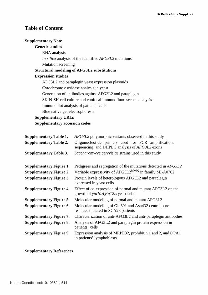
Table of Content
Supplementary Note
Genetic studies
RNA analysis
In silico analysis of the identified AFG3L2 mutations
Mutation screening
Structural modeling of AFG3L2 substitutions
Expression studies
AFG3L2 and paraplegin yeast expression plasmids
Cytochrome c oxidase analysis in yeast
Generation of antibodies against AFG3L2 and paraplegin
SK-N-SH cell culture and confocal immunofluorescence analysis
Immunoblot analysis of patients’ cells
Blue native gel electrophoresis
Supplementary URLs
Supplementary accession codes
Supplementary Table 1. AFG3L2 polymorphic variants observed in this study
Supplementary Table 2. Oligonucleotide primers used for PCR amplification, sequencing, and DHPLC analysis of AFG3L2 exons
Supplementary Table 3. Saccharomyces cerevisiae strains used in this study
Supplementary Figure 1. Pedigrees and segregation of the mutations detected in AFG3L2
Supplementary Figure 2. Variable expressivity of AFG3L2R702Q in family MI-A0762
Supplementary Figure 3. Protein levels of heterologous AFG3L2 and paraplegin expressed in yeast cells
Supplementary Figure 4. Effect of co-expression of normal and mutant AFG3L2 on the growth of yta10 yta12 yeast cells
Supplementary Figure 5. Molecular modeling of normal and mutant AFG3L2
Supplementary Figure 6. Molecular modeling of Glu691 and Asn432 central pore residues mutated in SCA28 patients
Supplementary Figure 7. Characterization of anti-AFG3L2 and anti-paraplegin antibodies
Supplementary Figure 8. Analysis of AFG3L2 and paraplegin protein expression in patients’ cells
Supplementary Figure 9. Expression analysis of MRPL32, prohibitin 1 and 2, and OPA1 in patients’ lymphoblasts
Supplementary References
Nature Genetics: doi:10.1038/ng.544

Di Bella et al. - Suppl. - 3
Supplementary Note
Genetic studies
RNA analysis
To verify that the mutations were present in the transcripts, when feasible, total RNA was
extracted from transformed lymphoblastoid cell lines derived from affected individuals using
the RNAeasy Mini Kit (Qiagen). cDNA synthesis was carried out using the Cloned AMV
First-Strand Synthesis Kit (Invitrogen) with an AFG3L2-specific primer. PCR primer pairs
were designed to amplify fragments spanning from exon 9 to exon 11 and exon 15 to exon 17.
In silico analysis of the identified AFG3L2 mutations
The four mutations in exon 16 cause the amino acid substitutions S674L, E691K, A694E, and
R702Q within the proteolytic domain, in a region that is highly conserved in m-AAA and m-
AAA-related proteins of multiple species, ranging from eubacteria to humans (Fig. 1). In
particular, conservation for Ser674, Arg702, Ala694, and Glu691 is 92%, 83%, 75%, and
66%, respectively. Notably, however, Glu691, Ala694, and Arg702 are conserved from the
ancestral eubacterial protein FtsH11. The N432T substitution, found in the sequence encoded
by exon 10, occurs in the ATPase (AAA) domain and also resides within an evolutionarily
highly conserved region. In particular, all m-AAA and m-AAA-related proteins from
eubacteria to humans exhibit an absolute conservation of a 5-amino acid motif (TLNQ)
encompassing AFG3L2 Asn432 (Fig. 1). By contrast, the H126Q substitution, caused by the
378C>G mutation in exon 4, occurs in the N-terminal part of the mature protein which
precedes the 1st transmembrane domain and protrudes into the matrix, a region of unknown
functional properties which exhibits high homology (100% identity between residue 113 and
residue 133) with rodent AFG3L2 but is not evolutionarily conserved in orthologs from more
distantly-related species (data not shown). Predictions on the effects of the substitutions on
protein function using the algorithms SIFT52 and PolyPhen53 did not give univocal results.
Both programs predicted the S674L, R702Q, and H126Q substitutions to be tolerated, and the
A694E substitution to affect protein function. By contrast, the E691K change was predicted to
be benign by PolyPhen but not tolerated by SIFT. Both programs predicted the N432T not to
be tolerated.
Nature Genetics: doi:10.1038/ng.544
Di Bella et al. - Suppl. - 4
Possible effects of the mutations on pre-mRNA splicing were assessed using the
algorithms ESEfinder54 (Release 3.0) for changes in exonic splicing enhancers (ESE) and
PESX55 for changes in putative exonic splicing silencers (PESS). None of the mutations is
predicted to change putative silencer sequences. No effect on ESE sequences is predicted for
the 4 mutations on exon 16. The 1296A>C mutation causing N432T is predicted to alter the
ESE pattern in exon 10 by abolishing one SF/ASF site and creating 3 novel ones. However,
RT-PCR analysis of AFG3L2 transcript in lymphoblastoid cells from patients carrying this
mutation or the mutations in exon 16 demonstrated equal dosage of normal and mutant
mRNA, with no evidence of aberrant splicing induced by the mutations. Two modest changes
were predicted for the 378C>G mutation (H126Q), namely, the mild attenuation of a putative
SRp40 ESE sequence along with the creation of a novel similar sequence with a just-above-
the-threshold score. Unfortunately, no mRNA source was available from the patient and the
effect of the 378C>G mutation (H126Q) on splicing could not be directly tested.
Mutation screening
For mutation screening of the selected patient population, PCR products were analyzed by
automated sequencing and/or Denaturing High-Performance Liquid Chromatography
(DHPLC)56,57. Sequences of the oligonucleotide primers and conditions used for PCR
amplification, DNA sequencing, and DHPLC analysis are detailed in Supplementary Table
2. For DHPLC analysis on the 3500HT WAVE® system (Transgenomic), melting profiles of
each PCR product sequence were predicted using the Navigator 6.4 software (Transgenomic).
For each exon, DHPLC mutation analysis was performed at at least two different
temperatures (Supplementary Table 2b), based on the melting profiles. PCR products
encompassing frequent polymorphic sites or PCR fragments not suitable for DHPLC analysis
were directly sequenced.
Structural modeling of AFG3L2 mutations
A three-dimensional model of AFG3L2 was built using the structure of the Th. thermophilus
(Tth.) AAA protease FtsH11,19 as a template (Fig. 5). This protein is a homohexamer (Fig. 5a)
which adopts a flat-cylinder-like shape divided into two disks (Fig. 5b). The lower disk,
containing the protease domain, forms a six-fold-symmetric structure with a Zn2+ binding site.
The upper disk is composed of six AAA+ domains, each of which contains ADP.
Nature Genetics: doi:10.1038/ng.544
Di Bella et al. - Suppl. - 5
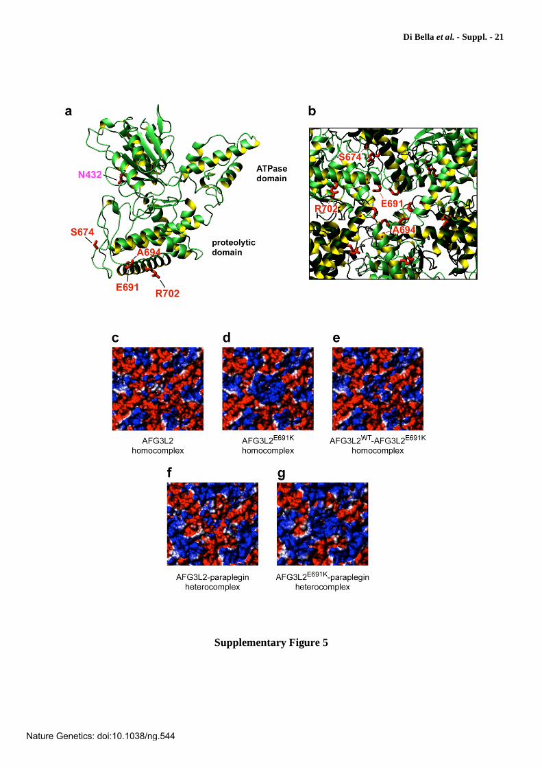
E691K. AFG3L2 Glu691 is also a glutamate (Glu537) in T. thermophilus FtsH19, but
is not conserved in other orthologs including paraplegin (Fig. 1). This residue is at the N-
terminus of helix 17 (Supplementary Figs. 5a and 6a) and sits in the middle of the central
pore formed by the six subunits surrounding the exit from the pore on the matrix side of the
proteolytic domain (Fig. 5a-b and Supplementary Figs. 5a-b and 6a). While overall exposed
and therefore not evidently contributing to subunit interactions or fold stability, this residue
could be relevant for protease specificity57. Substitution of this residue with a lysine as in
AFG3L2E691K could have severe consequences on protein function as it drastically changes
the electrostatic potential and the chemical characteristics of the pore (Fig. 5c-g and
Supplementary Fig. 5c-g). The change is evident in the AFG3L2WT-AFG3L2E691K compound
homohexamer (Supplementary Fig. 5e) but is greatest in both the homohexameric mutant
AFG3L2E691K-AFG3L2E691K (Supplementary Fig. 5d) and the heterohexameric
AFG3L2E691K-paraplegin (Supplementary Fig. 5g) in which the charge reversal of E691K is
not counteracted by the neutral residue (Gln693) that substitutes AFG3L2 Glu691 in
paraplegin.
A694E and R702Q, the other two AFG3L2 mutations which produce a variation in the
protein charge, appear to have smaller effects on the electrostatic potential of the protein
surface (not shown). Ala694 (Ala540 in FtsH) is also close to the pore and nonconserved.
Despite being rather superficial, its side chain points towards the pore inside and is overall
rather buried, therefore not influencing significantly the electrostatic potential. Substitution of
this alanine with a glutamate as in AFG3L2A694E requires burial of a charged group in the
protein interior which can be achieved only at a high energetic cost. We may therefore predict
that this mutation would have a strong effect on destabilizing the fold. Arg702 is Arg548 in
FtsH. This side chain packs with the next subunit, although it does not appear to establish
specific interactions with it. Also in this case, the effect of its replacement by a glutamine in
AFG3L2R702Q could be the destabilization of the assembly.
S674L. Ser674 is an alanine (A518) in FtsH. This residue is in a loop region between
16 and 9 and while it is exposed in the monomer (Supplementary Fig. 5a), it is buried in
the subunit interface. In FtsH, the side chain of A518 is sandwiched between the conserved
T498 and R494 of the adjacent subunit. While a serine can easily substitute the alanine,
mutation of this residue in the bulkier leucine as in AFG3L2S674L is expected to destabilize the
hexamer assembly.
Nature Genetics: doi:10.1038/ng.544
Di Bella et al. - Suppl. - 6
N432T. N432 is located within the ATPase domain in an evolutionarily conserved
region and is absolutely conserved from eubacterial Tth. FtsH (N280) to human AFG3L2
(Fig. 1). This residue, which lies in the middle of 7, between Walker B and SRH motifs, is
relatively exposed in the monomer (46 Å2 accessible surface area) (Supplementary Fig. 5a)
and is rather superficial also in the hexameric assembly (Fig. 5 and Supplementary Fig. 6b).
The side chain does not seem to form significant specific interactions with the surrounding.
Although replacement of Asn432 by threonine in AFG3L2N432T represents an exchange of
two amino acids of the same class (uncharged polar), the asparagine side chain could have an
important role in forming the required characteristics for substrate recognition and specificity.
In the hexameric assembly of T. thermophilus FtsH, the side chain of the corresponding
residue Asn280 is located in the pore and is near (~6 Å) the conserved Phe229 (Phe381 in
AFG3L2) of the alternate monomer (Supplementary Fig. 6b). Notably, this phenylalanine is
the crucial aromatic residue in the central pore loop motif @XG (pore-1 motif, where @ is an
aromatic residue and X is any residue) that is conserved in all subfamilies of the AAA
family20 and has been proposed to play an essential role for substrate recognition and ATP-
dependent translocation of proteins into the proteolytic chamber27,32.
Expression studies
AFG3L2 and paraplegin yeast expression plasmids
To generate yeast plasmids for heterologous expression of human AFG3L2, the sequence
encoding residues 35-797 of human AFG3L2 was amplified by PCR from a full-length
human AFG3L2 cDNA clone (clone IMAGp998I0513535Q1, RZPD Consortium), fused to
the sequence encoding the mitochondrial targeting peptide of Yta10p (amino acids 1-63), and
cloned into the BamHI/XbaI-digested low-copy-number centromeric vector pYC6/CT
(Invitrogen) which carries the blasticidin resistance gene (plasmid pYC6/CTGAL1-AFG3L2).
To obtain high constitutive levels of AFG3L2 expression under control of the ADH1
promoter, the glucose/galactose-regulated GAL1 promoter on pYC6/CT was substituted with
the 1-kb region upstream from the ADH1 translation initiation codon, amplified by PCR and
cloned into the SpeI/BamHI-digested pYC6/CTGAL1-AFG3L2 plasmid. This plasmid
[pYC6/CTADH1-Yta10p(1-63)-AFG3L2(35-797)-V5/HIS, abbreviated into pYC6/CTADH1-
AFG3L2-V5/HIS] was used in all expression experiments except when differently indicated.
For AFG3L2 expression under control of the weaker YTA10 promoter, plasmid
Nature Genetics: doi:10.1038/ng.544

Di Bella et al. - Suppl. - 7
pYC6/CTYTA10-AFG3L2-V5/HIS was likewise generated by replacing the pYC6/CT GAL1
promoter with a PCR fragment amplified from the 1-kb region upstream from the YTA10
translation initiation. To investigate dominance of the mutations, plasmid pYC2/CTADH1-
AFG3L2-V5/HIS carrying wild-type AFG3L2 and the URA3 gene for selection was
constructed by subcloning the SpeI/XbaI fragment [ADH1-Yta10p(1-63)-AFG3L2(35-797)]
from pYC6/CTADH1-AFG3L2-V5/HIS into the SpeI/XbaI-digested centromeric pYC2/CT
vector (Invitrogen).
The mutations causing the six substitutions identified in patients (AFG3L2H126Q,
AFG3L2S674L, AFG3L2E691K, AFG3L2A694E, AFG3L2R702Q, and AFG3L2N432T) or the control
proteolytic substitution AFG3L2E575Q (ref. 13) were introduced into the yeast AFG3L2
expression construct pYC6/CTADH1-AFG3L2-V5/HIS using the QuikChange XL Site-
Directed Mutagenesis Kit (Stratagene) and the following oligonucleotide primer pairs.
Mutagenized plasmids were verified by DNA sequencing.
Mutant Primer pairs (5'->3')
AFG3L2E575Q Forward
Reverse
CTGTGGCATACCACCAAGCAGGCCATGCGG
CCGCATGGCCTGCTTGGTGGTATGCCACAG
AFG3L2E691K Forward
Reverse
TATTGGAGAAACCTTACAGTAAAGCCACTGCAAGATTGATAGA
TCTATCAATCTTGCAGTGGCTTTACTGTAAGGTTTCTCCAATA
AFG3L2H126Q Forward
Reverse
GGCAAGAAAGTAGATTCTCAGTGGTGGTCCAGGTTTCAGAA
TTCTGAAACCTGGACCACCACTGAGAATCTACTTTCTTGCC
AFG3L2R702Q Forward
Reverse
GATGATGAAGTACAAATACTTATTAATGATG
CATCATTAATAAGTATTTGTACTTCATCATC
AFG3L2A694E Forward
Reverse
TTACAGTGAAGCCACTGAAAGATTGATAGATGATG
CATCATCTATCAATCTTTCAGTGGCTTCACTGTAA
AFG3L2S674L Forward
Reverse
GGTTGGGCAAATCTTATTTGACCTCCCACGTCAGG
CCTGACGTGGGAGGTCAAATAAGATTTGCCCAACC
AFG3L2N432T Forward
Reverse
GAGAACACACTCACCCAGCTGCTGGTGGAG
CTCCACCAGCAGCTGGGTGAGTGTGTTCTC
For human paraplegin expression, plasmid YCplac111ADH1-Yta10p(1-63)-
paraplegin(59-795)-HA (abbreviated into YCplac111ADH1-paraplegin-HA) was used, in which
the sequence corresponding to the mature form of paraplegin (amino acids 59-795) is tagged
Nature Genetics: doi:10.1038/ng.544
Di Bella et al. - Suppl. - 8
at the C-terminus with the HA epitope, fused to the Yta10p mitochondrial leader peptide
(amino acids 1-63), and expressed under control of the ADH1 promoter59.
Plasmids and strains generated in this study are available upon request following the
execution of an MTA agreement.
Cytochrome c oxidase analysis in yeast
For in-vitro activity assay of cytochrome c oxidase (COX) or immunoblot analysis of its
subunits, yeast cells were grown at 28°C in YEP medium supplemented with 2% (wt/vol)
galactose-0.1% (wt/vol) glucose45,46. Following differential centrifugation of the cells, the
resulting mitochondrial pellet was resuspended in 10-mM potassium phosphate buffer and
freezed and thawed for three times. COX activity was determined spectrophotometrically at
30˚C following for 2 minutes the decrease of absorbance at 550 nm because of oxidation of
cytochrome cred (refs. 46,60). Activity was expressed as nanomoles of cytochrome c
red
oxidized per minute per milligram of protein. Protein concentration was determined by
Bradford microplate microassay (Bio-Rad) with bovine serum albumin as the standard.
In yeast, COX is composed of 11 subunits, three of which (Cox1p, Cox2p, and
Cox3p) are encoded in the mitochondrial genome and form the catalytic core of the enzyme61.
Immunoblot analysis with antibodies directed against the three mitochondrial-encoded
subunits and one nuclear-encoded subunit (Cox4p) demonstrated multi-subunit deficiency,
with a drastic reduction (80 - >95%) of Cox1p, Cox2p, and Cox4p protein levels and a milder
decrease (40-75%) of Cox3p levels (Fig. 3b). Upon co-expression of paraplegin, COX
activity (Fig. 3c) was partially recovered in mutants AFG3L2S674L (51%), AFG3L2A694E
(61%), and AFG3L2R702Q (84%) but remained significantly low (P 0.01 n=4) in mutants
AFG3L2E691K (7%), AFG3L2N432T (25%), or the control mutant AFG3L2E575Q (ref. 13) (4%),
consistently with the observed respiratory phenotypes (Fig. 2d and e). As illustrated by
immunoblot analysis in Fig. 3d, COX subunit protein levels paralleled enzyme activity,
returning nearly normal in mutants AFG3L2S674L, AFG3L2A694E, and AFG3L2R702Q (Fig. 3d,
lanes 5, 7, and 8), but remaining significantly low (P 0.01 n=4) in mutants AFG3L2E691K,
AFG3L2N432T, or the control mutant AFG3L2E575Q (Fig. 3d, lanes 6, 9, and 3, respectively).
Generation of antibodies against AFG3L2 and paraplegin
To raise rabbit polyclonal antisera against human AFG3L2 and paraplegin, GST fusion
proteins were generated with protein fragments of AFG3L2 and paraplegin showing no
Nature Genetics: doi:10.1038/ng.544
Di Bella et al. - Suppl. - 9
homology to each other. The regions encoding amino acids 67-305 of AFG3L2 and amino
acids 89-304 of paraplegin were PCR amplified from IMAGE clones (clone
IMAGp998I0513535Q1 for AFG3L2 and clone IMAGp998D1211693Q1 for SPG7, RZPD
Consortium) and subcloned into the pGEX-6P-1 vector (Amersham Biosciences), in order to
generate GST-AFG3L2 and GST-paraplegin fusion proteins. Recombinant proteins were
expressed in Escherichia coli and purified by glutathione affinity chromatography. After
preparative SDS-PAGE and electroelution62, recovered proteins were injected into rabbits
(200-300 g of protein per injection). The anti-AFG3L2 and anti-paraplegin antisera collected
from the animals showed no cross-reaction when tested against each protein individually
expressed in yta10 yta12 yeast cells (Supplementary Fig. 7a) nor when used in Western
blot analysis of protein extracts from human cells (Supplementary Fig. 7b).
Antibodies generated in this study are available upon request following the execution
of an MTA agreement.
SK-N-SH cell culture and confocal immunofluorescence analysis
Human neuroblastoma SK-N-SH cells63 were grown in Dulbecco’s modified Eagle’s medium
(DMEM) supplemented with nonessential amino acids and 10% fetal bovine serum and plated
on coverslip in 12-well plates. Prior to immunofluorescence (Supplementary Fig. 7), cells
were fixed in 4% (wt/vol) paraformaldehyde for 5 minutes, repeatedly rinsed in phosphate-
buffered saline (PBS), and incubated overnight at 4°C with the primary antisera48 (rabbit
polyclonal anti-AFG3L2 or anti-paraplegin, 1:500 dilution). For sequential double-labeling,
cells were incubated for 2 hours with Alexa Fluor 546-conjugated goat anti-rabbit IgG
(Molecular Probes; 1:2,000 dilution), then with monoclonal anti-prohibitin-1 (PHB1)
antibody (Santa Cruz Biotechnology; 1:50 dilution) as mitochondrial marker, followed by
Alexa Fluor 488-conjugated goat anti-mouse secondary antibody (Molecular Probes; 1:2,000
dilution). Finally, cells were repeatedly rinsed and coverslipped with Fluorsave (Calbiochem).
Immunofluorescence images were acquired on a Radiance 2100 confocal microscope (Bio-
Rad) at 1,024 1,024-pixel resolution.
Immunoblot analysis of patients’ cells
Epstein-Barr-virus-stabilized lymphoblastoid cell lines from patients, their relatives and
control subjects were established and cultured as previously described42. For Western blot
analysis of patients’ lymphoblasts (Supplementary Figs. 8a and 9), 50-150 g of
Nature Genetics: doi:10.1038/ng.544
Di Bella et al. - Suppl. - 10
lymphoblastoid cell lysate from each line were electrophoresed on 10%-15%-SDS-
polyacrylamide gels and transferred to a nitrocellulose membrane (Amersham Biosciences)
by electroblotting. Filters were probed with polyclonal antibodies as indicated in the text and
in the figure legends, and developed by HRP-conjugated secondary antibodies using a
chemiluminescent substrate (ECL Kit, Amersham Biosciences) followed by autoradiography.
The signals were normalized by probing filters with antibodies directed against tubulin (Santa
Cruz Biotechnology) as a loading control protein.
Blue native gel electrophoresis
Blue native polyacrylamide gel electrophoresis (BNE) (Supplementary Fig. 8b) was carried
out essentially as described64. To obtain mitochondria-enriched extracts, lymphoblastoid cell
pellets were resuspended in a cold digitonin solution (20-mM MOPS, 250-mM sucrose, pH
7.4, 0.2-mg/ml digitonin), kept on ice for 10 min, and centrifuged at 600 g for 10 min at
4°C. Supernatants were centrifuged at 7000 g for 7 min at 4°C. The resulting pellets were
solubilized in NativePAGE™ Sample Buffer (Invitrogen) with 1% (wt/vol) digitonin, 1-mM
ATP, 5-mM -amino-n-caproic acid, and COMPLETE™ Protease Inhibitor Cocktail (Roche).
The solution was incubated on ice for 90 min and then centrifuged at 125,000 g for 30 min
at 4°C. After addition of NativePAGE™ 5%-G-250 Sample Additive (Invitrogen), samples
were loaded onto 3-12% NativePAGE™ Novex® Bis-Tris Gels (Invitrogen). Electrophoresis
was performed at 4°C according to the manufacturer’s protocol.
Supplementary URLs
SIFT: http://blocks.fhcrc.org/sift/SIFT.html
PolyPhen: http://genetics.bwh.harvard.edu/pph/
ESEfinder: http://rulai.cshl.edu/tools/ESE
PESX: http://cubweb.biology.columbia.edu/pesx/
Supplementary accession codes
GenBank (NCBI): Saccharomyces cerevisiae YTA10/Yta10p, NC_001137.2
(GI:7276232)/NP_010933.1 (GI:6320854); Saccharomyces cerevisiae ADH1, NC_001147.5
(GI:84626310).
Nature Genetics: doi:10.1038/ng.544
Di Bella et al. - Suppl. - 11
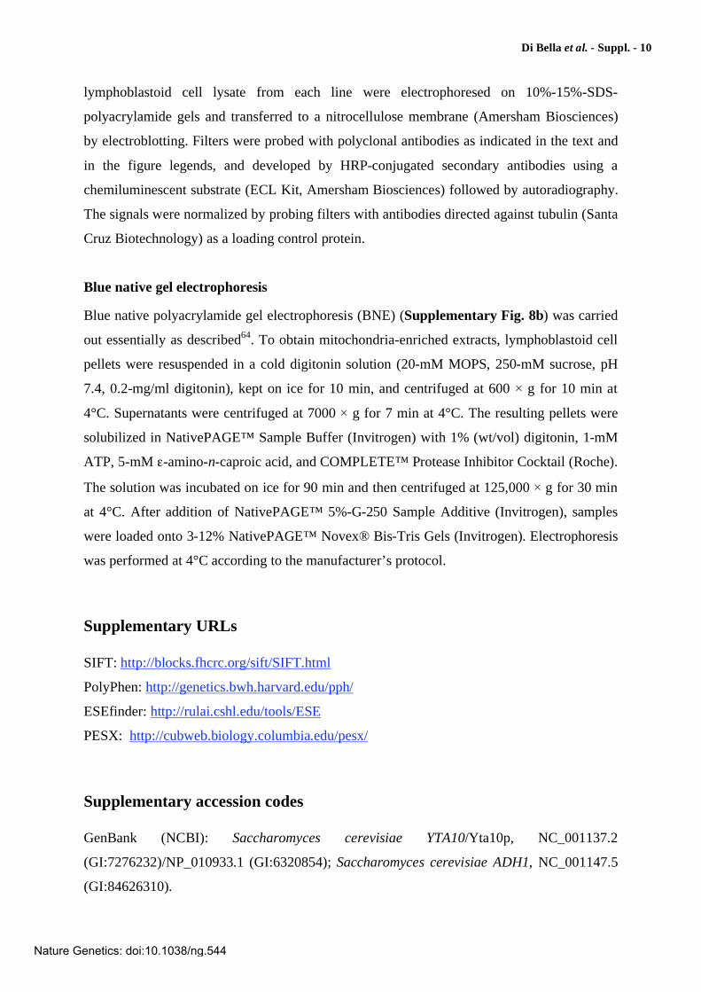
Supplementary Table 1. AFG3L2 polymorphic variants observed in this study
Nucleotide
changea
Amino acid
changeb
NCBI SNP Reference
Cluster IDc
Allele frequency (%)
(n=300)
-96G>C rs12327346 G=97.4; C=2.6
293-61A>G rs8093375 ndd
400-95G>A rs2298542 ndd
400-14C>G not reported C=99.4; G=0.6
752+6C>T rs8097342 C=18.4; T=81.6
753-55T>C rs7407640 ndd
1026+8G>A rs8091858 G=93.9; A=6.1
1165-21T>A rs9966470 ndd
1319-59G>T not reported G=99.7; T=0.3
1319-55T>G not reported T=99.1; G=0.9
1389G>A L463L rs11080572 G=32; A=68
1650A>G E550E not reported A=18; G=82
1664-39G>A not reported G=98.1; A=1.9
1664-9T>C not reported T=99.7; C=0.3
2394G>C rs1129115 ndd
aNucleotide numbering refers to the AFG3L2 cDNA [GenBank accession No. NM_006796.1 (GI:5802969)].
Nucleotides are numbered so that the first nucleotide (nt) of the first in-frame ATG codon is nucleotide +1. bAmino acids are numbered so that methionine encoded by the first in-frame ATG codon is Met1. c http://www.ncbi.nlm.nih.gov/projects/SNP/. dnd, not determined.
Nature Genetics: doi:10.1038/ng.544
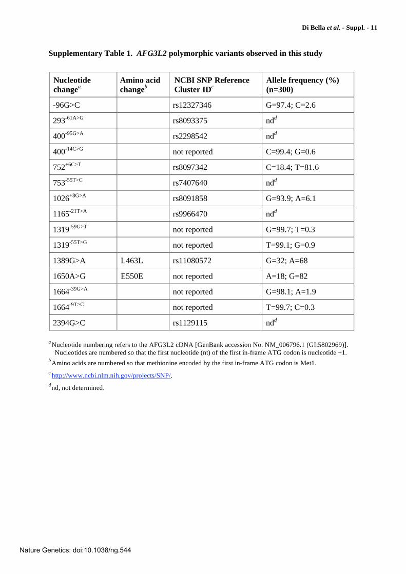
Di Bella et al. - Suppl. - 12
Supplementary Table 2. Oligonucleotide primers used for PCR amplification, DNA
sequencing, and DHPLC analysis of AFG3L2 exons
2a. DNA sequence analysis
Exon
amplicons Primer pairs (5'->3')
PCR annealing
temperaturea
1 Forward Reverse
TTGAGAGCTTGGGCTCCT GTCATCTCGGCCCAAAAG
57°C
2-3 Forward Reverse
TTATGACCAGGAAATGAAGC CTTTGTTCAGTGGAAACTACC
56°C
4-5 Forward Reverse
AGCCTCCCTGATTGGTAAG GCTGACTGTCACTTCTTTGGT
58°C
6 Forward Reverse
TGGGGGCATCTTTATCTG AGGCAGGTTTTCCTTTCAG
58°C
7 Forward Reverse
AATGAGTGACATTTAATCACC GGACAGAACACAGTGAACC
57°C
8 Forward Reverse
GCCTTTGAAGAACACTTGC TGACCCAAAACGATCCTC
56°C
9 Forward Reverse
AATGTTCTACCATAGCTCAGATG AGCACTCTAGGGGGAAGG
57°C
10 Forward Reverse
GGCCGATTTATTTCATTTCT CCGAAACACACCACTCA
56°C
11-12 Forward Reverse
GCTATGAATTTGCAGTGCTC AGGAAGCCCACAGTAAACAA
56°C
13 Forward Reverse
ACTATGGATTTGGCTGTCC TGGATACACTTTCTTTGCTTCT
57°C
14 Forward Reverse
TTGTGATAGGCAGCTCAGTC CTTTGCAGGAGTGTAGCTTG
58°C
15 Forward Reverse
CCACTAAGGCTGATGAACT TCCTTGCCTAAAAAGCCTAA
57°C
16 Forward Reverse
TGGGATTTGCGTCCTAAC GCAGACAACGAAACATCAGAAC
59°C
17 Forward Reverse
TGGGGTCACCTGTAAATAAAA TCCTGTAGAAAACCATTCCA
56°C
aPCR conditions included an initial denaturation step at 95°C for 3 min, followed by 35 cycles of
denaturation at 94°C for 1 min, annealing for 45 s at the temperature indicated in the table, and extension at 72°C for 1 min, with a final extension step at 72°C for 10 min.
Nature Genetics: doi:10.1038/ng.544
Di Bella et al. - Suppl. - 13
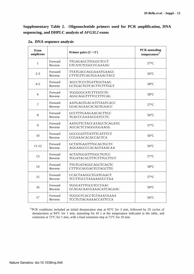
2b. DHPLC analysis
Exon
amplicons Primer pairs (5'->3')
PCR annealing
temperaturea
DHPLC
analysis
temperatures
4 Forward Reverse
GCTGAGAGAGCTAAAACCTTGC AATGCCTCCCAACCTTCTCT
55°C 58.8°C 60°C 62°C
9 Forward Reverse
AATGTTCTACCATAGCTCAGATG AGCACTCTAGGGGGAAGG
57°C 53°C 61°C 62°C
10 Forward Reverse
GGCCGATTTATTTCATTTCT GCAGTTAAAGATACAAAAGC
49°C 60°C 61°C
61.5°C
14 Forward Reverse
TTGTGATAGGCAGCTCAGTC CTTTGCAGGAGTGTAGCTTG
58°C 57.1°C 61.2°C 62.5°C
15 Forward Reverse
CCACTAAGGCTGATGAACT TCCTTGCCTAAAAAGCCTAA
57°C 56.3°C 57°C
16 Forward Reverse
TTGTCTGGTTAAAGAACAATCA AACTGTAAAGAATTATTCCCACAA
55°C 57°C
57.4°C 58.5°C
17 Forward Reverse
TGGGGTCACCTGTAAATAAAA GACTGAGATGGCCTCCCT
52°C 54.6°C 58.2°C 61°C
aPCR conditions included an initial denaturation step at 95°C for 3 min, followed by 35 cycles of
denaturation at 94°C for 1 min, annealing for 45 s at the temperature indicated in the table, and extension at 72°C for 1 min, with a final extension step at 72°C for 10 min.
Nature Genetics: doi:10.1038/ng.544
Di Bella et al. - Suppl. - 14
Supplementary Table 3. Saccharomyces cerevisiae strains used in this study
Strain Relevant genotypea Source
K699 (source: W303)
MATa ade2-1 trp1-1 can1-100 leu2-3,112 his3-11,15 ura3-52 Refs. 43,65
yDDB64b K699 yta10::NAT yta12::KanMX6 This study
yDDB79 yDDB64 (pYC6/CTADH1-AFG3L2-V5/HIS) This study
yDDB94 yDDB64 (pYC6/CTYTA10-AFG3L2-V5/HIS) This study
yDDB111b yDDB64 (pYC6/CTYTA10-AFG3L2-V5/HIS) (YCplac111YTA10-paraplegin-HA) This study
yDDB122 yDDB64 (pYC6/CTADH1-AFG3L2H126Q-V5/HIS) This study
yDDB123 yDDB64 (pYC6/CTADH1-AFG3L2E691K-V5/HIS) This study
yDDB124 yDDB64 (pYC6/CTADH1-AFG3L2A694E-V5/HIS) This study
yDDB125 yDDB64 (pYC6/CTADH1-AFG3L2R702Q-V5/HIS) This study
yDDB126 yDDB64 (pYC6/CTADH1-AFG3L2S674L-V5/HIS) This study
yDDB158 yDDB64 (pYC6/CTADH1-AFG3L2N432T-V5/HIS) This study
yDDB127 yDDB64 (pYC6/CTADH1-AFG3L2E575Q-V5/HIS) This study
yDDB109 yDDB64 (pYC2/CTADH1-AFG3L2-V5/HIS) This study
yDDB190 yDDB64 (pYC6/CTADH1-AFG3L2E691K-V5/HIS) (pYC2/CTADH1-AFG3L2-V5/HIS) This study
yDDB191 yDDB64 (pYC6/CTADH1-AFG3L2A694E-V5/HIS) (pYC2/CTADH1-AFG3L2-V5/HIS) This study
yDDB192 yDDB64 (pYC6/CTADH1-AFG3L2R702Q-V5/HIS) (pYC2/CTADH1-AFG3L2-V5/HIS) This study
yDDB189 yDDB64 (pYC6/CTADH1-AFG3L2S674L-V5/HIS) (pYC2/CTADH1-AFG3L2-V5/HIS) This study
yDDB201 yDDB64 (pYC6/CTADH1-AFG3L2N432T-V5/HIS) (pYC2/CTADH1-AFG3L2-V5/HIS) This study
yDDB138 yDDB64 (YCplac111ADH1-paraplegin-HA) This study
yDDB165 yDDB64 (pYC6/CTADH1-AFG3L2-V5/HIS) (YCplac111ADH1-paraplegin-HA) This study
yDDB174 yDDB64 (pYC6/CTADH1-AFG3L2R702Q-V5/HIS) (YCplac111ADH1-paraplegin-HA) This study
yDDB75b yDDB64 (pYC6/CTADH1-AFG3L2H126Q-V5/HIS) (YCplac111ADH1-paraplegin-HA) This study
yDDB200 yDDB64 (pYC6/CTADH1-AFG3L2A694E-V5/HIS) (YCplac111ADH1-paraplegin-HA) This study
yDDB166 yDDB64 (pYC6/CTADH1-AFG3L2E691K-V5/HIS) (YCplac111ADH1-paraplegin-HA) This study
yDDB129 yDDB64 (pYC6/CTADH1-AFG3L2E575Q-V5/HIS) (YCplac111ADH1-paraplegin-HA) This study
yDDB167 yDDB64 (pYC6/CTADH1-AFG3L2S674L-V5/HIS) (YCplac111ADH1-paraplegin-HA) This study
yDDB175 yDDB64 (pYC6/CTADH1-AFG3L2N432T-V5/HIS) (YCplac111ADH1-paraplegin-HA) This study
a
See Supplementary Note for plasmid description.
byta10 yta12 parental strain generated using the one-step PCR strategy (refs. 43,66).
Nature Genetics: doi:10.1038/ng.544
Di Bella et al. - Suppl. - 15
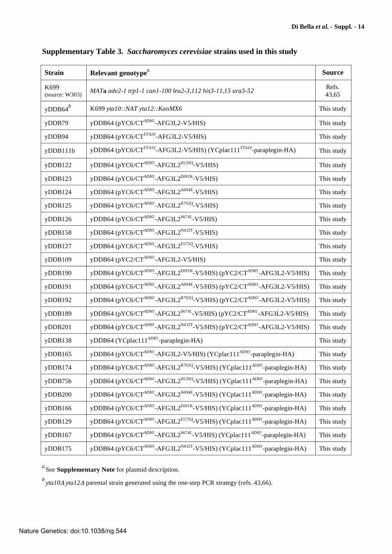
Supplementary Figure 1
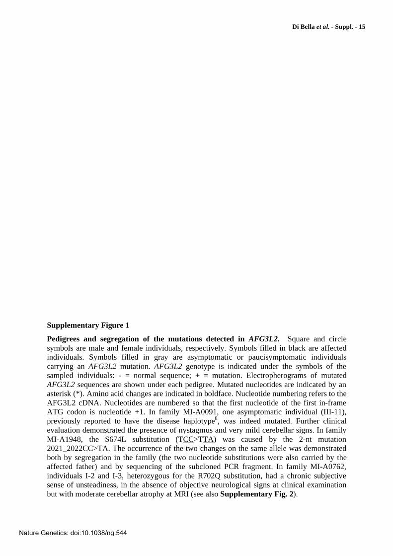
Pedigrees and segregation of the mutations detected in AFG3L2. Square and circle symbols are male and female individuals, respectively. Symbols filled in black are affected individuals. Symbols filled in gray are asymptomatic or paucisymptomatic individuals carrying an AFG3L2 mutation. AFG3L2 genotype is indicated under the symbols of the sampled individuals: - = normal sequence; + = mutation. Electropherograms of mutated AFG3L2 sequences are shown under each pedigree. Mutated nucleotides are indicated by an asterisk (*). Amino acid changes are indicated in boldface. Nucleotide numbering refers to the AFG3L2 cDNA. Nucleotides are numbered so that the first nucleotide of the first in-frame ATG codon is nucleotide +1. In family MI-A0091, one asymptomatic individual (III-11), previously reported to have the disease haplotype8, was indeed mutated. Further clinical evaluation demonstrated the presence of nystagmus and very mild cerebellar signs. In family MI-A1948, the S674L substitution (TCC>TTA) was caused by the 2-nt mutation 2021_2022CC>TA. The occurrence of the two changes on the same allele was demonstrated both by segregation in the family (the two nucleotide substitutions were also carried by the affected father) and by sequencing of the subcloned PCR fragment. In family MI-A0762, individuals I-2 and I-3, heterozygous for the R702Q substitution, had a chronic subjective sense of unsteadiness, in the absence of objective neurological signs at clinical examination but with moderate cerebellar atrophy at MRI (see also Supplementary Fig. 2).
Nature Genetics: doi:10.1038/ng.544
Di Bella et al. - Suppl. - 16
Supplementary Figure 1
Nature Genetics: doi:10.1038/ng.544
Di Bella et al. - Suppl. - 17
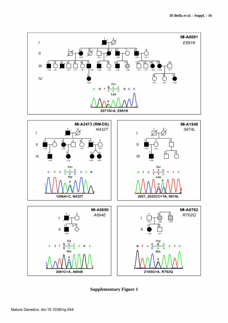
Supplementary Figure 2
Variable expressivity of AFG3L2R702Q
in family MI-A0762. Pedigree of family MI-A0762 (see also Supplementary Fig. 1) showing segregation of the R702Q substitution. Symbols are as in Supplementary Fig. 1. AFG3L2 genotype is indicated under the symbols of the tested individuals (- = normal sequence; + = mutated sequence). The index case (II-1) is a 40-year-old woman with a full-blown cerebellar phenotype that manifested at 28 years of age with progressive gait and limb ataxia. She now presents severe ataxia and dysarthria, ophthalmoplegia, and pyramidal signs with increased muscle tone, brisk reflexes, and Babinski sign. MRI shows the presence of marked atrophy of the vermis and the cerebellar hemispheres. Her 78-years-old father, who does not carry the AFG3L2R702Q substitution, is completely asymptomatic and does not exhibit any clinical sign at neurological examination. MRI is negative (not shown). AFG3L2R702Q is carried in heterozygous form by the mother (I-2, 76 years old) and the maternal uncle (I-3, 74 years old). Both are negative at neurological examination, exhibiting none of the clinical signs observed in the index case II-1. In particular, there are no abnormalities of gait and speech, and no signs of corticospinal involvement. Despite negative neurological examination, though, both report to have been suffering of a chronic subjective sense of unsteadiness since many years. Interestingly, in both subjects, MRI shows the presence of a moderate cerebellar atrophy in comparison to age-matched controls.
Nature Genetics: doi:10.1038/ng.544
Di Bella et al. - Suppl. - 18
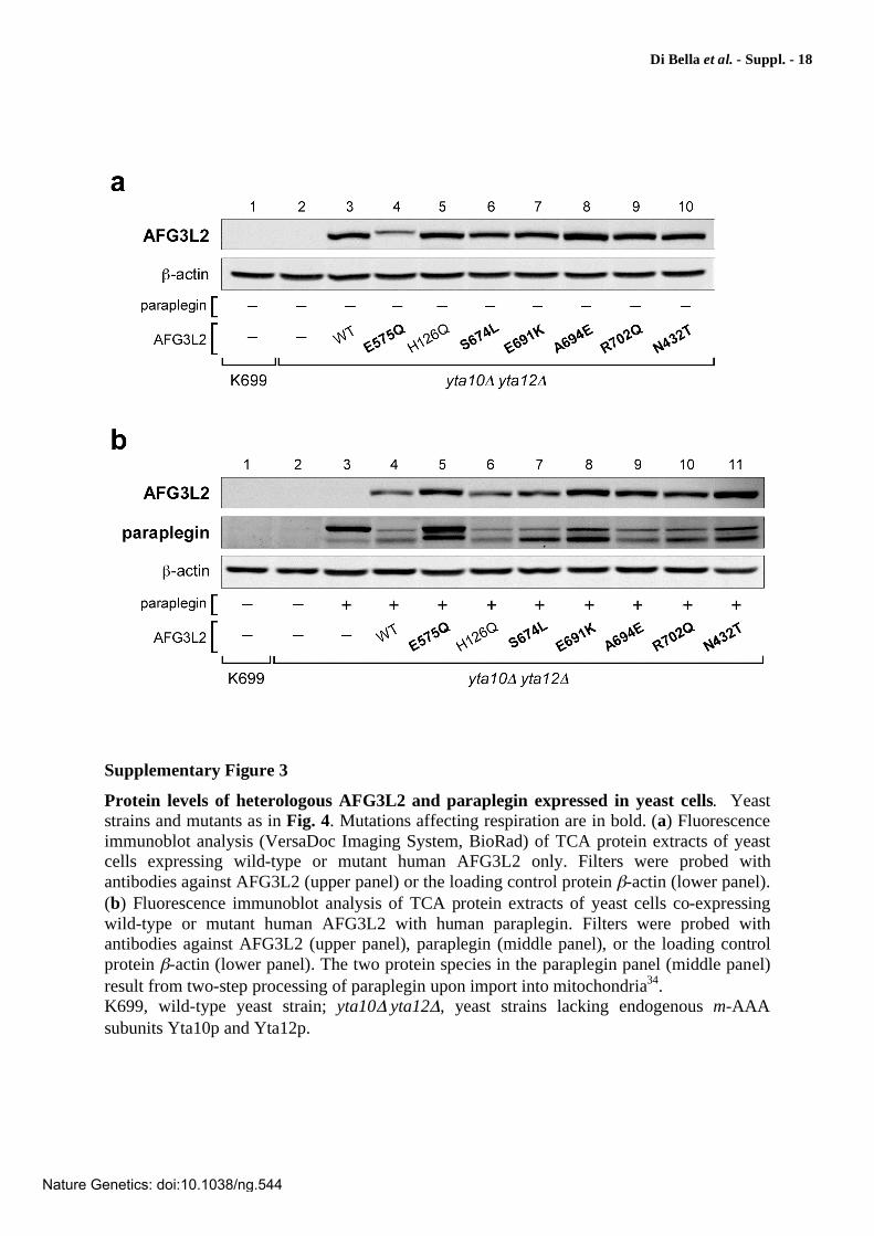
Supplementary Figure 3
Protein levels of heterologous AFG3L2 and paraplegin expressed in yeast cells. Yeast strains and mutants as in Fig. 4. Mutations affecting respiration are in bold. (a) Fluorescence immunoblot analysis (VersaDoc Imaging System, BioRad) of TCA protein extracts of yeast cells expressing wild-type or mutant human AFG3L2 only. Filters were probed with antibodies against AFG3L2 (upper panel) or the loading control protein -actin (lower panel). (b) Fluorescence immunoblot analysis of TCA protein extracts of yeast cells co-expressing wild-type or mutant human AFG3L2 with human paraplegin. Filters were probed with antibodies against AFG3L2 (upper panel), paraplegin (middle panel), or the loading control protein -actin (lower panel). The two protein species in the paraplegin panel (middle panel) result from two-step processing of paraplegin upon import into mitochondria34. K699, wild-type yeast strain; yta10 yta12 , yeast strains lacking endogenous m-AAA subunits Yta10p and Yta12p.
Nature Genetics: doi:10.1038/ng.544
Di Bella et al. - Suppl. - 19
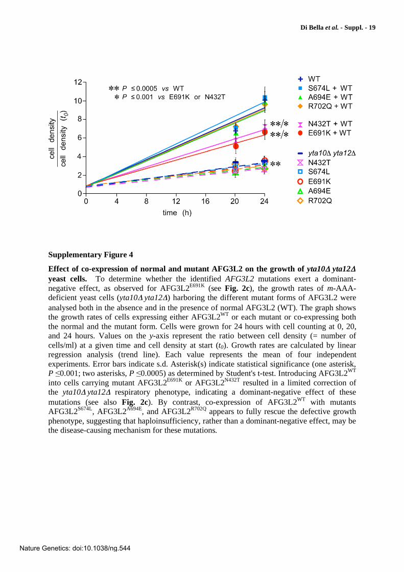
Supplementary Figure 4
Effect of co-expression of normal and mutant AFG3L2 on the growth of yta10 yta12
yeast cells. To determine whether the identified AFG3L2 mutations exert a dominant-negative effect, as observed for AFG3L2E691K (see Fig. 2c), the growth rates of m-AAA-deficient yeast cells (yta10 yta12 ) harboring the different mutant forms of AFG3L2 were analysed both in the absence and in the presence of normal AFG3L2 (WT). The graph shows the growth rates of cells expressing either AFG3L2WT or each mutant or co-expressing both the normal and the mutant form. Cells were grown for 24 hours with cell counting at 0, 20, and 24 hours. Values on the y-axis represent the ratio between cell density (= number of cells/ml) at a given time and cell density at start (t0). Growth rates are calculated by linear regression analysis (trend line). Each value represents the mean of four independent experiments. Error bars indicate s.d. Asterisk(s) indicate statistical significance (one asterisk, P 0.001; two asterisks, P 0.0005) as determined by Student's t-test. Introducing AFG3L2WT into cells carrying mutant AFG3L2E691K or AFG3L2N432T resulted in a limited correction of the yta10 yta12 respiratory phenotype, indicating a dominant-negative effect of these mutations (see also Fig. 2c). By contrast, co-expression of AFG3L2WT with mutants AFG3L2S674L, AFG3L2A694E, and AFG3L2R702Q appears to fully rescue the defective growth phenotype, suggesting that haploinsufficiency, rather than a dominant-negative effect, may be the disease-causing mechanism for these mutations.
Nature Genetics: doi:10.1038/ng.544
Di Bella et al. - Suppl. - 20
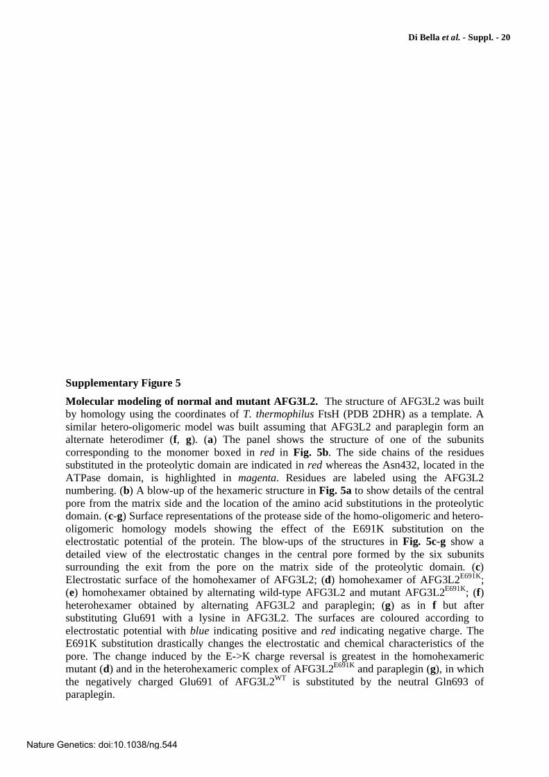
Supplementary Figure 5
Molecular modeling of normal and mutant AFG3L2. The structure of AFG3L2 was built by homology using the coordinates of T. thermophilus FtsH (PDB 2DHR) as a template. A similar hetero-oligomeric model was built assuming that AFG3L2 and paraplegin form an alternate heterodimer (f, g). (a) The panel shows the structure of one of the subunits corresponding to the monomer boxed in red in Fig. 5b. The side chains of the residues substituted in the proteolytic domain are indicated in red whereas the Asn432, located in the ATPase domain, is highlighted in magenta. Residues are labeled using the AFG3L2 numbering. (b) A blow-up of the hexameric structure in Fig. 5a to show details of the central pore from the matrix side and the location of the amino acid substitutions in the proteolytic domain. (c-g) Surface representations of the protease side of the homo-oligomeric and hetero-oligomeric homology models showing the effect of the E691K substitution on the electrostatic potential of the protein. The blow-ups of the structures in Fig. 5c-g show a detailed view of the electrostatic changes in the central pore formed by the six subunits surrounding the exit from the pore on the matrix side of the proteolytic domain. (c) Electrostatic surface of the homohexamer of AFG3L2; (d) homohexamer of AFG3L2E691K; (e) homohexamer obtained by alternating wild-type AFG3L2 and mutant AFG3L2E691K; (f) heterohexamer obtained by alternating AFG3L2 and paraplegin; (g) as in f but after substituting Glu691 with a lysine in AFG3L2. The surfaces are coloured according to electrostatic potential with blue indicating positive and red indicating negative charge. The E691K substitution drastically changes the electrostatic and chemical characteristics of the pore. The change induced by the E->K charge reversal is greatest in the homohexameric mutant (d) and in the heterohexameric complex of AFG3L2E691K and paraplegin (g), in which the negatively charged Glu691 of AFG3L2WT is substituted by the neutral Gln693 of paraplegin.
Nature Genetics: doi:10.1038/ng.544
Di Bella et al. - Suppl. - 21
Supplementary Figure 5
Nature Genetics: doi:10.1038/ng.544
Di Bella et al. - Suppl. - 22
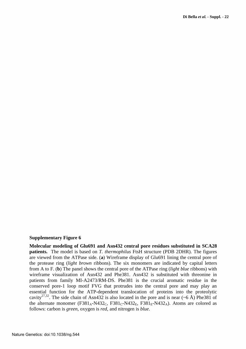
Supplementary Figure 6
Molecular modeling of Glu691 and Asn432 central pore residues substituted in SCA28
patients. The model is based on T. thermophilus FtsH structure (PDB 2DHR). The figures are viewed from the ATPase side. (a) Wireframe display of Glu691 lining the central pore of the protease ring (light brown ribbons). The six monomers are indicated by capital letters from A to F. (b) The panel shows the central pore of the ATPase ring (light blue ribbons) with wireframe visualization of Asn432 and Phe381. Asn432 is substituted with threonine in patients from family MI-A2473/RM-DS. Phe381 is the crucial aromatic residue in the conserved pore-1 loop motif FVG that protrudes into the central pore and may play an essential function for the ATP-dependent translocation of proteins into the proteolytic cavity27,32. The side chain of Asn432 is also located in the pore and is near (~6 Å) Phe381 of the alternate monomer (F381A-N432C, F381C-N432E, F381E-N432A). Atoms are colored as follows: carbon is green, oxygen is red, and nitrogen is blue.
Nature Genetics: doi:10.1038/ng.544
Di Bella et al. - Suppl. - 23
Supplementary Figure 6
Nature Genetics: doi:10.1038/ng.544
Di Bella et al. - Suppl. - 24
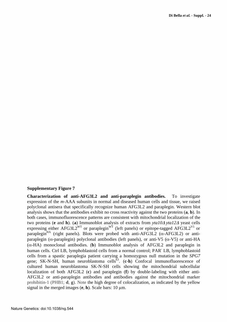
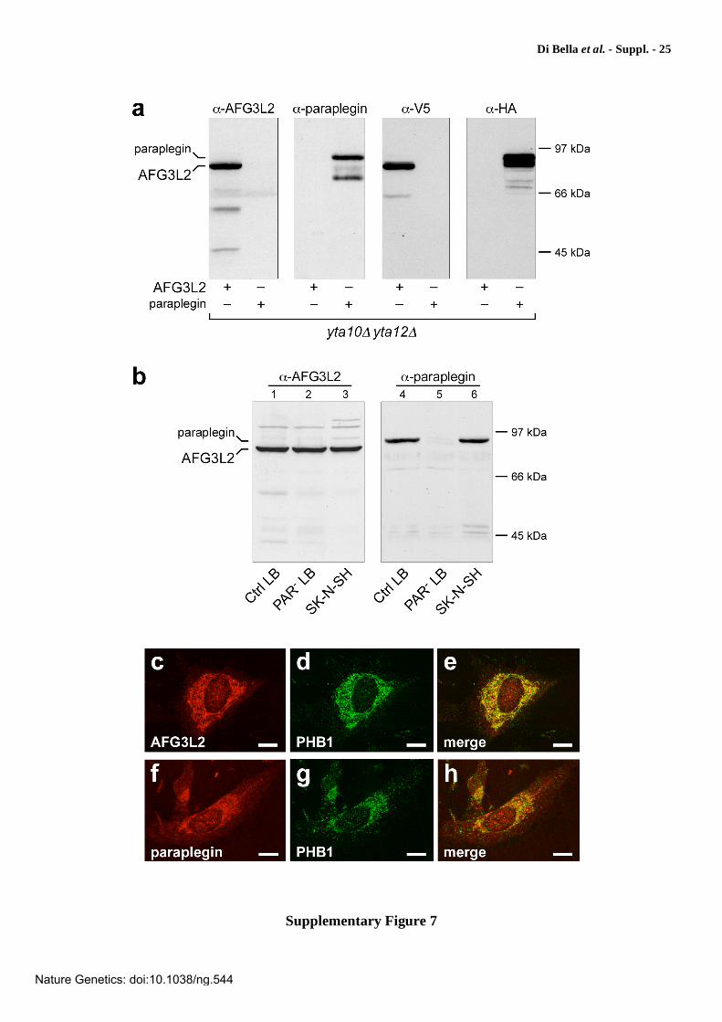
Supplementary Figure 7
Characterization of anti-AFG3L2 and anti-paraplegin antibodies. To investigate expression of the m-AAA subunits in normal and diseased human cells and tissue, we raised polyclonal antisera that specifically recognize human AFG3L2 and paraplegin. Western blot analysis shows that the antibodies exhibit no cross reactivity against the two proteins (a, b). In both cases, immunofluorescence patterns are consistent with mitochondrial localization of the two proteins (e and h). (a) Immunoblot analysis of extracts from yta10 yta12 yeast cells expressing either AFG3L2WT or parapleginWT (left panels) or epitope-tagged AFG3L2V5 or parapleginHA (right panels). Blots were probed with anti-AFG3L2 ( -AFG3L2) or anti-paraplegin ( -paraplegin) polyclonal antibodies (left panels), or anti-V5 ( -V5) or anti-HA ( -HA) monoclonal antibodies. (b) Immunoblot analysis of AFG3L2 and paraplegin in human cells. Ctrl LB, lymphoblastoid cells from a normal control; PAR- LB, lymphoblastoid cells from a spastic paraplegia patient carrying a homozygous null mutation in the SPG7 gene; SK-N-SH, human neuroblastoma cells63. (c-h) Confocal immunofluorescence of cultured human neuroblastoma SK-N-SH cells showing the mitochondrial subcellular localization of both AFG3L2 (c) and paraplegin (f) by double-labeling with either anti-AFG3L2 or anti-paraplegin antibodies and antibodies against the mitochondrial marker prohibitin-1 (PHB1; d, g). Note the high degree of colocalization, as indicated by the yellow signal in the merged images (e, h). Scale bars: 10 m.
Nature Genetics: doi:10.1038/ng.544
Di Bella et al. - Suppl. - 25
Supplementary Figure 7
Nature Genetics: doi:10.1038/ng.544
Di Bella et al. - Suppl. - 26
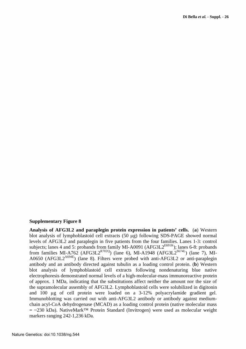
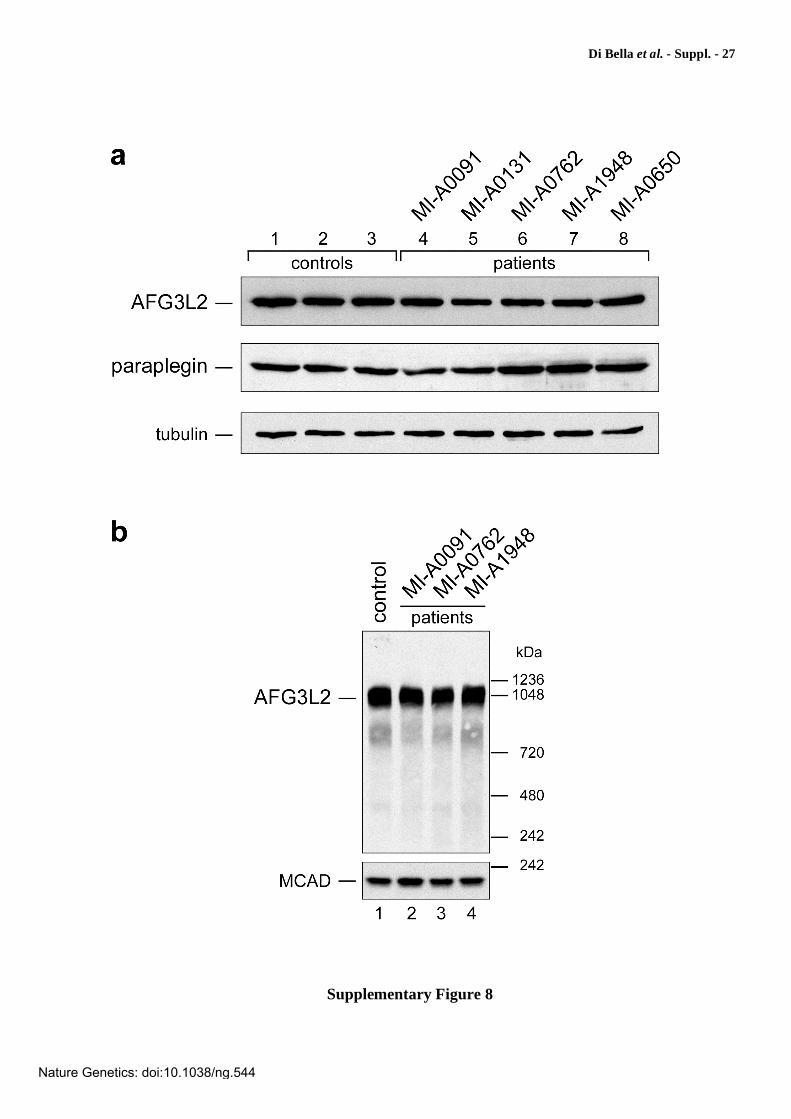
Supplementary Figure 8
Analysis of AFG3L2 and paraplegin protein expression in patients’ cells. (a) Western blot analysis of lymphoblastoid cell extracts (50 g) following SDS-PAGE showed normal levels of AFG3L2 and paraplegin in five patients from the four families. Lanes 1-3: control subjects; lanes 4 and 5: probands from family MI-A0091 (AFG3L2E691K); lanes 6-8: probands from families MI-A762 (AFG3L2R702Q) (lane 6), MI-A1948 (AFG3L2S674L) (lane 7), MI-A0650 (AFG3L2A694E) (lane 8). Filters were probed with anti-AFG3L2 or anti-paraplegin antibody and an antibody directed against tubulin as a loading control protein. (b) Western blot analysis of lymphoblastoid cell extracts following nondenaturing blue native electrophoresis demonstrated normal levels of a high-molecular-mass immunoreactive protein of approx. 1 MDa, indicating that the substitutions affect neither the amount nor the size of the supramolecular assembly of AFG3L2. Lymphoblastoid cells were solubilized in digitonin and 100 g of cell protein were loaded on a 3-12% polyacrylamide gradient gel. Immunoblotting was carried out with anti-AFG3L2 antibody or antibody against medium-chain acyl-CoA dehydrogenase (MCAD) as a loading control protein (native molecular mass = ~230 kDa). NativeMark™ Protein Standard (Invitrogen) were used as molecular weight markers ranging 242-1,236 kDa.
Nature Genetics: doi:10.1038/ng.544
Di Bella et al. - Suppl. - 27
Supplementary Figure 8
Nature Genetics: doi:10.1038/ng.544
Di Bella et al. - Suppl. - 28
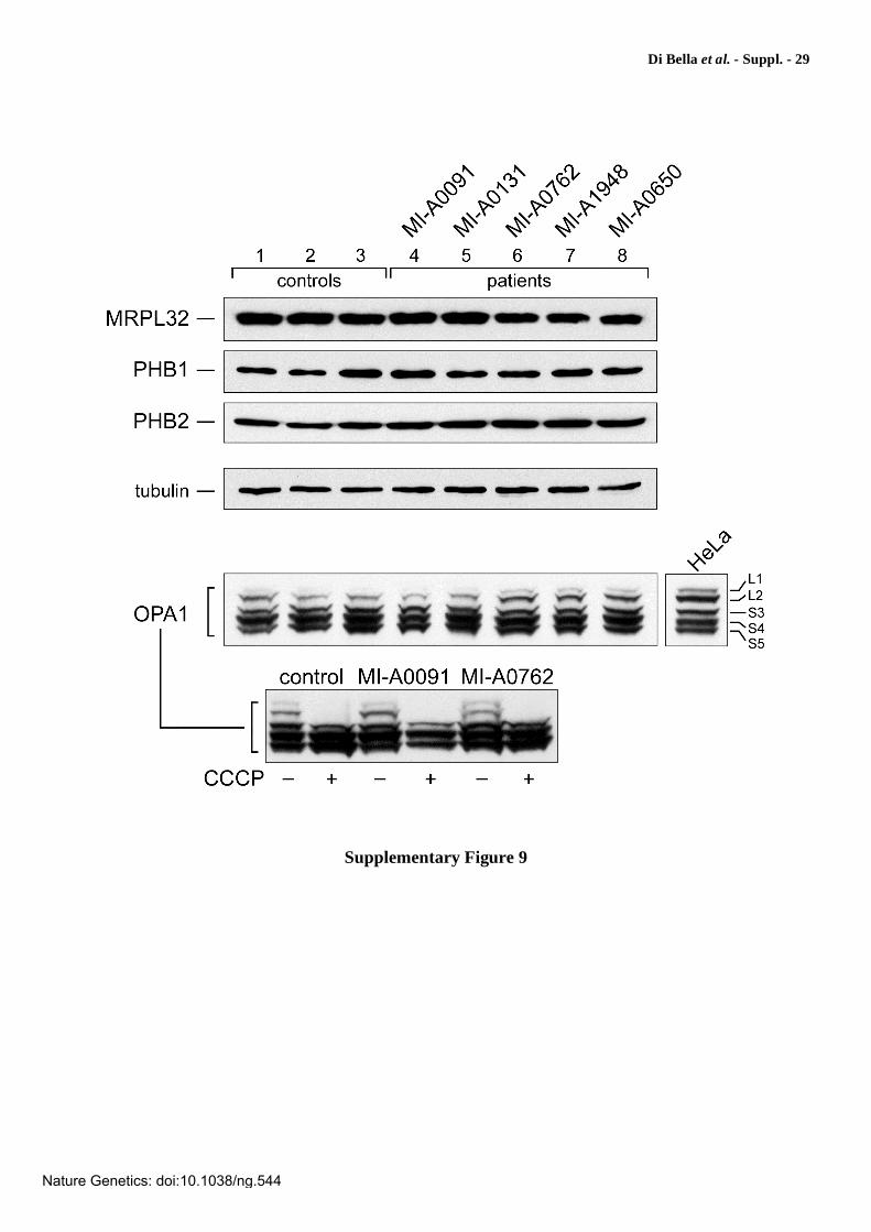
Supplementary Figure 9
Expression analysis of MRPL32, prohibitin 1 and 2, and OPA1 in patients’
lymphoblasts. To examine whether mutations affecting AFG3L2 could induce secondary abnormalities of other proteins known for being either partners or substrates of the m-AAA complex, we investigated the expression of prohibitin 1 (PHB1) and 2 (PHB2), MRPL32, and OPA1, observing no differences both in the protein levels and in the migration pattern as compared to normal controls. MRPL32 is a subunit of human mitochondrial ribosomes, homolog of yeast MrpL32, a previously reported substrate of m-AAA (ref. 16); prohibitin 1 (PHB1) and 2 (PHB2) have been shown to form ring-shaped assemblies that associate with m-AAA in a supercomplex of ~1.2 MDa and modulate m-AAA proteolytic activity12; OPA1, a dynamin-like GTPase that causes human dominant optic atrophy and functions in mitochondrial fusion and inner membrane remodeling, has been recently proposed to be regulated by the m-AAA protease22,67,68. Cell extracts were subjected to Western blotting with the antibody indicated. HeLa cell extracts were used as a control for OPA1 processing. Expression of eight OPA1 splice variants and proteolytic processing leads to the formation of at least five different isoforms of OPA1, two long forms designated L1 and L2, which can be proteolytically converted into three short forms, designated S3-S567,68. Dissipation of mitochondrial membrane potential, as that caused by the uncoupler carbonyl cyanide 3-chlorophenylhydrazone (CCCP), stimulates OPA1 processing67,68 and may thereby reveal impairment of processing, if any.
Nature Genetics: doi:10.1038/ng.544
Di Bella et al. - Suppl. - 29
Supplementary Figure 9
Nature Genetics: doi:10.1038/ng.544
Di Bella et al. - Suppl. - 30
Supplementary References
52. Ng, P. C. & Henikoff, S. SIFT: Predicting amino acid changes that affect protein function. Nucleic Acids Res 31, 3812-3814 (2003).
53. Ramensky, V., Bork, P. & Sunyaev, S. Human non-synonymous SNPs: server and survey. Nucleic Acids Res 30, 3894-3900 (2002).
54. Cartegni, L., Wang, J., Zhu, Z., Zhang, M. Q. & Krainer, A. R. ESEfinder: A web resource to identify exonic splicing enhancers. Nucleic Acids Res 31, 3568-3571 (2003).
55. Zhang, X. H. & Chasin, L. A. Computational definition of sequence motifs governing constitutive exon splicing. Genes Dev 18, 1241-1250 (2004).
56. Xiao, W. & Oefner, P. J. Denaturing high-performance liquid chromatography: A review. Hum Mutat 17, 439-474 (2001).
57. Takashima, H., Boerkoel, C. F. & Lupski, J. R. Screening for mutations in a genetically heterogeneous disorder: DHPLC versus DNA sequence for mutation detection in multiple genes causing Charcot-Marie-Tooth neuropathy. Genet Med 3, 335-342 (2001).
58. Bieniossek, C. et al. The molecular architecture of the metalloprotease FtsH. Proc Natl
Acad Sci U S A 103, 3066-3071 (2006).
59. Atorino, L. et al. Loss of m-AAA protease in mitochondria causes complex I deficiency and increased sensitivity to oxidative stress in hereditary spastic paraplegia. J Cell Biol 163, 777-787 (2003).
60 Rimoldi, M. et al. Cytochrome-c-oxidase deficiency in muscles of a floppy infant without mitochondrial myopathy. J Neurol 227, 201-207 (1982).
61. Fontanesi, F., Soto, I. C. & Barrientos, A. Cytochrome c oxidase biogenesis: new levels of regulation. IUBMB Life 60, 557-568 (2008).
62 Plumari, M., Gellera, C. & Taroni, F. Production of polyclonal antibodies against protein antigens purified by electroelution from SDS-polyacrylamide gel. Nat. Protoc.
published online, doi:10.1038/nprot.2010.27 (7 March 2010).
63. Ciccarone, V., Spengler, B. A., Meyers, M. B., Biedler, J. L. & Ross, R. A. Phenotypic diversification in human neuroblastoma cells: expression of distinct neural crest lineages. Cancer Res 49, 219-225 (1989).
64. Schägger, H. Blue-native gels to isolate protein complexes from mitochondria. Methods
Cell Biol 65, 231-244 (2001).
65. Nasmyth, K., Adolf, G., Lydall, D. & Seddon, A. The identification of a second cell cycle control on the HO promoter in yeast: cell cycle regulation of SW15 nuclear entry. Cell 62, 631-647 (1990).
66. Longtine, M. S. et al. Additional modules for versatile and economical PCR-based gene deletion and modification in Saccharomyces cerevisiae. Yeast 14, 953-961 (1998).
67. Ishihara, N., Fujita, Y., Oka, T. & Mihara, K. Regulation of mitochondrial morphology through proteolytic cleavage of OPA1. EMBO J 25, 2966-2977 (2006).
68. Duvezin-Caubet, S. et al. OPA1 processing reconstituted in yeast depends on the subunit composition of the m-AAA protease in mitochondria. Mol Biol Cell 18, 3582-3590 (2007).
Nature Genetics: doi:10.1038/ng.544