mutation report deletions of nf1 gene and exons detected ... · phy (dhplc)15 27 with varying...
TRANSCRIPT
MUTATION REPORT
Deletions of NF1 gene and exons detected by multiplexligation-dependent probe amplificationA De Luca, I Bottillo, M C Dasdia, A Morella, V Lanari, L Bernardini,L Divona, S Giustini, L Sinibaldi, A Novelli, I Torrente, A Schirinzi,B Dallapiccola. . . . . . . . . . . . . . . . . . . . . . . . . . . . . . . . . . . . . . . . . . . . . . . . . . . . . . . . . . . . . . . . . . . . . . . . . . . . . . . . . . . . . . . . . . . . . . . . . . . . . . . . . . . . . . . . . . . . . . . . . . . . . . . . . . .
J Med Genet 2007;44:800–808. doi: 10.1136/jmg.2007.053785
To estimate the contribution of single and multi-exon NF1 genecopy-number changes to the NF1 mutation spectrum, weanalysed a series of 201 Italian patients with neurofibromatosistype 1 (NF1). Of these, 138 had previously been found, usingdenaturing high-performance liquid chromatography or pro-tein truncation test, to be heterozygous for intragenic NF1 pointmutations/deletions/insertions, and were excluded from thisanalysis. The remaining 63 patients were analysed usingmultiplex ligation-dependent probe amplification (MLPA),which allows detection of deletions or duplications encompass-ing >1 NF1 exons, as well as entire gene deletions. MLPAresults were validated using real-time quantitative PCR (qPCR)or fluorescent in situ hybridisation. MLPA screening followed byreal-time qPCR detected a total of 23 deletions. Of thesedeletions, six were single exon, eight were multi-exon, and ninewere of the entire NF1 gene. In our series, deletionsencompassing >1 NF1 exons accounted for ,7% (14/201)of the NF1 gene mutation spectrum, suggesting that screeningfor these should now be systematically included in genetictesting of patients with NF1.
Neurofibromatosis type 1 (NF1; OMIM 162200) is anautosomal dominant disorder with a prevalence ofapproximately 1 in 3000–4000 individuals worldwide.
NF1 is clinically characterised by cutaneous neurofibromas,cafe-au-lait spots, iris hamartomas (Lisch nodules), andfreckling of the axillary and inguinal regions, present in.90% of patients at puberty. Other features occurring in fewerpatients include plexiform neurofibromas, optic gliomas,scoliosis, pseudoarthrosis, short stature, macrocephaly, learningdisabilities, cardiovascular disease and an increased risk ofcertain malignancies.1 Diagnosis is based on National Institutesof Health (NIH) consensus clinical criteria defined in 1987 andrevised in 1997.2 3
NF1 is caused by mutations in the NF1 gene, which covers280 kb of genomic DNA, is divided into 61 exons and encodes atranscript of approximately 12 kb.4–6 The NF1 gene product,neurofibromin, is a ubiquitously expressed protein, withstructural and functional similarities to the mammalianGTPase-activating protein (GAP)-related protein family, agroup of evolutionarily conserved proteins.7–9 The most highlyconserved region of the protein is the NF1 GAP-related domain(GRD), which is encoded by NF1 exons 20–27a and functionsby downregulating Ras.10 Two additional domains of neurofi-bromin have been described: the cysteine–serine rich domain(CSRD) and the Sec14p domain.11 12
The NF1 gene is thought to be a tumour suppressor gene, asloss of function mutations are associated with benign andmalignant tumours in tissues derived from the neural crest, andby myeloid malignancies.13 14 Most (,90%) of these mutations
are small lesions, such as intraexonic deletions or insertions,splicing mutations, and nonsense or missense mutations.11 15–17
In these cases, the intrafamilial and interfamilial clinicalvariability of all symptoms is marked, precluding any prognosisregarding patient outcome even if the disease-causing mutationis known, and thus preventing unambiguous moleculardiagnosis.18 A minority (,4%) of patients carry typical 1.2–1.4 Mb deletions that delete the NF1 gene and its flankingregions.19 20 These patients generally exhibit a severe phenotypecharacterised by more neurofibromas at an earlier age, a lowerIQ, non-familial facial dysmorphisms, and possibly a higherincidence of malignant peripheral nerve sheath tumours.21–24
Usually, NF1 diagnostic screening strategies employ PCR-based screening methods such as the protein truncation test(PTT),17 25 single-strand conformational polymorphism(SSCP),26 or denaturing high-performance liquid chromatogra-phy (DHPLC)15 27 with varying degrees of sensitivity for eachmethod. Direct DNA sequencing is then used to confirm andcharacterise mutations detected by each of these approaches,and fluorescence in situ hybridisation (FISH) is used to detectlarge NF1 deletions.21 28 29 These techniques detect whole genedeletions and small intraexonic deletions/insertions or pointmutations. However, they are rarely able to detect deletions andduplications encompassing >1 NF1 exons. These lesions havebeen associated with several conditions, such as Fanconianaemia group A,30 hereditary non-polyposis colorectal cancer31
and hereditary breast–ovarian cancer syndrome.32 Only recentlydid Wimmer et al use reverse transcriptase PCR in combinationwith multiplex ligation-dependent probe amplification (MLPA)to screen a large cohort of patients with NF1.33 Single andmulti-exon copy number changes were found in approximately2% of patients with NF1. To re-evaluate the frequency of singleand multi-exon copy-number changes in the NF1 population,and to characterise the NF1 mutation spectrum, we used MLPAto screen a large series of Italian patients with NF1, who werenegative for NF1 point mutations or small insertions/dele-tions.15 16 Taking into account all the cases tested for NF1mutations in our laboratory, we estimated the contribution ofsingle and multi-exon rearrangements to the NF1 mutationspectrum in the Italian NF1 population.
MATERIALS AND METHODSPatientsBetween 2000 and 2005, our laboratory tested 201 NF1unrelated patients by DHPLC and/or PTT, and found 138
Abbreviations: CSRD, cysteine-serine rich domain; DHPLC, denaturinghigh-performance liquid chromatography; FISH, fluorescence in situhybridisation; GAP, GTPase-activating protein; MLPA, multiplex ligation-dependent probe amplification; NIH, National Institutes of Health; NF1,neurofibromatosis type 1; OMIM, Online Mendelian Inheritance in Man;PTT, protein truncation test; qPCR, quantitative PCR; SSCP, single-strandconformational polymorphism
This paper is freely available onlineunder the BMJ Journals unlocked scheme,see http://jmg.bmj.com/info/unlocked.dtl
800
www.jmedgenet.com
on 24 February 2019 by guest. P
rotected by copyright.http://jm
g.bmj.com
/J M
ed Genet: first published as 10.1136/jm
g.2007.053785 on 30 Novem
ber 2007. Dow
nloaded from
subjects who were positive for NF1 mutations.15 16 The currentstudy group includes the remaining 63 people in whommutation analysis did not find any pathogenic NF1 mutation.All patients were diagnosed with NF1 according to NIHdiagnostic criteria,2 3 except for three sporadic patients (patients131, 182 and 18), who presented only cafe-au-lait spots at theages of 1, 2 and 5 years, respectively. All participants wereinformed about the study and their consent was obtained.
MLPA analysisGenomic DNA was purified from peripheral blood leucocytes aspreviously described.15 Screening for NF1 single and multi-exondeletions was perfomed using the SALSA P081/082 NF1 V.04MLPA assay (MRC-Holland, Amsterdam, The Netherlands), asinstructed by the manufacturer. This assay consists of tworeaction mixes containing probes for all constitutive NF1 exons,with the exception of exons 5, 7, 17, 19a, 45, and 47. An aliquotof ,100 ng of denatured genomic DNA was used in theovernight annealing of the exon-specific probes and subsequentligation reaction. PCR was carried out with FAM-labelledprimers using 10 ml of ligation reaction. Separation and relativequantification of the amplification products were carried outusing an ABI Prism 3100 Genetic Analyzer (Applied Biosystem,Foster City, California, USA). The peak area for each fragmentwas measured with GeneScan Analysis software V.3.7 (AppliedBiosystems), and normalised by dividing it by the combinedarea of all peaks in that lane. This normalised peak area wasthen divided by the average normalised peak area from fivenormal controls. With this method, the results given are allelecopy numbers compared with normal controls, and a ratio of,1 should be obtained if both alleles are present. A reduction orincrease in the peak area values to ,0.7 or .1.3 was consideredan indication of a deletion or a duplication, respectively. DNAsamples showing such a reduction or increase in the MLPApeak area values were reanalysed by MLPA, and only thesamples showing consistent results between the two experi-ments were considered positive for a deletion or duplication.Another MLPA (SALSA P122 NF1 area) assay specificallydesigned to detect whole NF1 gene deletions was also used, andthe same procedure was followed. In particular, the SALSAP122 NF1 area assay contained four probes centromeric to NF1(in genes CRLF3, FLJ12735, CENTA2, and RNF135), fiveintragenic probes (NF1 exons 1, 12b, 23–2, 40, 48), and threeprobes telomeric to the NF1 gene (in HCA66, JJAZ1 and theKIAA0563-related gene).
Real-time PCRDNA copy-number changes identified by MLPA were confirmedusing an ABI 7000 Sequence Detection System (Applied
Biosystems) and the DNA-binding dye SYBR Green(Invitrogen Corporation, Carlsbad, California, USA). To accountfor possible variation related to DNA input amounts or thepresence of PCR inhibitors, the reference gene ZNF80 wassimultaneously quantified in a separate tube for each patientsample. SYBR Green amplification mixtures (25 ml) containedSYBR Green master mix, 150 nmol/l of each forward andreverse primer, and 60 ng of template DNA. The PCR cyclingconditions were as follows: 2 minutes at 50 C, 2 minutes at95 C, 40 cycles of 95 C for 15 seconds and 60 C for 30 seconds,and a final step at 72 C for 30 seconds; primer sequences areshown in table 1. After PCR amplification, a melting curve wasgenerated for every PCR product to check the specificity of thePCR reaction (absence of primer dimers or other nonspecificamplification products). Each assay included a no-templatecontrol, 60 ng of a normal control DNA used as a calibrator, andapproximately 10 ng of test DNA (in triplicate). Each samplewas combined with two non-deleted negative controls (intriplicate) and three deleted positive controls (in triplicate). Thedeleted control had previously been proven to carry a wholeNF1 gene deletion by FISH using probes specific for the NF1locus (data not shown). The threshold cycle (Ct) values of SDSsoftware V.2.3 (Applied Biosystems) were exported to Excel(Microsoft Corp., Seattle, Washington, USA) for furtheranalysis. The DDCt calculation for the relative quantificationof target was used as followsDDCt = (Ct, target NF1 exon – Ct, ZNF80)x – (Ct, target NF1
exon – Ct, ZNF80)y,where x = unknown NF1 sample and y = calibrator.
Results for each sample were expressed in N-fold changes in xNF1 gene copies, and normalised to ZNF80 relative to the copynumber of the NF1 gene in the calibrator according to thefollowing equation: amount of target = 22DDCt.34
Cases showing N-fold ( the maximum N-fold copy numberobserved among the deleted positive controls were considereddeleted. Cases showing N-fold . the maximum N-fold copynumber observed among the deleted positive controls and , theminimum N-fold copy number observed among the non-deletednegative controls were considered equivocal. Cases showing N-fold > the minimum N-fold copy number observed among thenon-deleted negative controls were considered non-deleted.
Fluorescence in situ hybridisationFISH analysis was undertaken using four probes (RP11-353O18, RP11-17I16, CTD-2283L18 and CTD-3060L5)selected from a public database (http//genome.ucsc.edu). TheRP11-353O18 clone spans from NF1 intron 1 to NF1 intron 27b.The RP11-17I16 probe covers the residual area of NF1 and partof the flanking RAB11-FIP4 gene. The CTD-2283L18 and
Table 1 Primer sequences used for quantitative real-time PCR reactions
Exon Forward primer Reverse primer
3 TTTCACTTTTCAGATGTGTGTTG CTTTGTGAATTTGATCTTGAG4a GTTTGAAAATTTTCATAATAGAAA CTCACAGCAGCTTTGACCTCC10a CTACAGTGATAAACAGAGCAT ATTCCTGCTGCTTTGGTT11 GAAAGAGCTCAATTTCTTAGC ACCATAAAACCTTTGGAAGTG15 ACTTGGCTGTAGCTGATTGA TCAAGAGTCGCTCAGTAAAGT22 TGCTACTCTTTAGCTTCCTAC GGCTGATTGTCTTCTTTTAAGG23.1 TTTGTATCATTCATTTTGTGTGTA CTTTTCACATAGAACCGCTGTTTTTT26 GCTTTGTCTAATGTCAAGTCA GATAGTGAACACTCTCCGTTTAA27a ATGGTCCTGAGGTCTTTTTG GCCACCAGGCCACTTGTTAGOMG GGGTAGAACATGGAGTCCC AGTTCCAACCAACATGCCC30 GAAAAAATTTTGGAACTATAAGG TAACAATTATTCTAAGAGAATTCAAAG34 TTCTAAATTCAAAATGAAACATGG AAAAACACTTGCATGGACTG35 GCATGGACTGTGTTATTGGTA TCTGTGGATCTTTTAATTGCA36 GCTGGACCAGTGGACAGAAC GACGTTTAAATTTGAGGTCAATGA
Deletions of NF1 gene and exons detected by MLPA 801
www.jmedgenet.com
on 24 February 2019 by guest. P
rotected by copyright.http://jm
g.bmj.com
/J M
ed Genet: first published as 10.1136/jm
g.2007.053785 on 30 Novem
ber 2007. Dow
nloaded from

CTD-3060L5 probes encompass the JJAZ1 and LRRC37B genes,respectively. Clones were obtained from the Sanger Institute(http://www.sanger.ac.uk). Probe labelling and hybridisationwere carried out as previously reported.35
RESULTSMolecular analysisIn total, 63 subjects who tested negative for NF1 pointmutations and intragenic insertions/deletions were analysed
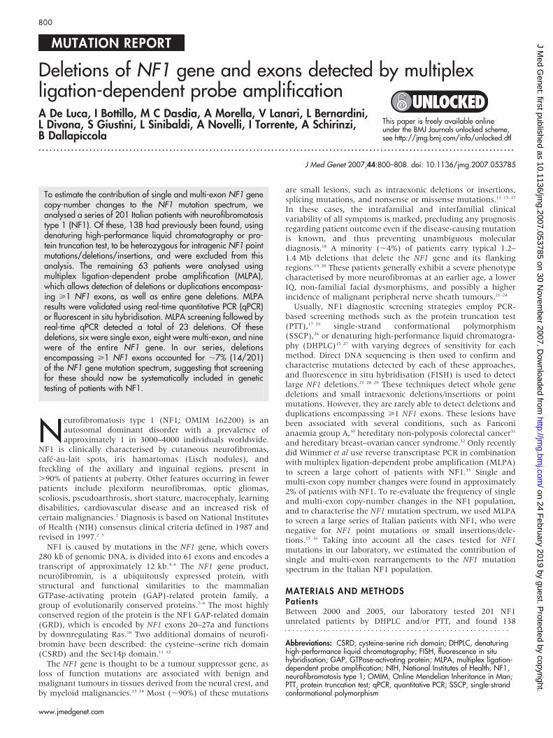
Figure 1 Single deletions detected by multiplex ligation-dependent probe amplification and confirmed by quantitative real-time PCR. Normalised relativepeak areas of all NF1 gene-specific and control probes are shown. Sequences present in two copies of the genome have a relative peak area value ofapproximately 1.0. A reduction in the peak area values to ,0.7 indicates a deletion (black arrows).
802 De Luca, Bottillo, Dasdia, et al
www.jmedgenet.com
on 24 February 2019 by guest. P
rotected by copyright.http://jm
g.bmj.com
/J M
ed Genet: first published as 10.1136/jm
g.2007.053785 on 30 Novem
ber 2007. Dow
nloaded from

using the SALSA P081/082 NF1 MLPA assay for NF1copy-number changes. Gene electropherograms showed reduc-tions of specific MLPA fluorescence signals in 23 casescompared with controls. After MLPA testing, all cases showingabnormal signals were reanalysed by real-time PCR using SYBR
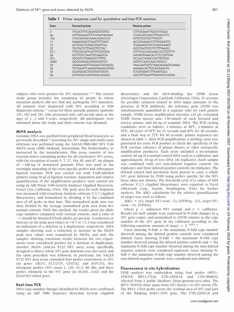
Green as the detection system. Single exon deletions werecorroborated by a single real-time PCR assay corresponding tothe deleted exon, and multi-exon deletions were confirmed byreanalysing, using two separate real-time PCR assays, the mostdistal and the most proximal exons encompassed by the
Figure 1 cont’d Multi-exon deletions detected by multiplex ligation-dependent probe amplification and confirmed by quantitative real-time PCR.Normalised relative peak areas of all NF1 gene-specific and control probes are shown. Sequences present in two copies of the genome have a relative peakarea value of approximately 1.0. A reduction in the peak area values to ,0.7 indicates a deletion (black arrows).
Deletions of NF1 gene and exons detected by MLPA 803
www.jmedgenet.com
on 24 February 2019 by guest. P
rotected by copyright.http://jm
g.bmj.com
/J M
ed Genet: first published as 10.1136/jm
g.2007.053785 on 30 Novem
ber 2007. Dow
nloaded from
deletions predicted by MLPA. All putative deletions identifiedby MLPA were confirmed by real-time PCR. In total, MLPAfollowed by real-time quantitative PCR (qPCR) detected 23 NF1deletions including 6 single exon deletions, 8 multi-exondeletions, and 9 large deletions encompassing the entire NF1gene. In patient 111, MLPA gave ambiguous results with allexons showing area values higher than those of deleted exons,but lower than undeleted exons, suggesting the presence of amosaic whole-gene deletion. In patient 307, carrying a multi-exon deletion, the real-time confirmation for the most proximalMLPA probe (OMG gene) gave ambiguous results (22DDCt
= 0.57). However, results were borderline compared withdeleted positive controls (0.49,22DDCt ,0.56) and clearlybelow non-deleted negative controls (0.92,22DDCt ,0.99).Furthermore, the most distal MLPA probe (exon 30) of thisdeletion was consistently deleted by real-time PCR, thusconfirming this deletion. In total, single and multi-exon NF1deletions were found in 14/201 (,7%) patients with NF1 (fig 1),whereas whole NF1 gene deletions were detected in 9/201(,4.5) NF1 individuals. Real-time qPCR results are reported intable 2; a list of all single and multi-exon deletions detected byMLPA and confirmed by quantitative real-time PCR is reportedin table 3. To verify the absence of a point mutation residingwithin the corresponding MLPA probe, the DNA from allpatients carrying single exon deletions or the recurrent deletionof exons 3 and 4a were sequenced. Sequence analysis did notreveal any point mutation in the DNA fragment recognised byMLPA probe in these exons (supplementary fig 1; available athttp://jmg.bmj.com/supplemental).
The SALSA P081/082 NF1 MLPA assay detected nine casescarrying a whole NF1 gene deletion. These results werecorroborated using the SALSA P122 NF1 area assay, whichconsists of 12 probes covering the entire NF1 gene and itsflanking genes. The SALSA P122 NF1 area assay was previouslyproven to distinguish between the 1.4 Mb deletions (type I)encompassing 14 genes, with breakpoints in the NF1 low-copyrepeats, and the 1.2 Mb deletions (type II), which cover 13
genes and are mediated by recombination between the JJAZ1gene and its pseudogene.33 36 The SALSA P122 AREA MLPAassay confirmed each of the whole gene deletions identified bySALSA P081/082 NF1 MLPA assay. In particular, SALSA P122NF1 area revealed six cases carrying a type I deletion and threecases with a type II deletion (table 4).
All deletions covering the entire NF1 gene detected by MLPAwere confirmed by FISH using a set of probes previously provento distinguish between type I and II deletions. FISH wasperformed on peripheral blood cells by analysing a total of 30metaphases for each case (table 4). In patient 111, FISHanalysis detected a type I mosaic deletion in which 66% of 50metaphases showed a single chromosome 17 signal, whereasthe remaining 34% had two signals.
In all, single and multi-exon deletions were found in 14/201(,7%) cases, whereas entire gene deletions were detected in 9/201 (,4.5%) patients.
Clinical resultsAfter genetic testing, the clinical charts of patients with singleand multi-exon deletions and of patients carrying whole genedeletions were reviewed. The group of patients with single andmulti-exon deletions comprised 14 unrelated patients (12female and 2 male); 9 cases were sporadic and 5 had a familyhistory of NF1. Mean age at the time of the examination was23.9 (range 1–48) years. A mean of 2.7 diagnostic criteria waspresent in each patient. All patients with single and multi-exondeletions fulfilled the NIH Consensus Criteria for NF1, exceptfor two sporadic patients (131 and 18), who presented onlycafe-au-lait spots at the age of 1 and 5 years, respectively. In thesubjects with whole gene deletions (four males and five femalepatients, median age of 27.2 (range 8–47) years), a mean of 3.1diagnostic criteria was observed. In this group, only one subjecthad a family history of NF1. The clinical manifestationsidentified in patients with either single and multi-exon NF1deletions or whole NF1 gene deletions are summarised intable 5.
Table 2 Quantitative real-time PCR results*
Patient no Exon
Sample� Non-deleted controls` Deleted controls1
Mean Min Max Mean Min Max Mean
175 3 0.70 0.96 1.03 1.00 0.69 0.72 0.704a 0.46 0.86 1.14 0.99 0.44 0.53 0.50
113 3 0.55 0.96 1.03 1.00 0.69 0.72 0.704a 0.26 0.86 1.14 0.99 0.44 0.53 0.50
34 4a 0.44 0.86 1.14 0.99 0.44 0.53 0.50201 3 0.58 0.96 1.03 1.00 0.69 0.72 0.70
4a 0.37 0.86 1.14 0.99 0.44 0.53 0.5061 10a 0.68 0.81 0.95 0.89 0.64 0.68 0.67
15 0.32 0.85 1.00 0.95 0.44 0.56 0.4972 10a 0.65 0.89 1.05 0.98 0.64 0.68 0.67117 11 0.69 0.89 0.97 0.93 0.67 0.70 0.6918 22 0.48 0.84 0.92 0.87 0.49 0.55 0.51
23.1 0.49 0.87 1.09 0.96 0.47 0.53 0.49131 26 0.43 0.87 1.20 1.01 0.44 0.53 0.47
27a 0.57 0.83 0.94 0.89 0.47 0.57 0.51227 34 0.65 0.95 1.02 0.97 0.64 0.69 0.66307 OMG 0.57 0.92 0.99 0.95 0.49 0.56 0.53
30 0.56 0.94 0.96 0.95 0.52 0.58 0.5616 35 0.60 0.88 0.96 0.92 0.59 0.65 0.61190 35 0.33 0.88 0.96 0.92 0.59 0.65 0.61
36 0.55 0.74 1.20 0.90 0.54 0.59 0.56196 36 0.55 0.74 1.20 0.90 0.54 0.59 0.56
Min, minimum; Max, maximum.*Results for unknown NF1 samples, non-deleted negative controls, and deleted positive controls are expressed in N-fold changes in NF1 gene copies, normalised toZNF80 relative to the copy number of the NF1 gene in the calibrator, according to the following equation: amount of target = 22DDCt.34
The DDCt calculation used for the relative quantification of target was as follows: DDCt = (Ct, target NF1 exon – Ct, ZNF80)x–(Ct, target NF1 exon–Ct, ZNF80)y, wherex = unknown NF1 sample (�), nondeleted negative control (`) or deleted positive control (1), and y = calibrator.
804 De Luca, Bottillo, Dasdia, et al
www.jmedgenet.com
on 24 February 2019 by guest. P
rotected by copyright.http://jm
g.bmj.com
/J M
ed Genet: first published as 10.1136/jm
g.2007.053785 on 30 Novem
ber 2007. Dow
nloaded from
DISCUSSIONHundreds of mutations have been reported in the NF1 gene,although no clear genotype–phenotype correlation has beenidentified to date.37 The only exceptions are deletions of theentire NF1 gene, which are present in approximately 4% ofpatients with NF1, generally associated with a severe form ofthe disease,21 22 24 and a 3-bp deletion in NF1 exon 17, which hasbeen recently associated with the absence of neurofibromas.38
Although entire gene deletions have been intensely studied,smaller rearrangements encompassing >1 NF1 exons have beeninvestigated to a lesser extent as they are difficult to detectusing standard molecular genetics techniques. To betterinvestigate smaller NF1 rearrangements, we performed MLPAscreening of a large series of patients affected by NF1 for whomthe presence of point mutations, small deletions and insertionshad been previously excluded by DHPLC and/or PTT.15 16
Single and multi-exon NF1 copy-number changes, exclu-sively represented by intragenic deletions in our series, werefound in approximately 7% of the patients with NF1. Thisfrequency is lower than in a previous smaller study, in whichsingle or multi-exon deletions were found in 3/30 (10%) ofpatients with NF1 with high/low grade malignant peripheralnerve sheath tumours,39 but is higher than in a second verylarge study in which single and multi-exon copy-numberchanges were detected in only 25/1100 (,2%) of the cases.33 Inthe latter study, most of the samples were screened using anRNA-based approach, whereas all our samples were screenedby MLPA using genomic DNA as a starting material. Veryrecently, the same authors reported the identification of 5/97(,5%) intragenic deletions in a cohort of Austrian patientswith NF1 meeting the NIH criteria,40 a result more commensu-rate with our findings.
The spectrum of genomic rearrangements disclosed by MLPAwas characterised by a wide range of single and multi-exondeletions, distributed along almost the entire sequence of theNF1 gene. Most of the deletions were unique, although thedeletion of exons 3 and 4a was found in several patients. Thepresence of this lesion was confirmed by two independent real-time qPCR assays using primers for exon 3 and exon 4. No pointmutations that might alter the binding of the MLPA probe togenomic DNA were detected in exons 3 and 4a in any of thepatients carrying this lesion. Alone, deletion of exons 3 and 4arepresents 13% of all lesions detected by MLPA. However,considering all the participants of this study, this lesion isresponsible for only 1.5% of patients with NF1, and thereforepriority screening for this lesion is unnecessary, in our opinion.With the exception of the multi-exon deletion of exon 22 and23.1, which has been previously reported,33 all other lesionsdetected in this study were new. Most of the deletionbreakpoints were unique, although some introns harbouredmore breakpoints than did others. For example, four break-points were mapped to intron 4a, three to introns 2 and 34, andtwo to intron 36. The fine characterisation of these breakpointsis ongoing in our laboratory with the intention of determiningthe molecular mechanisms underlying these deletions and ofdesigning long-range PCR assays for their rapid confirmation.
Table 3 Single and multi-exon deletions detectedby multiplex ligation-dependent probeamplification and confirmed by real-time PCR
Patient no Deleted exons Type of deletion
175 3 and 4a* Multi-exon113 3 and 4a* Multi-exon34 4a* Single exon201 3 and 4a* Multi-exon61 10a to 15 Multi-exon72 10a* Single exon117 11* Single exon18 22 and 23.1 Multi-exon131 26 and 27a Multi-exon307 IVS27b(OMG) to 30 Multi-exon227 34* Single exon16 35* Single exon190 35 and 36 Multi-exon196 36* Single exon
*These deletions were sequenced and none of thecorresponding exons were found to carry a point mutationwithin the corresponding multiplex ligation-dependent probeamplification (MLPA) probe (supplementary fig 1; available athttp://jmg.bmj.com/supplemental).
Table 4 MLPA and FISH results showing type I and type II deletions of the entire NF1 genedetected in patients with NF1
Patients 55 248 208 45 71 111 27 318 305
MLPACentromeric probes
CRLF3 – – – – – – – – –FLJ12735 – – – – – – – – –CENTA2 – – – – – – – – –RNF135 – – – – – – – – –
Intragenic probesExon 1 – – – – – – – – –Exon 12B – – – – – – – – –Exon 23–2 – – – – – – – – –Exon 40 – – – – – – – – –Exon 48 – – – – – – – – –
Telomeric probesHCA66 – – – – – – – – –JJAZ1 + – – + – – – + –KIAA0563–related gene + – – + – – – + –
FISHRP11–353O18 Del Del Del Del Del Del Del Del DelRP11–17I16 Del Del Del Del Del Del Del Del DelCTD–2283L18 Non-Del Del Del Non-Del Del Del Del Non-Del DelCTD–3060L5 Non-Del Del Del Non-Del Del Del Del Non-Del DelDeletion type II I I II I I I II I
+, MLPA probes showing peak area values between 0.7 and 1.3; 2, MLPA probes showing peak area values ,0.7.Del, deleted; Non-del, non-deleted.
Deletions of NF1 gene and exons detected by MLPA 805
www.jmedgenet.com
on 24 February 2019 by guest. P
rotected by copyright.http://jm
g.bmj.com
/J M
ed Genet: first published as 10.1136/jm
g.2007.053785 on 30 Novem
ber 2007. Dow
nloaded from
Patients carrying whole NF1 gene deletions are usuallyaffected by a more severe form of NF1, characterised by a highnumber of neurofibromas and plexiform neurofibromas, facialdysmorphism, mental retardation and a higher risk ofmalignancies.21 22 24 These patients represent approximately 4%of the entire NF1 population, according to a large FISH study.20
Using MLPA, we were able to detect a whole gene deletion in 9/201 (4.5%) patients with neurofibromatosis type 1, indicatingthat MLPA sensitivity for whole gene deletions is quitecomparable with FISH. Most of the whole NF1 gene deletionsare of two types: (1) type I is a 1.4 Mb germline deletion, withbreakpoints mapping in low-copy repeats termed NF1-LCR,41
and (2) type II spans 1.2 Mb and is caused by aberrantrecombination of the JJAZ1 gene and its pseudogene.36 Mostpatients with sporadic NF1 who have type II deletions aremosaic with normal cells and usually show a less severephenotype than patients with type I deletions.36 Consequently,the development of sensitive, reliable and easy to use methodsto differentiate between type I and type II deletions hasimportant clinical implications for the management of patientswith NF1. In our study, the SALSA P122 NF1 area assay, whichis specifically designed to detect and characterise whole genedeletions, was able to distinguish between type I and IIdeletions. The MLPA results were corroborated by FISH using aprobe set previously proven to distinguish between type I and IIdeletions. Similarly, another study has previously shown thatthe SALSA P122 NF1 area assay can be used to distinguishbetween type I and II deletions.33 Previous and current resultsconfirm that the SALSA P122 NF1 area assay could be used as areliable alternative to identify whole gene deletions in NF1laboratories where FISH is not available. Both MLPA and FISH
were not able to confirm the mosaic status of type II deletionsin our cases, but MLPA did detect a mosaic for a type I deletion,which was further confirmed by FISH. However, this type Ideletion mosaic affected 66% of peripheral blood cells, whereasmosaics for type II deletions usually involve .90% of peripheralblood cells,36 and thus would have been detected with difficultyby our FISH, which was performed by analysing 30 peripheralblood metaphases.
In general, subjects with whole gene deletions presented witha more severe phenotype than those carrying single and multi-exon deletions. For example, plexiform neurofibromas werefound in 44% of patients with a whole gene deletion, butoccurred in only 25% of the cases with single and multi-exondeletions. Similarly, scoliosis was found at higher frequency inpatients with whole gene deletions compared with those withpartial gene deletions. Facial dysmorphism and mentalretardation, which are hallmarks of whole gene deletions, wereobserved in two and three patients with a whole gene deletion,respectively, but were not observed in patients with single ormulti-exon deletions. The severe NF1 phenotype associated towhole gene deletions is usually explained by the large size ofthe lesion, which spans approximately 1.2–1.4 Mb of genomicDNA, resulting in the haploinsufficiency of 14 differentgenes.36 41 In comparison, single and multi-exon deletions ofNF1 are relatively small lesions, usually involving no genesother than NF1. Interestingly, all patients with mentalretardation (patients 248, 208 and 305) carried a type Igermline deletion, but none had a type II deletion. Thisobservation is in accordance with the notion that patients withNF1 with type I deletions generally show a more severephenotype than patients with type II deletions.36 In one case
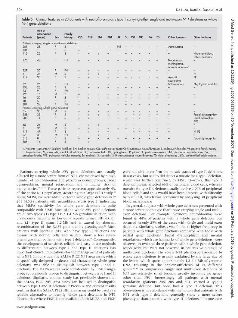
Table 5 Clinical features in 23 patients with neurofibromatosis type 1 carrying either single and multi-exon NF1 deletions or wholeNF1 gene deletions
Patients
Age atobservation(years) Sex
Familyhistory CLS CNf SNf PNf AF Sc OG MR PA TD Other tumours Other features
Patients carrying single or multi-exon deletions201 28 F S + – – + NE – – – – – Astrocytoma –131 1 F S + – – – – – – – – – – –113 26 F S + – + – + – – – – + – Hypothyroidism,
UBOs, seizures175 48 F FH + – + – – + – – – – Neurinoma,
meningioma,adrenal adenoma
–
227 30 F FH + + – – + – – – – – – –61 37 M S + + + + – – – – – – – H117 30 F S + + – – – – – – – – Acoustic
neurinomaPE
72 26 F FH + + – + + + – – – – Schwannoma BN, thyroid nodules196 25 F S + – – – + – – – – – – –34 9 F FH + – – – – – – – – – – –190 15 M S + – – – + – – – – – – –16 20 F S + + – – – – + – – – – –18 5 F S + – – – – + – – – – – –307 34 F FH + – + – + – – – – – – –Patients carrying whole gene deletions55 35 M S + + + + + + – – – – – –248 35 F S + + + + + + – + – – – Facial dysmorphism208 11 F S + + – – + + – + – – – Chest anomalies,
PVS45 26 M S + + + – + – – – – – – P71 36 M S + + – + + + – – – – – –111 47 F S + + – – + + – – – – – H, PE27 33 M FH + – + + + + – – + + – E318 8 F S + – – – + – – – – – – Facial dysmorphism305 14 F S + + – – + – – + – – – –
+, Present; –, absent; AF, axillary freckling; BN, Becker naevus; CLS, cafe-au-lait spots; CNf, cutaneous neurofibromas; E, epilepsy; F, female; FH, positive family history;H, hypertension; M, male; MR, mental retardation; NE, not evaluated; OG, optic glioma; P, ptosis; PE, pectus excavatum; PNf, plexiform neurofibromas; PA,pseudoarthrosis; PVS, pulmonar valvular stenosis; Sc, scoliosis; S, sporadic; SNf, subcutaneous neurofibromas; TD, tibial dysplasia; UBOs, unidentified bright objects.
806 De Luca, Bottillo, Dasdia, et al
www.jmedgenet.com
on 24 February 2019 by guest. P
rotected by copyright.http://jm
g.bmj.com
/J M
ed Genet: first published as 10.1136/jm
g.2007.053785 on 30 Novem
ber 2007. Dow
nloaded from
(patient 307), we identified a deletion involving three NF1exons and the small OMG gene in NF1 intron 27b. This womanpresented with cafe-au-lait spots, axillary freckling and sub-cutaneous neurofibromas at 34 years of age, suggesting thatOMG is unlikely to be the gene responsible for the NF1complications (ie mental retardation and facial dysmorphism)observed in whole gene deletion carriers. Accordingly, aprevious screening of OMG gene for point mutations in patientswith non-syndromic mental retardation failed to demonstratenucleotide variants of clear significance.42 Patient 208, carryinga whole NF1 gene deletion, had Watson syndrome, presentingwith cafe-au-lait spots, neurofibromas, mental retardation,thoracic abnormalities and pulmonary stenosis. Watson syn-drome has been reported previously in a patient carrying anNF1 tandem duplication, as well as in other patients carryingsmall deletions and point mutations,37 thus showing that thissyndrome is associated with a wide range of NF1 geneabnormalities.
In our hands, the MLPA technique gave several false positiveresults, including reduced MLPA signals for exons 13a and 18,and for both probes in intron 27b recognising the OMG gene.For this reason, it is our recommendation to (1) reassess everyMLPA-positive sample by a second MLPA experiment, and (2)confirm every putative lesion identified by MLPA with analternative technique. In this regard, quantitative real-time PCRwith SYBR Green has several advantages, including therequirement for small amounts of DNA and low costs owingto use of the same primers used for DHPLC and sequenceanalysis. Furthermore, samples carrying whole gene deletionscan be used as reliable positive controls for real-time PCR. Wealso suggest running each sample with a non-deleted negativecontrol as normal DNA reference for real-time PCR. It has alsobeen reported that false positive MLPA signals could be due tothe presence of subtle point mutations under the MLPA probe,which may impair probe hybridisation and mimic the presenceof a deletion.33 To exclude this possibility, we suggestsequencing the MLPA probe corresponding region in all casescarrying recurrent deletions or single exon deletions.
In our series, single and multi-exon NF1 deletions wereresponsible for ,7% of the mutations in our patient group. Thespectrum of these lesions was heterogeneous, with a similarproportion of single and multi-exon deletions. In previousstudies, whole gene deletions were found in 4% of thepatients,20 and using DHPLC followed by direct sequencing,we identified a point mutation or a small deletion/insertion in138/201 (68.7%) patients with NF1. By combining the muta-tions previously identified by DHPLC with the genomicrearrangements detected in this study by MLPA, we have beenable to find the disease-causing lesion in 161/201 (80.1%) of ourpatients with NF1. We hypothesise that mutations affectingregulatory portions of the gene might also have a pathogenicrole in NF1. Although mutations affecting the NF1 promoterhave not been reported to date, lesions in other non-codingportions are to be expected.43 Furthermore, we cannot excludethe possibility that some mutations have not been detected byour protocol.
In conclusion, MLPA analysis followed by real-time PCRrevealed 23 genomic rearrangements in a series of 201 patientswith NF1. These data suggest the possibility of adding MLPA toDHPLC in routine diagnostic procedures for patients with NF1.Furthermore, to reduce the time of analysis, MLPA should beused as a priority in patients presenting with severe NF1,possibly reflecting deletion of the entire NF1 gene.
ACKNOWLEDGEMENTSWe thank the patients and their families who enrolled in this study andphysicians who referred these subjects.
Supplementary material is available on the JMGwebsite at http://jmg.bmj.com/supplemental
Authors’ affiliations. . . . . . . . . . . . . . . . . . . . . . .
A De Luca, IRCCS-CSS, San Giovanni Rotondo and CSS-Mendel Institute,Rome, ItalyA De Luca, Department of Pathology and Laboratory Medicine, Universityof British Columbia, Vancouver, British Columbia, CanadaMCDasdia, A Morella, V Lanari, L Bernardini, A Novelli, I Torrente,IRCCS-CSS, San Giovanni Rotondo and CSS-Mendel Institute, Rome, ItalyL Divona, S Giustini, Department of Dermatology-Venereology and Plasticand Reconstructive Surgery, University of Rome ‘‘La Sapienza’’, Rome, ItalyL Sinibaldi, A Schirinzi, B Dallapiccola, I Bottillo, IRCCS-CSS, SanGiovanni Rotondo and CSS-Mendel Institute, Rome, ItalyL Sinibaldi, A Schirinzi, B Dallapiccola, I Bottillo, Department ofExperimental Medicine and Pathology, University of Rome ‘‘La Sapienza’’,Rome, Italy
Funding: This work was supported by the Italian Ministry of Health (RicercaCorrente 2006–2007). A. De Luca is also supported by the Michael SmithFoundation for Health Research.
Competing interests: none declared.
Correspondence to: Professor Bruno Dallapiccola, Dipartimento diMedicina Sperimentale e Patologia, Universita degli Studi di Roma ‘‘LaSapienza’’, Viale Regina Margherita 261–00198 Rome, Italy;[email protected]
Received 10 August 2007Revised 10 August 2007Accepted 13 August 2007
REFERENCES1 Riccardi VM. Neurofibromatosis: phenotype, natural history, and pathogenesis,
2nd ed. Baltimore: Johns Hopkins University Press, 1992.2 Gutmann DH, Aylsworth A, Carey JC, Korf B, Marks J, Pyeritz RE, Rubenstein A,
Viskochil D. The diagnostic evaluation and multidisciplinary management ofneurofibromatosis 1 and neurofibromatosis 2. JAMA 1997;278:51–7.
3 Stumpf DA, Alksne JF, Annegers JF, Brown SS, Conneally PM, Housman D,Leppert MF, Miller JP, Moss ML, Pileggi AJ, Rapin I, Strohman RC, Swanson LW,Zimmerman A. Neurofibromatosis. Conference statement. National Institutes ofHealth Consensus Development Conference. Arch Neurol 1988;45:575–8.
4 Cawthon RM, O’Connell P, Buchberg AM, Viskochil D, Weiss RB, Culver M,Stevens J, Jenkins NA, Copeland NG, White R. Identification andcharacterization of transcripts from the neurofibromatosis 1 region: the sequenceand genomic structure of EVI2 and mapping of other transcripts. Genomics1990;7:555–65.
5 Viskochil D, Buchberg AM, Xu G, Cawthon RM, Stevens J, Wolff RK, Culver M,Carey JC, Copeland NG, Jenkins NA, White R, OConnell P. Deletions and atranslocation interrupt a cloned gene at the neurofibromatosis type 1 locus. Cell1990;62:187–92.
6 Wallace M, Marchuk D, Anderson L, Letcher R, Odeh H, Saulino A, Fountain J,Brereton A, Nicholson J, Mitchell A, Brownstein B, Collins F. Type 1neurofibromatosis gene: identification of a large transcript disrupted in three NF1patients. Science 1990;249:181–6.
7 Danglot G, Regnier V, Fauvet D, Vassal G, Kujas M, Bernheim A.Neurofibromatosis 1 (NF1) mRNAs expressed in the central nervous system aredifferentially spliced in the 59 part of the gene. Hum Mol Genet 1995;4:915–20.
8 Li Y, O’Connell P, Breidenbach HH, Cawthon R, Stevens J, Xu G, Neil S,Robertson M, White R, Viskochil D. Genomic organization of theneurofibromatosis 1 gene (NF1). Genomics 1995;25:9–18.
9 Xu GF, O’Connell P, Viskochil D, Cawthon R, Robertson M, Culver M, Dunn D,Stevens J, Gesteland R, White R, Weiss R. The neurofibromatosis type 1 geneencodes a protein related to GAP. Cell 1990;62:599–608.
10 Martin GA, Viskochil D, Bollag G, McCabe PC, Crosier WJ, Haubruck H,Conroy L, Clark R, O’Connell P, Cawthon RM, Innis MA, McCormick F. The GAP-related domain of the neurofibromatosis type 1 gene product interacts with rasp21. Cell 1990;63:843–9.
11 Fahsold R, Hoffmeyer S, Mischung C, Gille C, Ehlers C, Kucukceylan N, Abdel-Nour M, Gewies A, Peters H, Kaufmann D, Buske A, Tinschert S, Nurnberg P.Minor lesion mutational spectrum of the entire NF1 gene does not explain its highmutability but points to a functional domain upstream of the GAP-relateddomain. Am J Hum Genet 2000;66:790–818.
12 D’Angelo I, Welti S, Bonneau F, Scheffzek K. A novel bipartite phospholipid-binding module in the neurofibromatosis type 1 protein. EMBO Reports2006;7:174–9.
Deletions of NF1 gene and exons detected by MLPA 807
www.jmedgenet.com
on 24 February 2019 by guest. P
rotected by copyright.http://jm
g.bmj.com
/J M
ed Genet: first published as 10.1136/jm
g.2007.053785 on 30 Novem
ber 2007. Dow
nloaded from
13 Li Y, Bollag G, Clark R, Stevens J, Conroy L, Fults D, Ward K, Friedman E,Samowitz W, Robertson M, Bradley P, McCormick F, White R, Cawthon R.Somatic mutations in the neurofibromatosis 1 gene in human tumors. Cell1992;69:275–81.
14 Shannon KM, O’Connell P, Martin GA, Paderanga D, Olson K, Dinndorf P,McCormick F. Loss of the normal NF1 allele from the bone marrow of childrenwith type 1 neurofibromatosis and malignant myeloid disorders. N Engl J Med1994;330:597–601.
15 De Luca A, Buccino A, Gianni D, Mangino M, Giustini S, Richetta A, Divona L,Calvieri S, Mingarelli R, Dallapiccola B. NF1 gene analysis based on DHPLC.Hum Mutat 2003;21:171–2.
16 De Luca A, Schirinzi A, Buccino A, Bottillo I, Sinibaldi L, Torrente I, Ciavarella A,Dottorini T, Porciello R, Giustini S, Calvieri S, Dallapiccola B. Novel and recurrentmutations in the NF1 gene in Italian patients with neurofibromatosis type 1. HumMutat 2004;23:629.
17 Messiaen LM, Callens T, Mortier G, Beysen D, Vandenbroucke I, Van Roy N,Speleman F, Paepe AD. Exhaustive mutation analysis of the NF1 gene allowsidentification of 95% of mutations and reveals a high frequency of unusualsplicing defects. Hum Mutat 2000;15:541–55.
18 Easton DF, Ponder MA, Huson SM, Ponder BA. An analysis of variation inexpression of neurofibromatosis (NF) type 1 (NF1): evidence for modifyinggenes. Am J Hum Genet 1993;53:305–13.
19 Cnossen MH, van der Est MN, Breuning MH, van Asperen CJ, Breslau-Siderius EJ, van der Ploeg AT, de Goede-Bolder A, van den Ouweland AM,Halley DJ, Niermeijer MF. Deletions spanning the neurofibromatosis type 1 gene:implications for genotype-phenotype correlations in neurofibromatosis type 1?Hum Mutat 1997;9:458–64.
20 Kluwe L, Siebert R, Gesk S, Friedrich RE, Tinschert S, Kehrer-Sawatzki H,Mautner VF. Screening 500 unselected neurofibromatosis 1 patients for deletionsof the NF1 gene. Hum Mutat 2004;23:111–16.
21 Tonsgard JH, Yelavarthi KK, Cushner S, Short MP, Lindgren V. Do NF1 genedeletions result in a characteristic phenotype? Am J Med Genet 1997;73:80–6.
22 Upadhyaya M, Cooper DN. Neurofibromatosis type 1 from genotype tophenotype. Oxford: Bios Scientific, 1998.
23 De Raedt T, Brems H, Wolkenstein P, Vidaud D, Pilotti S, Perrone F, Mautner V,Frahm S, Sciot R, Legius E. Elevated risk for MPNST in NF1 microdeletionpatients. Am J Hum Genet 2003;72:1288–92.
24 Kayes LM, Burke W, Riccardi VM, Bennett R, Ehrlich P, Rubenstein A, Stephens K.Deletions spanning the neurofibromatosis 1 gene: identification and phenotypeof five patients. Am J Hum Genet 1994;54:424–36.
25 Heim RA, Kam-Morgan LN, Binnie CG, Corns DD, Cayouette MC, Farber RA,Aylsworth AS, Silverman LM, Luce MC. Distribution of 13 truncating mutations inthe neurofibromatosis 1 gene. Hum Molec Genet 1995;4:975–81.
26 Gasparini P, D’Agruma L, Pio de Cillis G, Balestrazzi P, Mingarelli R, Zelante L.Scanning the first part of the neurofibromatosis type 1 gene by RNA-SSCP:identification of three novel mutations and of two new polymorphisms. HumGenet 1996;97:492–5.
27 Han SS, Cooper DN, Upadhyaya MN. Evaluation of denaturing highperformance liquid chromatography (DHPLC) for the mutational analysis of theneurofibromatosis type 1 ( NF1) gene. Hum Genet 2001;109:487–97.
28 Wu BL, Austin MA, Schneider GH, Boles RG, Korf BR. Deletion of the entire NF1gene detected by the FISH: four deletion patients associated with severemanifestations. Am J Med Genet 1995;59:528–35.
29 Riva P, Corrado L, Natacci F, Castorina P, Wu BL, Schneider GH, Clementi M,Tenconi R, Korf BR, Larizza L. NF1 microdeletion syndrome: refined FISHcharacterization of sporadic and familial deletions with locus-specific probes.Am J Hum Genet 2000;66:100–9.
30 Callen E, Tischkowitz MD, Creus A, Marcos R, Bueren JA, Casado JA,Mathew CG, Surralles J. Quantitative PCR analysis reveals a high incidence oflarge intragenic deletions in the FANCA gene in Spanish Fanconi anemiapatients. Cytogenet GenomeRes 2004;104:341–5.
31 Nakagawa H, Hampel H, de la Chapelle A. Identification and characterizationof genomic rearrangements of MSH2 and MLH1 in Lynch syndrome (HNPCC) bynovel techniques. Human Mutat 2003;22:258.
32 Hogervorst FB, Nederlof PM, Gille JJ, McElgunn CJ, Grippeling M, Pruntel R,Regnerus R, van Welsem T, van Spaendonk R, Menko FH, Kluijt I, Dommering C,Verhoef S, Schouten JP, van’t Veer LJ, Pals G. Large genomic deletions andduplications in the BRCA1 gene identified by a novel quantitative method. CancerRes 2003;63:1449–53.
33 Wimmer K, Yao S, Claes K, Kehrer-Sawatzki H, Tinschert S, De Raedt T, Legius E,Callens T, Beiglbock H, Maertens O, Messiaen L. Spectrum of single- andmultiexon NF1 copy number changes in a cohort of 1,100 unselected NF1patients. Gene Chromosomes Cancer 2006;45:265–76.
34 Livak KJ, Schmittgen TD. Analysis of relative gene expression data using real-time quantitative PCR and the 2(-Delta Delta C(T)) method. Methods2001;25:402–8.
35 De Luca A, Bernardini L, Ceccarini C, Sinibaldi L, Novelli A, Giustini S, Daniele I,Calvieri S, Mingarelli R. Fluorescence in situ hybridization analysis of alleliclosses involving the long arm of chromosome 17 in NF1-associatedneurofibromas. Cancer Genet Cytogenet 2004a;150:168–72.
36 Kehrer-Sawatzki H, Kluwe L, Sandig C, Kohn M, Wimmer K, Krammer U,Peyrl A, Jenne DE, Hansmann I, Mautner VF. High frequency of mosaicismamong patients with neurofibromatosis type 1 (NF1) with microdeletions causedby somatic recombination of the JJAZ1 gene. Am J Hum Genet2004;75:410–23.
37 Castle B, Baser ME, Huson SM, Cooper DN, Upadhyaya M. Evaluation ofgenotype-phenotype correlations in neurofibromatosis type 1. J Med Genet2003;40:e109.
38 Upadhyaya M, Huson SM, Davies M, Thomas N, Chuzhanova N, Giovannini S,Evans DG, Howard E, Kerr B, Griffiths S, Consoli C, Side L, Adams D, Pierpont M,Hachen R, Barnicoat A, Li H, Wallace P, Van Biervliet JP, Stevenson D,Viskochil D, Baralle D, Haan E, Riccardi V, Turnpenny P, Lazaro C, Messiaen L.An absence of cutaneous neurofibromas associated with a 3-bp inframe deletionin exon 17 of the NF1 gene (c.2970-2972 delAAT): evidence of a clinicallysignificant NF1 genotype-phenotype correlation. Am J Hum Genet2007;80:140–51.
39 Upadhyaya M, Spurlock G, Majounie E, Griffiths S, Forrester N, Baser M,Huson SM, Gareth Evans D, Ferner R. The heterogeneous nature of germlinemutations in NF1 patients with malignant peripheral serve sheath tumours(MPNSTs). Hum Mutat 2006;27:716.
40 Wimmer K, Roca X, Beiglbock H, Callens T, Etzler J, Rao AR, Krainer AR,Fonatsch C, Messiaen L. Extensive in silico analysis of NF1 splicing defectsuncovers determinants for splicing outcome upon 59 splice-site disruption. HumMutat 2007;28:599–612.
41 Lopez-Correa C, Dorschner M, Brems H, Lazaro C, Clementi M, Upadhyaya M,Dooijes D, Moog U, Kehrer-Sawatzki H, Rutkowski JL, Fryns JP, Marynen P,Stephens K, Legius E. Recombination hotspot in NF1 microdeletion patients. HumMolec Genet 2001;10:1387–92.
42 Venturin M, Moncini S, Villa V, Russo S, Bonati MT, Larizza L, Riva P. Mutationsand novel polymorphisms in coding regions and UTRs of CDK5R1 and OMGgenes in patients with non-syndromic mental retardation. Neurogenetics2006;7:59–66.
43 Raponi M, Upadhyaya M, Baralle D. Functional splicing assay shows apathogenic intronic mutation in neurofibromatosis type 1 (NF1) due to intronicsequence exonization. Hum Mutat 2006;27:294–5.
Stay a step ahead with Online First
We publish all our original articles online before they appear in a print issue. This means that thelatest clinical research papers go straight from acceptance to your browser, keeping you at thecutting edge of medicine. We update the site weekly so that it remains as topical as possible.Follow the Online First link on the home page and read the latest research.
808 De Luca, Bottillo, Dasdia, et al
www.jmedgenet.com
on 24 February 2019 by guest. P
rotected by copyright.http://jm
g.bmj.com
/J M
ed Genet: first published as 10.1136/jm
g.2007.053785 on 30 Novem
ber 2007. Dow
nloaded from