mutationalanalysis nm23-h2/ndp structural domains critical · mutationalanalysis...
TRANSCRIPT
Proc. Natl. Acad. Sci. USAVol. 93, pp. 6892-6897, July 1996Biochemistry
Mutational analysis of NM23-H2/NDP kinase identifies thestructural domains critical to recognition of a c-mycregulatory element
(PuF/transcription factor/metastasis)
EDITH H. POSTEL*, VALERIE H. WEISSt, JUTIA BENEKENt, AND AJAY KIRTANE§Department of Molecular Biology, Princeton University, Princeton, NJ 08544-1014
Communicated by Arnold J. Levine, Princeton University, Princeton, NJ, February 20, 1996 (received for review December 6, 1995)
ABSTRACT NM23-H2, a presumed regulator of tumor me-tastasis in humans, is a hexameric protein with both enzymatic(NDP kinase) and regulatory (transcriptional activation) activ-ity. While the structure and catalytic mechanisms have been wellcharacterized, the mode of DNA binding is not known. Weexamined this latter function in a site-directed mutational studyand identified residues and domains essential for the recognitionof a c-myc regulatory sequence. Three amino acids, Arg-34,Asn-69, and Lys-135, were found among 30 possibilities to becritical for DNA binding. Two of these, Asn-69 and Lys-135, arenot conserved between NM23 variants differing in DNA-bindingpotential, suggesting that DNA recognition resides partly innonconserved amino acids. All three DNA-binding defectivemutant proteins are active enzymatically and appear to be stablehexamers, suggesting that they perform at the level of DNArecognition and that separate functional domains exist forenzyme catalysis and DNA binding. In the context of the knowncrystal structure of NM23-H2, the DNA-binding residues arelocated within distinct structural motifs in the monomer, whichare exposed to the surface near the 2-fold axis of adjacentsubunits in the hexamer. These findings are explained by a modelin which NM23-H2 binds DNA with a combinatorial surfaceconsisting ofthe "outer" face ofthe dimer. Chemical crosslinkingdata support a dimeric DNA-binding mode by NM23-H2.
Altered nm23 gene expression is associated with a multitudeof phenotypes including inhibition of tumor metastasis (1-5),oncogenesis (6, 7), cellular proliferation (8, 9), development,differentiation (10-14), and apoptosis (14). Although nm23was discovered a decade ago, there are still no molecular orbiochemical explanations for these data. The nature of theseobservations suggest, however, that NM23 is involved in theregulation of other genes important to cell growth and differ-entiation. Indeed, such a role for human NM23-H2 has beenrecognized in c-myc gene transcription (15-17).nm23 genes encode the 17-kDa subunits of nucleoside
diphosphate kinases (NDPKs), housekeeping enzymes thatcatalyze the transfer of y-phosphates between nucleoside tri-and diphosphates (18). The nm23 gene family is large andhighly conserved among species (19). Human nm23-HI (1) andnm23-H2 (3) are 88% homologous and are closely linked onchromosome 17q21 near the BRCA-1 gene locus (20). TheNM23-H1 protein is more closely associated with metastasisinhibition and signal transduction (1, 2, 21), has an acidic pI,and is also known as NDPK-A (22). The second variant,NM23-H2 (3), is a basic protein identical to NDPK-B (22) andto the human PuF factor, a transcriptional activator of thec-myc protooncogene (23, 24). A third gene located on chro-mosome 16, known as DR-nm23, is 70% identical to Hi andH2and may play a role in normal hematopoiesis and in the inductionof apoptosis (14). Overexpression of all three human nm23 genesis postulated to contribute to differentiation arrest.
The 3-dimensional structures of several NM23/NDPKs areknown and the catalytic mechanisms have been well character-ized (25-29). All eukaryotic NM23/NDPKs are hexameric, aconformation which is required for stability as well as for efficientenzyme catalysis (25-29). In contrast, the biochemical and mo-lecular bases of the of NM23 regulatory functions are not known.As the developmental and the metastatic functions of NM23appear to be independent of the NDPK enzymatic activity (2, 11,30, 31), it seems likely that at least some of the biologicalproperties are consequences of the transcriptional function, par-ticularly because one target of NM23-H2 is the c-myc gene, itselfa regulator of cell proliferation and differentiation (8, 15, 17,32-34). This notion is partly supported by the finding that DNAbinding and transcriptional activation, at least in vitro, can occurin the absence of the phosphotransferase activity (35).To attempt a fuller understanding of the NM23 function, we
have undertaken a study of its transcriptional activity. As astarting point for this analysis, we examined in a previous study(35) the relevance of the NDPK activity to the DNA-bindingfunction and concluded that, whereas a missense substitutionof the catalytic residue (His-118 with phenylalanine) inacti-vated the phosphotransferase function, the mutation had noeffect on DNA binding or in vitro transcription. Thus, with asingle point mutation we essentially have separated the cata-lytic and the DNA-binding functions, although the physiolog-ical consequences of this finding are not known yet. Here weused a systematic site-directed mutational analysis to identifyresidues and domains of NM23-H2 that are involved in DNAbinding. On the basis of these analyses, a model for theNM23-H2 DNA-binding domain is presented.
MATERIALS AND METHODSMutagenesis. Point substitution mutations were introduced
into nm23-H2 cDNA by the Unique Site Elimination methodas described (35). The mutagenic primers were 27-39 baseslong and had a greater than 50% GC content. The sequenceof the oligonucleotides used to generate the DNA-bindingdefective mutants were, with-base changes indicated in bold-face letters, as follows: 5'-gagcagaagggattcgccctcgtggccat-gaag-3' (R34A); 5'-gcagaagggattcgacctcgtggccatg-3'; (R34D);5-'cagaagggattcggcctcgtggccatg-3' (R34G); 5'- gggctggtgaag-tacatgcactcagggccggttgtggcc-3' (N69H); 5'-cagcctatggtttcac-cctgaagaactggttgac-3' (K135H).
Protein Production and DNA Binding. Mutant and wild-typeproteins were overproduced in Escherichia coli and purified byammonium sulfate fractionation and hydroxylapatite chromatog-raphy as described (15). DNA binding was assessed in gel
Abbreviation: NDPK, nucleoside diphosphate kinase.*To whom reprint requests should be addressed.tPresent address: Department of Biological Chemistry and MolecularPharmacology, Harvard Medical School, 240 Longwood Avenue,Boston, MA 02115.iPresent address: Program in Biochemistry, Cell and Molecular Bi-ology, Johns Hopkins University School of Medicine, 725 NorthWolfe Street, Baltimore, MD 21205-2185.§Present address: College of Physicians and Surgeons, ColumbiaUniversity, New York, NY 10032.
6892
The publication costs of this article were defrayed in part by page chargepayment. This article must therefore be hereby marked "advertisement" inaccordance with 18 U.S.C. §1734 solely to indicate this fact.
Dow
nloa
ded
by g
uest
on
Mar
ch 9
, 202
0
Proc. Natl. Acad. Sci. USA 93 (1996) 6893
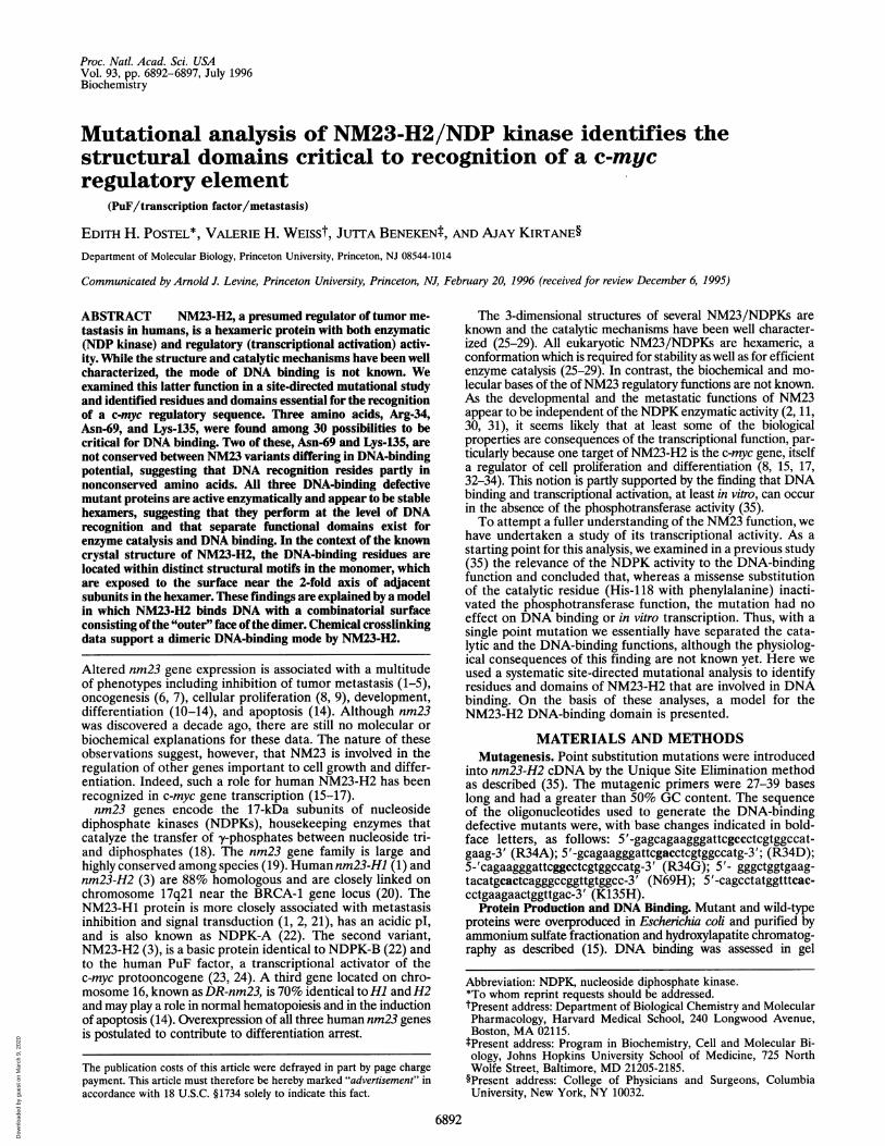
Head <-Kpn loop-->- C-terminal segment
PI aO al P2 aA a2 P3 a3 a4 P4 a5
I I I I I I I I I I I I I I I1 1 20 30 40 50 60 70 80 90 1001 l 120 130 140 1
B 18 30 50 70RGLVGEIIKRFEQKGFRLVAMKFLRASEEHLKQHYIDLKDRPFFPGLVKYMNSGPVVAMNW
90 110 130 148
EGLNVVKTGRVMLGETNPADSKPGTIRGDFCIQVGRNIIHGSDSVKSAEKEISLWFKPEELVDYKSCAHD
electrophoretic mobility shift assays (15, 24, 35). Standard reac-tions were carried out with [32P]-end-labeled c-myc fragmentscontaining the -164 to -110 nuclease-hypersensitive element(NHE), the known site of interaction with PuF/NM23-H2 (15,24). One fragment was 105 bp long and generated by PCR (15,35); the other was a 34 bp double-stranded synthetic oligonucle-otide with the sequence 5'-ctccccaccttccccaccctccccaccctcccca-3'. The two DNA fragments provided qualitatively similar resultsalthough, as noted before (24), higher affinities were observedwith the longer probe. Stoichiometric amounts of DNA andprotein (generally 1 ng probe: 200-2000 ng of hexameric protein)were mixed together in 10-p,l reactions also containing 100 ngpoly(dA-dT) in 0.1 M HM buffer (20 mM Hepes, pH 7.9/5 mMMgCl2/0.1 mM EDTA/0.1 M KCl/1 mM DTT/20% glycerol/protease inhibitors; refs. 15 and 24) and incubated 20 min on ice.The NM23/DNA complexes were resolved on 5% native poly-acrylamide gels and detected by autoradiography as described(15, 35).
Size Exclusion Chromatography. The molecular size ofwild-type NM23-H2 was initially determined using a Pharma-cia FPLC Superose 12 column (29 x 1.5 cm), equilibrated with0.1 HM buffer and calibrated several times with low and highmolecular weight protein standards (Pharmacia). A standardcurve was constructed by plotting K5V versus log Mr. Ka, wascalculated from the elution volumes Ve, from total columnvolume Vt, and from the void volume Vo using the equation K5,,= Ve - VO/Vt - V0. The void volume (Vo) was determined bymeasuring the eluted volume of blue dextran. From the FPLCcolumn the molecular weight of recombinant NM23-H2 wasdetermined to be 100,000 + 10,000. Based on the amino acidsequence (15), the calculated molecular weight of the hexameris 105,000 (22). In subsequent experiments, a conventional 27x 1.5 cm Sephacryl S-200 HR (Pharmacia) column was used,from which the same Mr was obtained for the wild-typehexamer. This column was recalibrated frequently and used forthe sizing of the mutant proteins.NDPK Enzyme Activity. NDPK activity was measured spec-
trophotometrically in a coupled pyruvate kinase-lactate de-hydrogenase assay and the specific activities calculated asdescribed (35).
Glutaraldehyde Crosslinking. Wild-type NM23-H2 (200 ng)was incubated with or without DNA in a standard DNA-bindingreaction, then treated with glutaraldehyde (Sigma) freshly dilutedin water. Crosslinking was allowed to proceed for 30 min at 25°Cand was terminated by the addition of 1 ,ul of 1 M lysine. Asindicated, reactions were treated with 1 unit of DNase (Promega)for 10 min at 37°C, after which samples were boiled in SDS samplebuffer and electrophoresed in 15% polyacrylamide gels. To detectDNA associated with proteins, the wet gels were exposed to x-rayfilm at 4°C overnight prior to detection of proteins by immuno-blotting. We used mouse polyclonal anti-NM23-H2 antiserumand horseradish peroxidase-conjugated secondary antibodies(Vector Laboratories) as described (15).
RESULTSRationales for Designing the Mutations. Because the crystal
structure of NM23 has not suggested a mode of DNA binding(25-29), the design of mutations was, within the context of the3-dimensional structure, based on the biochemical and bio-logical properties of NM23 including: (i) the presence of
FIG. 1. Location of amino acidsubstitution mutations in the pri-mary sequence of NM23-H2/NDPK. (A) Schematic representa-tion of NM23-H2 (hatched box)indicating major secondary struc-tural features. The "head," the Kpnloop, and the C- terminal segmentare exposed to the outer surface inthe hexamer (25-29). (B) Partialamino acid sequence of NM23-H2showing the mutated residues inboldface type.
transcription factor motifs in the protein sequence and likelyregulatory domains, (ii) identification of functional domainsusing monoclonal antibodies, (iii) a 12% sequence diversitybetween NM23-H1 and H2 and the demonstration that Hidoes not bind to c-myc promoter DNA, and (iv) mutationsoccurring naturally in nm23 that are associated with cancer ordevelopmental defects.
Transcription factor motifs as targets ofmutagenesis. nm23-H2encodes a potential bZIP-like motif involving helices cal andaA (ref. 3; Fig. 1). Consistent with a heterodimerization-basedtranscriptional activation mechanism is the behavior of en-
dogenous PuF/NM23-H2 in HeLa cell fractions (15) and a
Leu -* Val substitution at position 48 (in the middle leucine)in an aggressive case of childhood neuroblastoma (7). Althoughthe "basic" region of the putative zipper motif in NM23 is part ofhelix al (Fig. 1), which constitutes the protein interface-oligomerization domain between two adjacent monomers (25-29), we nonetheless mutated these residues (R18, K26, R27, K31,R34, and K39) because of the possibility thatDNA may dissociatethe interacting helices prior to binding. A similar model has beenproposed by Janin and coworkers (26).
Because the aA helix contains the leucines (at positions 41,48, and 55) and because it is ideally located on the surface ofthe hexamer (25-29), this helix could be a protein interactingdomain. Alternatively, the caA helix could be considered theDNA-recognition helix, for it is rich in charged residues thatcould stably interact with DNA (e.g., R42, E46, H47, and Q50).In fact, the caA helix in NM23-H2, unlike in Hi, has an overallbasic charge. Finally, the aiA helix could function in a dualcapacity by binding both protein and DNA.
A H2 HI M kDa
W 43
26
B Hl Hl H2 HZ
18
14
6
p -a
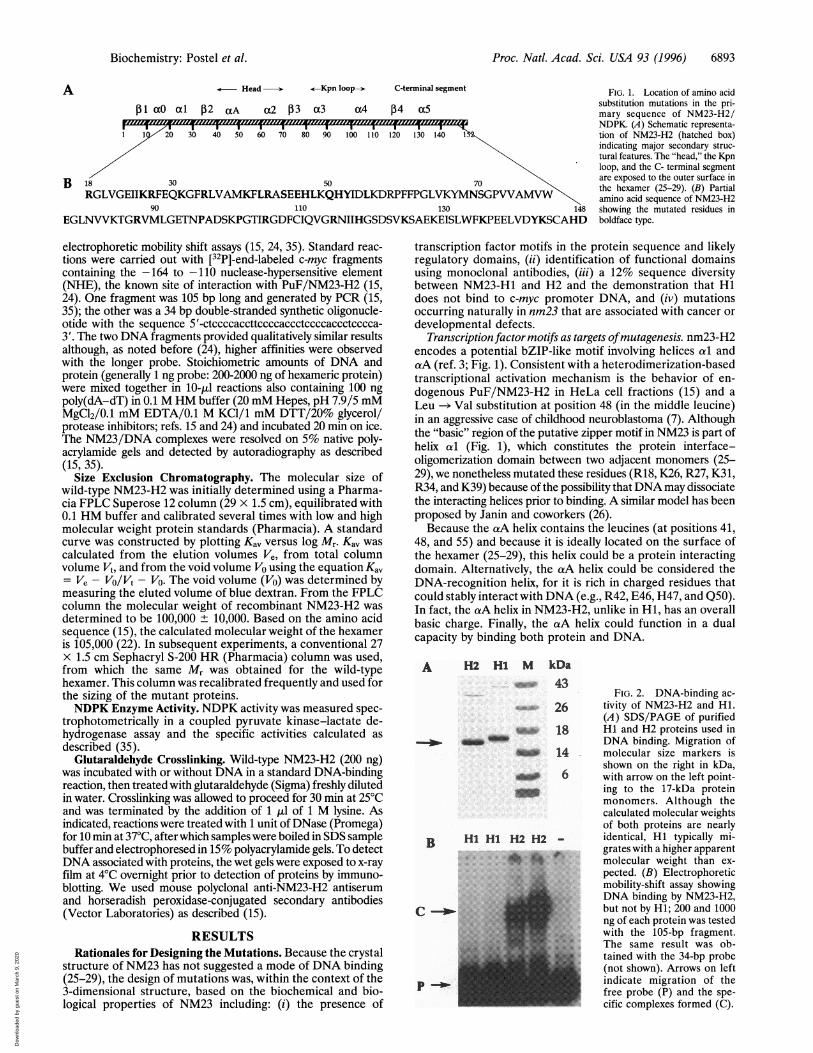
FIG. 2. DNA-binding ac-
tivity of NM23-H2 and Hi.(A) SDS/PAGE of purifiedHi and H2 proteins used inDNA binding. Migration ofmolecular size markers isshown on the right in kDa,with arrow on the left point-ing to the 17-kDa proteinmonomers. Although thecalculated molecular weightsof both proteins are nearlyidentical, Hi typically mi-grates with a higher apparentmolecular weight than ex-
pected. (B) Electrophoreticmobility-shift assay showingDNA binding by NM23-H2,but not by H1; 200 and 1000ng of each protein was testedwith the 105-bp fragment.The same result was ob-tained with the 34-bp probe(not shown). Arrows on leftindicate migration of thefree probe (P) and the spe-cific complexes formed (C).
A
Biochemistry: Postel et al.
Dow
nloa
ded
by g
uest
on
Mar
ch 9
, 202
0

Proc. Natl. Acad. Sci. USA 93 (1996)
Table 1. Properties of NM23-H2/NDPK missense mutants
MutationWild typeR18GK26GK26QR27GR27QR27VK26G + R27GK31GK31A + P101GR34GR34AR34DK39AL41ML41M + R42QR42QL41M + L48VL48VL48V + L55VL41M + L48V + L55VS44AE46LH47DQ50EH51FY52VStop69N69H
E79AE79A + P62AR88AP96SPlolsP101GR1O5AR114AH118FK124EK124E + K135HK135HY142AK143TH147FD148N
Expressionand stability DNAin E. coli* bindingt
+ +
+ +
+ +
+ +
+
+
+_
+_
+ +
+ ++ ++ +
+ ++ ++ ++ +
+ ++ ++ ++ ++ +_ +
+++ +
+ ++ ++ ++ ++ ++ ++ +
+ +
+++ ++ +
NDPK Oligomerizactivityt state§
+ Hexamer
Mixed
Hexamer
Hexamer
+
+ Hexamer
+ Hexamer
+
Hexamer
- Ref. 35
+ Hexamer
+ Hexamer
*Normal expression is indicated by +, its absence by -. Reduced or
abnormal protein expression/and or stability is indicated by ±.
tApproximately normal DNA binding measured as specific activity(intensity of sequence specific shifted complex/amount of protein inelectrophoretic mobility-shift assay) is indicated by +; completeabsence of DNA binding or a residual activity <5 percent of the WTis indicated by -. All proteins, with the few noted exceptions, showedwild-type levels of DNA binding.tNDPK activity was measured in a coupled assay as described (35). +,
specific activities comparable to wild-type levels. See Table 2 forspecific activity values of DNA-binding mutants.§As determined by SEC. All proteins examined were determined to behexameric, with the exception of the K31A/P101G mutant, which wasa mixture of different order oligomers.
Another DNA-binding mode is suggested by the presence ofa potential HTH motif, comprising the aA-turn-a2 structuraldomains (Fig. 1). These two helices, referred to as the "head"of the NM23-H2 molecule (29), in fact protrude on the sideand, although not properly oriented for DNA binding (25),they could, after minor conformational changes, directly ac-
cess the DNA substrate. In the HTH model, the aA helixwould be considered the probe helix for DNA binding.Combined functional and immunological approach points to
a2/j33 as involved in DNA binding. As a preliminary step to
mutagenesis we used monoclonal antibodies raised againstNM23-H2 to identify functionally important domains. Onemonoclonal antibody, mAb3E4, which inhibited DNA bindingby both HeLa and recombinant NM23-H2, recognized anepitope consisting of residues 63-79 located to the a2/03(E.H.P. and C. Ferrone, unpublished results). This findingfurther raised interest in the HTH motif and the possibility thathelix a2 is involved in DNA recognition, either alone, or as partof another as yet unidentified motif.Sequence diversity between NM23-Hl andH2 may account for
their disparity in DNA binding. The possibility that the closelyrelated NM23 variants Hi and H2 might differ in theirDNA-binding potential was suggested by their differences incharge (22), regulation (3), and amino acid sequence ofpotential regulatory regions, including helix aA and the ex-tended C-terminal fragment (25-29). These assumptions wereborne out by experimental evidence. First, we tested purifiedhuman erythrocyte NDPK A/Hi and B/H2 enzymes (22) andobserved significant-differences in terms of their DNA-bindingpotential to c-myc promoter DNA (E.H.P., unpublished re-sults). Second, Hildebrandt et al. (36) reported the absence ofbinding by NM23-H1/NDPK-A to a c-myc NHE oligonucle-otide (unpublished results). Third, we demonstrate in thisreport that NM23-Hl does not bind to the - 164 to -110 c-mycpromoter sequence (Fig. 2). We therefore substituted all of thenonconserved H2 residues with the potential for H-bondingwith DNA, with Hi amino acids, including R42Q, E46L,H47D, Q50E (aA helix) and K124E, K135H, K143T, H147F,and D148N (C terminus). Interestingly, one of the uncon-served residues, Asn-69, located at the 3' end of the a2 helix,is within the critical DNA-binding epitope recognized bymAb3E4. Asn-69 is replaced by His-69 in NM23-Hl, is on theprotein surface (26, 28, 29), and is capable of sequence specificcontact with DNA (37). Based on these considerations we madethe Asn-69 -* His-69 substitution a major target of our studies.
Naturally occurring mutations associated with cancer or de-velopment may have a DNA-binding defect. Because most of thenaturally occurring mutations in the tumor suppressor proteinp53 disrupt DNA-binding interactions (38), we might expectthat similar mutations in NM23, a putative metastasis sup-pressor, may also be involved in DNA binding. To date, onlya handful of NM23 mutations have been identified that areassociated with cancer or abnormal development. One ofthese, discussed above, is the L48V mutation found in neuro-blastoma (7). Another possibility is the conditional lethal killerofprune (kpn) mutation in Awd, the Drosophila NM23 homo-logue (11). Kpn is a Pro -- Ser substitution at position 97 (P96in NM23-H2), located within a structural motif called the "Kpnloop" (named after the mutation). The Kpn loops are consid-ered critical for trimeric interactions with neighboring subunitsand are exposed on the top and bottom surface of the hexamer(25-29). The involvement of the Kpn loops in DNA bindingwould thus require substantial conformational changes. Inaddition to P96S, we also targeted residues R88, P101, andR114 in the loop, because of their potential, based on charge,of influencing DNA binding. Another residue of interest wasSer-44, whose phosphorylation has been associated with themetastatic phenotype (39), and, although conserved betweenHi and H2, this serine is near the surface of the molecule andcould thus readily make contact with DNA.
Properties of Site-Directed Mutations. A total of 30 indi-vidual amino acids were targeted for mutagenesis and, includ-ing double and triple substitutions, over 40 mutant proteinswere examined for DNA-binding activity. Those found rele-vant or interesting were also examined for NDPK activity andoligomerization state. The mutations (Table 1) can be classi-fied according to the following phenotypes: (i) mutations thathave no effect on protein stability and activity (the majority),(ii) those that affect intrinsic stability and solubility of theprotein (six), and (iii) mutations that disrupt DNA binding(three). Mutations belonging to category 2 involving residuesK31, Y52, and P96 affected solubility (salting out by ammo-nium sulfate) and/or stability (hexameric conformation) to
6894 Biochemistry: Postel et al.
Dow
nloa
ded
by g
uest
on
Mar
ch 9
, 202
0
Proc. Natl. Acad. Sci. USA 93 (1996) 6895
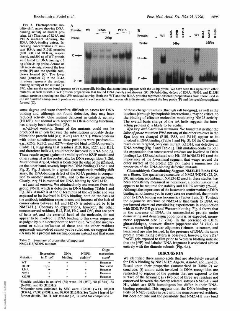
FIG. 3. Electrophoretic mo- Ability-shift assays showing DNA- otein: Wt R34A PlOlS B pbinding activity of mutant pro- t nBteins. (A) Titration of R34A and prbe 34bp Protein: Wil'PlOlS mutants showing theR34A DNA-binding defect. In- Probe 34bp lOSbpcreasing concentrations of mu-tant R34A and P1O1S proteins MN 1......(100, 300, and 1000 ng, respec- ;!kF
tively), and 500 ng ofWT protein C..........-were tested for DNA binding to 1 M.......ng of the 34-bp probe. Arrows onleft indicate migration of the freeprobe (P) and the specific com- r.plexes formed (C). The lowerband (complex C) in the R34A p-wtitrations represent the residual Pbinding activity of the mutant (<5%), whereas the upper band appears to be nonspecific binding that sometimes appears with the 34-bp probe. We have seen this signal with othermutants, as well as with a WT protein preparation that bound DNA poorly (not shown). (B) DNA-binding defect of R34A, N69H, and K135Hmutant proteins showing less than 5% residual activity. Both the WT and the R34A proteins represent different preparations from those used inA. Five hundred nanograms of protein were used in each reaction. Arrows on left indicate migration of the free probe (P) and the specific complexesformed (C).
some degree and were therefore difficult to assess for DNAbinding and, although not found defective, they may havereduced activity. One mutant deficient in catalytic activity(H118F), but normal with respect to DNA-binding functions,has already been described (35).
al-fP2-aA mutants. Some of the mutants could not beproduced in E. coli because the substitutions probably desta-bilized the protein fold (e.g., K26Q and R27G). When proteinswith other substitutions at these positions were produced-e.g., K26G, R27Q, and R27V-they did bind to DNA normally(Table 1), suggesting that residues R18, K26, R27, and K31,and therefore helix al, could not be involved in DNA binding.These results argue against the validity of the bZIP model andothers using cai as the probe helix for DNA recognition (3, 26).Mutations in Arg-34, which is located on the edge of the (32 sheet,on the other hand, severely impaired DNA binding (Table 1 andFig. 3). Fig. 3 shows, in a typical electrophoretic mobility-shiftassay, the DNA-binding defect of the R34A protein in compar-ison to another mutant, PlOlS, and to the wild-type proteins.Clearly, Arg-34 is essential for DNA binding by NM23-H2.
caA turn a2 mutants. We obtained only one mutant from thisgroup, N69H, which is defective in DNA binding (Table 1 andFig. 3B). Asn-69 is at the C terminus of the a2 helix and wasexpected to be involved in DNA binding, both on the basis ofthe antibody inhibition experiments and because of the lack ofconservation between Hi and H2 (N is substituted by H inNM23-H1). Contrary to expectations, however, the othernonconserved residues, R42, E46, H47, and Q50, that are partof helix caA and the external head of the molecule, do notappear to be involved in DNA binding to this c-myc sequenceas judged by our electrophoretic technique. While the involve-ment or subtle influence of these and the other residuesapparently uninvolved cannot yet be ruled out, we suggest thatcaA may be a protein interacting domain instead and that some
Table 2. Summary of properties of importantNM23-H2/NDPK mutants
Oligo-Expression DNA NDPK merization
Mutation in E. coli binding activity* statetWT + + + HexamerH118F +/ x + - Not testedR34A + - + HexamerN69H + - + HexamerK135H + - + Hexamer
*Specific activities in units/ml (35) were 105 (WT), 90 (R34A), 84(N69H), and 83 (K135H).
tMolecular sizes estimated by SEC were: 102,000 (WT), 105,000(R34A), 97,000 (N69H), and 98,000 (K135H). See Table 1 legend forfurther details. The H118F mutant (35) is listed for comparison.
of these charged residues (through salt bridging), as well as theleucines (through hydrophobic interactions), may be critical tothe binding of effector molecules modulating NM23 activity.The overall basic charge of the ciA helix suggests the inter-acting protein(s) is likely to be acidic.Kpn loop and C-terminal mutations. We found that neither the
killer-of-prune mutation P96S nor any of the other residues in theKpn loop we changed (PlOi, R88, and R114) appear to beinvolved in DNA binding (Table 1 and Fig. 3). Of the C-terminalresidues we targeted, only one mutant, K135H, was defective inDNA binding (Fig. 3 and Table 1). This mutation confirms boththe expectation that unconserved residues are involved in DNAbinding (Lys-135 is substituted with His-135 in NM23-Hl) and theimportance of the C-terminal segment that wraps around theouter surface of the protein (28, 29). Table 2 summarizes theproperties of the DNA-binding defective mutants.
Glutaraldehyde Crosslinking Suggests NM23-H2 Binds DNAas a Dimer. The quatemary structure of NM23/NDPK (22, 28,29), including recombinant NM23-H2 used in these studies, hasbeen consistently measured as hexameric, a conformation thatappears to be required for stability and NDPK activity (26-28).Although the importance of the hexameric conformation to DNAbinding is not known yet, in every case we examined, the proteinprior to DNA binding was hexameric (Tables 1 and 2). To studythe oligomeric structure of NM23-H2 that binds to DNA weperformed chemical crosslinking experiments in conjunctionwith SDS/PAGE gels and Western blotting. Fig. 4 shows thatin the abscence of DNA, the uncrosslinked protein underdissociating and denaturing conditions is, as expected, mono-meric (apparent size 17 kDa). In the presence of 0.01%glutaraldehyde, however, dimers (apparent size 34 kDa), aswell as some higher order oligomers (trimers, tetramers, andhexamers) are also formed. In the presence of DNA, the sameprotein crosslinking pattern is observed; however, the SDS/PAGE gels exposed to film prior to Western blotting indicatethat the [32P]-end-labeled DNA fragment is associated almostentirely with the dimeric subunit (Fig. 4A).
DISCUSSIONWe identified three amino acids that are absolutely essentialfor DNA binding by NM23-H2: Arg-34, Asn-69, and Lys-135.Based upon their properties (summarized in Table 2) weconclude: (i) amino acids involved in DNA recognition arerestricted to regions of the protein that are exposed to thesurface of the hexamer; (ii) two out of three are residues notconserved between the closely related isotypes NM23-H2 andHi, which are 88% homologous but differ in their DNA-binding potential. This suggests that the DNA-binding speci-ficity of NM23 resides in part with the nonconserved residues,but does not rule out the possibility that NM23-Hl may bind
Biochemistry: Postel et al.
Dow
nloa
ded
by g
uest
on
Mar
ch 9
, 202
0

Proc. Natl. Acad. Sci. USA 93 (1996)
ADNA + + - _ BDNAGlutarald. - + - + Glutarald.
wells _-
+ +- + _ +
wells -woH _
D -
i--em'
M_-11 j_ _.M -.
digestedDNA -w
to a different DNA sequence specified by the histidines. (iii)All three DNA-binding defective mutant proteins are activeenzymatically and appear to be stable hexamers, implying thatArg-34, Asn-69, and Lys-135 perform at the level of DNArecognition and not through stabilization of the NM23 fold.Although the Arg-34 side chain is not fully exposed (ref. 28;Fig. 5), it could still contact DNA directly after minor con-formational changes such as an induced curvature on theprotein, or it could make contact with a protruding structuralcomponent of the DNA. Another possibility is that R34 isimportant to DNA binding for structural reasons, e.g., byholding the C terminus in place, as was suggested by Y. W. Xuand J. Janin (personal communication) on the basis of mod-elling of Arg-34 side chain interactions with Y142 (C terminus)
FIG. 4. Glutaraldehyde crosslinking ofNM23-H2 to DNA shows dimeric binding. Aglutaraldehyde titration experiment (notshown), using concentrations between 0.001-0.1%, indicated that the 0.01% range was op-timal; at the higher concentrations of glutaral-dehyde NM23-H2 formed artifactual crosslinksthat resisted electrophoresis and also degradedthe DNA. (A) Autoradiograph of wet SDS/PAGE gel prior to immunoblotting. Arrows onleft indicate expected positions of monomers(M) and dimers (D). (B) Immunoblot of thesame gel. Arrows on left indicate the expectedpositions of monomers (M), dimers (D), trim-ers (Tr), tetramers (Te), and hexamers (H)based on the migration of molecular size mark-ers (not shown).
and E79 (132). (iv) All three mutants function normally asNDP kinases, thus confirming our earlier conclusions, drawnfrom the properties of the active site H118 mutation, thatthere exist two separate functional domains for NM23/NDPK: a catalytic domain and a DNA-recognition domain.These data also imply that NDPK activity and DNA bindingare not mutually exclusive functions, thus allowing for apossible interaction of the two domains in vivo in a capacitythat requires both DNA binding and nucleotide phosphor-ylation, such as might occur during DNA and RNA synthesis.Experiments using cell transfection assays (16) are currentlyin progress to assess the requirement of the NDPK functionand DNA binding in the transactivation of a c-myc promoterin the cell.
FIG. 5. Space-filling model of the NM23-H2 hexamer. Residues implicated in DNA binding are in red, GDP bound to the NDPK active sitein cyan (28). The two subunits of the front dimer are shown in green and purple, the other two dimers are in grey. The model was drawn withthe GRASP computer graphics program.
6896 Biochemistry: Postel et al.
Dow
nloa
ded
by g
uest
on
Mar
ch 9
, 202
0

Proc. Natl. Acad. Sci. USA 93 (1996) 6897
Although each of the residues critical for DNA binding arelocated on separate structural motifs (Arg-34 at the edge of (32,Asn-69 on the C terminus of the a2 helix, and K135 on theunstructured C-terminal segment; Fig. 1), they line up alongthe equator on the "outer" face of the hexamer (Fig. 5). Amodel for these findings is provided on the basis of the knowncrystal structure (28, 29). The NM23/NDPK hexamer consistsof three vertical dimers around a 3-fold axis, or two horizontaltrimers along a 2-fold axis. The dimers are held together by twoat helices that are in antiparallel contact with each other.DNA-binding residues Arg-34, Asn-69, and Lys-135 are clus-tered around the interface of the dimer (Fig. 5). As our geneticdata exclude the dimer interface from participating in DNAbinding, we suggest a model in which DNA makes contact withresidues of two adjacent monomers near the 2-fold axis and onthe outer surface of the hexamer. This creates a combinatorialDNA-binding surface with possibly a single active site com-posed of residues donated by both monomers of a dimer.We have previously reported that HeLa PuF/Nm23-H2
makes contact with the major groove of DNA (24). Themethylation interference contacts GGGTGGG (repeated 3times within the c-myc promoter fragments used in thesestudies) are consistent with asymmetric binding and the pos-sibility that some of the DNA-binding residues make contactwith opposite sides of the major groove of DNA. The dimen-sions of the dimer interface ("30 A) are compatible withcontact by a single turn of B-DNA. However, as the structureof the c-myc promoter DNA to which NM23-H2 binds is unor-thodox and is essentially unknown, modelling with B-DNA maynot be fruitful in this case. The structure of a cocrystal ofNM23-H2 with the c-myc promoter sequence should clarify theseinteractions and any others not detected by this genetic study.
Chemical crosslinking data support the present model bysuggesting that at least two subunits of NM23-H2, i.e., a dimer,may be required for making contacts with DNA (Fig. 4);although on the basis of our present data we cannot rule out thepresence of higher order complexes on the DNA. Photocrosslink-ing of endogenous NM23-H2 in B-cell extracts (17) have alsoindicated that a 34-kDa species (dimer) is making contact withc-myc DNA, as well as the active component in the deregulationof the c-myc gene by NM23-H2 in Burkitt lymphoma cells.The model for DNA binding by NM23-H2 presented here is
inconsistant with theoretical models proposed earlier (3, 26), aswell as being incompatible with a more recent model suggestedby Webb et al. (29), in which DNA binding occurs on the side ofthe molecule covered by helices aA and a2 also involving theC-terminal fragment, and with that of Morera et al. (28), in whichDNA binds to a palm-like domain involving ,3 sheets, helices tA,a2, and o4. These latter models imply the existence of sixDNA-binding domains and/or require major conformationalchanges in the hexamer. The model we present here has anexperimental basis as well as being simpler, in that DNA bindingrequires no major conformational changes and the functionalsubunit making contact with DNA is the stable dimer. Our modelalso implies that in addition to the physical separation of thecatalytic and the DNA-binding domains, these two functions maybe modulated by the oligomerization state of the protein: forexample, DNA binding could be accomplished by the dimer, butfor enzyme catalysis, the hexamer may be the active structure(30). If the DNA is presented with a hexamer but presumably onlyone of the dimers makes contact, this may explain the relativelyhigh (,tM) dissociation constant that we have observed in all ofour DNA-binding studies.
In summary, we investigated the DNA-binding mechanismof NM23-H2/NDPK. Our results, based on site-directed mu-tational analysis, suggest that the DNA-binding specificity ofNM23-H2 resides in nonconserved surface residues located ona different functional domain from that used for enzymecatalysis. In our model, NM23-H2 binds DNA as a dimer, withthe DNA-binding region consisting of residues donated by twoadjacent monomers. This provides a combinatorial DNA-binding surface located on the front "face" and along the
2-fold axis of the dimer (Fig. 5). Chemical crosslinking datasupport a dimeric DNA-binding mode.We thank J. Janin for helpful discussions, for the modelling of R34
interactions, and for structural information including the coordinatesfor NM23-H2. We also thank I. Lascu for suggestions and forerythrocyte NDPKs and M. J. Lacombe for the NDPK-AcDNA clone.We are grateful to F. Hughson for making Fig. 5 and for discussionsand S. J. Flint for review of the manuscript. The technical assistanceof C. F. Conroy is acknowledged. This work was supported by NationalInstitutes of Health Grant ROI CA5584 to E.H.P.1. Steeg, P. S., Bevilacqua, G., Kopper, L., Thorgeirsson, U. P., Talmadge,
J. E., Liotta, L. A. & Sobel, M. E. (1988) J. Natl. Cancer Inst. 80, 199-206.2. Steeg, P. S., De La Rosa, A., Flatow, U., MacDonald, N. J., Benedict, M.
& Leone, A. (1993) Breast Cancer Res. Treatment 25, 175-187.3. Stahl, J. A., Leone, A., Rosengard, A. M., Porter, L., King, C. R. & Steeg,
P. S. (1991) Cancer Res. 52, 445-449.4. Nakayama, T., Ohtsuru, A., Nakao, K., Shima, M., Nakata, K., Watanabe,
K., Ishii, N., Kumura, N. & Nagataki, S. (1992) J. Natl. Cancer Inst. 84,1349-1354.
5. Nakayama, H., Yasui, W., Yokozaki, H. & Tahara, E. (1993) Jpn. J. CancerRes. 84, 184-190.
6. Hailat, N., Keim, D. R., Melhem, R. F., Zhu, X., Eckerskorn, C., Broudeur,G. M., Reynolds, C. P., Seeger, R. C., Lottspeich, F., Strahler, J. R. &Hanash, S. M. (1991) J. Clin. Invest. 88, 342-345.
7. Leone, A., Seeger, R. C., Hong, C. M., Hu, Y. Y., Arboleda, M. J., Broderu,G. M., Stram, D., Slamon, D. J. & Steeg, P. S. (1993) Oncogene 8,855-865.
8. Keim, D., Hailat, N., Melhem, R., Zhu, X. X., Lascu, I., Veron, M.,Strahler, J. & Hanash, S. M. (1992) J. Clin. Invest. 89, 919-924.
9. Ohneda, K., Fukuda, M., Shimada, N., Ishikawa, N., Ichou, T., Kaji, K.,Toyota, T. & Kimura, N. (1994) FEBS Lett. 348, 273-277.
10. Dearolf, C. R., Tripoulas, N., Biggs, J. & Shearn, A. (1988) Dev. Biol. 129,169-178.
11. Biggs, J., Tripoulas, N., Hersperger, E., Dearolf, C. & Sheam, A. (1988)Genes Dev. 2, 1333-1343.
12. Okabe-Kado, J., Kasukabe, T., Honma, Y., Hayashi, M., Henzel, W. J. &Hozumi, M. (1992) Biochem. Biophys. Res. Commun. 182, 987-994.
13. Okabe-Kado, J., Kasukabe, T., Hozumi, M., Honma, Y., Kimura, N., Baba,H., Urano, T. & Hiroshi, S. (1995) FEBS Lett. 363, 311-315.
14. Venturelli, D., Martinez, R., Melotti, P., Casella, I., Peschle, C., Cucco, C.,Spampinato, G., Darzynkiewicz, Z. & Calabretta, B. (1995) Proc. Natl.Acad. Sci. USA 92, 7435-7439.
15. Postel, E. H., Berberich, S. J., Flint, S. J. & Ferrone, C. A. (1993) Science261, 478-480.
16. Berberich, S. J. & Postel, E. H. (1995) Oncogene 10, 2343-2347.17. Ji, L., Arcinas, M. & Boxer, L. M. (1995) J. Biol. Chem. 270, 13392-13398.18. Agarwal, R. P., Robinson, B. & Parks. R. E. (1978) Methods Enzymol. 51,
376-386.19. Postel, E. H. (1996) Attempts to Understand Metastasis Formation: Vol II,
Regulatory Factors, Current Topics In Microbiology and Immunology, inpress.
20. Backer, J. K., Mendola, C. E., Kovesdi, I., Fairhurst, J. L., O'Hara, B. &Eddy, R. L., Jr. (1993) Oncogene 8, 497-502.
21. Howlett, A. R., Petersen, 0. W., Steeg, P. S. & Bissell, M. J. (1994) J. Natl.Cancer Inst. 86, 1838-1844.
22. Gilles, A.-M., Presecan, E., Vonica, A. & Lascu, I. (1991)J. Biol. Chem. 266,8784-8789.
23. Arcinas, M. & Boxer, L. M. (1994) Oncogene 9, 2699-2706.24. Postel, E. H., Mango, S. E. & Flint, S. J (1989 MoL Cell. Biol. 9,5123-5133.25. Dumas, C., Lascu, I., Morera, S., Glaser, P., Fourme, R., Wallet, V.,
Lacombe, M.-L., Veron, M. & Janin, J. (1992) EMBO J. 11, 3203-3208.26. Chiadmi, M., Morera, S., Lascu, I., Dumas, C., Le Bras, G., Veron, M. &
Janin, J. (1993) Structure 1, 283-293.27. Mordra, S., LeBras, G., Lascu, I., Lacombe, M. L., Veron, M. & Janin, J.
(1994) J. Mol. Biol. 243, 873-890.28. Mor6ra, S., Lacombe, M.-L., Yingwu, X., LeBras, G. & Janin, J. (1995)
Structure 3, 1307-1314.29. Webb, P. A., Perisic, O., Mendola, C. E., Backer, J. M. & Williams, R. L.
(1995) J. Mol. Biol. 251, 574-587.30. Lascu, I., Chaffotte, A., Limbourg-Bouchon, B. & Veron, M. (1992)J. Biol.
Chem. 268, 20268-20275.31. Sastre-Garau, X., Lacombe, M. L., Jouve, M., Veron, M. & Magdelenat, J.
(1992) Int. J. Cancer 50, 533-538.32. Spencer, C. A. & Groudine, M. (1991) Adv. Cancer Res. 56, 1-48.33. Marcu, K. B., Bossone, S. A. & Patel, A. J. (1992) Annu. Rev. Biochem. 61,
809-860.34. Prendergast, G. C. & Ziff, E. B. (1992) Trends Genet. 8, 91-97.35. Postel, E. H. & Ferrone, C. A. (1994) J. Biol. Chem. 269, 8627-8630.36. Hildebrandt, M., Lacombe, M.-L., Passeron, S. & Veron, M. (1995) Nucleic
Acids Res. 23, 3858-3864.37. Suzuki, M. & Yagi, N. (1994) Proc. Natl. Acad. Sci. USA 91, 12357-12361.38. Cho, Y., Gorina, S., Jeffrey, P. D. & Pavletich, N. P. (1994) Science 265,
346-355.39. McDonald, N. J., De La Rosa, A., Benedict, M. A., Freije, J. M. P., Kritch,
H. & Steeg, P. S. (1993) J. Biol. Chem. 268, 25780-25789.
Biochemistry: Postel et al.
Dow
nloa
ded
by g
uest
on
Mar
ch 9
, 202
0