prune camp phosphodiesterase binds nm23-h1 and ... prune camp phosphodiesterase binds nm23-h1 and...
TRANSCRIPT
A R T I C L E
Prune cAMP phosphodiesterase binds nm23-H1 and promotescancer metastasis
Anna D’Angelo,1 Livia Garzia,1 Alessandra Andre,1 Pietro Carotenuto,1 Veruska Aglio,1
Ombretta Guardiola,1 Gianluigi Arrigoni,2 Antonio Cossu,3 Giuseppe Palmieri,4 L. Aravind,5
and Massimo Zollo1,*
1Telethon Institute of Genetics and Medicine, Via Pietro Castellino 111, 80131 Naples, Italy2 Department of Anatomy and Pathology, Ospedale San Raffaele, HSR, Milan, Italy3 Azienda A.S.L. n� 1, Sassari, Department of Pathology, University of Sassari, Italy4 Istituto di Chimica Biomolecolare, Sezione di Sassari, Consiglio Nazionale delle Ricerche Tramariglio, Alghero, Italy5 Computational Biology Branch, National Institutes of Health, National Center for Biotechnology Information, The NationalLibrary of Medicine, Bethesda, Maryland 20894*Correspondence: [email protected]
Summary
We identify a new enzymatic activity underlying metastasis in breast cancer and describe its susceptibility to therapeuticinhibition. We show that human prune (h-prune), a phosphoesterase DHH family appertaining protein, has a hithertounrecognized cyclic nucleotide phosphodiesterase activity effectively suppressed by dipyridamole, a phosphodiesteraseinhibitor. H-prune physically interacts with nm23-H1, a metastasis suppressor gene. The h-prune PDE activity, suppressedby dipyridamole and enhanced by the interaction with nm23-H1, stimulates cellular motility and metastasis processes. Outof 59 metastatic breast cancer cases analyzed, 22 (37%) were found to overexpress h-prune, evidence that this novelenzymatic activity is involved in promoting cancer metastasis.
Introduction domains. The first four motifs are the most conserved portionsof the N-terminal domain and define the active site of theseenzymes, whereas the group-specific fifth motif maps to theThe human prune (h-prune) protein belongs to the DHH super-
family, which includes several phosphoesterases, such as the divergent C-terminal domains.The Drosophila prune gene was originally characterizedRecJ nuclease from bacteria and the pyrophosphatases from
yeast and bacteria (Aravind and Koonin, 1998b). The DHH su- based on its mutant phenotype, which showed a brownish-purple eye color due to the reduction of drosopterins, in contrastperfamily can be divided into two main groups on the basis of
a C-terminal motif that is very well conserved within each group to the bright red eye of the wild-type fly (Timmons and Shearn,1996). While homozygous prune mutants are viable and fertile,but not across the groups. All of the members of this superfamily
possess four other motifs that contain highly conserved charged they are synthetically lethal, developing pseudomelanotic tu-mors in the presence of even a single copy of the gain-of-residues that are predicted to be responsible for binding ions
and catalyzing the phosphoesterase reaction. The most charac- function mutation in the abnormal wing disc gene (awd/K-pn;also named Killer-of-Prune). Humans encode up to eight or-teristic of these is the third motif, with the signature DHH (Asp-
His-His), after which this superfamily was named. The RecJ thologs of awd (nm23s), at least four of which are active nucleo-side diphosphate kinases (NDPKs), which catalyze the phos-protein is a DNA repair protein and, along with other nucleases
and poorly characterized bacterial proteins, belongs to the first phoryl transfer from a nucleoside triphosphate to a nucleosidediphosphate (Lombardi et al., 2000). One of these, nm23-H1group. Prune and the polyphosphatases belong to the second
group. The recent availability of the structures of the RecJ pro- (NDPK-A), is a predominantly cytoplasmic protein with antimet-astatic properties, and mutations in it are associated with thetein (Yamagata et al., 2002) and the bacterial pyrophosphatases
(PPASEs) (Ahn et al., 2001) reveal that these two major classes progression of cancers. Numerous tumors and highly prolifera-tive cells overexpress nm23-H1 mRNA and protein, and in mostof DHH proteins share an N-terminal �/� domain with a five-
stranded parallel sheet but have somewhat different C-terminal cases this overexpression is linked to early stages of cancer,
S I G N I F I C A N C E
H-prune is frequently overexpressed, and nm-23 is often downregulated in metastatic cancers in different tissues. Our work providesmechanistic insight into how the interaction between these molecules can control cancer metastasis. A major pathway identifiedby this work is the binding of nm23-H1 and h-prune with physiological consequences. By identifying a novel enzymatic activity ofh-prune required for its metastasis-promoting potential, these results open the possibility of therapeutic intervention. Dipyridamole,a selective inhibitor of h-prune activity, is already used in the clinic, which could facilitate clinical trials to address its potential useas a suppressor of metastasis.
CANCER CELL : FEBRUARY 2004 · VOL. 5 · COPYRIGHT 2004 CELL PRESS 137

A R T I C L E
with a loss of expression in more advanced and aggressive line clone MDA-C100 has been shown to significantly reduceits metastatic phenotype both in vitro and in vivo (Hartsoughstages. In breast cancer and in melanomas, high expression of
human nm23-H1 is associated with a decreased metastatic and Steeg, 2000; Mao et al., 2001; Tseng et al., 2001), we showthat h-prune is able to change the “low” motility phase to apotential (Florenes et al., 1992). In contrast, in other cancers,
such as prostate, non-Hodgkin lymphomas, and neuroblasto- “high” motility phase. In a panel of eight PDE selective inhibitorstested, dipyridamole, an anti-platelet aggregation drug, wasmas, high nm23-H1 expression is associated with an adverse
outcome (Hartsough and Steeg, 2000; Niitsu et al., 2001). To found to inhibit h-prune PDE activity both in vitro and in ourcellular breast model. Further in vivo analyses in a large numberdate, several mutations that affect nm23s folding, oligomeriza-
tion, DNA binding, or NDPK activity have been described for of metastatic breast tumors showed a direct correlation betweenh-prune protein increased levels and lower regulation of nm23-nm23-H1. In particular, the nm23H1-P96S mutation, a Drosoph-
ila developmental mutation homolog (awd/K-pn), exhibits auto- H1 expression, thus leading to the formation of distal metastasisas reported in our clinical follow-up records. A direct correlationphosphorylation and NDPK normal function, while it is deficient
for phosphotransferase activity and shows failure in folding between an increased h-prune cAMP-PDE activity and cellularmotility, due to a physical protein-protein interaction with nm23-properties associated to oligomeric nm23-H1 protein complexes
(Freije et al., 1997a). A second mutation, nm23H1-S120G, associ- H1, was found in the breast cancer model.These findings throw light on the interaction betweenated with stage IV-S neuroblastoma (Chang et al., 1994) shows
a positive effect on tumor cell motility (MacDonald et al., 1996). h-prune and nm23-H1, altering its protective function in cellularproliferation and tumor metastasis processes. Thus, this repre-In breast cancer, metastatic spread is responsible for virtu-
ally all cancer deaths. Metastasis is a highly complex molecular sents a novel cellular pathway that is involved in terminal differ-entiation and malignancy of tumors.and cellular process (Sleeman, 2000). To become invasive, tu-
mor cells need to change their adhesive properties in order tolose contact with other cells in the primary tumor and make Resultsnew contacts with the extracellular matrix of host cells that theyencounter as they invade. They also need to be able to penetrate Structural and functional analysis of h-prune
An iterative protein database search (PSI-BLAST) initiated oninto the surrounding host tissue, and here the modulation ofprotease activity in the vicinity of the tumor cells plays a critical the h-prune protein recovered, with statistically significant ex-
pectation values, the eukaryotic orthologs of prune, followedrole. To migrate away from the primary tumor, tumor cells alsoneed to gain motility functions. These same properties are also by the inorganic pyrophosphatases from a variety of bacteria
and diverse DHH proteins from various organisms, includingthought to be important when circulating tumor cells exit thecirculatory system and start metastatic colonization in second- the RecJ nucleases (Figures 1A and 1C). A clustering of the DHH
proteins using the BLASTCLUST program and a phylogeneticary organs. To date, several metastasis suppressor genes havebeen isolated and characterized (Steeg, 2003). Within this group, analysis using the maximum likelihood method show that prune
proteins (human and Drosophila) belong to the second DHHnm23 plays a major role for its ability to induce low motilitycellular processes if overexpressed in aggressive breast cancer family, along with the inorganic pyrophosphatases (Figure 1A).
While the two families of DHH proteins share a commoncells (Freije et al., 1997a, 1997b; Hartsough et al., 2001), influ-encing anchorage-independent colonization and induction to N-terminal domain that contains the four conserved motifs typi-
cal of the DHH superfamily, they are distinguished from eachdifferentiation (Kantor et al., 1993; Leone et al., 1993a; Howlettet al., 1994; Hartsough and Steeg, 1998; Lombardi et al., 2000). other by their C-terminal domains. The shared N-terminal do-
main has an �/� fold with a parallel � sheet and contains theOf note is a new role of nm23-H1, alternatively named GzmA-activated Dnase, which creates single-stranded DNA nicks in motifs with the absolutely conserved signatures of the form DXD
(Motif-I), D (Motif-II), DHH (Motif-III), and D/E (Motif-IV) (Figuresthe nucleus in a caspase-independent apoptosis pathway (Fanet al., 2003). 1A and 1B). These residues are all on the same face of this
domain and together form the catalytic site that chelates at leastOur group has previously demonstrated the interaction be-tween h-prune and nm23-H1 and the disruption of this interac- two divalent cations. The prune proteins contain the form DHR
as substitution for the canonical DHH (Motif-III) that is observedtion by the nm23H1-S120G mutation (Reymond et al., 1999).In addition, we have demonstrated that amplification of h-prune in all other members of this family (Figure 1A). In both DHH
families, the C-terminal domain contains a core sheet of fivecopy numbers induces cell proliferation and that high levels ofh-prune expression, compared to the moderate or low nm23- strands, four of which form two � strand hairpins. However,
differences in the C-terminal domains between the first and theH1 levels, is correlated to aggressiveness of sarcoma andbreast carcinoma tumors, thus postulating an inhibitory role of second families of DHH proteins may contribute prominently to
substrate specificities of the two superfamilies. Additionally,h-prune versus the suppressor of metastasis nm23 functionin vivo (Forus et al., 2001). Given the h-prune prediction of a C-terminal to the DHH module, mammalian prune contains a
nonglobular extension, within which there are some conservedphosphoesterase activity and its association with NDPK familyproteins, we have investigated its precise biochemical proper- serines that may serve as a site for regulation through phosphor-
ylation.ties and its potential role in metastasis. We demonstrate thath-prune has phosphodiesterase (PDE) activity, with a preferen- Structural analysis of h-prune showed similarities to RecJ
(Yamagata et al., 2002) and pyrophosphatases proteins (Ahn ettial affinity for cAMP over cGMP as substrate. PDEs belong toa diverse superfamily that catalyze the hydrolysis of 3�,5�-cyclic al., 2001), thus suggesting potentially similar activities to those
proteins. However, their strong synergistic interactions with thenucleotides (cAMP; cGMP) to their corresponding nucleoside5�-monophosphates (Beavo and Brunton, 2002). In addition, as awd/K-pn-like NDPKs suggested that prune proteins might have
alternative substrates, such as nucleotides. Evolutionary studiesoverexpression of nm23-H1 in the aggressive breast cancer cell
138 CANCER CELL : FEBRUARY 2004
A R T I C L E
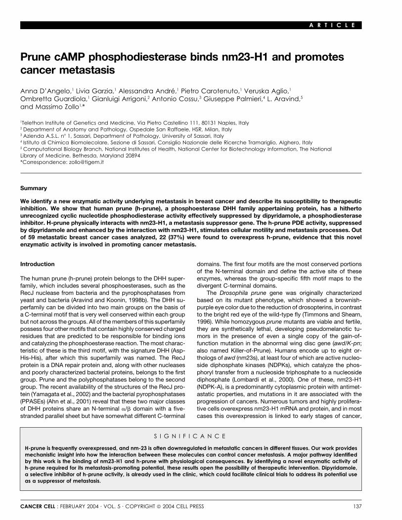
Figure 1. Alignment of DHH family phosphoesterases
A: Multiple alignment of the DHH family phosphoesterases, showing separately the four generic motifs (I–IV) and the motifs diagnostic of the two distinctsubfamilies that map to the second domain. The position of the first aligned residue in each protein sequence and the distances between the motifs areindicated by numbers. The Gene Identification (GI) numbers in the NCBI/GenBank protein sequence database are indicated to the right of each sequence.Species name abbreviations: Af, Archaeoglobus fulgidus; Bb, Borrelia burgdorferi; Bs, Bacillus subtilis; Dm, Drosophila melanogaster; Ec, Escherichia coli; Hi,Hameophilus influenzae; Hp, Helicobacter pylori; Lm, Leishmania major; Mg, Mycoplasma genitalium; Mj, Methanococcus jannaschii; Mp, Mycoplasmapneumoniae; Mt, Mycobacterium tuberculosis; Sc, Saccharomyces cerevisiae; Sg, Streptococcus gordonii; Ssp, Synechocystis species.B: Ribbon structure of the h-prune protein based on the crystal structure of PPASE and the RecJ protein. Red balls indicate potential cofactor ions (Mg2�
and/or Mn2�) and the region of binding to motif III. Arrows indicate the aspartic acids (D). Aspartic acids of the four DHH motifs are represented, indicatingthe potential catalytic site of DHH protein family.C: Ribbon structure of the RecJ protein. Red balls indicate cofactor ions (Mg2� and/or Mn2�) and the region of binding to motif III. Arrows indicate theaspartic acids (D) characteristic of DHH protein family.
CANCER CELL : FEBRUARY 2004 139

A R T I C L E
on the DHH family have shown that phosphoesterases are de- alternative phosphodiesterase assay (Fisher et al., 1998) (datarived from a number of protein folds that encode diverse phos- not shown). Thus, we present evidence of a cyclic nucleotidephoesterases and hydrolases. These include the HD fold, from phosphodiesterase activity for a protein of the DHH superfamily.which the classic signaling PDEs are recognized (PDE1-11) (Ara- To study the buffer influence on h-prune PDE activity, we testedvind and Koonin, 1998a, 1998b; Galperin et al., 1999); the met- Tris-HCl and HEPES buffers in the presence of the same salt,allo-�-lactamase fold (Galperin et al., 1999; Aravind, 1999), from and we observed a higher PDE activity in the presence of Tris-which the PdsA-like PDEs have derived; and the calcineurin- HCl buffer (Figure 2E). Considering the ion dependence of DHHlike phosphoesterase fold (Aravind and Koonin, 1998b), from proteins, we investigated the Mg2� and Mn2� ion dependencywhich Icc-like PDEs have derived. While the DHH catalytic do- of h-prune in the PDE cAMP assay. Although the higher activitymain has a very distinct fold from these other families, it contains of h-prune was found in Tris-HCl buffer, we performed PDEseveral analogous metal chelating residues (aspartates and his- assays, using increasing concentrations of two different divalenttidines) and could potentially define an entirely new class of ions, in the presence of HEPES buffer to avoid oxido-reductionPDEs. In order to test this hypothesis, we expressed, purified, reactions. Although some PDE activity was measured in theand assayed h-prune for potential PDE activity. no-ion buffer, Mg2� stimulated h-prune PDE activity (Figure 2E).
In contrast, in the presence of MnCl2, this activity is inhibitedIdentification and characterization of the h-prune (Figure 2F). In addition, the h-prune� mutant is not activatedphosphodiesterase activity by the Mg2� ions as well as the wild-type protein (Figure 2F),To determine the ability of h-prune to disrupt the phosphoester thus indicating that the motif III, modified in h-prune� mutant,bonds in cyclic nucleotides (cAMP and cGMP), we cloned and is necessary for phophodiesterase activity. In conclusion, weexpressed h-prune using the Baculovirus expression system. show here that h-prune cAMP-PDE activity is influenced posi-His-tagged h-prune and h-prune�, a mutation created in the tively by the Mg2� ion concentration.motif III region (DHRP126-129AAAA), were purified by affinitychromatography. We used the PDE assay to characterize puri- Stable breast MDA h-prune clones and correlationfied h-prune catalytic activity and to determine the specific sub- to cellular motilitystrate. As shown in Figure 2A, h-prune possesses significant To study the h-prune function in regulating nm23-H1 antimotilityPDE activity that is higher for cAMP than for cGMP as substrate, and suppressor metastasis activities, we have taken advantagewhile h-prune� shows a 40% reduction of this activity. As posi-
of the breast cancer cellular models MDA-C100 and H1-177tive control, we used PDE2. The negative controls were both
(Hartsough et al., 2001; Mao et al., 2001; Tseng et al., 2001).h-prune preincubated with the A59-specific polyclonal antibody
We produced several stable clones overexpressing theand h-prune� (Figure 2A). To confirm the PDE activity found,h-prune cDNA (clones #3 and #4), the h-prune� cDNA (cloneswe overexpressed h-prune and h-prune� transiently in human#10 and #11), the h-prune4D� cDNA (clones #19 and #20), andHEK-293 cells, followed by immunoprecipitation and then per-the PDE5A cDNA (clones #14 and #16) in MDA-C100 cells. Weformed a PDE assay on immunoprecipitated proteins (data notstabilized the h-prune cDNA in MDA-H1-177 overexpressingshown). These results indicate that h-prune protein, purifiednm23-H1 (clones #7 and #8), in MDA overexpressing nm23H1-both from insect and human cells, shows PDE activity.P96S (clones #4 and #5), and in MDA overexpressing nm23H1-To identify amino acids potentially involved in the catalyticS120G (clones #2 and #3). Several of these clones were charac-site, a mutation analysis at single and multiple sites affectingterized to determine the expression level of h-prune mRNA usingh-prune PDE activity was conducted. All the aspartic acids ofreal-time PCR analysis by TaqMan technology (Figure 3A). Wethe DHH characteristic motifs (Figure 1B) were mutated aloneselected four clones for their level of mRNA expression by quan-and in combination (Figure 2B). We expressed the mutants usingtitative analysis and copy number extrapolation, as comparedthe Baculovirus expression system and purified them to homo-to gene reference targets (GAPDH) (Figure 3B). In addition,geneity (with an 80% yield of purification). We tested the pro-Western blot analyses were performed to identify the expressionteins for cAMP-PDE activity and observed an 80% decrease inlevels of the h-prune, nm23, and PDE5A proteins (Figure 3C).the h-prune4D� (D28A, D106A, �, D179A) mutant. In summary,The stable clones produced were then assayed for cellular motil-D28, D126, H127, R128, P129, and D179 amino acids wereity using the Trans-well cell culture chambers (Freije et al.,found to be essential for h-prune PDE activity, thus indicating1997a). Six independent clones (MDA-C100; MDA-prune clonesthat they are most likely part of the catalytic site. Instead, D106A#3 and #4; MDA-H1-177-prune clones #7 and #8; MDA-H1-mutation in motif II did not influence h-prune PDE activity.177) were assayed. Overall, the MDA-prune clones have a 2-foldTo define Km values, we purified the His-tagged h-pruneincrease in motility as compared to the control cell line MDA-protein to homogeneity by another step of purification using anC100 (Figure 3D). The values observed for the MDA-H1-177-ion-exchange chromatography (Mono-Q column) with a highprune clones are increased 2.2-fold as compared to the cellyield of purification (90%). The Km and Vmax values were deter-line MDA-H1-177, overexpressing nm23-H1 alone (Figure 3D).mined by measuring nucleotides hydrolysis with a fixed amountThe clone MDA-H1-177 value observed is reduced by a meanof purified enzyme in a range of substrate concentrations (0.05–of almost 40% compared to the MDA-C100 cell line, as it was10.0 �M) and taking those data points in the linear part of thedescribed previously (Hartsough et al., 2001), thus confirmingreaction. Both cAMP and cGMP are substrates for h-prune,the role of nm23-H1 in the inhibition of cellular motility.with Km values of 0.9 � 0.03 �M and 2.3 � 0.11 �M, respectively
In order to study the contribution of h-prune PDE activity(Figures 2C and 2D). The maximal rates of turnover of substrateto cell motility, we performed the motility assay on MDA-C100,(Vmax) were found to be 12.8 � 0.5 pmol min1 �g1 andMDA-H1-177, MDA-prune (clones #3 and #4), MDA-prune�16.1 � 0.8 pmol min1 �g1 purified enzyme for cAMP
and cGMP, respectively. These data were also verified by an (clones #10 and #11), and MDA-prune4D� (clones #19 and #20).
140 CANCER CELL : FEBRUARY 2004

A R T I C L E
Figure 2. Identification of h-prune PDE activity
A: H-prune PDE activity for cAMP and cGMP assubstrates. Negative controls, h prune preincu-bated with specific antibody (A59), h-prune�, amutant protein in the motif III characteristic ofDHH protein family (p � 0.03). Positive control,purified PDE2 protein.B: Mutational analyses in the potential catalyticsite of h-prune protein. Single and multiple muta-tions in all the amino acids of the DHH character-istic motifs and their cAMP-PDE activities are re-ported in the histogram (p � 0.03).C and D: Lineweaver-Burk plots to determine Km
and Vmax for both cAMP and cGMP as substrate,respectively.E: cAMP-PDE activity measured in the presenceof two different buffers at increasing concentra-tions of Mg2�.F: cAMP-PDE activity measured in the presenceof increasing concentrations of Mg2� (blackpoints) or Mn2� (white points). Activity plots ofboth h-prune (solid lines) and h-prune� (scat-tered lines) are shown. In all the assays pre-sented, the activities values are arithmeticalmeans � SD for five independent assays, eachconducted in triplicate.
We chose these mutants because of their different abilities to (Figure 3E), thus indicating that only the h-prune PDE activityis able to induce cell motility in this conventional cellular model.influence h-prune PDE activity. Compared to MDA-prune stable
clones (Figure 3E), we observed a 40% decrease in cell motility In addition, it has been reported (MacDonald et al., 1996;Freije et al., 1997a) that the nm23H1-S120G (a mutant showingin MDA-prune� clones and almost a complete decrease (90%)
in MDA-prune4D�. To verify whether h-prune PDE activity con- an impaired interaction with h-prune) (Reymond et al., 1999) andnm23H1-P96S (a mutant that retains its ability to bind h-prune)tributes alone to cell motility in breast cancer cell lines, we tested
the clones overexpressing a well-characterized PDE (PDE5A) in proteins are able to induce cellular motility. We investigated therole of h-prune in cellular motility overexpressing mutants aloneMDA-C100 (MDA-PDE5A clones #14 and #16). No increase in
cell motility was observed in both PDE5A overexpressing clones and together with h-prune and correlated this to cellular motility.
CANCER CELL : FEBRUARY 2004 141

A R T I C L E
Figure 3. Stable clone analyses and in vitro motility assay
A: mRNA expression of h-prune and nm23-H1 genes by real-time PCR quantitative analysis. Relative �Ct values are indicated. Histograms and copy numberswere extrapolated by real-time software analysis.B: Real-time mRNA detection performed with TaqMan technology. Copy number of stable clones are represented. The mRNA copy number measuredvalues are arithmetical means � SD for three independent assays, each conducted in triplicate.
142 CANCER CELL : FEBRUARY 2004

A R T I C L E
The MDA-nm23H1-S120G-prune clones show an almost 60%increase in motility as compared to the MDA-C100 control cellline, while the MDA-nm23H1-P96S-prune clones show a 200%increase in motility when compared to MDA-C100 cells (Figure 3F).
In conclusion, our findings indicate that overexpressionof h-prune in MDA-C100 cells increases their cellular motility.H-prune is able to revert the antimotility effect of nm23-H1, thuspromoting cellular motility. This effect is not observed whenh-prune is overexpressed in the presence of the impaired inter-acting nm23H1-S120G mutant, thus postulating a role of nm23-H1-h-prune complex in increasing cellular motility.
In vitro and in vivo h-prune PDE activityConsidering that h-prune and nm23-H1 physically interact (Mac-Donald et al., 1996), we investigated whether nm23s may influ-ence the PDE activity of h-prune and the biochemical signifi-cance of the nm23-h-prune interaction. This was achieved bypreincubating nm23-H1 with h-prune purified protein and mea-suring the cAMP-PDE activity in vitro. H-prune PDE activityshowed up to a 2-fold increase in the presence of nm23-H1 overthe control (Figure 4A). In addition, to verify that this increasedactivity was due to a physical interaction, we tested differentnm23 mutants. The noninteracting mutant nm23H1-S120G (Fig-ure 4A) was not able to increase h-prune PDE activity. In con-trast, the interacting mutant nm23H1-P96S increased h-prunePDE activity almost like the wild-type nm23-H1 did, althoughto a lesser extent. This is possible because of the lower bindingaffinity to h-prune previously reported (Reymond et al., 1999).As a further control experiment, we tested h-prune� PDE activityin the presence of nm23-H1 protein. These two proteins donot interact by coimmunoprecipitation assays (data not shown).Indeed, there is no increase in h-prune� PDE activity measured Figure 4. In vitro and in vivo h-prune PDE activity(Figure 4A). Altogether, these results demonstrate a correlation A: H-prune and h-prune� cAMP-PDE activity in the presence of nm23 pro-between the direct physical interaction of h-prune and nm23- teins (p � 0.03). Negative control, nm23-H1 and h-prune� purified proteins.
Positive control, h-prune purified protein. The activities values are arithmeti-H1 and the increase of h-prune PDE activity. Furthermore, eachcal means � SD for five independent assays, each conducted in triplicate.stable clone used in the motility assay was analyzed for specificB: H-prune PDE activity measured as pmol min1 �g1 on whole proteinh-prune cAMP-PDE activity on immunoprecipitated protein (Fig- cell lysates immunoprecipitated with the specific anti-h-prune antibody
ure 4B). Through these analyses, the MDA-prune clones have (A59) at 4�C overnight. The activity values are arithmetical means � SD forfive independent assays, each conducted in triplicate. Cellular motility wasan 8-fold increase of cAMP-PDE activity as compared to themeasured after attraction by 0.5% FCS, as the number of cells subjectedMDA-C100 clone. Instead, the MDA-prune� clones have a 0.5-to motility and counted under the microscope. Cell motility measured val-fold decrease of cAMP-PDE activity as compared to the MDA-ues are arithmetical means � SD for three independent assays, each con-
prune clones, and this correlates to their cell motility properties ducted in duplicate.(Figure 4B). In addition, we found that h-prune PDE activity inMDA-prune compared to double stable clones MDA-H1-177-prune is increased 1.4-fold (Figure 4B). These results show adirect correlation of h-prune PDE activity to cell motility. Further-
C: Western blot analyses using Abs specific for h-prune, nm23-H1, and His-tag (for PDE5A) indicate the amount of proteins expressed in each individualcell clone. Twenty micrograms total protein cell lysate were loaded in each lane. Purified baculovirus h-prune protein was used as a positive control.D: Cellular motility of MDA C-100 (control cell line), MDA H1-177, MDA-prune, and MDA-H1-177-prune cell lines, overexpressing h-prune (clones #3 and #4)alone or h-prune and nm23-H1 (clones #7 and #8), respectively, was measured after attraction by two different chemoattractors as the number of cellssubjected to motility and counted under the microscope (p � 0.05).E: Cellular motility of MDA-C100 (control cell line), MDA-prune (clones #3 and #4), MDA-prune� (clones #10 and #11), MDA-prune4D� (clones #19 and#20), MDA-PDE5A (clones #14 and #16), and MDA-H1-177 cell lines was measured after attraction by 0.5% FCS as the number of cells subjected to motilityand counted under the microscope (MDA-C100/MDA-prune p � 0.01; MDA-prune�/MDA-prune p � 0.025; MDA-prune4D�/MDA-prune p � 0.001; MDA-prune/MDA-PDE5A p � 0.004).F: Cellular motility of MDA-C100 (control cell line) and MDA-nm23H1-S120G, MDA-nm23H1-S120G-prune (clones #2 and #3), MDA-nm23H1-P96S, MDA-nm23H1-P96S-prune (clones #4 and #5) cell lines, overexpressing nm23-H1 mutants alone or with h-prune was measured after attraction by 0.5% FCS asthe number of cells subjected to motility and counted under the microscope (MDA-C100/MDA-nm23H1-S120G-prune p � 0.008; MDA-C100/MDA-nm23H1-P96S-prune p � 0.005; MDA-nm23H1-P96S-prune/MDA-nm23H1-S120G-prune p � 0.003). All the motility assay histograms represent the number of cells andthe arithmetical means � SD for three independent experiments performed in duplicate.
CANCER CELL : FEBRUARY 2004 143

A R T I C L E
more, the MDA-nm23H1-S120G-prune clones have a 3-folddecrease of h-prune PDE activity as compared to the MDA-nm23H1-P96S-prune clones, thus implying a direct correlationbetween h-prune cAMP-PDE activity, cellular motility, and pro-tein-protein interactions. In conclusion, we noted a direct corre-lation between h-prune PDE function and protein-protein inter-actions, resulting in a significant influence on cellular motility.
PDE inhibitor studies of h-prune and influenceon cellular motilityTo identify the physiological role(s) of h-prune cAMP PDE activ-ity in the cell, we tested a panel of selective PDE inhibitors andverified whether any were affecting h-prune protein activity. Theability of h-prune to hydrolyze cAMP was inhibited selectivelyby dipyridamole (already known to act against PDE5, PDE6,PDE9, PDE10, and PDE11). The IC50 measured for dipyridamoleinhibition of h-prune PDE activity was 0.78 � 0.05 �M, andthis value is lower (higher specificity) if compared to the otherdipyridamole selective PDEs (PDE5, PDE9, PDE10). Only PDE6and PDE11 have a lower IC50 compared to h-prune IC50 value(Figure 5A). H-prune was also moderately sensitive to IBMX(IC50: 40.2 � 0.8 �M), a nonselective specific PDE inhibitor,and to vinpocetine (IC50: 22.3 � 1.1 �M), a PDE1C-specificinhibitor. Several other inhibitors used in this study did not affecth-prune hydrolysis of cAMP, even when applied at 100-foldhigher concentrations than those defined as their IC50 valuesmeasured against other PDEs. The results of the inhibitor studiesare summarized in Figure 5A.
The data presented above were further verified in the MDA-MB-435 breast cancer cell line in order to study the physiologicalinhibition in vitro. We choose to use h-prune overexpressingMDA clones and, as additional controls, MDA-prune� clonesbecause of a partial reduction (40%) of h-prune PDE activity, Figure 5. PDE inhibitor studies and cellular motilityas discussed above (Figures 2A and 2B), to verify the extent A: Eight inhibitors were tested for h-prune PDE activity. In second and third,to which dipyridamole was able to inhibit their activities and columns for each inhibitor are listed for the specific or selective PDEs and
their respective IC50 values. On the last column, h-prune IC50 is reported forcorrelate them to cellular motility. Both the MDA-prune andthe most sensitive compounds: dipyridamole, IBMX, and vinpocetine. Theprune� clones were incubated with dipyridamole (8 �M, aactivity values presented are arithmetical means � SD for three indepen-10-fold higher concentration with respect to its IC50) for 24 hr dent assays, each conducted in triplicate.
to obtain the complete enzyme inactivation, and then the motility B: MDA-C100 (control cell line), MDA-prune (clones #3 and #4), and MDA-assay was repeated as described above. After treatment with prune� (clones #10 and #11) cell lines were treated with 8.0 �M dipyridam-
ole for 24 hr. Cellular motility was measured after attraction by 0.5% FCS asdipyridamole, the MDA-prune and MDA-prune� clones showedvisualized and counted under the microscope (MDA-prune clones #3 andan average reduction of 40% and 20% in motility, respectively,# 4, p � 0.04). Motility assay histograms represent the number of cells asshowing that the inhibitor acts against h-prune PDE activity, arithmetical means � SD for three independent assays, each conducted
thus inferring a substantial decrease in cellular motility (Figure in duplicate.5B). These results are of pharmacological impact because ofpartial success in the use of dipyridamole in combination withother drugs in clinical trials in breast (Budd et al., 1990, 1994)
and definitively no amplification was observed in a total of 55and gastric and intestinal carcinoma (Hejna et al., 1999).cases analyzed using a PAC containing nm23-H1 as probe(Figure 6A). In addition, immunohistochemical analyses on TMABreast carcinoma study on metastases-affected patientswere performed using two antibodies (A59, K73) that recognizeTo verify the oncogenic role of h-prune in vivo, we have randomlyh-prune and nm23-H1, respectively. FISH and immunohisto-selected 59 cases for which metastasis has been reportedchemical analyses of normal as well as nonmetastatic cancer(TxNxM1 according to TNM classification which describes thetissues were reported (see the Supplemental Data at http://anatomical extent of disease). Analysis was performed on awww.cancercell.org/cgi/content/full/5/2/137/DC1).tissue multiple array (TMA) containing primary tumor tissue
According to immunohistopathology grading, all 22 casescases that showed metastasis at the time of diagnosis or during(37%) with cytogenetic amplification of the h-prune chromo-follow-up (at least 5 years of follow-up; date of diagnosis: 1992–somal region also presented high h-prune protein expression1997). FISH analyses, using a PAC containing h-prune (279-in contrast to the low or moderate expression level of nm23-H19) as probe, revealed 22 out of 59 (37%) tumor cases withH1, thus suggesting that about one third of breast metastasistrisomy or higher copy number, indicating amplification offormation may be due to both h-prune amplification and overex-the h-prune genomic region (a normal diploid signal was found
using the centromeric control probe), whereas only disomy pression with concurrent diminished level of the nm23-H1 sup-
144 CANCER CELL : FEBRUARY 2004

A R T I C L E
Figure 6. In vivo analysis of breast metastasis-associated tumors
A: FISH analysis on multiple tissue array (MTA)shows copy number amplification of h-prune(left) and an example of nm23-H1 (right). FISHresults were obtained using PAC 279-H19 (red,left panel), PAC nm23-H1 (red, right panel), andthe control pUC177 (green, both panels) asprobes.B and C: 100 and 200 magnification of immu-nohistochemical analysis on two tumors cohortswith high expression (���) of h-prune (left) com-pared to a very moderate and low (0/�) nm23-H1 expression (right).D: Summary table of both FISH and IHC analysesperformed on 59 TxNxM1 breast cancer cases.
pressor metastasis function. In addition, seven cases (12%) do pyrophosphatase, suggested that h-prune is likely to possessa metal ion-dependent phosphoesterase activity. Because ofnot present h-prune amplification but possess high h-prunethe DHH family structural analysis and evolutionary protein fold-protein level while nm-23H1 level is low (Figure 6D). This sug-ing studies of phosphoesterases, we demonstrated that mam-gests the presence of an alternative mechanism of h-prunemalian h-prune possesses a cyclic nucleotide phosphodiester-overexpression independent from gene amplification.ase activity. Since h-prune interacts with the nm23-H1 isoformFor the remaining 37 out of 59 (63%) cases with TxNxM1that has NDPK activity, which is known to function on nucleo-tumors, we can hypothesize the involvement of an alternativetides, we focused our attention on the characterization of thepathogenetic pathway that is responsible for metastasis forma-cyclic nucleotides phosphodiesterase activity and its role intion. These data indicate a metastasis-promoting role of h-prunecellular signaling. The h-prune� mutant that contains substitu-protein in breast carcinoma.tions in the conserved motif III of the DHH family shows a
Discussion reduction of the PDE activity (Figures 2A, 2B, and 2F). Mutationanalysis at single and multiple sites indicate that only mutationsin histidine (127), argine (128), and proline (129) of motif III andStructural analyses based on the two proteins belonging to the
DHH superfamily, namely the RecJ nuclease and the inorganic in the aspartic acids D28, D126, and D179 present, respectively,
CANCER CELL : FEBRUARY 2004 145

A R T I C L E
in motif I, III, and IV, were found to substantially affect the ity effect of the nm23-H1 wild-type protein (Freije et al., 1997a;MacDonald et al., 1996). Additionally, we show that breast can-h-prune PDE activity, most likely indicating their involvement in
the h-prune PDE catalytic site. cer cells overexpressing h-prune in a high nm23-H1-S120Gexpression background have lower cellular motility in compari-The conserved motif III region is responsible for the binding
of Mg2� ions as predicted by protein modeling (Figure 1B), and, son to cells overexpressing h-prune in a high nm23-H1-P96Sbackground, thus further indicating that the physical interactionin addition, in an in vitro functional assay, we observed that
h-prune is also able to function in the absence (Figure 2E) or between these two proteins may be responsible for the motility-promoting role. Furthermore, the h-prune-nm23-H1-S120Gat low concentrations of metal ions (Figure 2F).
We have previously demonstrated that h-prune and the clone has almost 66% lower PDE activity compared to the PDEvalue observed in the clone overexpressing both h-prune andnm23s protein levels are unbalanced in sarcoma and breast
carcinoma tumors, suggesting that h-prune may negatively reg- nm23-H1-P96S (Figure 4B), thus definitively indicating a correla-tion between protein-protein interaction, h-prune cAMP-PDEulate nm23-H1 antimetastatic function. An increase in h-prune
expression is a direct correlation with the aggressiveness of activity, and cellular motility effects.In order to understand whether any known PDE inhibitorsthose tumors and cancer progression (Forus et al., 2001). Since
several reports have postulated that the antimetastatic activity were able to function selectively against h-prune PDE activity,we tested a panel of eight PDE inhibitors and found that dipyri-of nm23-H1 is independent of the NDPK activity (Wagner et al.,
1997), we investigated what influence h-prune has on cellular damole was able to inhibit h-prune PDE activity with a significantIC50 value. In addition, the use of dipyridamole severely reducedmotility, which represents one of the first cellular functions to
be acquired by the cancer cell to migrate away from the primary the motility of the stable h-prune breast clones and, to a lesserextent, the h-prune� overexpressing clones (Figure 5B). Al-tumor site.
In order to confirm that h-prune PDE activity is directly though dipyridamole might not be the most effective h-prunecAMP PDE inhibitor, and further experiments have to be per-involved in these phenomena, we overexpressed h-prune,
h-prune�, and h-prune4D� in a breast cancer model and ob- formed to identify a new highly specific compound, these find-ings are of pharmacological interest. It is common opinion thatserved that overexpression of the wild-type protein induces
cell motility, while a decrease of its PDE function (h-prune�, anticoagulants such as dipyridamole and similar drugs exerttheir function by interfering with the blood-clotting pathway acti-h-prune4D�) corresponds to a decrease in cell motility. Indeed,
we observed an 80% reduction of PDE activity for the mutant vation through inhibition of adhesion of metastatic cells to capil-lary walls. These results are an added value to those presentedh-prune4D�, and overexpression in MDA clone shows no sig-
nificant increase in cell motility, thus excluding that other poten- in vivo (Haaz et al., 1996) describing the positive effect of dipyri-damole and fluorouracil (FU) combination in chemotherapy andtial h-prune activities are responsible for increasing motility. To
date, even though no other intracellular characterized PDEs in several clinical trials. Examples are in breast (Budd et al.,1994) and gastric and intestinal carcinoma (Hejna et al., 1999).have been associated with cell migration, we investigated
whether the increment of PDE activity in MDA stable clones Dipyridamole in combination with protease inhibitors, interferon,and 5-fluoroUracil inhibits metastasis formation. In view of themight contribute to cell motility. In order to verify this, we overex-
pressed PDE5A in the same cellular model and tested for cell results reported here, we believe that the use of dipyridamolemight hold promise for the prevention and treatment of breastmotility. PDE5A, chosen for its sensitivity to dipyridamole (IC50
0.9 �M), did not affect MDA breast cell motility, thus indicating metastases spread, and a large controlled investigation studymight provide further evidence.that only h-prune PDE function is responsible for increasing cell
motility in breast cancer cells. Moreover, we confirmed in vivo the data observed on cellularmotility activation in vitro, using a significant number of breastIn addition, we observed that overexpression of h-prune in
a high nm23-H1 expression background displays a decreased cancer tissues from TxNxM1 patients. In 59 tumors from casespresenting distal metastasis, h-prune was found amplified inmotility phenotype and a lower h-prune PDE activity compared
to the cells overexpressing h-prune alone. Although h-prune copy number and overexpressed in 22 cases (37%), whereasnm23-H1 was found expressed at lower levels in all analyzedPDE activity is increased in vitro upon interaction with nm23-
H1 (Figure 4A), this effect is not observed in vivo. These phenom- cases (Figures 6B–6D). The data presented indicate that h-pruneupregulates genes involved in metastasis, and its activity in vivoena can be explained by the presence of a high amount of
nm23-H1 in MDA-H1-177-prune as compared to MDA-prune increases the risk of more aggressive tumor behavior, contribut-ing negatively to the clinical outcome in breast cancer patients.clones (Figure 3B). This can negatively influence the prune-
nm23H1 complex formation, which might depend on the pres- The results reported here have important pharmacological con-sequences, as drugs that are able to selectively inhibit h-pruneence of different oligomeric and/or posttranslationally modified
nm23-H1 forms (for example, serine phosphorylation) (Steeg, PDE activity can be used in the treatment of breast carcinomain order to block h-prune prometastasis malignancy function.2003).
In order to test the hypothesis that the negative regulation Furthermore, we have analyzed by array expression analysesand validated by real-time PCR 11 genes involved in oncogene-of h-prune on the nm23 antimetastatic function is due to an
increase in PDE activity as a result of the protein-protein interac- sis and metastasis processes whose expression was found tobe altered in breast MDA-H1-177-prune clone #8 compared totion, we investigated what effect two nm23-H1 mutants hold
on h-prune PDE activity. These protein mutants are nm23H1- MDA-H1-177 clone. The results presented are additional evi-dence of the aggressiveness and metastatic potential of theP96S, which is able to physically interact with h-prune, and
nm23H1-S120G, which does not interact with h-prune (Rey- MDA breast cancer cell line upon h-prune overexpression (seethe Supplemental Data at http://www.cancercell.org/cgi/content/mond et al., 1999). Both mutants transfected in breast cancer
cells (MDA-435) are able to suppress the endogenous antimotil- full/5/2/137/DC1).
146 CANCER CELL : FEBRUARY 2004
A R T I C L E
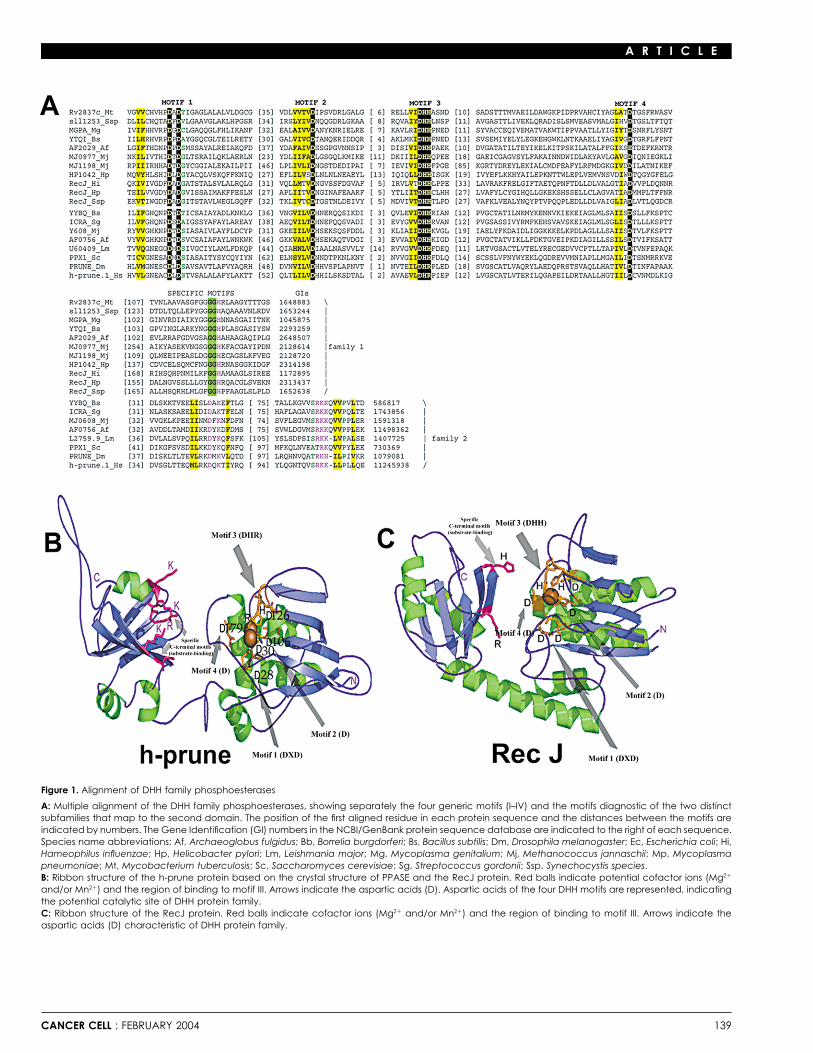
Figure 7. A model representing h-prune prometastatic function in breast cancer
In the cytoplasm of the cell, h-prune and nm23-H1 may exist in free forms and/or in a protein complex. In a cancer-committed cell, chromosomal 1q gainoccurs, increasing h-prune copy number and protein content, thus influencing cellular motility and h-prune PDE activity and promoting cancer metastasis.In contrast, the overexpression of nm23-H1 in breast cancer cells translates in reducing cellular motility and enhancing its potential to suppress metastasesformations.
trees were constructed using the Fitch program of the Phylip and the ProtMLOverall, these experiments shed light on the role of h-pruneprogram of the Molphy package. Homology modeling of protein structuresin the promotion of cellular motility and metastases negativelywas performed using the SWISS-MODEL server (Guex and Peitsch, 1997).influencing the nm23-H1 antimetastatic function. Our model ofThe target was threaded to the template using the SWISS-PDBviewer soft-the role of h-prune in cancer progression invokes, in general,ware, and the alignment with the template was manually adjusted to minimize
its amplification in tumor cells. The amplification leads to an the clashes of the protein backbone. The energy minimization was carriedincreased h-prune PDE activity in the cytoplasmic compart- out using the GROMOS program that uses a Sippl-like force field. The ribbonment, thus negatively influencing the suppressor of metastasis diagrams of the structures were generated using the MOLSCRIPT program.function of the nm23s. The activation of the h-prune PDE activity
Cell cultureis due to a physical interaction with the nm23-H1 protein. TheHEK-293 and MDA-MB-435 cells were cultured in Dulbecco’s modified Ea-complex formation results in a substantial decrease of the levelgle’s medium supplemented with 10% fetal bovine serum, 100 Units/mlof free nm23-H1 forms, thus influencing cell proliferation, cellularpenicillin, and 100 �g/ml streptomycin at 37�C with 5% CO2.motility, and metastatic processes (Figure 7).
How the h-prune-nm23 protein complex influences nm23-Protein expression and purification in Baculovirus
H1 metastasis suppressor function and apoptosis by promoting Protein expression was performed using Baculovirus Expression Systemterminal differentiation and malignancy is not yet known. These (Invitrogen). In brief, the cDNAs coding for h-prune, nm23-H1, and the nm23-questions and their following experimental plans will be aims H1 (MacDonald et al., 1996) and h-prune mutants h-prune�, D28A, D106A,of future research efforts. D179A, D28A-D106A, D28A-D106A-D126A, D28A-�, and 4D� (D28A-
D106A-�-D179A) were subcloned into the EcoRI/XhoI-digested pFastBac-Further specific investigations to address the biologicalHta vector. In order to produce the h-prune mutants cDNA, site-directedfunctions of h-prune and nm23 in other model organisms shouldmutageneses of the h-prune construct were performed using the Quik-help uncover the potential significance of this pathway in meta-Change III Kit (Stratagene) according to the manufacturer’s instructions (seestasis as well as in cellular motility and development.the Supplemental Data at http://www.cancercell.org/cgi/content/full/5/2/137/DC1). Virus infection and purification conditions are described in GarziaExperimental procedureset al. (2003). Histidine-tagged h-prune and h-prune� were further purifiedon a MonoQ HR 5/5 column (Amersham) using 10 mM Tris-HCl (pH 8.0)Protein sequence analysisbuffer. Column elution was performed with a linear gradient from 0 to 0.8 MThe nonredundant database of protein sequences at the National CenterNaCl over 20 min and at a flow rate of 1 ml/min. The fractions were furtherfor Biotechnology Information (NIH, Bethesda, MD) was iteratively searcheddialysed against 10 mM Tris-HCl (pH 8.0) buffer, and tested for activity.using the PSI-BLAST program (Altschul et al., 1997). Multiple alignments ofThe purity of isolated proteins was measured by electrophoresis SDS pageprotein sequences were constructed using the T-coffee program (Notredame
et al., 2000) and corrected on the basis of PSI-BLAST results. Phylogenetic analysis.
CANCER CELL : FEBRUARY 2004 147

A R T I C L E
Identification and characterization of the h-prunephosphodiesterase activity
Received: June 26, 2003PDE activity was measured by a cAMP/cGMP detection assay, as describedRevised: November 1, 2003by Fisher et al. (1998), and with a scintillation proximity assay (Amersham-Accepted: November 24, 2003Pharmacia Biotech). Samples were diluted as required and incubated atPublished: February 23, 200430�C in 100 �l assay buffer (50 mM Tris-HCl [pH 7.4], 8.3 mM MgCl2, 1.7 mM
EGTA) containing the desired concentrations of cAMP or cGMP as substrateReferences(3:1 ratio unlabeled to 3H-labeled). All reactions, including buffer-only blanks,
were conducted in triplicate and allowed to proceed for an incubation timeAhn, S., Milner, A.J., Futterer, K., Konopka, M., Ilias, M., Young, T.W., andgiving �25% substrate turnover (empirically determined). Reactions wereWhite, S.A. (2001). The “open” and “closed” structures of the type-C inor-terminated by adding 50 �l Yttrium silicate SPA beads (Amersham). Enzymeganic pyrophosphatases from Bacillus subtilis and Streptococcus gordonii.activities were calculated for the amount of radiolabeled product detectedJ. Mol. Biol. 313, 797–811.according to the manufacturer’s protocol. As negative controls, we used
h-prune preincubated with the A59 polyclonal antibody, raised against the Altschul, S.F., Madden, T.L., Schaffer, A.A., Zhang, J., Zhang, Z., Miller, W.,motif III region (Apotech Corporation, CH) and h-prune� mutant. In particular, and Lipman, D.J. (1997). Gapped BLAST and PSI-BLAST: a new generationfor h-prune and h-prune mutants PDE activity, 200 ng of purified enzymes of protein database search programs. Nucleic Acids Res. 25, 3389–3402.were incubated for 10 min at 30�C. Lineweaver-Burk plots with Km and Vmax Aravind, L. (1999). An evolutionary classification of the metallo-beta-lacta-values were determined by measuring hydrolysis with a range of substrate
mase fold proteins. In Silico Biol. 1, 69–91.concentrations (0.05–10.0 �M) and a fixed amount of diluted enzyme over
Aravind, L., and Koonin, E.V. (1998a). The HD domain defines a new super-a time course of 5–40 min. Initial rates were calculated at each substratefamily of metal-dependent phosphohydrolases. Trends Biochem. Sci. 23,concentration and plotted against substrate concentration, from which the469–472.kinetic parameters were determined. To study the influence of different
buffers and of nm23 activity on h-prune PDE activity and to perform inhibitor Aravind, L., and Koonin, E.V. (1998b). A novel family of predicted phos-studies, we modified PDE assays as described in Supplemental Data (http:// phoesterases includes Drosophila prune protein and bacterial RecJ exo-www.cancercell.org/cgi/content/full/5/2/137/DC1). nuclease. Trends Biochem. Sci. 23, 17–19.
Beavo, J.A., and Brunton, L.L. (2002). Cyclic nucleotide research—still ex-In vitro cell motility assay panding after half a century. Nat. Rev. Mol. Cell Biol. 3, 710–718.Stable MDA clones overexpressing h-prune, h-prune�, h-prune4D�, and
Budd, G.T., Jayaraj, A., Grabowski, D., Adelstein, D., Bauer, L., Boyett, J.,human PDE5A were produced and analyzed as described in SupplementalBukowski, R., Murthy, S., and Weick, J. (1990). Phase I trial of dipyridamoleData (http://www.cancercell.org/cgi/content/full/5/2/137/DC1). The controlwith 5-fluorouracil and folinic acid. Cancer Res. 50, 7206–7211.MDA-C100 breast cancer cell line was used in the “cell motility” assay, as
described (Leone et al., 1993a, 1993b), with the cell line overexpressing Budd, G.T., Herzog, P., and Bukowski, R.M. (1994). Phase I/II trial of dipyri-nm23-H1 (MDA-H1-177), which shows an inhibition of metastasis processes damole, 5-fluorouracil, leukovorin, and mitoxantrone in metastatic breast
cancer. Invest. New Drugs 12, 283–287.in vivo. Cellular motility was determined using the transwell technology(6-well, Corning-Costar) using 0.25%, 0.5% FCS, 2.5, and 5.0 ng/ml fibro-
Chang, C.L., Zhu, X.X., Thoraval, D.H., Ungar, D., Rawwas, J., Hora, N.,nectin (Sigma) final concentrations as chemoattractants (for details, see Strahler, J.R., Hanash, S.M., and Radany, E. (1994). Nm23–H1 mutation inSupplemental Data). The in vitro h-prune inhibition motility assay was per- neuroblastoma. Nature 370, 335–336.formed as follows. The MDA-prune (clones #3 and #4) and MDA-prune�
Fan, Z., Beresford, P.J., Oh, D.Y., Zhang, D., and Lieberman, J. (2003).(clones #10 and #11) were incubated with dipyridamole (8 �M, a 10-foldTumor suppressor NM23–H1 is a granzyme A-activated DNase during CTL-higher concentration with respect to its IC50) for 24 hr to obtain the completemediated apoptosis, and the nucleosome assembly protein SET is its inhibi-
enzyme inactivation, and then the motility assay was repeated as describedtor. Cell 112, 659–672.
above.Fisher, D.A., Smith, J.F., Pillar, J.S., St Denis, S.H., and Cheng, J.B. (1998).Isolation and characterization of PDE9A, a novel human cGMP-specificStatistical analysesphosphodiesterase. J. Biol. Chem. 273, 15559–15564.All the assays, including PDE activity and cellular motility, were validated
using the unpaired t test method available at http://www.graphpad.com/ Florenes, V.A., Aamdal, S., Myklebost, O., Maelandsmo, G.M., Bruland,quickcalcs/index.cfm. O.S., and Fodstad, O. (1992). Levels of nm23 messenger RNA in metastatic
malignant melanomas: inverse correlation to disease progression. CancerRes. 52, 6088–6091.FISH and IHC analyses—tumor case collection and TNM selection
For FISH and IHC analyses, tumor case collection, and TNM selection, see Forus, A., D’Angelo, A., Henriksen, J., Merla, G., Maelandsmo, G.M., Flor-the Supplemental Data (http://www.cancercell.org/cgi/content/full/5/2/ enes, V.A., Olivieri, S., Bjerkehagen, B., Meza-Zepeda, L.A., del Vecchio137/DC1). Blanco, F., et al. (2001). Amplification and overexpression of PRUNE in
human sarcomas and breast carcinomas-a possible mechanism for alteringthe nm23–H1 activity. Oncogene 20, 6881–6890.Acknowledgments
Freije, J.M., Blay, P., MacDonald, N.J., Manrow, R.E., and Steeg, P.S.Our special thanks go to Dr. P.S. Steeg for sharing the MDA-C100, MDA- (1997a). Site-directed mutation of Nm23–H1. Mutations lacking motility sup-H1-177, MDA-nm23H1-P96S, and MDA-nm23H1-S120G clones; the DNA pressive capacity upon transfection are deficient in histidine-dependent pro-TIGEM-IIGB Sequencing Core for developing sequencing reactions; Dr. Keni tein phosphotransferase pathways in vitro. J. Biol. Chem. 272, 5525–5532.Omori for PDE5A gene construct; the MicroArray Core facility and Dr. Coc-
Freije, J.M., Lawrence, J.A., Hollingshead, M.G., De la Rosa, A., Narayanan,chia for help on data mining; Dr. Budroni for breast data collection analysis;V., Grever, M., Sausville, E.A., Paull, K., and Steeg, P.S. (1997b). Identification
Drs. Beavo, Iolascon, Santoro, and Venkitaraman for critical discussions of of compounds with preferential inhibitory activity against low-Nm23-the manuscript; and Drs. Meroni and Diez-Roux for supportive ideas on expressing human breast carcinoma and melanoma cell lines. Nat. Med. 3,project development. This work was supported by an AIRC-FIRC Research 395–401.Fellowship to A.D., a Molecular Oncology and Pharmacology PhD program
Galperin, M.Y., Natale, D.A., Aravind, L., and Koonin, E.V. (1999). A special-(University of Ferrara, Italy) to V.A., a 2002 AIRC-FIRC grant (G.P, M.Z.), aized version of the HD hydrolase domain implicated in signal transduction.Compagnia San Paolo Torino 2002 grant (M.Z.), a Regione Autonoma dellaJ. Mol. Microbiol. Biotechnol. 1, 303–305.
Sardegna 2001-03 grant (A.C., G.P.), a FIRB-MIUR-RBAU01RW82 grant(M.Z.), and a 2001 TIGEM-Telethon Regione Campania grant (M.Z.). Garzia, L., Andre, A., Amoresano, A., D’Angelo, A., Martusciello, R., Cirulli,
148 CANCER CELL : FEBRUARY 2004
A R T I C L E
C., Tsurumi, T., Marino, G., and Zollo, M. (2003). Method to express and Lombardi, D., Lacombe, M.L., and Paggi, M.G. (2000). Nm23: unraveling itsbiological function in cell differentiation. J. Cell. Physiol. 182, 144–149.purify nm23–H2 protein from Baculovirus infected cells. Biotechniques 35,
384–391.MacDonald, N.J., Freije, J.M., Stracke, M.L., Manrow, R.E., and Steeg, P.S.(1996). Site-directed mutagenesis of nm23–H1. Mutation of proline 96 orGuex, N., and Peitsch, M.C. (1997). SWISS-MODEL and the Swiss-serine 120 abrogates its motility inhibitory activity upon transfection intoPdbViewer: an environment for comparative protein modeling. Electrophore-human breast carcinoma cells. J. Biol. Chem. 271, 25107–25116.sis 18, 2714–2723.
Mao, H., Liu, H., Fu, X., Fang, Z., Abrams, J., and Worsham, M.J. (2001).Haaz, M.C., Fischel, J.L., Formento, P., Renee, N., Etienne, M.C., and Milano,Loss of nm23 expression predicts distal metastases and poorer survival forG. (1996). Impact of different fluorouracil biochemical modulators on cellularbreast cancer. Int. J. Oncol. 18, 587–591.dihydropyrimidine dehydrogenase. Cancer Chemother. Pharmacol. 38,
52–58. Niitsu, N., Okabe-Kado, J., Okamoto, M., Takagi, T., Yoshida, T., Aoki, S.,Hirano, M., and Honma, Y. (2001). Serum nm23–H1 protein as a prognosticHartsough, M.T., and Steeg, P.S. (1998). Nm23–H1: genetic alterations andfactor in aggressive non-Hodgkin lymphoma. Blood 97, 1202–1210.expression patterns in tumor metastasis. Am. J. Hum. Genet. 63, 6–10.Notredame, C., Higgins, D.G., and Heringa, J. (2000). T-Coffee: A novelHartsough, M.T., and Steeg, P.S. (2000). Nm23/nucleoside diphosphatemethod for fast and accurate multiple sequence alignment. J. Mol. Biol. 302,
kinase in human cancers. J. Bioenerg. Biomembr. 32, 301–308.205–217.
Hartsough, M.T., Clare, S.E., Mair, M., Elkahloun, A.G., Sgroi, D., Osborne, Reymond, A., Volorio, S., Merla, G., Al-Maghtheh, M., Zuffardi, O., Bulfone,C.K., Clark, G., and Steeg, P.S. (2001). Elevation of breast carcinoma A., Ballabio, A., and Zollo, M. (1999). Evidence for interaction between humanNm23–H1 metastasis suppressor gene expression and reduced motility by PRUNE and nm23–H1 NDPKinase. Oncogene 18, 7244–7252.DNA methylation inhibition. Cancer Res. 61, 2320–2327.
Sleeman, J.P. (2000). The lymph node as a bridgehead in the metastaticHejna, M., Raderer, M., and Zielinski, C.C. (1999). Inhibition of metastases dissemination of tumors. Recent Results Cancer Res. 157, 55–81.by anticoagulants. J. Natl. Cancer Inst. 91, 22–36.
Steeg, P.S. (2003). Metastasis suppressors alter the signal transduction ofHowlett, A.R., Petersen, O.W., Steeg, P.S., and Bissell, M.J. (1994). A novel cancer cells. Nat. Rev. Cancer 3, 55–63.function for the nm23–H1 gene: overexpression in human breast carcinoma
Timmons, L., and Shearn, A. (1996). Germline transformation using a prunecells leads to the formation of basement membrane and growth arrest. J.cDNA rescues prune/killer of prune lethality and the prune eye color pheno-Natl. Cancer Inst. 86, 1838–1844.type in Drosophila. Genetics 144, 1589–1600.
Kantor, J.D., McCormick, B., Steeg, P.S., and Zetter, B.R. (1993). InhibitionTseng, Y.H., Vicent, D., Zhu, J., Niu, Y., Adeyinka, A., Moyers, J.S., Watson,
of cell motility after nm23 transfection of human and murine tumor cells.P.H., and Kahn, C.R. (2001). Regulation of growth and tumorigenicity of
Cancer Res. 53, 1971–1973. breast cancer cells by the low molecular weight GTPase Rad and nm23.Cancer Res. 61, 2071–2079.Leone, A., Flatow, U., VanHoutte, K., and Steeg, P.S. (1993a). Transfection
of human nm23-H1 into the human MDA-MB-435 breast carcinoma cell line: Wagner, P.D., Steeg, P.S., and Vu, N.D. (1997). Two-component kinase-likeeffects on tumor metastatic potential, colonization and enzymatic activity. activity of nm23 correlates with its motility-suppressing activity. Proc. Natl.Oncogene 8, 2325–2333. Acad. Sci. USA 94, 9000–9005.
Leone, A., Seeger, R.C., Hong, C.M., Hu, Y.Y., Arboleda, M.J., Brodeur, Yamagata, A., Kakuta, Y., Masui, R., and Fukuyama, K. (2002). The crystalG.M., Stram, D., Slamon, D.J., and Steeg, P.S. (1993b). Evidence for nm23 structure of exonuclease RecJ bound to Mn2� ion suggests how its charac-RNA overexpression, DNA amplification and mutation in aggressive child- teristic motifs are involved in exonuclease activity. Proc. Natl. Acad. Sci.
USA 99, 5908–5912.hood neuroblastomas. Oncogene 8, 855–865.
CANCER CELL : FEBRUARY 2004 149