mrsa: a tale of three types – 15 years of survey data from agar
TRANSCRIPT
NOVEMBER 2016
MRSA: A Tale of Three Types 15 years of survey data from AGAR John Turnidge, Geoffrey Coombs, Denise Daley, Graeme Nimmo and the Australian Group on Antimicrobial Resistance (AGAR) participants, 2000–14 have prepared this report on behalf of the Australian Commission on Safety and Quality in Health Care.
AURA TECHNICAL SERIES: NO.1
Published by the Australian Commission on Safety and Quality in Health Care Level 5, 255 Elizabeth Street, Sydney NSW 2001 Phone: (02) 9126 3600 Fax: (02) 9126 3613 Email: [email protected] Website: www.safetyandquality.gov.au
© Commonwealth of Australia 2016 All material and work produced by the Australian Commission on Safety and Quality in Health Care is protected by Commonwealth copyright. It may be reproduced in whole or in part for study or training purposes, subject to the inclusion of an acknowledgement of the source. The Commission’s preference is that you attribute this publication (and any material sourced from it) using the following citation:
Turnidge, J., Coombs, G., Daley, D., Nimmo, G., Australian Group on Antimicrobial Resistance (AGAR) participants, 2000–14. MRSA: A Tale of Three Types 15 years of survey data from AGAR. Sydney: ACSQHC; 2016
Enquiries regarding the use of this publication are welcome and can be sent to [email protected].
Introduction Staphylococcus aureus (S. aureus) is a frequent coloniser of humans and a major human bacterial pathogen. It is the cause of a wide range of infections from benign self-limiting conditions (including boils, bullous impetigo and folliculitis), to more serious infections (including cellulitis, post-surgical wound infection, acute and chronic osteomyelitis, septic arthritis, infections of intravascular lines, and prosthetic joint and other device infections), to life-threatening infections (including septicaemia, meningitis, post-viral pneumonia and endocarditis).1 It is carried by about 30% of the population at any one time, and for the great majority of people it causes no harm.2 Nevertheless, it has the capacity to cause outbreaks of infection in hospitals from a common source or through poor hand hygiene practices. Soon after the introduction of (benzyl) penicillin into clinical medicine in the 1940s, strains of penicillin-resistant S. aureus emerged that produced penicillinase – the first β-lactamase enzyme to be described.3 By the late 1950s, chemists began working on chemical modifications of the penicillin nucleus in the hope of finding a modification that would protect the antimicrobial medicine from degradation by penicillinase. The first successful modification was methicillin, which was marketed for the treatment of staphylococcal infection in 1960 in the United Kingdom. Other modifications followed (such as nafcillin, oxacillin, cloxacillin, flucloxacillin and dicloxacillin), each developed specifically for treating staphylococcal infection caused by penicillinase-producing strains. Jevons4 first reported resistance to methicillin in clinical isolates of S. aureus (methicillin-resistant S. aureus – MRSA) from a London hospital in 1961. Of note, the isolates were also resistant to streptomycin and tetracycline, highlighting the staphylococcal propensity for accumulating resistance to multiple agents. Since that time, and slowly at first, MRSA has become a global phenomenon, and has taken many forms. The first report of MRSA in Australia was from Sydney in 1968.5 By the late 1970s a particular type of multi-resistant MRSA had become established in public hospitals on the eastern Australian seaboard.6 That clone, now called Aus-2/3 (multi-locus sequence type ST239-MRSA-III), has now become established in tertiary care hospitals in most parts of Australia. By the mid-1980s, community strains of MRSA started appearing in Western Australia, and since that time have developed into an Australia-wide problem.6
The mechanism of resistance to methicillin in staphylococci differs from resistance to penicillin. Rather than being mediated by a β-lactamase, methicillin resistance is due to the production of an additional so-called penicillin-binding protein, pbp-2a, which is encoded by the mecA gene. This protein is a variant of one of the essential cell-wall synthetic enzymes, pbp-2 (a transpeptidase). A methicillin-resistant isolate retains its original pbp-2, but also produces pbp-2a, which retains its transpeptidase function but has much lower affinity for β-lactams generally, including penicillins, cephalosporins and carbapenems. Because the resistance mechanism is not that of a β-lactamase, MRSA are also resistant to the combinations of β-lactamase inhibitors with β-lactams. Treatment of MRSA infections depends on severity. Superficial skin infections can be managed with drainage and/or topical agents. More serious infections will require systemic antimicrobials: vancomycin is most commonly used when intravenous therapy is needed, and only a limited range of agents is effective when oral therapy is needed. Recent evidence has confirmed that vancomycin is suboptimal treatment for staphylococcal infection compared to β-lactams when the infection is caused by strains susceptible to methicillin.7 The implication is that vancomycin is
1
suboptimal therapy for MRSA infections, but unfortunately there is no evidence that any alternative intravenous agent is superior.
Tracking MRSA emergence and spread through typing There have been several systems for typing strains of S. aureus. Prior to the advent of multi-locus sequence typing (MLST), the most popular system was phage typing, a system that used a collection of different viruses (called bacteriophages) that attack and kill specific strains of this bacterial species. The patterns of killing by the different phages defined the ‘phage type’. Phage typing was supplanted by pulsed-field gel electrophoresis (PFGE) in the 1980s, a technique that created a ‘bar code’ of the bacterial DNA after fragmenting it with specific enzymes. PFGE was valuable at the local level, but due to technical variation, could not easily be used to compare results from different laboratories locally or internationally.
MLST was developed in the late 1990s, and applied to a range of bacterial species, including S. aureus, soon after. MLST involves the DNA sequencing of seven so-called housekeeping genes, which are found in all strains of the species, and whose sequences are known not to vary significantly over time. Each unique gene sequence is called an ‘allele’, and the combination of the seven allelic sequences defines the allelic profile, which is considered to be a distinct clone. MLST has the advantage of being stable and readily portable, meaning that sequences are stored and can then be compared from anywhere in the world (MLST online database and analysis website). It can also provide information about the evolution of individual clones. The sequence type is designated by a number, preceded by ‘ST’. Related sequence types belong to families called ‘clonal complexes’.
The sequence type provides the base information of a clone, but an additional piece of genetic typing is required to identify an MRSA clone. This additional information is called the ‘SCCmec’ type (Staphylococcal Cassette Chromosome mec). It is a piece of mobile DNA (meaning it can be transferred to other staphylococci) that has been acquired by a staphylococcus and inserted into its chromosome. Possession of SCCmec makes the staphylococcus resistant to methicillin because it contains the mec gene, which codes for the additional penicillin-binding protein pbp2a, and the mec gene’s associated regulatory genes. There are at least 12 varieties of SCCmec, which vary in size and content, and each type is designated by an upper case roman numeral. This is added to the sequence type to provide the full designation of a clone – for example, the Aus-2/3 clone is ST239-III, meaning it is sequence type 239 and SCCmec type III. Larger SCCmecs possess additional resistance genes besides mec, and are the main contributors to multi-resistance. The acquisition of SCCmec converts a methicillin-susceptible clone of S. aureus into an MRSA clone.
PFGE still plays a role in discriminating specific genetic lineages amongst strains with the sequence and SCCmec type. It forms the basis of the Western Australian typing nomenclature that has wide currency across Australia. For instance, at least five different clones have been identified in the ST5-IV type, only one of which (PFGE pattern WA-3) has become more common in recent years.
As costs come down, whole genome sequencing (WGS) is now being used with increasing frequency for tracking clones of resistant bacteria. Within a few years, it is likely that the additional information generated by WGS will refine S. aureus typing.
AGAR surveillance The Australian Group on Antimicrobial Resistance (AGAR) has been tracking MRSA infections across the country since 1985 (AGAR website). While participation in AGAR has been voluntary, the group has managed to maintain contributions from at least 24 laboratories, with almost all
2
states and territories regularly represented. With the introduction of MLST, the activities of AGAR have greatly enhanced our understanding of MRSA epidemiology, or more correctly, epidemiologies. We now recognise MRSA comes in three ‘types’: healthcare-associated, community-associated and livestock-associated, and each with a range of different clones. Table 1 below shows the key features of the healthcare-associated and the community-associated types and clones that have been tracked in Australia since 2000. From 2000−12, AGAR conducted period prevalence surveys on all types of S. aureus infections (see note below about AGAR data for figures). In 2001, isolates from hospital emergency departments were also included. From 2013, AGAR switched to continuous surveillance of blood culture isolates, and included those with community-onset (from blood cultures collected prior to or within 48 hours of admission), and hospital-onset (from blood cultures collected more than 48 hours after admission) infections. Figures 1 and 2 show the combined incidence of the major clones since 2000 in hospital-onset and community-onset infections respectively. The vertical blue dotted line in these figures indicates where the sampling method changed. There has been a noticeable decline in the proportion of hospital-onset S. aureus infections that are MRSA since 2009 (Figure 1). This trend appears to have continued despite the change in sampling method. By contrast, the prevalence of MRSA in community-onset infections has remained reasonably stable since 2008 (Figure 2). Note about AGAR data for figures Community-onset infection: For the biennial AGAR surveys from 2000–12, each participating institution contributed 100 consecutive clinical isolates causing infection of any type from outpatients and those attending emergency departments. Hospital-onset infection: For the biennial AGAR surveys from 2001–11, each participating institution contributed 100 consecutive clinical isolates causing infection of any type from patients who had been hospitalised for at least 48 hours. In 2001 and 2003, the sample also included clinical isolates from patients attending an emergency department. From 2013 onwards: The AGAR survey method changed in 2013 to the continuous collection of isolates from blood cultures (invasive disease). Both community-onset and hospital-onset episodes were included, defined by the ‘48-hour rule’ (onset > 48h after admission = hospital-onset, otherwise community-onset).
3

Figure 1: Hospital-onset MRSA, all clones, 2001–2014; percentage of all S. aureus
Figure 2: Community-onset MRSA, all clones, 2000–2014; percentage of all S. aureus
0%
5%
10%
15%
20%
25%
30%
35%
2001 2003 2005 2007 2009 2011 2013 2014Year of testing
Other CA WA-84 WA-2 WA-3 QldSWP WA-1 Other HA UK EMRSA-15 Aus-2/3
0%2%4%6%8%
10%12%14%16%18%20%
2000 2002 2004 2006 2008 2010 2012 2013 2014Year of testing
Other CA WA-84 WA-2 WA-3 QldSWP WA-1 Other HA UK EMRSA-15 Aus-2/3
4

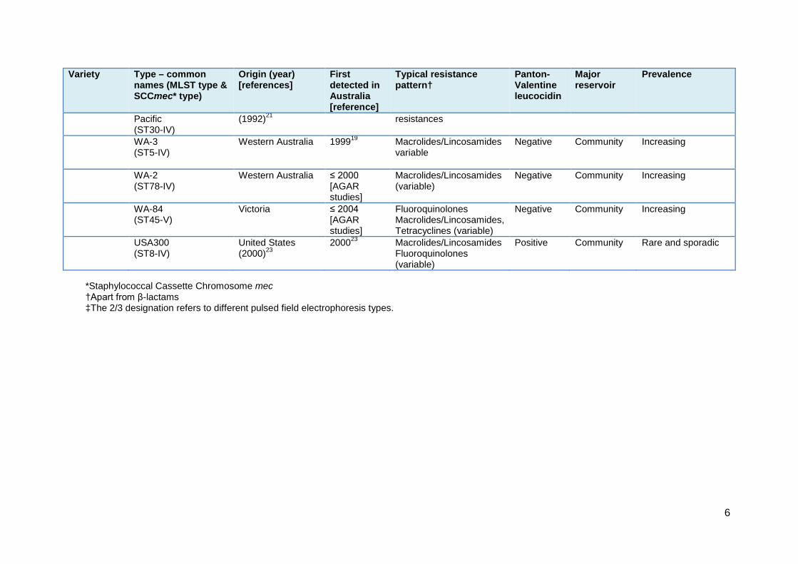
Table 1: Important MRSA clones that have been identified in Australia 2000–14 Variety Type – common
names (MLST type & SCCmec* type)
Origin (year) [references]
First detected in Australia [reference]
Typical resistance pattern†
Panton-Valentine leucocidin
Major reservoir
Prevalence
Healthcare-associated
Aus-2/3‡ (ST239-III)
Australia or USA (1976)8,9,10
19769 Macrolides/Lincosamides Tetracyclines Trimethoprim-sulfamethoxazole Gentamicin Fluoroquinolones
Negative Hospital ‘frequent flyers’
Was common in many countries around the world Was very common in NSW, Qld, Vic. and SA, now diminishing
UK EMRSA-15 (ST22- IV)
England (1991)11 199712 Fluoroquinolones Macrolides/Lincosamides variable
Negative Long-term care facilities13
Increasingly common
New York / Japan or USA100 (ST5-II)
Japan (1982)14 200515 Macrolides/Lincosamides Fluoroquinolones
Negative None Very common in the USA, Japan and Korea Rare and sporadic in Australia
UK EMRSA-16 or USA200 (ST36-II)
Southern England (1991)16
200217 Macrolides/Lincosamides Fluoroquinolones Mupirocin
Negative None Rare and sporadic
Community-associated
WA-1 (ST1-IV)
Northern Western Australia18,19
198918 Typically no additional resistances Macrolides/Lincosamides variable Fusidic acid variable
Negative Community Common
Queensland (ST93-IV)
South-eastern Queensland (2000)20
200020 Typically no additional resistances
Positive Community Common
Oceania/Southwest New Zealand 199722 Typically no additional Positive Community Common
5
Variety Type – common names (MLST type & SCCmec* type)
Origin (year) [references]
First detected in Australia [reference]
Typical resistance pattern†
Panton-Valentine leucocidin
Major reservoir
Prevalence
Pacific (ST30-IV)
(1992)21 resistances
WA-3 (ST5-IV)
Western Australia 199919 Macrolides/Lincosamides variable
Negative Community Increasing
WA-2 (ST78-IV)
Western Australia ≤ 2000 [AGAR studies]
Macrolides/Lincosamides (variable)
Negative Community Increasing
WA-84 (ST45-V)
Victoria ≤ 2004 [AGAR studies]
Fluoroquinolones Macrolides/Lincosamides, Tetracyclines (variable)
Negative Community Increasing
USA300 (ST8-IV)
United States (2000)23
200023 Macrolides/Lincosamides Fluoroquinolones (variable)
Positive Community Rare and sporadic
*Staphylococcal Cassette Chromosome mec †Apart from β-lactams ‡The 2/3 designation refers to different pulsed field electrophoresis types.
6

TYPE 1 MRSA – healthcare-associated (HA-MRSA) Two healthcare-associated clones have dominated healthcare-acquired S. aureus infections in Australia: Aus-2/3 and EMRSA-15 – the first possibly ‘home-grown’ and the second imported.
Aus-2/3 – the first major multi-resistant clone in Australian hospitals The current evidence suggests that the first strains of Aus-2/3 originated in Australia or possibly the USA;8,9,10 the earliest known Australian strain was isolated in 1976.9 By the late 1970s this clone had become established in many Melbourne teaching hospitals.24 By the time of the first surveys conducted by the AGAR in 1985, Aus-2/3 was found to cause 12–25% of all S. aureus infections in large public capital-city hospitals along the eastern seaboard, in Brisbane, Sydney, Canberra and Melbourne, and to a lesser extent in Adelaide.25 Ultimately, the only state not to become a reservoir for Aus-2/3 was Western Australia, which introduced intensive efforts at screening and segregating patients and staff coming from eastern states hospitals.26 It remains very uncommon there.27
Recent detailed studies of the Aus-2/3 sequence and SCCmec type in Australia (ST239-III) using detailed genomic and phenotypic methods have shown that what is currently identified as Aus-2/3 may actually represent a mixture of two clades.28 One clade represents the long-established (many decades) Aus-2/3 HA-MRSA, while in 2001 a new clade appeared in Victoria which appears to have originated in Asia. Whether this explains the boost in the prevalence of Aus-2/3 between 2001 and 2005 in Victoria, New South Wales and Queensland (see Figure 9 in the geographic section), in an otherwise previously well-established clone, is not clear. A decline of Aus-2/3 in hospital-onset infections began 2009, and has continued rapidly (Figure 3). Aus-2/3 has also contributed to community-onset infections but almost always occurs in patients who have been previously hospitalised in facilities where the clone is known to be prevalent (Figure 4).29
It is believed that the decline in hospital-onset Aus-2/3 reflects improving infection control efforts in all hospitals, and especially the introduction of a national hand hygiene program in June 2008,30 supported by the national pubic reporting of healthcare-associated S. aureus bacteraemia.31 These initiatives would have been boosted by the 2012 implementation of the National Safety and Quality Health Service (NSQHS) Standards, linked to accreditation, released by the Australian Commission on Safety and Quality in Health Care, and in particular Standard 3: Preventing and Controlling Healthcare Associated Infections.32
7
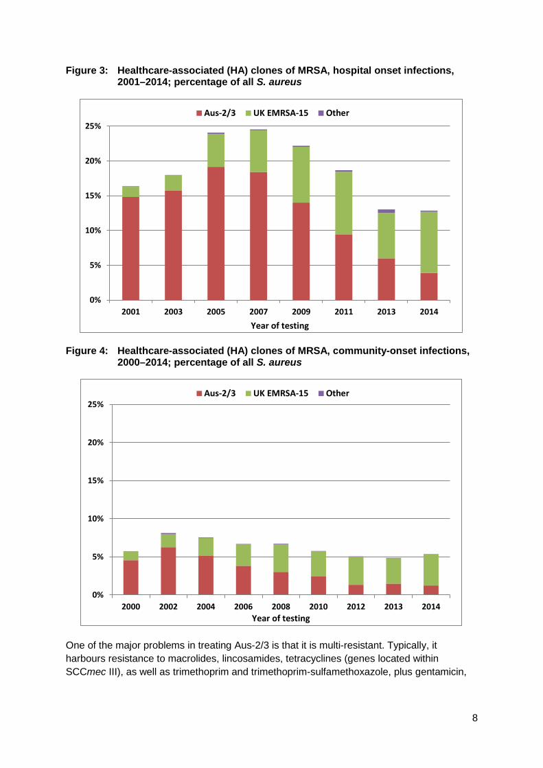
Figure 3: Healthcare-associated (HA) clones of MRSA, hospital onset infections, 2001–2014; percentage of all S. aureus
Figure 4: Healthcare-associated (HA) clones of MRSA, community-onset infections,
2000–2014; percentage of all S. aureus
One of the major problems in treating Aus-2/3 is that it is multi-resistant. Typically, it harbours resistance to macrolides, lincosamides, tetracyclines (genes located within SCCmec III), as well as trimethoprim and trimethoprim-sulfamethoxazole, plus gentamicin,
0%
5%
10%
15%
20%
25%
2001 2003 2005 2007 2009 2011 2013 2014Year of testing
Aus-2/3 UK EMRSA-15 Other
0%
5%
10%
15%
20%
25%
2000 2002 2004 2006 2008 2010 2012 2013 2014Year of testing
Aus-2/3 UK EMRSA-15 Other
8

and frequently acquired resistance to ciprofloxacin and rifampicin. Few agents are available for treatment.
UK EMRSA-15 – the first imported healthcare-associated clone to spread widely The EMRSA-15 clone emerged in the United Kingdom in 1991. It has a distinct resistance profile, being resistant consistently to ciprofloxacin and often to macrolides/lincosamides but typically to no other classes of antimicrobial beyond the β-lactams. It was first documented in Western Australia in 1997, in British healthcare workers who were screened as part of that state’s MRSA containment policy.12 By 2000, it was seen nationally in AGAR surveys, most prominently in Sydney.17 Since that time its prevalence has continued to grow steadily. In the AGAR 2014 survey, EMRSA-15 comprised 30% of all MRSA and 5% of all S. aureus isolates, was more than twice as common as Aus-2/3, and was the most frequently identified type of MRSA overall. It contributed significantly to both hospital- and community-onset bacteraemia (Figures 3 and 4).29
The EMRSA-15 clone appears to have a significant reservoir in residential aged-care facilities.13,27 At one tertiary hospital in Melbourne, 33% of EMRSA-15 infections occurred in patients admitted from residential aged-care facilities, compared to only 5% of cases caused by Aus-2/3.13 Apart from the first detection in UK healthcare workers in Western Australia, the link between the foreign healthcare workers and the repeated introduction of EMRSA-15 remains speculative. Medical and nursing staff born in the UK made up 13% and 26% of all those who were foreign-born in 2011, falling from 2001 levels of 20% and 36% respectively.33 Nevertheless, they are still the largest group of foreign-born healthcare workers in both categories, and active recruitment from the UK continues. Another feature of EMRSA-15 contributing to its ongoing spread is its capacity to colonise healthcare workers, being four-fold more likely to colonise healthcare workers in an outbreak than Aus-2/3.34 It is likely therefore that the reservoir for EMRSA-15 has components in frequently hospitalised patients, in residents of residential aged-care facilities, and in some healthcare workers. The increasing prevalence of EMRSA-15 is occurring in Australia at a time when it has declined significantly in England.35
Other healthcare-associated clones Other healthcare-associated clones have remained rare in Australia. The AGAR surveys have regularly detected the New York / Japan clone (ST5-II and also called USA100) at very low levels since 2005 (1-3 isolates annually). It has been isolated in four states so far. The New York / Japan clone was first described in Australia in an outbreak in Western Australia that could traced to an Australian healthcare worker who had been previously hospitalised in New York. However, this clone has remained uncommon afterwards in that state.15 Despite its high prevalence in the United States, Japan and Korea,36 consistent with a high capacity for spread, ST5-II has not become established in Australia. The picture is one of intermittent introduction from overseas, but without local spread. The New York / Japan clone has a similar resistance profile to EMRSA-15, including to macrolides/lincosamides and ciprofloxacin, but is less multi-resistant than Aus-2/3.
9
A similar story applies to UK EMRSA-16 (ST36-II and also called USA200). This is second only to EMRSA-15 in prevalence in the United Kingdom. It has been seen intermittently in AGAR surveys, but shows no signs of becoming established. Other healthcare-associated clones that have been found in the AGAR surveillance include ST250-I (so-called classical MRSA), ST247-I (UK EMRSA-17), ST8-VI (Irish 2), and ST228-I (Southern German). These fit the pattern of sporadic introduction into Australia, with no or very limited spread.
TYPE 2 – community-associated MRSA (CA-MRSA) Community-associated MRSA clones first emerged in Australia in the 1980s and since that time have both diversified and increased in prevalence. They now exceed HA-MRSA as a cause of community-onset infection and rival them as a cause of hospital-onset infections. CA-MRSA clones are totally different from those found to cause cross-infection in healthcare settings. They tend not to be resistant to the non-beta-lactam antibiotics, although with the passage of time, some CA-MRSA clones are accumulating more resistances. A characteristic frequently associated with some clones of CA-MRSA is the possession of the Panton-Valentine Leucocidin (PVL) toxin – details are provided in the next section. CA-MRSA now play a substantial role in hospital-onset S. aureus infections. This is not surprising, as a significant proportion of healthcare-associated staphylococcal infections are caused by strains that are carried by the patient at the time of the intervention that led to the infection.37 In 2014, almost half of CA-MRSA (48%) bacteraemias were hospital-onset, and CA-MRSA rivalled HA-MRSA as a cause of hospital-onset MRSA infection (45% CA-MRSA).
WA-1 – the first established CA-MRSA in Australia Community-associated strains of MRSA were first noted in Western Australia in 1984, and became a particular problem in the Kimberley region by 1989.38 By 2004, it had become clear that a number of new clones of CA-MRSA had emerged over the years in Western Australia, and that one particular clone, WA-1 (ST1-IV), had come to dominate the CA-MRSA picture there, and that it had spread to almost all other states and territories.17,19 WA-1 is still one of the three most important CA-MRSA clones isolated across Australia (Figures 5 and 6). WA-1 is usually susceptible to non-β-lactam agents. Around half of the strains harbour at least one additional resistance mechanism, most commonly to macrolides/lincosamides and/or fusidic acid. WA-1 does not usually produce PVL, although 10% of ST1-IV isolates possess this toxin.29
10
Figure 5: AGAR Surveys: Community-associated clones of MRSA, community-onset infections, 2000–2014; percentage of all S. aureus
Figure 6: AGAR Surveys: Community-associated clones of MRSA, hospital-onset
infections, 2001–2014; percentage of all S. aureus
0%
2%
4%
6%
8%
10%
12%
14%
2000 2002 2004 2006 2008 2010 2012 2013 2014Year of testing
Other CA WA-84 WA-2 WA-3 SWP Qld WA-1
0%
2%
4%
6%
8%
10%
12%
14%
2001 2003 2005 2007 2009 2011 2013 2014Year of Testing
Other CA WA-84 WA-2 WA-3 SWP Qld WA-1
11
Figure 7: AGAR Surveys: Community-associated MRSA, trends of the three dominant clones in community-onset infections, 2000–2014; percentage of all S. aureus
Figure 8: AGAR Surveys: Community-associated MRSA, trends of other clones in
community-onset infections, 2000–2014; percentage of all S. aureus
0%
1%
1%
2%
2%
3%
3%
4%
4%
5%
5%
2000 2002 2004 2006 2008 2010 2012 2013 2014Year of testing
WA-1 Qld SWP
0%
1%
1%
2%
2%
3%
3%
4%
4%
5%
5%
2000 2002 2004 2006 2008 2010 2012 2013 2014Year of testing
WA-3 WA-2 WA-84 Other CA
12
The Southwest Pacific (SWP) clone – an introduced CA-MRSA The SWP clone (ST30-IV) first appeared in Auckland, New Zealand, and was frequently isolated from patients of Western Samoan origin, leading to its designation initially as WSPP (Western Samoan phage pattern).39 The first descriptions were in 1998.40 Soon after it was recognised that ST30-IV was also present in Australia; again people of Pacific Island nation origin in the southwest Pacific (Western Samoa and Tonga) were disproportionally affected.22,41 More recent studies have shown that the SWP clone had its origins as far back as 1953,42 when its methicillin-susceptible and PVL-producing ancestor, then called ‘phage type 80/81’, was associated with major outbreaks of infections in neonatal nurseries in Australia43 and subsequently many other parts of the world. The SWP clone is almost always susceptible to non-β-lactam classes of antimicrobial – approximately 10% have additional resistance(s). Most notably, SWP produces PVL (see next section ‘The importance of Panton-Valentine leucocidin’).
The Queensland (Qld) clone – a home-grown and increasing problem The Queensland clone (ST93-IV) has become the dominant CA-MRSA in Australia (see Figure 7). First recognised amongst Caucasians (and not Pacific Islanders) in southern Queensland in 2000,20 it has since spread to become the dominant CA-MRSA in Queensland, Northern Territory, and South Australia, on equal terms with WA-1 in New South Wales, and Victoria, and second only to WA-1 in Western Australia.29 AGAR surveys show that it was actually first detected in five states/territories in 2000, mostly notably in New South Wales. Like the SWP clone, the Queensland clone tends to be susceptible to non-β-lactam classes of antimicrobial, and importantly it also produces PVL (see ‘The importance of Panton-Valentine leucociden’). The reasons for its success remain unclear despite extensive molecular studies, and it harbours very few virulence factors besides the genes encoding PVL.44
It is noticeable that the proportion of the PVL-producing clones, the Queensland and SWP clones, decreased with the change to bacteraemia-only surveillance in 2013 (Figure 7). This is consistent with the observation that PVL-producing strains are less likely to cause invasive infection.45,46
Other CA-MRSA clones – ever increasingly diversity Following the introduction of molecular typing of MRSA, the AGAR surveys have witnessed an ever increasing number of new CA-MRSA clones across Australia. A few have gained a foothold and are seen regularly (Figure 8). These include:
• WA-3 (ST5-IV): first documented in New South Wales and South Australia in 2000,17 but now seen Australia wide;47 largely PVL negative
• WA-2 (ST78-IV): first documented in 2000, predominantly in Western Australia,17 but since spread Australian-wide; 47 largely PVL negative
13
• the Victorian clone (WA-84, ST45-V): first documented in Victoria in 2004 and increasingly prominent there ever since; 47 PVL negative and multi-resistant.
More than 100 clones on CA-MRSA have now been detected in Australia since the year 2000. These are a mixture of both locally arising and international clones. For the locally arising clones, it suggests that although SCCmec is not believed to spread easily from MRSA to other methicillin-susceptible S. aureus clones, it does happen with some frequency. This increasing diversity is reflected in Figure 8 which shows that ‘other CA-MRSA’ have collectively outnumbered WA-1, SWP and Queensland since 2006. In 2014 for instance, there were 17 clones (community- plus hospital-onset) detected in addition to the common three clones. Thus, while SCCmec has the ability to be inserted into many chromosomal backgrounds, most of the resulting clones are not particularly well adapted for transmission and carriage compared to the dominant clones. Nevertheless, there are two clones − WA-3 (ST5-IV) and the Victorian clone (WA-84, ST45-V) − which appear to be on the rise (Figure 8). Another clone of interest is USA300 (ST8-IV). This is a clone of CA-MRSA that has become a major cause of S. aureus infection in the community in the USA, accounting for up to 50% of all community infections in many parts of country.48 It was first recognised in 1999, and after spread across the US community, is an important cause of infection in at least some parts of the USA.49 This clone has been detected in AGAR surveys from at least 2000. However, its numbers remain low, and present another example of the sporadic introduction of a clone that has failed to become established yet in Australia, despite success elsewhere.50 Like the Southwest Pacific and Queensland clones, USA300 produces PVL (see ‘The importance of Panton-Valentine leucociden’). There have also been sporadic appearances of CA-MRSA from other countries, including the Taiwan clone (ST59-V), the Bengal Bay clone (ST772-V), and the European clone (ST80-IV)). Again, none of these are showing signs of becoming established.
14
The importance of Panton-Valentine leucocidin Panton-Valentine leucocidin (PVL) was first described in 1932 by Panton and Valentine in S.aureus causing purulent infections.51 Its importance was unrecognised until 2003 when a strong association was noted between certain clones of CA-MRSA and PVL52 by French and other international investigators. Both the SWP and Queensland clones, as well as USA300, were identified as carrying PVL in that study. It has been shown subsequently that PVL is not confined to CA-MRSA, but can be found in methicillin-susceptible strains.53
PVL-producing strains of S. aureus are associated with a variety of distinctive clinical manifestations: excessively purulent skin and soft tissue infections that frequently require drainage/surgical intervention,54 recurrent furunculosis (boils),56 multifocal osteomyelitis associated with early bone necrosis and deep vein thrombosis,57 and necrotising pneumonia.58 By contrast, PVL-producing strains are less likely to cause bacteraemia or other invasive disease and the same or lower mortality than that caused by PVL negative S. aureus.45,59 Overall, PVL-producing S. aureus are not more life-threatening than other S. aureus but cause more morbidity, hospitalisation and infections that require surgical intervention. They appear to be over-represented in skin and soft tissue infections requiring hospital care, when compared to PVL-negative strains.45,59 The high degree of purulence of these infections may contribute to higher densities of organisms on the skin available for transmission to other susceptible individuals.
Infections caused by PVL-producing MRSA strains are more challenging to treat in the community, not just because of the frequent need for surgical invention, but also because the availability of effective antimicrobials in the community is limited.
TYPE 3 MRSA – livestock-associated There have been sporadic isolates of MRSA internationally for many years, but the first major problem emerged in Europe in 2003, with the description of ST398 in pigs and subsequently other livestock.61 To date, this livestock-associated clone has not been detected in any AGAR surveys. In 2013, it was detected in a nasal swab from an Australian pig veterinarian,62 and subsequently detected at very low levels in pig herds across Australia.63 A distinctive clone often carried by equine veterinarians in Australia (WA-20 – ST612-IV),64 has been found only once in the 2013 AGAR survey causing community-onset bloodstream infection.
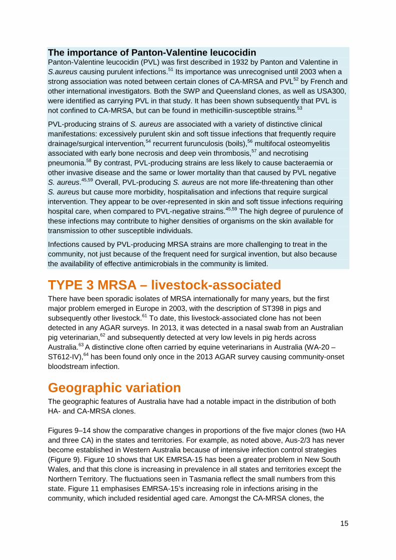
Geographic variation The geographic features of Australia have had a notable impact in the distribution of both HA- and CA-MRSA clones. Figures 9–14 show the comparative changes in proportions of the five major clones (two HA and three CA) in the states and territories. For example, as noted above, Aus-2/3 has never become established in Western Australia because of intensive infection control strategies (Figure 9). Figure 10 shows that UK EMRSA-15 has been a greater problem in New South Wales, and that this clone is increasing in prevalence in all states and territories except the Northern Territory. The fluctuations seen in Tasmania reflect the small numbers from this state. Figure 11 emphasises EMRSA-15’s increasing role in infections arising in the community, which included residential aged care. Amongst the CA-MRSA clones, the
15
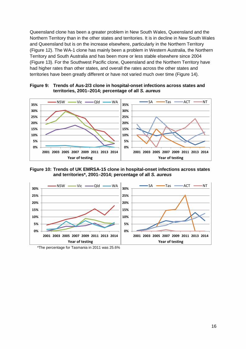
Queensland clone has been a greater problem in New South Wales, Queensland and the Northern Territory than in the other states and territories. It is in decline in New South Wales and Queensland but is on the increase elsewhere, particularly in the Northern Territory (Figure 12). The WA-1 clone has mainly been a problem in Western Australia, the Northern Territory and South Australia and has been more or less stable elsewhere since 2004 (Figure 13). For the Southwest Pacific clone, Queensland and the Northern Territory have had higher rates than other states, and overall the rates across the other states and territories have been greatly different or have not varied much over time (Figure 14). Figure 9: Trends of Aus-2/3 clone in hospital-onset infections across states and
territories, 2001–2014; percentage of all S. aureus
Figure 10: Trends of UK EMRSA-15 clone in hospital-onset infections across states
and territories*, 2001–2014; percentage of all S. aureus
*The percentage for Tasmania in 2011 was 25.6%
0%
5%
10%
15%
20%
25%
30%
35%
2001 2003 2005 2007 2009 2011 2013 2014Year of testing
NSW Vic Qld WA
0%
5%
10%
15%
20%
25%
30%
35%
2001 2003 2005 2007 2009 2011 2013 2014Year of testing
SA Tas ACT NT
0%
5%
10%
15%
20%
25%
30%
2001 2003 2005 2007 2009 2011 2013 2014Year of testing
NSW Vic Qld WA
0%
5%
10%
15%
20%
25%
30%
2001 2003 2005 2007 2009 2011 2013 2014Year of testing
SA Tas ACT NT
16
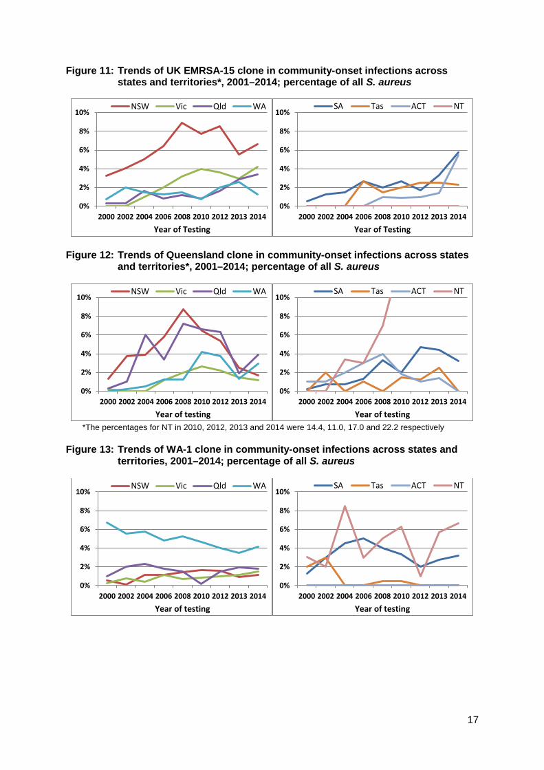
Figure 11: Trends of UK EMRSA-15 clone in community-onset infections across states and territories*, 2001–2014; percentage of all S. aureus
Figure 12: Trends of Queensland clone in community-onset infections across states
and territories*, 2001–2014; percentage of all S. aureus
*The percentages for NT in 2010, 2012, 2013 and 2014 were 14.4, 11.0, 17.0 and 22.2 respectively
Figure 13: Trends of WA-1 clone in community-onset infections across states and
territories, 2001–2014; percentage of all S. aureus
0%
2%
4%
6%
8%
10%
2000 2002 2004 2006 2008 2010 2012 2013 2014Year of Testing
NSW Vic Qld WA
0%
2%
4%
6%
8%
10%
2000 2002 2004 2006 2008 2010 2012 2013 2014Year of Testing
SA Tas ACT NT
0%
2%
4%
6%
8%
10%
2000 2002 2004 2006 2008 2010 2012 2013 2014Year of testing
NSW Vic Qld WA
0%
2%
4%
6%
8%
10%
2000 2002 2004 2006 2008 2010 2012 2013 2014Year of testing
SA Tas ACT NT
0%
2%
4%
6%
8%
10%
2000 2002 2004 2006 2008 2010 2012 2013 2014Year of testing
NSW Vic Qld WA
0%
2%
4%
6%
8%
10%
2000 2002 2004 2006 2008 2010 2012 2013 2014Year of testing
SA Tas ACT NT
17
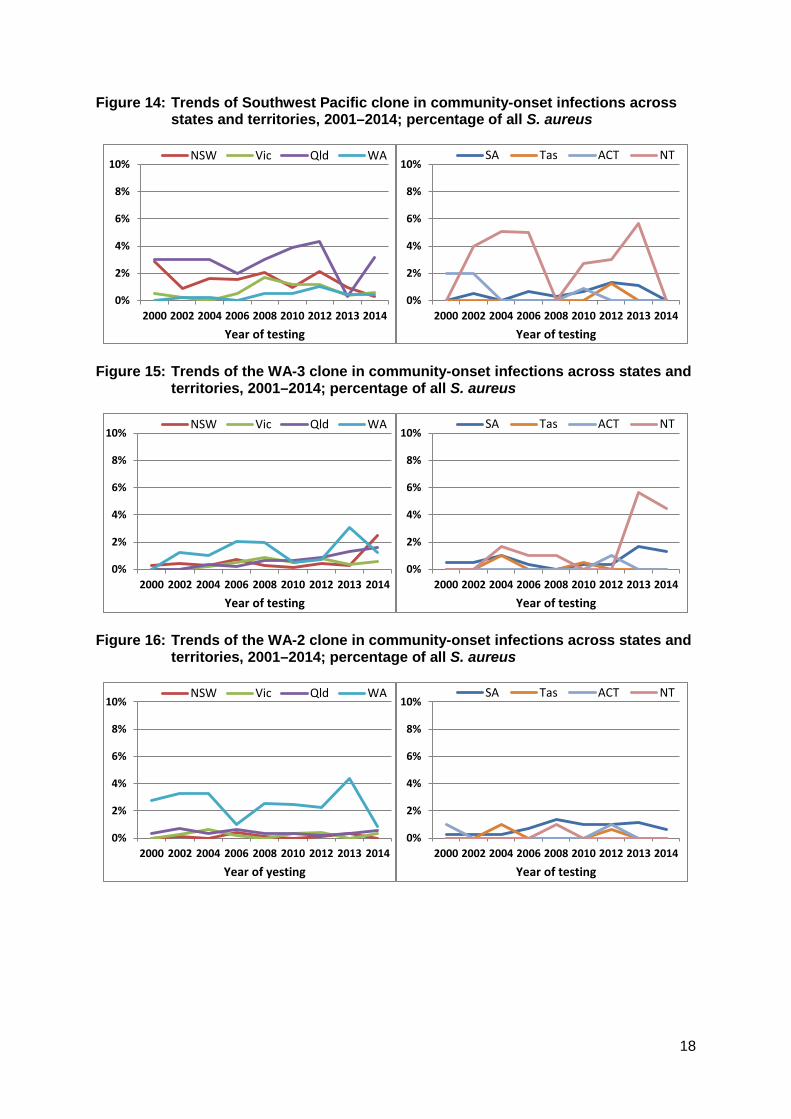
Figure 14: Trends of Southwest Pacific clone in community-onset infections across states and territories, 2001–2014; percentage of all S. aureus
Figure 15: Trends of the WA-3 clone in community-onset infections across states and
territories, 2001–2014; percentage of all S. aureus
Figure 16: Trends of the WA-2 clone in community-onset infections across states and
territories, 2001–2014; percentage of all S. aureus
0%
2%
4%
6%
8%
10%
2000 2002 2004 2006 2008 2010 2012 2013 2014Year of testing
NSW Vic Qld WA
0%
2%
4%
6%
8%
10%
2000 2002 2004 2006 2008 2010 2012 2013 2014Year of testing
SA Tas ACT NT
0%
2%
4%
6%
8%
10%
2000 2002 2004 2006 2008 2010 2012 2013 2014Year of testing
NSW Vic Qld WA
0%
2%
4%
6%
8%
10%
2000 2002 2004 2006 2008 2010 2012 2013 2014Year of testing
SA Tas ACT NT
0%
2%
4%
6%
8%
10%
2000 2002 2004 2006 2008 2010 2012 2013 2014Year of yesting
NSW Vic Qld WA
0%
2%
4%
6%
8%
10%
2000 2002 2004 2006 2008 2010 2012 2013 2014Year of testing
SA Tas ACT NT
18

Figure 17: Trends of the WA-84 clone in community-onset infections across states and territories, 2001–2014; percentage of all S. aureus
Commentary As this analysis has shown, the situation with MRSA in Australia in the 15 years from 2000 to 2014 has been changing significantly. Importantly, the hospital burden has grown and then decreased, while that in the community has continued to grow steadily. The main implication of these changes is the potential impact on empirical treatment of S. aureus infections, accounting for whether they arise in patients who are hospitalised, come from residential aged care, or come from the community. For serious infections, vancomycin is the mainstay of treatment. With the now significant prevalence of CA-MRSA in the community, the empirical treatment of common minor infections is not as certain, due to the variable number of additional resistances in the CA-MRSA clones. Oral options for treatment in the community include the macrolides and clindamycin, tetracyclines (expect in young children), or trimethoprim-sulfamethoxazole (except in the elderly) if the local clones are susceptible. The good news is the rapid decline in the prevalence of the multi-resistant Aus-2/3 HA-MRSA clone after so many decades. This is despite the introduction of a related clade in 2001. The reasons for this decline, as mentioned above, may have been a combination of improvements in infection-control measures and the introduction of a national hand hygiene program. A further factor in reducing the importance in S. aureus infections may have been a decline in virulence as measured in vitro and in vivo.28
By contrast, the expansion of the UK EMRSA-15 clone continues and is concerning. It is hypothesised that this is due in part to increased rates of colonisation rates in residential aged care.13 A recent survey undertaken by the National Centre for Antimicrobial Stewardship in residential aged-care facilities, and supported by the Commission, shows high rates of antimicrobial use,54 much of it inappropriate, which could act as a driver to colonisation and subsequent infection. The most notable feature of CA-MRSA between 2000 and 2014 has been the emergence and spread of the Queensland clone. It was already present in most states and territories in very low numbers in the 2000 survey, mostly in New South Wales. By 2008 it had become dominant in New South Wales and Queensland but since then has subsequently declined in those states. In the other states and territories, it started off more slowly but has steadily
0%
2%
4%
6%
8%
10%
2000 2002 2004 2006 2008 2010 2012 2013 2014Year of testing
NSW Vic Qld WA
0%
2%
4%
6%
8%
10%
2000 2002 2004 2006 2008 2010 2012 2013 2014Year of testing
SA Tas ACT NT
19
increased in prevalence. It has been suggested that part of the success of this clone can be attributed to the possession of Panton-Valentine leucocidin, analogous to the great success of the USA300 clone in the United States. However, if this were the case, then the Southwest Pacific clone, which also possesses this toxin and preceded the emergence of the Queensland clone, might have been expected to become more prominent earlier. Other factors such as different accessory gene content may have played a part. There have been no overall significant trends in the SWP clone of CA-MRSA over the same time period. It is most common in Queensland and the Northern Territory. Its prevalence in New Zealand by contrast, where it was first described, is slowly declining (Annual Survey of MRSA, 2013 online) The WA-1 clone has been present in Australia since 1989. It was identified first in Western Australia, and has since slowly expanded eastwards, and still remains important in South Australia and the Northern Territory. Since 2000, its role has been declining in Western Australia, but it is still the commonest CA-MRSA there. All states except Tasmania see this clone on a regular basis (Figure 13). Although dominated by three clones, community-associated MRSA are continuing to diversify. Three clones are gaining prominence in parts of Australia: WA-3 (ST5-IV), WA-2 (ST78-IV) and the Victorian clone (WA-84 – ST45-V) (Figures 15–17). The WA-3 clone has a similar distribution to WA-1, being most common in Western Australia and to a lesser extent South Australia and the Northern Territory. It is now found throughout Australia except for the Australian Capital Territory. By contrast, WA-2 has remained prominent, mainly in Western Australia, although it is slowly increasing in South Australia. Again, it is found across Australia now, except for the Australian Capital Territory. The Victorian clone is the newest of the three additional CA-MRSA clones. It first emerged in Victoria in 2004 and subsequently in New South Wales and South Australia. It is seen intermittently in the Australian Capital Territory, Queensland and South Australia. To date, the livestock-associated clone of MRSA, ST398, has not been detected in any AGAR surveys. The ST398 MRSA clone emerged in Europe in 2003, initially associated with pigs and pig farmers, and subsequently in other livestock.61 In 2013, it was detected in a nasal swab from an Australian pig veterinarian,62 and subsequently detected at very low levels in pig herds across Australia.63 A distinctive clone often carried by equine veterinarians in Australia, (WA-20 – ST612-IV),64 has been found only once in the 2013 survey, causing a community-onset bloodstream infection. There is no doubt that MRSA types and clones will continue to evolve in Australia. SCCmec is transmitted between strains of S. aureus, and probably between all staphylococcal species including the many species of coagulase-negative staphylococci that colonise humans.60 Each clone varies in its ability to spread and become a problem, and each requires specific measures for control. Control of HA-MRSA is possible through effective infection control, hand hygiene and antimicrobial stewardship programs in hospitals and aged care facilities. More challenging is finding effective control measures for the spread of CA-MRSA.
20

References 1. Tong SY, Davis JS, Eichenberger E, Holland TL, Fowler VG Jr. Staphylococcus aureus
infections: epidemiology, pathophysiology, clinical manifestations, and management. Clin Microbiol Rev. 2015; 28(3):603-61.
2. Sollid JU, Furberg AS, Hanssen AM, Johannessen M. Staphylococcus aureus: determinants of human carriage. Infect Genet Evol. 2014 Jan;21:531-41.
3. Munch-Petersen E, Boundy C. Yearly incidence of penicillin-resistant staphylococci in man since 1942. Bull World Health Organ. 1962; 26(2): 241-252.
4. Jevons MP. ‘Celbenin’-resistant staphylococci. Br Med J 1961; 1:125-6 5. Rountree PM, Beard MA. Hospital strains of Staphylococcus aureus, with particular
reference to methicillin-resistant strains. Med J Aust 1968; 2(26):1163-8. 6. Turnidge JD, Bell JM. Methicillin-resistant Staphylococcus aureus evolution in Australia
over 35 years. Microb Drug Resist 2000; 6(3):223-9 7. Turnidge JD, Kotsanas D, Munckhof W, Roberts S, Bennett CM, Nimmo GR, Coombs
GW, Murray RJ, Howden B, Johnson PD, Dowling K; Australia New Zealand Cooperative on Outcomes in Staphylococcal Sepsis. Staphylococcus aureus bacteraemia: a major cause of mortality in Australia and New Zealand. Med J Aust. 2009 Oct 5;191(7):368-73
8. Harris SR, Feil EJ, Holden MTG, et al. Evolution of MRSA during hospital transmission and intercontinental spread. Science 2010; 327:469-74.
9. Lancashire JF, Jones A, Bergh H, Huygens F, Nimmo GR. Typing early Australian health-associated MRSA: confirmation of major clones and emergence of ST1-MRSA-IV and novel ST2249-MRSA-III. Pathology 2013; 45(5):492-4
10. Gray RR, Tatem AJ, Johnson JA, Alekseyenko AV, Pybus OG, Suchard MA, Salemi M. Testing spatiotemporal hypothesis of bacterial evolution using methicillin-resistant Staphylococcus aureus ST239 genome-wide data with a Bayesian Framework. Mol Biol Evol 2011 May;28(5):1593-603
11. Holden MTG, Hsu L-Y, Kurt K, Weinert LA, Mather AE, Harris SR, Strommenger B, Layer F, Witte W, de Lencastre H, Skov R, Westh H, Zemlicková H, Coombs G, Kearns AM, Hill RL, Edgeworth J, Gould I, Gant V, Cooke J, Edwards GF, McAdam PR, Templeton KE, McCann A, Zhou Z, Castillo-Ramírez S, Feil EJ, Hudson LO, Enright MC, Balloux F, Aanensen DM, Spratt BG, Fitzgerald JR, Parkhill J, Achtman M, Bentley SD, Nübel U. et al. A genomic portrait of the emergence, evolution. And global spread of a methicillin-resistant Staphylococcus aureus pandemic. Genome Res 2013; 23(4):653-64.
12. Pearman JW, Coombs GW, Grubb WB, O’Brien F. A British epidemic strain of methicillin-resistant Staphylococcus aureus (UK EMRSA-15) in Western Australia. Med J Aust 2001; 174:662.
13. Jeremiah CJ, Kandiah JP, Spelman DW, Giffard PM, Coombs GW, Jenney AW, Tong SY. Differing epidemiology of two major healthcare-associated meticillin-resistant Staphylococcus aureus clones. J Hosp Infect. 2016; 92(2):183-90.
14. Oliveira DC, Tomasz A, de Lencastre H. The evolution of pandemic clones of methicillin-resistant Staphylococcus aureus: identification of two ancestral genetic backgrounds and the associated mec elements. Microb Drug Resist. 2001; 7(4):349-61.
15. Coombs GW, Van Gessel H, Pearson JC, Godsell MR, O'Brien FG, Christiansen KJ. Controlling a multicenter outbreak involving the New York/Japan methicillin-resistant Staphylococcus aureus clone. Infect Control Hosp Epidemiol. 2007 Jul;28(7):845-52.
21
16. McAdam PR, Templeton KE, Edwards GF, et al. Molecular tracking of the emergence, adaptation, and transmission of hospital-associated methicillin-resistant Staphylococcus aureus. Proc Natl Acad Sci 2012; 109(23):9107-12.
17. Coombs GW, Nimmo GR, Bell JM, Huygens F, O’Brien FG, Malkowski MJ, Pearson JC, Stephens AJ, Giffard PM, and the Australian Group on Antimicrobial Resistance. Genetic diversity among community methicillin-resistant Staphylococcus aureus infections in Australia. J Clin Microbiol 2004; 42(10):4735-43.
18. Udo EE, Pearman JW, Grubb WB. Genetic analysis of community isolates of methicillin-resistant Staphylococcus aureus in Western Australia. J Hosp Infect 1993; 25(2):97-108.
19. O’Brien FG, Lim TT, Chong FN, Coombs GW, Enright MC, Robinson DA, Monk A, Saïd-Salim B, Kreiswirth BN, Grubb WG. Diversity among community isolates of methicillin-resistant Staphylococcus aureus in Australia. J Clin Microbiol 2004; 42(7):3185-90.
20. Munckhof WJ1, Schooneveldt J, Coombs GW, Hoare J, Nimmo GR. Emergence of community-acquired methicillin-resistant Staphylococcus aureus (MRSA) infection in Queensland, Australia. Int J Infect Dis. 2003;7(4):259-64.
21. Adhikari RP, Cooks GM, Lamont I, Lang S, Heffernan H, Smith JMB. Phenotypic and molecular characterization of community occurring, Western Samoan phage pattern methicillin-resistant Staphylococcus aureus. J Antimicrob Chemother 2002; 50(6):825-31.
22. Collignon P, Gosbell I, Vickery A, Nimmo G, Stylianopoulos T, Gottlieb T. Community-acquired meticillin-resistant Staphylococcus aureus in Australia. Lancet 1998; 352(9122):146-7.
23. Centers for Disease Control and Prevention. Methicillin-resistant Staphylococcus aureus infections in correctional facilities–Georgia, California and Texas, 2001–2003. MMWR Morb Mort Wkly Rep 2003; 52:992-6.
24. Pavillard R, Harvey K, Douglas D, Hewstone A, Andrew J, Collopy B, Asche V, Carson P, Davidson A, Gilbert G, Spicer J, Tosolini F. Epidemic of hospital-acquired infection due to methicillin-resistant Staphylococcus aureus in major Victorian hospitals. Med J Aust. 1982;1(11):451-4.
25. Turnidge J, Lawson P, Munro R, Benn R. A national survey of antimicrobial resistance in Staphylococcus aureus in Australian teaching hospitals. Med J Aust. 1989 Jan 16;150(2):65, 69-72.
26. Dailey L, Coombs GW, O'Brien FG, Pearman JW, Christiansen K, Grubb WB, Riley TV. Methicillin-resistant Staphylococcus aureus, Western Australia. Emerg Infect Dis. 2005 Oct;11(10):1584-90.
27. Gram-positive Bacteria Typing Laboratory, Microbiology Department, Fiona Stanley Hospital, PathWest Laboratory Medicine – WA, and Australian Collaborating Centre for Enterococcus and Staphylococcus Species (ACCESS) Typing and Research, School of Veterinary and Life Sciences, Murdoch University. Western Australian methicillin-resistant Staphylococcus aureus (MRSA) epidemiology and typing report, July 1 2014 to June 30 2015. Western Australian MRSA epidemiology and typing report, July 1 2014 to June 30 2015 online. Accessed June 2016
28. Baines SL, Holt KE, Schultz MB, Seemann T, Howden BO, Jensen SO, van Hal SJ, Coombs GW, Firth N, Powell DR, Stinear TP, Howden BP. Convergent adaptation in the dominant global hospital clone DT239 of methicillin-resistant Staphylococcus aureus. 2015; 6(2) e00080.
22
29. Coombs GW, Daley DA, Lee YT, Pearson JC, Robison JO, Nimmo GR, Collignon P, Howden BP, Bell JM, Turnidge JD. Australian Group on Antimicrobial Resistance (AGAR) Australian Staphylococcus aureus Sepsis Outcome Programme (ASSOP) annual report, 2014. Commun Dis Intell 2016; 40(2):E244-254.
30. Hand Hygiene Australia–Australian Commission on Safety and Quality in Health Care. National Hand Hygiene Initiative: Commission flyer – National Hand Hygiene Initiative. Accessed April 2016.
31. Australian Institute of Health and Welfare. Staphylococcus aureus bacteraemia in Australian public hospitals 2013–14. AIHW report online. Staphylococcus aureus bacteraemia in Australian public hospitals 2013–14. Accessed April 2016.
32. Australian Commission on Safety and Quality in Health Care, National Safety and Quality Health Service Standards (September 2012). Sydney. ACSQHC, 2012.
33. Australian Bureau of Statistics. Australian social trends, April 2013. Doctors and Nurses. ABS online report – Australian social trends, April 2013. Doctors and Nurses. Accessed April 2016.
34. Hart J, Christiansen KJ, Lee R, Heath CH, Coombs GW, Robinson JO. Increased EMRSA-15 health-care worker colonization demonstrated in retrospective review of EMRSA hospital outbreaks. Antimicrob Resist Infect Control 2014; 3:7. p1-6.
35. Public Health England. Voluntary surveillance of Staphylococcus aureus bacteraemia in England, Wales and Northern Ireland: 2007-2014. Health Protection Report weekly report, 2015; Volume 9, number 29. Public Health England online – Health Protection Report weekly report, 2015. Accessed June 2016
36. Chen CJ, Huang YC. New epidemiology of Staphylococcus aureus infection in Asia. Clin Microbiol Infect. 2014 Jul;20(7):605-23.
37. Wertheim HF, Vos MC, Ott A, van Belkum A, Voss A, Kluytmans JA, van Keulen PH, Vandenbroucke-Grauls CM, Meester MH, Verbrugh HA. Risk and outcome of nosocomial Staphylococcus aureus bacteraemia in nasal carriers versus non-carriers. Lancet. 2004 Aug 21-27;364(9435):703-5.
38. Riley TV, Rouse IL. Methicillin-resistant Staphylococcus aureus in Western Australia, 1983-1992. J Hosp Infect 1995; 29:177-88.
39. Survey of non-multiresistant and multiresistant MRSA, July 2000. In ESR Lablink Volume 8: No 2 June 2001. ESR Lablink online – Survey of non-multiresistant and multiresistant MRSA, July 2000. Accessed April 2016.
40. Riley D, MacCulloch D, Morris A. Methicillin-resistant S. aureus in the suburbs. New Zealand Medical Journal 1998; 111:59.
41. Nimmo GR, Coombs GW. Community-associated methicillin-resistant Staphylococcus aureus in Australia. Int J Antimicrob Agents 2008; 31:401-10.
42. Robinson DA, Kearns AM, Holmes A, Morrison D, Grindman H, Edwards G, O’Brien FG, Tenover FC, McDougal LK, Monk AB, Enright MC. Re-emergence of early pandemic Staphylococcus aureus as a community-acquired methicillin-resistant clone. Lancet 2005; 365:1256-58.
43. Rountree PM, Beard MA. Further observations on infection with phage type 80 staphylococci in Australia. Med J Aust 1958; 2:789-95.
44. Coombs GW, Goering RV, Chua KY, Monecke S, Howden BP, Stinear TP, Ehricht R, O'Brien FG, Christiansen KJ. The molecular epidemiology of the highly virulent ST93 Australian community Staphylococcus aureus strain. PLoS One. 2012;7(8):e43037
23
45. Shallcross LJ, Fragaszy E, Johnson AM, Hayward AC. The role of the Panton-Valentine leucocidin toxin in staphylococcal disease: a systematic review and meta-analysis. Lancet Infect Dis. 2013 Jan;13(1):43-54.
46. Laabei M, Uhlemann AC, Lowy FD, Austin ED, Yokoyama M, Ouadi K, Feil E, Thorpe HA, Williams B, Perkins M, Peacock SJ, Clarke SR, Dordel J, Holden M, Votintseva AA, Bowden R, Crook DW, Young BC, Wilson DJ, Recker M, Massey RC. Evolutionary trade-offs underlie the multi-faceted virulence of Staphylococcus aureus. PLoS Biol. 2015 Sep 2;13(9):e1002229
47. Coombs G, Pearson J, Daley D, Robinson O, Nimmo G, Turnidge J. Staphylococcus aureus programme 2012 (SAP 2012) community survey. MRSA epidemiology and typing report. Australian Group on Antimicrobial Resistance. AGAR – MRSA epidemiology and typing report online. Accessed April 2016.
48. Tenover FC, Goering RV. Methicillin-resistant Staphylococcus aureus strain USA300: origin and epidemiology. J Antimicrob Chemother 2009; 64:441-6.
49. Pardos de la Gandara M, Curry M, Berger J, Burstein D, Della-Latta P, Kopetz V, Quale J, Spitzer E, Tan R, Urban C, Wang G, Whittier S, de Lencastre H, Tomasz A. MRSA causing infections in hospitals in greater metropolitan New York; major shift in the dominant clonal type between 1996 and 2014. PLoS One 2016; 11(6):e0156924.
50. Nimmo GR. USA300 abroad: global spread of a virulent strain of community-associated MRSA. Clin Microbiol Infect 2012; 18:725-34
51. Panton PN, Valentine FCO. Staphylococcal toxin. Lancet 1932; 219:506-8. 52. Vandenesch F, Naimi T, Enright MC, Nimmo GR, Heffernan H, Liassine N, Bes M,
Greenland T, Reverdy ME, Etienne J. Community-acquired methicillin-resistant Staphylococcus aureus carry Panton-Valentine leukocidin genes: worldwide emergence. Emerg Infect Dis 2003; 9:978-84.
53. Monecke S, Slickers P, Ellington MJ, Kearns AM, Ehricht R. High diversity of Panton-Valentine leukocidin-positive, methicillin-susceptible isolates of Staphylococcus aureus and implications for the evolution of community-associated methicillin-resistant S. aureus. Clin Microbiol Infect. 2007 Dec;13(12):1157-64
54. National Centre for Antimicrobial Stewardship and the Australian Commission on Safety and Quality in Health Care. Antimicrobial prescribing and infections in Australian residential aged care facilities: Results of the 2015 Aged Care National Antimicrobial Prescribing Survey pilot. Sydney: ACSQHC, 2016.
55. Moran GJ, Krishnadasan A, Gorwitz RJ, Fosheim GE, McDougal LK, Carey RB, Talan DA; EMERGEncy ID Net Study Group. Methicillin-resistant S. aureus infections among patients in the emergency department. N Engl J Med. 2006; 355(7):666-74
56. Masiuk H, Kopron K, Grumann D, Goerke C, Kolata J, Jursa-Kulesza J, Giedrys-Kalemba S, Bröker BM, Holtfreter S. Association of recurrent furunculosis with Panton-Valentine leukocidin and the genetic background of Staphylococcus aureus. J Clin Microbiol. 2010 May;48(5):1527-35.
57. Sheikh HQ, Aqil A, Kirby A, Hossain FS. Panton-Valentine leukocidin osteomyelitis in children: a growing threat. Br J Hosp Med (Lond). 2015; 76(1):18-24.
58. Hidron AI, Low CE, Honig EG, Blumberg HM. Emergence of community-acquired meticillin-resistant Staphylococcus aureus strain USA300 as a cause of necrotising community-onset pneumonia. Lancet Infect Dis. 2009 Jun;9(6):384-92.
59. Wehrhahn MC1, Robinson JO, Pearson JC, O'Brien FG, Tan HL, Coombs GW, Pascoe EM, Lee R, Salvaris P, Salvaris R, New D, Murray RJ. Clinical and laboratory features of invasive community-onset methicillin-resistant Staphylococcus aureus infection: a
24
prospective case-control study. Eur J Clin Microbiol Infect Dis. 2010 Aug;29(8):1025-33. doi: 10.1007/s10096-010-0973-4. Epub 2010 Jun 12.
60. Martínez-Meléndez A, Morfín-Otero R, Villarreal-Trevino L, González-González G, Llaca-Díaz J, Rodríguez-Noriega E, Camacho-Ortíz A, E. Garza-González E. Staphylococcal Cassette Chromosome mec (SCCmec) in coagulase negative staphylococci. Medicina Universitaria. 2015;17(69):229-33
61. van Cleef BA, Monnet DL, Voss A, Krziwanek K, Allerberger F, Struelens M, Zemlickova H, Skov RL, Vuopio-Varkila J, Cuny C, Friedrich AW, Spiliopoulou I, Pászti J, Hardardottir H, Rossney A, Pan A, Pantosti A, Borg M, Grundmann H, Mueller-Premru M, Olsson-Liljequist B, Widmer A, Harbarth S, Schweiger A, Unal S, Kluytmans JA. Livestock-associated methicillin-resistant Staphylococcus aureus in humans, Europe. Emerg Infect Dis. 2011; 17(3):502-5.
62. Trott D, Jordan D, Barton M. Abraham S, Groves M. Vets versus pets: methicillin-resistant Staphylococcus aureus in Australian animals and their doctors. Microbiol Aust 2013; 34: 25-7.
63. Groves MD, O'Sullivan MV, Brouwers HJ, Chapman TA, Abraham S, Trott DJ, Al Jassim R, Coombs GW, Skov RL, Jordan D. Staphylococcus aureus ST398 detected in pigs in Australia. J Antimicrob Chemother. 2014 May;69(5):1426-8.
64. Groves MD, Crouch B, Coombs GW, Jordan D, Pang S, Barton MD, Giffard P, Abraham S, Trott DJ. Molecular Epidemiology of Methicillin-Resistant Staphylococcus aureus Isolated from Australian Veterinarians. PLoS One. 2016;11(1):e0146034.
25
Australian Commission on Safety and Quality in Health Care Level 5, 255 Elizabeth Street, Sydney NSW 2001 GPO Box 5480, Sydney NSW 2001 Phone: (02) 9126 3600 Fax: (02) 9126 3613 Email: [email protected] Website: www.safetyandquality.gov.au