motor neuron mitochondrial dysfunction in spinal muscular ... · original article motor neuron...
TRANSCRIPT
O R I G I N A L A R T I C L E
Motor neuron mitochondrial dysfunction in spinal
muscular atrophyNimrod Miller1, Han Shi1, Aaron S. Zelikovich1 andYong-Chao Ma1,*1Department of Pediatrics, Northwestern University Feinberg School of Medicine, Ann & Robert H. LurieChildren’s Hospital of Chicago, Chicago, IL, USA
*To whom correspondence should be addressed at: Tel: þ773-755-6393; Fax: þ773-755-6581; Email: [email protected]
AbstractSpinal muscular atrophy (SMA), the leading genetic cause of infant mortality, predominantly affects high metabolic tissuesincluding motor neurons, skeletal muscles and the heart. Although the genetic cause of SMA has been identified,mechanisms underlying tissue-specific vulnerability are not well understood. To study these mechanisms, we carried out adeep sequencing analysis of the transcriptome of spinal motor neurons in an SMA mouse model, in which we unexpectedlyfound changes in many genes associated with mitochondrial bioenergetics. Importantly, functional measurement of mito-chondrial activities showed decreased basal and maximal mitochondrial respiration in motor neurons from SMA mice. Usinga reduction-oxidation sensitive GFP and fluorescence sensors specifically targeted to mitochondria, we found increased oxi-dative stress level and impaired mitochondrial membrane potential in motor neurons affected by SMA. In addition, mito-chondrial mobility was impaired in SMA disease conditions, with decreased retrograde transport but no effect on anterogradetransport. We also found significantly increased fragmentation of the mitochondrial network in primary motor neurons fromSMA mice, with no change in mitochondria density. Electron microscopy study of SMA mouse spinal cord revealed mitochon-dria fragmentation, edema and concentric lamellar inclusions in motor neurons affected by the disease. Intriguingly, thesefunctional and structural deficiencies in the SMA mouse model occur during the presymptomatic stage of disease, suggestinga role in initiating SMA. Altogether, our findings reveal a critical role for mitochondrial defects in SMA pathogenesis and sug-gest a novel target for improving tissue health in the disease.
IntroductionSpinal Muscular Atrophy (SMA) is an autosomal recessive neu-romuscular disease that occurs in one of every 10,000 live births(1–3), ranking as the leading genetic cause of infant mortality.SMA is caused by mutation in survival motor neuron 1 (SMN1)gene (4). In addition to SMN1, humans have SMN2, which is dif-ferent from SMN1 by a cytosine (C) to thymine (T) change in itsexon 7. This single nucleotide change disrupts the efficientsplicing of SMN2 gene. Consequently, only about 10% of SMNprotein generated by SMN2 is full-length and functional. In SMApatients, genetic mutations cause a complete loss of SMN
production from SMN1 (4), leading to reduced levels of SMN pro-tein, which correlate inversely with disease severity (5). Patientswith a severe form of disease exhibit disease onset around 5months of age, with death from respiratory distress within 2years (5). SMN is implicated in regulating a variety of biologicalfunctions, including small nuclear ribonucleoprotein (snRNP)biogenesis and pre-mRNA splicing (6–9). Although SMN proteinis ubiquitously expressed, SMA disease is characterized by thepredominant loss of lower alpha motor neurons in the spinalcord (10–12). Other tissues affected in SMA include skeletal mus-cles and the heart, all of which require high levels of energy
Received: May 16, 2016. Revised: July 25, 2016. Accepted: July 28, 2016
VC The Author 2016. Published by Oxford University Press.All rights reserved. For permissions, please email: [email protected]
3395
Human Molecular Genetics, 2016, Vol. 25, No. 16 3395–3406
doi: 10.1093/hmg/ddw262Advance Access Publication Date: 3 August 2016Original Article
supply (13–16). Mechanisms underlying tissue-specific vulnera-bility in SMA are poorly understood.
Tissues with high-energy demand are particularly enrichedin mitochondria, whose primary function is to supply cells withATP generated by oxidative phosphorylation (17). Depending onthe cell type, hundreds and sometimes thousands of mitochon-dria populate a cell. Neurons and muscles, which expend moreenergy than other cell types, have a particularly high require-ment of mitochondrial functions. Within a cell mitochondriaare dynamically transported to and localized in regions that uti-lize more energy, such as the axon hillock and presynaptic ter-minal of neurons (18,19). Mutations in genes codingcomponents of the respiratory chain of oxidative phosphoryla-tion lead to mitochondrial diseases (18,20,21), which preferen-tially affect tissue types with high bioenergetic requirement.SMA share many common features with mitochondrial diseasesincluding the specific vulnerability of tissues with high energydemand, suggesting that mitochondria might be functionallyimpaired in SMA. Clinical studies have indicated fatty acid me-tabolism defects in SMA patient plasma (22–24), and impairedmyogenesis and mitochondrial biogenesis in SMA patient’smuscles (25). However, potential defects of mitochondrial struc-ture and function in motor neurons of SMA mouse models havenot been explored.
In this study, we report that mitochondria in motor neuronsaffected by SMA are functionally and structurally defective be-fore the onset of disease symptoms. We found that mitochon-dria in motor neurons from SMA mice were functionallyimpaired and fragmented, with reduced mitochondrial respira-tion, decreased mitochondrial ATP synthesis, defective retro-grade mitochondrial transport, decreased mitochondrialmembrane potential and increased oxidative stress level. In ad-dition, electron micrographs of lumbar level spinal cord motorneurons from SMA mice show significant mitochondrial frag-mentation and edema at a presymptomatic stage, suggesting arole for mitochondrial defects in initiating SMA pathogenesis.
ResultsImpaired mitochondrial bioenergetics
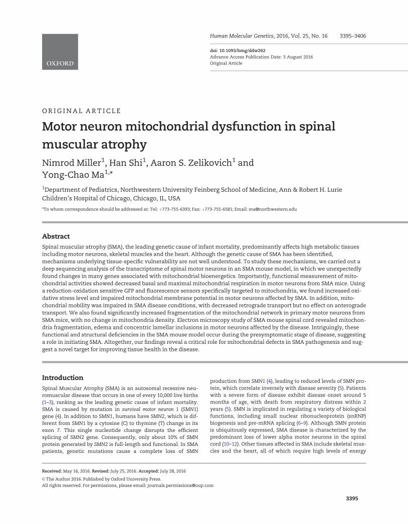
To better understand why motor neurons are specifically vul-nerable in SMA, we decided to isolate spinal motor neuronsfrom SMA and control mice and study the difference betweentheir RNA expression profiles. We crossed the Hung-Li SMAmouse model with the Hb9:GFP mice, which specifically labelmotor neurons with GFP (26). After micro-dissection and enzy-matic dissociation, GFP-expressing spinal motor neurons fromSMA (SMA; Hb9:GFP) mice and control (Hb9:GFP) mice were iso-lated by fluorescence activated cell sorting (FACS). Then RNAdeep sequencing (RNA-seq) was used to analyse RNA expres-sion profiles in these purified motor neurons. Analysis of theRNA-seq data using GSEA (Gene Set Enrichment Analysis) andthe hallmark gene sets showed that many mitochondriabioenergetics-related genes were significantly dysregulated inmotor neurons affected by SMA (Fig. 1A and B, SupplementaryMaterial, Table S1) . Therefore, we decided to test whether mito-chondrial functions were changed in motor neurons from SMAmouse models.
The primary function of mitochondria in cells is to generateATP through oxidative phosphorylation. To test whether mito-chondrial bioenergetics were compromised in SMA disease con-ditions, we isolated and cultured motor neurons from the spinalcord of D7 SMA mice and control littermates (Fig. 1C).
Mitochondrial respiration of primary spinal motor neurons wasmeasured using a Seahorse analyzer. Motor neurons affected bySMA showed significantly lower basal mitochondrial respirationrate/oxygen consumption rate (OCR) than non-disease motorneurons (Fig. 1D and F). In addition, maximal mitochondrial res-piration rate/OCR induced by mitochondrial uncoupler carbonylcyanide m-chlorophenyl hydrazone (CCCP), which is an indica-tion of the highest mitochondrial functional capacity understress conditions, was also significantly reduced (Fig. 1D and G).Importantly, coupled mitochondrial respiration rate/OCR usedfor ATP synthesis, as well as the OCR for counteracting protonleaking across the mitochondrial inner membrane were bothsignificantly decreased (Fig. 1D,H,I). To test whether the ob-served mitochondrial respiration deficiency is specific to motorneurons, we examined primary mouse midbrain neurons fromSMA and wild type mice. Midbrain neurons from SMA miceshowed no change of mitochondrial basal, maximal, coupled orproton leak OCR, compared to those of midbrain neurons fromwild type mice (Fig. 1E–I). These results suggest that mitochon-drial bioenergetics are specifically impaired in motor neuronsaffected by SMA.
Increased mitochondrial oxidative stress and reducedmitochondrial membrane potential
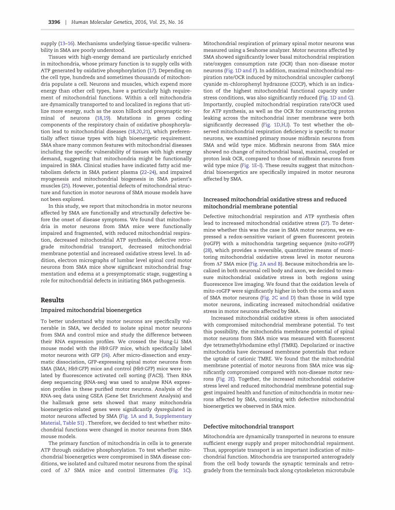
Defective mitochondrial respiration and ATP synthesis oftenlead to increased mitochondrial oxidative stress (27). To deter-mine whether this was the case in SMA motor neurons, we ex-pressed a redox-sensitive variant of green fluorescent protein(roGFP) with a mitochondria targeting sequence (mito-roGFP)(28), which provides a reversible, quantitative means of moni-toring mitochondrial oxidative stress level in motor neuronsfrom D7 SMA mice (Fig. 2A and B). Because mitochondria are lo-calized in both neuronal cell body and axon, we decided to mea-sure mitochondrial oxidative stress in both regions usingfluorescence live imaging. We found that the oxidation levels ofmito-roGFP were significantly higher in both the soma and axonof SMA motor neurons (Fig. 2C and D) than those in wild typemotor neurons, indicating increased mitochondrial oxidativestress in motor neurons affected by SMA.
Increased mitochondrial oxidative stress is often associatedwith compromised mitochondrial membrane potential. To testthis possibility, the mitochondria membrane potential of spinalmotor neurons from SMA mice was measured with fluorescentdye tetramethylrhodamine ethyl (TMRE). Depolarized or inactivemitochondria have decreased membrane potentials that reducethe uptake of cationic TMRE. We found that the mitochondrialmembrane potential of motor neurons from SMA mice was sig-nificantly compromised compared with non-disease motor neu-rons (Fig. 2E). Together, the increased mitochondrial oxidativestress level and reduced mitochondrial membrane potential sug-gest impaired health and function of mitochondria in motor neu-rons affected by SMA, consisting with defective mitochondrialbioenergetics we observed in SMA mice.
Defective mitochondrial transport
Mitochondria are dynamically transported in neurons to ensuresufficient energy supply and proper mitochondrial repairment.Thus, appropriate transport is an important indication of mito-chondrial function. Mitochondria are transported anterogradelyfrom the cell body towards the synaptic terminals and retro-gradely from the terminals back along cytoskeleton microtubule
3396 | Human Molecular Genetics, 2016, Vol. 25, No. 16
Figure 1. Impaired mitochondrial bioenergetics in spinal motor neurons affected by SMA. (A) Heat map depicting differential gene expression in spinal motor neurons
isolated by fluorescence activated cell sorting (FACS) from SMA; Hb9:GFP mice and littermate wild type (WT) mice (n¼4). (B) Gene ontology analysis of RNA-seq results
using GSEA revealed that several categories of genes, whose expression changed most significantly in SMA motor neurons, were related to mitochondrial function and
energy metabolism (fold change cutoff ¼ 1.5, P< 0.05, FDR: False Discovery Rate). (C) Immunostaining of motor neurons cultured from E12.5 mouse spinal cords. Cells
were stained with antibodies recognizing neuronal marker Tuj1 (green) and motor neuron marker HB9 (red). (D) Mitochondrial oxygen consumption rates (OCR) of mo-
tor neurons from D7 SMA and wild type control mice. OCR measured by a Seahorse XF24 Analyzer showed impaired mitochondrial bioenergetics in motor neurons af-
fected by SMA. (E) Midbrain neuron mitochondrial OCR was not changed in D7 SMA mice. (F) Basal mitochondrial OCR was significantly decrease in spinal motor
neurons (P¼0.0019, WT n¼27; SMA n¼28), but not in midbrain neurons (P¼0.78, WT n¼20; SMA n¼16), isolated from SMA mice compared to those from wild type
(WT) control mice. (G) Maximal mitochondrial OCR measured in the presence of CCCP was significantly reduced in SMA spinal motor neurons (P¼0.0029) but not in
midbrain neurons (P¼0.84). (H) Coupled mitochondrial OCR used for ATP synthesis was significantly reduced in SMA spinal motor neurons (P¼ 0.024) but not in mid-
brain neurons (P¼0.69). (I) Mitochondrial proton leak OCR was also specifically reduced in SMA spinal motor neurons (P¼ 0.0049) but not in midbrain neurons (P¼0.73).
Results are mean 6 SEM, from at least three independent experiments, were normalized to cell number. n.s.: not significant; *P< 0.05; **P< 0.01; ***P < 0.001, Student’s
t test.
3397Human Molecular Genetics, 2016, Vol. 25, No. 16 |
tracks (29). We recently found that hyper-phosphorylation ofthe microtubule-associated protein Tau, a key regulator of cellu-lar trafficking, contributed to the specific loss of motor neuronsin SMA (30). This finding motivated us to explore the possibilitythat mitochondrial transport is impaired in SMA motor neu-rons. We used time-lapse confocal live imaging to measure mi-tochondrial transport in primary motor neurons transfectedwith mitochondria targeting sequence-tagged DsRed (mito-DsRed) (Fig. 3A) (31). We found that mitochondria travelled sig-nificantly shorter distance in axons of D7 SMA motor neuronscompared to wild type neurons (Fig. 3B). Further analysis re-vealed that retrograde travel distance (Fig. 3E) of mitochondriawas also decreased in motor neurons affected by SMA, whichwas caused by decreased speed (Fig. 3D) and time (Fig. 3C) spenton retrograde transport. There was also a significant increase inthe time spent in the immobile state (Fig. 3C) by mitochondriain SMA motor neurons, but no significant effect on any aspectsof anterograde transport. To test whether defective mitochon-drial transport is unique to the D7 SMA mice, we performedthese studies using a different SMA model, the Hung-Li SMAmice. Both of these mouse models were generated by express-ing low levels of human SMN transgene in Smn knock-out miceto recapitulate hallmarks of SMA pathogenesis at the molecular,cellular and behaviour levels, with similar average lifespan ofaround 13 days (32,33). Similarly, mitochondria in spinal motorneurons from the Hung-Li SMA mice also showed significantlyreduced total travel distance, decreased retrograde transportdistance, speed and time, as well as increased percent oftime in the immobile state (Supplementary Material, Fig. S1).
By contrast, every aspect of mitochondrial transport appears tobe normal in midbrain neurons isolated from SMA mice (Fig. 3F–J),suggesting defective mitochondrial transport is specific formotor neurons. Given the particularly long axons of lower alphamotor neurons, it is conceivable that detrimental consequencesof defective mitochondrial transport may be exacerbated inmotor neurons, thus contributing to motor neuron-specificdegeneration in SMA pathogenesis.
Increased mitochondria fragmentation
Mitochondrial functions are closely related to its size and mor-phology. Elongated mitochondria have higher density of cristaewith better efficiency in ATP production; whereas short, frag-mented mitochondria are more likely to have compromisedmembrane potential, with lower levels of the dimeric form ofATPase and decreased cristae density, both of which are associ-ated with the impaired ATP synthesis (34–36). To test whethermitochondria fragmentation is associated with the functionaldefects observed in SMA disease conditions, we compared themitochondria length in motor neurons from the D7 SMA miceand control mice transfected with mito-DsRed. Confocal liveimaging of mitochondria located in motor neuron axonsshowed significantly reduced mitochondria length in SMA motorneurons, with no change of mitochondria density (Fig. 4A–C).In addition, we also tested mitochondria fragmentation in mo-tor neurons from the Hung-Li SMA mice. The length of mito-chondria was also significantly reduced in motor neurons from
Figure 2. Increased mitochondrial oxidative stress and compromised membrane potential in spinal motor neurons affected by SMA. (A) A primary mouse spinal motor
neuron expressing redox-sensitive green fluorescent protein with mitochondria targeting sequence (mito-roGFP). The contour of motor neuron was marked by dashed
lines. (B) Measurement of mitochondrial oxidation level. The intensities of mito-roGFP in motor neurons were measured with a live imaging fluorescent microscope.
DTT were applied to fully reduce (red trace) and aldrithiol were used to fully oxidize (green trace) roGFP for calculating relative oxidation levels as described in
Methods. (C, D) Mitochondrial oxidative stress was significantly increased in both the soma (C) (P¼0.00013) and axon (D) (P¼0.0003) of spinal motor neurons from D7
SMA mice. Mitochondrial roGFP oxidation levels in the soma and axon (at least 50 mm away from the motor neuron soma) were recorded in 5–7 experimental repeats
for each group. (E) Mitochondrial membrane potential was significantly reduced (P¼0.0069) in SMA motor neurons. The membrane potentials of mitochondria on 42
samples of D7 SMA motor neurons and 42 wild type samples in three independent experiments were measured using fluorescent dye tetramethylrhodamine ethyl
(TMRE) and normalized to cell number. **P< 0.01; ***P < 0.001, Student’s t test.
3398 | Human Molecular Genetics, 2016, Vol. 25, No. 16
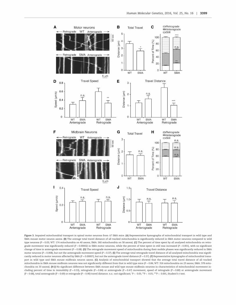
Figure 3. Impaired mitochondrial transport in spinal motor neurons from D7 SMA mice. (A) Representative kymographs of mitochondrial transport in wild type and
SMA mouse motor neuron axons. (B) The average total travel distance of all tracked mitochondria is significantly reduced in SMA motor neurons compared to wild
type neurons (P¼0.03; WT: 174 mitochondria on 40 axons; SMA: 260 mitochondria on 58 axons). (C) The percent of time spent by all analysed mitochondria on retro-
grade movement was significantly reduced (P¼0.00002) in SMA motor neurons, while the percent of time spent in still was increased (P¼0.001), with no significant
change of time in anterograde movement (P¼0.08). (D) The retrograde movement speed of mitochondria during their mobile phases was significantly reduced in SMA
motor neurons (P¼0.008), but not the anterograde movement speed (P¼0.37). (E) The average total retrograde travel distance of all analysed mitochondria was signifi-
cantly reduced in motor neurons affected by SMA (P¼0.00007), but not the anterograde travel distance (P¼0.37). (F) Representative kymographs of mitochondrial trans-
port in wild type and SMA mouse midbrain neuron axons. (G) Analysis of mitochondrial transport showed that the average total travel distance of all tracked
mitochondria in SMA mouse midbrain neurons was not significantly different from that in wild type mice (P¼ 0.84; WT: 316 mitochondria on 23 axons; SMA: 378 mito-
chondria on 33 axons). (H-J) No significant difference between SMA mouse and wild type mouse midbrain neurons in characteristics of mitochondrial movement in-
cluding percent of time in immobility (P¼ 0.53), retrograde (P¼0.84) or anterograde (P¼0.47) movement, speed of retrograde (P¼ 0.80) or anterograde movement
(P¼0.68), total anterograde (P¼0.69) or retrograde (P¼0.90) travel distance. n.s.: not significant; *P< 0.05; **P< 0.01; ***P< 0.001, Student’s t test.
3399Human Molecular Genetics, 2016, Vol. 25, No. 16 |
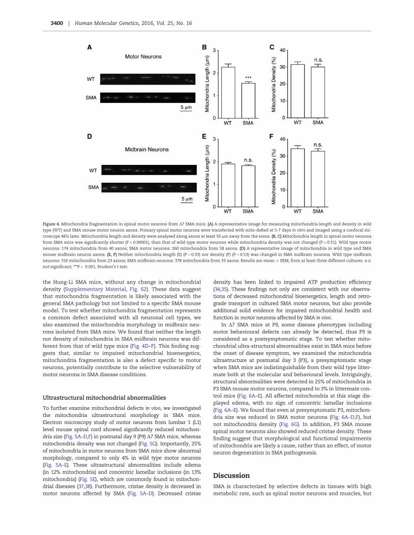
the Hung-Li SMA mice, without any change in mitochondrialdensity (Supplementary Material, Fig. S2). These data suggestthat mitochondria fragmentation is likely associated with thegeneral SMA pathology but not limited to a specific SMA mousemodel. To test whether mitochondria fragmentation representsa common defect associated with all neuronal cell types, wealso examined the mitochondria morphology in midbrain neu-rons isolated from SMA mice. We found that neither the lengthnor density of mitochondria in SMA midbrain neurons was dif-ferent from that of wild type mice (Fig. 4D–F). This finding sug-gests that, similar to impaired mitochondrial bioenergetics,mitochondria fragmentation is also a defect specific to motorneurons, potentially contribute to the selective vulnerability ofmotor neurons in SMA disease conditions.
Ultrastructural mitochondrial abnormalities
To further examine mitochondrial defects in vivo, we investigatedthe mitochondria ultrastructural morphology in SMA mice.Electron microscopy study of motor neurons from lumbar 1 (L1)level mouse spinal cord showed significantly reduced mitochon-dria size (Fig. 5A–D,F) in postnatal day 9 (P9) D7 SMA mice, whereasmitochondria density was not changed (Fig. 5G). Importantly, 25%of mitochondria in motor neurons from SMA mice show abnormalmorphology, compared to only 4% in wild type motor neurons(Fig. 5A–E). These ultrastructural abnormalities include edema(in 12% mitochondria) and concentric lamellar inclusions (in 13%mitochondria) (Fig. 5E), which are commonly found in mitochon-drial diseases (37,38). Furthermore, cristae density is decreased inmotor neurons affected by SMA (Fig. 5A–D). Decreased cristae
density has been linked to impaired ATP production efficiency(34,35). These findings not only are consistent with our observa-tions of decreased mitochondrial bioenergetics, length and retro-grade transport in cultured SMA motor neurons, but also provideadditional solid evidence for impaired mitochondrial health andfunction in motor neurons affected by SMA in vivo.
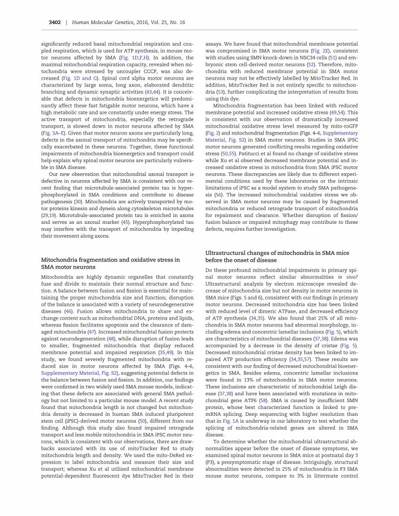
In D7 SMA mice at P9, some disease phenotypes includingmotor behavioural defects can already be detected, thus P9 isconsidered as a postsymptomatic stage. To test whether mito-chondrial ultra-structural abnormalities exist in SMA mice beforethe onset of disease symptom, we examined the mitochondriaultrastructure at postnatal day 3 (P3), a presymptomatic stagewhen SMA mice are indistinguishable from their wild type litter-mate both at the molecular and behavioural levels. Intriguingly,structural abnormalities were detected in 25% of mitochondria inP3 SMA mouse motor neurons, compared to 3% in littermate con-trol mice (Fig. 6A–E). All affected mitochondria at this stage dis-played edema, with no sign of concentric lamellar inclusions(Fig. 6A–E). We found that even at presymptomatic P3, mitochon-dria size was reduced in SMA motor neurons (Fig. 6A–D,F), butnot mitochondria density (Fig. 6G). In addition, P3 SMA mousespinal motor neurons also showed reduced cristae density. Thesefinding suggest that morphological and functional impairmentsof mitochondria are likely a cause, rather than an effect, of motorneuron degeneration in SMA pathogenesis.
DiscussionSMA is characterized by selective defects in tissues with highmetabolic rate, such as spinal motor neurons and muscles, but
Figure 4. Mitochondria fragmentation in spinal motor neurons from D7 SMA mice. (A) A representative image for measuring mitochondria length and density in wild
type (WT) and SMA mouse motor neuron axons. Primary spinal motor neurons were transfected with mito-dsRed at 5-7 days in vitro and imaged using a confocal mi-
croscope 48 h later. Mitochondria length and density were analysed along axons at least 50 mm away from the soma. (B, C) Mitochondria length in spinal motor neurons
from SMA mice was significantly shorter (P¼0.00001), than that of wild type motor neurons while mitochondria density was not changed (P¼0.51). Wild type motor
neurons: 174 mitochondria from 40 axons; SMA motor neurons: 260 mitochondria from 58 axons. (D) A representative image of mitochondria in wild type and SMA
mouse midbrain neuron axons. (E, F) Neither mitochondria length (E) (P¼0.33) nor density (F) (P¼0.53) was changed in SMA midbrain neurons. Wild type midbrain
neurons: 316 mitochondria from 23 axons; SMA midbrain neurons: 378 mitochondria from 33 axons. Results are mean 6 SEM, from at least three different cultures. n.s:
not significant; ***P< 0.001, Student’s t test.
3400 | Human Molecular Genetics, 2016, Vol. 25, No. 16
mitochondrial dysfunction in motor neurons during SMA path-ogenesis has not been systematically explored. Using mousemodels of SMA, we found that mitochondria in SMA motor neu-rons were functionally impaired, with reduced respiration rate,decreased membrane potential, increased oxidative stress level,defective transport along axons and fragmented morphology.Electron microscopy analysis of motor neurons from SMA miceshowed substantial mitochondrial fragmentation and edema,which occur prior to the onset of disease symptoms, suggestinga role for mitochondrial dysfunction in contributing to SMApathogenesis.
Functional defects of mitochondria and motor neuron-specific vulnerability in SMA
Previous clinical studies of infants with severe forms of SMAshowed abnormal fatty acid metabolism (22,23,39,40). Musclebiopsies from SMA patients also showed metabolic deficits (24).In addition, depletion of mitochondrial DNA and disintegratedmitochondria were found in muscles affected by SMA (25,41,42).However, all these works are primarily clinical studies focusingon metabolic or mitochondrial abnormalities in muscles. Herewe systematically investigated functional defects of mitochon-dria in motor neurons using SMA mouse models. We found
Figure 5. Ultrastructural abnormalities of mitochondria in motor neurons in D7
SMA mice. (A-D) Representative low-magnification (A, B) and high-magnifica-
tion (C, D) electron micrographs of mitochondria from lumbar 1 (L1) level spinal
cord motor neurons in wild type and SMA mice at postnatal day 9 (P9).
Mitochondria in SMA mice displayed concentric lamellar inclusions (arrows in
B, D) and edema (arrow heads in B) with decreased cristae density compared to
mitochondria in wild type mice. (E) 25% mitochondria in L1 level motor neurons
in P9 SMA mice exhibited ultrastructural abnormalities compared to 4% mito-
chondria in P9 wild type mouse L1 level motor neurons (P¼0.0001; WT: 200 mi-
tochondria from three wild type mice, SMA: 230 mitochondria from three SMA
mice). (F) Average size of mitochondria in L1 level motor neurons of P9 SMA
mice was significantly smaller than mitochondria in P9 wild type L1 level motor
neurons (P¼0.0009, WT: 200 mitochondria from three wild type mice, SMA: 230
mitochondria from three SMA mice). (G) Mitochondria density in L1 level motor
neurons in P9 SMA mice was similar to that in P9 wild type mice (P¼0.26, WT:
200 mitochondria from three wild type mice, SMA: 230 mitochondria from three
SMA mice). Results are mean 6 SEM, from at least three independent experi-
ments. n.s: not significant; ***P< 0.001, Student’s t test.
Figure 6. Mitochondria show ultrastructural abnormalities in D7 SMA mice be-
fore the onset of disease symptom. (A-D) Representative low-magnification
(A, B) and high-magnification (C, D) electron micrographs of mitochondria from
lumbar 1 (L1) level spinal cord motor neurons in wild type and SMA mice at pre-
symptomatic stage postnatal day 3 (P3). Mitochondria in P3 SMA mice showed
edema (arrow heads in B, D), but not concentric lamellar inclusions, with de-
creased cristae density. (E) 25% mitochondria in L1 level motor neurons in pre-
symptomatic P3 SMA mice displayed edema compared to 3% mitochondria in P3
wild type mice (P¼0.00001; WT: 326 mitochondria from three wild type mice,
SMA: 883 mitochondria from three SMA mice). (F) Average size of mitochondria
in L1 level motor neurons in P3 SMA mice was significantly smaller than that in
P3 wild type mice (P¼0.02, WT: 326 mitochondria from three wild type mice,
SMA: 883 mitochondria from three SMA mice). (G) Mitochondria density in L1
level motor neurons in presymptomatic P3 SMA mice was similar to that in P3
wild type mice (P¼0.85, WT: 326 mitochondria from three wild type mice, SMA:
883 mitochondria from three SMA mice). Results are mean 6 SEM, from at least
three independent experiments. n.s: not significant; *P< 0.05; ***P< 0.001,
Student’s t test.
3401Human Molecular Genetics, 2016, Vol. 25, No. 16 |
significantly reduced basal mitochondrial respiration and cou-pled respiration, which is used for ATP synthesis, in mouse mo-tor neurons affected by SMA (Fig. 1D,F,H). In addition, themaximal mitochondrial respiration capacity, revealed when mi-tochondria were stressed by uncoupler CCCP, was also de-creased (Fig. 1D and G). Spinal cord alpha motor neurons arecharacterized by large soma, long axon, elaborated dendriticbranching and dynamic synaptic activities (43,44). It is conceiv-able that defects in mitochondria bioenergetics will predomi-nantly affect these fast fatigable motor neurons, which have ahigh metabolic rate and are constantly under energy stress. Theactive transport of mitochondria, especially the retrogradetransport, is slowed down in motor neurons affected by SMA(Fig. 3A–E). Given that motor neuron axons are particularly long,defects in the axonal transport of mitochondria may be specifi-cally exacerbated in these neurons. Together, these functionalimpairments of mitochondria bioenergetics and transport couldhelp explain why spinal motor neurons are particularly vulnera-ble in SMA disease.
Our new observation that mitochondrial axonal transport isdefective in neurons affected by SMA is consistent with our re-cent finding that microtubule-associated protein tau is hyper-phosphorylated in SMA conditions and contribute to diseasepathogenesis (30). Mitochondria are actively transported by mo-tor proteins kinesin and dynein along cytoskeleton microtubules(29,19). Microtubule-associated protein tau is enriched in axonsand serves as an axonal marker (45). Hyperphosphorylated taumay interfere with the transport of mitochondria by impedingtheir movement along axons.
Mitochondria fragmentation and oxidative stress inSMA motor neurons
Mitochondria are highly dynamic organelles that constantlyfuse and divide to maintain their normal structure and func-tion. A balance between fusion and fission is essential for main-taining the proper mitochondria size and function; disruptionof the balance is associated with a variety of neurodegenerativediseases (46). Fusion allows mitochondria to share and ex-change content such as mitochondrial DNA, proteins and lipids,whereas fission facilitates apoptosis and the clearance of dam-aged mitochondria (47). Increased mitochondrial fusion protectsagainst neurodegeneration (48), while disruption of fusion leadsto smaller, fragmented mitochondria that display reducedmembrane potential and impaired respiration (35,49). In thisstudy, we found severely fragmented mitochondria with re-duced size in motor neurons affected by SMA (Figs. 4–6,Supplementary Material, Fig. S2), suggesting potential defects inthe balance between fusion and fission. In addition, our findingswere confirmed in two widely used SMA mouse models, indicat-ing that these defects are associated with general SMA pathol-ogy but not limited to a particular mouse model. A recent studyfound that mitochondria length is not changed but mitochon-dria density is decreased in human SMA induced pluripotentstem cell (iPSC)-derived motor neurons (50), different from ourfinding. Although this study also found impaired retrogradetransport and less mobile mitochondria in SMA iPSC motor neu-rons, which is consistent with our observations, there are draw-backs associated with its use of mitoTracker Red to studymitochondria length and density. We used the mito-DsRed ex-pression to label mitochondria and measure their size andtransport; whereas Xu et al utilized mitochondrial membranepotential-dependent fluorescent dye MitoTracker Red in their
assays. We have found that mitochondrial membrane potentialwas compromised in SMA motor neurons (Fig. 2E), consistentwith studies using SMN knock-down in NSC34 cells (51) and em-bryonic stem cell-derived motor neurons (52). Therefore, mito-chondria with reduced membrane potential in SMA motorneurons may not be effectively labelled by MitoTracker Red. Inaddition, MitoTracker Red is not entirely specific to mitochon-dria (53), further complicating the interpretation of results fromusing this dye.
Mitochondria fragmentation has been linked with reducedmembrane potential and increased oxidative stress (49,54). Thisis consistent with our observation of dramatically increasedmitochondrial oxidative stress level measured by mito-roGFP(Fig. 2) and mitochondrial fragmentation (Figs. 4–6, SupplementaryMaterial, Fig. S2) in SMA motor neurons. Studies in SMA iPSCmotor neurons generated conflicting results regarding oxidativestress (50,55). Patitucci et al found no change of oxidative stresswhile Xu et al observed decreased membrane potential and in-creased oxidative stress in mitochondria from SMA iPSC motorneurons. These discrepancies are likely due to different experi-mental conditions used by these laboratories or the intrinsiclimitations of iPSC as a model system to study SMA pathogene-sis (56). The increased mitochondrial oxidative stress we ob-served in SMA motor neurons may be caused by fragmentedmitochondria or reduced retrograde transport of mitochondriafor repairment and clearance. Whether disruption of fission/fusion balance or impaired mitophagy may contribute to thesedefects, requires further investigation.
Ultrastructural changes of mitochondria in SMA micebefore the onset of disease
Do these profound mitochondrial impairments in primary spi-nal motor neurons reflect similar abnormalities in vivo?Ultrastructural analysis by electron microscope revealed de-crease of mitochondria size but not density in motor neurons inSMA mice (Figs. 5 and 6), consistent with our findings in primarymotor neurons. Decreased mitochondria size has been linkedwith reduced level of dimeric ATPase, and decreased efficiencyof ATP synthesis (34,35). We also found that 25% of all mito-chondria in SMA motor neurons had abnormal morphology, in-cluding edema and concentric lamellar inclusions (Fig. 5), whichare characteristics of mitochondrial diseases (37,38). Edema wasaccompanied by a decrease in the density of cristae (Fig. 5).Decreased mitochondrial cristae density has been linked to im-paired ATP production efficiency (34,35,57). These results areconsistent with our finding of decreased mitochondrial bioener-getics in SMA. Besides edema, concentric lamellar inclusionswere found in 13% of mitochondria in SMA motor neurons.These inclusions are characteristic of mitochondrial Leigh dis-ease (37,38) and have been associated with mutations in mito-chondrial gene ATP6 (58). SMA is caused by insufficient SMNprotein, whose best characterized function is linked to pre-mRNA splicing. Deep sequencing with higher resolution thanthat in Fig. 1A is underway in our laboratory to test whether thesplicing of mitochondria-related genes are altered in SMAdisease.
To determine whether the mitochondrial ultrastructural ab-normalities appear before the onset of disease symptoms, weexamined spinal motor neurons in SMA mice at postnatal day 3(P3), a presymptomatic stage of disease. Intriguingly, structuralabnormalities were detected in 25% of mitochondria in P3 SMAmouse motor neurons, compare to 3% in littermate control
3402 | Human Molecular Genetics, 2016, Vol. 25, No. 16
mice. All affected mitochondria at this stage displayed edema,with no sign of concentric lamellar inclusions (Fig. 6A–E).Mitochondria in presymptomatic P3 SMA motor neuronsalso displayed decreased size and reduced cristae density(Fig. 6A–D, F). Decreased mitochondria size and cristae densityhas been linked to impaired ATP synthesis (34,35,57). Our find-ings imply that ATP production in SMA motor neurons is likelyimpaired at a presymptomatic stage. These findings stronglysuggest that mitochondrial defects contribute to SMA diseasepathogenesis, rather than being a secondary effect of motorneuron degeneration.
As another prevalent neuromuscular disease, amyotrophiclateral sclerosis (ALS) is also characterized by motor neuron de-generation and muscle atrophy. In ALS, retrograde movementof mitochondria is impaired (59–61). Ultrastructural abnormali-ties of mitochondria were also observed in ALS by electron mi-croscopy. However, in ALS there were reports of enlargedmitochondria (60,62,63) whereas we found mitochondria of re-duced size in motor neurons affected by SMA. In addition, la-mellar inclusions, which are characteristic of mitochondriadiseases and not reported in ALS, were observed by us in SMAmotor neurons. These differences suggest that distinct aetiolo-gies underlying SMA and ALS cause different mitochondrial im-pairments, but the high sensitivity of motor neuron tomitochondrial dysfunction leads to the similar symptom of mo-tor neuron degeneration.
Mitochondrial health and function are crucial for maintain-ing cellular bioenergetic demand. Impairment of mitochondrialfunctions in spinal motor neurons may contribute to their spe-cific vulnerability in SMA disease. Our findings reveal a criticalrole for mitochondrial defects in SMA pathogenesis and suggesta novel target for improving tissue health in the disorder.
Materials and MethodsSMA mouse models
The D7 SMA mouse model (Smn-/-;SMN2tg/tg;SMNΔ7tg/tg orJackson Laboratory #005025), the Hung-Li SMA mouse model(Smn-/-;SMN2Hungtg/- or Jackson Laboratory #005058) and theHb9:GFP transgenic mice (Hb9:GFP or Jackson Laboratory#005029) were obtained from the Jackson Laboratory. GenomicDNA extracted from tail samples was used for PCR-based geno-typing as reported (26,32,33). Both male and female mice wereused.
Motor neurons purification and RNA-seq analysis
Smn-/-;SMN2Hungtg/- SMA mice were crossed with the Hb9:GFPmice to label spinal motor neurons with GFP. Spinal cords fromP9 SMA;Hb9:GFP mice and control Hb9:GFP mice were isolatedand dissociated using Accutase (MP Biomedicals). GFP-expressing spinal motor neurons were purified using BDFacsAria SORP 4-Laser fluorescence-activated cell sorter (BDBiosciences) and lysed into Trizol LS (Thermo Fisher Scientific).Total RNA from sorted motor neurons was purified usingRNeasy Mini kit (Qiagen). RNA-seq libraries were constructedusing the strand specific dUTP method (64) with minor modifi-cations. Briefly, 3 ug of DNAse treated RNA was depleted ofrRNA using Ribozero (Epicentre). Two batches of rRNA-depletedsamples were combined, cleaned by RiboMinus concentrationmodule (Invitrogen) and fragmented at 90 �C for 3 min (NEB frag-mentation buffer). First strand synthesis was followed bycleanup with RNAClean XP SPRI beads (Agencourt). Second
strand synthesis incorporated dUTP, followed by sample cleanup with MinElute PCR purification Kit (Qiagen). Fragment endswere repaired, adenylated, then ligated to True-Seq barcodedadaptors and cleaned up with AMPure XP SPRI beads(Agencourt). The libraries were then amplified by PCR for 12 cy-cles and cleaned up with AMPure XP SPRI beads. Illumina se-quencing (1650 bp read length) was performed on a HiSeq 2000.Sequence analyses were carried out using Gene Set EnrichmentAnalysis (GSEA) with the hallmark gene sets and other softwarepackages by the NUSeq Core Facility at Northwestern UniversityCenter for Genetic Medicine.
Mouse primary spinal motor neuron and midbrainneuron culture
Primary neurons from mouse spinal cord and ventral midbrainwere cultured in Neurobasal (Life Technologies) supplementedwith B27 (Life Technologies) as previously described (30). Briefly,spinal cords and midbrains from E12.5 mouse embryos weredissected out and dissociated with 0.25% trypsin. After enrich-ing motor neurons with Optiprep (Sigma) density gradient cen-trifugation and BSA cushion, cells were seeded on glass coverslips coated with 20lg/ml Poly-L-Lysine (PLL) (Sigma) and 8 lg/mlLaminin (Sigma), and grown in the presence of 50 lg/ml BDNF,50lg/ml CNTF and 25 lg/ml GDNF (PeproTech). Neurons weretransfected with Lipofectamine 2000 (Life Technologies) follow-ing manufacturer’s instructions.
Mitochondrial oxygen consumption rate measurements
Mitochondrial oxygen consumption rates (OCR) were measuredusing a XF24 Seahorse Biosciences Extracellular Flux Analyzer.Mouse spinal motor neurons and midbrain neurons were platedonto a Seahorse 24-well plate at 180,000 neurons per well andgrown for 7–9 days. Culture media were changed to 500 ll offresh Neurobasal 30 min before the assay. Seahorse analyzer in-jection ports contained 1 lM ATP synthase inhibitor oligomycinA, or 5 lM mitochondrial uncoupler carbonyl cyanide m-chloro-phenyl hydrazine (CCCP), or 1 lM mitochondrial respiratorycomplex I inhibitor rotenone plus 1 lM complex III inhibitor an-timycin A. Basal mitochondrial oxygen consumption rate (OCR)was determined by subtracting the mitochondrial respirationfollowing antimycin A and rotenone treatment from the baseline OCR. Maximal mitochondrial OCR was calculated by sub-tracting the mitochondrial respiration following antimycin Aand rotenone treatment from the CCCP-induced OCR. Coupledmitochondrial OCR was determined by subtracting oligomycinA-induced OCR from the basal mitochondrial OCR. After assays,neurons were immediately fixed for further immunofluores-cence staining analysis. Oxygen consumption rates were nor-malized to cell number quantified by DAPI staining.
Transmission electron microscopy
SMA mice at 3 days and 9 days of age were euthanized andtranscardially perfused with 2% paraformaldehyde and 2.5%glutaraldehyde (EM grade freshly prepared) in 0.05M sodiumPhosphate buffer pH 7.4. Dissected lumber level spinal cordsamples were fixed overnight in fresh fixative at 4 �C. A PelcoBiowave microwave with a Cold Spot and vacuum chamber wasused for all processing steps. Second exchange of fixative wasprocessed in the microwave at 250 Watts (all other steps wereprocessed at 100 Watts). Spinal cord samples were washed in
3403Human Molecular Genetics, 2016, Vol. 25, No. 16 |
0.05M sodium phosphate buffer, and post-fixed in reduced 1.5%osmium tetroxide, followed with three double-deionized waterwashes and an acetone dehydration series. EMBed 812 Resinwas used for embedding. Blocks were polymerized in a 60 �Coven for 24 h. Thin sections (90 nm) were cut with a diamondknife on a Leica Ultracut S ultramicrotome and retrieved on 200mesh copper grids. Grids were stained with 3% uranyl acetateand Reynold’s lead citrate at 4 �C. Micrographs were obtainedusing a Gatan Orius camera on a JEOL 1230 transmission elec-tron microscope with an accelerating voltage of 80kV. Motorneurons were identified by their location, size and morphologyon mouse spinal cord ventral horn ultrathin sections. The iden-tity of motor neurons was confirmed by immunostaining ofthick sections prepared from spinal cord tissue adjacent to EMultrathin sections with antibodies recognizing motor neuronmarker HB9. Motor neurons were photographed and examinedin blinded experimental conditions. Mitochondria density wascalculated by dividing the total area of mitochondria with thearea of the cytoplasm in the same view field.
Mitochondrial oxidative stress measurement
Primary mouse neurons cultured on glass coverslips for 3–4days were infected with adeno-associated virus expressing aredox-sensitive variant of green fluorescent protein (roGFP)with a mitochondria targeting sequence (AAV-mito-roGFP) fromDr. Jyothisri Kondapalli of the Surmeier laboratory (28). 96 h af-ter transduction, cultured neurons were transferred to an imag-ing chamber with inverted epifluorescence microscope(Olympus IX71) using a 40X/NA 1.35 oil-immersion objective(Olympus). The imaging chamber was perfused with artificialcerebrospinal fluid (ACSF) containing 125 mM NaCl, 3 mM KCl,1.25 mM NaH2PO4, 25 mM NaHCO3, 1 mM MgCl2, 2 mM CaCl2 and25 mM D-glucose, pH 7.4. Cultures were kept in ACSF for 10 minto allow fluid environment reaching equilibrium before imag-ing. All experiments were performed at 35–36 �C. Two excitationwavelengths (410 nm and 470 nm) were used with emissionmonitored at 535 nm. Region of interests (ROIs) were selectedusing imaging software SlideBook (Intelligent ImagingInnovations) on the soma or axon (at least 50 mm from the neu-ronal cell body). Images were taken every 30 s using a cooledCCD camera (I-PentaMax, Princeton Instruments) and fluores-cent intensities were measured ratiometrically. After initialbasal measurements, cultures were treated with 2 mM dithio-threitol (DTT) to get fully reduced, followed by 100 mM aldrithioltreatment to reach maximal oxidation. Fluorescent intensitieswere measured correspondingly to determine the range ofroGFP signal. The relative mitochondrial oxidative levels werethen calculated as 1� [(F� FAld)/(FDTT�FAld)], in which F, FDTT
and FAld represent measured intensities at basal, reduced andoxidized states, respectively.
Mitochondrial membrane potential measurement
Mitochondrial membrane potential was measured with thepotential-dependent fluorescent dye tetramethylrhodamineethyl (TMRE) (Molecular Probes). 150,000 primary motor neuronswere plated into each well of a BD-Falcon 96-well black plate(with clear bottom) and cultured for 7 days. Neurons werelabelled with 200 nM TMRE for 30 min, followed by washing withPBS plus 0.2% BSA before measuring the fluorescence intensitywith a Wallac Victor plate reader. Background fluorescence wasmeasured after treating neurons with 40 mM FCCP for 15 min.
Neurons were fixed immediately after the assay for immunoflu-orescence staining. TMRE intensity was normalized to cell num-ber quantified by DAPI staining.
Live imaging and data analysis of mitochondriatransport, density and fragmentation
Time-lapse live imaging by confocal microscope was used tomeasure axonal mitochondrial transport, density and fragmen-tation. After culturing for 5–7 days, primary mouse neuronswere transfected with mitochondria targeting sequence-taggedDsRed (mito-DsRed). 48 h after transfection, images were ac-quired using a Zeiss LSM 700 confocal microscope equipped witha 63X/NA 1.15 water LD C-Apochromat objective lense and atemperature (37�C) and CO2 (5%) controlled stage. Images werecaptured every 2 s for a period of 2 min using Zen 2009 software.The 561 nm laser intensity was set at 0.2 mW to minimize dam-age, and pinholes were opened maximally to allow the entirethickness of the axon to be imaged. Axon fragments of50-100 mm in length located at least 50 mm away from the cellbody were selected for analysis. A custom-made Image J plug-inswere used to generate kymographs and analyse mitochondriamotility (31). Motility was assessed based on three parameters,Total Travel Distance, Travel Speed and Percent of Time in mo-tion. Total Travel Distance was defined as the average of totaldistance travelled by each mitochondrion in 1 min; Travel speedwas defined as the average of speed travelled in each direction;Percent of Time in motion was defined as the average of timespent mobile in each direction. Mitochondria that moved contin-uously in one direction were scored as 100% of Time in motionfor that direction, while those that were entirely stationary oronly moved in the opposite direction were scored as 0% Time inmotion for that direction. Mitochondria length and density weremeasured by using the first frame of each time-lapse recordingon selected axons, and analysed with Imaris software (Bitplane).Mitochondria density was calculated by dividing the total lengthof mitochondria with the length of axon in the same view field.
Immunohistochemistry
Primary motor neurons were fixed for 20 min in freshly made4% PFA, followed by three washes with PBS and permeabiliza-tion in PBST buffer (PBS with 0.05% Tween-20). Samples werethen blocked with 5% donkey serum and 5% goat serum in PBSTbefore incubated with primary antibodies overnight at 4 �C,washed with PBST, incubated with secondary antibodies,washed with PBST and mounted in Aquamount (FisherScientific). All images were taken with a Zeiss LSM510 confocalmicroscope. Primary antibodies used in this study are as fol-lows: HB9 (1:10000, Dr. Samuel Pfaff, rabbit polyclonal), Tuj1(1:1000, Covance mouse monoclonal). Secondary antibodies arefrom Jackson ImmunoResearch and used at 1:500 dilution.
Supplementary MaterialSupplementary Material is available at HMG online.
Acknowledgements
We thank Dr. Qinwen Mao for her help with analysing EM data,Drs. Benjamin W. Okaty and Jesse M. Gray for their help withRNA-seq and data analysis, Dr. Jyothisri Kondapalli for provid-ing the AAV-mito-roGFP, Cathy Su for her help on analysing
3404 | Human Molecular Genetics, 2016, Vol. 25, No. 16
mitochondrial transport, Samuel Weinberg and Dr. NavdeepChandel for their help with using the Seahorse analyzer.
Conflict of Interest statement. None declared.
FundingThis work was supported by National Institutes of Health [grantnumber R01NS094564] and grants from The Hartwell Foundation,Cure SMA and Whitehall Foundation to Y.C.M. Y.C.M. is AnnMarie and Francis Klocke M.D. Research Scholar supported by theJoseph and Bessie Feinberg Foundation.
References1. Sugarman, E.A., Nagan, N., Zhu, H., Akmaev, V.R., Zhou, Z.,
Rohlfs, E.M., Flynn, K., Hendrickson, B.C., Scholl, T., Sirko-Osadsa, D.A., et al. (2012) Pan-ethnic carrier screening andprenatal diagnosis for spinal muscular atrophy: clinical lab-oratory analysis of> 72,400 specimens. Eur. J. Hum. Genet.,20, 27–32.
2. Prior, T.W., Snyder, P.J., Rink, B.D., Pearl, D.K., Pyatt, R.E.,Mihal, D.C., Conlan, T., Schmalz, B., Montgomery, L., Ziegler,K., et al. (2010) Newborn and carrier screening for spinalmuscular atrophy. Am. J. Med. Genet. A, 152A, 1608–1616.
3. Pearn, J. (1978) Incidence, prevalence, and gene frequencystudies of chronic childhood spinal muscular atrophy. J.Med. Genet., 15, 409–413.
4. Lefebvre, S., Burglen, L., Reboullet, S., Clermont, O., Burlet, P.,Viollet, L., Benichou, B., Cruaud, C., Millasseau, P., Zeviani,M., et al. (1995) Identification and characterization of a spinalmuscular atrophy-determining gene. Cell, 80, 155–165.
5. Faravelli, I., Nizzardo, M., Comi, G.P. and Corti, S. (2015)Spinal muscular atrophy–recent therapeutic advances foran old challenge. Nat. Rev. Neurol., 11, 351–359.
6. McWhorter, M.L., Monani, U.R., Burghes, A.H. and Beattie,C.E. (2003) Knockdown of the survival motor neuron (Smn)protein in zebrafish causes defects in motor axon outgrowthand pathfinding. J. Cell Biol., 162, 919–931.
7. Rossoll, W., Jablonka, S., Andreassi, C., Kroning, A.K., Karle,K., Monani, U.R. and Sendtner, M. (2003) Smn, the spinalmuscular atrophy-determining gene product, modulatesaxon growth and localization of beta-actin mRNA in growthcones of motoneurons. J. Cell Biol., 163, 801–812.
8. Pellizzoni, L., Kataoka, N., Charroux, B. and Dreyfuss, G.(1998) A novel function for SMN, the spinal muscular atro-phy disease gene product, in pre-mRNA splicing. Cell, 95,615–624.
9. Akten, B., Kye, M.J., Hao le, T., Wertz, M.H., Singh, S., Nie, D.,Huang, J., Merianda, T.T., Twiss, J.L., Beattie, C.E., et al. (2011)Interaction of survival of motor neuron (SMN) and HuD pro-teins with mRNA cpg15 rescues motor neuron axonal defi-cits. Proc. Natl. Acad. Sci. U S A, 108, 10337–10342.
10. Burghes, A.H. and Beattie, C.E. (2009) Spinal muscular atro-phy: why do low levels of survival motor neuron proteinmake motor neurons sick? Nat. Rev. Neurosci., 10, 597–609.
11. Monani, U.R. (2005) Spinal muscular atrophy: a deficiency ina ubiquitous protein; a motor neuron-specific disease.Neuron, 48, 885–896.
12. Murray, L.M., Comley, L.H., Thomson, D., Parkinson, N.,Talbot, K. and Gillingwater, T.H. (2008) Selective vulnerabil-ity of motor neurons and dissociation of pre- and post-synaptic pathology at the neuromuscular junction in mouse
models of spinal muscular atrophy. Hum. Mol. Genet., 17,949–962.
13. Hamilton, G. and Gillingwater, T.H. (2013) Spinal muscularatrophy: going beyond the motor neuron. Trends Mol. Med,19, 40–50.
14. Rudnik-Schoneborn, S., Heller, R., Berg, C., Betzler, C., Grimm, T.,Eggermann, T., Eggermann, K., Wirth, R., Wirth, B. andZerres, K. (2008) Congenital heart disease is a feature ofsevere infantile spinal muscular atrophy. J. Med. Genet., 45,635–638.
15. Shababi, M., Habibi, J., Yang, H.T., Vale, S.M., Sewell, W.A.and Lorson, C.L. (2010) Cardiac defects contribute to the pa-thology of spinal muscular atrophy models. Hum. Mol. Genet.,19, 4059–4071.
16. Lunn, M.R. and Wang, C.H. (2008) Spinal muscular atrophy.Lancet, 371, 2120–2133.
17. Mishra, P. and Chan, D.C. (2014) Mitochondrial dynamicsand inheritance during cell division, development and dis-ease. Nat. Rev. Mol. Cell Biol., 15, 634–646.
18. Schon, E.A. and Przedborski, S. (2011) Mitochondria: the next(neurode)generation. Neuron, 70, 1033–1053.
19. Sheng, Z.H. and Cai, Q. (2012) Mitochondrial transport inneurons: impact on synaptic homeostasis and neurodegen-eration. Nat. Rev. Neurosci., 13, 77–93.
20. DiMauro, S. and Schon, E.A. (2003) Mitochondrialrespiratory-chain diseases. N. Engl. J. Med., 348, 2656–2668.
21. DiMauro, S., Schon, E.A., Carelli, V. and Hirano, M. (2013) Theclinical maze of mitochondrial neurology. Nat. Rev. Neurol., 9,429–444.
22. Tein, I., Sloane, A.E., Donner, E.J., Lehotay, D.C., Millington,D.S. and Kelley, R.I. (1995) Fatty acid oxidation abnormalitiesin childhood-onset spinal muscular atrophy: primary or sec-ondary defect(s)? Pediatr. Neurol., 12, 21–30.
23. Crawford, T.O., Sladky, J.T., Hurko, O., Besner-Johnston, A.and Kelley, R.I. (1999) Abnormal fatty acid metabolism inchildhood spinal muscular atrophy. Ann. Neurol., 45, 337–343.
24. Harpey, J.P., Charpentier, C., Paturneau-Jouas, M., Renault, F.,Romero, N. and Fardeau, M. (1990) Secondary metabolic de-fects in spinal muscular atrophy type II. Lancet, 336, 629–630.
25. Ripolone, M., Ronchi, D., Violano, R., Vallejo, D., Fagiolari, G.,Barca, E., Lucchini, V., Colombo, I., Villa, L., Berardinelli, A.,et al. (2015) Impaired Muscle Mitochondrial Biogenesis andMyogenesis in Spinal Muscular Atrophy. JAMA Neurol., 72,666–675.
26. Wichterle, H., Lieberam, I., Porter, J.A. and Jessell, T.M. (2002)Directed differentiation of embryonic stem cells into motorneurons. Cell, 110, 385–397.
27. Nicholls, D.G. (2008) Oxidative stress and energy crises inneuronal dysfunction. Ann. N Y Acad. Sci., 1147, 53–60.
28. Dryanovski, D.I., Guzman, J.N., Xie, Z., Galteri, D.J., Volpicelli-Daley, L.A., Lee, V.M., Miller, R.J., Schumacker, P.T. andSurmeier, D.J. (2013) Calcium entry and alpha-synuclein in-clusions elevate dendritic mitochondrial oxidant stress indopaminergic neurons. J. Neurosci., 33, 10154–10164.
29. Millecamps, S. and Julien, J.P. (2013) Axonal transport deficitsand neurodegenerative diseases. Nat. Rev. Neurosci., 14, 161–176.
30. Miller, N., Feng, Z., Edens, B.M., Yang, B., Shi, H., Sze, C.C.,Hong, B.T., Su, S.C., Cantu, J.A., Topczewski, J., et al. (2015)Non-aggregating tau phosphorylation by cyclin-dependentkinase 5 contributes to motor neuron degeneration in spinalmuscular atrophy. J. Neurosci., 35, 6038–6050.
31. Pekkurnaz, G., Trinidad, J.C., Wang, X., Kong, D. and Schwarz,T.L. (2014) Glucose regulates mitochondrial motility viaMilton modification by O-GlcNAc transferase. Cell, 158, 54–68.
3405Human Molecular Genetics, 2016, Vol. 25, No. 16 |
32. Le, T.T., Pham, L.T., Butchbach, M.E., Zhang, H.L., Monani,U.R., Coovert, D.D., Gavrilina, T.O., Xing, L., Bassell, G.J. andBurghes, A.H. (2005) SMNDelta7, the major product of thecentromeric survival motor neuron (SMN2) gene, extendssurvival in mice with spinal muscular atrophy and associ-ates with full-length SMN. Hum. Mol. Genet., 14, 845–857.
33. Hsieh-Li, H.M., Chang, J.G., Jong, Y.J., Wu, M.H., Wang, N.M.,Tsai, C.H. and Li, H. (2000) A mouse model for spinal muscu-lar atrophy. Nat. Genet., 24, 66–70.
34. Strauss, M., Hofhaus, G., Schroder, R.R. and Kuhlbrandt, W.(2008) Dimer ribbons of ATP synthase shape the inner mito-chondrial membrane. Embo J., 27, 1154–1160.
35. Gomes, L.C., Di Benedetto, G. and Scorrano, L. (2011) Duringautophagy mitochondria elongate, are spared from degrada-tion and sustain cell viability. Nat. Cell Biol., 13, 589–598.
36. Roy, M., Reddy, P.H., Iijima, M. and Sesaki, H. (2015)Mitochondrial division and fusion in metabolism. Curr. Opin.Cell. Biol., 33, 111–118.
37. Pronicki, M., Matyja, E., Piekutowska-Abramczuk, D.,Szymanska-Debinska, T., Karkucinska-Wieckowska, A.,Karczmarewicz, E., Grajkowska, W., Kmiec, T., Popowska, E.and Sykut-Cegielska, J. (2008) Light and electron microscopycharacteristics of the muscle of patients with SURF1 gene mu-tations associated with Leigh disease. J. Clin. Pathol., 61, 460–466.
38. Zick, M., Rabl, R. and Reichert, A.S. (2009) Cristae formation-linking ultrastructure and function of mitochondria. Biochim.Biophys. Acta, 1793, 5–19.
39. Kelley, R.I. and Sladky, J.T. (1986) Dicarboxylic aciduria in aninfant with spinal muscular atrophy. Ann. Neurol., 20, 734–736.
40. Bach, J.R. (2007) Medical considerations of long-term survivalof Werdnig-Hoffmann disease. Am. J. Phys. Med. Rehabil., 86,349–355.
41. Berger, A., Mayr, J.A., Meierhofer, D., Fotschl, U., Bittner, R.,Budka, H., Grethen, C., Huemer, M., Kofler, B. and Sperl, W.(2003) Severe depletion of mitochondrial DNA in spinal mus-cular atrophy. Acta Neuropathol., 105, 245–251.
42. Voigt, T., Meyer, K., Baum, O. and Schumperli, D. (2010)Ultrastructural changes in diaphragm neuromuscular junc-tions in a severe mouse model for Spinal Muscular Atrophyand their prevention by bifunctional U7 snRNA correctingSMN2 splicing. Neuromuscul. Disord., 20, 744–752.
43. Kanning, K.C., Kaplan, A. and Henderson, C.E. (2010) Motorneuron diversity in development and disease. Annu. Rev.Neurosci., 33, 409–440.
44. Cullheim, S., Fleshman, J.W., Glenn, L.L. and Burke, R.E. (1987)Three-dimensional architecture of dendritic trees in type-identified alpha-motoneurons. J. Comp. Neurol., 255, 82–96.
45. Kosik, K.S. and Finch, E.A. (1987) MAP2 and tau segregateinto dendritic and axonal domains after the elaboration ofmorphologically distinct neurites: an immunocytochemicalstudy of cultured rat cerebrum. J. Neurosci., 7, 3142–3153.
46. Itoh, K., Nakamura, K., Iijima, M. and Sesaki, H. (2013)Mitochondrial dynamics in neurodegeneration. Trends CellBiol., 23, 64–71.
47. Pernas, L. and Scorrano, L. (2016) Mito-morphosis: mito-chondrial fusion, fission, and cristae remodeling as key me-diators of cellular function. Annu Rev Physiol., 78:505–531.
48. Chen, H., McCaffery, J.M. and Chan, D.C. (2007)Mitochondrial fusion protects against neurodegeneration inthe cerebellum. Cell, 130, 548–562.
49. Chen, H., Chomyn, A. and Chan, D.C. (2005) Disruption of fu-sion results in mitochondrial heterogeneity and dysfunc-tion. J. Biol. Chem., 280, 26185–26192.
50. Xu, C.C., Denton, K.R., Wang, Z.B., Zhang, X. and Li, X.J. (2016)Abnormal mitochondrial transport and morphology as earlypathological changes in human models of spinal muscularatrophy. Dis. Model Mech., 9, 39–49.
51. Acsadi, G., Lee, I., Li, X., Khaidakov, M., Pecinova, A., Parker,G.C. and Huttemann, M. (2009) Mitochondrial dysfunction ina neural cell model of spinal muscular atrophy. J. Neurosci.Res., 87, 2748–2756.
52. Wang, Z.B., Zhang, X. and Li, X.J. (2013) Recapitulation ofspinal motor neuron-specific disease phenotypes in a hu-man cell model of spinal muscular atrophy. Cell Res., 23,378–393.
53. Song, W., Song, Y., Kincaid, B., Bossy, B. and Bossy-Wetzel, E.(2013) Mutant SOD1G93A triggers mitochondrial fragmenta-tion in spinal cord motor neurons: neuroprotection by SIRT3and PGC. Neurobiol. Dis., 51, 72–81.
54. Lin, M.T. and Beal, M.F. (2006) Mitochondrial dysfunctionand oxidative stress in neurodegenerative diseases. Nature,443, 787–795.
55. Patitucci, T.N. and Ebert, A.D. (2015) SMN deficiency does notinduce oxidative stress in SMA iPSC-derived astrocytes ormotor neurons. Hum. Mol. Genet., 25:514–523.
56. Edens, B.M., Ajroud-Driss, S., Ma, L. and Ma, Y.C. (2015)Molecular mechanisms and animal models of spinal muscu-lar atrophy. Biochim. Biophys. Acta, 1852, 685–692.
57. Cogliati, S., Frezza, C., Soriano, M.E., Varanita, T., Quintana-Cabrera, R., Corrado, M., Cipolat, S., Costa, V., Casarin, A.,Gomes, L.C., et al. (2013) Mitochondrial cristae shape deter-mines respiratory chain supercomplexes assembly and re-spiratory efficiency. Cell, 155, 160–171.
58. Rak, M., Tetaud, E., Godard, F., Sagot, I., Salin, B., Duvezin-Caubet, S., Slonimski, P.P., Rytka, J. and di Rago, J.P. (2007)Yeast cells lacking the mitochondrial gene encoding the ATPsynthase subunit 6 exhibit a selective loss of complex IV andunusual mitochondrial morphology. J. Biol. Chem., 282,10853–10864.
59. Magrane, J., Sahawneh, M.A., Przedborski, S., Estevez, A.G.and Manfredi, G. (2012) Mitochondrial dynamics and bioen-ergetic dysfunction is associated with synaptic alterationsin mutant SOD1 motor neurons. J. Neurosci., 32, 229–242.
60. Wang, W., Li, L., Lin, W.L., Dickson, D.W., Petrucelli, L.,Zhang, T. and Wang, X. (2013) The ALS disease-associatedmutant TDP-43 impairs mitochondrial dynamics and func-tion in motor neurons. Hum. Mol. Genet., 22, 4706–4719.
61. Magrane, J., Cortez, C., Gan, W.B. and Manfredi, G. (2014)Abnormal mitochondrial transport and morphology arecommon pathological denominators in SOD1 and TDP43ALS mouse models. Hum. Mol. Genet., 23, 1413–1424.
62. Fornai, F., Longone, P., Cafaro, L., Kastsiuchenka, O., Ferrucci,M., Manca, M.L., Lazzeri, G., Spalloni, A., Bellio, N., Lenzi, P.,et al. (2008) Lithium delays progression of amyotrophic lateralsclerosis. Proc. Natl. Acad. Sci. U S A, 105, 2052–2057.
63. Parone, P.A., Da Cruz, S., Han, J.S., McAlonis-Downes, M.,Vetto, A.P., Lee, S.K., Tseng, E. and Cleveland, D.W. (2013)Enhancing mitochondrial calcium buffering capacity redu-ces aggregation of misfolded SOD1 and motor neuron celldeath without extending survival in mouse models of in-herited amyotrophic lateral sclerosis. J. Neurosci., 33,4657–4671.
64. Parkhomchuk, D., Borodina, T., Amstislavskiy, V., Banaru,M., Hallen, L., Krobitsch, S., Lehrach, H. and Soldatov, A.(2009) Transcriptome analysis by strand-specific sequencingof complementary DNA. Nucleic Acids Res., 37, e123.
3406 | Human Molecular Genetics, 2016, Vol. 25, No. 16