molecular laboratory design, qa/qc considerations - aphl · molecular laboratory design, qa/qc...
TRANSCRIPT

Rachel Lee, Ph.D.Texas Department of State Health Services
NBS Molecular Training WorkshopMarch 7, 2017
Molecular Laboratory Design, QA/QC Considerations

Laboratory Regulatory and Accreditation Guidelines
US Food and Drug Administration (FDA): Approves kits and reagents for use in clinical testing Proposed oversight for Laboratory Developed Test
Clinical Laboratory Improvement Amendments (CLIA): Regulations passed by Congress 1988 to establish quality standards for all laboratory testing to
ensure the accuracy, reliability and timeliness of patient test results regardless of where the test was performed
College of American Pathologists (CAP): Molecular Pathology checklist
International Organization for Standardization standards ISO 17025:2005 General requirements for the competence of testing and calibration
laboratories ISO 15189:2012 Medical laboratories -- Requirements for quality and competence
State Specific Regulations NY Clinical Laboratory Evaluation Program (CLEP)

Professional Guidelines American College of Medical Genetics (ACMG)

Professional Guidelines Clinical and Laboratory Standards Institute (CLSI)

Professional Guidelines Clinical and Laboratory Standards Institute (CLSI)

Professional Guidelines Association of Molecular Pathologists

Molecular Laboratory Design

Contamination
Introduction of unwanted nucleic acids into specimen the sensitivity of PCR techniques makes them vulnerable to contamination
Repeated amplification of the same target sequence leads to accumulation of amplification products in the laboratory environment A typical PCR generates as many as 109 copies of target sequence Aerosols from pipettes will contain as many as 106 amplification products Buildup of aerosolized amplification products will contaminate laboratory
reagents, equipment, and ventilation systems
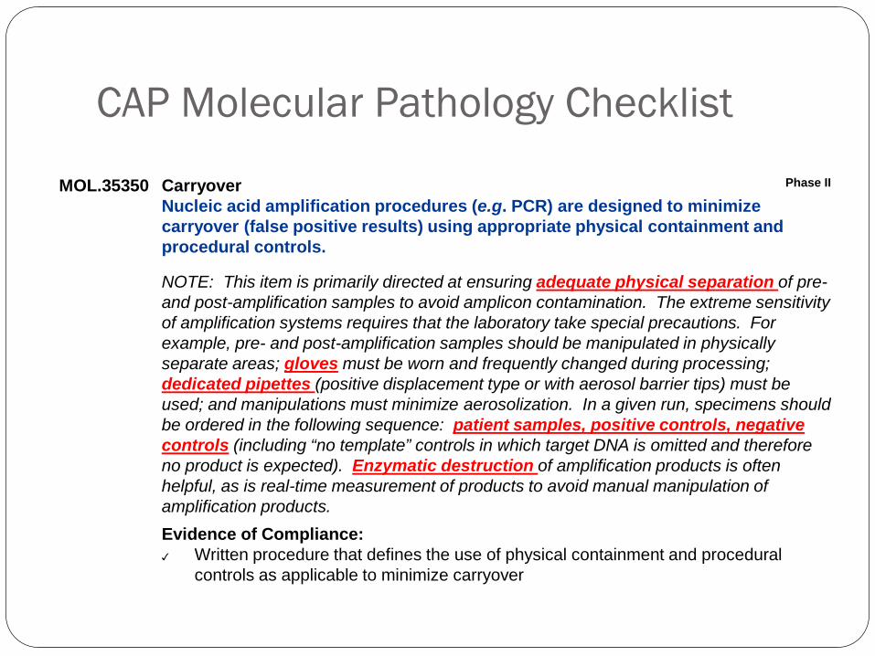
MOL.35350 Carryover Phase II
Nucleic acid amplification procedures (e.g. PCR) are designed to minimize carryover (false positive results) using appropriate physical containment and procedural controls.
NOTE: This item is primarily directed at ensuring adequate physical separation of pre-and post-amplification samples to avoid amplicon contamination. The extreme sensitivity of amplification systems requires that the laboratory take special precautions. For example, pre- and post-amplification samples should be manipulated in physically separate areas; gloves must be worn and frequently changed during processing; dedicated pipettes (positive displacement type or with aerosol barrier tips) must be used; and manipulations must minimize aerosolization. In a given run, specimens should be ordered in the following sequence: patient samples, positive controls, negative controls (including “no template” controls in which target DNA is omitted and therefore no product is expected). Enzymatic destruction of amplification products is often helpful, as is real-time measurement of products to avoid manual manipulation of amplification products.Evidence of Compliance:✓ Written procedure that defines the use of physical containment and procedural
controls as applicable to minimize carryover
CAP Molecular Pathology Checklist

Potential Sources of Contamination
Cross contamination between specimens
Amplification product contamination
Laboratory surfaces
Ventilation ducts
Reagents/supplies
Hair, skin, saliva, and clothes of lab personnel

What Happens If Lack Of Contamination Control
Incorrect results
Require extensive cleanup
Loss of creditability
Financial and performance impact

How to Control Contamination
Laboratory design
Laboratory practices
Chemical and enzymatic controls

Setting Up a Molecular Laboratory
Mechanical barriers to prevent contamination Spatial separation of pre- and post-amplification work
areas Area 1 – Reagent preparation Area 2 – Specimen/control preparation, PCR set-up Area 3 – Amplification/product detection, plasmid
preparation
Physically separated and, preferably, at a substantial distance from each other

Unidirectional Flow
Both personnel, including cleaning personnel, and specimens
Amplification product-free to product-rich
Remove PPE before leaving one area
Avoid or limit reverse direction Reusable supplies in the reverse
direction need to be bleached.

CLSI MM19-A Establishing Molecular Testing in Clinical Laboratory Environments

Features of the 3 Areas Each area has separate sets of equipment and supplies
Refrigerator/freezer (manual defrost) Pipettes, filtered tips, tubes, and racks Centrifuge, timers, vortex Lab coat (color-coded), disposable gloves, safety glasses, and other PPE Cleaning supplies Office supplies Ventilation system
Dead air box with UV light – serves as a clean bench area

Features of the 3 Areas
Air pressure Reagent Prep – Positive Sample Prep - Negative Postamplification - Negative
Reagent Prep – Single entrance, reagents used for amplification should not be exposed to other areas
Specimen Prep – Specimens should not be exposed to post-amplification work areas
Size of each area should consider space for equipment and bench space needed for preparation

Laboratory Design Examplehttp://fx.damasgate.com/the-pcr-laboratory/

Two Areas Only Area 1 – Reagent prep, specimen prep, and target loading –
use of laminar-flow hoods
Area 2 – Amplification/product detection

Alternative to Spatial Separation
Class II biological safety cabinet
Dedicated areas for each work phase
Unidirectional
Automated specimen processing station/closed-tube amplification and detection system
Core Laboratory Concept On site – e.g. combine with microbiological testing Off site – e.g. academic institute

Other Laboratory Design Considerations
Temperature and humidity requirements Exhaust ventilation Water quality Electric outlet Back-up power system Eye wash Ergonomic assessment Need for storage area Need for waste disposal area

Laboratory Practices
Use of positive displacement pipettes and disposable filtered pipette tips
Avoid production of aerosols when pipetting
Use of sterilized single-use plasticware
Use of cleanroom sticky floor mats
Minimizes the risk of amplicon carry-over on clothing, hair and skin Hairnet
Dedicated safety glasses
Disposable labcoat/gown, color-coded preferred
Gloves, need to change periodically
Shoe covers


More Laboratory Practices Clean punches between samples Use of nuclease free or autoclaved water Aliquot oligonucleotides – multiple freeze thaws will cause
degradation Always include a blank (no template) control to check for
contamination Use of electronic data system (flow of paper) Wipe test (swab test)
Monthly Detect, localize, and remove contamination Identify the source of the contamination
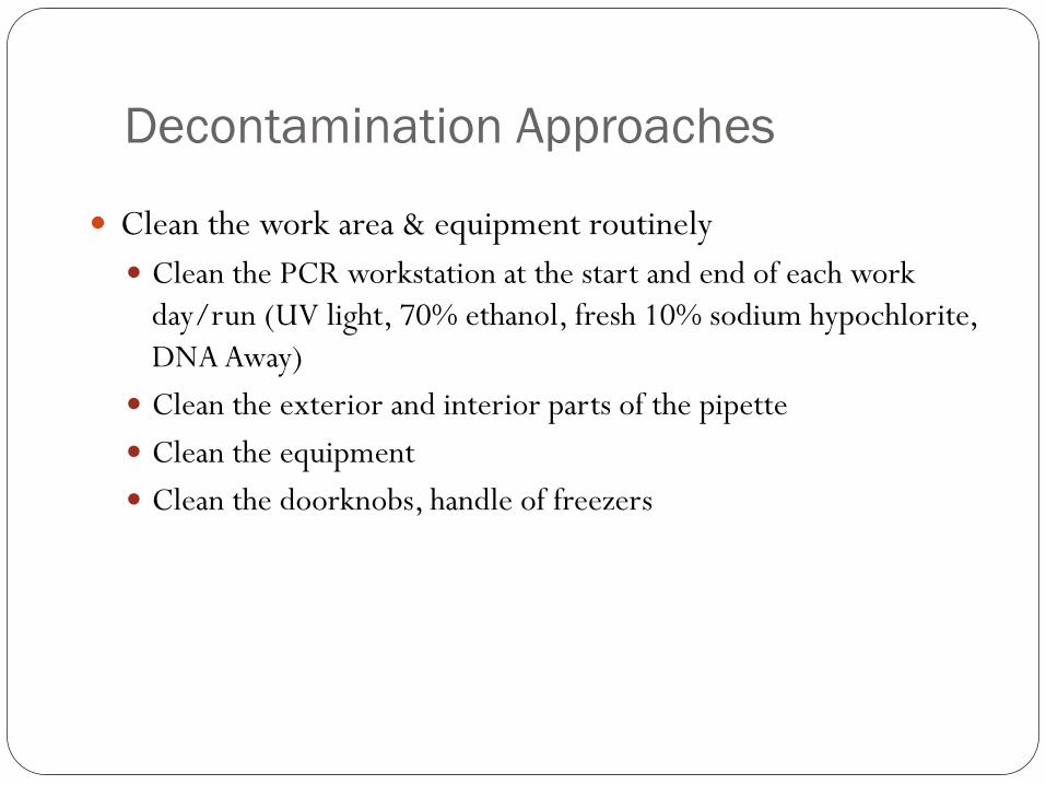
Decontamination Approaches
Clean the work area & equipment routinely Clean the PCR workstation at the start and end of each work
day/run (UV light, 70% ethanol, fresh 10% sodium hypochlorite, DNA Away)
Clean the exterior and interior parts of the pipette Clean the equipment Clean the doorknobs, handle of freezers
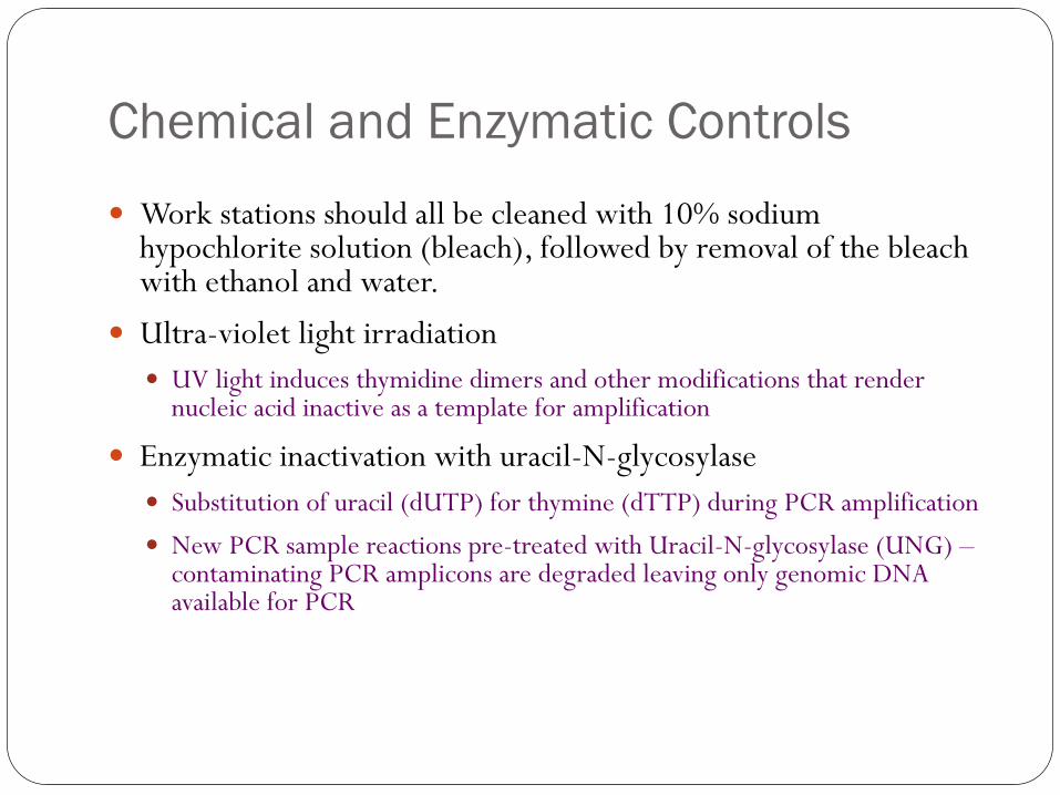
Chemical and Enzymatic Controls
Work stations should all be cleaned with 10% sodium hypochlorite solution (bleach), followed by removal of the bleach with ethanol and water.
Ultra-violet light irradiation UV light induces thymidine dimers and other modifications that render
nucleic acid inactive as a template for amplification
Enzymatic inactivation with uracil-N-glycosylase Substitution of uracil (dUTP) for thymine (dTTP) during PCR amplification
New PCR sample reactions pre-treated with Uracil-N-glycosylase (UNG) –contaminating PCR amplicons are degraded leaving only genomic DNA available for PCR

Quality ControlsMonitor all steps of analytical procedure Types of Control Frequency and Number of Controls Evaluation of Controls and Calibrators

Types of Controls
Internal Control Internal positive amplification controls to detect failure of DNA extraction or
PCR amplification Reagent or equipment issues
Integrity of DNA sample Presence of inhibitory substance
External Control Positive control Negative control (normal, wild type) No template control (extraction blank) Blank

Internal ControlsReference gene (e.g. RNaseP)

External Controls Positive and negative controls:
Inhibitors Component failure Interpretation of results Sources:
Residual DBS PT samples QC materials
No template controls and Blanks: Nucleic acid contamination during extraction Nucleic acid contamination during PCR

Frequency and Number of External Controls
Based on risk Ideally should represent each target allele and include in each run, but
may not be feasible when: Highly multiplex genotypes
o Systematic rotation of different alleles as positiveso Specimens representing short and long amplification products to control for differential
amplification Rare alleles
Quantitative PCR External controls should represent more than one concentration, e.g. low and high positives
and negative, covering the analytical measurement range Daily run or with each runs
After equipment maintenance, new operator, new reagent lot/shipment

Calibrators Calibrator copy levels should cover analytic cut-offs

Size Marker Include in each run that involves size separation

Evaluation of Controls and Calibrators
Pass/Fail Criteria – established during validation study Parameters
Specific PCR product bands Specific DNA fragments Quantity or Ct of reference gene Quantity or Ct of targeted marker Slope, R2, and Y-intercept of Calibrator curve
Threshold Presence or absence of DNA bands Above or below LoB Above or below cut-offs Within Mean± 2SD, Mean ± 3SD, or Mean± 10%
% of controls acceptable Impact the entire run or only affected samples

Allele drop-out (ADO) The failure of a molecular test to amplify or detect one or more alleles
Potential causes: DNA template concentration
Incomplete cell lysis
DNA degradation
Non-optimized assay conditions
Unknown polymorphisms in target sites
Reagent component failure
Interfering substance, http://www.aphl.org/aphlprograms/newborn-screening-and-genetics/Pages/Assuring-Laboratory-Quality.aspx
Major concern for screening laboratories
Confirmation of mutation inheritance in families may not an option
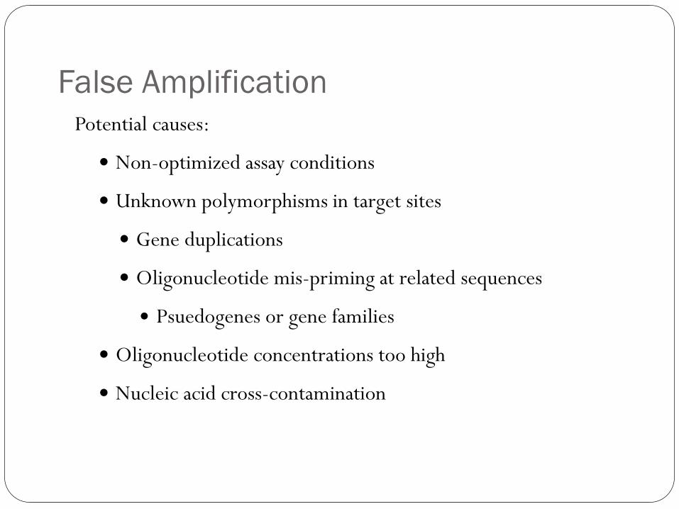
False AmplificationPotential causes:
Non-optimized assay conditions
Unknown polymorphisms in target sites
Gene duplications
Oligonucleotide mis-priming at related sequences
Psuedogenes or gene families
Oligonucleotide concentrations too high
Nucleic acid cross-contamination

What to do if control fails?

Proficiency Testing Assessment of the Competence in Testing
Required for all CLIA/CAP certified laboratories
Performed twice a year
If specimens are not commercially available alternative proficiency testing program has to be established (specimen exchange etc.)

Molecular Assay Proficiency Testing and Reference Material Sources
CDC NSQAP
CAP
In-house samples
Round-robin with other NBS laboratories
Corielle
European Collection of Authenticated Cell Cultures (ECACC)
United Kingdom National External Quality Assessment Service (UK NEQAS)
EuroGentest
Acrometrix
Advanced Biotechnologies
Asuragen
Diagnostic Hybrids
Horizon Discovery
Invivoscribe
LGC Standards
Maine Molecular Quality Controls
Molecular Controls
QIAGEN Marseille (formerly Ipsogen)
Qnostics
Seracare Life Sciences
Zeptometrix
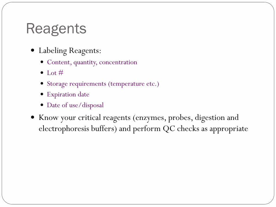
Reagents Labeling Reagents:
Content, quantity, concentration Lot # Storage requirements (temperature etc.) Expiration date Date of use/disposal
Know your critical reagents (enzymes, probes, digestion and electrophoresis buffers) and perform QC checks as appropriate

Critical Molecular Assay Components
Nucleic Acids: Prepare aliquots appropriate to workflow to limit freeze-thaw cycles Primers and probes dNTPs Genomic DNA 4-8°C -15 to -25°C
Enzymes Benchtop coolers recommended
Fluorescent reporters Limit exposure to light Amber storage tubes or wrap in shielding (foil)

CAP Requirement on TAT CBG.20140 Out-of-Range/Invalid Results Phase IIThere is a policy for reporting positive (out of range) or invalid results to the submitting location and other appropriate entities to allow for patient follow-up within a timeframe appropriate to ensure maximum health benefit.NOTE: Positive results include those results that are outside of the expected range of testing results established for a particular condition. Invalid results include situations where the laboratory is unable to complete the screening process due to an unsuitable specimen, test, or incomplete information. The findings must be communicated in a manner consistent with the urgency of the intervention needed. For situations requiring repeat screening or confirmatory testing, the laboratory must clearly communicate the timing of the actions to be taken.Results must be reported to the submitting location (at minimum) within 7 days of specimen receipt and within 3 days for specimens received for tests requiring additional action (e.g. invalid or positive).The records should indicate when results were reported and who received the results. In cases where the testing laboratory is responsible for documenting that a return specimen has been received and analyzed, appropriate records should attest to specimen receipt, testing and result reporting.

Other QA/QC Considerations
Validation studies Sample acceptance and tracking Specimen storage Laboratory Cleanliness, and Waste Disposal Instrument Maintenance and Calibration Instrument/Method Comparison Document Management Personnel Training and Competency Periodic Review of QA/QC COOP Plan

Take Home Messages Separate laboratory spaces for Reagent Prep, Sample Prep, and
Amplification and Detection
Precautions and special laboratory practices must be made to minimize the risk of contamination
A Quality Control Plan to monitor the quality of testing process and detect errors should be in placed for each new test before it’s implemented.
Continuous quality improvement is essential