module 4 : mechanism of immune response -...
TRANSCRIPT
NPTEL – Biotechnology – Cellular and Molecular Immunology
Joint initiative of IITs and IISc – Funded by MHRD Page 1 of 23
Module 4 : Mechanism of immune response
Lecture 23: Cytokines (Part I)
Cytokines are the proteins secreted by the cells of immune system that control the
immune responses by interaction between the neighboring cells. In other words
Cytokines are the signaling molecules like hormones and are the end products of
interaction among immune cells. Cytokines like other signaling molecules tend to bind
the specific receptors on the target cells but the structure of cytokines and their receptors
is very different. Once the cytokines bind to their receptors, transcription factors are
produced as a result of changes in the cell behavior by the process called as signal
transduction. Transcription factors stimulate the selected genes for transcription which
then secrete new cytokines or signaling molecules.
23.1 Features of cytokines
1) Cytokines show redundancy i.e. different cytokines may have similar function, e.g.
IL-1 and IL-6 both induce fever by acting on brain.
2) They are secreted in multiple numbers, e.g. macrophages secrete more than five
interleukins and tumor necrosis factor.
3) Cytokines produce their effect on many cell types i.e. they are pleiotropic.
4) Cytokines need extreme regulation and are toxic in high doses.
23.2 Cytokine nomenclature
Most of the cytokines are named according to the Interleukin nomenclature subcommittee
of the international union of immunological societies. Although the definition of
cytokines is quite broad, but together they can be classified as lymphokines,
interleukins, interferons, chemokines etc. depending on their function, cell of secretion,
or target of action. The name interleukin was coined initially for those cytokines that
mediate signaling between lymphocytes and leukocytes. In other words leukocytes were
thought to be the principal target for interleukins but now this definition is no longer in
function and the name interleukin is given to any new cytokine discovered routinely.
Cytokines are produced mainly in response to viral infection or in response to immune
attack. Interferons are the cytokines that interfere with viral RNA and protein synthesis to
prevent viral replication.
NPTEL – Biotechnology – Cellular and Molecular Immunology
Joint initiative of IITs and IISc – Funded by MHRD Page 2 of 23
Types of interferon – Interferons are mainly divided into two types.
Type I Interferons --- Interferon-α (IFN- α) and interferon-β (IFN- β).
Type II interferon --- Interferon-γ
Tumor necrosis factors (TNFs) can kill tumor cells and colony stimulating factors assist
in regulation of stem cell activities. Chemokines play role in leukocyte recruitment and
circulation.
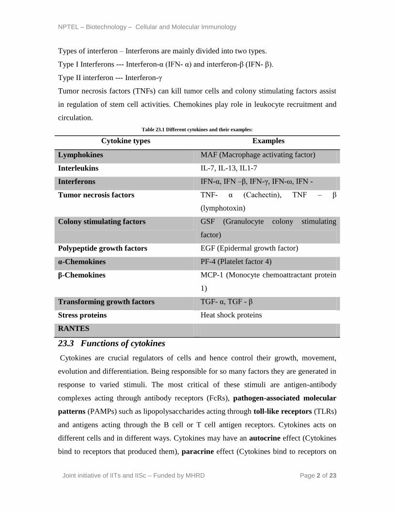
Table 23.1 Different cytokines and their examples:
Cytokine types Examples
Lymphokines MAF (Macrophage activating factor)
Interleukins IL-7, IL-13, IL1-7
Interferons IFN-α, IFN –β, IFN-γ, IFN-ω, IFN -
Tumor necrosis factors TNF- α (Cachectin), TNF – β
(lymphotoxin)
Colony stimulating factors GSF (Granulocyte colony stimulating
factor)
Polypeptide growth factors EGF (Epidermal growth factor)
α-Chemokines PF-4 (Platelet factor 4)
β-Chemokines MCP-1 (Monocyte chemoattractant protein
1)
Transforming growth factors TGF- α, TGF - β
Stress proteins Heat shock proteins
RANTES
23.3 Functions of cytokines
Cytokines are crucial regulators of cells and hence control their growth, movement,
evolution and differentiation. Being responsible for so many factors they are generated in
response to varied stimuli. The most critical of these stimuli are antigen-antibody
complexes acting through antibody receptors (FcRs), pathogen-associated molecular
patterns (PAMPs) such as lipopolysaccharides acting through toll-like receptors (TLRs)
and antigens acting through the B cell or T cell antigen receptors. Cytokines acts on
different cells and in different ways. Cytokines may have an autocrine effect (Cytokines
bind to receptors that produced them), paracrine effect (Cytokines bind to receptors on
NPTEL – Biotechnology – Cellular and Molecular Immunology
Joint initiative of IITs and IISc – Funded by MHRD Page 3 of 23
neighboring or cells which are very close) or endocrine effect (Cytokines flow
throughout the body) while acting on the target cells. Cytokines also play role in
apoptosis and control various stages of replication. They have the capability for being
sensitive markers of chemically induced perturbations in function, but this claim is not
accepted from the toxicological point of view. The reasons behind this being that
cytokines are released locally with plasma measures being unreliable and they have short
half-lives which need precise timing to find.
23.4 Cytokine structure
Cytokines have distinct structures but can be broadly divided into following.
Group I cytokines are involved in immune regulation --- includes four α-helices in
bundles, Interferon subfamily and the IL-10 subfamily.
Group II cytokines are engaged in growth and regulation of cells, cell death and
inflammation --- IL-1 family, TGF-β
Group III cytokines are involved in inflammation--- Chemokines
Group IV cytokines --- IL-12
NPTEL – Biotechnology – Cellular and Molecular Immunology
Joint initiative of IITs and IISc – Funded by MHRD Page 4 of 23
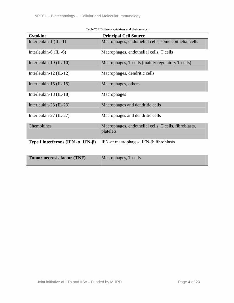
Table 23.2 Different cytokines and their source:
Cytokine Principal Cell Source
Interleukin-1 (IL -1) Macrophages, endothelial cells, some epithelial cells
Interleukin-6 (IL -6) Macrophages, endothelial cells, T cells
Interleukin-10 (IL-10) Macrophages, T cells (mainly regulatory T cells)
Interleukin-12 (IL-12) Macrophages, dendritic cells
Interleukin-15 (IL-15) Macrophages, others
Interleukin-18 (IL-18) Macrophages
Interleukin-23 (IL-23) Macrophages and dendritic cells
Interleukin-27 (IL-27) Macrophages and dendritic cells
Chemokines Macrophages, endothelial cells, T cells, fibroblasts,
platelets
Type I interferons (IFN -α, IFN-β) IFN-α: macrophages; IFN-β: fibroblasts
Tumor necrosis factor (TNF) Macrophages, T cells
NPTEL – Biotechnology – Cellular and Molecular Immunology
Joint initiative of IITs and IISc – Funded by MHRD Page 5 of 23
Lecture 24: Cytokines (Part II)
24.1 Cytokine receptors
Cytokine receptors can be distinguished based on their structure and consist basically of
two units, one for the ligand binding and the other for signal transduction. Broad
classification of cytokine receptors can be listed as under –
Proteins that acts as Tyrosine kinases and function as growth factors.
One class of receptors is membrane-bound Guanosine triphosphate – binding proteins
called as G proteins. In active state these bind GTP.
Channel linked receptors.
This class of receptor stimulates a neutral sphingomyelinase.
24.2 Cytokine regulation
The three major ways for regulating the cytokine signaling are –
By specific binding proteins
By changes in receptor expression and
By cytokines that exert opposite effects.
Concept behind understanding the cytokine regulation is that as the single cell receives
signals from numerous cytokine receptors, a unification of multiple signals is required to
generate a coherent response.
24.3 Signal transduction and pathways
During the signal transduction the first step is the binding of cytokines to its receptors.
Once the binding occurs, the receptor transmits a signal to the cell to alter its behavior.
This phenomenon involving conversion of an extracellular signal into a series of
intracellular events is called signal transduction. As Cell signaling needs to be precise and
quick, enzyme cascade serve this function by the amplification of the responses rapidly.
The important features of signal transduction include binding of cytokine to a cell surface
receptor, stimulation of a transducer protein by the receptor, secondary activation of
various enzymes, production of new transcription factors and ultimately gene stimulation
leading to changed cell behavior.
Although there are many pathways of signal transduction but three pathways are of
utmost significance to the immune system.
NPTEL – Biotechnology – Cellular and Molecular Immunology
Joint initiative of IITs and IISc – Funded by MHRD Page 6 of 23
The NF-kB pathway
The NF-AT pathway
The JAK/STAT pathway
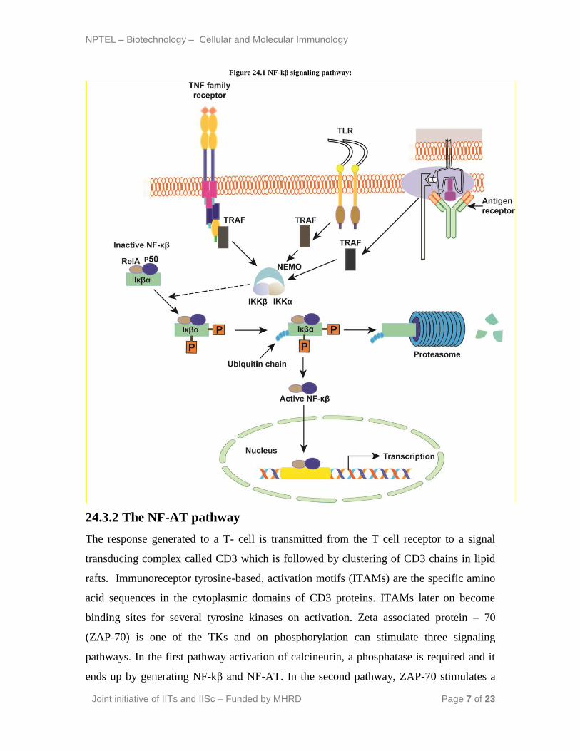
24.3.1 The NF-kβ pathway
NF-kβ has its origins in the family of five transcription factors that play a crucial role in
inflammation and immunity. There are more than 150 genes that get expressed on NF-kβ
stimulation. There are three major pathways for NF-kβ activation. These are the classical
pathway, alternative pathway and the third pathway that is stimulated by DNA
damaging drugs and Ultraviolet light. The classical pathway involves proinflammatory
signaling and gets activated by inflammatory cytokines, TLRs and antigen receptors
(figure 24.1). IKK (Ikβ) complex is the central regulator of NF-kβ and all the signals
induced by various stimuli focus on the same complex IKK. IKK complex is rich with
kinase activity and on activation IKK complex phosphorylates Ikβ. Consequently Ikβ
dissociates from the NF-kβ and the newly formed Ikβ becomes ubiquinated and is
destroyed by proteasomes. Dissociation between Ikβ and NF-kβ liberates NF-kβ and it
enters the nucleus to activate kβ motif containing promoters on DNA. This stimulates
many genes and cytokines IL-1β, IL-6, IL-18, IL-33, TNF-α, GM-CSF, and IL-4. The
second pathway or alternative pathway is required for lymphocyte development and is
stimulated by a subset of TNF receptors. The third pathway does not require any IKK
activation.
IKK- Ikβ kinase
Ikβ- Inhibitor of NF-kβ
NEMO- NF-κB essential modulator
TRAF- Tumor necrosis factor receptor-associated factor
NPTEL – Biotechnology – Cellular and Molecular Immunology
Joint initiative of IITs and IISc – Funded by MHRD Page 7 of 23
Figure 24.1 NF-kβ signaling pathway:
24.3.2 The NF-AT pathway
The response generated to a T- cell is transmitted from the T cell receptor to a signal
transducing complex called CD3 which is followed by clustering of CD3 chains in lipid
rafts. Immunoreceptor tyrosine-based, activation motifs (ITAMs) are the specific amino
acid sequences in the cytoplasmic domains of CD3 proteins. ITAMs later on become
binding sites for several tyrosine kinases on activation. Zeta associated protein – 70
(ZAP-70) is one of the TKs and on phosphorylation can stimulate three signaling
pathways. In the first pathway activation of calcineurin, a phosphatase is required and it
ends up by generating NF-kβ and NF-AT. In the second pathway, ZAP-70 stimulates a
NPTEL – Biotechnology – Cellular and Molecular Immunology
Joint initiative of IITs and IISc – Funded by MHRD Page 8 of 23
protein kinase C which is required for activation of NF-kβ. The third pathway activates
ras, a GTP-binding protein, fos and jun.
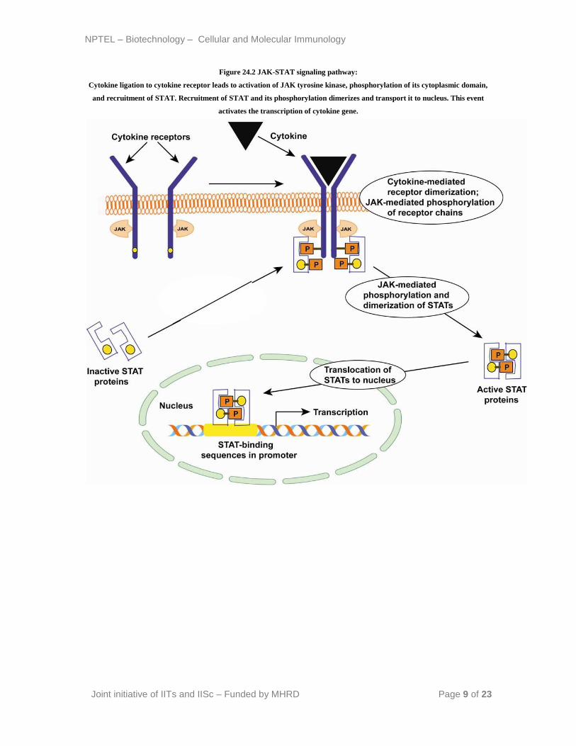
24.3.3 The JAK-STAT pathway
More than 40 cytokines use the JAK-STAT pathway. The stimulated JAK (Janus kinase)
molecules phosphorylate tyrosine residues on one of several STAT proteins (signal
transducers and activators of transcription). Phosphorylated STAT proteins then dimerize
and dissociate from JAK and get shifted to the nucleus where they regulate the expression
of target genes.
NPTEL – Biotechnology – Cellular and Molecular Immunology
Joint initiative of IITs and IISc – Funded by MHRD Page 9 of 23
Figure 24.2 JAK-STAT signaling pathway:
Cytokine ligation to cytokine receptor leads to activation of JAK tyrosine kinase, phosphorylation of its cytoplasmic domain,
and recruitment of STAT. Recruitment of STAT and its phosphorylation dimerizes and transport it to nucleus. This event
activates the transcription of cytokine gene.

NPTEL – Biotechnology – Cellular and Molecular Immunology
Joint initiative of IITs and IISc – Funded by MHRD Page 10 of 23
Lecture 25: Mechanism of cell mediated immune
response (Part I)
The recruitment of the leukocyte and its activation is largely governed by the subset of
CD4+ cells. Each leukocyte is specified for certain job and the cooperation of leukocyte
to the lymphocytes is an important connection between innate and adaptive immune
response.
In general Th1 cells activate macrophages, Th2 activates eosinophils, and Th17 activates
neutrophils. The Th1 cells activate the macrophage mediated phagocytosis, Th2
stimulates the production of IgE in response to a helminthic infection, and Th17 activates
neutrophils to extracellular microbes including bacteria and fungi. Cytotoxic T
lymphocytes (CTLs) kill the microbes which escape the phagocytosis and replicate in the
cytoplasm.
25.1 Migration of lymphocyte at the site of infection
Majority of the leukocyte and lymphocyte are migrated towards the site of inflammation
and infection, respectively in order to nullify the effect. Once the naïve lymphocyte
encounters an antigen presented by the MHC molecule over the antigen presenting cells,
they differentiate and migrate as an effector cells through the blood vessels to different
parts of the body. The migration of lymphocyte is an interesting phenomenon
orchestrated by different cytokines and specific receptors present over the surface of the
lymphocytes. Naïve T lymphocytes express L-selectin and chemokine receptor CCR7
which help them to adhere to the lymph node and its surrounding tissues. After antigen
stimulation the naïve T lymphocyte decrease the expression of L-selectin and CCR7 and
increase the expression of sphingosine 1-phosphate receptors (S1PR1), which help
their migration into the blood vessels. Th1 cells produces large amount of ligands that
binds to E and P-selectin which recognize selectins secreted at the site of bacterial
infection and trigger a strong innate immune response. Th2 cells express different
chemokine receptors such as CCR3, CCR4 and CCR8 which recognize specific
Chemokines secreted at the site of helminthic infection. Similarly, Th17 migration to the
site of fungal and bacterial infection is modulated by CCR6 and other cytokines. Usually
the subset of T lymphocytes is directed to the site of action with the help of different

NPTEL – Biotechnology – Cellular and Molecular Immunology
Joint initiative of IITs and IISc – Funded by MHRD Page 11 of 23
chemokines and their respective ligands. Similarly memory cells express different
integrins that binds to the fibronectins over the extracellular matrices and help in their
migration.
25.2 Functions of CD4+ helper cells
The function of CD4+ cells are divided into four distinct stages.
Leukocyte recruitment- T lymphocytes produces many cytokines that helps in the
recruitment of neutrophils, eosinophils and monocytes at the site of infection or
injury.
Leukocyte activation- Leukocytes gets activated by the binding of CD40L on
lymphocyte to the CD40 express over the antigen presenting cells.
Expansion- The activation response gets expanded by the secretion of cytokines by
the T lymphocytes.
Control- The response of the immune system is controlled by some inhibitory
cytokines such as IL-10.
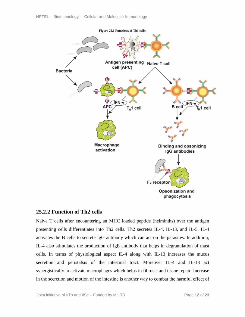
25.2.1 Function of Th1 cells
Naïve T cells after encountering an MHC loaded peptide over the antigen presenting cells
differentiates into Th1 cells. Th1 cells secretes interferon-γ (IFN-γ) which is a key
cytokine involve in its functioning. IFN-γ acts on the activated macrophages and
increases its phagocytosis efficiency and microbial killing. In addition IFN-γ also
stimulates the B cells to secrete the IgG antibody that helps in opsonization and activation
of complement pathway. Occasionally they also produce tumor necrosis factor that
activate the neutrophils and promotes inflammation.
NPTEL – Biotechnology – Cellular and Molecular Immunology
Joint initiative of IITs and IISc – Funded by MHRD Page 12 of 23
Figure 25.1 Functions of Th1 cells:
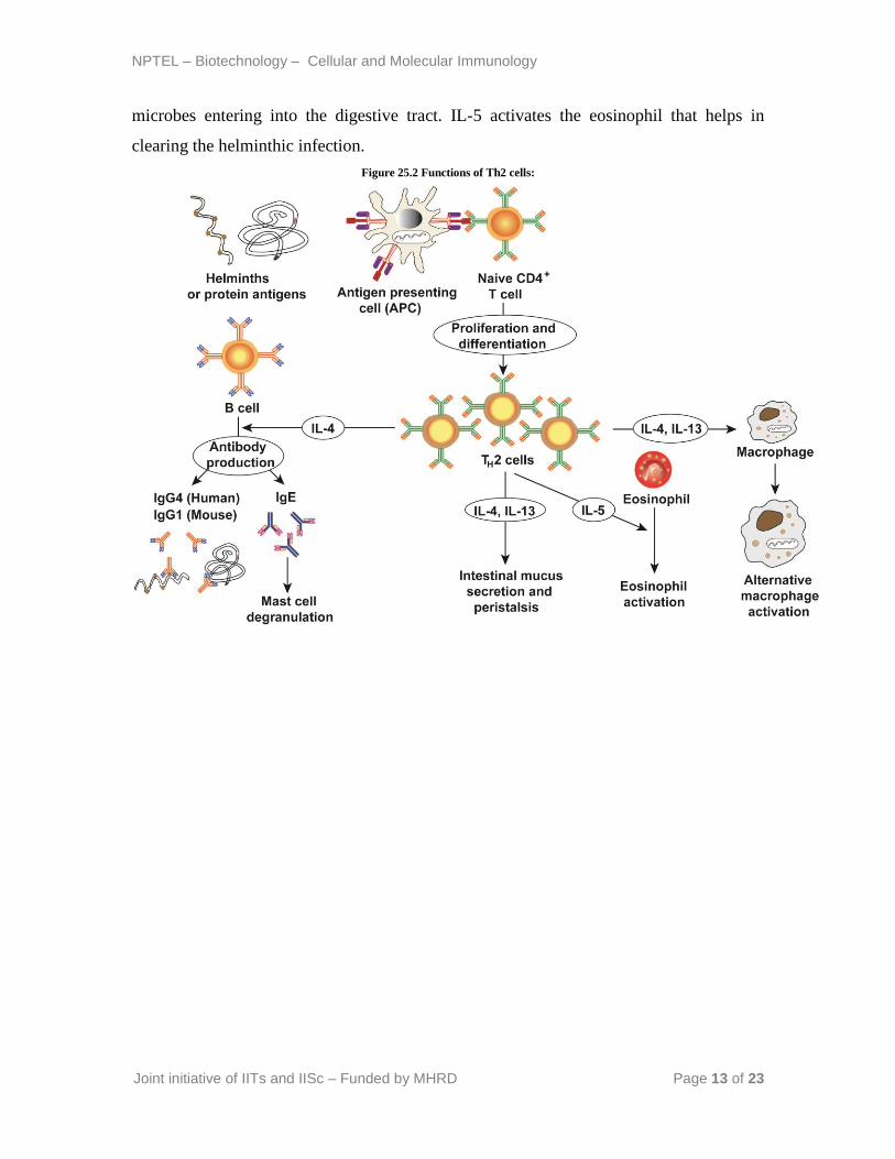
25.2.2 Function of Th2 cells
Naïve T cells after encountering an MHC loaded peptide (helminths) over the antigen
presenting cells differentiates into Th2 cells. Th2 secretes IL-4, IL-13, and IL-5. IL-4
activates the B cells to secrete IgG antibody which can act on the parasites. In addition,
IL-4 also stimulates the production of IgE antibody that helps in degranulation of mast
cells. In terms of physiological aspect IL-4 along with IL-13 increases the mucus
secretion and peristalsis of the intestinal tract. Moreover IL-4 and IL-13 act
synergistically to activate macrophages which helps in fibrosis and tissue repair. Increase
in the secretion and motion of the intestine is another way to combat the harmful effect of
NPTEL – Biotechnology – Cellular and Molecular Immunology
Joint initiative of IITs and IISc – Funded by MHRD Page 13 of 23
microbes entering into the digestive tract. IL-5 activates the eosinophil that helps in
clearing the helminthic infection.
Figure 25.2 Functions of Th2 cells:
NPTEL – Biotechnology – Cellular and Molecular Immunology
Joint initiative of IITs and IISc – Funded by MHRD Page 14 of 23
Lecture 26: Mechanism of cell mediated immune
response (Part II)
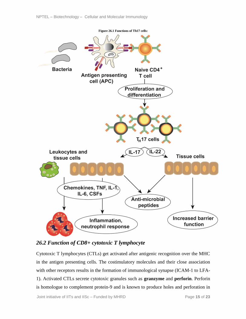
26.1 Function of Th17 cells
After differentiation of naïve T cells into Th17 effector cells, they start secreting IL-17
and IL-22. IL-17 is a unique cytokine that do not have any homologue in the body. IL-17
stimulates the production of chemokines, TNF, IL-1, IL-6, and granulocyte-colony
stimulating factors (G-CSF) which take part in inflammatory reactions. In addition, it is
also involved in the production of defensins from the epithelial surface which in turn acts
as a barrier for the invading pathogens. IL-22 is a relatively new interleukin and its
function is not well established. However IL-22 is known to maintain the epithelial
integrity, increase barrier function and involve in the repair activity following
inflammation.
NPTEL – Biotechnology – Cellular and Molecular Immunology
Joint initiative of IITs and IISc – Funded by MHRD Page 15 of 23
Figure 26.1 Functions of Th17 cells:
26.2 Function of CD8+ cytotoxic T lymphocyte
Cytotoxic T lymphocytes (CTLs) get activated after antigenic recognition over the MHC
in the antigen presenting cells. The costimulatory molecules and their close association
with other receptors results in the formation of immunological synapse (ICAM-1 to LFA-
1). Activated CTLs secrete cytotoxic granules such as granzyme and perforin. Perforin
is homologue to complement protein-9 and is known to produce holes and perforation in
NPTEL – Biotechnology – Cellular and Molecular Immunology
Joint initiative of IITs and IISc – Funded by MHRD Page 16 of 23
the target cell. Perforin polymerize to form an aqueous pore in the target cells through
which granzyme enters the target cells. Granzyme is serine proteases which is responsible
for the cleavage and activation of caspase in the cells leading to the apoptosis of the
target cells. In addition, activated CTLs also secrete granules such as serglycin which are
known to assemble the granzyme and perforin over the surface of target cells.
CTLs also kill the target cells in granule independent fashion by Fas Ligand mediated
pathway. The Fas ligand binds to the death receptors that are expressed in many cell
types and activates the caspase mediated apoptosis pathway.
The infection of intracellular microbes to host needs participation of CTLs because it will
eliminate the reservoir of infection by killing the infected cell. Hepatitis B and C virus
infection to a host activates the CTLs in order to kill the liver cells to eliminate the
reservoir.
26.3 γδ T cells
These are clonally distinct cells containing heterodimer of γ and δ chains, which are
homologous to α and β chains in αβ T cells. Usually less than 5% of total circulating
lymphocytes are composed of γδ T cell receptor. The γδ T cells present in the epithelial
surface of bowel are called as intraepithelial lymphocytes. The γδ T cells do not
recognize the MHC loaded peptide and are MHC independent. They require protein and
non-proteinous antigen that do not require processing by MHC molecules.
26.4 Natural killer cells
T cells population that express the CD56 marker over their surface are called natural
killer or NK cells. NK cells recognizes the lipid bound to CD1 receptor (MHC class I
homologue). NK cell secretes IL-4 and interferon-γ after activation and help in the
production of antibody by B cells. NK cell plays an important role in the protection
against mycobacterium infection.
26.5 Points to remember
Cell mediated immunity is a type of adaptive immune response that is stimulated by
microbes and can be transferred to naïve animal by T cells.
Differentiation and migration of lymphocytes are orchestrated by different cytokines.
Both CD4+ and CD8+ cells are responsible for cell mediated immunity
NPTEL – Biotechnology – Cellular and Molecular Immunology
Joint initiative of IITs and IISc – Funded by MHRD Page 17 of 23
CD4+ cells are comprised of Th1, Th2, and Th17 subsets and have different effector
functions.
CD8+ cells are converted into cytotoxic T lymphocyte upon differentiation and help
in eradication of infection by killing the target cells.
NPTEL – Biotechnology – Cellular and Molecular Immunology
Joint initiative of IITs and IISc – Funded by MHRD Page 18 of 23
Lecture 27: Mechanism of antibody medicated immune
response (Part I)
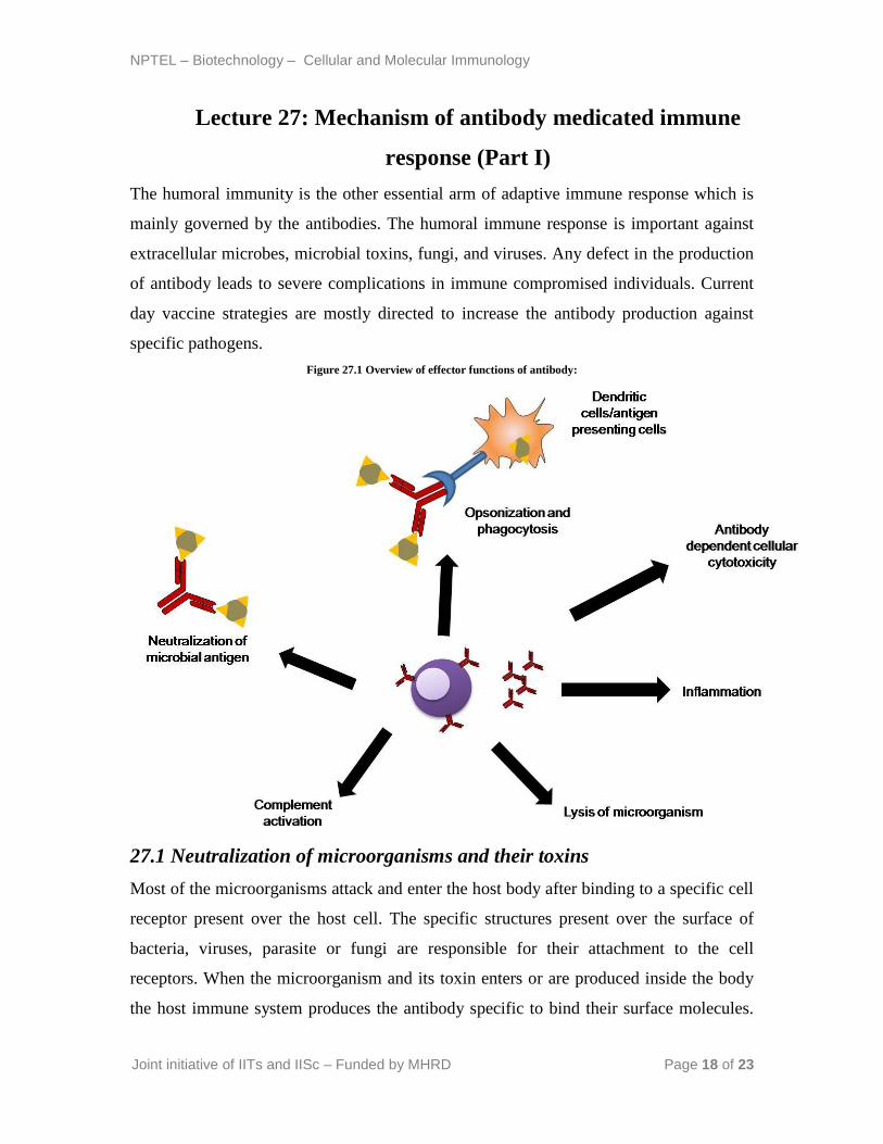
The humoral immunity is the other essential arm of adaptive immune response which is
mainly governed by the antibodies. The humoral immune response is important against
extracellular microbes, microbial toxins, fungi, and viruses. Any defect in the production
of antibody leads to severe complications in immune compromised individuals. Current
day vaccine strategies are mostly directed to increase the antibody production against
specific pathogens.
Figure 27.1 Overview of effector functions of antibody:
27.1 Neutralization of microorganisms and their toxins
Most of the microorganisms attack and enter the host body after binding to a specific cell
receptor present over the host cell. The specific structures present over the surface of
bacteria, viruses, parasite or fungi are responsible for their attachment to the cell
receptors. When the microorganism and its toxin enters or are produced inside the body
the host immune system produces the antibody specific to bind their surface molecules.
NPTEL – Biotechnology – Cellular and Molecular Immunology
Joint initiative of IITs and IISc – Funded by MHRD Page 19 of 23
The binding of specific antibodies to these molecules in the microbes and toxin makes it
inaccessible to the host cell and hence neutralized. The host cell produces antibodies
against the surface glycoprotein hemagglutinin in influenza virus infection in order to
inhibit their binding to the sialic acid receptor present on the respiratory epithelium.
Similarly, tetanus toxin binds to the cellular receptors over the neurotransmitter junction
leading to paralysis and lockjaw condition. The antibody produced in Clostridium tetani
infection neutralizes the toxins and inhibits its binding to receptors present in the motor
end plates. Neutralization of the microbes and their toxins only requires antigen binding
region of the antibody, i.e Fab or F (ab) 2 fragments. Most of the neutralizing antibodies
in the blood are of IgG and IgA types in the mucosal surface.
27.2 Opsonization and phagocytosis
Coating of microbes by the IgG type antibody helps in their phagocytosis by
macrophages and neutrophils, the phenomenon is called opsonization. The Fc receptor of
the IgG containing microbes binds to the neutrophils and participates in their intracellular
degradation and killing. In addition to IgG molecules, complement protein C3b also helps
in the opsonization and phagocytosis of the microbes by binding to leukocytes. The
substance that helps in opsonization and phagocytosis including antibodies and
complements are called as opsonins.
NPTEL – Biotechnology – Cellular and Molecular Immunology
Joint initiative of IITs and IISc – Funded by MHRD Page 20 of 23
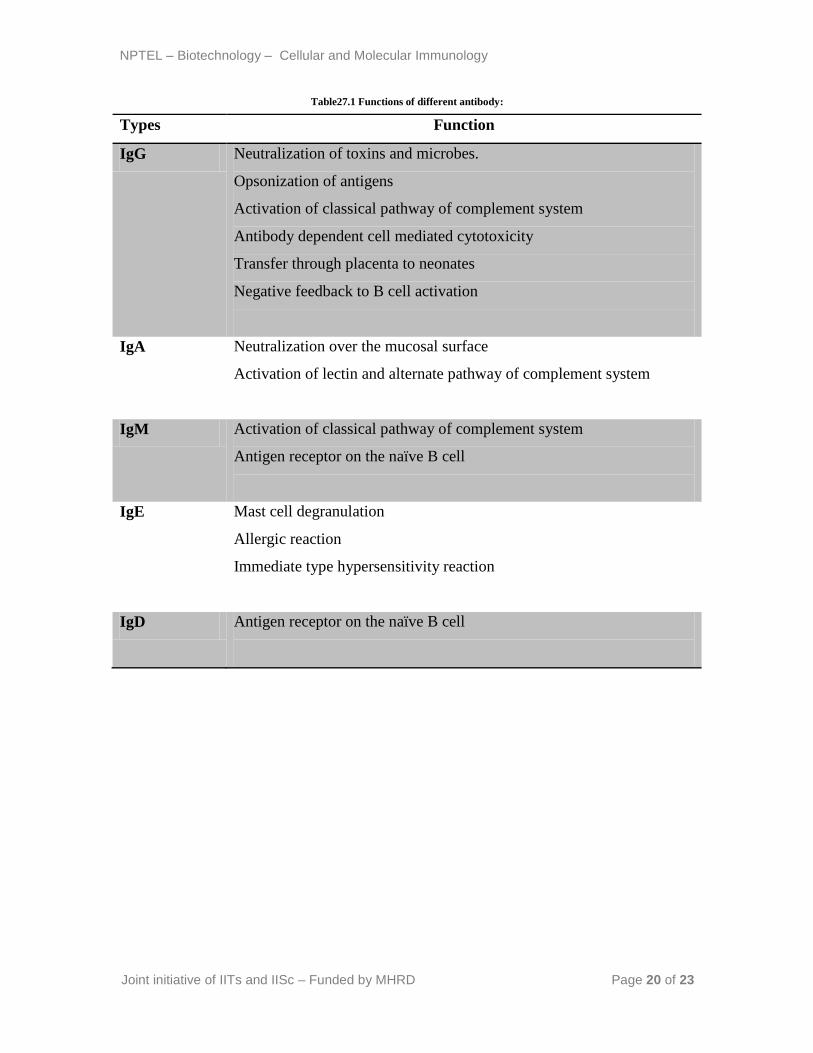
Table27.1 Functions of different antibody:
Types Function
IgG Neutralization of toxins and microbes.
Opsonization of antigens
Activation of classical pathway of complement system
Antibody dependent cell mediated cytotoxicity
Transfer through placenta to neonates
Negative feedback to B cell activation
IgA Neutralization over the mucosal surface
Activation of lectin and alternate pathway of complement system
IgM Activation of classical pathway of complement system
Antigen receptor on the naïve B cell
IgE Mast cell degranulation
Allergic reaction
Immediate type hypersensitivity reaction
IgD Antigen receptor on the naïve B cell
NPTEL – Biotechnology – Cellular and Molecular Immunology
Joint initiative of IITs and IISc – Funded by MHRD Page 21 of 23
Lecture 28: Mechanism of antibody mediated immune
response (Part II)
28.1 Antibody dependent cell-mediated cytotoxicity
Many subclasses of IgG antibodies bind to infected cells and are recognized by the Fc
receptors of NK cell. NK cell contains Fc receptor called FcγRIII (CD16) that binds to
the antibody coated infected cells. Many therapeutic monoclonal antibodies against tumor
cells are working on the same principle by coating the tumor cells which facilitates
their killing by NK cells.
28.2 Antibody dependent killing of helminths
Helminths are larger in size and cannot be processed by phagocytic cells of the immune
system. Antibodies like IgE, IgG, and IgA coats the surface of helminths which can bind
to the Fc receptor present over the eosinophils. Eosinophils contain granules in their
cytoplasm which are responsible for the secretion of microbicidal major basic proteins.
Binding of the antibody coated helminths to Fc receptor degranulates the eosinophils and
secretion of major basic protein which is responsible for their killing. Mast cells also take
part in the clearance of helminths from the body. Mast cell releases many cytokines and
chemokines which attracts and helps in the degranulation of eosinophils and in turns
secretion of major basic protein.
28.3 The complement system
The complement system consists of different serum proteins that works through different
pathways and interacts with each other in order to kill the invading microbes. The
complement system is activated by the microbial antigen and the antibody produced
against them by the host immune system. The proteins of the complement system
attaches sequentially over the surface of microbes and help in their lysis. The
complement system comprises of classical, alternate, and lectin pathways. The
classical pathway is activated by the antibody binding with an antigen, alternate pathway
activates in absence of antibody, and lectin pathway activates in response of lectin that
binds to mannose. The final step in all these three pathways leads to the formation of
membrane attack complex. These complexes are around 100 Å in diameter and forms, a
channel that allows the free flow of water and ion into the microbial cells. The movement

NPTEL – Biotechnology – Cellular and Molecular Immunology
Joint initiative of IITs and IISc – Funded by MHRD Page 22 of 23
of water and ions inside the cells increases the osmotic pressure of the cells and its
rupture. The pore formed by complement proteins are similar to what we observe in case
of NK cells, where instead of water and ions, perforin and granzyme enters and kills the
cell.
28.3.1 Function of complement system
Promotes phagocytosis of the microbes by opsonization.
Stimulates inflammation by degranulation of mast cells.
Lysis of microbes by forming a membrane attack complex.
Solubilization of those complexes that are unwanted for the body and helps in
their clearance.
Figure 28.1 Different complement pathways:
NPTEL – Biotechnology – Cellular and Molecular Immunology
Joint initiative of IITs and IISc – Funded by MHRD Page 23 of 23
28.4 Neonatal immunity
The body of neonate is sterile and passive transfer of antibody takes place by placenta
before birth and through milk immediately after birth. The passive transfer of antibodies
to neonates is important in providing immunity during the early phase of life when the
immune system is not fully functional. Transfer of antibody through the gut in early life
phase is called transcytosis. The maternal IgG is transferred through placenta and IgG
and IgA through milk.