modulating hair follicle size with wnt10bdkk1 during hair …cmchuong/2014wnt10b-dkk1.pdf · ·...
TRANSCRIPT

This article has been accepted for publication and undergone full peer review but has not been through the copyediting, typesetting, pagination and proofreading process, which may lead to differences between this version and the Version of Record. Please cite this article as doi: 10.1111/exd.12416
This article is protected by copyright. All rights reserved.
Received Date : 08-Aug-2013
Accepted Date : 15-Apr-2014
Article type : Regular Article
Modulating hair follicle size with Wnt10b/DKK1 during hair
regeneration
Mingxing Lei1, 2, Haiying Guo3, Weiming Qiu3, Xiangdong Lai1, 2, Tian Yang3, Randall B. Widelitz4,
Cheng-Ming Chuong4, Xiaohua Lian3*, Li Yang1, 2*
1 Key Laboratory of Biorheological Science and Technology, Ministry of Education, Bioengineering College, Chongqing University,
Chongqing 400044, China; 2 '111' Project Laboratory of Biomechanics and Tissue Repair, Bioengineering College, Chongqing University,
Chongqing 400044, China; 3 Department of Cell Biology, the Third Military Medical University, Chongqing, 400038, People’s Republic
of China; 4 Department of Pathology, University of Southern California, Los Angeles, CA 90033, USA.
Correspondence: Li Yang, Key Laboratory of Biorheological Science and Technology, Ministry of Education, Bioengineering College,
Chongqing University, Chongqing 400044, China, Tel.: +86-023-65111802, e-mail: [email protected] (LY); Xiaohua Lian,
Department of Cell Biology, the Third Military Medical University, Chongqing, 400038, People’s Republic of China,
Tel.:+86-023-68752264, e-mail:[email protected]
Abstract: Hair follicles have characteristic sizes corresponding to their cycle specific stage.
However, how the anagen hair follicle specifies its size remains elusive. Here, we show that in
response to prolonged ectopic Wnt10b-mediated β-catenin activation, regenerating anagen hair
follicles grow larger in size. In particular, the hair bulb, dermal papilla and hair shaft become
This article is protected by copyright. All rights reserved.
enlarged. While the formation of different hair types (Guard, Awl, Auchene, and Zigzag) is
unaffected. Interestingly, we found the effect of exogenous WNT10b was mainly on Zigzag and
less on the other kinds of hairs. We observed dramatically enhanced proliferation within the
matrix, DP and hair shaft of the enlarged AdWnt10b-treated hair follicles compared with those
of normal hair follicles at P98. Furthermore, expression of CD34, a specific hair stem cell
marker, was increased in its number to the bulge region after AdWnt10b treatment. Ectopic
expression of CD34 throughout the ORS region was also observed. Many CD34 positive hair
stem cells were actively proliferating in AdWnt10b-induced hair follicles. Importantly,
subsequent co-treatment with the Wnt inhibitor, DKK1, reduced hair follicle enlargement,
decreased proliferation and ectopic localization of hair stem cells. Moreover, injection of DKK1
during early anagen significantly reduced the width of prospective hairs. Together, these findings
strongly suggest that Wnt10b/DKK1 can modulate hair follicle size during hair regeneration.
Key words: Wnt10b – DKK1 –hair follicle size – hair regeneration – hair stem cells
Introduction
Hair follicles display different shapes, lengths and thickness during anagen, catagen and telogen
phases of the hair cycle (1, 2). Furthermore, in mouse dorsal skin, there are four distinct hair
follicle types with different shapes and sizes that successively emerge during hair development.
These include Guard (primary hair), Auchene and Awl (secondary hair) and Zigzag (tertiary
hair) hairs (3). In general, primary and secondary hair follicles are larger than tertiary hair
follicles during anagen (4).

This article is protected by copyright. All rights reserved.
Hair size is strictly controlled by reciprocal epithelial–mesenchymal interactions. Epithelial
keratinocytes of the hair matrix supply the hair shaft progenitor cells, while the dermal papilla
(DP) is the critical mesenchymal signaling center that regulates epithelial behavior (5). In both
human and murine hair follicles, hair size correlates with DP cell numbers (4, 6, 7). A recent
study showed that hairs can switch progressively from smaller to larger types during the hair
cycle due to increased DP cell numbers (4).
At the molecular level, hair size can be regulated by multiple factors that influence epithelium
and/or mesenchyme. Ectopic epidermal expression of the BMP antagonist, Noggin, causes a
marked increase in anagen hair follicle size and changes Zigzag hairs to become larger, Awl-like
hair follicles (8). Epidermal Eda/Edar is required for primary hair placode formation (9-12),
while Sox2+ cell depletion resulted in a loss of primary and secondary hair induction (3).
Knocking out Sox2 in the dermal papilla reduced the overall rate of hair growth (13). Sox18
deletion reduced Zigzag hair numbers (14, 15). FGF20 signaling activity also plays a role in
primary and secondary hair induction by influencing dermal condensation formation (16). The
deletion or up-regulation of each of these molecules not only results in changes of hair size but
also hair types. It remains elusive whether or not the hair size can be controlled by molecular
mechanisms independent of those regulating the switch of hair types.
Wnts and their DKK antagonists regulate the morphology of ectodermal organs (17-20).
Wnt10b is a canonical Wnt member (21) that is expressed during hair induction and hair
reconstitution (22, 23). Recent studies have shown that adenovirus mediated Wnt10b expression
(AdWnt10b) leads to epithelial and melanocyte cell differentiation and elongated hair shafts
(24-26). In our previous studies, we reported that WNT10b over-expression resulted in growth of
This article is protected by copyright. All rights reserved.
vibrissae in vitro and early induction of hair follicles in vivo (27-29). In the present study, we
apply multiple injections of Wnt10b-expressing adenoviruses to regenerating hair follicles, with
or without the presence of a Wnt antagonist, DKK1. We show that WNT10b treatment leads to
enlargements of hair follicles during regeneration through the proliferation and scattering of hair
stem cells, which can be partially rescued by DKK1. These findings shed new light on how
external macroenvironmental signaling communicates with the hair follicle to specify organ size
at the cellular and molecular levels.
Materials and methods
Mice
Animal maintenance and utilization were approved by the Third Military Medical University in
China. Female C57BL/6J mice at 8 weeks of age, corresponding to the second telogen phase of
the hair cycle (28), were used for the adenovirus injection study. Female C57BL/6J mice at
postnatal day98 (P98) were used as controls.
Adenovirus and plasmid.
Adenoviruses including Adwnt10b and AdGFP (control) used in this study were a gift from Dr.
T.C. He, University of Chicago, USA. The adenoviruses were propagated in HEK293 cells to a
final titer of 1×10⁸ according to the published protocol (30). Full length DKK1 CDS sequence
was cloned into a pEGFP-N1 vector at Kpn I and Hind III restriction enzyme sites, with the
following primers Sense: 5'-CCCAAGCTTATGATGGTTGTGTGTGCAGCGG-3',
Antisense:-5' GGGGTACCTTGTGTCTCTGGCAGGTGTGGAGC-3'. pEGFP-N1 plasmid
information and expression in skin after injection were presented in our previous studies (31, 32).

This article is protected by copyright. All rights reserved.
Intradermal injection of Adenovirus in vivo.
For adenovirus injection, 40ul 1×10⁸ AdWnt10b or AdGFP adenoviruses were injected
intradermally once a week for four weeks (Schematic drawing in Fig. 1a and Fig. S1a). The
diffusion and expression of adenoviruses were observed by detecting LacZ and GFP in the
adenovirus injected area as previously described (28). In the present study, we also observed
WNT10b was increased in hair bulb, DP and hair shaft (Fig. S2a). Adenovirus injection
experiments were repeated at least 40 times, with a hair regeneration rate of about 85%.
Transfection of naked plasmid in vivo.
Naked plasmid was applied to the adenovirus treated area when hair follicles entered anagen
(pigmentation appears; Schematic drawing in Fig. 3a and Fig. S4a) or later, during early anagen
(P91, schematic drawing in Fig. 4a). 20ul DKK1 or pEGFP-N1 empty vector plasmid was
injected at a concentration of 600 ug/ml (12ug total) to a 12.6 mm2 area in the center of the
pigmented region (31, 32). Plasmid injection experiments were repeated five times. Authenticity
of the naked plasmid intradermal injection was confirmed by PCR, immunostaining and direct
fluorescence as described in our previous studies (31, 32). In the present study, most hair
follicles (60.5±6.8%, n=100) in the plasmid injected skin were positive for the encoded GFP and
DKK1 one week after DKK1 treatment (Fig. S4c). The AdWnt10b+DKK1 plasmid treated skin
samples were harvested two weeks after plasmid injection (Fig. 3a). All hair follicles in the
collected samples remained in anagen phase as evaluated by TUNEL staining (Fig. S1i and Fig.
S4d). Skin samples were harvested one week after receiving a single DKK1 plasmid injection
(Fig. 4a). The size of the central hair bulb and the middle hair shaft width were determined. Hair

This article is protected by copyright. All rights reserved.
shaft length was measured from the epidermis to the tip of the hair bulb. BrdU diluted in PBS
(100 mg/kg) was injected to the abdomen 4 hours before euthanasia.
Histology and immunofluorescence.
Harvested samples were fixed in 4% paraformaldehyde (PFA) and embedded in paraffin.
Sections were cut at 5um and stained with hematoxylin and eosin (H&E) for 3min. The Adobe
Photoshop CS3 ruler tool was used to analyze follicle width. For immunostaining, antigen
retrieval was carried out by microwaving the tissues for 10 min in boiled citrate acid plus sodium
citrate buffer. Then samples were incubated with primary antibodies against WNT10b (Goat,
1:100, Santa Cruz, CA, USA), β-catenin (Rabbit, Boster, Wuhan, China), Lef1 (Goat, 1:100,
Santa Cruz, CA, USA), BrdU (mouse, 1:100, Sigma-Aldrich, St. Louis, MO, USA), Ki67
(mouse, 1:100, Sigma-Aldrich, St. Louis, MO, USA), AE15 (1:2, gift), CD34 (Rabbit, Boster,
Wuhan, China), Sox2 (Goat, 1:100, R&D Systems, MN, USA), or β1-integrin (Rabbit, 1:100,
Bioss, Beijing, China) overnight at 4°C and with Cy3-labeled fluorescent secondary antibodies
(Beyotime, Nantong, China) for 2 hrs at 37°C. Sections were counterstained with DAPI (1:1000,
Sigma-Aldrich, St. Louis, MO, USA). Fluorescence was checked by fluorescence microscopy
(Nikon, Japan). The relative intensity ofβ1-integrin protein was measured using Image J.
Statistic analysis
40 hair follicles for each group of AdWnt10b, AdGFP, P98, AdWnt10b+DKK1 and
AdWnt10b+N1 were analyzed. All experiments were repeated at least 3 times and the
determinations were performed in triplicate. Statistical significance was determined using the
Student’s t-test (SPSS 13.0, SPSS Inc.; P<0.05). Results are shown as the mean ± SD.

This article is protected by copyright. All rights reserved.
Results
Prolonged Wnt10b over-expression increased the size of regenerated hair follicles.
To determine whether WNT10b regulates hair follicle size during hair regeneration, we
subcutaneously injected AdWnt10b into dorsal skin of the mouse once a week beginning in
refractory telogen at P56. The regenerated hairs reached their largest size approximate 4 weeks
after the initiation of AdWnt10b treatments (P84; Schematic drawing in Fig. 1a and Fig. S1a),
and resembled anagen VI hairs found in P98 control mice. H&E staining (Fig. 1b) showed the
width of AdWnt10b-induced hair bulbs and hair shafts were remarkably increased compared to
untreated controls (Fig. 1c). In contrast, skin treated with AdGFP remained at telogen phase at
P84 (Fig. 1b). The width of medulla cells in the hair fiber was also significantly broadened (Fig.
1e and Fig. S1c-d). The DP of AdWnt10b-treated hair follicles were significantly enlarged
compared to those of the P98 normal mouse hair follicles (Fig. 1b-c). AE15 immunostaining
showed that the inner root sheath (IRS) of AdWnt10b-infected hair follicles was significantly
thicker than those of control hair follicles (Fig. 1d-e). Furthermore, compared to the control
follicle, where the outer root sheath (ORS) is composed of one or two cell layers, the ORS of
AdWnt10b-infected hair follicles increased to three to five layers (Fig. S1b red line).
Next, we examined whether the larger regenerating follicles originated from primary or
secondary hairs. H&E staining showed that there was only one column of medulla cells
indicating that these were Zigzag hairs (Fig. S1b). Zigzag hairs have three bends while Guard
hairs don’t have any bends (Fig S1i). The ratio of the four different hair types remained similar by
AdWnt10b treatment compared to the controls which are in the third anagen cycle (Fig. S1g). So it
is unlikely that AdWnt10b induced further conversion of tertiary hair types into the primary hair

This article is protected by copyright. All rights reserved.
type. In fact, among the four types of regenerated hair follicles, Zigzag hairs were most affected
by Wnt10b overexpression (Fig. S1e). About 65% of Zigzag hair follicles had a larger size in
mice treated 4-weeks with AdWnt10b compared to controls (Fig. 1f), while the size of other hair
types were less affected (Fig. 1f). Unaffected Zigzag hair follicles were comparable in size to
normal P98 hair follicles (Fig. S1f). Staining for Sox2, a marker of primary hairs, was not
detected in most of the enlarged hair follicles, indicating that the enlarged AdWnt10b treated
hairs were not converted to primary hairs (Fig. 1g). Further, the lengths of hair follicles were
mostly unchanged compared with those of control mice at P98 (Fig. S1h), and the overall shape
of all hair types remained normal after continuous AdWnt10b treatment (Fig. S1i), suggesting
that AdWnt10b treatment primarily enlarged the size of Zigzag hairs rather than changing the
lengths, shapes, and types of hairs. We hereafter focused on the AdWnt10b treatment-induced
changes in the Zigzag hairs.
Aberrant and over-activation of Wnt signaling in AdWnt10b-induced hair follicles.
We next confirmed that 4 weeks of treatment with AdWnt10b induced ectopic Wnt10b signaling.
Normally, WNT10b expression was observed in the hair matrix, with very weak expression in
the ORS at P98. Prolonged treatment of the skin with ectopic AdWnt10b increased WNT10b
expression levels significantly in the hair matrix, hair shaft, and DP (Fig. S2a). Nuclear β-catenin
expression within the matrix was also elevated compared to control animals at P98 (Fig. 2a-b and
Fig. S2b). Interestingly, β-catenin also accumulated in nuclei of the hair shaft and DP of
WNT10b-induced hair follicles compared to controls (Fig. 2a-b). No nuclear β-catenin was
detected in the AdGFP-treated hair follicles (Fig. 2a-b). Subsequently, we found that the number
of Lef1 positive nuclei was dramatically increased in the matrix and DP of enlarged
This article is protected by copyright. All rights reserved.
AdWnt10b-induced hair follicles, compared to those of P98 control animals (Fig. 2c and Fig.
S2c). While in the hair follicle of the AdGFP-treated group, Lef1 was only expressed in the DP
region (Fig. S2c).
AdWnt10b induced hair follicle components show excessive proliferation
To investigate the mechanisms underlying enlargement of AdWnt10b-induced hair follicles, we
examined proliferative activities of hair follicles using BrdU labeling. More BrdU labeled cells
were located in the hair shaft (especially in the ORS), matrix and DP of AdWnt10b-treated mice
(Fig. 2d-e and Fig. S3c). Ki67 staining confirmed that proliferation was increased in
AdWnt10b-treated mice (Fig. S3a-b), while AdGFP-treated hair follicles remained in telogen
and contained very few BrdU+ or Ki67+ cells (Fig. S3a-b).
Increased and aberrant distribution cells with hair stem markers in AdWnt10b induced
hair follicles.
To examine progenitor cells that replenish the hyperproliferative hair follicles, we examined the
expression of hair stem cell markers. Six days after the first AdWnt10b injection, expression of
β1-integrin, a hair stem cell marker (33), was significantly enhanced in the bulge and the
interfollicular epidermis compared to similar regions in the AdGFP treated group and normal
telogen hair follicle (P56) (Fig. 2f and Fig. S3d). Moreover, CD34+ cells were also increased in
the bulge region of the AdWnt10b-treated hair follicles compared with AdGFP-treated hair
follicles (Fig. S3f-g). Interestingly, Ki67 immunostaining showed that the bulge, the DP and
especially the second hair germ (HG) all displayed increased cell proliferation (Fig. 3g and Fig.
S3e).

This article is protected by copyright. All rights reserved.
Surprisingly, CD34 expression was detected in the hair shaft of regenerated AdWnt10b-treated
hair follicles from the bulge to the upper hair bulb, with particularly strong expression observed
in the ORS (Fig. 2h). Double staining showed that CD34+/BrdU+ cells were colocalized both in
the bulge and ORS of the regenerated AdWnt10b-treated hair follicles, compared to the P98 hair
follicles which had very few cells that expressed both markers (Fig. 2i and Fig. S3h-i).
DKK1 decreased hair follicle sizes by blocking AdWnt10b induced activation of Wnt
signaling
H&E staining showed that the size of most hair bulbs and hair shafts after DKK1 treatment were
similar to those of P98 control animals (Fig. 3b). Quantification of hair follicle widths showed a
significant decrease in AdWnt10b+DKK1-treated animals compared to AdWnt10b+N1-treated
animals (Fig. 3c). Moreover, the DP size of the AdWnt10b+DKK1-treated group decreased
compared to the AdWnt10b+N1-treated group and was similar to that of P98 controls (Fig. 3c).
However, AdWnt10b+DKK1-treated hair follicles were similar in length to those of P98 control
animals (Fig. S4b). Additionally, the proportion of enlarged hair follicles was reduced in the
AdWnt10b+DKK1-treated group compared to the AdWnt10b+N1-treated group (Fig. 3d).
We next explored whether DKK1 suppression of Wnt induced β-catenin expression blocked
hair follicle enlargement after AdWnt10b treatment. Immunostaining revealed that numbers of
cells with β-catenin positive nuclei in the AdWnt10b+N1-treated hair matrix and DP were
greater than those found in either the AdWnt10b+DKK1-treated mice or P98 control mice. In
addition, β-catenin expression was only associated with the membrane of the hair shaft cells of
the AdWnt10b+DKK1-treated hair follicles (Fig. S5a, c). Moreover, Lef1 expression increased in
This article is protected by copyright. All rights reserved.
animals treated with AdWnt10b+N1 but not in AdWnt10b+DKK1 treated or control P98 mice
(Fig. S5b, d).
DKK1 treatment after AdWnt10b induction partially rescued the phenotypes
It appears that the emaciated hair follicle phenotype observed following treatment with
AdWnt10b+DKK1 was due to perturbed proliferation or differentiation. The AdWnt10b+N1
treated group displayed hyperproliferative hair matrix, DP and hair shaft cells (Fig. 3e-f and Fig.
S6a), while treatment with AdWnt10b+DKK1 restored proliferation nearly to control levels (Fig.
3e-f and Fig. S6a), which was confirmed by Ki67 immunostaning (Fig. 3g and Fig. S6b).
Moreover, proper hair differentiation events (Fig. S7a-b) and hair stem cell localization (Fig.
3h-i) were also partially rescued by DKK1 overexpression.
Decrease of hair widths after DKK1 treatment
To further test the influence of DKK1 on hair size regulation, we subcutaneously injected DKK1
at early anagen when the skin became pigmented and examined hair size one week later (Fig.
4a). Most types of hairs displayed decreased widths after DKK1 treatment. Such reduction was
particularly clear in secondary hairs and tertiary hairs (Fig. 4b). Unexpectedly, we observed that
Awl hairs were significantly shorter after DKK1 treatment; whereas, other hair types retained
normal lengths that were comparable to hairs of P98 control animals (Fig. 4b lower panels).
Discussion
The precise control of organ size is regulated by complex biological processes involving
interactions that coordinate a response to autonomous factors and the extrinsic environment. The

This article is protected by copyright. All rights reserved.
roles of multiple signals including Eda/Edar, BMP, FGF, Sox2/Sox18 in specifying hair type and
size have been well characterized (3, 8, 12, 14-16). In the present study, we demonstrate that
Wnt10b/DKK1 co-operation could modulate anagen hair follicle sizes, including the hair bulb
width, DP sizes as well as the overall thickness of hair shafts. Notably, these events occur
without altering hair types.
Injecting up to four weekly doses of highly concentrated AdWnt10b into dorsal skin to
continuously activate Wnt signaling significantly increased the size of regenerated hair follicles.
It was recently reported that epidermal over-expression of Noggin inhibited BMP signaling and
led to increased hair bulb size. This also resulted in the conversion of kinked Zigzag and
Auchene hairs into straight Awl-like hairs (8). Another study reported that some Zigzag and
Auchene hairs can transform to larger Awl hair types in the following hair cycle (4). In contrast,
we show that WNT10b treatment leads to the enlargement of regenerating Zigzag hair follicles
but not a switch of hair types.
We also determined how WNT10b exerted its effect on hair follicle enlargement. WNT10b
was reported as a very important activator of primary hair follicle development and growth (11),
and we also reported WNT10b over-expression resulted in early induction of hair follicles in vivo
(26, 28). However, based on the previous and our current studies, we infer that WNT10b has
different roles during hair development, regeneration and growth. During development and
regeneration, WNT10b functions as an activator to initiate hair induction. After that, WNT10b is
strongly expressed in the hair matrix region of anagen VI hair follicles, during the most active
growth period of the hair cycle, but was expressed at lower levels in the early anagen, catagen or
telogen (26, 28). WNT10b was also shown to promote growth of vibrissae in vitro (29). These

This article is protected by copyright. All rights reserved.
studies indicate that WNT10b might facilitate hair follicle proliferation. Indeed, we demonstrated
that the enlarged hair follicles contained greater numbers of nuclear β-catenin, Lef1+, BrdU+ and
Ki67+ cells. Further, hair matrix progenitor cells differentiate from hair stem cells during hair
regeneration (34, 35). We found aberrant localization of hyperproliferative hair stem cells during
WNT10b induction. suggesting that WNT10b might stimulate hair stem cells to proliferate and
migrate from the bulge region to replenish hair matrix cells. Taken together, these data suggest
that WNT10b may have a proliferative effect on hair matrix and DP cells and that interactive
signaling between extra epithelial and mesenchymal cells may later lead to the enlarged hair
follicle phenotype. Furthermore, since the hair fiber and IRS are derived from hair keratinocyte
precursors (36), differentiation of the increased matrix cell progenitor pool or their progeny may
lead to a thickened hair shaft.DKK1 is a specific endogenous Wnt antagonist (37). When hair
ORS or DP cells were cultured in nevus sebaceus sebocyte-conditioned media, DKK1 was
increased but WNT10b was decreased (38). DKK1 was also reported to promote hair follicle
regression (39). Here, by applying DKK1 after AdWnt10b treatment, we found the number of
enlarged anagen VI hair follicles was dramatically decreased, suggesting that WNT10b and
DKK1 can modulate hair follicle size. Such regulation might act directly at the stem cell levels,
since WNT10b treatment leads to stem cell proliferation and scattering, which is suppressed by
DKK1 treatment.
How does WNT10b enlarge hair size but not switch the hair type? At the current stage, we do
not have a clear molecular explanation. Previous study showed over activation of β-catenin could
lead to telogen-anagen transition of hair follicles, with broadened ORS generation (40).
However, even though all Wnt-mediated canonical Wnt signaling pathway activation leads to the
nuclear accumulation of β-catenin, different Wnt ligands have different functions to regulate hair

This article is protected by copyright. All rights reserved.
follicle activities due to their temporal and spatial expression patterns. For instance, WNT3a
functions more on melanocyte differentiation (41), while WNT7b and WNT1 have more
influence on regulating hair follicle stem cell homeostasis and hair follicle cycling (42, 43).
Although WNT10b is expressed in all the hair types, we might speculate that WNT10b could
interact with or regulate those molecules uniquely expressed in Zigzag hair follicles. Second, DP
size correlated with hair follicle size. It was reported that epidermal activation of β-catenin
results in ectopic hair formation associated with increased fibroblast proliferation (44). It was
also shown that inactivation of β-catenin within the developing hair follicle DP leads to reduced
proliferation (5). Therefore, it is possible that the WNT10b-induced canonical Wnt/β-catenin
pathway could also directly promote proliferation and enlargement of the DP. In addition,
WNT10b was reported to promote differentiation of mesenchymal cells toward myofibroblasts
(45). However, WNT10b is not expressed in the skin dermal-lineage under normal conditions
(26, 28). It may function by mediating epithelial – mesenchymal interactions but not by
reprogramming DP cell properties that determines hair types. Furthermore, several lines of
evidence suggest that the hair type could be identified by DP markers. Sox2 marks the DPs of the
larger primary and secondary hair follicles, while Sox18 marks the DPs of the smaller tertiary
Zigzag hair follicles. So we might speculate that different types of DP have different sensitivities
to ectopic WNT10b, which led to the larger Zigzag hairs but not those of primary and secondary
hair follicles.
Taken together, our data provide compelling evidence that Wnt10b/DKK1 can modulate hair
follicle sizes by regulating hair matrix, DP and hair stem cell behaviors, including cell
proliferation, differentiation and migration (Fig. 4c). Most importantly, our results suggest
This article is protected by copyright. All rights reserved.
potential mechanisms for the control of hair follicle miniaturization which may be utilized during
aging or androgenetic alopecia and provide future directions to study the hair follicle response to
external insults such as environmental pollution and radiation that target hair stem cells, hair
matrix and DP cells.
Acknowledgements
This study was supported by grants 30972645, 11172338 and 11032012 from the National
Nature Science Foundation of China and CSTC, Program for New Century Excellent Talents in
University (NCET-10-0879), Innovation and Attracting Talents Program for College and
University (‘111’ Project) (B06023), China. RW and CMC are supported by US NIH grant AR
42177 and AR 60306. We thank Dr. T.C. He (The University of Chicago) for the generous gifts
of Wnt10b and control Adenoviruses. We thank Dr. Chin-Lin Guo (California Institute of
Technology), Dr. Eve Kandyba (University of Southern California) and Dr. Lishi Li (The
Rockefeller University) for carefully revising the manuscript.
Author contributions
ML, HYG, WQ, XDL performed the experiments. ML, LY, TY and XHL designed the research.
ML, CMC and RBW wrote the manuscript.
Conflict of interests
The authors have declared no conflict of interest.
This article is protected by copyright. All rights reserved.
Reference
1. Stenn K S, Paus R. Controls of hair follicle cycling. Physiol Rev 2001: 81: 449-494. 2. Plikus M V, Mayer J A, de la Cruz D, et al. Cyclic dermal BMP signalling regulates stem cell activation during hair regeneration. Nature 2008: 451: 340-344. 3. Driskell R R, Giangreco A, Jensen K B, Mulder K W, Watt F M. Sox2-positive dermal papilla cells specify hair follicle type in mammalian epidermis. Development 2009: 136: 2815-2823. 4. Chi W, Wu E, Morgan B A. Dermal papilla cell number specifies hair size, shape and cycling and its reduction causes follicular decline. Development 2013: 140: 1676-1683. 5. Enshell-Seijffers D, Lindon C, Kashiwagi M, Morgan B A. beta-catenin activity in the dermal papilla regulates morphogenesis and regeneration of hair. Dev Cell 2010: 18: 633-642. 6. Elliott K, Stephenson T J, Messenger A G. Differences in hair follicle dermal papilla volume are due to extracellular matrix volume and cell number: implications for the control of hair follicle size and androgen responses. J Invest Dermatol 1999: 113: 873-877. 7. Van Scott E J, Ekel T M. Geometric relationships between the matrix of the hair bulb and its dermal papilla in normal and alopecic scalp. J Invest Dermatol 1958: 31: 281-287. 8. Sharov A A, Sharova T Y, Mardaryev A N, et al. Bone morphogenetic protein signaling regulates the size of hair follicles and modulates the expression of cell cycle-associated genes. Proc Natl Acad Sci U S A 2006: 103: 18166-18171. 9. Cui C Y, Kunisada M, Piao Y, Childress V, Ko M S, Schlessinger D. Dkk4 and Eda regulate distinctive developmental mechanisms for subtypes of mouse hair. PLoS One 2010: 5: e10009. 10. Fliniaux I, Mikkola M L, Lefebvre S, Thesleff I. Identification of dkk4 as a target of Eda-A1/Edar pathway reveals an unexpected role of ectodysplasin as inhibitor of Wnt signalling in ectodermal placodes. Dev Biol 2008: 320: 60-71. 11. Zhang Y, Tomann P, Andl T, et al. Reciprocal requirements for EDA/EDAR/NF-kappaB and Wnt/beta-catenin signaling pathways in hair follicle induction. Dev Cell 2009: 17: 49-61. 12. Mou C, Jackson B, Schneider P, Overbeek P A, Headon D J. Generation of the primary hair follicle pattern. Proc Natl Acad Sci U S A 2006: 103: 9075-9080. 13. Clavel C, Grisanti L, Zemla R, et al. Sox2 in the dermal papilla niche controls hair growth by fine-tuning BMP signaling in differentiating hair shaft progenitors. Dev Cell 2012: 23: 981-994. 14. James K, Hosking B, Gardner J, Muscat G E, Koopman P. Sox18 mutations in the ragged mouse alleles ragged-like and opossum. Genesis 2003: 36: 1-6. 15. Pennisi D, Gardner J, Chambers D, et al. Mutations in Sox18 underlie cardiovascular and hair follicle defects in ragged mice. Nat Genet 2000: 24: 434-437. 16. Huh S H, Narhi K, Lindfors P H, et al. Fgf20 governs formation of primary and secondary dermal condensations in developing hair follicles. Genes Dev 2013: 27: 450-458. 17. Chodankar R, Chang C H, Yue Z, et al. Shift of localized growth zones contributes to skin appendage morphogenesis: role of the Wnt/beta-catenin pathway. J Invest Dermatol 2003: 120: 20-26. 18. Haara O, Fujimori S, Schmidt-Ullrich R, Hartmann C, Thesleff I, Mikkola M L. Ectodysplasin and Wnt pathways are required for salivary gland branching morphogenesis. Development 2011: 138: 2681-2691. 19. Li A, Chen M, Jiang T X, et al. Shaping organs by a wingless-int/Notch/nonmuscle myosin module which orients feather bud elongation. Proc Natl Acad Sci U S A 2013: 110: E1452-1461. 20. Mukhopadhyay M, Gorivodsky M, Shtrom S, et al. Dkk2 plays an essential role in the corneal fate of the ocular surface epithelium. Development 2006: 133: 2149-2154. 21. Siar C H, Nagatsuka H, Han P P, et al. Differential expression of canonical and non-canonical Wnt ligands in ameloblastoma. J Oral Pathol Med 2012: 41: 332-339. 22. Reddy S, Andl T, Bagasra A, et al. Characterization of Wnt gene expression in developing and postnatal hair follicles and identification of Wnt5a as a target of Sonic hedgehog in hair follicle morphogenesis. Mech Dev 2001: 107: 69-82.

This article is protected by copyright. All rights reserved.
23. Sriwiriyanont P, Lynch K A, Maier E A, Hahn J M, Supp D M, Boyce S T. Morphogenesis of chimeric hair follicles in engineered skin substitutes with human keratinocytes and murine dermal papilla cells. Exp Dermatol 2012: 21: 783-785. 24. Ouji Y, Yoshikawa M, Moriya K, Ishizaka S. Effects of Wnt-10b on hair shaft growth in hair follicle cultures. Biochem Biophys Res Commun 2007: 359: 516-522. 25. Ouji Y, Yoshikawa M, Moriya K, Nishiofuku M, Matsuda R, Ishizaka S. Wnt-10b, uniquely among Wnts, promotes epithelial differentiation and shaft growth. Biochem Biophys Res Commun 2008: 367: 299-304. 26. Ye J, Yang T, Guo H, et al. Wnt10b promotes differentiation of mouse hair follicle melanocytes. Int J Med Sci 2013: 10: 691-698. 27. Lei M X, Chuong C M, Widelitz R B. Tuning Wnt signals for more or fewer hairs. J Invest Dermatol 2013: 133: 7-9. 28. Li Y H, Zhang K, Yang K, et al. Adenovirus-mediated Wnt10b overexpression induces hair follicle regeneration. J Invest Dermatol 2013: 133: 42-48. 29. Li Y H, Zhang K, Ye J X, Lian X H, Yang T. Wnt10b promotes growth of hair follicles via a canonical Wnt signalling pathway. Clinical and experimental dermatology 2011: 36: 534-540. 30. He T C, Zhou S, da Costa L T, Yu J, Kinzler K W, Vogelstein B. A simplified system for generating recombinant adenoviruses. Proc Natl Acad Sci U S A 1998: 95: 2509-2514. 31. Lei M, Bai X, Yang T, et al. Gsdma3 is a new factor needed for TNF-alpha-mediated apoptosis signal pathway in mouse skin keratinocytes. Histochem Cell Biol 2012: 138: 385-396. 32. Lei M, Gao X, Yang L, Yang T, Lian X. Gsdma3 gene is needed for the induction of apoptosis-driven catagen during mouse hair follicle cycle. Histochem Cell Biol 2011: 136: 335-343. 33. Tumbar T, Guasch G, Greco V, et al. Defining the epithelial stem cell niche in skin. Science 2004: 303: 359-363. 34. Li J, Jiang T X, Hughes M W, et al. Progressive alopecia reveals decreasing stem cell activation probability during aging of mice with epidermal deletion of DNA methyltransferase 1. J Invest Dermatol 2012: 132: 2681-2690. 35. Xiong Y, Li W, Shang C, et al. Brg1 governs a positive feedback circuit in the hair follicle for tissue regeneration and repair. Dev Cell 2013: 25: 169-181. 36. Cai J, Lee J, Kopan R, Ma L. Genetic interplays between Msx2 and Foxn1 are required for Notch1 expression and hair shaft differentiation. Dev Biol 2009: 326: 420-430. 37. Bafico A, Liu G, Yaniv A, Gazit A, Aaronson S A. Novel mechanism of Wnt signalling inhibition mediated by Dickkopf-1 interaction with LRP6/Arrow. Nat Cell Biol 2001: 3: 683-686. 38. Lee W J, Cha H W, Lim H J, Lee S J, Kim do W. The effect of sebocytes cultured from nevus sebaceus on hair growth. Exp Dermatol 2012: 21: 796-798. 39. Kwack M H, Kim M K, Kim J C, Sung Y K. Dickkopf 1 promotes regression of hair follicles. J Invest Dermatol 2012: 132: 1554-1560. 40. Van Mater D, Kolligs F T, Dlugosz A A, Fearon E R. Transient activation of beta -catenin signaling in cutaneous keratinocytes is sufficient to trigger the active growth phase of the hair cycle in mice. Genes Dev 2003: 17: 1219-1224. 41. Guo H, Yang K, Deng F, et al. Wnt3a promotes melanin synthesis of mouse hair follicle melanocytes. Biochem Biophys Res Commun 2012: 420: 799-804. 42. Castilho R M, Squarize C H, Chodosh L A, Williams B O, Gutkind J S. mTOR mediates Wnt-induced epidermal stem cell exhaustion and aging. Cell Stem Cell 2009: 5: 279-289. 43. Kandyba E, Kobielak K. Wnt7b is an important intrinsic regulator of hair follicle stem cell homeostasis and hair follicle cycling. Stem Cells 2013. 44. Collins C A, Kretzschmar K, Watt F M. Reprogramming adult dermis to a neonatal state through epidermal activation of beta-catenin. Development 2011: 138: 5189-5199. 45. Wei J, Melichian D, Komura K, et al. Canonical Wnt signaling induces skin fibrosis and subcutaneous lipoatrophy: a novel mouse model for scleroderma? Arthritis Rheum 2011: 63: 1707-1717.

This article is protected by copyright. All rights reserved.
Figures and Legends
Figure 1. AdWnt10b treatment enlarges hair follicle size without shifting its type. a. Schematic
drawing showing the timing of multiple AdWnt10b injections, hair cycle events and check
points. b-c. H&E staining and statistical chart showing that the width of HB, DP and HS was
significantly increased after continuous AdWnt10b treatment. d-e. AE15 immunostaining and
statistical chart revealed the IRS was broadened (Red line), and the hair fiber did not differentiate
properly in the AdWnt10b-induced hair follicles (yellow arrow). f. About 65% of Zigzag hair
follicles became enlarged, while only few Guard (G), Awl (A), Auchene (Au) hair follicles
expanded in size. g. Wnt10b-induced larger Zigzag hair follicles were Sox2 negative while the
normal Guard hairs were Sox2 positive. Epi, epidermis; SG, sebaceous gland; HFi, hair fiber;
Bu, bulge; HB, hair bulb; HM, hair matrix; DP, dermal papilla; HS, hair shaft. *P < 0.05.
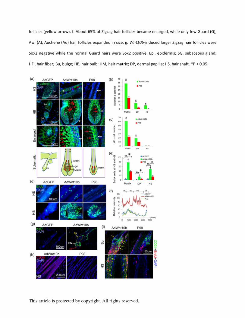
Figure 2. Wnt10b-induced hair follicles were accompanied by excessive activation of the
Wnt/β-catenin pathway, increased proliferation, and hair stem cell activation and migration. a-b.
Immunostaining and statistical chart showed nuclear β-catenin, a key mediator of the Wnt
signaling pathway, was dramatically increased in the Matrix, HS and DP in the
AdWnt10b-treated group compared within AdGFP and normal P98 hair follicle controls (Red
arrow, nuclear β-catenin; pink arrow, cytoplasm β-catenin; white arrow, membrane β-catenin). c.
Statistical chart of Lef1, a downstream Wnt signaling target, was also markedly augmented in the
Matrix and DP region, but not in the HS. d-e. Immunostaining and statistical chart reveals
BrdU-labeled proliferating cells were increased in Wnt10b-induced enlarged hair follicles. f.
Statistical graph showing β1-integrin expression was significantly increased in the HG, Bu and
This article is protected by copyright. All rights reserved.
IFE regions of AdWnt10b-treated hair follicles. g. Ki67+ proliferating cells were located in the
bulge, second hair germ and DP region 6d after the first AdWnt10 treatment. h. CD34 was
unexpectedly localized to the hair shaft, especially in the ORS region. i. Double staining
displayed colocalization of CD34 and BrdU both in the bulge and hair shaft of
AdWnt10b-induced hair follicles but not in those of normal control hair follicles. Bu, bulge; HB,
hair bulb; DP, dermal papilla; HS, hair shaft; HG, second hair germ; IFE, interfollicular
epidermis; EB, epidermal basal layer. *P < 0.05.
Figure 3. Sequential AdWnt10b+DKK1 hair follicle treatment decreased Wnt/β-catenin pathway
activation, reduced proliferation in the hair matrix, DP and hair shaft, but maintained the proper
localization of hair stem cells. a. Schematic drawing showing the timing of two injections of
AdWnt10b followed by DKK1 treatment, hair cycle events and check points. b-c. H&E staining
and statistical chart presenting the significantly decreased width of HB, DP and HS after
AdWnt10b-DKK1 treatment. The results were similar to those of the P98 normal hair follicles. d.
The enlarged hair follicles were significantly reduced from 54.3±4.3 (%) in the
AdWnt10b+N1-treated group to 9.8±3.8 (%) in the AdWnt10b+DKK1-treated group. Note, not
all hair follicles decreased in size (Red and green arrows in b). e-g. Immunostaining and
statistical chart revealed BrdU+ and Ki67+ proliferating cells were decreased in
AdWnt10b+DKK1-treated hair follicles compared within the AdWnt10b+N1-treated hair
follicles, especially in the hair matrix, DP and hair shaft. The proliferative index was recovered
to the P98 normal state. h. Immunostaining for CD34 indicated that after AdWnt10b+DKK1
treatment, hair stem cells were only located in the bulge region of the regenerated hair follicles. i.
CD34+ cells were widespread in the hair shaft, especially the ORS region of AdWnt10b+N1

This article is protected by copyright. All rights reserved.
while not in those of AdWnt10b+DKK1 treated group. N1, control plasmid; Epi, epidermis; SG,
sebaceous gland; HB, hair bulb; HM, hair matrix; DP, dermal papilla; HS, hair shaft. *P < 0.05;
# no statistical difference.
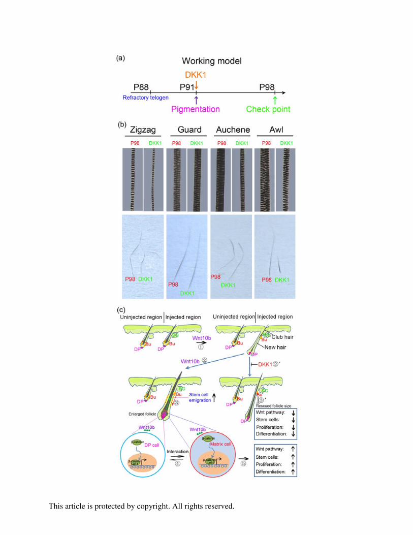
Figure 4. DKK1 treatment decreased hair width. a. Schematic drawing showing the timing of
DKK1 injection, hair follicle status and check points. b. DKK1 treatment narrowed the width of
Zigzag, Auchene, and Awl hairs, and shortened the length of Awl hairs. c. Summary diagram
showing that hair regeneration could be induced by ectopic WNT10b. Prolonged activation of
Wnt signaling in the hair follicle would lead to greater interaction between the hair matrix
epithelial cells and the DP mesenchymal cells, producing more proliferation and differentiation
and broadening the HB, DP and HS. WNT10b could also promote migration of hair stem cells to
sustain matrix proliferation. Interestingly, these processes can be rescued by giving DKK1 to
inhibit regenerating hair follicles. Bu, bulge; DP, dermal papilla; HS, hair shaft.
Supplementary information
Figure S1. Prolonged AdWnt10b treatment enlarged hair follicle size without changing the hair
length. a. Schematic drawing of working model showing that a subcutaneous injection of
AdWnt10b could induce hair regeneration. b. H&E staining shows regenerated hair fibers have
only one column of medulla cells, indicating that the Zigzag hair type was not changed in
Wnt10b-induced regenerated hair follicles. Increased ORS cell layers were present compared
with the normal hair follicles (red line). c-d. WNT10b increased medulla width of regenerated
hairs compared with the normal Zigzag hairs. e. WNT10b only increased the width of Zigzag
hair fibers. f. The regenerated hair follicles unaffected by AdWnt10b treatment maintained a
This article is protected by copyright. All rights reserved.
normal hair size (Red arrow). g. The ratio of the four different hair types was unchanged after
AdWnt10b treatment compared to the P98 controls. h. The length of the WNT10b-induced
regenerated hair follicles was not significantly shifted. i. The overview of all hair types were
normal compared with AdWnt10b-induced hair fibers and P98 normal hair fibers. j. The absence
of TUNEL positive cells in AdWnt10b-induced hair follicles and P98 normal hair follicles
revealed collected samples were still in anagen. Data are reported as mean ±SD. *P < 0.01. # no
statistical difference.
Figure S2. AdWnt10b treatment increases the activation of the Wnt/β-catenin pathway. a.
WNT10b was expressed in the hair matrix and a little bit at the ORS in normal P98 hair follicles.
Prolonged AdWnt10b treatment led to increased Wnt10b expression in the hair matrix and
ectopic expression in the DP and hair shaft. Wnt10b was not expressed in the AdGFP-treated
telogen hair follicles. b. Immunostaining showing overview of β-catenin expression related to
Fig. 2a. c. Immunostaining of Lef1, a downstream Wnt signaling target, was also markedly
augmented in the Matrix and DP region, but not in the HS. Lef1 was only expressed in the DP
region of AdGFP treated hair follicles.
Figure S3. Wnt10b-induced hair follicles were accompanied by hair stem cell activation,
proliferation and migration. a-b. Immunostaining and statistical chart showing Ki67+ cells were
increased in number in the HM, DP and HS region of AdWnt10b-induced hair follicles. c.
Overview of BrdU immunostaining related to Fig. 2d. d. Six days after AdWnt10b treatment,
β1-integrin immunoreactivity was significantly increased compared with the AdGFP or the
normal P56 control hair follicles. e. Ki67+ proliferating cells were increased in the bulge, second
hair germ and DP region 6d after the first AdWnt10 treatment. f-g. Immunostaining and

This article is protected by copyright. All rights reserved.
statistical chart showing that the number of CD34+ bulge stem cells was markedly increased 6d
after AdWnt10 treatment, compared with the control group. h-i. Double staining displayed CD34
and BrdU colocalization both in the bulge and hair shaft of AdWnt10b-induced hair follicles but
not in those of normal control hair follicles.
Figure S4. Hair regeneration after AdWnt10b+DKK1 sequential treatment. a. Schematic drawing
showing our experimental approach to test the role of Wnt10b/�-catenin and DKK1 signaling on
hair regeneration. DKK1 was injected subcutaneously in the center of the pigmented region of
hair follicles that had regenerated two weeks after AdWnt10b treatment. b. Measurement of
follicle length after AdWnt10b+DKK1 co-treatment. AdWnt10b+DKK1 treatment didn’t change
the length of the regenerated hair follicles. Follicle lengths were determined at the anagen VI
stage in AdWntWnt10b +N1, AdWnt10b+DKK1 and normal P98 groups. c. GFP fluorescence of
DKK1-GFP plasmid injected and uninjected skin. Immunostaining showed GFP and DKK1 were
increased in the DKK1-GFP plasmid injected skin. d. The absence of TUNEL positive cells in
AdWnt10b+N1 and AdWnt10b+DKK1-induced hair follicles revealed the collected samples
were still in anagen. *P < 0.01.
Figure S5. Sequential AdWnt10b plus DKK1 treatment decreased Wnt/β-catenin pathway
activation. a and c. Immunostaining and statistical chart revealed nuclear β-catenin expression
was dramatically reduced in the HM, HS and DP in the AdWnt10b+DKK1-treated hair follicles
compared with the AdWnt10b+N1-treated hair follicles. The expression pattern between the
AdWnt10b+DKK1-treated group and normal P98 group were most similar to each other (Red
arrow, nuclear β-catenin; pink arrow, cytoplasm β-catenin; white arrow, membrane β-catenin). b

This article is protected by copyright. All rights reserved.
and d. Immunostaining for Lef1 showed Lef1+ cell numbers were also markedly decreased in the
HM, DP region. N1, control plasmid; Epi, epidermis; HB, hair bulb; HM, hair matrix; DP,
dermal papilla; HS, hair shaft. *P < 0.05; # no statistical difference.
Figure S6. a. Overview of BrdU immunostaining related to Fig. 3f. b. AdWnt10b+DKK1-treated
hair follicles have reduced proliferation in hair matrix, DP and hair shaft. Immunostaining
showing Ki67+ cells were prominently diminished in the HM, DP and HS regions of
AdWnt10b+DKK1 treated hair follicles. The proliferative index was recovered to the P98
normal state.
Figure S7. Recovered IRS thickness after DKK1 treatment. a-b. AE15 immunostaining for the
IRS and statistical chart comparing IRS width. The IRS of AdWnt10b+N1 treated hair follicles
(yellow arrow and red line) was broadened. The width was attenuated to P98 level after DKK1
treatment (pink arrow and red line). Some hair follicles that were not rescued show aberrant
AE15 expression in the hair fiber medulla cells (Green arrow). *P < 0.05; # no statistical
difference.

This article is protected by copyright. All rights reserved.
Figure 1. AdWnt10b treatment enlarges hair follicle size without shifting its type. a. Schematic drawing
showing the timing of multiple AdWnt10b injections, hair cycle events and check points. b-c. H&E
staining and statistical chart showing that the width of HB, DP and HS was significantly increased after
continuous AdWnt10b treatment. d-e. AE15 immunostaining and statistical chart revealed the IRS was
broadened (Red line), and the hair fiber did not differentiate properly in the AdWnt10b-induced hair
This article is protected by copyright. All rights reserved.
follicles (yellow arrow). f. About 65% of Zigzag hair follicles became enlarged, while only few Guard (G),
Awl (A), Auchene (Au) hair follicles expanded in size. g. Wnt10b-induced larger Zigzag hair follicles were
Sox2 negative while the normal Guard hairs were Sox2 positive. Epi, epidermis; SG, sebaceous gland;
HFi, hair fiber; Bu, bulge; HB, hair bulb; HM, hair matrix; DP, dermal papilla; HS, hair shaft. *P < 0.05.

This article is protected by copyright. All rights reserved.
Figure 2. Wnt10b-induced hair follicles were accompanied by excessive activation of the Wnt/β-catenin
pathway, increased proliferation, and hair stem cell activation and migration. a-b. Immunostaining and
statistical chart showed nuclear β-catenin, a key mediator of the Wnt signaling pathway, was
dramatically increased in the Matrix, HS and DP in the AdWnt10b-treated group compared within AdGFP
and normal P98 hair follicle controls (Red arrow, nuclear β-catenin; pink arrow, cytoplasm β-catenin;
white arrow, membrane β-catenin). c. Statistical chart of Lef1, a downstream Wnt signaling target, was
also markedly augmented in the Matrix and DP region, but not in the HS. d-e. Immunostaining and
statistical chart reveals BrdU-labeled proliferating cells were increased in Wnt10b-induced enlarged hair
follicles. f. Statistical graph showing β1-integrin expression was significantly increased in the HG, Bu
and IFE regions of AdWnt10b-treated hair follicles. g. Ki67+ proliferating cells were located in the bulge,
second hair germ and DP region 6d after the first AdWnt10 treatment. h. CD34 was unexpectedly
localized to the hair shaft, especially in the ORS region. i. Double staining displayed colocalization of
CD34 and BrdU both in the bulge and hair shaft of AdWnt10b-induced hair follicles but not in those of
normal control hair follicles. Bu, bulge; HB, hair bulb; DP, dermal papilla; HS, hair shaft; HG, second hair
germ; IFE, interfollicular epidermis; EB, epidermal basal layer. *P < 0.05.

This article is protected by copyright. All rights reserved.

This article is protected by copyright. All rights reserved.
Figure 3. Sequential AdWnt10b+DKK1 hair follicle treatment decreased Wnt/β-catenin pathway
activation, reduced proliferation in the hair matrix, DP and hair shaft, but maintained the proper
localization of hair stem cells. a. Schematic drawing showing the timing of two injections of AdWnt10b
followed by DKK1 treatment, hair cycle events and check points. b-c. H&E staining and statistical chart
presenting the significantly decreased width of HB, DP and HS after AdWnt10b-DKK1 treatment. The
results were similar to those of the P98 normal hair follicles. d. The enlarged hair follicles were
significantly reduced from 54.3±4.3 (%) in the AdWnt10b+N1-treated group to 9.8±3.8 (%) in the
AdWnt10b+DKK1-treated group. Note, not all hair follicles decreased in size (Red and green arrows in b).
e-g. Immunostaining and statistical chart revealed BrdU+ and Ki67+ proliferating cells were decreased in
AdWnt10b+DKK1-treated hair follicles compared within the AdWnt10b+N1-treated hair follicles,
especially in the hair matrix, DP and hair shaft. The proliferative index was recovered to the P98 normal
state. h. Immunostaining for CD34 indicated that after AdWnt10b+DKK1 treatment, hair stem cells were
only located in the bulge region of the regenerated hair follicles. i. CD34+ cells were widespread in the
hair shaft, especially the ORS region of AdWnt10b+N1 while not in those of AdWnt10b+DKK1 treated
group. N1, control plasmid; Epi, epidermis; SG, sebaceous gland; HB, hair bulb; HM, hair matrix; DP,
dermal papilla; HS, hair shaft. *P < 0.05; # no statistical difference.
This article is protected by copyright. All rights reserved.
This article is protected by copyright. All rights reserved.
Figure 4. DKK1 treatment decreased hair width. a. Schematic drawing showing the timing of DKK1
injection, hair follicle status and check points. b. DKK1 treatment narrowed the width of Zigzag,
Auchene, and Awl hairs, and shortened the length of Awl hairs. c. Summary diagram showing that hair
regeneration could be induced by ectopic WNT10b. Prolonged activation of Wnt signaling in the hair
follicle would lead to greater interaction between the hair matrix epithelial cells and the DP
mesenchymal cells, producing more proliferation and differentiation and broadening the HB, DP and HS.
WNT10b could also promote migration of hair stem cells to sustain matrix proliferation. Interestingly,
these processes can be rescued by giving DKK1 to inhibit regenerating hair follicles. Bu, bulge; DP,
dermal papilla; HS, hair shaft.