mitotic figures – large intestine walls
DESCRIPTION
Notes on mitotic figures about the large intestine wallsTRANSCRIPT
Mitotic Figures – Large Intestine wallsWithin nucleus’: Dark heterochromatin, light euchromatin
Nucleolus usually 2µm diameter, and one or more per nucleus
Nuclear envelope is the surrounding wall of the nucleus. Nuclear pores is a small gap for things to move from within the nucleus to the outside cytoplasm or viseversa.
Homeostatis (Balance)
Cell accumulation disordersCancer Lupus Glomerulonephritis (Inflammatory disorder of the kidney)Viral Infections
Cell loss disordersAUDSAlzheimer’s Parkinson’sAplastic AnemiaMyocardial infarctions.
Killing off cells in the body (Necrosis and Apoptosis)
NecrosisBacteria punching holes in the nucleus membrane, causing the plasma membrane to take in water causing it to explode (Lysis, caused by membrane breakdown).
When an infection ruptures a cellInjury SwellingLysisCell death + inflammation.
Apoptosis
Cell becomes smaller/concentrated, causing then membrane to bleb, causing small Apoptotic bodies to be formed, no content of the cells is released
DNA FragmentationCell Volume lossMembrane BlebbingApoptotic bodies formed.

Cytoskeleton (Internal framework of filaments & Tubules)
FunctionGiving the cell it’s shape and allowing it to move. Help move things within the cell, like secretion, and also help control metabolism.
Microtubules are made up of tubulin protein always being 22nm diameter and 5nm thick walls. They are dynamic (Reform and breakdown) and hollow cylinders.Function – Intracellular transport,cell shape cilia movement, chromosome arrangement to create 2 daughter cells.
Microtubule structure
Centrioles is what makes up microtubulesThey are the roots of the microtubules from these there are triplets
Microfilaments are made up of Actic, a helical array with a 6nm diameter (due to size of protein molecules) and is flexibleFound in Microvilli.Function Microvilli struction, and an extension of cell processes.

Intermediant filaments (Most likely not in exam)
Mitochrondria:
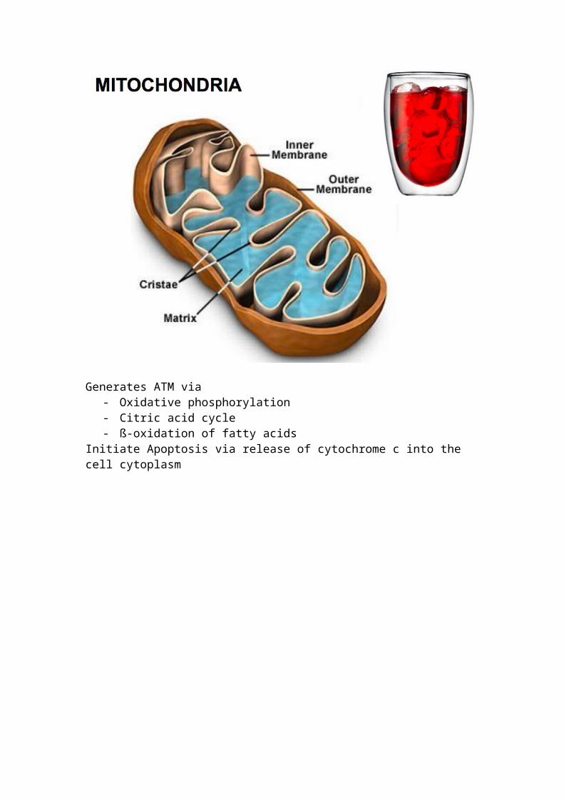
Generates ATM via - Oxidative phosphorylation- Citric acid cycle- ß-oxidation of fatty acids
Initiate Apoptosis via release of cytochrome c into the cell cytoplasm