miguel a. garcía-díaz, manuel ruiz-castrofile.scirp.org/pdf/ijcm_2017050416170861.pdf · central...
TRANSCRIPT

International Journal of Clinical Medicine, 2017, 8, 227-235 http://www.scirp.org/journal/ijcm
ISSN Online: 2158-2882 ISSN Print: 2158-284X
Ultrasound-Guided Infraclavicular Axillary Vein Cannulation
Miguel A. García-Díaz, Manuel Ruiz-Castro
Department of Anesthesia, University Hospital Príncipe de Asturias, Madrid, Spain
Abstract Background and Objective: Percutaneous central venous cannulation is a common invasive procedure. In comparison with an external landmark tech-nique, the advantages of ultrasound-guided venous access include direct visua-lization of the anatomy and in vivo visualization of venous cannulation. Me-thods: We evaluated an ultrasound-guided technique for infraclavicular axillary vein cannulation, focusing on its ease of use, success rate and complications rate. One hundred and twenty patients who submitted to central venous cathe-ter placement were punctured using our technique. The patients were posi-tioned so that their ipsilateral upper limb was abducted at 90˚ to the longitudin-al axis, which makes it possible to visualize the infraclavicular vessels due to the elevation of the clavicle, thereby improving accessibility. Results: Cannulation was successful in all patients. The median time from the start of the first punc-ture (of the skin) until the aspiration of blood was 15 s (range 7 - 135 s). Both infraclavicular axillary veins were cannulated, and the vein was punctured suc-cessfully at the first attempt in 95% of the patients, without complications dur-ing the procedure. Conclusion: We propose an ultrasound-guided infraclavi-cular approach of the axillary vein, with a high success rate and no complica-tions in the present cohort.
Keywords Ultrasonography, Catheterization, Central Venous, Central Venous Catheter, Ultrasound-Guided Technique, Infraclavicular Axillary Vein
1. Introduction
Central venous catheterization (CVC) is a common invasive procedure. Com-plications associated with CVC are often related to blind passage of the needle, and include bleeding, arterial puncture, pneumothorax, nerve damage, pain and
How to cite this paper: García-Díaz, M.A. and Ruiz-Castro, M. (2017) Ultrasound- Guided Infraclavicular Axillary Vein Can-nulation. International Journal of Clinical Medicine, 8, 227-235. https://doi.org/10.4236/ijcm.2017.84022 Received: January 24, 2017 Accepted: April 27, 2017 Published: April 30, 2017 Copyright © 2017 by authors and Scientific Research Publishing Inc. This work is licensed under the Creative Commons Attribution International License (CC BY 4.0). http://creativecommons.org/licenses/by/4.0/
Open Access
DOI: 10.4236/ijcm.2017.84022 April 30, 2017

M. A. García-Díaz, M. Ruiz-Castro
other less common complications. Most authors [1] [2] [3] [4] [5], currently recommend the use of ultrasound
(US) for placing catheters in adults. In comparison with the use of superficial anatomical landmarks, the advantages of US-guided venous access include direct visualization of the anatomy and the in vivo visualization of venous cannulation [6] [7]. The use of real-time US technology to guide needle insertion has im-proved success rates and reduced the incidence of complications [1] [2].
It has recently been shown that US is beneficial for catheterizing the internal jugular vein (IJV), as well as the axillary and mid-arm basilica and cephalic veins. The IJV is more popular due to the easier access and perceived lower risk of complications, however, subclavian vein (SCV) catheter placement is asso-ciated with a lower incidence of infection and thrombosis [8] [9] [10].
Most authors contend that infraclavicular approaches are difficult to visualize with US due to the SCV being located under the clavicle and the inability to place the linear array probe in a position that allows the vessel to be visualized. For this reason, US-guided SCV access is often described using a supraclavicular [8], or a distal approach.
We have developed an US-guided infraclavicular approach for axillary cathe-ter placement using a standard high-frequency linear probe. The location of the axillary vein approach is very proximal, close to, or even over the lip of the first rib, where the SCV is renamed, and hence this approach could be comparable to the approach of the SCV.
2. Method
Ethical approval for this prospective study (protocol number: PI/422010), was provided by the Ethical Committee of the Príncipe de Asturias University Hos-pital, Alcalá de Henares, Madrid, Spain (chairperson: Prof. Elvira Poves Martínez) on 25 November 2010. Written informed consent to participate was obtained from each patient.
A GE Healthcare Venue 40 devices and a GE 12L-SC multifrequency linear transducer at 5 - 13 MHz (GE Healthcare, a division of General Electric Com-pany, Piscataway, New Jersey, USA) were used. Both authors performed this procedure in sixty patients each. The patients were assigned to the study in a consecutive way. The objective of this work was to make a purely descriptive study, so it was not considered to perform the comparison with the conventional technique without ultrasonography, given the high success rate in the US-guided puncture. Therefore, the size of the sample was intended to be representative, although we did not perform a calculation of the sample size when not having to observe differences with a control group.
The position of the patient and the anesthesiologist varies depending upon the standard external-anatomical-landmark-guided catheterization technique. The patient should be positioned so that the ipsilateral upper limb is abducted at 90˚ to the longitudinal axis, which makes it possible to visualize the infraclavicular vessels due to the elevation of the clavicle, thereby improving accessibility. This
228

M. A. García-Díaz, M. Ruiz-Castro
improves the US visualization of the infraclavicular axillary vein (IAV), making almost its entire length visible. If the probe is moved laterally, it is possible to vi-sualize the course of the axillary vein, while if the probe is moved medially with a slight cranial angulation, it is possible to visualize SCV.
Moreover, placing the patient in the Trendelenburg position increases venous engorgement and ease of viewing.
The anesthesiologist puts the patient’s head and locates the sterile field and the equipment needed for the cannulation cephalad to the patient’s arm or beside it, allowing intraoperative puncture. The anesthesiologist’s non-dominant hand holds the US probe, while the dominant hand performs the cannulation.
Initial exploration of the area with US is essential for placing the probe infracla-vicularly, parallel and in close contact with the bottom edge of the clavicle, in the infraclavicular fossa, just lateral to the clavicular head of the sternocleidomastoid muscle Figure 1.
A slight cranial angulation of the probe will facilitate the visualization of the in-fraclavicular vessels, which will be visible as long as the probe is moving parallel to the clavicle, medially and laterally. The vessels on the left side are caudal to those on the right side and the required degree of cranial angulation of the US probe is lower on the left side.
The image obtained from the IAV is medial and superficial to the infraclavicular axillary artery (IAA), and has an elongated shape, close to the long axis view Fig-ure 2.
Furthermore, the pressure of the US probe on the patient can partially collapse the vein. In US imaging, the IAA is normally viewed in cross section, and it appears rounded, pulsatile, and incompressible under the pressure of the US probe.
In-plane venipuncture is achieved from the lateral end of the probe, in the same plane as the US beam, which displays the progress of the needle as it moves
Figure 1. Artistic drawing of the location and placement of the linear array probe (LP) and the needle (N) during the infraclavicular ultrasound-guided approach to can-nulation of the right axillary vein.
229

M. A. García-Díaz, M. Ruiz-Castro
(a)
(b)
Figure 2. Ultrasonographic image obtained at the infraclavi-cular level. (a) Blank image. (b) Labelled image. SCT, subcu-taneous cellular tissue; PM, pectoralis major muscle; SM, sub-clavian muscle; SCV, subclavian vein; SCA, subclavian artery; Arrowheads, first rib.
into the lumen of the vein. It is of paramount importance that the operator ob-serves the needle advancement in real time. To improve visualization of the puncture needle, it is helpful to exert a slight pressure on the inner edge of the US probe so that the angle between the US beams and the needle is as close as possible to 90˚.
While inserting the needle, mild aspiration is performed with the syringe, so
230
M. A. García-Díaz, M. Ruiz-Castro
that blood is observed entering the syringe when the tip of the needle reaches the vessel lumen. Real-time visualization of the extent of the venous lumen demon-strates that the needle tip is at the correct location.
The venous wall is more elastic in younger patients, and so the tip of the needle can be pushed against the wall without perforating it. Aspiration of blood will be necessary to show that the needle tip has reached the vein lumen, as the progress of the needle tip will be visible on the US image. Only minimal further progression from this point will cause the needle tip to enter the lumen without piercing the posterior venous wall.
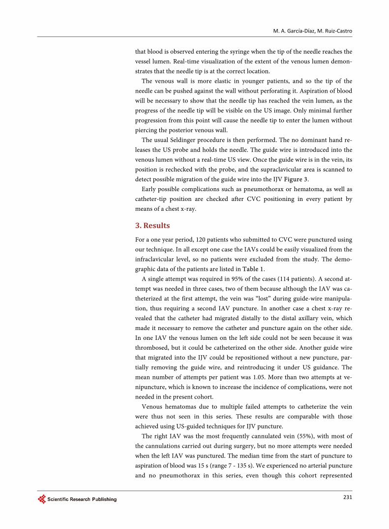
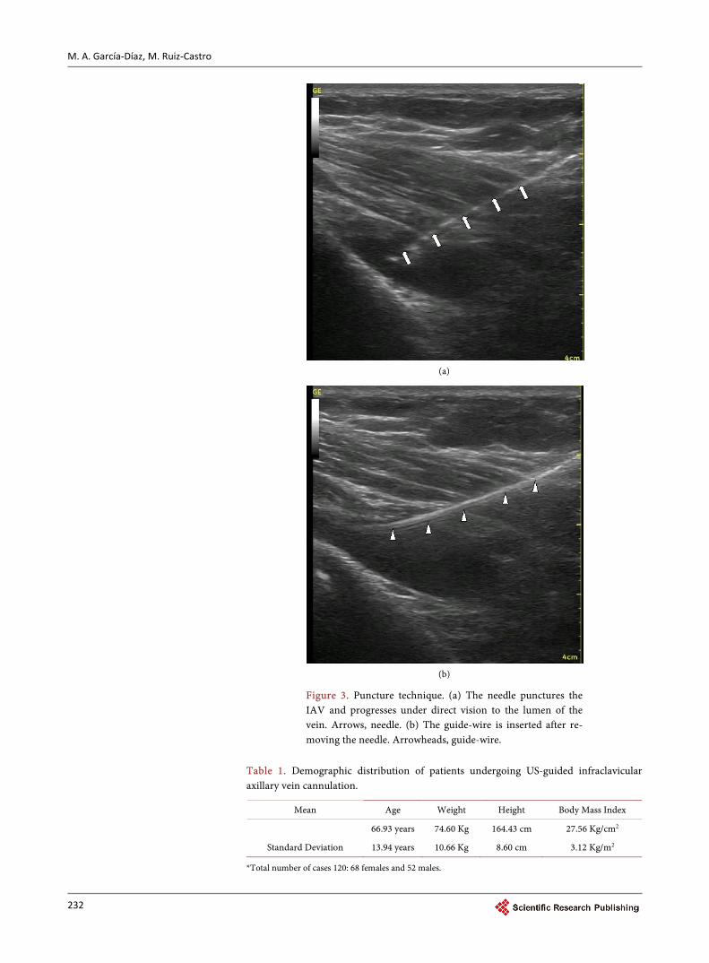
The usual Seldinger procedure is then performed. The no dominant hand re-leases the US probe and holds the needle. The guide wire is introduced into the venous lumen without a real-time US view. Once the guide wire is in the vein, its position is rechecked with the probe, and the supraclavicular area is scanned to detect possible migration of the guide wire into the IJV Figure 3.
Early possible complications such as pneumothorax or hematoma, as well as catheter-tip position are checked after CVC positioning in every patient by means of a chest x-ray.
3. Results
For a one year period, 120 patients who submitted to CVC were punctured using our technique. In all except one case the IAVs could be easily visualized from the infraclavicular level, so no patients were excluded from the study. The demo-graphic data of the patients are listed in Table 1.
A single attempt was required in 95% of the cases (114 patients). A second at-tempt was needed in three cases, two of them because although the IAV was ca-theterized at the first attempt, the vein was “lost” during guide-wire manipula-tion, thus requiring a second IAV puncture. In another case a chest x-ray re-vealed that the catheter had migrated distally to the distal axillary vein, which made it necessary to remove the catheter and puncture again on the other side. In one IAV the venous lumen on the left side could not be seen because it was thrombosed, but it could be catheterized on the other side. Another guide wire that migrated into the IJV could be repositioned without a new puncture, par-tially removing the guide wire, and reintroducing it under US guidance. The mean number of attempts per patient was 1.05. More than two attempts at ve-nipuncture, which is known to increase the incidence of complications, were not needed in the present cohort.
Venous hematomas due to multiple failed attempts to catheterize the vein were thus not seen in this series. These results are comparable with those achieved using US-guided techniques for IJV puncture.
The right IAV was the most frequently cannulated vein (55%), with most of the cannulations carried out during surgery, but no more attempts were needed when the left IAV was punctured. The median time from the start of puncture to aspiration of blood was 15 s (range 7 - 135 s). We experienced no arterial puncture and no pneumothorax in this series, even though this cohort represented
231
M. A. García-Díaz, M. Ruiz-Castro
(a)
(b)
Figure 3. Puncture technique. (a) The needle punctures the IAV and progresses under direct vision to the lumen of the vein. Arrows, needle. (b) The guide-wire is inserted after re-moving the needle. Arrowheads, guide-wire.
Table 1. Demographic distribution of patients undergoing US-guided infraclavicular axillary vein cannulation.
Mean Age Weight Height Body Mass Index
66.93 years 74.60 Kg 164.43 cm 27.56 Kg/cm2
Standard Deviation 13.94 years 10.66 Kg 8.60 cm 3.12 Kg/m2
*Total number of cases 120: 68 females and 52 males.
232

M. A. García-Díaz, M. Ruiz-Castro
our learning curve for this technique.
4. Discussion
US-guided venous access has been described and widely recommended for IJV cannulation [11]. However, few studies have investigated US-guided SCV punc-ture, most of which have focused on paediatric venous cannulation [6] [7]. One paper reports the use of US to puncture the SCV via a supraclavicular approach with an endocavitary probe with good results [8], but we describe here a tech-nique that uses a standard linear array probe to puncture the IAV, in close proximity to the SCV. The boundary between the two is defined by the edge of the first rib, so we believe that there are no clinically significant differences.
In previous studies, the venous approach was performed more laterally, even at the level of the mid-axillary vein, where mobility is greater, but fixation of the catheter is worse.
Only a few articles [12] [13] have reported on US-guided for axillary vein puncture. Two studies imaged both the vein and the needle transversely, which offers poorer needle visualization. Another paper [14] describes a more lateral approach to the axillary vein, where longitudinal visualization of both the vein and needle was possible. Our longitudinal in-line puncture technique offers a clear view of both the needle and the vein, which should produce consistently good results. The risk of accidental puncture of pleura or other structures (e.g., arteries) is lower, regardless of how deep the needle is, since the advancement of the needle, the vessel, and the surrounding structures can all be viewed in real time. Moreover, the place where the SCV passes over the first rib has been de-fined as a safe spot for its cannulation, since at this location, the first rib protects the lung from accidental puncture and the direction of the subclavian artery dif-fers from that of the SCV, passing behind the anterior scalene muscle whereas the vein passes in front of it.
Abduction of the upper limb and the clavicle with it provides a US view of the infraclavicular vessels at this level, allowing safe puncture and cannulation.
Puncture time was measured in this study. The preparation time is known to be longer with US-guided techniques than with landmark-guided techniques. With experience, anatomical screening, and appropriate equipment preparation, the time should not exceed 5 min. Because the anatomical (vein position) and pathological (vein thrombosis) variations are now diagnosed by US, the time from skin puncture to guide-wire placement is reduced.
Furthermore, the very time-consuming vicious circle of multiple attempts, venous or arterial hematoma, failed puncture, and need to change to another central venous insertion site is probably minimized by US guidance.
The entire procedure is likely to be completed faster than when using the ana-tomical-landmark-based technique. It should also be noted that the procedure can be carried out during surgery because the abducted position of the upper extremi-ties is common in the operating room, thus further reducing the time required. This is a clear advantage over the traditional external landmark technique where the adducted position of the upper extremity is required and forces the anesthesi-
233

M. A. García-Díaz, M. Ruiz-Castro
ologist to cannulate the IJV in the operating setting, associated with a higher inci-dence of infection and thrombosis [8] [9] [10].
Another advantage of US is the early detection of incorrect guide-wire cathe-terization or migration of the guide wire into the IJV. In our study, we expe-rienced a single case of misplacement of the guide-wire, perhaps because the abduction of the upper limb directs the IAV toward the territory of the superior vena cava.
In a completely sterile setting, the assistance of a third hand is usually not needed because the probe can be left in the sterile field during maneuvers that require two hands, such as clavicle palpation for needle insertion or guide-wire advancement.
The use of US not only provides needle guidance but also makes it easy to check the patency of the IAV, regardless of whether the cases are hypovolemic patients whose veins have little filler, or are well hydrated (e.g., with engorged veins). Lumen vein in hypovolemic patients is very narrow, being very difficult to place the tip of the needle inside. When it is closed to the venous wall, a Val-salva maneuver can distend the vein and facilitate the puncture.
The learning curve of this approach, as for every US-guided technique, will probably be highly operator-dependant. Prior to the start of the study, each of the anaesthesiologists cannulated at least three IAV with this US-guided tech-nique, to familiarize themselves with it. Then, during the study, each of the anesthesiologists performed this procedure in sixty patients. Both of them had previous experience in US-guided puncture techniques (regional anesthesia and CVC) and in IJV and SCV anatomical-landmark-based cannulation.
We consider that it takes a minimum of five cases to perform the procedure safely, with many more being needed in the absence of prior experience in man-aging US. For inexperienced clinicians, residents, or fellows, the US-guided IAV cannulation should be considered an advanced technique given the complexity and risk of complications with this approach.
In conclusion US-guided IAV cannulation can be performed by using a li-near-array probe and ipsilaterally abducting the patient’s upper limb at 90˚. We propose an infraclavicular approach when performing this technique, since it was associated with a high success rate and no complications in this the present cohort. This procedure is safe and easy to learn for CVC, and we recommend this technique even in an early learning stage. Moreover, the procedure can be performed in an intraoperative setting.
Acknowledgements
We thank M. Sanz, MD, F. Barrios and M. Ayuso for their assistance with the study. This work was supported by the Department of Anesthesiology, Univer-sity Hospital Príncipe de Asturias, Alcalá de Henares, Madrid, Spain.
References [1] Waldman, C.S. (2001) Use of Central Venous Catheters (CVC)—An Algorithm.
234

M. A. García-Díaz, M. Ruiz-Castro
Care of the Critically Ill, 17, 148-149.
[2] Randolph, A.G., Cook, D.J., Gonzales, C.A. and Pribble, C.G. (1996) Ultrasound Guidance for Placement of Central Venous Catheters: A Meta-Analysis of the Lite-rature. Critical Care Medicine, 24, 2053-2058. https://doi.org/10.1097/00003246-199612000-00020
[3] Maslove, D. and Mihm, F. (2010) Ultrasound-Guided Internal Jugular Vein Cannu-lation. The New England Journal of Medicine, 363, 796. https://doi.org/10.1056/NEJMc1006062
[4] Levitov, A.B., Aziz, S. and Slonim, A.D. (2009) Before We Go Too Far: Ultra-sound-Guided Central Catheter Placement. Critical Care Medicine, 37, 2473-2474. https://doi.org/10.1097/CCM.0b013e3181a9f694
[5] Barash, P. (2009) Ultrasound-Guided Central Venous Access. F1000 Medicine Reports, 1, 65. https://doi.org/10.3410/m1-65
[6] Hind, D., Calvert, N., McWilliams, R., et al. (2003) Ultrasonic Locating Devices for Central Venous Cannulation: Meta-Analysis. BMJ, 327, 361. https://doi.org/10.1136/bmj.327.7411.361
[7] Theodoro, D., Bausano, B., Lewis, L., Evanoff, B. and Kollef. M. (2010) A Descrip-tive Comparison of Ultrasound-Guided Central Venous Cannulation of the Internal Jugular Vein to Landmark-Based Subclavian Vein Cannulation. Academic Emer-gency Medicine, 17, 416-422. https://doi.org/10.1111/j.1553-2712.2010.00703.x
[8] Mallin, M., Louis, H. and Madsen, T. (2010) A Novel Technique for Ultra-sound-Guided Supraclavicular Subclavian Cannulation. American Journal of Emergency Medicine, 28, 969-969.
[9] McGee, D.C. and Gould, M.K. (2003) Preventing Complications of Central Venous Catheterization. The New England Journal of Medicine, 348, 1123-1133. https://doi.org/10.1056/NEJMra011883
[10] Stone, M.B., Moon, C., Sutijono, D. and Blaivas, M. (2010) Needle Tip Visualization during Ultrasound-Guided Vascular Access: Short-Axis vs. Long-Axis Approach. American Journal of Emergency Medicine, 28, 343-347.
[11] Gualtieri, E., Deppe, S.A., Sipperly, M.E., et al. (1995) Subclavian Venous Cathete-rization: Greater Success Rate for Less Experienced Operators Using Ultrasound Guidance. Critical Care Medicine, 23, 692. https://doi.org/10.1097/00003246-199504000-00018
[12] Sharma, A., Bodenham, A.R. and Mallick, A. (2004) Ultrasound-Guided Infraclavi-cular Axillary Vein Cannulation for Central Venous Access. British Journal of Anaesthesia, 93, 188-192. https://doi.org/10.1093/bja/aeh187
[13] Sandhu, N.S. (2004) Transpectoral Ultrasound-Guided Catheterization of the Axil-lary Vein: An Alternative of Standard Catheterization of the Subclavian Vein. Anesthesia & Analgesia, 99, 183-187. https://doi.org/10.1213/01.ANE.0000117283.09234.2C
[14] Stone, M.B., Nagdev, A., Murphy, M.C. and Sisson, C.A. (2010) Ultrasound Detec-tion of Guidewire Position during Central Venous Catheterization. American Jour-nal of Emergency Medicine, 28, 82-84.
235

Submit or recommend next manuscript to SCIRP and we will provide best service for you:
Accepting pre-submission inquiries through Email, Facebook, LinkedIn, Twitter, etc. A wide selection of journals (inclusive of 9 subjects, more than 200 journals) Providing 24-hour high-quality service User-friendly online submission system Fair and swift peer-review system Efficient typesetting and proofreading procedure Display of the result of downloads and visits, as well as the number of cited articles Maximum dissemination of your research work
Submit your manuscript at: http://papersubmission.scirp.org/ Or contact [email protected]