microstereolithography of tissue scaffolds using a ... · microstereolithography of tissue...
TRANSCRIPT

Microstereolithography of Tissue Scaffolds Using a Biodegradable Photocurable Polyester
Nicholas A. Chartrain1,4
, Maria Vratsanos2, Dung T. Han
1, Justin M. Sirrine
3,4, Allison
Pekkanen3,4
, Timothy E. Long3,4
, Abby R. Whittington1,4
, Christopher B. Williams2,4
1Department of Materials Science & Engineering, Virginia Tech, Blacksburg, VA 24061
2Department of Mechanical Engineering, Virginia Tech, Blacksburg, VA 24061
3Department of Chemistry, Virginia Tech, Blacksburg, VA 24061
4Macromolecules and Interfaces Institute, Virginia Tech, Blacksburg, VA 24061
Correspondence: Nicholas Chartrain [email protected]
Keywords: Tissue Scaffold, Regenerative Medicine, Stereolithography, Biodegradable, Polymer
Abstract
Due to its ability to create complex cellular geometries with extremely fine resolution, mask
projection microstereolithography (MPμSL) can be useful for fabricating designed tissue
scaffolds and other biological constructs for use in Tissue Engineering and Regenerative
Medicine. However, few photocurable materials with low cytotoxicity, adequate cell adhesion,
and degradability can be processed with MPμSL. In this work, we present the fabrication of
biocompatible and biodegradable tissue scaffolds with 50 μm feature sizes from a novel
polyester using MPμSL. Poly(tri(ethylene glycol)adipate) dimethacrylate (PTEGA-DMA) was
synthesized and evaluated for its printability. The curing parameters for printing were identified
and scaffolds were fabricated. Optical and electron microscopy were used to determine the
achievable feature sizes and accuracy of printed parts using the polymer in the MPμSL system.
MC3T3-E1 mouse preosteoblasts were seeded on PTEGA-DMA films to assess adhesion and
biocompatibility.
1. Introduction
1.1. The Need for Tissue Scaffolds
Millions of patients suffer from damaged or diseased tissue resulting from a wide variety of
diseases, conditions, and accidents. Current treatments generally consist of using drugs, wound
dressings or biomedical devices to alleviate symptoms, but do not replace or repair damaged
tissue with healthy tissue [1, 2]. Thousands of patients receive transplant tissues and organs each
year, but others must wait many years before a transplant becomes available [3]. For many other
conditions, using transplant tissue is not practical. Patients suffering from diabetic foot ulcers,
skin burns and wounds, organ failure, and bone fractures would benefit from a greater
availability and variety of replacement tissue.
Tissue Engineering aims to use tissue scaffolds in conjunction with cells as well as
chemical, mechanical, or electrical stimuli to construct functional tissue that can be used to repair
or replace damaged or diseased tissues [1, 2, 4, 5]. Tissue scaffolds are sponge or network-like
devices that provide a three-dimensional environment upon which cells can attach, grow, and
proliferate [6]. Scaffolds must maintain sufficient porosity for nutrients to flow into the scaffolds
1732
Solid Freeform Fabrication 2016: Proceedings of the 26th Annual InternationalSolid Freeform Fabrication Symposium – An Additive Manufacturing Conference
Reviewed Paper
Solid Freeform Fabrication 2016: Proceedings of the 27th Annual International

while also providing structural support for the cells [7]. Fulfilling these two goals requires
creative scaffold design as structures with greater porosities tend to have less mechanical
strength. In addition, the incorporation of biochemical factors into tissue scaffolds, such as
growth factors, small molecules, or even minerals, can significantly enhance cell adhesion,
viability, and differentiation [8-10]. Mechanical exercising and electrical stimulation of certain
cell types has also been shown to improve cell differentiation and function [11]. Finally, tissue
scaffolds require vascularization, or the incorporation of a system for blood flow throughout the
scaffold. While many have reported tissue scaffolds that provide mechanical robustness,
adequate porosity, and the incorporation of chemical factors, success at incorporating
vascularization into tissue scaffolds has been limited [12, 13]. Creating the foundation for a
vascular system in a tissue scaffold is undoubtedly the greatest challenge in Tissue Engineering
and has hindered the fabrication of large tissue scaffolds for the replacement of solid tissues and
organs [13, 14].
1.2. Benefits of AM in Fabricating Tissue Scaffolds
Tissue scaffolds resulting from traditional manufacturing techniques feature stochastic
distribution of pores. Specifically, these techniques such as gas foaming, particulate leeching,
and electrospinning (Figure 1) have the ability to control pore size and density, but pore
placement occurs randomly within the scaffold [15]. The lack of ability to control the precise
mesostructure of the scaffold affects the repeatability of the process, and makes it very difficult
to incorporate vasculature into the structure [13, 14]. Without vasculature, cells that migrate to
the center of the tissue scaffold will not have sufficient access to nutrients provided by blood
[13]. Such scaffolds feature healthy cells on the surface, while apoptosis, programmed cell death,
occurs in the center of the scaffold.
Figure 1. Electrospun scaffold used for tissue engineering [16].
Additive Manufacturing systems, often referred to as 3D printers, have the ability to
precisely control material placement in three dimensional space [17]. This allows 3D printers to
repeatedly fabricate complex designed structures that could not be fabricated by other means.
The ability of 3D printers to construct complex geometries with designed macro and
mesostructure makes them ideal for fabricating tissue scaffolds that incorporate vasculature [18-
21].
1733

1.3. Advantages of Mask Projection Microstereolithography
While all AM systems are able to fabricate complex designed structures, some are more
suited for fabricating tissue scaffolds that others. The AM system chosen should be able to
fabricate features with sizes on the order of a cell diameter (~10 μm) so that surface area of the
tissue scaffold can be maximized [6]. Achieving such fine features has proven difficult with most
AM systems [17]. The printed resolution of techniques such as filament-based extrusion and
Powder Bed Fusion are limited by extrusion tip diameter, powder particle size, and other
physical constraints [22]. As a result, these systems are generally not able to produce parts with
feature sizes below several hundred microns [18, 23]. These large feature sizes allow less surface
area for cells to attach and require greater amounts of time for degradation of the part to occur.
Bioprinting systems, occasionally referred to as bioplotters, are subject to a similar constraint.
While they are able to directly place both material and cells in a scaffold, bioplotters are limited
by the nozzle diameter through which they can extrude cells [18]. As extrusion devices have
difficulty with precise start/stop motion, they are limited to printing “log-cabin” cellular
topologies that feature extruded serpentine paths (with large offsets between roads). In addition,
thin nozzles that provide high resolution features normally result in shear stresses on cells that
significantly reduce their viability. Contrary to these other AM systems, the printing resolution
of vat photopolymerization is limited only by the wavelength of light and quality and tuning of
optical components [17]. Mask projection microstereolithography (MPμSL) systems able to
fabricate feature sizes below 50 μm have been demonstrated by several groups [19, 24-27]. Such
resolution would enable the fabrication of scaffold geometries with high porosity, large surface
area, and pores of appropriate size for cell proliferation [28].
Tissue scaffolds must also be fabricated from material(s) that are biocompatible and
biodegradable [2]. Most AM systems are not yet able to fabricate biocompatible and
biodegradable materials [29]. For example, cells can be grown on scaffolds fabricated using
filament-based extrusion processes, but these materials are not often easily dissolved in
physiological conditions. Bioprinters, another extrusion AM process, are able to extrude a
variety of natural and synthetic polymers, many of which are both biocompatible and
biodegradable. Unfortunately, many of these materials have little mechanical robustness and do
not serve well for large tissue scaffolds, particularly for hard tissue replacement [18, 30, 31]. In
addition, bioprinters face the difficulty of keeping cells viable during the printing process, which
does not usually occur in media.
Researchers have demonstrated the fabrication of tissue scaffolds with vat
photopolymerization using several biocompatible synthetic polymers. These polymers often
contain carbonate or ester groups that can be easily hydrolyzed under physiological conditions
[32]. The most widely reported printable photopolymer for tissue scaffolds, poly(propylene
fumarate) (PPF), shows good biocompatibility and demonstrates promise for use in scaffolds for
hard tissue replacement [33-39]. Additional polymers reported include poly(ε-caprolactone) [40],
poly(ethylene glycol) diacrylate [41], trimethylene carbonate [42], and poly (D,L-lactide) [43].
However, these polymers have a limited range of mechanical and chemical properties making
them appropriate for tissue scaffolds for only certain types of tissue replacement. Future
successes in using vat photopolymerization for the fabrication of tissue scaffolds with relevant
1734

feature sizes hinges on the development of novel biocompatible and biodegradable
photopolymers.
In an effort to expand the palette of materials from which tissue scaffolds can be fabricated,
this research investigated the printability and biocompatibility of the novel polyester, PTEGA-
DMA. A successful candidate material must a) permit fabrication of feature sizes below 100 μm,
b) demonstrate good cell adhesion and viability, c) exhibit mechanical properties similar to those
of human tissue, and d) allow degradation in physiologically relevant conditions. To evaluate
whether PTEGA-DMA is a valid candidate material for fabricating tissue scaffolds via MPμSL,
three primary research goals were devised and investigated:
- to develop process parameters for the fabrication of PTEGA-DMA tissue scaffolds using
MPμSL.
- to determine the minimum feature sizes, accuracy, and resolution that could be achieved
when fabricating PTEGA-DMA parts using the MPμSL machine.
- to gain understanding of the thermomechanical and degradation properties of PTEGA-
DMA as well as assessing cell response to the material to evaluate its viability as a tissue
scaffold material.
2. Experimental Techniques
2.1. Synthesis of PTEGA-DMA
Synthesis of tri(ethylene glycol) adipate was achieved through the melt polycondensation of
tri(ethylene glycol) and adipic acid as described previously [44] (Figure 2). Functionalization to
allow UV-induced crosslinking necessary for vat photopolymerization was realized through the
addition of dimethacrylate end-groups via reaction of the PTEGA diol with 2-isocyanatoethyl
methacrylate. Proton nuclear magnetic resonance (1H NMR) was used to verify chemical purity
of the PTEGA-DMA and determine that the product’s Mn was 1,600 g/mol.
1735
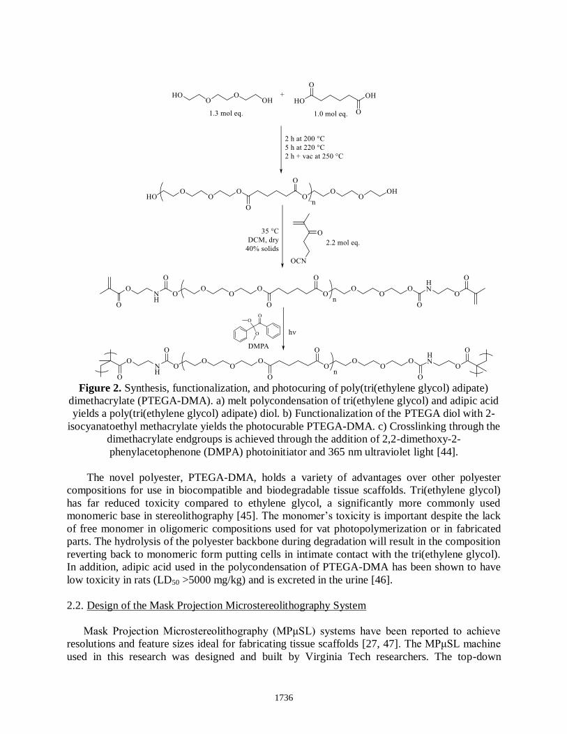
Figure 2. Synthesis, functionalization, and photocuring of poly(tri(ethylene glycol) adipate)
dimethacrylate (PTEGA-DMA). a) melt polycondensation of tri(ethylene glycol) and adipic acid
yields a poly(tri(ethylene glycol) adipate) diol. b) Functionalization of the PTEGA diol with 2-
isocyanatoethyl methacrylate yields the photocurable PTEGA-DMA. c) Crosslinking through the
dimethacrylate endgroups is achieved through the addition of 2,2-dimethoxy-2-
phenylacetophenone (DMPA) photoinitiator and 365 nm ultraviolet light [44].
The novel polyester, PTEGA-DMA, holds a variety of advantages over other polyester
compositions for use in biocompatible and biodegradable tissue scaffolds. Tri(ethylene glycol)
has far reduced toxicity compared to ethylene glycol, a significantly more commonly used
monomeric base in stereolithography [45]. The monomer’s toxicity is important despite the lack
of free monomer in oligomeric compositions used for vat photopolymerization or in fabricated
parts. The hydrolysis of the polyester backbone during degradation will result in the composition
reverting back to monomeric form putting cells in intimate contact with the tri(ethylene glycol).
In addition, adipic acid used in the polycondensation of PTEGA-DMA has been shown to have
low toxicity in rats (LD50 >5000 mg/kg) and is excreted in the urine [46].
2.2. Design of the Mask Projection Microstereolithography System
Mask Projection Microstereolithography (MPμSL) systems have been reported to achieve
resolutions and feature sizes ideal for fabricating tissue scaffolds [27, 47]. The MPμSL machine
used in this research was designed and built by Virginia Tech researchers. The top-down
1736

projection system passes an ultraviolet LED light source (365 nm; 5.0 mJ/cm2 intensity at resin
surface) onto a digital micromirror device (DMD), which serves as a dynamic mask, through a
series of conditioning and imaging optics, and onto a build stage mounted on a linear actuator,
which dips into a resin vat (Figure 3). The system’s DMD, a TI Instruments 1080p DLP 6500
chip, provides a 3.78 x 3.78 μm effective pixel projection size at the resin surface when
implemented with the selected imaging optics. It is able to fabricate structures with feature sizes
on the order of tens of microns across a build volume of up to 4 x 6 x 36 mm. The operation of
the MPμSL system has been previously reported [48].
Figure 3. Schematic of the Mask Projection Microstereolithography machine.
2.3. Curing Parameters and Part Fabrication
To fabricate accurate and detailed parts using stereolithography, it is essential to understand
the interaction of the photopolymer with the ultraviolet light that is curing it. As described by
Jacobs, two primary intrinsic material parameters control the photocrosslinking process: the
depth of penetration (DP) and the critical exposure (EC) [49]. The energy imparted to a
photopolymer (E) and the resulting thickness of the cured polymer film (CD) can be related to the
depth of penetration and the critical exposure through the ‘Working Curve’ (Equation 1).
C𝐷 = DPln (E
E𝐶) 𝐸𝑞𝑢𝑎𝑡𝑖𝑜𝑛 1
To determine the DP and EC of PTEGA-DMA, a slightly modified single-layer
‘Windowpane’ technique was used in which the build stage was removed and thin films of
PTEGA-DMA were cured on the surface of the photopolymer vat surface [17]. This was
repeated using several known exposure amounts and the resulting film thicknesses were
tabulated. This allowed the generation of a working curve and the determination of the material’s
DP and EC. Initial results showed that the PTEGA-DMA had a large DP. To reduce the depth of
1737

penetration of light into the polymer and improve resolution, avobenzone, a small molecule UV
absorber often used in sunscreen, was added at 0.05 wt%.
2.4. Resolution, Accuracy, and Feature Size Determination
A benchmark test part was designed and printed to quantitatively determine the resolution,
accuracy, and minimum feature sizes that could be achieved using the MPμSL machine in
conjunction with the PTEGA-DMA polyester [27]. The part (Figure 4), has cylinders ranging
from 3.5 to 150 μm in diameter on the top surface that allow the determination of minimum
feature size and resolution in the XY plane. Thin horizontal walls (20-150 μm thick) on the sides
of the part in the XZ and YZ planes reveal the resolution in these planes. The thickness of these
horizontal walls as well as the thickness of the primary horizontal crossbeams help determine the
extent of any undesirable crosslinking of liquid photopolymer in areas below the layer being
built (also referred to as “print-through”). The XY plane accuracy can be quantitatively
determined by measuring the distances between the crosshairs on the top plane of the part.
Figure 4. Schematic of the diagnostic test part used to determine accuracy, resolution, and
minimum feature sizes that could be achieved when fabricating PTEGA-DMA parts using
MPμSL (left). To demonstrate the fabrication of complex and physiologically relevant
geometries, a tissue scaffold (center) containing 400 μm pores and a Virginia Tech Hokie
bird (right) were fabricated.
To evaluate the dimensional accuracy of the test part, the samples were rinsed with
isopropanol (IPA), dried, and then imaged using a Dinolight USB digital microscope. Additional
images of some samples were taken with a JEOL NeoScope JCM-5000 desktop SEM. Sputter
coating the samples was not necessary for SEM imaging.
2.5. Characterization of Printed PTEGA-DMA Parts
In order to evaluate the feasibility of using PTEGA-DMA for the fabrication of tissue
scaffolds, it is essential to understand both the thermal, mechanical and degradation properties of
the material. For example, soft tissue cells respond more favorably to soft materials while cells
from bone have higher viability when in contact with harder materials [50]. In addition, the
1738

degradation of the material should happen slowly enough that cells have enough time to secrete
extracellular matrix that will provide mechanical stability to the forming tissue [6].
Dynamic Mechanical Analysis (DMA) was performed to determine the glass transition
temperature (Tg) as well as the thermomechanical properties of the PTEGA-DMA at a range of
temperatures. Bars of PTEGA-DMA photoinitiated with 2 wt % DMPA were printed (1.4 mm
thick, 4.35 mm wide, 30 mm long) and extracted in consecutive sonicated THF and EtOH baths
for 30 minutes each to remove any uncured oligomer. The bars were tested in tension on a TA
Instruments Q800 DMA with a temperature ramp of 3 °C/min and a 15 μm strain amplitude at 1
Hz. A temperature sweep between -100 °C and +100 °C determined both the Tg and the storage
modulus of the PTEGA-DMA at various temperatures. The storage modulus at 37 °C was
compared to those of various human tissues to determine what types of tissues and tissue
scaffolds the material might be most suitable for.
The PTEGA-DMA polymer backbone contains ester bonds that can be cleaved through
hydrolysis. Degradation kinetics of the polyester was determined by soaking printed scaffolds
(Figure 4) in minimum essential media at 37 °C, for 4 h, 1 day, or 5 days. Scaffolds in media
were expected to degrade at a rate similar to what would be observed in in vivo or in vitro cell
culture conditions. Before soaking, the scaffolds were cleaned with IPA and vacuum dried at 50
°C for 12 hours, and weighed. After soaking in media, the scaffolds were again vacuum dried at
50 °C for 12 hours and reweighed to determine mass loss.
2.6. Cytotoxicity Testing of PTEGA-DMA
Cell cultures were conducted on polyester films in order to determine the cytotoxicity of the
PTEGA-DMA. DMPA photoinitiator and avobenzone were dissolved in acetone and added to
PTEGA-DMA at 2 wt% and 0.05 wt% respectively. Thin films were cast and photocrosslinked
using a 6 W handheld UV-A lamp (Spectroline EA-160) for 5 min in a 24-well untreated
polystyrene plate. The films were swelled in reverse osmosis filtered water overnight. Three
70:30 v/v EtOH/H2O extractions were performed for 60 minutes each to sterilize the films and
remove any uncrosslinked oligomer. Then, two phosphate buffered saline (PBS) and one
minimum essential cell media washes of 60 minutes each were used to remove residual ethanol.
MC3T3-E1 mouse preosteoblasts were cultured in minimum essential media containing 10%
fetal bovine serum and 1% penicillin/streptomycin until 80% confluent. The cells were lifted
from the cell culture flasks using 0.5% Trypsin-EDTA, counted, and seeded onto the PTEGA-
DMA films and tissue culture treated polystyrene 24 well plates at a density of 50,000 cells/well.
Cell viability was determined after 24 hours using a CellTiter-Glo luminescence assay and a
BioTek Synergy Mx plate reader in absorbance mode. Viability was normalized to the tissue
culture treated polystyrene plate. Cells were fixed using a formaldehyde solution (0.5% Triton
X-100, 4% formaldehyde, 5% sucrose in PBS) in preparation for fluorescence imaging. 205 μL
of 165 nM Texas Red-X Phalloidin solution and 300 μL of 300 nM DAPI (4',6-diamidino-2-
phenylindole, dihydrochloride) solution, both in PBS, were added to each well. Fluorescence
images were taken using a Zeiss Axio Observer.Z1 microscope.
1739

3. Results & Discussion
3.1. UV Curing Parameters and Part Fabrication
Using the modified Windowpane technique described in Section 2.3, the intrinsic resin
properties, EC and DP, for PTEGA-DMA with 2 wt% DMPA were determined (Table 1). Due to
the very large DP of the PTEGA-DMA, parts printed without any UV blocker had significant
print-through resulting in poor layer and feature definition (Figure 5). The addition of 0.05 wt%
avobenzone dissolved in acetone served to significantly reduce the DP and allow for the
fabrication of parts with thin layers (<50 μm). EC and DP were re-determined for the resin
containing avobenzone. The increase in EC (from 6.32 to 9.27 mJ/cm2; a 47% increase) and
decrease in DP (from 453 to 107 μm; a 76% decrease) more than doubled the exposure time for
curing a 50 μm layer. However, total print time was not greatly affected as the slow recoating
step contributes the largest amount of time to the print duration.
Table 1. Curing Parameters of the
PTEGA-DMA polyester
Figure 5. The working curve of PTEGA-DMA containing 2 wt% DMPA and 0.05 wt%
avobenzone demonstrates a significantly lower Dp but slightly higher Ec than the sample without
avobenzone.
To determine the accuracy and resolution that could be achieved with the PTEGA-DMA on
the MPμSL system, the diagnostic test parts described in Section 3.2 was fabricated. Parts were
made using 50 μm layer thicknesses, washed in isopropanol, and then imaged and measured
using a DinoLight USB camera. The dimensions of three parts were averaged to determine
minimum feature size achievable as well as the accuracy in each of the three axes. Vertical
pillars with diameters as small as 30 μm were successfully fabricated. The observed dimensions
in all three axes were slightly smaller than intended (Table 2). This could be due to part
shrinkage observed during the curing of acrylates as well as errors in the optical setup that could
change the effective size of pixels on the resin surface.
Avobenzone
concentration
0 wt% 0.05
wt%
Ec (mJ/cm2) 6.32 9.27
Dp (μm) 453 107
50 μm layer
print time (s)
1.42 2.97
1740

Table 2. Printing accuracy and feature sizes achieved when fabricating the diagnostic part on
the on the MPμSL system using PTEGA-DMA.
To demonstrate the fabrication of complex structures using PTEGA-DMA, a Hokie bird and
tissue scaffold with square pores were built using 2 wt% DMPA and 0.05 wt% avobenzone
(Figure 6). The 4x4x8 mm scaffold contains pores that are 400 μm by 800 μm. The Hokie bird is
approximately 7 mm tall. Each part was printed with 100 μm layers irradiated for 7.5 s at an
intensity of 5 mW/cm2. A scaffold printed from material without avobenzone demonstrates poor
resolution and feature definition. Each 100 μm layer was irradiated for just 1.57 s.
Figure 6. Image and SEM micrographs of PTEGA-DMA scaffolds and Hokie bird fabricated
using MPμSL. a,b,c) contain 2 wt% DMPA and 0.05 wt% avobenzone. Each 100 μm layer was
irradiated for 7.5 s at an intensity of 5 mW/cm2. d) was initiated with 2 wt% DMPA but without
avobenzone. Despite the shorter irradiation time of 1.57 s per 100 μm layer, significant “print-
through” is observed.
xy accuracy - 8.5%
xz and yz accuracy - 3.5%
Minimum feature size (xy axis) 30 μm
1741

3.2. Characterization of PTEGA-DMA
DMA run in triplicate showed a single phase transition and a glass transition temperature (Tg)
of PTEGA-DMA to be approximately 3.6 °C ± 3.6 °C (Figure 7). Because the majority of the
softening occurs well below the physiological temperature of 37 °C, small temperature
fluctuations will have little effect on the storage modulus of the material. At 37 °C, PTEGA-
DMA is relatively soft and has a storage modulus of 11.3 ± 3.5 MPa. In comparison, porcine
skin has a storage modulus of approximately 2 MPa while soft spongy bone tissue found in
humans have moduli in the 40-250 MPa range [51, 52]. The storage modulus of PTEGA-DMA,
which falls in the range of these two tissue types, could make it a good candidate for connective
tissue and spongy bone tissue scaffolds. Particularly, the incorporation of porosity in a scaffold
will further reduce the storage moduli [53].
Future mechanical testing will focus on determining elastic moduli and compressive strength
of both printed dogbone samples and tissue scaffolds. Testing will also be done on scaffolds after
cell culture to observe how mechanical properties change with both scaffold degradation and the
secretion of extracellular matrix by cells.
Figure 7. Dynamic Mechanical Analysis (DMA) reveals a Tg between at approximately 3.5 °C
(measured at the Tan Delta peak).
Hydrolytic cleavage of the polyester backbone occurred gradually in the presence of
minimum essential media. A rapid 3% decrease in mass of the scaffolds was observed after just
four hours, but the rate of mass loss slowed after this initial drop (Figure 8). After five days in
media at 37 °C, the scaffolds had lost approximately 8% of their initial mass. SEM images of the
scaffolds after five days revealed that the vertical beams degraded far more than the horizontal
beams (Figure 9). In some scaffolds, this resulted in delamination of the horizontal layers. The
higher surface to volume ratio and greater number of 3D printed interfaces likely contributed to
the faster degradation of the vertical beams. This testing demonstrates that the PTEGA-DMA
may have the chemical robustness to provide long-term structural support in a tissue scaffold
0.001
0.01
0.1
1
1
10
100
1000
10000
-100 -50 0 50 100
Tan δ
Sto
rage
Modulu
s (M
Pa)
Temperature (°C)
Storage ModulusTan Delta
1742

while still permitting degradation to occur over an extended period of time. Further degradation
testing will be completed at extended time points of several weeks. In addition, degradation
following in vitro cell culture will be investigated.
Figure 8. Hydrolysis testing of PTEGA-DMA showed a 3% mass loss after just four hours, but
the rate of mass loss slowed after this initial drop.
Figure 9. PTEGA-DMA scaffolds after 5 day soak in minimum essential media at 37 °C
3.3. Cell Culture and Viability
Cell viability of MC3T3-E1 mouse preosteoblasts on the PTEGA-DMA polyester films was
normalized and compared to that of tissue culture treated polystyrene (Figure 10). After one day,
cell viability on the PTEGA-DMA films was higher than on tissue culture treated polystyrene.
One-way ANOVA shows that the higher cell viability of the polyester films as compared to the
tissue culture treated polystyrene was statistically significant at the p < 0.05 level. Texas Red
90
92
94
96
98
100
0 20 40 60 80 100 120 140
Mas
s R
eam
inin
g
Hours
1743

Phalloidin and DAPI staining revealed good adhesion and spreading of the cells on the PTEGA-
DMA films.
Although these preliminary results are encouraging, a variety of additional tests will be
performed to ensure long-term cytocompatibility of the material. Further tests will include
extended time points of up to a week on films with both MC3T3-E1 preosteoblasts and 3T3
mouse fibroblasts. However, two-dimensional cell culture does not adequately replicate
conditions found in vivo. Scaffolds similar to those demonstrated in Figure 4 will have cells
seeded on them using a perfusion bioreactor. In addition to creating a more physiologically
relevant environment, this will allow the evaluation of scaffold degradation during cell culture.
Figure 10. Cell viability determined via MTS assay is normalized to tissue culture treated
polystyrene (TCPS). Cell viability on the polyester films were significantly higher than those on
tissue culture treated polystyrene (p < 0.05). Texas Red Phalloidin and DAPI fluorescent stains
were imaged using a Zeiss Axio Observer.Z1 microscope.
4. Summary and Future Work
Characterization and 3D printing of PTEGA-DMA, a novel photocurable polyester, has
demonstrated that the material is a viable candidate for the fabrication of connective tissue and
spongy bone tissue scaffolds using MPμSL. With the addition of a photoinitiator and the UV
absorber avobenzone, tissue scaffolds with feature sizes below 100 μm can be fabricated using
MPμSL. In addition, PTEGA-DMA has thermomechanical properties suitable for tissue
scaffolds designed for the regeneration of connective tissue and spongy bone. Cell viability
studies using MC3T3-E1 mouse preosteoblasts indicate good cell adhesion and significantly
higher cell viability compared to tissue culture treated polystyrene. Hydrolysis studies show that
the polyester backbone degrades in minimum essential cell media but does so relatively slowly.
The degradation rate observed will allow the PTEGA-DMA to provide sufficient mechanical
support for developing tissue. Future work will focus on determining cell viability of both mouse
0
20
40
60
80
100
120
140
160
TCPS 1 Day PTEGA 1 Day
Cel
l V
iabil
ity (
%)
1744

preosteoblasts and fibroblasts at extended time points. In addition, dynamic culture using a
perfusion bioreactor will be employed to create a more in vivo like environment and allow for
three-dimensional cell culture on printed scaffolds. This setup will allow the investigation that
the effects of scaffold geometry (e.g. pore size and shape) have on cell response and
differentiation.
1745

References
1. Atala, A., Regenerative medicine strategies. Journal of pediatric surgery, 2012. 47(1): p.
17-28.
2. Langer, R. and J. Vacanti, Tissue engineering. Science, 1993. 260(5110): p. 920-926.
3. OPTN/SRTR 2012 Annual data report. Introduction. Am J Transplant, 2014. 14 Suppl 1:
p. 8-10.
4. Atala, A., Engineering organs. Current opinion in biotechnology, 2009. 20(5): p. 575-92.
5. Vacanti, J.P. and C.A. Vacanti, The History and Scope of Tissue Engineering. 2014. p. 3-
8.
6. Hollister, S.J., Porous scaffold design for tissue engineering. Nature Materials, 2005.
4(July).
7. Hollister, S.J., R.D. Maddox, and J.M. Taboas, Optimal design and fabrication of
scaffolds to mimic tissue properties and satisfy biological constraints. Biomaterials,
2002. 23(20): p. 4095-103.
8. Ronca, A., L. Ambrosio, and D.W. Grijpma, Preparation of designed poly(D,L-
lactide)/nanosized hydroxyapatite composite structures by stereolithography. Acta
Biomater, 2013. 9(4): p. 5989-96.
9. Seol, Y.-j., et al., Fabrication of a hydroxyapatite scaffold for bone tissue regeneration
using microstereolithography and molding technology. Microelectronic Engineering,
2009. 86(4-6): p. 1443-1446.
10. Kim, J.Y., et al., Development of a bone scaffold using HA nanopowder and micro-
stereolithography technology. Microelectronic Engineering, 2007. 84(5-8): p. 1762-1765.
11. Badylak, S.F., et al., Engineered whole organs and complex tissues. Lancet, 2012.
379(9819): p. 943-52.
12. Nguyen, L.H., et al., Vascularized Bone Tissue Engineering : Approaches for Potential
Improvement. Tissue Engineering: Part B, 2012. 18(5).
13. Novosel, E.C., C. Kleinhans, and P.J. Kluger, Vascularization is the key challenge in
tissue engineering. Advanced drug delivery reviews, 2011. 63(4-5): p. 300-11.
14. Phelps, E.a. and A.J. García, Engineering more than a cell: vascularization strategies in
tissue engineering. Current opinion in biotechnology, 2010. 21(5): p. 704-9.
15. Subia, B., J. Kundu, and S.C. Kundu, Biomaterial scaffold fabrication techniques for
potential tissue engineering applications, in Tissue Engineering, D. Eberli, Editor. 2008.
p. 141-159.
16. Lee, S.J., et al., Development of a composite vascular scaffolding system that withstands
physiological vascular conditions. Biomaterials, 2008. 29(19): p. 2891-8.
17. Gibson, I., D. Rosen, and B. Stucker, Additive Manufacturing Technologies Rapid
Prototyping to Direct Digital Manufacturing. 2010: Springer.
18. Melchels, F.P.W., et al., Additive manufacturing of tissues and organs. Progress in
Polymer Science, 2012. 37(8): p. 1079-1104.
19. Bertsch, A., et al., Microstereolithography: a Review. Rapid Prototyping Technologies,
2003. 758: p. 1-13.
20. Kang, H.W., et al., 3-D organ printing technologies for tissue engineering applications.
2014: p. 236-253.
21. Peltola, S.M., et al., A review of rapid prototyping techniques for tissue engineering
purposes. Ann Med, 2008. 40(4): p. 268-80.
1746

22. Kim, J.Y., et al., Fabrication of a SFF-based three-dimensional scaffold using a
precision deposition system in tissue engineering. Journal of micr, 2008. 055027.
23. Arthur, J.-h.S. and J. Kim, Effect of solid freeform fabrication-based
polycaprolactone/poly (lactic-co-glycolic acid)/collagen scaffolds on cellular activities of
human adipose-derived stem cells and rat primary hepatocytes. Journal of Materials
Science, 2013. 24: p. 1053-1065.
24. Bertsch, A., et al., Ceramic microcomponents by microstereolithography. Mems 2004:
17th Ieee International Conference on Micro Electro Mechanical Systems, Technical
Digest, 2004: p. 725-728.
25. Cho, D.W. and H.W. Kang, Microstereolithography-based computer-aided
manufacturing for tissue engineering. Methods Mol Biol, 2012. 868: p. 341-56.
26. Choi, J.W., et al., Design of microstereolithography system based on dynamic image
projection for fabrication of three-dimensional microstructures. Journal of Mechanical
Science and Technology, 2006. 20(12): p. 2094-2104.
27. Philip Lambert, Nicholas Chartrain, Alison Schultz, Shelley Cooke, Timothy Long, Abby
Whittington, Christopher Williams, Mask Projection Microstereolithography of Novel
Biocompatible Polymers, in International Solid Freeform Fabrication Symposium. 2014:
Austin, Texas. p. 974-990.
28. Gauvin, R., et al., Microfabrication of complex porous tissue engineering scaffolds using
3D projection stereolithography. Biomaterials, 2012. 33(15): p. 3824-34.
29. Yeong, W.-Y., et al., Rapid prototyping in tissue engineering: challenges and potential.
Trends in biotechnology, 2004. 22(12): p. 643-52.
30. Murphy, S.V. and A. Atala, 3D bioprinting of tissues and organs. Nat Biotechnol, 2014.
32(8): p. 773-85.
31. Xu, T., et al., Principles of Bioprinting Technology. 2014: p. 67-79.
32. Croll, T.I., et al., Controllable surface modification of poly(lactic-co-glycolic acid)
(PLGA) by hydrolysis or aminolysis I: physical, chemical, and theoretical aspects.
Biomacromolecules, 2004. 5(2): p. 463-73.
33. Cho, D.-W., et al., Estimation of cell proliferation by various peptide coating at the
PPF/DEF 3D scaffold. Microelectronic Engineering, 2009. 86(4-6): p. 1451-1454.
34. Kempen, D.H.R., et al., Development of biodegradable poly(propylene
fumarate)/poly(lactic-co-glycolic acid) blend microspheres. II. Controlled drug release
and microsphere degradation. Journal of biomedical materials research. Part A, 2004.
70(2): p. 293-302.
35. Lee, J.W., et al., Development of nano- and microscale composite 3D scaffolds using
PPF/DEF-HA and micro-stereolithography. Microelectronic Engineering, 2009. 86(4-6):
p. 1465-1467.
36. Lee, J.W., et al., Scaffold Fabrication with Biodegradable Poly(propylene fumarate)
Using Microstereolithography. Key Engineering Materials, 2007. 342-343: p. 141-144.
37. Lee, J.W., et al., 3D scaffold fabrication with PPF/DEF using micro-stereolithography.
Microelectronic Engineering, 2007. 84(5-8): p. 1702-1705.
38. Lee, J.W., et al., Fabrication and characteristic analysis of a poly(propylene fumarate)
scaffold using micro-stereolithography technology. J Biomed Mater Res B Appl
Biomater, 2008. 87(1): p. 1-9.
1747

39. Lan, P.X., et al., Development of 3D PPF / DEF scaffolds using micro-stereolithography
and surface modification. Journal of materials science. Materials in medicine, 2009. 20:
p. 271-279.
40. Skoog, S.A., P.L. Goering, and R.J. Narayan, Stereolithography in tissue engineering. J
Mater Sci Mater Med, 2014. 25(3): p. 845-56.
41. Han, L.-H., et al., Projection Microfabrication of Three-Dimensional Scaffolds for Tissue
Engineering. Journal of Manufacturing Science and Engineering, 2008. 130(2): p.
021005-021005.
42. Lee, S.-J., et al., Application of microstereolithography in the development of three-
dimensional cartilage regeneration scaffolds. Biomedical microdevices, 2008. 10(2): p.
233-41.
43. Melchels, F.P.W., et al., Photo-Cross-Linked Poly(dl-lactide)-Based Networks. Structural
Characterization by HR-MAS NMR Spectroscopy and Hydrolytic Degradation Behavior.
Macromolecules, 2010. 43(20): p. 8570-8579.
44. Sirrine, J.M., et al., 3D-Printable Biodegradable Polyester Tissue Scaffolds for Cell
Adhesion. Australian Journal of Chemistry, 2015. 68(9): p. 1409.
45. Borron SW, Baud FJ, and G. R., Intravenous 4-methylpyrazole as an antidote for
diethylene glycol and triethylene glycol poisoning: a case report. Veternary and Human
Toxicology, 1997. 39(1): p. 26-28.
46. Jr., G.L.K., Toxicity of Adipic Acid. Drug and Chemical Toxicology, 2002. 25(2): p. 191-
202.
47. Schultz, A.R., et al., 3D Printing Phosphonium Ionic Liquid Networks with Mask
Projection Microstereolithography. ACS Macro Letters, 2014. 3(11): p. 1205-1209.
48. Lambert, P.M., E.A.C. III, and C.B. Williams. Design Considerations for Mask
Projection Microstereolithography Systems. in Solid Freeform Fabrication Symposium.
2013. Austin, Texas.
49. Jacobs, P.F. Fundamentals of Stereolithography. in Solid Freeform Fabrication
Symposium. 1992.
50. Discher, D.E., P. Janmey, and Y.L. Wang, Tissue cells feel and respond to the stiffness of
their substrate. Science, 2005. 310(5751): p. 1139-43.
51. Ronca, D., et al., Critical analysis on dynamic-mechanical performance of spongy bone:
the effect of an acrylic cement. Hard Tissue, 2014. 3(1): p. 9-16.
52. Xu, F., et al. Characterization of Thermomechanical Behaviour of Skin Tissue II.
Viscoelastic Behaviour. in World Congress on Engineering. 2007.
53. Moroni, L., J.R. de Wijn, and C.A. van Blitterswijk, 3D fiber-deposited scaffolds for
tissue engineering: influence of pores geometry and architecture on dynamic mechanical
properties. Biomaterials, 2006. 27(7): p. 974-85.
1748