micro-surgical endodontics - hodsoll housenon-surgical endodontic retreatment is the treatment of...
TRANSCRIPT

Micro-surgical endodonticsS. Eliyas,*1 J. Vere,2 Z. Ali2 and I. Harris3
However, as long as the tooth is restorable (with adjunctive crown lengthening surgery where appropriate), failure of the endodon-tic treatment should not result in the tooth being considered untreatable. It is important to appreciate that non-surgical endodontic retreatment, to eliminate residual microor-ganisms and establish an adequate coronal seal, preventing re-infection, is the treatment of choice for endodontically treated teeth with recurrent disease.2
Recently published success rates for non-surgical endodontic retreatment vary from 76% to 86% depending upon the criteria used to determine success.3.4 Survival is defined as the presence of the tooth in the mouth regardless of the presence or absence of infection. Four-year survival has been stated as 93-95% and eight to ten year sur-vival as 87%.5,6 Success is determined by the absence of clinical signs and symptoms (such as pain, swelling, tenderness to percussion, sinus, mobility) and evidence of radiographic healing. When assessing radiographic heal-ing, studies often differentiate between strict (presence of a normal periodontal ligament space) and loose (reduction in apical radiolu-cency) criteria. Ng et al. (2011) reported that the success rate of non-surgical endodontic retreatment was 85% using strict criteria and 86% using loose criteria at 2-4 years.4
Systematic reviews by Del Fabbro et al. (2008) and Torbinejad et al. (2009) have com-pared the success rates of non-surgical and surgical endodontic treatment. These results should be interpreted cautiously because they are influenced by case selection and study inclusion criteria. Surgically treated cases appear to show higher success rates after one year. However, after 2-4 years
INTRODUCTIONNon-surgical endodontic treatment may fail for a number of reasons.1 However, it is important to appreciate that the major-ity of cases fail due to the persistence or re-entry of microorganisms into the root canal system.1
Many consider maintaining their natural dentition for life as an important priority. When a tooth is of strategic importance, all viable options for maintenance of that tooth in a disease free state should be considered. A tooth may be considered of strategic impor-tance, for example, when it is an anterior tooth in a patient with a high lip line and thin soft tissues where implant success may be difficult to predict and achieve, or when the tooth is a terminal abutment where extraction would leave the patient with an unbounded saddle and implant therapy would be contra-indicated due to lack of bone or difficulty of access. There may also be financial limi-tations to replacing strategically important teeth, which may render extraction of the tooth unfavourable. Often such teeth are heavily restored and endodontically treated.
Non-surgical endodontic retreatment is the treatment of choice for endodontically treated teeth with recurrent or residual disease in the majority of cases. In some cases, surgical endodontic treatment is indicated. Successful micro-surgical endo-dontic treatment depends on the accuracy of diagnosis, appropriate case selection, the quality of the surgical skills, and the application of the most appropriate haemostatic agents and biomaterials. This article describes the armamentarium and technical procedures involved in performing micro-surgical endodontics to a high standard.
relative success rates appear equivalent or reversed.2,7 These findings have been attrib-uted to the slow healing of non-surgical cases and the late failures of surgical cases resulting from the slow egress of intra-radicular microorganisms persisting within the main body of the root canal system. Therefore, non-surgical endodontic retreat-ment should be considered before surgical endodontic treatment. This has the added advantage of potentially preventing the patient from requiring a surgical procedure.
In situations where surgical endodontic treatment is necessary, evidence suggests a modern approach to the procedure produces better outcomes. A meta-analysis compared the outcomes of traditional root end surgery (TRS) with endodontic microsurgery (EMS).8 Weighted pooled success rates were 59% (95% CI 55-63%) for TRS compared to 94% (95% CI 89-98%) for EMS, which is a statisti-cally significant difference (P <0.0005) with EMS 1.58 time more likely to succeed than TRS.8 The absence of pre-operative pain, a satisfactory root canal filling, a periapical radiolucency less than 5 mm in diameter and an experienced operator have been identi-fied as positive prognostic factors.2,9 Previous procedural errors and a previous surgical endodontic procedure have been identified as negative prognostic factors.2
The American Association of Endodontists guidance on surgical endodontic treatment (AAE 2010)10 and the Royal College of Surgeons of England Guidelines for Surgical Endodontics (RCS 2012)11 favour microsurgi-cal endodontic treatment. Surgical endodon-tics is an umbrella term that encompasses a variety of treatments including incision and drainage, biopsy, periradicular surgery,
1Oral Rehabilitation Fellow, Central Manchester Univer-sity Hospitals NHS Foundation Trust, Royal Liverpool & Broadgreen University Hospitals NHS Trust, and Aintree University Hospitals NHS Foundation Trust/Consultant in Restorative Dentistry (Endodontics), Sheffield Teach-ing Hospitals NHS Foundation Trust; 2Specialist Regis-trars in Restorative Dentistry, 3Consultant in Restorative Dentistry, Charles Clifford Dental Hospital, Sheffield Teaching Hospitals NHS Foundation Trust *Correspondence to: Shiyana Eliyas Email: [email protected]
Refereed Paper Accepted 4 November 2013 DOI: 10.1038/sj.bdj.2014.142 ©British Dental Journal 2014; 214: 169-177
• A step by step guide to micro-surgical endodontics in practice.
• Supports procedures with reference to the current evidence base.
• Provides clinical tips to help clinicians achieve optimal outcomes.
I N B R I E F
PRA
CTICE
BRITISH DENTAL JOURNAL VOLUME 216 NO. 4 FEB 21 2014 169
© 2014 Macmillan Publishers Limited. All rights reserved

PRACTICE
corrective surgery (perforations, root resec-tion, hemisection), surgical retreatment, regeneration and decompression. This paper details the technique of modern micro-surgical endodontics and the term surgi-cal endodontics will be used to describe periradicular surgery.
AIMS OF SURGICAL ENDODONTICS The aim of surgical endodontic treatment is to remove any associated extra-radicular infection and foreign bodies including the removal of soft tissue lesions such as persis-tent apical granulomas and cysts. The root canal system must then be sealed to block the escape of any persistent intra-radicular microbes and prevent the ingress of potential nutrients from the periapical tissues.
INDICATIONS FOR SURGICAL ENDODONTICS Decision-making should be based on best available evidence, clinical judgement and the patient’s preferences.12 The available evidence indicates that surgical endodontic treatment should only be considered when the root canal system is not accessible non-surgically. Impaired or inadequate access to the apical third of the root canal system may be as a result of non-negotiable canal block-ages (such as canal sclerosis, irretrievable fractured instruments in the apical third and non-negotiable ledges); internal resorption, where there is transportation of the canal to an extent that it cannot be corrected by non-surgical means; if extra-radicular microorganisms or a foreign body reaction is suspected; to repair perforations in the apical third; or if biopsy of the periapical region is required. Contrary to popular belief the pres-ence of a post is not an absolute indication for surgical endodontic treatment as most posts (Fig. 1) can be removed safely without risk of root fracture.13 However, it is impor-tant to appreciate that there are situations where risks, costs and time considerations favour surgical root treatment even when non-surgical treatment may seem possible. For example a patient may choose not to have a large span bridge dismantled for non-surgical endodontic re-treatment of a single abutment tooth, because of the financial implications. It is important to emphasise that this approach of accepting the existing restoration is only reasonable if the tooth in question contains a satisfactory root filling and coronal seal.
Endodontic surgery is not advised for teeth with an unfavourable crown to root ratio, poor periodontal support, poor root canal obturations and inadequate coronal restorations. Additional considerations are access to the site, local anatomy and the
general ability of the patient to undergo lengthy treatment. There are few absolute contraindications for surgical endodontics and the factors that merit consideration are listed in Table 1.
PRE-OPERATIVE CONSIDERATIONSA good quality radiograph is required before commencing surgical endodontics. The radiograph should show all roots, the entire extent of any associated lesion, any foreign
Table 1 Factors to be considered in assessing the suitability of patient for endodontic surgery
Factor Related considerations
Ability to tolerate treatment Patient’s cooperationPatient’s anxietyWill it be possible to complete treatment?
Medical history Heart conditions – surgery usually requires more LA with adrenalineElderly patients on numerous medications – cannot metabolise and excrete medication as efficiently (consider even when giving LA and analgesics)Anticoagulant therapy for example, Warfarin (check INR) – do not stop medi-cation, optimise local measures for haemostasisAspirin – do not stop medication, use local measures for haemostasisGinkgo biloba, ginger, garlic, ginseng, feverfew, and vitamin E inhibit platelet aggregationBleeding disordersImpaired liver function (secondary to alcohol consumption or drug abuse) can predispose patient to excessive bleeding during surgeryPatients who have undergone radiotherapy to the regionPatients on oral and IV bisphosphonates
Quality of existing root filling
Well condensedWithin 2 mm of radiographic apexPresence of technical errors(Completed under good isolation with appropriate irrigants?)
Quality of coronal restoration
Signs of leakage – poor margins, caries, decementation
Access to surgical site Can the surgical site be visualised (under operating microscope?)Can instruments reach site and be used in the correct way?Adjacent structures: mental nerve, ID nerve, lingual nerve (flap design and relieving incision, when retracting soft tissues), maxillary sinus and anterior palatine arteryAccess to lateral lesions especially if slightly ligually or palatally placedAccessing palatal roots – approaching palatally can make instrumentation difficult (high vaults are better than shallow flat palates).Access to upper anterior teeth is easier – be aware of long roots combined with a shallow vestibuleApicectomy of lower anterior teeth – lingual root inclination, shallow vesti-bule, prominent mental protuberance and proximity to adjacent teeth
Fig. 1 Removal of posts. (a) Pre-operative radiographs of upper central incisors with dentatus posts. (b) Post-operative radiograph of upper centrals restored with fibre posts and crowns
a b
170 BRITISH DENTAL JOURNAL VOLUME 216 NO. 4 FEB 21 2014
© 2014 Macmillan Publishers Limited. All rights reserved

PRACTICE
bodies, and local anatomical structures such as the inferior dental canal, mental foramen, incisive canal or maxillary sinus. Two radio-graphs at different angles may provide sup-plementary information. For large lesions where more than one tooth appears to be implicated, sensibility testing of adjacent teeth before surgery is essential. If it is nec-essary to carry out non-surgical endodon-tic treatment for adjacent non-vital teeth the need for endodontic surgery should be reconsidered after allowing time for signs of healing to become apparent. If the lesion persists with no signs of improvement surgi-cal endodontics can be considered (Fig. 2).
Patients should be warned about the possibility of post-operative pain, swell-ing, bleeding, bruising, infection, sutures, gingival recession, scarring and possible treatment failure as part of the informed consent process.
Non-steroidal anti-inflammatory drugs (NSAIDs) taken pre-operatively within 1-2 hours of surgery can enhance post-oper-ative pain relief as inflammatory mediators peak at 2-4 hours post-surgery.14 The use of paracetamol and an NSAID has been shown to offer superior pain control than either drug alone. Therefore the authors recom-mend alternating between paracetamol and ibuprofen 4-6 hourly ‘by the clock’ to avoid over use of either analgesic.15 Rinsing pre-operatively with chlorhexidine gluconate (0.12%) is advocated to reduce the microbial load in the surgical field as it has been shown to eliminate 85% of bacterial flora and last four hours.16 However, there is little evidence that this reduces the incidence of post-oper-ative infection. Antibiotics are not routinely prescribed either pre or post operatively as they are not efficacious for healthy patients.17
High success rates have been reported when surgical endodontic treatment is per-formed under magnification.8,18 However, it is not possible to quantify the benefits of magnification per se because it is used as
Table 2 Soft tissue incisions with their advantages and disadvantages
Flap design Advantages Disadvantages
Envelope flap (crevicular incision)
No relieving incisions therefore access is very poor – not recommended
Split thickness flap Poor accessDifficult to re-approximateScarring commonNot recommended
Full sulcular flap (crevicular incision)
Triangular Flap – one relieving incision - easier to reposition and less disruptive to the blood supplyEasy to extend if needed or change to rectangular flapEasy to suture
Access can be compromised especially when you may not be sure of the extent of the lesion If crevicular incision involves papillae recession is likely
Rectangular - two relieving incisions gives better access and visualisationMinimises flap tension and tearingIdeal if there is a limited width of attached gingivaeMost freedom of options – facilitates root amputation, guided tissue regeneration, extraction, as requiredMore difficult to suture
More disruptive to the blood sup-ply of flap (trapezoidal flap no longer recommended)If crevicular incision involves papillae recession is likely
Semi-lunar flap Keeps incision free of marginal tissue - minimises recessionFast and easy
Limited access to surgical site - incision line may lie over the defect therefore the wound cannot be closed over sound boneDisruption of the blood supplyCannot extendDifficult to get accurate re-approximation of flapsNot recommended
Submarginal flap (Ochsenbein-Leubke)
Most popular design described by Ochsenbein and LuebkeNeed at least a 2 mm zone of attached gingivae apical to probing depthKeeps incision free of marginal tissue - minimises recessionWhen concurrent non-surgical and surgi-cal endodontic treatment is considered, it may be appropriate to consider the Oshenbein-Leubke incision as the posi-tioning of the rubber dam may interfere with crevicular incisions.
Incision line could be inadvertently over the defect therefore the wound cannot be closed over sound bone, scar formation and the blood supply to the non-reflected gingivae is disrupted - small risk of tissue necrosis of the non-reflected gingivae with very serious consequencesPotential for significant scarringRoot fractures and periodontal defects may be missed
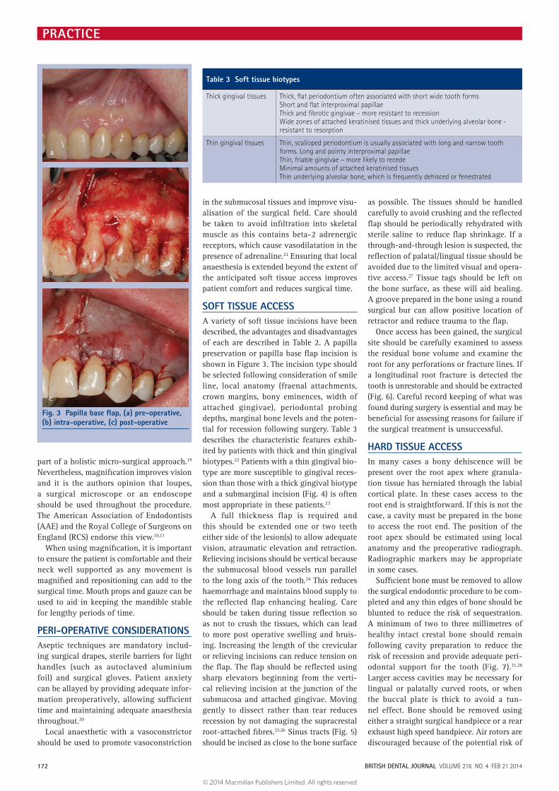
Papilla base flap Preserving the papilla when raising a flap reduces the risk of recession - Velvart P (2002) showed excellent healing with this technique with minimal recession and most sites resulting in no or minimal scarring
The papilla preservation flap requires two incisions: one at 90° to the outer contour of the marginal gingivae to a depth of 1.5 mm and the second is angulated apically towards the crestal bone margin which avoids creating a thin wedge of gin-givae (prevent necrosis and scar formation)
Fig. 2 Large lesions involving numerous teeth (a: pre-operative), completion of non-surgical endodontics (b: immediately post obturation) healing post non-surgical endodontic treatment (c: one -year follow-up)
a b c
BRITISH DENTAL JOURNAL VOLUME 216 NO. 4 FEB 21 2014 171
© 2014 Macmillan Publishers Limited. All rights reserved
PRACTICE
part of a holistic micro-surgical approach.19 Nevertheless, magnification improves vision and it is the authors opinion that loupes, a surgical microscope or an endoscope should be used throughout the procedure. The American Association of Endodontists (AAE) and the Royal College of Surgeons on England (RCS) endorse this view.10,11
When using magnification, it is important to ensure the patient is comfortable and their neck well supported as any movement is magnified and repositioning can add to the surgical time. Mouth props and gauze can be used to aid in keeping the mandible stable for lengthy periods of time.
PERI-OPERATIVE CONSIDERATIONSAseptic techniques are mandatory includ-ing surgical drapes, sterile barriers for light handles (such as autoclaved aluminium foil) and surgical gloves. Patient anxiety can be allayed by providing adequate infor-mation preoperatively, allowing sufficient time and maintaining adequate anaesthesia throughout.20
Local anaesthetic with a vasoconstrictor should be used to promote vasoconstriction
in the submucosal tissues and improve visu-alisation of the surgical field. Care should be taken to avoid infiltration into skeletal muscle as this contains beta-2 adrenergic receptors, which cause vasodilatation in the presence of adrenaline.21 Ensuring that local anaesthesia is extended beyond the extent of the anticipated soft tissue access improves patient comfort and reduces surgical time.
SOFT TISSUE ACCESSA variety of soft tissue incisions have been described, the advantages and disadvantages of each are described in Table 2. A papilla preservation or papilla base flap incision is shown in Figure 3. The incision type should be selected following consideration of smile line, local anatomy (fraenal attachments, crown margins, bony eminences, width of attached gingivae), periodontal probing depths, marginal bone levels and the poten-tial for recession following surgery. Table 3 describes the characteristic features exhib-ited by patients with thick and thin gingival biotypes.22 Patients with a thin gingival bio-type are more susceptible to gingival reces-sion than those with a thick gingival biotype and a submarginal incision (Fig. 4) is often most appropriate in these patients.23
A full thickness flap is required and this should be extended one or two teeth either side of the lesion(s) to allow adequate vision, atraumatic elevation and retraction. Relieving incisions should be vertical because the submucosal blood vessels run parallel to the long axis of the tooth.24 This reduces haemorrhage and maintains blood supply to the reflected flap enhancing healing. Care should be taken during tissue reflection so as not to crush the tissues, which can lead to more post operative swelling and bruis-ing. Increasing the length of the crevicular or relieving incisions can reduce tension on the flap. The flap should be reflected using sharp elevators beginning from the verti-cal relieving incision at the junction of the submucosa and attached gingivae. Moving gently to dissect rather than tear reduces recession by not damaging the supracrestal root-attached fibres.25,26 Sinus tracts (Fig. 5) should be incised as close to the bone surface
as possible. The tissues should be handled carefully to avoid crushing and the reflected flap should be periodically rehydrated with sterile saline to reduce flap shrinkage. If a through-and-through lesion is suspected, the reflection of palatal/lingual tissue should be avoided due to the limited visual and opera-tive access.27 Tissue tags should be left on the bone surface, as these will aid healing. A groove prepared in the bone using a round surgical bur can allow positive location of retractor and reduce trauma to the flap.
Once access has been gained, the surgical site should be carefully examined to assess the residual bone volume and examine the root for any perforations or fracture lines. If a longitudinal root fracture is detected the tooth is unrestorable and should be extracted (Fig. 6). Careful record keeping of what was found during surgery is essential and may be beneficial for assessing reasons for failure if the surgical treatment is unsuccessful.
HARD TISSUE ACCESSIn many cases a bony dehiscence will be present over the root apex where granula-tion tissue has herniated through the labial cortical plate. In these cases access to the root end is straightforward. If this is not the case, a cavity must be prepared in the bone to access the root end. The position of the root apex should be estimated using local anatomy and the preoperative radiograph. Radiographic markers may be appropriate in some cases.
Sufficient bone must be removed to allow the surgical endodontic procedure to be com-pleted and any thin edges of bone should be blunted to reduce the risk of sequestration. A minimum of two to three millimetres of healthy intact crestal bone should remain following cavity preparation to reduce the risk of recession and provide adequate peri-odontal support for the tooth (Fig. 7).21,28 Larger access cavities may be necessary for lingual or palatally curved roots, or when the buccal plate is thick to avoid a tun-nel effect. Bone should be removed using either a straight surgical handpiece or a rear exhaust high speed handpiece. Air rotors are discouraged because of the potential risk of
Fig. 3 Papilla base flap, (a) pre-operative, (b) intra-operative, (c) post-operative
a
b
c
Table 3 Soft tissue biotypes
Thick gingival tissues Thick, flat periodontium often associated with short wide tooth formsShort and flat interproximal papillaeThick and fibrotic gingivae – more resistant to recessionWide zones of attached keratinised tissues and thick underlying alveolar bone - resistant to resorption
Thin gingival tissues Thin, scalloped periodontium is usually associated with long and narrow tooth forms. Long and pointy interproximal papillaeThin, friable gingivae – more likely to recedeMinimal amounts of attached keratinised tissuesThin underlying alveolar bone, which is frequently dehisced or fenestrated
172 BRITISH DENTAL JOURNAL VOLUME 216 NO. 4 FEB 21 2014
© 2014 Macmillan Publishers Limited. All rights reserved

PRACTICE
air embolism.29 Burs should be used with light pressure and adequate irrigation to reduce heat generation as this may lead to necrosis of bone.30
Once access to the root end has been achieved, the granulation tissue within the
bony crypt should be removed with sharp curettes. Speedy removal of granulation tissue will reduce haemorrhage and assist vision. It is advisable to send the soft tis-sue removed for histopathological analysis. Sometimes there is a need to remove the root tip to gain access to the soft tissue mass or to remove the buccal root tip to gain access to the palatal root. If there is a natural fen-estration of the buccal cortical plate because of root anatomy, apical root end prepara-tion beyond this area can allow remodel-ling of the bone over the root (for example in the case of the buccal roots of upper first premolars).
MANAGEMENT OF THE ROOT END
Root end resectionThe apical 3 mm of the root is usually removed because 75% of teeth have canal abnormalities in the apical 3 mm of the root,31,32 canal ramifications are removed 98% of the time and accessory canals are eliminated 93% of the time.33 However, a greater length of root may be removed if there are anatomical variations, separated instruments, transposed canal apices or where access to a second, more palatally or lingually placed root is necessary. It could be argued that root end resection is unnec-essary when the cause of non-healing is extraradicular, however it is only possible to determine this histologically therefore root end resection is always recommended. Methylene blue (1%) can be used to stain the periodontal ligament and apical anatomy to improve visualisation.
The root tip should be resected perpen-dicular to the long axis of the tooth giv-ing a zero degree bevel. This allows for a 90 degree cavosurface margin for the root end filling and ensures that lingual anatomy is accessed. Bevelling of the root tip is not recommended, as this exposes more dentine tubules, which allow the passage of residual intra-radicular microorganisms and extra-radicular nutrients.30 If the tooth has under-gone previous apical surgery, it may not be necessary or possible to remove 3 mm of the root end and still maintain a favour-able crown to root ratio. Equally it may not always be possible to resect 3 mm of the root end beyond a post and still maintain ade-quate space for a root end filling. Following resection the root end should be smoothed as this allows better assessment of ramifica-tions and infractions.
If the tooth has been obturated with a sil-ver point, care should be taken to resect the root around the silver point, without cut-ting the silver point. This will simplify sub-sequent removal of the silver point.
CRYPT MANAGEMENTFollowing resection of the root apex and curettage of soft tissue the bony crypt should be kept clean and dry to allow visu-alisation and management of the root end. Adequate haemostasis minimises surgical time, blood loss and post-operative haem-orrhage and swelling.26 A local anaesthetic containing a vasoconstrictor should be used to promote haemostasis. However, when this is insufficient, a variety of adjunctive options are available. These are summarised in Table 4. Cautery/electrosurgery is not recommended because it can delay heal-ing and cause necrosis, depending on the temperature and duration of application. Curetting the cavity and the bony walls to remove the remainder of any adhesive haemostatic agent used and causing bleed-ing of the bone, following placement of a root end filling, creates a blood clot which is the ideal for healing.34 An animal study assessed clinical and histological status of bone adjacent to bone wax, ferric sulphate (left for five minutess and left perma-nently), aluminium chloride (left for two minutes) and a combination of ferric sul-phate (five minutes) and aluminium chlo-ride (two minutess). The findings indicate a varying degree of inflammation occurs adjacent to all of the materials at 12 weeks post surgery.34 Choice of local haemostatic agents is a matter of operator preference. Biocompatibility, ease of use, efficacy, and ease of removal are likely to be the most important factors. The authors’ preference is to use three cotton wool pledgets soaked
Fig. 4 Submarginal flap. (a) Three days post-operatively at suture removal following surgical endodontics on the upper right central incisor. (b) One week review post surgical endodontics on the upper left central incisor
a
b
Fig. 5 Presence of a sinus tract (arrow)
Fig. 7 Maintain at least 2-3 mm of crestal bone to ensure adequate periodontal support for the tooth
Fig. 6 Root fracture seen following exploratory surgery with a view to surgical endodontics
BRITISH DENTAL JOURNAL VOLUME 216 NO. 4 FEB 21 2014 173
© 2014 Macmillan Publishers Limited. All rights reserved

PRACTICE
in vasoconstrictor-containing local anaes-thetic, applied with pressure one on top of the other and held with finger pressure for two minutes. One or two of the superfi-cial cotton wool pledgets are then removed leaving the last one undisturbed until after root end filling. This is often adequate for timely completion of the procedure (Fig. 8).
ROOT END PREPARATIONFollowing root end resection the apical 3 mm of the root canal system should be pre-pared to facilitate an adequate apical seal.35 The preparation must follow the anatomical canal space and develop adequate retention form. If there are two canals in a root, at the 4 mm level there will be a partial or complete isthmus 100% of the time.36,37 When there are two root canals within a root the isthmus between the canals should also be prepared to a depth of 3 mm as there are likely to be communications in this area.36,37 Therefore, a minimum of 6 mm of root length apical to a post is required for satisfactory root resec-tion and cavity preparation.
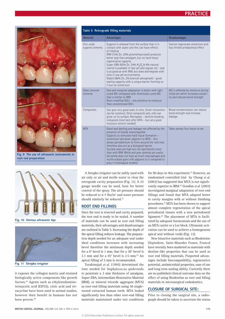
Ultrasonic instruments are preferred for root end cavity preparation because they are small, easy to manoeuvre and allow deeper prepara-tion of the root end than a round bur (Fig. 9). They permit cavity preparation perpendicular to the resected toot end. Ultrasonic tips are available in a variety of sizes and angulations (Fig. 10). A number of studies have shown higher success with ultrasonic preparation compared to preparation with burs.38–40 Some have suggested that the use of ultrasonics causes cracks within the root surface, how-ever studies on cadavers showed no increase in cracks with the use of ultrasonics as the periodontal ligament is thought to absorb the stresses.41 Ultrasonic tips can be used without irrigation to improve visualisation. However, when irrigant is not used, the authors recom-mend that the tips be used intermittently on a low power setting to prevent the risk of over-heating and extend the lifetime of these tips.
ROOT END CONDITIONINGNo evidence exists to support the asser-tion that smooth root ends promote better
healing however a smooth surface allows better assessment of ramifications and cracks. Burs that produce a smooth end usually produce less vibration and may be more comfortable for the patient. Removing the smear layer by conditioning provides a surface that may be more conducive to mechanical adhesion as well as the cellular mechanisms for growth and attachment as
Table 4 Haemostatic agents
Haemostatic agent Action
Collagen-based products
Stimulate platelet adhesion and aggregation
Surgicel - regenerated cellulose
Acts like a coagulum plug - sticky when in contact with blood. No enhanced effect on clotting cascadeRemains in wound with little evidence of resorption at 120 days. pH of 3 - can retard healing
Gelform Gelatine-based sponge, water insoluble, resorbableStimulates intrinsic clotting pathway
Bone wax Plugs all vascular openings – no effect on clotting mechanisms88% beeswax & 12% isopropyl palmitate – non-absorbableRetards bone healing, initiates a foreign body reaction, inflammation and increases risk of infectionMust be removed after root end filling and before closure of the surgical site
Ferric sulphate Good haemostasis via chemical reaction with the bloodConcentration of 35-72% - necrotising agent with low pHMust remove coagulum to avoid foreign body reaction and negative effects on osseous healingCan cause acute inflammation and necrosis of the surrounding soft tissuesCan be cytotoxic (although it is not absorbed systemically as the coagulum formed blocks the vasculature)
Calcium sulphate Plaster of Paris – biologically compatible, inexpensive, sterilisable (30 seconds in microwave on high setting or in oven for 1 hour at 200 °F), sets fast, easy to remove after useActs like a physical barrier, does not affect healing or cementum formationPlace in bony crypt, allow setting, partly carving away excess to get access to root tipAfter apicectomy the calcium sulphate can be removed or left in situ
Thrombin Dry powder formed from bovine prothrombinDirect effect on fibrinogenExpensive and difficult to handle and deliver to a wet site
Adrenaline impreg-nated pellets
Effect is topical - contains 0.45-0.55 mg racaemic adrenalineSimple and easily availableRemove granulation tissue, apply pellet and pack with sterile cotton wool pellets. Place pressure for 2-4 minutes. Remove the cotton pellets but not the adrenaline impregnated pelletRetention of cotton fibres in the surgical site can impair healing by causing inflam-mation and foreign body reaction
‘Touch and heat’ At highest setting to cauterise blood vessels
Fig. 8 Haemostasis with cotton wool soaked in adrenaline: (a) wet crypt, (b) one remaining cotton wool pledget, (c) removal of cotton wool post placement of apical seal (MTA)
a
b
c
174 BRITISH DENTAL JOURNAL VOLUME 216 NO. 4 FEB 21 2014
© 2014 Macmillan Publishers Limited. All rights reserved
PRACTICE
it exposes the collagen matrix and retained biologically active components like growth factors.42 Agents such as ethylenediamine-tetraacetic acid (EDTA), citric acid and tet-racycline have been used in animal studies, however their benefit in humans has not been proven.30
A Stropko irrigator can be safely used with air only or air and sterile water to clear the retrograde cavity preparation (Fig. 11). A 25 gauge needle can be used, bent for better control of the spray. The air pressure should be reduced to 4-7 lbs/in2 and water pressure should similarly be reduced.21
ROOT END FILLINGSOnce the root is resected and cavity prepared, the root end is ready to be sealed. A number of materials can be used as root end filling materials, their advantages and disadvantages are outlined in Table 5. Increasing the depth of the apical filling reduces leakage. The prepara-tion depth needed for an adequate seal under ideal conditions increases with increasing bevel therefore the minimum depth needed for a 0° bevel is 1 mm, that for a 30° bevel is 2.1 mm and for a 45° bevel is 2.5 mm.43 An apical filling of 3 mm is recommended.
Torabinejad et al. (1995) determined the time needed for Staphylococcus epidermidis to penetrate a 3 mm thickness of amalgam, Super-EBA, Intermediate Restorative Material (IRM), or mineral trioxide aggregate (MTA) as root-end filling materials using 56 single-rooted extracted human teeth. MTA leaked significantly less than other root-end filling materials maintained under test conditions
for 90 days in this experiment.35 However, an randomised controlled trial by Chong et al. (2003) has suggested that MTA is not signifi-cantly superior to IRM.44 Gondim et al. (2003) investigated marginal adaptation of root end fillings and found that MTA adapted better to cavity margins with or without finishing procedures.45 MTA has been shown to support almost complete regeneration of the apical periodontal tissues with a new periodontal ligament.46 The placement of MTA is facili-tated by adequate haemostasis and the use of an MTA carrier or a Lee block. Ultrasonic acti-vation can be used to achieve a homogenous apical seal without voids (Fig. 12).
New bioactive materials such as Biodentine (Septodont, Saint-Maurdes Fosses, France) have recently been marketed as materials with dentine-like properties that can be used as root end filling materials. Purported advan-tages include biocompatibility, regenerative potential, antimicrobial properties, ease of use and long term sealing ability. Currently there are no published clinical outcome data on the effect of using Biodentine as root end filling materials in microsurgical endodontics.
CLOSURE OF SURGICAL SITE:Prior to closing the surgical site, a radio-graph should be taken to ascertain the status
Fig. 9 The use of ultrasonic instruments in root end preparation
a
b
Fig. 10 Various ultrasonic tips
Fig. 11 Stropko irrigator
Table 5 Retrograde filling materials
Material Advantages Disadvantages
Zinc oxide eugenol cements
Eugenol is released from the surface that is in contact with water and this can have effects on healingIRM (75% Zn, 20% polymethacrylate) produces better seal than amalgam, but no hard tissue regenerative capacitySuper-EBA (60% Zn, 34% Al2O3 & 6% natural resins) is available in fast set and regular set - seal is as good as with IRM, but does disintegrate with time in low pH environmentsDiaket (98% Zn, 2% bismuth phosphate) - good sealing capacity with a unique barrier forming on it but no cementum
Cannot regenerate cementum and has limited antibacterial effect
Glass ionomer cements
Seal and marginal adaptation is better with light cured GIC compared with chemically cured GICSeal is similar to IRMResin modified GICs – less sensitive to moisture than conventional GICs
GIC is affected by moisture during initial set which increases solubil-ity and reduces bond strength
Composites Can give very good seals in vitro. Unset monomers can be cytotoxic. Once composite sets, cells can grow on its surface. Retroplast – dentine bonding composite (next best after MTA – but very good moisture control needed).
Blood contamination can reduce bond strength and increase leakage.
MTA Good seal (setting and leakage not affected by the presence of blood), biocompatibleCapacity to stimulate hard tissue formation – cementum laid down adjacent to MTA – this allows periodontium to form around the root end, therefore also act as a biological barrierSuccess rates are high but not significantly more than with IRM. White and grey varieties are availa-ble (white does not have as much macrophages and multinucleate giant cells adjacent to it compared to grey, in histological studies)
Takes almost four hours to set
BRITISH DENTAL JOURNAL VOLUME 216 NO. 4 FEB 21 2014 175
© 2014 Macmillan Publishers Limited. All rights reserved

PRACTICE
of the root end filling and ensure all for-eign objects have been removed. The crypt should be thoroughly irrigated with saline to remove any haemostatic agents and packing materials. The crypt should then be scraped with a sharp curette to encouraging bleeding and the formation of a blood clot.34 Bony crypts are usually filled with blood clots and natural bony infill, to varying degrees, occurs. Some have suggested the use of guided tissue regeneration for preventing soft tissue growth into the bony crypt by using a membrane for selective repopula-tion. This has mainly been for cases where there is a through-and-through perforation of the bone or where apical migration of the epithelium is a potential problem.47 No additional beneficial effect on healing is seen where there is a residual buccal cortical plate over the remainder of the root.48
The flap should be carefully replaced and sutured without tension as tension may lead to necrosis at the incision site with subse-quent scarring or recession.21 Small diameter sutures (5/0 or smaller) are recommended as they have smaller needles, cause less trauma and lead to thread breaking rather than tis-sue tearing.49 Non-resorbable monofilament sutures are recommended, as they are less supportive of bacterial growth.50 Gentle compression of the flap for one-minute post closure ensures fibrin adhesion and may prevent haematoma development.30 Post-operative antibiotics are not routinely prescribed unless surgery has been extraor-dinarily long or the patient is immunocom-promised.11 Removal of sutures at three days post operatively is recommended as epithe-lial bridging and collagen crosslinking is thought to happen within 21-28 hours.25
POST-OPERATIVE CONSIDERATIONSAs with most oral surgical procedures, the patient should be advised to take simple analgesia, reduce the swelling and prevent further swelling by using ice packs for 24 to 48 hours, maintain good oral hygiene and use chlorhexidine gluconate (0.12%) mouth-wash twice daily for a minimum of three days post operatively and warm saltwater mouth washes 4-5 times daily for seven days.
REVIEW AND ASSESSING SUCCESS OF SURGICAL ENDODONTIC TREATMENTSutures should be removed at three days post surgery and if histopathological results are available the patient should be informed of the findings. If healing is uneventful at this stage, the patient can be reassessed at one-year following surgical treatment.51 Clinical and radiographic examination is necessary to determine the extent of healing. If the
patient returns with signs of non-healing and frank infection, the cause of failure needs to be established. Repeated treatment without diagnosis of the cause of failure is unlikely to resolve the condition.
The options for failed surgical endodontic treatment are to consider non-surgical endo-dontic re-treatment, surgical re-treatment or extraction (± prosthetic replacement). The outcome of surgical re-treatment is lower with 36% being successful, 26% having uncertain healing and 38% having failed at one-year.52 Gagliani et al. (2005) have shown higher heal-ing rates for re-surgery with 59% being suc-cessful, 17% having uncertain healing and 23% having failed at one to five years.53
CONCLUSIONNon-surgical endodontic retreatment is the treatment of choice for endodontically
treated teeth with recurrent or residual dis-ease, however, surgical endodontic treatment is appropriate in selected cases. Clinicians require a thorough understanding of this treatment procedure and should appreciate the importance and rationale of the different stages detailed above. Optimal outcomes for surgical endodontic treatment can only be achieved if the diagnosis is accurate, appro-priate cases are selected and the procedure is completed to a high standard.
1. Nair P N. On the causes of persistent apical peri-odontitis: a review. Int Endod J 2006; 39: 249–281.
2. Torabinejad M, Corr R, Handysides R et al. Outcomes of non-surgical retreatment and endodontic surgery: a systematic review. J Endod 2009; 35: 930–937.
3. Ng YL, Mann V, Gulabivala K. Outcome of secondary root canal treatment: a systematic review of the literature. Int Endod J 2008; 41: 1026–1046.
4. Ng YL, Mann V, Gulabivala K. A prospective study of the factors affecting outcomes of non-surgical root canal treatment: part 1: periapical health. Int Endod J 2011a; 44: 583–609.
5. Ng YL, Mann V, Gulabivala K. Tooth survival follow-ing non-surgical root canal treatment: a systematic review of the literature. Int Endod J 2010; 43: 171–189.
6. Ng YL, Mann V, Gulabivala K. A prospective study of the factors affecting outcomes of non-surgical root canal treatment: part 2: Tooth survival. Int Endod J 2011b; 44: 610–625.
7. Del Fabbro M, Taschieri S, Testori T, Francetti L, Weinstein R L. Surgical vs non-surgical endodontic re-treatment for periradicular lesions. Cochrane Database Syst Rev 2007; 18: CD005511.
8. Setzer F C, Shah S B, Kohli M R, Karabucak B, Kim S. Outcome of endodontic surgery: a meta-analysis of the literature part 1: Comparison of traditional root-end surgery and endodontic microsurgery. J Endod 2010; 36: 1757–1765.
9. von Arx T, Penarrocha M, Jensen S. Prognostic fac-tors in apical surgery with root-end filling: a meta analysis. J Endod 2010; 36: 957–973.
10. American Association of Endodontists. Endodontics Colleagues for Excellence: Contemporary endodontic microsurgery: procedural advancements and treatment planning considerations. 2010. Available at: www.aae.org/uploadedfiles/publications_and_research/endodontics_colleagues_for_excel-lence_newsletter/ecfefall2010final.pdf (accessed 15 November 2013).
11. Evans G E, Bishop K, Renton T. Royal College of Surgeons guidelines for surgical endodontics. 2012.
12. Haynes, R B, Devereaux P, Guyatt G H. Clinical Expertise in the Era of Evidence Based Medicine and Patient Choice. ACP Journal Club 2002; 136: A11A14.
13. Abbott P V. Incidence of root fractures and methods used for post removal. Int Endod J 2002; 35: 63–67.
14. Roszkowski M T, Swift J Q, Hargreaves K M. Effects of NSAID administration on tissue levels of Immunoreactive prostaglandin E2, leukotriene B4 and (s)-flurbiprofen following extraction of impacted third molars. Pain 1997; 73: 339–346.
15. Ong C K S, Seymour R A, Lirk P, Merry A F. Combining paracetamol (acetaminophen) with non-steroidal anti-inflammatory drugs: a qualita-tive systematic review of analgesic efficacy for acute postoperative pain. Anesth Analg 2010; 110: 1170–1179.
16. Balbuena L, Stambaugh K I, Ramirez S G, Yeager C. Effects of topical oral antiseptic rinses on bacte-rial counts of saliva in healthy human subjects. Otolaryngol Head Neck Surg 1998; 118: 625–629.
17. Longman L P, Preston A J, Martin M V, Wilson N H F. Endodontics in the adult patient: the role of antibi-otics. J Dent 2000; 28: 539–548.
18. Tsesis I, Faivishevsky V, Kfir A et al. Outcome of sur-gical endodontic treatment performed by a modern technique: a meta-analysis of literature. J Endod 2009; 35: 1505–1511.
Fig. 12 Homogenous MTA apical seal. (a) Pre-operative radiograph 12. (b) Post-operative radiograph of 12
a
b
176 BRITISH DENTAL JOURNAL VOLUME 216 NO. 4 FEB 21 2014
© 2014 Macmillan Publishers Limited. All rights reserved

PRACTICE
19. Del Fabbro M, Taschieri S. Endodontic therapy using magnification devices: a systematic review. J Dent 2010; 38: 269–275.
20. Weiner R N, Forgione A G, Weiner L K. Survey examines patients’ fear of dental anxiety treatment. J Mass Dent Soc 1998; 47: 16–21, 36.
21. Stropko J J. Vol III Chapter 34 Micro-surgical endo-dontics. In Castellucci A. Endodontics. pp 1076–1145. Florence, Italy, 2009.
22. Sclar AG. Strategies for management of single-tooth extraction sites in aesthetic implant therapy. J Oral Maxillofac Surg 2004; 62: 90–105.
23. von Arx T, Salvi G E, Janner S, Jensen S S. Gingival recession following apical surgery in the aesthetic zone: a clinical study with 70 cases. Eur J Esthet Dent. 2009; 4: 28–45.
24. Macphee T C, Cowley G. Essentials of periodontol-ogy and periodontics, 3rd ed. Oxford: Blackwell Scientific, 1981.
25. Harrison J W, Jurosky K A. Wound healing in the tissues of the periodontium following periradicular surgery. I. The incisional wound. J Endod 1991; 12: 425.
26. Gutmann J L, Harrison J W. Surgical endodontics. St Louis: Ishiyaku EuroAmerica, 1994.
27. Ingle J I, Bakland L K. Endodontics, 5th ed. London: BC Decker Inc, 2002.
28. Song M, Kim S G, Shin S-J, Kim H-C, Kim E. The influence of bone tissue deficiency on the outcome of endodontic microsurgery: a prospective study. J Endod 2013; 39: 1341–1345.
29. McKenzie W S, Rosenberg M. Iatrogenic subcutane-ous emphysema of dental and surgical origin: a literature review. J Oral Maxillofac Surg. 2009; 67: 1265–1268.
30. Cohen S, Hargreaves K M. Pathways of the pulp, 10th ed. Mosby Elsevier, 2011.
31. De Deus Q D. Frequency, location and direction of the lateral, secondary and accessory canals. J Endod 1975; 1: 361–366.
32. Seltzer S, Soltanoff W, Bender I B, Ziontz M. Biologic aspects of endodontics. Part 1: histological observa-tions of the anatomy and morphology of root apices and surroundings. Oral Surg Oral Med Oral Pathol
1996; 22: 375.33. Kim S, Pecora G, Rubenstein R, Dorscher-Kim
J. Colour atlas of microsurgery in endodontics. Philadelphia: WB Saunders, 2001.
34. von Arx T, Jensen S S, Hänni S, Schenk R K. Haemostatic agents used in periradicular surgery: an experimental study of their efficacy and tissue reactions. Int Endod J 2006; 39: 800–806.
35. Torabinejad M, Rastegar A F, Kettering J D, Pitt Ford T R. Bacterial leakage of mineral trioxide aggregate as a root-end filling material. J Endod 1995; 21: 109–112.
36. Teixeira F B, Sano C L, Gomez B P, Zara A A, Ferraz C C, Souza-Filho F J. A preliminary in vitro study of the incidence and position of the root canal isthmus in maxillary and mandibular first molars. Int Endod J 2003; 36: 276–280.
37. Weller R N, Niemczyk S P, Kim S. Incidence and position of the canal isthmus. Part 1: mesiobuccal root of the maxillary first molar. J Endod 1995; 21: 380–383.
38. Shearer J, McManners J. Comparison between the use of an ultrasonic tip and a microhead handpiece in periradicular surgery: a prospective randomised trial. Br J Oral Maxillofac Surg. 2009; 47: 386–388.
39. De Lange J, Putters T, Baas E M et al. Ultrasonic root-end preparation in apical surgery: a prospective randomised study. Oral Surg Oral Med Oral Pathol Oral Radiol Endod 2007; 104: 841–845.
40. Vallecillo Capilla M, Muñoz Soto E, Reyes Botella C, Prados Sáchez E, Olmedo Gaya M V. Periapical surgery of 29 teeth. A comparison of conventional technique, microsaw and ultrasound. Med Oral 2002; 7: 49–49, 50–53.
41. Kim S, Kratchman S. Modern endodontic surgery concepts and practice: a review. J Endod 2006; 32: 601–623.
42. Galler K M, D’Souza R N, Federlin M et al. Dentine conditioning codetermines cell fate in regenerative endodontics. J Endod 2011; 37: 1536–1541.
43. Gilheany P A, Figdor D, Tyas M J. Apical dentine permeability and microleakage associated with root end resection and retrograde filling. J Endod 1994;
20: 22-26.44. Chong B S, Pitt Ford T R, Hudson M B. A prospective
clinical study of MTA and IRM when used as a root end filling material in endodontic surgery. Int Endod J 2003; 36: 520–526.
45. Gondim E Jr, Zaia A A, Gomes B P F A, Ferraz C C, Teixeira F B, Souza-Filho F J. Investigation of the marginal adaptation of root-end filling materials in root-end cavities prepared with ultrasonic tips. Int Endod J 2003; 36: 491–499.
46. Regan J D, Gutmann J L, Witherspoon D E. Comparison of Diaket and MTA when used as root-end filling materials to support regeneration of the periradicular tissues. Int Endod J 2002; 35: 840–847.
47. Dietrich T, Zunker P, Diertich D, Bernimoulin J P. Periapical and periodontal healing after osseous grafting and guided tissue regeneration treatment of apicomarginal defects in periradicular surgery; results after 12 months. Oral Surg Oral Med Oral Pathol Oral Radiol Endod. 2003; 95: 474–482.
48. Garrett K, Kerr M, Hartwell G, O’Sullivan S, Mayer P. The effect of a bioresorbable matrix barrier in endodontic surgery on the rate of periapical healing: an in vivo study. J Endod 2002; 28: 503–506.
49. Burkhardt R, Preiss A, Joss A, Lang N P. Influence of suture tension to the tearing characteristics of the soft tissues: an in vitro experiment. Clin Oral Implants Res 2008; 19: 314–319.
50. Banche G, Roana J, Mandras N et al. Microbial adherence on various intraoral suture materials in patients undergoing dental surgery. J Oral Maxillofac Surg 2007; 65: 1503–1507.
51. European Society of Endodontology. Quality Guidelines for endodontic treatment: consensus report of the European Society of Endodontology. Int Endod J 2006; 39: 921–930.
52. Peterson J, Gutmann J L. The outcome of endodontic re-surgery: a systematic review. Int Endod J 2001; 34: 169–175.
53. Gagliani M M, Gorni, F G M, Strohmenger L. Periapical. Resurgery versus periapical surgery: a 5-year longitudinal comparison. Int Endod J 2005; 38: 320–327.
BRITISH DENTAL JOURNAL VOLUME 216 NO. 4 FEB 21 2014 177
© 2014 Macmillan Publishers Limited. All rights reserved