restoration of endodontically treated teeth: the seven ...€¦ · restoration of endodontically...
TRANSCRIPT
Restoration of endodontically treated teeth: The seven keys to success By Nadim Z. Baba, DMD, MSD Charles J. Goodacre, DDS, MSD Tony Daher, DDS, MSEd Featured in General Dentistry, November/December 2009 Pg. 596-603
Posted on Friday, November 06, 2009
Preservation of tooth integrity and strength is important for the long-term survival of endodontically treated teeth. Endodontic treatment and post space preparation requires reduction of the remaining supportive tooth structure. Restorative modalities following root canal therapy must provide sufficient strength for the prosthetic material and tooth structures. This article presents seven key factors that should be taken into consideration to ensure clinical success when restoring an endodontically treated tooth. Received: May 4, 2009 Accepted: June 8, 2009 Several factors play a role in the long-term survival of endodontically treated teeth and associated restorations. This article presents seven key factors that affect tooth and restoration survival. Fiber-reinforced resin posts should be used with caution until more long-term data are available For many years, the standard method for restoring endodontically treated teeth has involved either a custom cast post
prefabricated metal post with a restorative material core.1-3 A nationwide survey of dentists in 1994 reported that 40% of general practitioners
used prefabricated posts, the most popular being the parallel-sided serrated metal post.4 It is likely that prefabricated post usage has increased substantially since that 1994 survey. The high demand for esthetic restorations and all-ceramic crowns has led to the development of a variety of nonmetallic prefabricated post
systems as alternatives to metal posts.5-8 In addition to the esthetic advantages of nonmetallic posts, laboratory studies have shown that
these posts offer favorable physical and mechanical properties and less root fracture compared to metal posts.9-13
fiber-reinforced posts have produced a wide range of reported failure percentages, ranging from 0% (after a mean period of 2.3 years) to
11.4% (after two years).14-23 The most commonly reported complications are post loosening and root fracture (Fig. 1 and 2).
core and the final restoration both depend on the retentive capacity of the post.27
Page 1 of 13AGD - Academy of General Dentistry
1/26/2011http://proxy.library.upenn.edu:4752/publications/articles/?ArtID=6504

Given the wide range of reported failure percentages, it appears that more long-term clinical data are needed to determine the efficacy of fiber-reinforced posts. Crowns should be placed on most endodontically treated posterior teeth to enhance their longevity Clinicians have observed a difference between endodontically treated teeth and vital teeth. Endodontically treated teeth fracture more often
than vital teeth. They tend to break during extraction; in addition, pulpless molars without crowns can fracture.28,29
Multiple studies have shown that endodontically treated teeth benefit from the placement of crowns. Aquilino and Caplan reported that
endodontically treated teeth with crowns had a survival rate six times greater than that of teeth without crowns (Fig. 3).evaluated 116 teeth that had failed and were extracted; the authors reported that endodontically treated teeth without crowns were lost after
an average of 50 months, while endodontically treated teeth with crowns were lost after an average of 87 months.
Page 2 of 13AGD - Academy of General Dentistry
1/26/2011http://proxy.library.upenn.edu:4752/publications/articles/?ArtID=6504

According to several clinical studies, fixed partial dentures have increased clinical failure when they are supported by endodontically treated
abutment teeth rather than vital abutment teeth.28,32-35 One study determined that crowns significantly improved the success of endodontically treated posterior teeth but did not improve the success of anterior teeth, indicating that intact endodontically treated anterior teeth do not need complete crown coverage unless they are weakened by large and/or multiple coronal restorations or they require significant changes to their
color or form.36
Conversely, Mannocci et al evaluated endodontically treated premolars that had been restored (both with and without complete coverage) by
either a post or direct composite resin restorations and reported similar success rates for both.37 A similar retrospective cohort study by Nagasiri and Chitmongkolsuk indicated that endodontically treated molars that are intact (except for the access opening) could be restored
successfully using composite resin restorations.38
After considering the available data, the authors recognize the potential benefits of using composite resin to restore posterior teeth that are intact except for the access opening. However, since wear is an indicator of the forces that will be brought to bear on the teeth, more clinical data are needed to determine the long-term success of these teeth when varying degrees of occlusal wear are present in the mouth. The authors recommend using crowns that encompass the cusps because they will help cusps that have been weakened by previous tooth structure removal to withstand the occlusal forces of everyday mastication. Conversely, it may be possible to avoid placing crowns on some previously restored posterior teeth, such as mandibular first premolars with small, poorly developed lingual cusps that would not be subjected to the wedging effect from opposing cusps. With these first premolars, there is little chance that occlusal forces will separate the cusps, so the access opening can be restored without the need for a coronal
coverage crown.39
Posts weaken endodontically treated teeth rather than enhance their clinical longevity Historically, the use of posts has been based on the concept that they reinforce teeth; however, nearly every laboratory study has reported that posts either fail to increase the fracture resistance of extracted endodontically treated teeth or that they decrease the fracture resistance
of the tooth when force is applied via a mechanical testing machine.40-50 Pontius and Hutter reported that maxillary incisors without posts
resisted higher failure loads than those with posts and crowns.42 Gluskin et al found that mandibular incisors with intact natural crowns
exhibited greater resistance to transverse loads than teeth with posts and cores.43 These studies showed no evidence that posts strengthen or reinforce teeth (Fig. 4).
Page 3 of 13AGD - Academy of General Dentistry
1/26/2011http://proxy.library.upenn.edu:4752/publications/articles/?ArtID=6504

Clinical studies also have failed to provide definitive support for the concept that posts strengthen endodontically treated teeth.longitudinal radiographic study, Eckerbom et al evaluated the radiographs of 200 consecutively treated patients several years after
endodontic treatment and reported that teeth with posts had significantly more apical periodontitis.29 In a 2003 analysis of data from multiple
clinical studies, Goodacre et al noted fractures in 3% of teeth with posts, with no evidence that posts enhanced the survival of teeth.have had little effect on the clinical success of fixed partial denture abutments but they have been reported to improve the clinical success of
removable partial denture abutments compared to endodontically treated abutments that did not use posts.36
Clinical and laboratory data indicate that teeth are not strengthened by posts; rather, their purpose is to retain a core that will provide adequate support for the definitive crown or prosthesis. Unfortunately, this primary purpose has not been completely recognized. A 1995 survey showed that 24% of general dentists felt that posts strengthen teeth.53 A year earlier, Morgano et al reported that 62% of dentists over
age 50 believed that a post reinforces the tooth, compared to only 41% of dentists under age 41.4 Thirty-nine percent of part
of full-time faculty, and 56% of non-faculty practitioners felt that posts reinforce teeth.4 In a survey conducted in Sweden, Eckerbom et al
found that 29% of general dental practitioners felt that a post reinforced the tooth, compared to 17% of board-certified prosthodontists. Since posts do not appear to reinforce teeth, they should be used only when the core cannot be retained by any other means. To ensure an adequate apical seal, 5 mm of gutta-percha should be retained After an endodontically treated tooth is prepared for a post, the remaining gutta-percha at the apex is the only barrier against bacteria passing into the periapical area. Several studies showed that leakage increased when only 2–3 mm of gutta-percha was present; however, preserving
4–5 mm of gutta-percha ensures an adequate seal.54-60 Although multiple studies indicate that 4 mm produces an adequate seal, stopping at precisely 4 mm is difficult and radiographic variations in angulation could lead to retention of less than 4 mm. With that in mind, 5 mm appears to be a safer minimal radiographic length. The best method for preserving the apical seal during preparation of a post space is to use the working length determined during endodontic treatment; the same reference point used on the tooth during endodontic therapy should be used during post preparation. In addition, a canal preparation instrument with an appropriate diameter should be used with a rubber stopper placed around the instrument at the proper location to help ensure that an adequate amount of gutta-percha is retained apically. Three methods have been advocated for removing gutta-percha when preparing a post space without disturbing the apical seal: chemical,
thermal, and mechanical.54,57,61-64 According to the literature, both hot hand instruments and rotary instruments can be used to safely remove
adequately condensed gutta-percha, provided 5 mm is retained apically.54,57,62-64 Several studies have determined that removing gutta
percha immediately after root canal treatment has no detrimental effect on the apical seal.55,56,58,61,65
Short posts should be avoided The appropriate length for a post should minimize the potential for damage to the tooth, optimize post retention, and maintain an appropriate
apical seal for the root canal restoration. Several guidelines for determining the length have been proposed.2,66-69
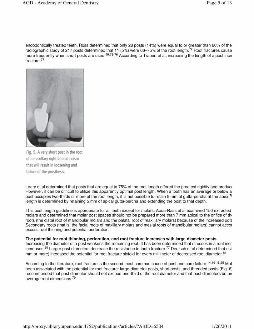
While short posts have never been advocated, they have been observed frequently on radiographs (Fig. 5). A 1993 study by Grieve and
McAndrew examined 327 posts and found that only 111 (34%) were as long as the incisocervical length of the crown.
Page 4 of 13AGD - Academy of General Dentistry
1/26/2011http://proxy.library.upenn.edu:4752/publications/articles/?ArtID=6504
endodontically treated teeth, Ross determined that only 28 posts (14%) were equal to or greater than 66% of the root length.
radiographic study of 217 posts determined that 11 (5%) were 66–75% of the root length.72 Root fractures caused by high stresses occur
more frequently when short posts are used.49,73-76 According to Trabert et al, increasing the length of a post increases the resistance to root
fracture.77
Leary et al determined that posts that are equal to 75% of the root length offered the greatest rigidity and produced the least root deflection.However, it can be difficult to utilize this apparently optimal post length. When a tooth has an average or below average root length and the
post occupies two-thirds or more of the root length, it is not possible to retain 5 mm of gutta-percha at the apex.78
length is determined by retaining 5 mm of apical gutta-percha and extending the post to that depth. This post length guideline is appropriate for all teeth except for molars. Abou-Rass et al examined 150 extracted maxillary and mandibular molars and determined that molar post spaces should not be prepared more than 7 mm apical to the orifice of the root canal in the primary
roots (the distal root of mandibular molars and the palatal root of maxillary molars) because of the increased potential for root perforation.Secondary roots (that is, the facial roots of maxillary molars and mesial roots of mandibular molars) cannot accommodate 7 mm posts without excess root thinning and potential perforation. The potential for root thinning, perforation, and root fracture increases with large-diameter posts Increasing the diameter of a post weakens the remaining root. It has been determined that stresses in a root increase as the post diameter
increases.80 Larger post diameters decrease the resistance to tooth fracture.77 Deutsch et al determined that using large
mm or more) increased the potential for root fracture sixfold for every millimeter of decreased root diameter.81
According to the literature, root fracture is the second most common cause of post and core failure.14,16-18,25 Multiple types of posts have
been associated with the potential for root fracture: large-diameter posts, short posts, and threaded posts (Fig. 6).recommended that post diameter should not exceed one-third of the root diameter and that post diameters be proportionally related to
average root dimensions.79
Page 5 of 13AGD - Academy of General Dentistry
1/26/2011http://proxy.library.upenn.edu:4752/publications/articles/?ArtID=6504

The post should be between 0.6–1.2 mm in diameter, depending on the tooth being restored.84-86 Only post preparation instruments that match the desired diameter of the post space should be used. When using a particular brand of post, make sure that the the drill and the post are made by the same manufacturer. Understanding dental anatomy, the configuration of the roots and their variations, and appropriate instrument angulations can help dentists to avoid root thinning and perforation. Instruments should be angled so that they follow the canal. Figure 7 is an example of a distal canal that was perforated because the instruments were at an improper angle when preparing the post space.
When posts are needed in premolars, it is best to place them in the palatal root of the maxillary premolar and in the straightest root of any mandibular premolar with multiple roots. Root taper, curvature, and depressions should be reviewed prior to post preparation. When posts are needed in molars, they should be placed in roots with the greatest dentin thickness. These roots (the palatal roots of
Page 6 of 13AGD - Academy of General Dentistry
1/26/2011http://proxy.library.upenn.edu:4752/publications/articles/?ArtID=6504

maxillary molars and the distal roots of mandibular molars) are known as the primary roots. However, it is important to remember that
extending a post more than 7 mm apical to the root canal orifice in primary canals increases the risk of perforation.mandibular molars and the facial roots of maxillary molars should be avoided, if possible. Dentists also should avoid placing instrument pressure on the root surface toward the furcation, as this surface is thinner than the outer surface due to root curvature.
For all teeth, the apical 5 mm of the roots should be avoided because most root curvatures occur within 5 mm of the root apex.into this area increases the risk of excessive root thinning or perforation. A cervical ferrule should engage tooth structure to prevent root fracture Ferrules can be established by the core engaging tooth structure (known as the core ferrule) or by the overlying encompassing sound tooth
structure (known as the crown ferrule).88-97 The data indicate that crown ferrules are more effective than core ferrules and that they increase
the tooth’s resistance to fracture.89,91-93,98 Although the data indicate the benefit of a crown ferrule, not all practitioners recognize its value; according to a survey by Morgano et al, 56% of general dentists, 67% of prosthodontists, and 73% of board-certified prosthodontists believed
that core ferrules increased a tooth’s fracture resistance.4
Different lengths and forms of the ferrule have been studied in the literature.92,94,95,99 The length and form are essential for the success of the ferrule effect. When possible, encompassing 2.0 mm of intact tooth structure around the entire circumference of a core creates an optimally effective crown ferrule. Ferrule effectiveness is enhanced by grasping larger amounts of tooth structure. The amount of tooth structure engaged by the overlying crown appears to be more important than the length of the post in increasing a tooth’s resistance to fracture. Figure 8 presents a case in which the length of the post was appropriate; however, the lack of a ferrule caused the restoration to fail.
If cervical tooth structure is insufficient for developing a ferrule, surgical crown lengthening or orthodontic extrusion should be considered to expose more tooth structure. It may be prudent to extract a tooth and replace it with an implant and crown when a ferrule cannot be developed, when crown lengthening would create an unacceptable esthetic environment or produce a furcation defect, or when a short root is present that would not allow for the development of appropriate post length. Summary Due to the wide range of reported failure rates in available clinical studies, fiber-reinforced resin posts should be used with caution until more long-term clinical data become available. Crowns are not needed for intact or minimally restored anterior teeth unless substantial color or form changes cannot be accomplished by more conservative means. Crowns should be placed on most endodontically treated posterior teeth to enhance their long-term survival. Some data indicate that posterior teeth that are intact except for the access opening can be restored satisfactorily with composite resin rather than a crown; however, the long-term success of this more conservative treatment in the presence of heavy occlusal forces is unknown. Posts weaken teeth and should be used only when the core cannot be adequately retained by any other means. An adequate apical seal is retained by preserving 5 mm of gutta-percha. Short posts should be avoided, as they increase the potential for root fracture. For all teeth except molars, optimal post length is determined by retaining 5 mm of apical gutta-percha and extending the post to that depth. For molars,
Page 7 of 13AGD - Academy of General Dentistry
1/26/2011http://proxy.library.upenn.edu:4752/publications/articles/?ArtID=6504

posts should be placed only in the primary roots (palatal roots of maxillary molars and distal roots of mandibular molars) and should not extend more than 7 mm apical to the orifice of the root canal due to the potential for root thinning or perforation. To minimize root thinning and the potential for root fracture, the diameter of posts should not exceed one-third of the root diameter. Post preparation instrument diameter should be matched to root diameter. Since crown ferrules increase the resistance of teeth to fracture, crowns placed on endodontically treated teeth should encompass 2.0 mm of tooth structure apical to the core whenever possible. Author information Drs. Baba and Daher are associate professors, Department of Restorative Dentistry, Loma Linda University, School of Dentistry, Loma Linda, California, where Dr. Goodacre is a professor and dean. References 1. Kurer PF. The Kurer Anchor system for the post crown restoration. J Ont Dent Assoc 1968;45:57- 2. Baraban DJ. A simplified method for making posts and cores. J Prosthet Dent 1970;24(3):287-297. 3. Musikant BL. A new prefabricated post and core system. J Prosthet Dent 1984;52(5):631-634. 4. Morgano SM, Hashem AF, Fotoohi K, Rose L. A nationwide survey of contemporary philosophies and techniques of restoring endodontically treated teeth. J Prosthet Dent 1994;72(3):259-267. 5. Akkayan B. An in vitro study evaluating the effect of ferrule length on facture resistance of endodontically treated teeth restored with fiber-reinforced and zirconia dowel systems. Int J Prosthodont 2004;92(2):155-162. 6. Deliperi S, Bardwell DN, Coiana C. Reconstruction of devital teeth using direct fiber-reinforced composite resins: A case report. J Adhes Dent 2005;7(2):165-171. 7. Michalakis KX, Hirayama H, Sfolkos J, Sfolkos K. Light transmission of posts and cores used for the anterior esthetic region. Int J Periodontics Restorative Dent 2004;24(5):462-469. 8. Carossa S, Lombardo S, Pera P, Corsalini M, Rastello ML, Preti PG. Influence of posts and cores on light transmission through different all-ceramic crowns: spectrophotometric and clinical evaluation. Int J Prosthodont 2001;14(1):9-14. 9. King PA, Setchell DJ. An in vitro evaluation of a prototype CFRC prefabricated post developed for the restoration of pulpless teeth. J Oral Rehabil 1990;17(6):599-609. 10. Cormier CJ, Burns DR, Moon P. In vitro comparison of the fracture resistance and failure mode of fiber, ceramic and conventional post systems at various stages of restoration. J Prosthodont 2001;10(1):26-36. 11. Fokkinga WA, Kreulen CM, Vallittu PK, Creugers NH. A structured analysis of in vitro failure loads and failure modes of fiber, metal, and ceramic post-and-core systems. Int J Prosthodont 2004;17(4):476-482. 12. Galhano GA, Valandro LF, de Melo RM, Scotti R, Bottino MA. Evaluation of the flexural strength of carbon fiberand glass fiber-based posts. J Endod 2005;31(3):209-211. 13. Pfeiffer P, Schulz A, Nergiz I, Schmage P. Yield strength of zirconia and glass fibre-reinforced posts. J Oral Rehabil 2006;33(1):70-74. 14. Hatzikyriakos AH, Reisis GI, Tsingos N. A 3-year postoperative clinical evaluation of posts and cores beneath existing crowns. J Prosthet Dent 1992;67:454-458. 15. Turner CH. The utilization of roots to carry post-retained crowns. J Oral Rehabil 1982;9(3):193-202. 16. Bergman B, Lundquist P, Sjogren U, Sundquist G. Restorative and endodontic results after treatment with cast post and cores. J Prosthet Dent 1989;61(1):10-15. 17. Mentink AG, Meeuwissen R, Kayser AF, Mulder J. Survival rate and failure characteristics of the all metal post and core restoration. J Oral Rehabil 1993;20:455-461. 18. Torbjorner A, Karlsson S, Odman PA. Survival rate and failure characteristics for two post designs. J Prosthet Dent 1995;73(5):439-444. 19. Sorensen JA, Martinoff JF. Clinically significant factors in dowel design. J Prosthet Dent 1984; 52(1):28 20. Creugers NH, Mentink AG, Kayser AF. An analysis of durability data on post and core restorations. J Dent 1993;21(5):281284. 21. Weine FS, Wax AH, Wenckus CS. Retrospective study of tapered, smooth post systems in place for 10 years or more. J Endod 1991;17(6):293-297. 22. Roberts DH. The failure of retainers in bridge prostheses. An analysis of 2000 retainers. Br Dent J 1970;128(3):117 23. Wallerstedt D, Eliasson S, Sundstrom F. A follow-up study of screwpost-retained amalgam crowns. Swed Dent J 1984;8(4):165-170. 24. Goodacre CJ, Spolnik KJ. The prosthodontic management of endodontically treated teeth: A literature review. Part I. Success and failure data, treatment concepts. J Prosthodont 1994;3(4):243-250. 25. Lewis R, Smith BG. A clinical survey of failed post retained crowns. Br Dent J 1988;165(3):95-97. 26. Turner CH. Post-retained crown failure: A survey. Dent Update 1982;9(4):221-229. 27. Dietschi D, Romelli M, Goretti A. Adaptation of adhesive posts and cores to dentin after fatigue testing. Int J Prosthodont 1997;10(6):498-507. 28. Reuter JE, Brose MO. Failures in full crown retained dental bridges. Br Dent J 1984;157(2):61-63.
Page 8 of 13AGD - Academy of General Dentistry
1/26/2011http://proxy.library.upenn.edu:4752/publications/articles/?ArtID=6504

29. Eckerbom M, Magnusson T, Martinsson T. Prevalence of apical periodontitis, crowned teeth and teeth with posts in a Swedish population. Endodont Dent Traumatol 1991;7(5):214-220. 30. Aquilino S, Caplan D. Relationship between crown placement and a survival of endodontically treated teeth. J Prosthet Dent 2002;87(3):256-263. 31. Vire DE. Failure of endodontically treated teeth: Classification and evaluation. J Endod 1991;17 (7):338 32. Randow K, Glantz PO, Zoger B. Technical failures and some related clinical complications in extensive fixed prosthodontics. Acta Odontol Scand 1986;44(4):241-255. 33. Karlsson S. A clinical evaluation of fixed bridges, 10 years following insertion. J Oral Rehab 1986;13(5):423 34. Palmqvist S, Swartz B. Artificial crowns and fixed partial dentures 18 to 23 years after placement. Int J Prosthodont 1993;6(3):279-285. 35. Sundh B, Odman P. A study of fixed prosthodontics performed at a university clinic 18 years after insertion. Int J Prosthodont 1997;10(6):513-519. 36. Sorensen JA, Martinoff JT. Endodontically treated teeth as abutments. J Prosthet Dent 1985; 53(5):631 37. Mannocci F, Bertelli E, Sherriff M, Watson TF, Ford TR. Three-year clinical comparison of survival of endodontically treated restored with either full cast coverage or with direct composite restoration. J Prosthet Dent 2002;88(3):297-301. 38. Nagasiri R, Chitmongkolsuk S. Long-term survival of endodontically treated molars without crown coverage: A retrospective cohort study. J Prosthet Dent 2005;93(2):164-170. 39. Hansen EK, Asmussen E, Christiansen NC. In vivo fractures of endodontically treated posterior teeth restored with amalgam. Endod Dent Traumatol 1990;6(2):49-55. 40. Lovdahl PE, Nicholls JI. Pin-retained amalgam cores vs. cast-gold dowel-cores. J Prosthet Dent 1977;38(5):507 41. Lu YC. [A comparative study of fracture resistance of pulpless teeth] [article in Chinese]. Zhonghua Ya Yi Xue Hui Za Zhi 1987;6(1):26-31. 42. Pontius O, Hutter JW. Survival rate and fracture strength of incisors restored with different post and core systems and endodontically treated incisors without coronoradicular reinforcement. J Endod 2002;28(10):710-715. 43. Gluskin AH, Radke RA, Frost SL, Watanabe LG. The mandibular incisor: Rethinking guidelines for post and core design. J Endod 1995;21(1):33-37. 44. McDonald AV, King PA, Setchell DJ. In vitro study to compare impact fracture resistance of intact rootJ 1990;23(6): 304-312. 45. Eshelman EG Jr, Sayegh FS. Dowel materials and root fracture. J Prosthet Dent 1983;50(3):342- 46. Guzy GE, Nicholls JI. In vitro comparison of intact endodontically treated teeth with and without endoProsthet Dent 1979;42(1):39-44. 47. Leary JM, Aquilino SA, Svare CW. An evaluation of post length within the elastic limits of dentin. J Prosthet Dent 1987;57(3):277-281. 48. Trope M, Maltz DO, Tronstad L. Resistance to fracture of restored endodontically treated teeth. Endod Dent Traumatol 1985;1(3):108-111. 49. Hunter AJ, Feiglin B, Williams JF. Effects of post placement on endodontically treated teeth. J Prosthet Dent 1989;62(2):166172. 50. Ko CC, Chu CS, Chung KH, Lee MC. Effects of posts on dentin stress distribution in pulpless teeth. J Prosthet Dent 1992;68(3):421-427. 51. Morfis AS. Vertical root fractures. Oral Surg Oral Med Oral Pathol 1990;69(5):631-635. 52. Goodacre CJ, Bernal G, Rungcharassaeng K, Kan JY. Clinical complications in fixed prosthodontics. J Prosthet Dent 2003;90(1):31-41. 53. Hussey DL, Killough SA. A survey of general dental practitioners’ approach to the restoration of root1995;28(2):91-94. 54. Camp LR, Todd MJ. The effect of dowel preparation on the apical seal of three common obturation techniques. J Prosthet Dent 1983;50(5):664-666. 55. Portell FR, Bernier WE, Lorton L, Peters DD. The effect of immediate versus delayed dowel space preparation on the integrity of the apical seal. J Endod 1982;8(4):154-160. 56. Zmener O. Effect of dowel preparation on the apical seal of endodontically treated teeth. J Endodont 1980;6(8):687 57. Nixon C, Vertucci FJ, Swindle R. The effect of post space preparation on the apical seal of root canal obturated teeth. TodayFDA 1991; 3(8):1-6C. 58. Madison S, Zakariasen KL. Linear and volumetric analysis of apical leakage in teeth prepared for posts. J Endodont 1984;10(9):422-427. 59. Neagley RL. The effect of dowel preparation on the apical seal of endodontically treated teeth. Oral Surg Oral Med Oral Path 1969;28(5):739-745. 60. Mattison GD, Delivanis PD, Thacker RW Jr, Hassell KJ. Effect of post preparation on the apical seal. J Prosthet Dent 1984;51(6):785-789. 61. Bourgeois RS, Lemon RR. Dowel space preparation and apical leakage. J Endod 1981;7(2):66-69. 62. Suchina JA, Ludington JR Jr. Dowel space preparation and the apical seal. J Endod 1985;11(1):11
Page 9 of 13AGD - Academy of General Dentistry
1/26/2011http://proxy.library.upenn.edu:4752/publications/articles/?ArtID=6504
63. Hiltner RS, Kulild JC, Weller RN. Effect of mechanical versus thermal removal of gutta-percha on the quality of the apical seal following post space preparation. J Endod 1992;18(9):451-454. 64. Haddix JE, Mattison GD, Shulman CA, Pink FE. Post preparation techniques and their effect on the apical seal. J Prosthet Dent 1990;64(5):515-519. 65. Schnell FJ. Effect of immediate dowel space preparation on the apical seal of endodontically filled teeth. Oral Surg Oral Med Oral Pathol 1978;45(3):470-474. 66. Mondelli J, Piccino AC, Berbert A. An acrylic resin pattern for a cast dowel and core. J Prosthet Dent 1971;25(4):413 67. Pickard HM. Variants of the post crown. Br Dent J 1964;117:517-526. 68. Henry PJ. Photoelastic analysis of post core restorations. Aust Dent J 1977;22(3):157-159. 69. Silverstein WH. Reinforcement of weakened pulpless teeth. J Prosthet Dent 1964;14:372-381. 70. Grieve AR, McAndrew R. A radiographic study of post-retained crowns in patients attending a dental hospital. Br Dent J 1993;174(6):197-201. 71. Ross IF. Fracture susceptibility of endodontically treated teeth. J Endod 1980;6(5):560-565. 72. Martin N, Jedynakiewicz N. A radiographic survey of endodontic post lengths [IADR Abstract No. 418]. J Dent Res 1989;68 (Special Issue):919. 73. Standlee JP, Caputo AA. The retentive and stress distributing properties of split threaded endodontic dowels. J Prosthet Dent 1992;68(3):436-442. 74. Standlee JP, Caputo AA, Collard EW, Pollack MH. Analysis of stress distribution by endodontic posts. Oral Surg Oral Med Oral Pathol 1972; 33(6):952-960. 75. Davy DT, Dilley GL, Krejci RF. Determination of stress patterns in root-filled teeth incorporating various dowel designs. J Dent Res 1981;60(7):1301-1310. 76. Peters MC, Poort HW, Farah JW, Craig RG. Stress analysis of a tooth restored with a post and core. J Dent Res 1983;62(6):760-763. 77. Trabert KC, Caput AA, Abou-Rass M. Tooth fracture—A comparison of endodontic and restorative treatments. J Endod 1978;4(11):341-345. 78. Zillich RM, Corcoran JF. Average maximum post lengths in endodontically treated teeth. J Prosthet Dent 1984;52(4):489 79. Abou-Rass M, Jann JM, Jobe D, Tsutsui F. Preparation of space for posting: Effect on thickness of canal walls and incidence of perforation in molars. J Am Dent Assoc 1982;104(6):834-837. 80. Mattison GD. Photoelastic stress analysis of cast-gold endodontic posts. J Prosthet Dent 1982;48(4):407 81. Deutsch AS, Musikant BL, Cavallari J, Silverstein L, Lepley J, Ohlen K, Lesser M. Root fracture during insertion of prefabricated posts related to root size. J Prosthet Dent 1985;53(6):786-789. 82. Standlee JP, Caputo AA, Holcomb JP. The Dentatus screw: Comparative stress analysis with other endodontic dowel designs. J Oral Rehab 1982;9(1):23-33. 83. Standlee JP, Caputo AA, Holcomb J, Trabert KC. The retentive and stress-distributing properties of a threaded endodontic dowel. J Prosthet Dent 1980;44(4):398-404. 84. Shillingburg HT Jr, Kessler JC, Wilson EL. Root dimensions and dowel size. Calif Dent Assoc J 1982;10(10):43 85. Tilk MA, Lommel TJ, Gerstein H. A study of mandibular and maxillary root widths to determine dowel size. J Endodont 1979;5(3):79-82. 86. Raiden G, Costa L, Koss S, Hernandez J, Acenolaza V. Residual thickness of root in first maxillary premolars with post space preparation. J Endod 1999;25(7):502-505. 87. Perez E, Zillich R, Yaman P. Root curvature localizations as indicators of post length in various tooth groups. Endod Dent Traumatol 1986;2(2):58-61. 88. Barkhordar RA, Radke R, Abbasi J. Effect of metal collars on resistance of endodontically treated teeth to root fracture. J Prosthet Dent 1989;61(6):676-678. 89. Tjan AH, Whang SB. Resistance to root fracture of dowel channels with various thicknesses of buccal dentin walls. J Prosthet Dent 1985;53(4):496-500. 90. Saupe WA, Gluskin AH, Radke RA Jr. A comparative study of fracture resistance between morphologic dowel and cores and a resin-reinforced dowel system in the intraradicular restoration of structurally compromised roots. Quintessence Int 1996;27(7):483 91. Loney RW, Kotowicz WE, McDowell GC. Three-dimensional photoelastic stress analysis of the ferrule effect in cast post and cores. J Prosthet Dent 1990;63(5):506-512. 92. Libman WJ, Nicholls JI. Load fatigue of teeth restored with cast posts and cores and complete crowns. Int J Prosthodont 1995;8(2):155-161. 93. Milot P, Stein RS. Root fracture in endodontically treated teeth related to post selection and crown design. J Prosthet Dent 1992;68(3):428-435. 94. Pereira JR, de Ornelas F, Conti PC, do Valle AL. Effect of a crown ferrule on the fracture resistance of endodontically treated teeth restored with prefabricated posts. J Prosthet Dent 2006; 95(1):50-54. 95. Ng CC, Dumbrigue HB, Al-Bayat MI, Griggs JA, Wakefield CW. Influence of remaining coronal tooth structure location on the fracture resistance of restored endodontically treated anterior teeth. J Prosthet Dent 2006;95(4):290-296. 96. Akkayan B. An in vitro study evaluating the effect of ferrule length on fracture resistance of endodontically treated teeth
Page 10 of 13AGD - Academy of General Dentistry
1/26/2011http://proxy.library.upenn.edu:4752/publications/articles/?ArtID=6504

restored with fiber-reinforced and zirconia dowel systems. J Prosthet Dent 2004;92(2):155-162. 97. Tan PL, Aquilino SA, Gratton DG, Stanford CM, Tan SC, Johnson WT, Dawson D. In vitro fracture resistance of endodontically treated central incisors with varying ferrule heights and configurations. J Prosthet Dent 2005;93(4):331 98. Sorensen JA, Engelman MJ. Ferrule design and fracture resistance of endodontically treated teeth. J Prosthet Dent 1990;63(5):529-536. 99. Zhi-Yue L, Yu-Xing Z. Effects of post-core design and ferrule on fracture resistance of endodontically treated maxillary central incisors. J Prosthet Dent 2003;89(4):368-373.
Page 11 of 13AGD - Academy of General Dentistry
1/26/2011http://proxy.library.upenn.edu:4752/publications/articles/?ArtID=6504

Page 12 of 13AGD - Academy of General Dentistry
1/26/2011http://proxy.library.upenn.edu:4752/publications/articles/?ArtID=6504

General Dentistry, November/December 2009 , Volume 57 , Issue 6
Rate it! Rate & Comment | Rate
Average (0 votes)
Page 13 of 13AGD - Academy of General Dentistry
1/26/2011http://proxy.library.upenn.edu:4752/publications/articles/?ArtID=6504