biomimetic restoration of endodontically treated posterior...
TRANSCRIPT

Biomimetic Restoration of Endodontically Treated Posterior Teeth
PhD Thesis
András Forster, DMD
Supervisor:
Márk Fráter, DMD, PhD
University of Szeged,
Faculty of Dentistry,
Szeged, Hungary
2019
!1

“Whether it be the sweeping eagle in his flight, or the open apple-blossom, the toiling
work-horse, the blithe swan, the branching oak, the winding stream at its base, the
drifting clouds, over all the coursing sun, form ever follows function, and this is the
law. Where function does not change, form does not change. The granite rocks, the
ever-brooding hills, remain for ages; the lightning lives, comes into shape, and dies,
in a twinkling.
It is the pervading law of all things organic and inorganic, of all things physical and
metaphysical, of all things human and all things superhuman, of all true
manifestations of the head, of the heart, of the soul, that the life is recognisable in its
expression, thatform ever follows function. This is the law."
L!" Su#ivan
!2
TABLE OF CONTENTS
1. List of publications providing the basis of and related to the topic of the thesis 4
2. Introduction
2.1. Marginal ridges 5
2.2. Pericervical dentine 7
2.3. Ferrule 9
3. Methods
3.1. Mechanical changes resulting from different MOD cavity preparations 10
3.2. Restoration of endodontically treated premolar teeth with occlusal cavity preparation 13
3.3. Restoration of endodontically treated premolar teeth without ferrule effect 18
4. Results
4.1. Mechanical changes resulting from different MOD cavity preparations 22
4.2. Restoration of endodontically treated premolar teeth with occlusal cavity preparation 23
4.3. Restoration of endodontically treated premolar teeth without ferrule effect 25
5. Discussion 27
6. Conclusion 34
7. Acknowledgements 35
8. References 36
9. Other Publications of the Author 43
10. List of Abbreviations 45
11. Appendix 46
!3
1. List of publications providing the basis of and related to the topic of the thesis
Publication providing the basis of the thesis:
I. Fráter M, Forster A, Jantyik Á, Braunitzer G, Nagy K, Grandini S. In vitro fracture
resistance of premolar teeth restored with fibre-reinforced composite posts using a single or a multi-
post technique. Aust Endod J. 2017 Apr;43(1):16-22. IF: 0.838 (2017)
II. Forster A, Sáry T, Braunitzer G, Fráter M. In vitro fracture resistance of endodontically
treated premolar teeth restored with a direct layered fiber-reinforced composite post and core. J
Adhes Sci Technol. 2016 Nov: 1454-1466 IF: 1.073 (2016)
III. Forster A, Braunitzer G, Tóth M, Szabó P. B, Fráter M. In Vitro Fracture Resistance of
Adhesively Restored Molar Teeth with Different MOD Cavity Dimensions. Journal of
Prosthodontics. J Prosthodont. 2019 Jan;28(1):e325-e331. IF: 1.452 (2018)
!4
2. INTRODUCTION
It is one of the everyday challenges of restorative dentistry to restore and reinforce
endodontically treated teeth (ETT). It is well known and accepted that ETT are susceptible to
fracture during masticatory function. In the past and still among many practitioners this
phenomenon is attributed to the supposedly changed and more rigid structure of the remaining
dental tissues. It has been proven to some extent that dentine quality is altered as root canal
treatment is carried out. Specifically, the water content of the tissue is reduced while collagen fibers
are partly dissolved by sodium hypochlorite resulting in a slightly less flexible structure(1). Even
though this phenomenon has been proven, it is understood that it does not play a significant part in
the fracture of root canal treated teeth.
The key factor to inferior mechanical resistance of the root canal treated teeth may be
found in the tissue loss related to restorative procedures (e.g.: cavity preparation, crown
preparation); tooth wear and autologous causes of mechanical damage (bruxism, attrition, erosion,
abfraction); and procedures priming or enabling disinfection of the root canal (access cavity
preparation, coronal flaring). As a result of dental tissue loss at neuralgic points of the tooth,
mechanical stability of the structure as a whole is reduced.
According to the authors clinical observations and thorough review of the scientific
literature it seems that there are three structural deficiencies that have to be examined, and if
possible, reconstructed or compensated for in order to successfully restore the biomechanical
resistance of posterior teeth and their masticatory function: 1. Condition of the marginal ridges 2.
Condition and quality of the pericervical dentine (PCD) 3. Preservation of the so-called ferrule. The
later can also be regarded as the prosthodontic entity of the PCD, also known as three dimensional
ferrule.
2.1. MARGINAL RIDGES
Once preparation of a tooth is commenced, regardless of instrument or degree of
invasion, the biomechanical integrity of the tooth is disrupted, therefore the tooth enters the so-
called restorative cycle (2). The degree of invasion seems to be an important issue and as minimal
invasive concepts are trending, professionals tend to focus on this at large, however, it seems that it
is more the locus of invasion in the coronal part of the tooth that is the key factor. Caries at young
age tends to attack the occlusal areas of posterior teeth in the pits and fissures mostly. Removal of !5
occlusal caries and resulting occlusal cavity preparation - in case if not invading the marginal and
cusp ridges - does not weaken the posterior teeth majorly (3,4). In case of an interproximal laesion
areas, caries undermines the marginal ridges and resulting treatment according to the classical - but
still clinically applied - Black principles involves occlusal entry to the interproximal area through
the marginal ridge. Loss of a single marginal ridge does introduce higher deflection of the cusps
during mastication, but it seems that the remaining single marginal ridge still serves well to stabilise
the cusps and prevent fracture in most situations (5). Loss of both marginal ridges seems to weaken
the posterior teeth to a great extent (6). As the masticatory forces push the cusps in an oral-
vestibular direction the "splinting" effect of these enamel structures is quintessential. According to
Black, the mesial and distal cavities serving to access and remove interproximal caries need to be
connected with an occlusal box as a retentive measure for non-adhesive restorations. With adhesive
restorations and slot cavity preparations connecting these two cavities needs to be prevented
whenever possible (7), to ensure oral-vestibular stability of the cusps by preserving occlusal
enamel+dentino-enamel junction (DEJ)+dentine truss. If not preventable either as a result of
excessive preparation or occlusal caries, the tooth “suffers” a so-called MOD (mesio-occlusal-
distal) cavity preparation. In these cases there are no more occlusal enamel areas connecting the oral
and vestibular cusps, therefore cuspal deflection is severely higher (8) and risk of a fracture is
induced. In the past it was the common preconception - conceived by opinion leaders but never
significantly proven - that while analysing MOD cavities it is the thickness measured at the base of
the cusp (9,10) that defines restorative indication. More specifically if the base of the cusp is thicker
than 2-2.5 mm, the cusp is considered stabile and no cusp coverage is needed according to Scotti et
al. (9) and Deliperi et al.(10). If the base is thinner, the cusp is considered unstable or vulnerable
and cusp coverage (overlay or crown) is indicated. This approach disregards the fact that the depth
of cavities is extremely variable. From the point that enamel connection of the oral and vestibular
cusps is eliminated it is a task of the remaining dentine to prevent fracture of the cusps. The
remaining cusps in these situations act like cantilever beams, therefore it is extrapolated according
to basic physics of cantilevers that cavity depth - which calculates as the length of the cantilever
beam - is not only to be considered but it is to be regarded as the most important influencer of
forces exerted at the base of the cusp (where fractures are likely to occur). Following this logic it is
easy to extrapolate and see that the weakness and fragility of ETT may partially be a result of deep
cavity preparation (access cavity).
!6

First objective of this thesis is to understand decision making in MOD cavity situations
and find a rationale for easily indicating a restoration which is minimally invasive and maximally
considers long term mechanical stability of the remaining hard tissues.
2.2 PERICERVICAL DENTINE
Pericervical dentine (PCD) is defined as the dentine below and above the alveolar crest
within 4 mm's (11). It is stated by Clark and Khademi in their groundbreaking paper on endodontic
access cavity that the PCD cannot be replaced by artificial material (11). Looking at the
development of endodontics towards supposedly more successful healing of the periapical areas, the
orifice flaring of the root canal (coronal third of the root canal system) became more and more
invasive, providing easier, straighter, strain free access to the so desired apical third of the root
canal. During this effort for many, the reason of root canal treatment was seemingly lost and
removing more and more pericervical tissue became state of the art. Step back technique, Crown
down techniques, Gates Glidden burs used excessively for coronal preflaring and finally
progressively tapered instruments, provide a simple rationale to access the apical area of the root
canal system and as a side effect promote removal of the healthy PCD. Up until today it is the gold
standard to use crown down methodology with rotary file systems (12) and it seems only a privilege
of the most well trained and elite group of endodontists to select instruments according to the needs
of each case. The usage of posts are becoming more and more debated but it is still the axiomic
approach of most clinicians to place a post in all ETT, and it is still the belief of many that posts
reinforce the tooth. With conventional metal posts it is clear that there is a reinforcing effect if
extended apically enough (13), but it should not be forgotten that once a critical load is obtained on
the tooth the rigidity of the metal post will almost surely lead to a catastrophic failure of the root
canal treated tooth (14,15). Metal posts and especially individually fabricated gold posts have
considerable advantages (elastic modulus close to dentine; good adaptation to the prepared root
canal space; individual shape; high load bearing capability; easy cementation) and numerous
disadvantages (more than one visit; technician involved; high price; often catastrophic failure). To
somewhat compensate for these disadvantages numerous concepts where applied. Two of these are
of great importance: Nayaar amalgam core (16) and fiber reinforced composite (FRC) post. Both of
these concepts hold significant improvements. In the authors view Nayaar core represents a good
concept as the core fills the whole of the prepared root canal space with a non shrinking monoblock
material which serves as the core material too. However, amalgam is too rigid and not tooth !7
coloured which are considerable disadvantages. Also the non adhesive nature of amalgam and its
final volumetric expansion should be considered as factors possibly negatively influencing or not
promoting the mechanical resistance of the restored tooth. FRC posts are an esthetic alternative to
previous posts and as a result of the glass fibers embedded into epoxy resin the flexural strength is
very similar to that of dentine (17-20). However these prefabricated posts fail to fill out the
prepared root canal space unless excess post-space preparation is done. Since it is well known that
healthy dentine removal has a major biologic cost, it is not recommended any more to carry out post
space preparation except for the removal of the guttapercha and root canal sealer materials (21-26).
This leaves a geometrically complex space to be filled out with a prefabricated post, therefore
resulting in mass bodies of luting composite occupying the rest of the root canal space. This single
phenomenon causes several problems: 1. As a result of the shrinkage of luting cement (27) and high
C-factor of the root canal space (28,29) bond strength to root canal dentine is low (30). 2. As a
result of the axial rigidity of the FRC posts (31) and low bonding strength on the luting cement-post
interface (32) it is common to have separation (33). These two major issues lead to a mechanically
compromised system not capable of strengthening the root canal treated teeth (21). These findings
could be caused by the mismatch between fiber posts and the root canal diameter since according to
modern minimal invasive principles post space preparation should not contribute to radicular
dentine removal (22,24). Following a minimally invasive post space preparation leaves us with
unique and irregular spaces (27) which are difficult to fill out with a single conventional or even a
flexible FRC post. A further problem with posts is that they are placed in the most central part of the
post space (neutral axis), leaving the space originally occupied by dentine to be filled by the
mechanically inferior luting composite material. A more effective reinforcement location
mechanically may be on the outer surfaces of the post space close to the dentine walls where the
highest tensile stresses occur (34). This is the space which, before the root canal preparation was
most likely obtained by healthy radicular and pericervical dentine. So to reach a reinforced,
mechanically homogenous unit it would be ideal if a restorative material could be directly bonded
to the root canal dentine and it would have mechanical properties similar to that of dentine.
In a 2007 investigation by Garoushi and coworkers it was found that anterior ETT
showed better load bearing capacity if restored with a short fiber reinforced composite (SFRC)
restorative material opposed to an FRC post (35). These findings open the debate on the necessity
of the FRC post concept and open a gateway for promotion of new types of endo-restorative
techniques.
!8
It seems clear that there is no currently accepted concept that is capable of substituting
PCD and therefore reinforcing the ETT. It is also evident that sound PCD needs to be preserved as
much as possible, so there is a demand for new minimally invasive techniques for root canal access,
root canal preparation and post endodontic reinforcement of these teeth.
Second objective of this thesis is to find and test endo-restorative solutions which may
be capable of reinforcing the PCD.
2.3. FERRULE
The prosthodontic ferrule is described as a minimum 2 mm high, minimum 1 mm thick
sound dentine area coronal to the preparation margin of a full veneer crown abutment (36).
Preparation margin in most cases with a healthy periodontium is designed to be subgingival or
epigingival for esthetic reasons. Given the fact that the marginal gingiva is approximately 2.8-3.5
mm distance from the alveolar crest (37) and the PCD is considered as the dentine volume 2 mm
above the alveolar crest, it can be stated that once prosthodontic crown preparation is carried out,
cervical enamel and dentine is removed to an extent of possibly compromising the PCD. Until today
it has not been described if PCD loss has similar mechanical effects if it is suffered from the
direction of the root canal space or if suffered from the external tooth surface. But it is clear that
missing a ferrule in case of crown preparation is more likely to result in mechanical failure of the
tooth as compared to a preparation with a ferrule (38,22). From this well know fact it can be
extrapolated that preserving dentine areas close to or invading the PCD are of key importance also
in case of crown preparations.
However in case of several structurally compromised teeth it is only possible to produce
a crown preparation without invading the biologic width if a proper ferrule is not prepared. It is one
of the greatest challenges of endo-restorative dentistry to try and restore these teeth with a
functionally stabile, long term prosthetic solution. Previously tested prosthodontic possibilities
include endo crowns; cast metal posts; adhesive restorations; and glass fiber posts with different
application methodologies (39) However until today there is no universally accepted solution to
restoring non-ferrule teeth.
The third objective of this thesis is to find and test endo-restorative solutions that are
capable of reinforcing teeth without a prosthodontic ferrule.
!9
3. METHODS
All procedures of the investigations presented where approved by the Regional Ethics
Committee for Human Medical and Biological Research (University of Szeged, Hungary) and the
studies where designed in accordance with the Declaration of Helsinki.
The series of investigations was designed to better understand the diagnostic aspects and
mechanical restorability of the posterior teeth examining some critical elements - perceived by the
authors to be - missing from contemporary scientific literature. By doing so it is expected that to
some extent, more adequate diagnostic and restorative procedures may emerge in the future
partially as a result of this thesis.
3.1. Mechanical changes resulting from different size MOD cavity preparations
The aim of this study is to assess the effect of different cavity configurations on the maximal
fracture load strength of restored molar teeth, without cuspal coverage. The null hypothesis was that
restored teeth are not weakened compared to the intact control specimens.
120 mandibular 3rd molars extracted for periodontal or orthodontic reasons were selected
for this investigation. The freshly extracted teeth were immediately placed in 5.25% NaOCl for 5
minutes and then stored in 0.9% saline solution at room temperature until use all within 2 months of
extraction. During specimen preparation, the soft tissue covering the root surface was removed with
hand scalers. The inclusion criteria were visual absence of caries or root cracks, absence of previous
endodontic treatment, posts or crown or resorptions. Teeth with severe polymorphism of the coronal
structures were excluded from the investigation.
Approximately eighty percent of the specimens ranged 10.0 to 10.9 mm in size, measured at the
widest bucco-lingual dimension, and the rest were between 11.0 and 12.0 mm. The mesio-distal
dimension of the samples was also measured and this parameter allowed a maximum deviation of
10% from the determined mean. The height was between 8.0 and 9.0 mm measured from the
cementoenamel-junction (CEJ). The 120 teeth were randomly distributed between 10 study groups
(n=12)including 9 restored groups (Table 1) and a control group of intact natural teeth.
Cavity preparation
MOD cavities with different wall thicknesses and with different depths (Figure 1.) were prepared by
the same trained operator in 9 of the groups.
!10
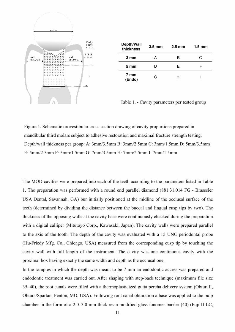
The MOD cavities were prepared into each of the teeth according to the parameters listed in Table
1. The preparation was performed with a round end parallel diamond (881.31.014 FG - Brasseler
USA Dental, Savannah, GA) bur initially positioned at the midline of the occlusal surface of the
teeth (determined by dividing the distance between the buccal and lingual cusp tips by two). The
thickness of the opposing walls at the cavity base were continuously checked during the preparation
with a digital calliper (Mitutoyo Corp., Kawasaki, Japan). The cavity walls were prepared parallel
to the axis of the tooth. The depth of the cavity was evaluated with a 15 UNC periodontal probe
(Hu-Friedy Mfg. Co., Chicago, USA) measured from the corresponding cusp tip by touching the
cavity wall with full length of the instrument. The cavity was one continuous cavity with the
proximal box having exactly the same width and depth as the occlusal one.
In the samples in which the depth was meant to be 7 mm an endodontic access was prepared and
endodontic treatment was carried out. After shaping with step-back technique (maximum file size
35–40), the root canals were filled with a thermoplasticized gutta percha delivery system (ObturaII,
Obtura/Spartan, Fenton, MO, USA). Following root canal obturation a base was applied to the pulp
chamber in the form of a 2.0–3.0-mm thick resin modified glass-ionomer barrier (40) (Fuji II LC,
!11
Depth/Wall thickness 3.5 mm 2.5 mm 1.5 mm
3 mm A B C
5 mm D E F
7 mm (Endo) G H I
Table 1. - Cavity parameters per tested group
Figure 1. Schematic orovestibular cross section drawing of cavity proportions prepared in
mandibular third molars subject to adhesive restoration and maximal fracture strength testing.
Depth/wall thickness per group: A: 3mm/3.5mm B: 3mm/2.5mm C: 3mm/1.5mm D: 5mm/3.5mm
E: 5mm/2.5mm F: 5mm/1.5mm G: 7mm/3.5mm H: 7mm/2.5mm I: 7mm/1.5mm
GC Europe, Leuven). This was cut back with a coarse diamond bur (801.36.6801 FG/Surg -
Brasseler USA Dental, Savannah, GA) to establish the 7mm final depth of the cavity. The
cavosurface margins were prepared perpendicular to the tooth surface. The cavity was rinsed with
water and air-dried with an air/water syringe. After application of a Tofflemire (1101C 0.035,
Hawe-Neos, Italy) matrix, the enamel was acid-etched selectively with 37% phosphoric acid for 15
seconds, rinsed with water and air-dried. The cavity was adhesive-treated with G-aenial Bond (GC
Europe, Leuven, Belgium) according to the manufacturer’s instructions. The adhesive was light-
cured for 40 s with an Optilux 501 halogen light (Kerr, Orange, CA, USA) operating in standard
mode at a light intensity of 740+/- 36mWcm2. In all groups, an approximately 0.5 mm-thin flow
composite layer (G-aenial Flo A2, GC Europe, Leuven) was applied on all walls of the cavity
(41-43). This layer was light-cured for 40 s. After applying the flowable layer, composite resin
(Gradia Direct Anterior A2, GC Europe, Leuven, Belgium) was placed in several consecutive 2
mm-thick oblique increments. Each increment was light cured from the occlusal surface for 40 s
each and after removal of the Tofflemire matrix band the mesial and distal sides were light cured for
20 s each (total 80 s). Light curing times chosen are the double of that recommended by the
manufacturer for each material used, in order to securely obtain maximal conversion at each layer.
The restorations were finished with a fine granular diamond burr (FG 7406-018, Jet Diamonds,
USA and FG 249-F012, Horico, Germany) and aluminum oxide polishers (OneGloss PS Midi,
Shofu Dental GmbH, Ratingen, Germany) and were stored in physiological saline solution (Isotonic
Saline Solution 0.9% B.Braun, Melsungen, Germany) before the fracture test.
To simulate the periodontal ligament, the root surface of each tooth was coated with a layer of
liquid latex separating material (Rubber-Sep, Kerr, Orange, CA) prior to embedding. Specimens
were embedded in methacrylate resin (Technovit 4004, Heraeus-Kulzer) at 2 mm from the CEJ to
simulate the bone level.
All specimens were tested for fracture strength within 24 hours of restoration, using a universal
loading device (5848 MicroTester1, Instron, Norwood, MA, USA). Each test was performed at a
cross-head speed of 2 mm/min and load was applied using a 6mm diameter stainless-steel ball-
shaped stylus which was positioned at the centre of the occlusal surface of the tooth between the
buccal and oral cusps in the central pit. A force vs. extension curve was dynamically plotted for
each tooth. Fracture threshold - defined as the load at which the tooth-restoration complex exhibited
the first fracture, resulting in a peak formation on the extension curve - was recorded in Newtons
(N).
!12

Statistical analysis was conducted in SPSS 21.0 (SPSS Inc., Chicago, USA). For the comparisons
between the groups, Kruskal-Wallis ANOVA with post-hoc pairwise comparisons was used. The
significance limit was set at α = 0.05.
3.2. Restoration of endodontically treated premolar teeth with occlusal cavity preparation
The aim of this investigation was to compare the mechanical properties of novel methods for
the reinforcement of ETT utilizing SFRC (EverX Posterior, GC Europe, Leuven); with previously
tested and accepted restorative methods. The null hypothesis was that 1, There would be no
difference in the maximal fracture resistance of the ETT restored teeth with the tested methods. 2,
There would be no difference in the fracture patterns of the ETT restored with the tested methods.
Seventy-two upper premolar teeth, extracted for periodontal or orthodontic reasons were
selected for this investigation. Specimen selection, exclusion criteria, root canal treatment protocol,
specimen preparation and mechanical testing were carried out as described by Frater et al. (44).
The freshly extracted teeth were immediately placed in 5.25% NaOCl for 5 minutes and stored in
0.9% saline solution at room temperature. Teeth were used within 6 months after extraction.
During specimen preparation the soft tissue covering the root surface was removed with hand
scalers. The inclusion criteria were absence of caries or root cracks, absence of previous endodontic
treatments, posts or crowns, resorptions, or evident lateral canals. Buccolingual and mesiodistal
radiographs of all teeth were taken and examined to evaluate root integrity and the number of canals
present. To standardize procedures and materials, all teeth used in this study had 1 root canal with a
curvature of less than 5°, evaluated by Schneider’s technique (45), and teeth with a root length of
15+/-1 mm and similar mesio-distal and bucco-lingual dimensions (+/-10%) were selected.
90% of the specimen ranged 9-10 mm in size, measured at the widest bucco-lingual dimension, and
the rest measured were 6.5 to 8 mm. Regarding the mesio-distal dimension, 90% of the specimen
ranged 7-7.5 mm, and the rest were 6.5 to 8 mm.
The teeth were randomly distributed over six study groups of 12 specimens each.
Access cavity was prepared by the same trained operator in five groups of the six, and one group
was left intact to serve as control (Group 6.).
Access cavity preparation was carried out with a round-end, tapered, medium grit, 0.8 mm tip
diameter, 10 mm length diamond bur (850-014M SSWhite, Lakewood, NJ, USA) with water
cooling in the approximated centre of the occlusal surface according to standardized parameters: the
!13
access cavity involved one-third of the intracuspal distance in the bucco-lingual dimension, and
one-third of the mesio-distal distance, measured at the level of the central fissure.
The working length was established with the direct method by subtracting 1 mm from the real root
length determined by introducing a no. 10 K-file (Maillefer-Dentsply, Ballaigues, Switzerland) until
it was visible through the apical foramen.The canals were instrumented using rotary ProTaper
Universal files (Dentsply, Maillefer, Ballaigues, Switzerland). The ProTaper sequence (S1, S2, F1,
F2) was used for the preparation at the working length.
Irrigation was performed after every instrument with 2 milliliters of 2.5% NaOCl solution and the
canal space was filled with irrigant during the instrumentation phase. After the shaping and cleaning
of the root canal the roots were dried with 96% alcohol and paper points. Root canal filling was
done by matched-single-cone obturation with a master cone (F2 gutta-percha, Maillefer-Dentsply,
Ballaigues, Switzerland) and sealer (AH plus; Dentsply De Trey GmbH, Konstanz, Germany). The
access cavity was temporarily filled with Fuji Triage Pink (GC Europe, Leuven, Belgium). Fuji
Triage Pink was applied to the apical part of the root in order to prevent leakage through the apex.
The teeth were stored in an incubator (mco-18aic, Sanyo, Japan) for 1 week (at 37 °C, 100%
relative humidity).
Group 1 and 2 received a minimal invasive post space preparation with a depth of 8 mm, as
measured from the CEJ on the buccal aspect of the tooth, but no post preparation drills
recommended by the manufacturer were used in order to preserve the individual anatomy of the
specimen teeth. Only the root canal filling was removed with size 3 Gates Glidden burs and ISO
standard Hedstrom files leaving a minimum apical seal of 6-8 mm of gutta-percha in the canal. The
No. 3 Gates Glidden bur was used on the full 8 mm length.
In Group 3.-5. the gutta-percha was only cut back 2 mm below the CEJ with an 0,1 mm diameter
ball shaped carbide bur (H1SM.205.010,Gebr. Brasseler GmbH & Co. KG, Lemgo, Germany) but
no post space preparation was performed. After cutting back the gutta-percha, the orifice was sealed
with resin modified glass ionomer cement (Fuji VIII, GC Europe, Leuven, Belgium).
All specimen received the same adhesive treatment. Prior to the adhesive treatment of the cavity
and the root canal, enamel was acid-etched selectively with 37% phosphoric acid for 15 s and rinsed
with water. The root canal and the coronal cavity were rinsed with 2 millilitres of water and dried
with paper points and air. For bonding, a dual-cure one-step self-etch adhesive system (Gradia Core
Self-Etching Bond, GC Europe, Leuven, Belgium) was used, according to the manufacturer’s !14
instructions using a microbrush-X disposable applicator (Pentron Clinical Technologies, LLC,
USA). Excess adhesive was removed by suction drying (Evacuation Tip - Starryshine, Anaheim,
CA, USA) within 0.5 cm from the occlusal cavity (without contact). Excess adhesive resin at the
bottom of the canal was removed with a paper point. The adhesive was light-cured for 60 s using an
Optilux 501 quartz-tungsten-halogen light-curing unit (Kerr Corp., Orange, CA, USA). The average
power density of the light source, measured with a digital radiometer (Jetlite light tester; J. Morita
USA Inc. Irvine, CA, USA) prior to the bonding procedure, was 840 +/-26.8 mW/cm2.
Five different techniques were used to restore the specimens (Figure 2):
Group 1: The teeth received a prefabricated, conventional FRC post (0,8 GC Fiber post, GC
Europe, Leuven, Belgium). Before the adhesive treatment the conventional translucent FRC posts
of 0.8 mm diameter (GC Fiber Post, GC Europe, Leuven, Belgium) was tried in and cut to a length
1 mm below the level of the occlusal cavity margins with a water-cooled diamond disc (Isomet
2000; Buehler Ltd., Lake Bluff, IL, USA) and cleaned with alcohol after try in. The posts received
silanization of the surface (Ceramic Primer, GC Europe, Leuven, Belgium) following the
manufacturer’s recommendation. After silanization, the post surface was bonded with the same
bonding agent used for the cavity. Luting of the posts and the core build-up was performed with a
dual-cure resin composite core material (Gradia Core, GC Europe, Leuven, Belgium). Gradia Core
was applied using its own automix cartridge with an ‘elongation tip’ for direct root canal
application. After insertion of the post, 5 minutes of chemopolymerization time was provided to
reduce polymerisation stress, then cement was polymerized with an Optilux 501 quartz tungsten-
halogen light-curing unit for 60 s from each side (a total of 240 s/tooth). The outlines of the
restoration were finished with dental composite (G-aenial Posterior P-JE, GC Europe, Leuven,
Belgium).
Group 2: The teeth were reconstructed with a novel method of building a direct layered fiber-
reinforced composite post and core (DLFRC post and core) from SFRC (EverX Posterior, GC
Europe, Leuven, Belgium). The DLFRC post and core was horizontally layered in 1-2 mm
segments. An increment of SFRC was packed to the apical portion of the post space using a
microbrush-X disposable applicator (Pentron Clinical Technologies, LLC, USA). A light
transmitting FRC post (0,8 GC Fiber post, GC Europe, Leuven, Belgium) was inserted into the post
space in order to aid the transmission of the light to the apically positioned layers. The “light
transmitting” post was withdrawn with 0,5-1 mm from the surface of the uncured SFRC layer not to !15

!16
Figure2. Restored study groups, layering concepts
and restorative materials applied.
have direct contact with it.
After each layer 80 seconds of light curing through the fiber post was carried out. After
incrementally filling the root canal to the level of the CEJ with repeating the previously described
procedure, SFRC was layered in the coronal cavity until 1 mm below the margin of the occlusal
cavity in a concave shape. Each increment was light cured from the occlusal surface for 40 seconds.
The outlines of the restoration were finished with dental composite (G-aenial Posterior P-JE, GC
Europe, Leuven, Belgium).
Group 3: The cavities were restored with SFRC material applied in an oblique incremental
technique. The material was placed in consecutive 2 mm thick increments. Each increment was
light cured from the occlusal surface for 40 seconds. The last 1 mm thick occlusal layer was
composite material (G-aenial Posterior PJ-E, GC Europe, Leuven, Belgium) covering the SFRC.
Group 4: The cavities were restored with micro hybrid composite restorative material (G-aenial
Posterior PJ-E, GC Europe, Leuven, Belgium) applied with an oblique incremental technique. The
material was placed in consecutive 2 mm thick increments. Each increment was light cured from the
occlusal surface for 40 seconds.
Group 5: The cavity walls were coated with flowable composite (G-aenial Flo, GC Europe, Leuven,
Belgium)and before curing, a piece of pre-impregnated glass fiber net (Everstick net, GC Europe,
Leuven, Belgium) (10 mm long, 3 mm width) was cut and embedded inside the flowable composite
first in buccal to lingual, then a mesial to distal direction. After curing for 40s, another glass fiber
band was adapted to the walls circumferentially, forming the FRC “box”. The remaining central part
of the cavity was restored with SFRC and a final layer of composite as described in Group 3.
Finally for all specimens glycerine gel (DeOx Gel, Ultradent Products Inc., Orange, CA, USA) was
applied and final polymerization from each side for 40 seconds was performed.The restorations
were finished with a fine granular diamond burr (FG 7406-018, Jet Diamonds, USA and FG 249-
F012, Horico, Germany) and aluminum oxide polishers (OneGloss PS Midi, Shofu Dental GmbH,
Ratingen, Germany).
After the restorative procedures, the specimens were stored in physiological saline solution
(Isotonic Saline Solution 0.9% B. Braun, Melsungen, Germany) in an incubator (mco-18aic, Sanyo, !17
Japan) for 1 week (at 37°C, 100% humidity) before the fracture strength test. Prior to embedding,
the root surface of each tooth was coated with a layer of liquid latex separating material (Ruber-
Sep, Kerr, Orange, CA, USA) to simulate the periodontal ligament [30]. Specimens were embedded
in methacrylate resin (Technovit 4004, Heraeus-Kulzer) at 2 mm from the CEJ to simulate the bone
level. After embedding, all specimens were immediately subjected to a fracture resistance test using
a universal loading device (5848 MicroTester1, Instron, Norwood, MA, USA). Each test was
performed at a cross-head speed of 0.5 mm/min and load was applied at 45° using a 4.8 mm
diameter stainless-steel ball-shaped stylus positioned to the central groove of the tooth providing 2
contacts with the triangular ridges and one with the more dominant marginal ridge. The maximum
failure load was recorded in Newton’s (N).
A force vs. extension curve was dynamically plotted for each tooth.
After mechanical testing, the specimens were examined for fracture patterns. According to Scotti
and co-workers, distinction was made between restorable or non-restorable fractures under optical
microscope with a two-examiner agreement. A restorable fracture is above the CEJ, meaning that in
case of fracture, the tooth can be restored, while a non-restorable fracture extends below the CEJ
and the tooth is likely to be extracted (46).
Statistical analysis was conducted with SPSS 22.0 (IBM, USA). As the data was not normally
distributed in all groups, the comparisons were performed with Kruskal-Wallis ANOVA with Dunn-
Bonferroni post-hoc pairwise comparisons. The level of significance was set at p<0.05.
3.3. Restoration of endodontically treated premolar teeth without ferrule effect
The goal of the present in vitro study was to determine and compare the fracture resistance
and fracture patterns of endodontically treated premolar teeth restored with different FRC posts in
different configurations. The null hypotheses were the following: 1.The fracture resistance of the
teeth restored with single or multiple posts would not be different. 2. The application of more elastic
posts would not result in more favourable fracture patterns.
Fifty upper premolar teeth extracted for periodontal or orthodontic reasons were selected for
this study. The inclusion criteria were absence of caries or root cracks, no previous endodontic
treatment, no posts or crowns, no resorption and the absence of lateral canals. Furthermore,
buccolingual and mesiodistal radiographs of all teeth were taken and examined to evaluate root
integrity and the number of canals present. To standardise procedures and materials, all teeth used in !18
this study had 1 root canal in each root with a curvature of less than 5°, evaluated by Schneider’s
technique (14), and root length of 15+/-1 mm and rather similar mesiodistal and buccolingual
dimensions (+/-10%) were selected.
The freshly extracted teeth were immediately placed in 5.25% NaOCl for 5 minutes and then stored
in 0.9% saline solution at room temperature. The teeth were used within 6 months after the
extraction. During specimen preparation, the soft tissue covering of the root surface was removed
with hand scalers.
Before root canal treatment, all crowns were sectioned at the level of the CEJ perpendicular to the
longitudinal axis, using a slow-speed, water-cooled diamond disc (40000 rpm).
At the beginning of the root canal treatment the working length was established using a direct
method, by subtracting 1 mm from the actual root length determined by introducing a no. 10 K-file
(Maillefer-Dentsply, Ballaigues, Switzerland) until it was visible through the apical foramen. A
crown down technique was used for instrumentation with Gates Glidden (Union Broach, York, PA)
#2 to #4 drills and then the canals were instrumented using rotary ProTaper files (Dentsply,
Maillefer, Ballaigues, Switzerland). The series of the ProTaper system (S1, S2, F1, F2, F3) was used
for the preparation at the working length.
Irrigation was performed after every change of instrument with 2 millilitres of 2.5% NaOCl solution
and the canal space was filled with irrigant during the instrumentation phase. A root canal lubricant
(Glyde, Dentsply-Maillefer, Konstanz, Germany) was only used during the shaping of the coronal
third. After shaping and cleaning, the roots were dried with 96% alcohol and paper points. Root
canal filling was performed by matched-single-cone obturation with a master cone (F3 gutta-percha,
Maillefer-Dentsply, Ballaigues, Switzerland) matching the final instrument used for preparation and
sealer (AH plus; Dentsply De Trey GmbH, Konstanz, Germany). The root access was temporarily
filled with Clearfil SE Bond and Clearfil AP-X (Kuraray, Tokyo, Japan). The same composite was
applied to the apical part of the root in order to prevent leakage through the apex. The teeth were
then stored in an incubator (mco-18aic, Sanyo, Japan) for 1 week (at 37 °C, 100% relative
humidity).
After 1 week of incubation, post space was prepared in the root portions of the teeth with a depth of
10 mm, as measured from the CEJ on the buccal aspect of the tooth, but no post space preparation
drill was used so that the individual anatomy could be preserved. Only the root canal filling was
removed with size 3 Gates Glidden burs and ISO standard Hedstrom files, leaving a minimum
apical seal of 4-6 mm of gutta-percha in the canal.
!19
For the restorations, two different types of FRC posts were used: a prefabricated, “rigid”
conventional FRC post (0,8 GC Fiber Post, GC Europe, Leuven, Belgium) and an elastic FRC post
(0,9 EverStick POST, GC Europe, Leuven, Belgium).
The conventional translucent FRC posts of 0.8 mm diameter (Fiber Post) were tried in and cleaned
with alcohol afterwards. The posts did not receive any surface treatment. The elastic FRC posts
were handled according to the manufacturer’s instructions, with sterile tweezers. Regardless of the
exact type, the main posts were placed in a way that 5.0 mm was left above the level of
decoronation, and 10.0 mm was inserted into the root canal. This way, a uniform 15.0 mm fiber
length was achieved.
The teeth were randomly distributed in 5 study groups, each group consisting of 10 teeth.
Group 1 received one single conventional FRC post (0.8 mm). Group 2 received one main
conventional FRC post and one collateral post (0.8 mm both) using a “multi-post technique”. The
collateral post was inserted next to the main post as apically as possible without causing manually
perceivable stress but it was always deep enough to wedge the main post in the canal. Group 3
received one single elastic FRC post (0.9 mm). According to the manufacturer's instructions, the
post was inserted into the root canal, and adapted to its form. Once adapted, the post was removed
from the root canal with a needle-nose plier and light cured for 40 seconds so that it would retain
the shape of the canal. Group 4 received one main elastic FRC post and one elastic collateral post
(0.9 mm both) using a multi-post technique. The collateral post was inserted next to the main post
as apically as possible without causing manually perceivable stress. The posts were removed as one
unit from the root canal with a needlenose plier and then light cured for 40 seconds maintaining
their position together in the canal. Group 5 received as many elastic FRC posts (0.9 mm) as
possible bundled according to the thickness of the root canal using the lateral condensation method
described by Hatta and co-workers (47). These posts were gently removed as one unit with a
needlenose plier from the root canal, and then light-cured for 40 seconds. It was confirmed in all
cases that the elastic FRC posts were repositioned to their original position into the canal after light-
curing. If resistance was met, the post surface was adjusted using carborundum point.
During the luting procedures all groups received the same adhesive treatment by the same trained
operator who completed a three year specialisation in restorative dentistry.
For bonding, a dual-cure one-step self-etch adhesive system (Gradia Core Self-Etching Bond, GC
Europe, Leuven, Belgium) was used, according to the manufacturer’s instructions. Luting of the
posts and the core build-up was performed with a dual-cure resin composite core material (Gradia
Core, GC Europe, Leuven, Belgium). Gradia Core was applied using its own automix cartridge with !20
an ‘elongation tip’ for direct root canal application. After the insertion of the post(s), the composite
core material was polymerised from the top of the post with an Optilux 501 quartz-tungsten-halogen
light-curing unit for 60 s from each side (a total of 240 s/tooth).
In order to ensure the uniformity of the specimens, the composite resin core build-ups were
standardised using cellulite core-forming matrices of the same size. These matrices were fabricated
as vacuum formed foils by a dental technician modelled on a healthy premolar tooth, which was
previously prepared for a crown with a one millimetre shoulder. The core build-up was
prepolymerised for 20 seconds, then glycerine gel (DeOx Gel, Ultradent Products Inc., Orange, CA)
was applied and final polymerisation was performed from each side for 40 s with an Optilux 501
quartz-tungsten-halogen light-curing unit. After the cementation procedures, the specimens were
stored in physiological saline solution (Isotonic Saline Solution 0.9% B. Braun, Melsungen,
Germany) in an incubator (mco-18aic, Sanyo, Japan) for 1 week (at 37°C, 100% humidity) before
the fracture test. The specimens were embedded as described by Frater et al.(48).
After embedding, all specimens were immediately subjected to a fracture resistance test using a
universal loading device (5848 MicroTester1, Instron, Norwood, MA, USA). Each test was
performed at a cross-head speed of 0.5 mm/min. Load was applied at 45° to the long axis of the
tooth by adjusting a stainless steel ball shaped stylus to the occlusal surface of the abutment in a
stabile position (49). The failure loads were recorded in Newtons (N). A force vs. extension curve
was dynamically plotted for each tooth.
After the mechanical testing, the specimens were examined for fracture patterns. A distinction was
made between restorable or non-restorable fractures, following the protocol proposed by Scotti et
al., under optical microscope with a two-examiner agreement (46). A restorable fracture was
recognised as one above the CEJ, meaning that in case of fracture, the tooth could be re-restored,
while a non-restorable fracture extends below the CEJ and extraction is likely to become necessary
(50). Statistical analysis was conducted with SPSS 17.0 (SPSS Inc., Chicago, USA). As the data
were not normally distributed in all groups, the comparisons were performed with Kruskal-Wallis
ANOVA with post-hoc pairwise comparisons. The level of significance was set at p<0.05.
!21
4. RESULTS
4.1. Mechanical changes resulting from different size MOD cavity preparations
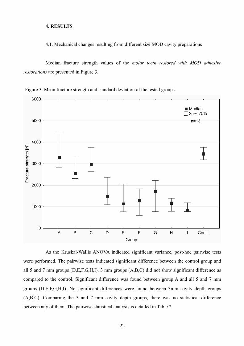
Median fracture strength values of the molar teeth restored with MOD adhesive
restorations are presented in Figure 3.
As the Kruskal-Wallis ANOVA indicated significant variance, post-hoc pairwise tests
were performed. The pairwise tests indicated significant difference between the control group and
all 5 and 7 mm groups (D,E,F,G,H,I). 3 mm groups (A,B,C) did not show significant difference as
compared to the control. Significant difference was found between group A and all 5 and 7 mm
groups (D,E,F,G,H,I). No significant differences were found between 3mm cavity depth groups
(A,B,C). Comparing the 5 and 7 mm cavity depth groups, there was no statistical difference
between any of them. The pairwise statistical analysis is detailed in Table 2.
!22
Figure 3. Mean fracture strength and standard deviation of the tested groups.
The null hypothesis was rejected as fracture strength of groups D,E,F,G,H,I where
significantly lower than that of the control group.
4.2. Restoration of endodontically treated premolar teeth with occlusal cavity preparation
Table 3 summarizes the fracture thresholds for the premolar study groups with only an
occlusal access cavity. The control group exhibited the highest fracture resistance. The application
of the DLFRC post and core technique yielded the highest fracture resistance among the restored
groups. The fracture resistance of Group 2 (DLFRC post and core group) did not differ significantly
(p=1.000) from the intact teeth (control group). The rest of the groups proved to be significantly
different from the control group in terms of fracture resistance. There was no statistically significant
difference when comparing the restored groups regarding their fracture resistance. Therefore, the
null hypothesis regarding fracture resistance was rejected.
!23
Group A B C D E F G H I Contr.
A 1.000000 1.000000 0.029247✽ 0.000970✽ 0.002154✽ 0.021271✽ 0.000389✽ 0.000170✽ 1.000000
B 1.000000 1.000000 0.923576 0.072278 0.132891 0.732700 0.035687✽ 0.018728✽ 1.000000
C 1.000000 1.000000 0.300701 0.017307✽ 0.034044✽ 0.231264 0.007930✽ 0.003899✽ 1.000000
D 0.029247✽ 0.923576 0.300701 1.000000 1.000000 1.000000 1.000000 1.000000 0.008013✽
E 0.000970✽ 0.072278 0.017307✽ 1.000000 1.000000 1.000000 1.000000 1.000000 0.000203✽
F 0.002154✽ 0.132891 0.034044✽ 1.000000 1.000000 1.000000 1.000000 1.000000 0.000478✽
G 0.021271✽ 0.732700 0.231264 1.000000 1.000000 1.000000 1.000000 1.000000 0.005670✽
H 0.000389✽ 0.035687✽ 0.007930✽ 1.000000 1.000000 1.000000 1.000000 1.000000 0.000076✽
I 0.000170✽ 0.018728✽ 0.003899✽ 1.000000 1.000000 1.000000 1.000000 1.000000 0.000032✽
Contr. 1.000000 1.000000 1.000000 0.008013✽ 0.000203✽ 0.000478✽ 0.005670✽ 0.000076✽ 0.000032✽
Table 2 - Kruskal-Wallis ANOVA pairwise statistical analysis (p<.0000). Significance indicated with ✽ symbol.
In terms of fracture patterns, the tested groups 2,3,4,5 were identical (Table 4.). Only the
control group and the FRC post showed dominantly repairable fractures. Therefore, the null
hypothesis regarding fracture patterns was also rejected.
!24
Group Sig. compared to
Control (p, post-hoc)
N Minimum (Newtons)
Maximum (Newtons)
Mean (Newtons)
Std. Deviation
Control - 12 605.85 1205.83 922.34 189.21
Group 1 .005 12 208.28 802.61 501.30 186.65
Group 2 1.000 12 352.85 1171.19 727.98 287.37
Group 3 .009 12 123.59 865.93 511.61 225.20
Group 4 .005 12 216.67 748.44 456.24 189.75
Group 5 .023 12 303.64 682.83 536.35 126.41
Table 3.: Fracture thresholds of studied groups and the significance of their difference
compared to the control group. Group 1: glass fibre- reinforced post; Group 2: direct layered
glass fibre-reinforced composite core; Group 3: SFRC applied by an oblique incremental
technique; Group 4: obliquely layered conventional composite; Group 5: torsion box with FRC
net. As there was no significant difference among the restored groups in this respect,
significances are shown as compared to the controls only.
Control Group 1 Group 2 Group 3 Group 4 Group 5
Reparable 0.66 0.58 0.33 0.33 0.33 0.33
Irreparable 0.33 0.42 0.66 0.66 0.66 0.66
Table 4.: Fracture patterns. The numbers indicate relative frequencies (n=12 in each group).
Group 1: glass fibre- reinforced post; Group 2: direct layered glass fibre-reinforced composite
core; Group 3: SFRC applied by an oblique incremental technique; Group 4: obliquely layered
4.3. Restoration of endodontically treated premolar teeth without ferrule effect
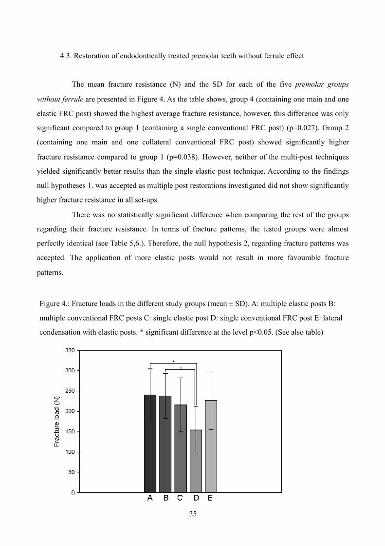
The mean fracture resistance (N) and the SD for each of the five premolar groups
without ferrule are presented in Figure 4. As the table shows, group 4 (containing one main and one
elastic FRC post) showed the highest average fracture resistance, however, this difference was only
significant compared to group 1 (containing a single conventional FRC post) (p=0.027). Group 2
(containing one main and one collateral conventional FRC post) showed significantly higher
fracture resistance compared to group 1 (p=0.038). However, neither of the multi-post techniques
yielded significantly better results than the single elastic post technique. According to the findings
null hypotheses 1. was accepted as multiple post restorations investigated did not show significantly
higher fracture resistance in all set-ups.
There was no statistically significant difference when comparing the rest of the groups
regarding their fracture resistance. In terms of fracture patterns, the tested groups were almost
perfectly identical (see Table 5,6.). Therefore, the null hypothesis 2, regarding fracture patterns was
accepted. The application of more elastic posts would not result in more favourable fracture
patterns.
!25
Figure 4.: Fracture loads in the different study groups (mean ± SD). A: multiple elastic posts B:
multiple conventional FRC posts C: single elastic post D: single conventional FRC post E: lateral
condensation with elastic posts. * significant difference at the level p<0.05. (See also table)

!26
Group N Mean Minimum Maximum SD
Multiple elastic posts 10 240.64 146.88 330.12 63.85
Multiple conventional FRC posts 10 237.99 165.93 361.63 55.26
Single elastic post 10 215.98 57.22 312.04 66.33
Single conventional FRC post 10 154.50 65.99 271.22 56.63
Lateral condensation with elastic posts 10 227.30 142.82 367.88 72.20
Table 5.: Descriptive statistics of the fracture loads measured in the different groups. The values
are given in Newtons.
Restorable Non-restorable
Multiple posts/elastic 7 (70%) 3 (30%)
Multiple posts/conventional 7 (70%) 3 (30%)
Single post/elastic 7 (70%) 3 (30%)
Single post/conventional 7 (70%) 3 (30%)
Lateral condensation/elastic 6 (60%) 4 (40%)
Table 6.: The distribution of fracture patterns among the study groups.
4. DISCUSSION
In the investigations of this thesis the two methods where utilised to examine the
mechanical properties of the specimens.
Static load to fracture is considered to be one of the effective means to study the
mechanical properties of posterior teeth despite the fact that functional loads are much higher and
load patterns do not resemble cyclic load of physiologic mastication(51). Using a 6 mm steel sphere
for loading is a common choice for the posterior region (52,53). The geometry of the sphere
provides possibility for tripodistic contacts on the triangular ridges of the posterior teeth involving
the functional and non-functional cusps, providing similar conditions to natural occlusion. For
molars it was considered that axial force exertion is the most clinically relevant, since a managed
occlusal scheme will exhibit load on the molars in maximal intercuspation thus providing a vertical
load on the coronal structures. For the premolar teeth a 45 degree load was applied as compared to
the long axis of the tooth. An oblique load appears to be the worst case scenario in terms of the
fracture resistance of ETT as described by Wandscher and co-workers (49). Applying this angle of
force to teeth places significant stress on the cervical aspect of the restored tooth (54) and heavy
shear forces on the post / luting agent / radicular dentine interfaces. This should represent a worst-
case occlusal loading scenario for these teeth and acid-test the integrity of the tested restorations
and tooth structure.
Fracture patterns where examined and categorised and restorability was assessed
according to Scotti et al (46). Examinations were done under an optical microscope with a two-
examiner agreement. A restorable fracture is above the CEJ, clinically meaning that the tooth can be
restored, while a non-restorable fracture extends below the CEJ clinically resulting in extraction of
the tooth unless surgical or orthodontic procedures are applied.
Marginal Ridges
If an MOD cavity is prepared in a molar tooth, both marginal ridges are lost and the
fracture resistance of the tooth is significantly reduced (55-57). It is widely accepted and published
that the defining measure of assessing the stability of the cusps in such cases is the thickness of the
cavity walls (9,10,58). According to several authors if the cavity wall is thicker than 2-2.5 mm the
cusps are considered stabile and if thinner than this measure they are considered to be fragile. The
results of this thesis suggest that cavity wall thickness is not a major influencing factor of cusp
stability. Comparing 3.5mm, 2.5mm and 1.5 mm cavity wall thickness in case of the 3, 5 or the 7 !27
mm cavity depth groups, mechanically similar behaviour was measured after being restored with a
conventional dental composite. In this thesis reducing only the wall thickness, without changing the
depth of the cavity, did not cause a significant reduction in fracture strength. 3 mm can be
considered a safe cavity depth for adhesive direct restorations. This is in accordance with Frater et
al. who found that when restoring shallow MOD cavities with direct techniques using oblique
layering, there was no significant difference between the restored groups and the intact control
group (48). These findings do not support the findings of S. Batalha-Silva et al. (50), who
concluded that 5mm deep cavities could safely be restored with direct techniques. These results
rather suggest that a cavity of 5mm depth is already in the "danger zone" when talking about direct
composite restorations without cusp coverage. From the results it seems that cavity wall thickness is
only secondary to cavity depth in molar MOD cavities in terms of fracture strength, as the change in
cavity wall thickness did not lead to a significant difference neither between the groups in the
“safety zone” nor between the “danger zone” groups. Within the limitations of this study, this leads
us to the conclusion that when the fracture safety of a cavity for a direct restoration without cusp
coverage is to be determined, cavity depth is the primary determining factor. This is in accordance
with the results of Morin et al. and Manhart et al. who found that the depth of the preparation is the
most critical factor in terms of future fractures (59,60). Since groups with cuspal coverage
restorations were not included in this study, the results do not offer direct guidance on cuspal
coverage. Also, since the cavities were only restored with a conventional adhesive direct composite
restoration, the results cannot be extrapolated to situations when, fiber-reinforced materials are
used. However if the cavity depth of molar teeth with an MOD cavity reaches or exceeds 5 mm,
cusp coverage with a direct or indirect adhesive restoration could be taken into consideration as a
safety measure. Future investigations with similar methodology - involving cusp coverage with
direct and indirect methods - need to be conducted to further our understanding on the effect of
MOD cavity dimensions on the restorability of molar teeth and limitations of adhesive restorations.
There is no statistical difference between the 5 and 7 mm results so endodontically
treated molars with an MOD cavity are not significantly weaker mechanically as compared to
molars with a vital pulp and intact pulp space with a 5 mm deep MOD cavity. Therefore root canal
treated molars are not weaker or more fragile by nature. They are weaker as a result of tissue loss
in the biomechanically sensitive anatomic locations such as the marginal ridges.
!28
Pericervical Dentine Reinforcement
For the isolated examination of the effect of PCD loss and possible options of restoring
the function of PCD, premolar teeth with a single occlusal access cavity where selected. It was the
intention of the authors to exclude the mechanical influencing effect of the loss of marginal ridges
and solely concentrate on the pericervical area. In premolar teeth the cervical part is the thinnest and
most vulnerable anatomic feature as a result of an unfavorable anatomy in crown volume and crown
to root proportion(61).Therefore reducing the PCD of these teeth is thought to have the most radical
effect. This is underlined clinically since root canal treated premolars are susceptible to fracture (51,
62). Restoration of premolars with a glass fiber post does not protect them from fracture but it
seems to prevent catastrophic fracture (21,63). So a premolar tooth with an occlusal access cavity,
intact marginal ridges and excess root canal preparation at the orifice of the canal and the coronal
third is possibly a good model for examining the biomechanical effects of PCD loss.
In this thesis, endodontically treated premolars restored with an oblique layered occlusal
composite restoration (Group 4) showed the lowest fracture resistance among all groups. This
justifies the model as if no attempted restoration of the pericervical tissues was done, the restored
tooth as a whole behaved in a mechanically inferior way and was significantly weaker than the
natural control group. However these restorations where not statistically significantly weaker than
the other types of tested restorations. This supports the findings of some authors, who claim that
posterior teeth with a single occlusal access preparation and no other structural loss may be restored
without post with a conservative direct bonded restoration(6,9,64,65). In this investigation a
minimally invasive access was prepared, which could give explanation to these findings, since it is
reported that additional tissue loss results in compromised structural integrity and as a result lower
fracture resistance of root canal treated teeth (1,62).
In the current investigation the group restored with obliquely layered SFRC (Group 3)
reached higher fracture resistance figures compared to the ones restored with conventional
composite (Group 4) or even the conventional FRC post group (Group 1), however, the difference
was statistically not significant. This has been previously described in molar teeth with MOD
cavities, where the SFRC restored groups yielded better results than the conventional composite
restored ones, yet the difference was not significant (48).
The threefold usage of FRC net (FRC box) (Group 5) together with the SFRC restorative
composite is aiming to reconstruct the integrity and strength of the opposing cavity walls. The
values of the FRC box restorations showed an increase compared to Group 1, 3 and 4, this
technique could also not emulate the values measured in case of sound premolar teeth. However in !29
this study there seems to be a clear tendency of increasing fracture resistance values towards the use
of individualized FRC materials compared to conventional techniques (composite restoration or
FRC post placement). This observation is in accordance with the findings of Bijelic et al (66).
Prefabricated FRC posts suffer from 2 main shortcomings in clinical settings:
Insufficient bonding of the interfaces (14,32,67) and the fact that the post position is in the neutral
axis of the root canal. Direct layering of SFRC into the root canal is intended to solve these
drawbacks. Seyam et al. and other authors showed that a transparent post can transfer the light and
aid the polymerization of composite resin in the root canal (68-70), enabling layering in the hollow
root canal space. However, there are investigations to oppose this statement (71,72). The DLFRC
post and core technique (Group 2) produced the highest fracture resistance values among the
restored groups in the present study. These results seem to be in accordance with Garoushi et al.
showing that the thicker the applied SFRC restoration the greater the fracture resistance is (73).
Although the numbers produced by group 2 were not significantly higher than the rest of the
restored groups, a positive tendency could be visible with the utilisation of SFRC materials.
Moreover, there was no statistically significant difference between the group 2 and the intact teeth.
This result suggests a move towards a biomimetic restorative concept. It has to be noted that the
reported advantages come at the price of increased application time and technically more
demanding clinical procedure as compared to Group 1. Development of materials, instruments and
light curing equipment specifically for such purposes could be promising and could resolve the
main shortcomings of the DLFRC post and core method as described in this investigation.
Regarding fracture pattern, the tested groups were identical with dominantly
unfavorable, irreparable fractures. Only the control group and Group 1 presented a shift towards
favorable, repairable fractures. Therefore the null hypothesis regarding fracture pattern was
rejected.
The DLFRC post and core technique according to the findings of this investigation
might hold the potential of reinforcing the root and particularly the pericervical area, which is
highly beneficial when shear forces are also present (f.e.: 45 degree loading). The DLFRC post and
core concept theoretically could present a possibility to compensate for most of the known
weaknesses of the presently accepted endo-restorative options with a not complicated, clinically
feasible and reproducible methodology.
!30
Substituting the Prosthodontic Ferrule
Since the middle of the 17th century when post borne restoration of non vital teeth was
described, it is a basic goal of restorative treatments to reinforce and retain ETT in function. Post
space preparation undermines the remaining tooth structure, which possibly increases the possibility
of fracture and tooth loss (74, 75). Post placement should not be carried out at the cost of sacrificing
radicular dentine (22, 24). Several publications seem to prove that up to 20% of ETT restored with a
radicular post suffer a vertical fracture (76-81).
It is even bigger of a challenge to restore ETT which are missing a prosthodontic ferrule
(82). In a non-ferrule situation there is not a currently known endo-restorative option that could
yield a long term clinical result reproducibly (83). According to Nam et al, conventional fibre posts
do not improve the fracture resistance of teeth without a dental ferrule (84). In this thesis premolar
teeth without a ferrule where restored to crown abutment shape with different FRC post application
techniques in order to distinguish which of them could be a mechanically more stabile solution in
case of non-ferrule conditions.
Gates-Glidden burs where used to obtain minimally invasive post-space preparation by
only removing guttapercha and root canal sealer, but not removing any sound dentine. This
technique leads to individual post spaces. Which can not be filled with one single symmetric post,
but multiple small diameter FRC posts or other means are needed. According to Sorensen et al. a
the fracture resistance of restored teeth significantly increased when posts where adapted closely to
the canal walls (85). Maceri and co-workers proved that a multi post technique may not only lead to
better adaptation, but possibly reduces pull out risk and induces durability to long term cyclic
loading (27). Therefore applying multiple posts in the same canal (multi-post technique) or using an
individual post is aiming to achieve a better fit to the individual, preserved root canal anatomy and
possibly enhancing long term clinical prognosis.
The results of this study appear to favour the use of multiple posts in the same root
canal. Both multi-post techniques (rigid FRC (group 2.) and elastic FRC (group 4)) yielded
significantly higher fracture resistance than the single post conventional FRC restoration (group 1)
It is interesting to note that neither of the two multi-post techniques yielded significantly
different fracture resistance from the single elastic post technique. The non significance between the
results can be explained by use of a minimally invasive post space preparation in case of using a
single elastic post and a multi-post technique, since even a single elastic post can achieve a good fit,
thus adequate stability in case of a preserved, relatively irregular root canal cross section. It is also
noted that minimally invasive post space preparation is likely to preserve mass amount of PCD !31
keeping the natural reinforcement of the tooth as compared to other techniques with an invasive
post space preparation. Therefore the null hypothesis regarding fracture resistance was partially
rejected (only for non-elastic FRC posts). A possible explanation is that the better adaptability of the
particular type of elastic post used in this study was enough to make up for the disadvantages of
using a single post only. However the limitations of this investigation cannot lead to this conclusion,
so further studies on the adaptive properties of the elastic FRC posts are necessary.
There are several methods of creating individual root canal posts (47, 86-88). In this
study the “lateral condensation method” of Hatta and colleagues was used (47). In the present study
the individual posts (group 5) yielded better results than restoration with a single FRC post (groups
1 and 3). However, the difference did not reach the level of statistical significance. These results are
in agreement with those of Hatta et al. (47) and Le Bell-Ronnlof et al. (34) in this respect.
Based on the results of this investigation, fracture resistances yielded by the individual
post technique and the two multi-post techniques were not significantly different. The reason might
be the minimally invasive post space preparation, as the number of posts insertable to
approximately the same depth is reduced by the limited space. In this case the cement-glass fiber
ratio of the single post techniques can be relatively similar to that of the multiple post techniques.
However most likely the preservation of tooth structure is the key influencing factor of mechanical
behaviour of ETT as supported by Wandscher et al. (49).
Limitations
In all three investigations of this thesis mechanical testing was carried out with a static
“load to fracture” methodology. Cyclic loading of dental specimens and ageing prior to testing
resemble the phisiological circumstances better. According to Taha et al, “In experimental studies,
fracture resistance to static loading has been used as a measure of the effect of cavity preparation
and/or restoration on tooth strength. Although the fracture load is typically much higher than
functional occlusal loads, it is still a valid method for comparing restorative materials and different
cavity designs” (51)
Non-ferrule conditions were tested without fabricating and cementing a crown on the
prepared abutments. The decision to not use crowns in this study was based on the observations of a
number of authors where subtle differences in post behaviour and performance may be masked
when teeth are definitively restored (22, 89-92). Despite these recommendations, the lack of a
crown makes it impossible to extrapolate the results of this investigation to a clinical situation and
there is a need for further experiments to bridge this gap. !32

In these investigations 10-12 specimens were prepared per group. By raising this number
the level of significance could be better established and discrete differences among restorative
techniques could be more pronounced.
!33
5. CONCLUSION
The investigations described in the thesis attempt to find a biomimetic rationale of
restoring posterior teeth by the means of applying novel diagnostic measures and utilising new endo
restorative techniques. Within the limitations of this thesis the following conclusions can be drawn:
Marginal ridges of molar teeth and PCD of premolar teeth are some of the key anatomic
features to be preserved in order to maintain the biomechanical integrity of the posterior root canal
treated teeth.
Molar teeth with a 3 mm or shallower MOD cavity are considered to be safely restorable
with conventional adhesive restorations. Molar teeth with MOD cavities of 5 mm or deeper -
including endodontically treated molar teeth - are considered to be in the “danger zone” if placing
adhesive composite restorations. In these situations cusp coverage should be considered. It is also
understood that cavity wall thickness does not significantly influence fracture resistance in the
described circumstances.
Natural premolar teeth exhibit higher fracture resistance then the ones that are
endodontically treated through an occlusal access and restored except for the DLFRC post and core
restored group. DLFRC post and core behaves mechanically similarly in the described conditions as
the natural control tooth therefore it can be considered a biomimetic endo-restorative solution. In
terms of fracture patterns conventional FRC posts exhibited more favourable fracture patterns then
the other restored groups. The direct layered short fibre-reinforced post and core is a promising
alternative to the currently accepted restorations of ETT, and as such should be further investigated.
Single rooted premolars restored in absence of a ferrule show significantly higher
fracture resistance, when a multi post technique or a single elastic post is applied as compared to a
conventional rigid single FRC post. Once utilizing a multipost technique the elasticity of the post
did not yield any significant difference in the described circumstances. Single canal teeth restored
with multiple posts achieved superior fracture resistance to teeth restored with single, conventional
FRC posts.
!34
6. ACKNOWLEDGEMENTS
This thesis represents my utmost gratitude to all those special people who helped me on
the way. Thankfully enough for my good fortune there are too many of them to mention. So for
those most special individuals:
First of all I have to thank my friend, past student and current supervisor Dr. Márk
Fráter. The fact that we made this happen together is amazing for both of us.
It would have never happened without the great mentors and colleagues who guided me
and inspired me: Dr. Paul Gerloczy who guided my first steps as a proper restorative dentist; Dr.
András Volom who taught me the first thoughts about biomechanics and fibre reinforcement; Prof.
István Urbán who gives me the best possibility I could have dreamt of to clinically fulfil my
potential and Prof. Pascal Magne who made me fall in love with biomimetic dentistry the first time
I read his book. I also owe a lot to Prof. Katalin Nagy, Dr. Kinga Turzó, Prof. Francesco Mangani,
Prof. Camillo D'Arcangelo, Prof. Simone Grandini, Dr. Gianfranco Politano, Dr. Jason Smithson,
Dr. János Grosz, János Makó MDT, Dr. Petra Borbély, Dr. Attila Halász, Dr. Tamás Würsching, Dr.
Ádám Lőrincz, Dr. Tibor Olasz, Dr. István Pelsőczy-Kovács, Dr. János Perényi, Dr. János
Hoppenthaler, Dr, Márk Antal, Dr. Gábor Braunitzer and the late "Facebook Dental Friends"
community.
Special thanks to my parents Dr. Mária "Babi" Faragó and Prof. Dr. Tamás Forster, for
guiding me to always think critically, never accept the status quo and dare to dream. I hope for once
you are proud.
Thank you Hanna and Ákos for loving me honestly like I was always there. One day
when you read this I hope you are proud. I believe that both of you are exceptional. Always follow
your heart as dad does. It will lead you to the best places.
Thank you to my brother Ádám Forster and my friends, Ákos Réder, Zsombor Holler,
Dave Winkler, Krisztián Rózsa, Róbert Lengyel. I am so lucky to know you guys.
They say that behind every successful man there is a smart woman. I always had the
privilege of having wonderful and smart women supporting me. Thank you for your belief Dr.
Krisztina Ungvári.
And of course all my love and gratitude goes to the unbelievable and exceptional Dr. Lili
Papp Fehérke.
"True success is reaching our potential without compromising our values."
Muhammad Ali!35
7. REFERENCES
1. Dietschi D, Duc O, Krejci I, Sadan A. Biomechanical considerations for the restoration of
endodontically treated teeth: a systematic review of the literature--Part 1. Composition and micro-
and macrostructure alterations. Quintessence Int. 2007 Oct;38(9):733-43.
2. Elderton RJ. Clinical studies concerning re-restoration of teeth. Adv Dent Res. 1990 Jun;4:4-9.
3. T.D. Larson, W.H. Douglas, R.E. Geistfeld. Effect of prepared cavities on the strength of teeth,
Oper Dent 1981;6:2-5.
4. Tay FR, Pashley DH. Monoblocks in root canals: a hypothetical or a tangible goal. J Endod. 2007
Apr;33(4):391-8.
5. Rocca GT, Krejci I. Crown and post-free adhesive restorations for endodontically treated
posterior teeth: from direct composite to endocrowns. Eur J Esthet Dent. 2013 Summer;8(2):
156-79.
6. Reeh ES, Messer HH, Douglas WH. Reduction in tooth stiffness as a result of endodontic and
restorative procedures. J Endod 1989;15:512-6.
7. Magne P, Oganesyan T. Premolar cuspal flexure as a function of restorative
material and occlusal contact location. Quintessence Int. 2009 May;40(5):363-70.
8. Lee MR, Cho BH, Son HH, Um CM, Lee IB. Influence of cavity dimension and
restoration methods on the cusp deflection of premolars in composite restoration.
Dent Mater. 2007 Mar;23(3):288-95.
9. Scotti N, Rota R, Scansetti M, Paolino DS, Chiandussi G, Pasqualini D, Berutti E. Influence of
adhesive techniques on fracture resistance of endodontically treated premolars with various residual
wall thicknesses. J Prosthet Dent. 2013 Nov;110(5):376-82.
10. Deliperi S, Bardwell DN. Multiple cuspal-coverage direct composite restorations: functional
and esthetic guidelines. J Esthet Restor Dent. 2008;20(5):300-8; discussion 309-12.
11. Clark D, Khademi J. Modern molar endodontic access and directed dentin conservation. Dent
Clin North Am. 2010 Apr;54(2):249-73.
12. Hargreaves K, Berman L. Cohen's Pathways of the Pulp 11th edition. Elsevier Publishing, 2nd
October 2015. Chapter 6.
13. Fragou T, Tortopidis D, Kontonasaki E, Evangelinaki E, Ioannidis K, Petridis H, Koidis P. The
effect of ferrule on the fracture mode of endodontically treated canines restored with fibre posts and
metal-ceramic or all-ceramic crowns. J Dent. 2012 Apr;40(4):276-85.
!36
14. Chieruzzi M, Pagano S, Pennacchi M, Lombardo G, D'Errico P, Kenny JM. Compressive and
flexural behaviour of fibre reinforced endodontic posts. J Dent. 2012 Nov;40(11):968-78
15. Fernandes AS, Shetty S, Coutinho I. Factors determining post selection: a literature review. J
Prosthet Dent 2003;90:556–62.
16. Nayyar A, Walton RE, Leonard LA. An amalgam coronal-radicular dowel and core technique
for endodontically treated posterior teeth. J Prosthet Dent. 1980 May;43(5):511-5.
17. Sorensen JA, Martinoff MD. Intracoronal reinforcement and coronal coverage: a study of
endodontically treated teeth. J Prosthet Dent 1984;51:780–4.
18. Lassila LVJ, Tanner J, Le Bell AM, Narva K, Vallittu PK. Flexural properties of fiber reinforced
root canal posts. Dent Mater 2004;20:29–36.
19. Drummond JL, Bapna MS. Static and cyclic loading of fiber-reinforced dental resin. Dent Mater
2003;19:226–31.
20. Mannocci F, Sherriff M, Watson TF. Three-point bending test of fiber posts. J Endod
2001;27:758–61.
21. Zicari F, Van Meerbeek B, Scotti R, Naert I. Effect of fibre post length and adhesive strategy on
fracture resistance of endodontically treated teeth after fatigue loading. J Dent. 2012 Apr;40(4):
312-21.
22. Meyenberg K. The ideal restoration of endodontically treated teeth - structural and esthetic
considerations: a review of the literature and clinical guidelines for the restorative clinician. Eur J
Esthet Dent. 2013 Summer;8(2):238-68. Review.
23. Al-Omiri MK, Mahmoud AA, Rayyan MR, Abu-Hammad O. Fracture resistance of teeth
restored with post-retained restorations: an overview. J Endod. 2010 Sep;36(9):1439-49.
24. Paolone G, Saracinelli M, Devoto W, Putignano A. Esthetic direct restorations in endodontically
treated anterior teeth. Eur J Esthet Dent. 2013 Spring;8(1):44-67.
25. Aurélio IL, Fraga S, Rippe MP, Valandro LF. Are posts necessary for the restoration of root
filled teeth with limited tissue loss? A structured review of laboratory and clinical studies. Int Endod
J. 2015 Sep 1.
26. Nicola S, Alberto F, Riccardo MT, Allegra C, Massimo SC, Damiano P, Mario A, Elio B. Effects
of fiber-glass-reinforced composite restorations on fracture resistance and failure mode of
endodontically treated molars. J Dent. 2016 Oct;53:82-7. 27. Maceri F, Martignoni M, Vairo G. Mechanical behaviour of endodontic
restorations with multiple prefabricated posts: a finite-element approach. J Biomech. 2007;40(11):
2386-98. !37

28. Aksornmuang J, Nakajima M, Senawongse P, Tagami J. Effects of C-factor and resin volume on
the bonding to root canal with and without fibre post insertion. J Dent. 2011 Jun;39(6):422-9.
29. Albashaireh ZS, Ghazal M, Kern M. Effects of endodontic post surface treatment, dentin
conditioning, and artificial aging on the retention of glass fiber-reinforced composite resin posts. J
Prosthet Dent. 2010 Jan;103(1):31-9.
30. Bouillaguet S, Troesch S, Wataha JC, Krejci I, Meyer JM, Pashley DH. Microtensile bond
strength between adhesive cements and root canal dentin. Dent Mater. 2003 May;19(3):199-205.
31. Silva NR, Castro CG, Santos-Filho PC, Silva GR, Campos RE, Soares PV, et al. Influence of
different post design and composition on stress distribution in maxillary central incisor: finite
element analysis. Indian Journal of Dental Research 2009;20:153–8.
32. Bell AM, Lassila LV, Kangasniemi I, Vallittu PK. Bonding of fibre-reinforced composite post to
root canal dentin. J Dent. 2005 Aug;33(7):533-9.
33. Zicari F, De Munck J, Scotti R, Naert I, Van Meerbeek B. Factors affecting the cement-post
interface. Dent Mater. 2012 Mar;28(3):287-97.
34. Le Bell-Ronnlof AM, Lassila LV, Kangasniemi I, Vallittu PK. Load-bearing capacity of human
incisor restored with various fiber-reinforced composite posts. Dent Mater 2011;27:e107-15
35. Garoushi S, Vallittu PK, Lassila LV. Direct restoration of severely damaged incisors using short
fiber-reinforced composite resin. J Dent 2007;35:731-6.
36. Juloski J, Radovic I, Goracci C, Vulicevic ZR, Ferrari M. Ferrule effect: a literature review. J
Endod. 2012 Jan;38(1):11-9.
37. Newman M, Takei H, Klokkevold P, Carranza F. Newman and Carranza's Clinical
Periodontology 13th Edition. Elsevier Publishing,17th July 2018. Part 2, Section 1.
38. Carvalho MA, Lazari PC, Gresnigt M, Del Bel Cury AA, Magne P. Current options concerning
the endodontically-treated teeth restoration with the adhesive approach. Braz Oral Res. 2018 Oct
18;32(suppl 1):e74.
39. Ree M, Schwartz RS. The endo-restorative interface: current concepts. Dent Clin North Am.
2010 Apr;54(2):345-74.
40. Magne P, Carvalho AO, Bruzi G, Anderson RE, Maia HP, Giannini M. Influence of no-ferrule
and no-post buildup design on the fatigue resistance of endodontically treated molars restored with
resin nanoceramic CAD/CAM crowns. Oper Dent. 2014;39:595-602.
41. Jayasooriya PR, Pereira PN, Nikaido T, Tagami J. Efficacy of a resin coating on bond strengths
of resin cement to dentin. J Aesthet Restor Dent 2003;15:105–13.
!38

42. Tay FR, Pashley DH. Have dentin adhesives become too hydrophilic? J Can Dent Assoc. 2003
Dec;69:726-31.
43. Pongprueksa P, Kuphasuk W, Senawongse P. Effect of elastic cavity wall and occlusal loading
on microleakage and dentin bond strength. Oper Dent. 2007;32:466-75.
44. Fráter M, Forster A, Jantyik Á, Braunitzer G, Nagy K. Fracture strength of elastic and
conventional fibre-reinforced composite intraradicular posts--an in vitro pilot study. Fogorv Sz.
2015 Dec;108(4):115-9.
45. Schneider SW. A comparison of canal preparations in straight and curved root canals. Oral Surg
Oral Med Oral Pathol 1971;32:271-5.
46. Scotti N, Coero Borga FA, Alovisi M, Rota R, Pasqualini D, Berutti E. Is fracture resistance of
endodontically treated mandibular molars restored with indirect onlay composite restorations
influenced by fiber post insertion? J Dent 2012;40:814-20.
47. Hatta M, Shinya A, Vallittu PK, Shinya A, Lassila LV. High volume individual fibre post versus
low volume fibre post: the fracture load of the restored tooth. J Dent. 2011;39(2011):65-71.
48. Frater M, Forster A, Kereszturi M, Braunitzer G, Nagy K. In vitro fracture resistance of molar
teeth restored with a short fibre-reinforced composite material. J Dent. 2014;42(2014):1143-50.
49. Wandscher VF, Bergoli CD, Limberger IF, Ardenghi TM, Valandro LF. Preliminary results of
the survival and fracture load of roots restored with intracanal posts: weakened vs nonweakened
roots. Operative dentistry. 2014;39(2014):541-55.
50. Batalha-Silva S, de Andrada MA, Maia HP, Magne P. Fatigue resistance and crack propensity of
large MOD composite resin restorations: direct versus CAD/CAM inlays. Dent Mater 2013;29(3):
324-31.
51. N.A. Taha, J.E. Palamara, H.H. Messer. Fracture strength and fracture patterns of root filled
teeth restored with direct resin restorations, J Dent 2011;39:527-35
52. D. Dietschi, M. Maeder, J.M. Meyer, J. Holz. In vitro resistance to fracture of porcelain inlays
bonded to tooth, Quintessence Int 1990;21:823-31.
53. C.J. Soares, L.R. Martins, J.M. Pfeifer, M. Giannini. Fracture resistance of teeth restored with
indirect-composite and ceramic inlay systems, Quintessence Int 2004;35:281-6.
54. Meira JB, Esposito CO, Quitero MF, Poiate IA, Pfeifer CS, Tanaka CB, Ballester RY. Elastic
modulus of posts and the risk of root fracture. Dental traumatology : official publication of
International Association for Dental Traumatology. 2009;25(2009):394-8.
55. Y. Wu, P. Cathro, V. Marino. Fracture resistance and pattern of the upper premolars with
obturated canals and restored endodontic occlusal access cavities, J Biomed Res 2010;24:474-8. !39
56. M.R. Lee, B.H. Cho, H.H. Son, C.M. Um, I.B. Lee. Influence of cavity dimension and
restoration methods on the cusp deflection of premolars in composite restoration, Dent Mater
2007;23:288-95.
57. N. Meredith, D.J. Setchell. In vitro measurement of cuspal strain and displacement in composite
restored teeth, J Dent 1997;25:331-7.
58. N. Scotti, C. Eruli, A. Comba, D.S. Paolino, M. Alovisi, D. Pasqualini et al. Longevity of class 2
direct restorations in root-filled teeth: A retrospective clinical study, J Dent 2015;43:499-505.
59. D. Morin, R. DeLong, W.H. Douglas. Cusp reinforcement by the acid-etch technique, J Dent
Res 1984;63:1075-8.
60. J. Manhart, K.H. Kunzelmann, H.Y. Chen, R. Hickel. Mechanical properties and wear behavior
of light-cured packable composite resins, Dent Mater 2000;16:33-40.
61. Soares PV, Santos-Filho PC, Martins LR, Soares CJ. Influence of restorative technique on the
biomechanical behavior of endodontically treated maxillary premolars. Part I: fracture resistance
and fracture mode. J Prosthet Dent 2008;99:30-7.
62. Mohammadi N, Kahnamoii MA, Yeganeh PK, Navimipour EJ. Effect of fiber post and cusp
coverage on fracture resistance of endodontically treated maxillary premolars directly restored with
composite resin. J Endod 2009;35:1428-32
63. Trope M, Maltz DO, Tronstad L. Resistance to fracture of restored endodontically treated teeth.
Endod Dent Traumatol 1985;1:108-11.
64. Yamada Y, Tsubota Y, Fukushima S. Effect of restoration method on fracture resistance of
endodontically treated maxillary premolars. Int J Prosthodont 2004;17:94-8.
65. Fokkinga WA, Le Bell AM, Kreulen CM, Lassila LV, Vallittu PK, Creugers NH. Ex vivo
fracture resistance of direct resin composite complete crowns with and without posts on maxillary
premolars. Int Endod J 2005;38:230-7.
66. Bijelic J, Garoushi S, Vallittu PK, Lassila LV. Short fiber reinforced composite in restoring
severely damaged incisors. Acta Odontol Scand. 2013;71:1221-31.
67. Belli S, Eraslan O, Eskitascioglu G, Karbhari V. Monoblocks in root canals: a finite elemental
stress analysis study. Int Endod J 2011;44:817-26.
68. Seyam RS, Mobarak EH. Reinforcement of teeth with simulated coronal fracture and immature
weakened roots using resin composite cured by a modified layering technique. Oper Dent
2014;39:128-36.
69. Galhano GA, de Melo RM, Barbosa SH, Zamboni SC, Bottino MA, Scotti R. Evaluation of
light transmission through translucent and opaque posts. Oper Dent 2008;33:321-24. !40
70. Aksornmuang J, Nakajima M, Panyayong W, Tagami J. Effects of photocuring strategy on
bonding of dual-cure one-step self-etch adhesive to root canal dentin. Dent Mater J 2009;28:133-41.
71. Goracci C, Corciolani G, Vichi A, Ferrari M. Light-transmitting ability of marketed fiber posts.
J Dent Res. 2008;87:1122-6.
72. Faria e Silva AL, Arias VG, Soares LE, Martin AA, Martins LR. Influence of fiber-post
translucency on the degree of conversion of a dual-cured resin cement. J Endod. 2007;33:303-5.
73. Garoushi S, Mangoush E, Vallittu M, Lassila L. Short fiber reinforced composite: a new
alternative for direct onlay restorations. Open Dent J. 2013;7:181-5.
74. Bolhuis HPB, De Gee AJ, Feilzer AJ, Davidson CL. Fracture strength of different core build-up
designs. American journal of dentistry. 2001;14(2001):286-90
75. Manning KE, Yu DC, Yu HC, Kwan EW. Factors to consider for predictable post and core
build-ups of endodontically treated teeth. Part I: Basic theoretical concepts. Journal. 1995;61(1995):
685-8, 90, 93-5.
76. Fuss Z, Lustig J, Katz A, Tamse A. An evaluation of endodontically treated vertical root
fractured teeth: impact of operative procedures. Journal of endodontics. 2001;27(2001):46-8.
77. Ferrari M, Vichi A, Garcia-Godoy F. Clinical evaluation of fiber-reinforced epoxy resin posts
and cast post and cores. American journal of dentistry. 2000;13(2000):15B-8B.
78. Butz F, Lennon AM, Heydecke G, Strub JR. Survival rate and fracture strength of
endodontically treated maxillary incisors with moderate defects restored with different post-and-
core systems: an in vitro study. The International journal of prosthodontics. 2001;14(2001):58-64.
79. Lertchirakarn V, Palamara JE, Messer HH. Patterns of vertical root fracture: factors affecting
stress distribution in the root canal. Journal of endodontics. 2003;29(2003):523-8.
80. Fuss Z, Lustig J, Tamse A. Prevalence of vertical root fractures in extracted endodontically
treated teeth. International endodontic journal. 1999;32(1999):283-6.
81. Tamse A, Fuss Z, Lustig J, Kaplavi J. An evaluation of endodontically treated vertically
fractured teeth. Journal of endodontics. 1999;25(1999):506-8.
82. Juloski J, Radovic I, Goracci C, Vulicevic ZR, Ferrari M. Ferrule effect: a literature review. J
Endod. 2012 Jan;38(1):11-9.
83. Jotkowitz A, Samet N. Rethinking ferrule--a new approach to an old dilemma. Br Dent J. 2010
Jul 10;209(1):25-33.
84. Nam SH, Chang HS, Min KS, Lee Y, Cho HW, Bae JM. Effect of the number of residual walls
on fracture resistances, failure patterns, and photoelasticity of simulated premolars restored with or
without fiber-reinforced composite posts. Journal of endodontics. 2010;36(2010):297-301. !41
85. Sorensen JA, Engelman MJ. Effect of post adaptation on fracture resistance of endodontically
treated teeth. The Journal of prosthetic dentistry. 1990;64(1990):419-24.
86. Cecchin D, de Almeida JF, Gomes BP, Zaia AA, Ferraz CC. Influence of chlorhexidine and
ethanol on the bond strength and durability of the adhesion of the fiber posts to root dentin using a
total etching adhesive system. Journal of endodontics. 2011;37(2011):1310-5.
87. Eskitascioglu G, Belli S, Kalkan M. Evaluation of two post core systems using two different
methods (fracture strength test and a finite elemental stress analysis). Journal of endodontics.
2002;28(2002):629-33.
88. Newman MP, Yaman P, Dennison J, Rafter M, Billy E. Fracture resistance of endodontically
treated teeth restored with composite posts. The Journal of prosthetic dentistry. 2003;89(2003):
360-7.
89. Heydecke G, Peters MC. The restoration of endodontically treated, single-rooted teeth with cast
or direct posts and cores: a systematic review. The Journal of prosthetic dentistry. 2002;87(2002):
380-6.
90. Ambica K, Mahendran K, Talwar S, Verma M, Padmini G, Periasamy R. Comparative
evaluation of fracture resistance under static and fatigue loading of endodontically treated teeth
restored with carbon fiber posts, glass fiber posts, and an experimental dentin post system: an in
vitro study. Journal of endodontics. 2013;39(2013):96-100.
91. Ozcan M, Valandro LF. Fracture strength of endodontically-treated teeth restored with post and
cores and composite cores only. Operative dentistry. 2009;34(2009):429-36.
92. Dietschi D, Duc O, Krejci I, Sadan A. Biomechanical considerations for the restoration of
endodontically treated teeth: a systematic review of the literature, Part II (Evaluation of fatigue
behavior, interfaces, and in vivo studies). Quintessence international. 2008;39(2008):117-29.
!42

8. OTHER PUBLICATIONS OF THE AUTHOR
Peer reviewed publications:
1.Forster A, Ungvári K, Györgyey Á, Kukovecz Á, Turzó K, Nagy K. Human Epithelial tissue
culture study on restorative materials. J Dent. 2014 Jan;42(1):7-14. Epub 2013 Nov 20. 2. Forster A, Velez R, Antal M, Nagy K. Width Ratios in the anterior maxillary region in a
hungarian population: addition to the golden proportions debate. J Prosthet Dent. 2013 Sep;110(3):
211-5.3. Fráter M, Forster A, Keresztúri M, Braunitzer G, Nagy K. In vitro fracture resistance of molar
teeth restored with a short fibre-reinforced composite material. J Dent. 2014 Sep;42(9):1143-50.
Epub 2014 May 21. 4. Antal M, Forster A, Zalai Z, Barabás K, Ramseier C, Nagy K. Attitudes of Hungarian dental
professionals to tobacco use and cessation. Cent Eur J Public Health 2012 Mar;20(1):45-49. 5. Mesmer, C, Forster A, Antal M, Nagy K: Alsó részleges kivehető fogpótlást viselő páciensek
mikrobiológiai és immunológiai vizsgálata peri-implantitiszes és egészséges kontrollcsoportba
tartozó esetekben. (12 hónapos utánkövetés). Fogorvosi Szemle 2012 Jun;105(2):59-64.
6. Antal M, Ramseier, A, Barabás K, Forster A, Zalai Zs, Nagy K: A dohányzás megelőzése és a
leszokás támogatásának lehetőségei. Fogorvosi Szemle 2012 Sep;105(3):99-103. 7. Antal M, Forster A, Zalai Z, Barabas K, Spangler J, Braunitzer G, Nagy K. A video feedback-
based tobacco cessation counselling course for undergraduates-preliminary results. Eur J Dent Educ
2013 Feb;17(1):e166-172.
8. Fráter M, Forster A, Jantyik Á, Braunitzer G, Nagy K. Fracture strength of elastic and
conventional fibre-reinforced composite intraradicular posts--an in vitro pilot study. Fogorv Sz.
2015 Dec;108(4):115-9.
Non peer reviewed publications:1. Forster A.: Üvegszálas csapok használata mechanikai elvek alapján. Dental Hírek 13; (4) 44-47,
2009.2. Forster A., Grosz J.: A fogorvosi fényképezés alapjai. 1. rész. Dental Hírek 13; (4) 64-69, 2009. 3. Forster A., Grosz J.: A fogorvosi fényképezés alapjai. 2. rész. Dental Hírek 13; (5) 42-46, 2009. 4. Nagy K., Barabás K., Antal M. Forster A.: A dohányzásról való leszoktatás lehetőségei a
fogorvosi praxisban. Dental Hírek 13; (6): 66-67, 2009. 5. Forster A., Grosz J.: A fogorvosi fényképezés alapjai. 3. rész. Dental Hírek 14; (1) 48-54, 2010.
!43

6. Forster A., Grosz J.: Preparálás nélküli héjak szerepe és alkalmzása a modern helyreállító
fogászatban Dental Hírek 15; (2) 38-42, 2011. 7. Forster A., Grosz J.: Preparálás nélküli héjak szerepe és alkalmazása a modern helyreállító
fogászatban. Fogtechnika 8; (4-5): 174-179, 2011. 8. Grosz J., Forster A.: Hátsó fogak ellátása indirekt adhezív restaurációval. Dental Hírek 15; (5):
40-44, 2011.
9. Handbook of dental hygienist; section 2.3 and 2.7 (http://www.tankonyvtar.hu/en/tartalom/
tamop412A/2011-0032_angol/adatok.html)
10. Fráter M., Forster A. Új generációs, rövid üvegszál- megerősítés alkalmazása a poszterior
régióban. Dental Hírek 19; (3): 18-20, 2015.
!44
9. LIST OF ABBREVIATIONS:
CEJ - Cemento-enamel junction
DEJ - Dentino-enamel junction
DLFRC - Direct layered fibre reinforced composite
ETT - Endodontically treated teeth
FRC - Fibre reinforced composite
MOD - mesio-occluso-distal
PCD - pericervical dentine
SFRC - Short fibre reinforced composite
!45

APPENDIX:
Copy of the publications providing the basis of the thesis
!46
In Vitro Fracture Resistance of Adhesively Restored MolarTeeth with Different MOD Cavity DimensionsAndras Forster, DMD ,1 Gabor Braunitzer, PhD,2 Mate Toth, DMD,1 Balazs P. Szabo, PhD,3 &Mark Frater, DDS1
1Department of Operative and Esthetic Dentistry, Faculty of Dentistry, University of Szeged, Szeged, Hungary2Department of Oral Surgery, Faculty of Dentistry University of Szeged, Szeged, Hungary3Faculty of Engineering, Department of Food Engineering, University of Szeged, Szeged, Hungary
KeywordsCavity dimensions; cusp coverage; maximalfracture strength; MOD cavity.
CorrespondenceAndras Forster, Faculty of Dentistry,University of Szeged, Tisza Lajos korut64.Szeged, H-6722, Hungary.E-mail: [email protected]
The authors deny any conflicts of interestrelated to this study.
Andras Forsterhttp://orcid.org/0000-0002-3495-7105
Accepted November 30, 2017
doi: 10.1111/jopr.12777
AbstractPurpose: Molar MOD (mesial-occlusal-distal) cavity preparation weakens relativecuspal stiffness by up to 63%, often resulting in cuspal fracture. This investigationinspects fracture resistance of MOD cavities restored using direct composite restora-tion.Materials and Methods: 120 extracted, intact mandibular molars were selected.MOD cavities with different depth/wall thickness were prepared in 9 groups (n =12): A: 3 mm/3.5 mm, B: 3 mm/2.5 mm, C: 3 mm/1.5 mm, D: 5 mm/3.5 mm, E: 5mm/2.5 mm, F: 5 mm/1.5 mm, G: 7 mm/3.5 mm, H: 7 mm/2.5 mm, I: 7 mm/1.5 mm.Specimens with 7 mm deep cavities received root canal treatment. The teeth wererestored with dental composite. Maximal fracture strength test was conducted. Intactnatural teeth were used as control. For statistical analysis Kruskal-Wallis ANOVAwith post-hoc pairwise comparisons was used (α = 0.05).Results: Significant difference was indicated between the control and groups D, E,F, G, H, and I. No significant differences were found between the A, B, C groups andthe control. Comparing the 5 and 7 mm cavity depth groups, there was no statisticaldifference between any of them.Conclusions: Within the limitations of this investigation, the following conclusionscan be drawn regarding molar teeth with a MOD cavity: 3 mm or shallower cavities canbe restored to the physiological fracture strength with direct composite restorations;5 mm or deeper cavities cannot be restored to the physiological fracture strength withdirect composite restorations. Cusp thickness does not significantly influence fracturestrength in molar MOD cavities with a direct composite restoration.
Loss of tooth structure as a result of caries, trauma, or restora-tive and endodontic procedures has a negative influence onthe fracture resistance of teeth and increases the risk of cuspfracture.1 Studies have pointed out that the primary reason fortooth fragility in the posterior region is the presence of extensivecavity preparations2 and endodontic therapy.3 Endodonticallytreated posterior teeth present specific restorative challengesdue to their more brittle behavior compared to vital teeth.4 Ac-cording to Dietschi et al5 this difference cannot be explained bytheir altered moisture content or dentine toughness, but ratherthe structural defect generated during tooth preparation; how-ever, according to several studies a conservative endodonticaccess cavity preparation in posterior teeth reduced the relativecuspal stiffness only by 5% to 20%.6,7 Meanwhile a standard-ized MOD cavity preparation in maxillary premolar teeth wasshown to result in an average loss of 63% in relative cuspalstiffness,8 which is related principally to the loss of marginal
ridge integrity.9 This causes a reduction in fracture strengthof approximately 54%.10,11 Complete cusp fracture of poste-rior teeth, especially with class II amalgam restorations, is acommon phenomenon,12 highlighting the importance of themarginal ridges regarding the stability of the remaining oppos-ing walls. It seems that posterior cavities with intact marginalridges are less susceptible to serious cusp deflection and re-sulting cuspal fracture than those with discontinued marginalridges.
Depth and design of the cavity preparation have beenshown to be the most critical factors for generating stress inenamel.13,14 Cuspal deflection increases with increasing cavitydimensions,15 meaning the larger the restoration’s volume, thehigher the stress generated in the remaining dental structure.Hood16 reported that the remaining cusp after cavity prepara-tion acts as a cantilever beam under occlusal load. The floorof the cavity serves as a fulcrum for cusp bending, and the
1Journal of Prosthodontics 0 (2018) 1–7 C⃝ 2018 by the American College of Prosthodontists

Fracture Resistance of Adhesively Restored MOD Cavities Forster et al
cantilever length increases with the depth of the cavity. Granathand Svensson17 found that cusp displacement was directly re-lated to the extent of cavity width and depth. The width ofthe cavity correlates with the width of remaining cavity walls.The residual cavity wall thickness represents the quantity ofremaining enamel and dentin and is directly correlated with theresidual sound tissue,1 thus, measuring this parameter could bea simple but effective way to aid the selection of the appropriatetype of future restoration.
Another important question in MOD (mesial-occlusal-distal)cavities both in vital and nonvital cases is the necessity of cuspalcoverage. In endodontically treated teeth the provision of cuspalcoverage is considered a key element in long-term success,18
since lowering of the remaining sound cusp to create cuspalcoverage for protection of the intact cusp increased the fractureresistance.19 There is evidence to suggest that large prepara-tions require cuspal coverage to prevent possible fracture.20,21
Since the point at which onlays should be preferred over bondedinlays is a matter of debate,22 this study aims to provide clini-cally useful and easily measurable information regarding cavityconfiguration and its prognosis on restorative decision making.
The question arises: What cavity depth and remaining wallthickness is restorable without cuspal coverage? The aim of thisstudy is to assess the effect of different cavity configurationson the maximal fracture load strength of restored molar teeth,without cuspal coverage.
Materials and methods
All procedures of the study were approved by the local EthicsCommittee, and the study was designed in accordance with theDeclaration of Helsinki. One hundred and twenty mandibularthird molars extracted for periodontal or orthodontic reasonswere selected for this investigation. The freshly extracted teethwere immediately placed in 5.25% NaOCl for 5 minutes andthen stored in 0.9% saline solution at room temperature untiluse, all within 2 months of extraction. During specimen prepa-ration, the soft tissue covering the root surface was removedwith hand scalers. The inclusion criteria were visual absence ofcaries or root cracks, absence of previous endodontic treatment,posts or crown, or resorptions. Teeth with severe polymorphismof the coronal structures were excluded from the investigation.
Approximately 80% of the specimens ranged 10.0 to10.9 mm in size, measured at the widest bucco-lingual di-mension, and the rest were between 11.0 and 12.0 mm. Themesio-distal dimension of the specimens was also measured,and this parameter allowed a maximum deviation of 10% fromthe determined mean. The height was between 8.0 and 9.0 mmmeasured from the cementoenamel-junction (CEJ). The 120teeth were randomly distributed between 10 study groups (n =12)3,18,22 including 9 restored groups (Table 1) and a controlgroup of intact natural teeth.
Cavity preparation
MOD cavities with different wall thicknesses and with differentdepths (Fig 1) were prepared by the same trained operator in9 of the groups. The MOD cavities were prepared into each ofthe teeth according to the parameters listed in Table 1.
Table 1 Cavity parameters per tested group
Depth/wall thickness 3.5 mm 2.5 mm 1.5 mm
3 mm A B C5 mm D E F7 mm (Endo) G H I
Figure 1 Schematic orovestibular cross section of cavity proportionsprepared in mandibular third molars subject to adhesive restorationand maximal fracture strength testing. Depth/wall thickness per group:A: 3 mm/3.5 mm, B: 3 mm/2.5 mm, C: 3 mm/1.5 mm, D: 5 mm/3.5 mm,E: 5 mm/2.5 mm, F: 5 mm/1.5 mm, G: 7 mm/3.5 mm, H: 7 mm/2.5 mm,I: 7 mm/1.5 mm.
2 Journal of Prosthodontics 0 (2018) 1–7 C⃝ 2018 by the American College of Prosthodontists

Forster et al Fracture Resistance of Adhesively Restored MOD Cavities
The preparation was performed with a round-end parallel di-amond bur (881.31.014 FG; Brasseler USA Dental, Savannah,GA) initially positioned at the midline of the occlusal surface ofthe teeth (determined by dividing the distance between the buc-cal and lingual cusp tips by two). The thickness of the opposingwalls at the cavity base was continuously checked during thepreparation with a digital caliper (Mitutoyo Corp., Kawasaki,Japan). The cavity walls were prepared parallel to the axis of thetooth. The depth of the cavity was evaluated with a 15 UNC pe-riodontal probe (Hu-Friedy Mfg. Co., Chicago, IL) measuredfrom the corresponding cusp tip by touching the cavity wallwith the full length of the instrument. The cavity was one con-tinuous cavity with the proximal box having exactly the samewidth and depth as the occlusal one.
In the specimens in which the depth was meant to be 7 mm, anendodontic access was prepared, and endodontic treatment wascarried out. After shaping with step-back technique (maximumfile size 35-40), the root canals were filled with a thermoplasti-cized gutta percha delivery system (ObturaII; Obtura/Spartan,Fenton, MO). Following root canal obturation a base was ap-plied to the pulp chamber in the form of a 2.0- to 3.0-mm-thick resin modified glass-ionomer (Fuji II LC; GC Europe,Leuven, Belgium). barrier.23 This was cut back with a coarsediamond bur (801.36.6801 FG/Surg; Brasseler USA Dental) toestablish the 7 mm final depth of the cavity. The cavosurfacemargins were prepared perpendicular to the tooth surface. Thecavity was rinsed with water and air-dried with an air/watersyringe. After application of a Tofflemire (1101C 0.038; Kerr,Bioggio, Switzerland) matrix, the enamel was acid-etched se-lectively with 37% phosphoric acid for 15 seconds, rinsed withwater, and air-dried. The cavity was adhesively treated withG-aenial Bond (GC Europe) according to the manufacturer’sinstructions. The adhesive was light-cured for 40 seconds withan Optilux 501 halogen light (Kerr, Orange, CA) operating instandard mode at a light intensity of 740 ± 36 mWcm2. In allgroups, an approximately 0.5 mm-thin flow composite layer(G-aenial Flo A2; GC Europe) was applied on all walls of thecavity.24-26
This layer was light-cured for 40 seconds. After the flowablelayer was applied, composite resin (Gradia Direct Anterior A2;GC Europe) was placed in several consecutive 2 mm-thickoblique increments. Each increment was light cured from theocclusal surface for 40 seconds each, and after removal of theTofflemire matrix band the mesial and distal sides were lightcured for 20 seconds each (total 80 seconds). Light-curing timeschosen were double that recommended by the manufacturerfor each material used, in order to securely obtain maximalconversion at each layer.
The restorations were finished with a fine granular diamondbur (FG 7406-018, Jet Diamonds, Kerr, and FG 249-F012;Horico, Berlin, Germany) and aluminum oxide polishers (One-Gloss PS Midi; Shofu Dental GmbH, Ratingen, Germany) andwere stored in physiological saline solution (Isotonic SalineSolution 0.9%; B. Braun, Melsungen, Germany) in an incuba-tor (mco-18aic; Sanyo, Moriguchi, Japan) at 37°C. Specimenswere subjected to a thermocycling process consisting of 500cycles between hot- and cold-water baths of 5° and 55°C for20 seconds each with an intermediate pause of 3 seconds’ trans-fer time in between.27
To simulate the periodontal ligament, the root surface of eachtooth was coated with a layer of liquid latex separating material(Rubber-Sep; Kerr) prior to embedding. Specimens were em-bedded in methacrylate resin (Technovit 4004; Heraeus-Kulzer,Hanau, Germany) at 2 mm from the CEJ to simulate the bonelevel.
All specimens were tested for fracture strength within24 hours of restoration, using a universal loading device (5848MicroTester1; Instron, Norwood, MA), according to the methoddescribed by Wu et al.28 Each test was performed at a 2 mm/mincrosshead speed, and load was applied using a 6 mm diameterstainless-steel ball-shaped stylus positioned at the center of theocclusal surface of the tooth between the buccal and oral cuspsin the central pit. A force vs. extension curve was dynamicallyplotted for each tooth. Fracture threshold—defined as the loadat which the tooth-restoration complex exhibited the first frac-ture, resulting in a peak formation on the extension curve—wasrecorded in Newtons (N).
Statistical analysis was conducted in SPSS 21.0 (SPSSInc., Chicago, IL). For the comparisons between the groups,Kruskal-Wallis ANOVA with post-hoc pairwise comparisonswas used. The significance limit was set at α = 0.05. The nullhypothesis was that restored teeth are not weakened comparedto the intact control specimens.
Results
Median fracture strength values are presented in Figure 2. As theKruskal-Wallis ANOVA indicated significant variance, post-hoc pairwise tests were performed. The pairwise tests indicatedsignificant difference between the control group and all 5 and7 mm groups (D, E, F, G, H, I). The 3 mm groups (A, B, C)did not show significant difference as compared to the control.Significant difference was found between group A and all 5and 7 mm groups (D, E, F, G, H, I). No significant differenceswere found between 3 mm cavity depth groups (A, B, C).Comparing the 5 and 7 mm cavity depth groups, there wasno statistical difference between any of them. The pairwisestatistical analysis is detailed in Table 2. The null hypothesiswas rejected, as the fracture strength of groups D, E, F, G, H,and I were significantly lower than that of the control group.
Discussion
A number of studies, with or without root canal preparation,have demonstrated that if the cavity involves the marginalridge(s), the fracture resistance of the tooth is significantlyreduced.28-30 In this study the fracture resistance of MOD cav-ities varying from small through medium to large sizes wastested. These latter ones are considered to be typical of anamalgam replacement with the indication of an indirect orlarge direct restoration, which is becoming more common.30
The reason for the placement of large direct restorations is notonly financial, but it is also a goal to preserve the tooth structureas much as possible, according to the principles of minimal in-tervention dentistry.32-35 As found in previous studies, the depthof the preparation is one of the most critical factors in termsof future fracture.14,36 In addition, the choice of the restorativemethod is commonly based on the cavity configuration and
Journal of Prosthodontics 0 (2018) 1–7 C⃝ 2018 by the American College of Prosthodontists 3

Fracture Resistance of Adhesively Restored MOD Cavities Forster et al
Figure 2 Mean fracture strength and standard deviation of the tested groups.
Table 2 Kruskal-Wallis ANOVA pairwise statistical analysis (p < 0.00001). Significance indicated with ✽ symbol
Group A B C D E F G H I Contr.
A 1.000000 1.000000 0.029247✽ 0.000970✽ 0.002154✽ 0.021271✽ 0.000389✽ 0.000170✽ 1.000000B 1.000000 1.000000 0.923576 0.072278 0.132891 0.732700 0.035687✽ 0.018728✽ 1.000000C 1.000000 1.000000 0.300701 0.017307✽ 0.034044✽ 0.231264 0.007930✽ 0.003899✽ 1.000000D 0.029247✽ 0.923576 0.300701 1.000000 1.000000 1.000000 1.000000 1.000000 0.008013✽
E 0.000970✽ 0.072278 0.017307✽ 1.000000 1.000000 1.000000 1.000000 1.000000 0.000203✽
F 0.002154✽ 0.132891 0.034044✽ 1.000000 1.000000 1.000000 1.000000 1.000000 0.000478✽
G 0.021271✽ 0.732700 0.231264 1.000000 1.000000 1.000000 1.000000 1.000000 0.005670✽
H 0.000389✽ 0.035687✽ 0.007930✽ 1.000000 1.000000 1.000000 1.000000 1.000000 0.000076✽
I 0.000170✽ 0.018728✽ 0.003899✽ 1.000000 1.000000 1.000000 1.000000 1.000000 0.000032✽
Contr. 1.000000 1.000000 1.000000 0.008013✽ 0.000203✽ 0.000478✽ 0.005670✽ 0.000076✽ 0.000032✽
dimensions and the number of residual cavity walls.37-39 In thisstudy the thickness of the cavity walls was measured with a den-tal caliper, and the depth was measured with a millimeter-scaleperiodontal probe.
Group A, having the most amount of dentine, showed thehighest median fracture strength among the restored groups,whereas group I, having the largest cavity, thus the least amountof dentine, showed the lowest fracture resistance. This is in ac-cordance with previous research by Goel et al40 and Torbjomeret al41 showing that preserving the anatomical structures anddentinal tissues is directly correlated with increased mechani-cal properties and less weakening of the tooth. The groups of3 mm cavity depth (group A, B, C) presented the highest frac-ture resistance of all the restored specimens, and we found nosignificant difference in this respect between these three groups.The 3 mm groups were also not significantly weaker comparedto the control teeth. The relatively small cantilever beam fromthe occlusal margin to the cavity floor, is a possible explana-
tion for this finding. According to Hood’s hypothesis, cusps ofteeth with MOD cavity preparations function as a cantileverbeam, with the extent of deflection under load influenced byboth beam thickness and length.16 It seems from the results thatwhen the cavity is shallow (3 mm deep), and thus the cantileverarm is small, the thickness of the walls is not a crucial factor infracture strength.
When the cavity depth increased to 5 mm, but access cavitypreparation was still not needed, the fracture strength droppedto approximately half of the value measured in group A. Thiscould be caused by the increased depth leading to a greater can-tilever effect in these groups. With deeper cavity preparationsthe length of the freely deflectable cusp increases, and the cus-pal deflection increases.28,29 Jantarat et al42 found that cuspsdo not deform as simple cantilever beams, which seeminglycontradicts the present findings.
In this study, cavity depth corresponding to an endodontictreatment (groups G, H, I) did not cause significant weaken-
4 Journal of Prosthodontics 0 (2018) 1–7 C⃝ 2018 by the American College of Prosthodontists
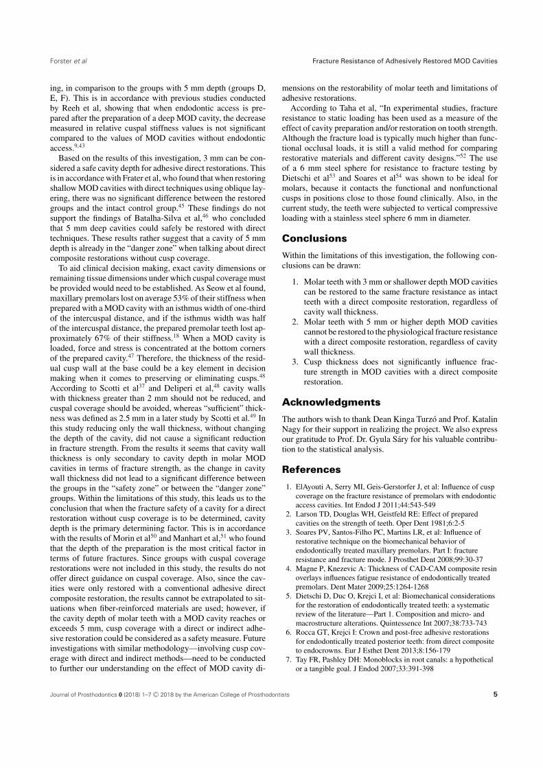
Forster et al Fracture Resistance of Adhesively Restored MOD Cavities
ing, in comparison to the groups with 5 mm depth (groups D,E, F). This is in accordance with previous studies conductedby Reeh et al, showing that when endodontic access is pre-pared after the preparation of a deep MOD cavity, the decreasemeasured in relative cuspal stiffness values is not significantcompared to the values of MOD cavities without endodonticaccess.9,43
Based on the results of this investigation, 3 mm can be con-sidered a safe cavity depth for adhesive direct restorations. Thisis in accordance with Frater et al, who found that when restoringshallow MOD cavities with direct techniques using oblique lay-ering, there was no significant difference between the restoredgroups and the intact control group.45 These findings do notsupport the findings of Batalha-Silva et al,46 who concludedthat 5 mm deep cavities could safely be restored with directtechniques. These results rather suggest that a cavity of 5 mmdepth is already in the “danger zone” when talking about directcomposite restorations without cusp coverage.
To aid clinical decision making, exact cavity dimensions orremaining tissue dimensions under which cuspal coverage mustbe provided would need to be established. As Seow et al found,maxillary premolars lost on average 53% of their stiffness whenprepared with a MOD cavity with an isthmus width of one-thirdof the intercuspal distance, and if the isthmus width was halfof the intercuspal distance, the prepared premolar teeth lost ap-proximately 67% of their stiffness.18 When a MOD cavity isloaded, force and stress is concentrated at the bottom cornersof the prepared cavity.47 Therefore, the thickness of the resid-ual cusp wall at the base could be a key element in decisionmaking when it comes to preserving or eliminating cusps.48
According to Scotti et al37 and Deliperi et al,48 cavity wallswith thickness greater than 2 mm should not be reduced, andcuspal coverage should be avoided, whereas “sufficient” thick-ness was defined as 2.5 mm in a later study by Scotti et al.49 Inthis study reducing only the wall thickness, without changingthe depth of the cavity, did not cause a significant reductionin fracture strength. From the results it seems that cavity wallthickness is only secondary to cavity depth in molar MODcavities in terms of fracture strength, as the change in cavitywall thickness did not lead to a significant difference betweenthe groups in the “safety zone” or between the “danger zone”groups. Within the limitations of this study, this leads us to theconclusion that when the fracture safety of a cavity for a directrestoration without cusp coverage is to be determined, cavitydepth is the primary determining factor. This is in accordancewith the results of Morin et al50 and Manhart et al,51 who foundthat the depth of the preparation is the most critical factor interms of future fractures. Since groups with cuspal coveragerestorations were not included in this study, the results do notoffer direct guidance on cuspal coverage. Also, since the cav-ities were only restored with a conventional adhesive directcomposite restoration, the results cannot be extrapolated to sit-uations when fiber-reinforced materials are used; however, ifthe cavity depth of molar teeth with a MOD cavity reaches orexceeds 5 mm, cusp coverage with a direct or indirect adhe-sive restoration could be considered as a safety measure. Futureinvestigations with similar methodology—involving cusp cov-erage with direct and indirect methods—need to be conductedto further our understanding on the effect of MOD cavity di-
mensions on the restorability of molar teeth and limitations ofadhesive restorations.
According to Taha et al, “In experimental studies, fractureresistance to static loading has been used as a measure of theeffect of cavity preparation and/or restoration on tooth strength.Although the fracture load is typically much higher than func-tional occlusal loads, it is still a valid method for comparingrestorative materials and different cavity designs.”52 The useof a 6 mm steel sphere for resistance to fracture testing byDietschi et al53 and Soares et al54 was shown to be ideal formolars, because it contacts the functional and nonfunctionalcusps in positions close to those found clinically. Also, in thecurrent study, the teeth were subjected to vertical compressiveloading with a stainless steel sphere 6 mm in diameter.
Conclusions
Within the limitations of this investigation, the following con-clusions can be drawn:
1. Molar teeth with 3 mm or shallower depth MOD cavitiescan be restored to the same fracture resistance as intactteeth with a direct composite restoration, regardless ofcavity wall thickness.
2. Molar teeth with 5 mm or higher depth MOD cavitiescannot be restored to the physiological fracture resistancewith a direct composite restoration, regardless of cavitywall thickness.
3. Cusp thickness does not significantly influence frac-ture strength in MOD cavities with a direct compositerestoration.
Acknowledgments
The authors wish to thank Dean Kinga Turzo and Prof. KatalinNagy for their support in realizing the project. We also expressour gratitude to Prof. Dr. Gyula Sary for his valuable contribu-tion to the statistical analysis.
References
1. ElAyouti A, Serry MI, Geis-Gerstorfer J, et al: Influence of cuspcoverage on the fracture resistance of premolars with endodonticaccess cavities. Int Endod J 2011;44:543-549
2. Larson TD, Douglas WH, Geistfeld RE: Effect of preparedcavities on the strength of teeth. Oper Dent 1981;6:2-5
3. Soares PV, Santos-Filho PC, Martins LR, et al: Influence ofrestorative technique on the biomechanical behavior ofendodontically treated maxillary premolars. Part I: fractureresistance and fracture mode. J Prosthet Dent 2008;99:30-37
4. Magne P, Knezevic A: Thickness of CAD-CAM composite resinoverlays influences fatigue resistance of endodontically treatedpremolars. Dent Mater 2009;25:1264-1268
5. Dietschi D, Duc O, Krejci I, et al: Biomechanical considerationsfor the restoration of endodontically treated teeth: a systematicreview of the literature—Part 1. Composition and micro- andmacrostructure alterations. Quintessence Int 2007;38:733-743
6. Rocca GT, Krejci I: Crown and post-free adhesive restorationsfor endodontically treated posterior teeth: from direct compositeto endocrowns. Eur J Esthet Dent 2013;8:156-179
7. Tay FR, Pashley DH: Monoblocks in root canals: a hypotheticalor a tangible goal. J Endod 2007;33:391-398
Journal of Prosthodontics 0 (2018) 1–7 C⃝ 2018 by the American College of Prosthodontists 5

Fracture Resistance of Adhesively Restored MOD Cavities Forster et al
8. El-Helali R, Dowling AH, McGinley EL, et al: Influence ofresin-based composite restoration technique and endodonticaccess on cuspal deflection and cervical microleakage scores. JDent 2013;41:216-222
9. Reeh ES, Douglas WH, Messer HH: Stiffness ofendodontically-treated teeth related to restoration technique. JDent Res 1989;68:1540-1544
10. Hannig C, Westphal C, Becker K, et al: Fracture resistance ofendodontically treated maxillary premolars restored withCAD/CAM ceramic inlays. J Prosthet Dent 2005;94:342-349
11. Seo DG, Yi YA, Shin SJ, et al: Analysis of factors associatedwith cracked teeth. J Endod 2012;38:288-292
12. Fennis WM, Tezvergil A, Kuijs RH, et al: In vitro fractureresistance of fiber reinforced cusp-replacing compositerestorations. Dent Mater 2005;21:565-572
13. Hansen EK, Asmussen E: In vivo fractures of endodonticallytreated posterior teeth restored with enamel-bonded resin. EndodDent Traumatol 1990;6:218-225
14. Lin CL, Chang CH, Ko CC: Multifactorial analysis of an MODrestored human premolar using auto-mesh finite elementapproach. J Oral Rehabil 2001;28:576-585
15. Taha NA, Palamara JE, Messer HH: Cuspal deflection, strain andmicroleakage of endodontically treated premolar teeth restoredwith direct resin composites. J Dent 2009;37:724-730
16. Hood JA: Biomechanics of the intact, prepared and restoredtooth: some clinical implications. Int Dent J 1991;41:25-32
17. Granath L, Svensson A: Elastic outward bending of loadedbuccal and lingual premolar walls in relation to cavity size andform. Scand J Dent Res 1991;99:1-7
18. Seow LL, Toh CH, Wilson NH: Strain measurements andfracture resistance of endodontically treated premolars restoredwith all-ceramic restorations. J Dent 2015;43:126-132
19. Fennis WM, Kuijs RH, Kreulen CM, et al: Fatigue resistance ofteeth restored with cuspal-coverage composite restorations. Int JProsthodont 2004;17:313-317
20. Burke FJ: Tooth fracture in vivo and in vitro. J Dent1992;20:131-139
21. Burke FJ, Wilson NH, Watts DC: The effect of cavity wall taperon fracture resistance of teeth restored with resin compositeinlays. Oper Dent 1993;18:230-236
22. St-Georges AJ, Sturdevant JR, Swift Jr EJ, et al: Fractureresistance of prepared teeth restored with bonded inlayrestorations. J Prosthet Dent 2003;89:551-557
23. Magne P, Carvalho AO, Bruzi G, et al: Influence of no-ferruleand no-post buildup design on the fatigue resistance ofendodontically treated molars restored with resin nanoceramicCAD/CAM crowns. Oper Dent 2014;39:595-602
24. Jayasooriya PR, Pereira PN, Nikaido T, et al: Efficacy of a resincoating on bond strengths of resin cement to dentin. J AesthetRestor Dent 2003;15:105–113
25. Tay FR, Pashley DH: Have dentin adhesives become toohydrophilic? J Can Dent Assoc 2003;69:726-731
26. Pongprueksa P, Kuphasuk W, Senawongse P: Effect of elasticcavity wall and occlusal loading on microleakage and dentinbond strength. Oper Dent 2007;32:466-475
27. Kivanc BH, Alacam T, Gorgul G: Fracture resistance ofpremolars with one remaining cavity wall restored using differenttechniques. Dent Mater J 2010;29:262-267
28. Wu Y, Cathro P, Marino V: Fracture resistance and pattern of theupper premolars with obturated canals and restored endodonticocclusal access cavities. J Biomed Res 2010;24:474-478
29. Lee MR, Cho BH, Son HH, et al: Influence of cavity dimensionand restoration methods on the cusp deflection of premolars incomposite restoration. Dent Mater 2007;23:288-295
30. Meredith N, Setchell DJ: In vitro measurement of cuspal strainand displacement in composite restored teeth. J Dent1997;25:331-337
31. Cara RR, Fleming GJ, Palin WM, et al: Cuspal deflection andmicroleakage in premolar teeth restored with resin-basedcomposites with and without an intermediary flowable layer. JDent 2007;35:482-489
32. Mount GJ, Ngo H: Minimal intervention: a new concept foroperative dentistry. Quintessence Int 2000;31:527-533
33. Yamada Y, Tsubota Y, Fukushima S: Effect of restorationmethod on fracture resistance of endodontically treated maxillarypremolars. Int J Prosthodont 2004;17:94-98
34. Cobankara FK, Unlu N, Cetin AR, et al: The effect of differentrestoration techniques on the fracture resistance ofendodontically-treated molars. Oper Dent 2008;33:526-533
35. Assif D, Gorfil C: Biomechanical considerations in restoringendodontically treated teeth. J Prosthet Dent 1994;71:565-567
36. Khers SC, Carpenter CW, Vetter JD, et al: Anatomy of cusps ofposterior teeth and their fracture potential. J Prosthet Dent1990;64:139-147
37. Scotti N, Rota R, Scansetti M, et al: Influence of adhesivetechniques on fracture resistance of endodontically treatedpremolars with various residual wall thicknesses. J Prosthet Dent2013;110:376-382
38. Nam SH, Chang HS, Min KS, et al: Effect of the number ofresidual walls on fracture resistances, failure patterns, andphotoelasticity of simulated premolars restored with or withoutfiber-reinforced composite posts. J Endod 2010;36:297-301
39. Mangold JT, Kern M: Influence of glass-fiber posts on thefracture resistance and failure pattern of endodontically treatedpremolars with varying substance loss: an in vitro study. JProsthet Dent 2011;105:387-393
40. Goel VK, Khera SC, Gurusami S, et al: Effect of cavity depth onstresses in a restored tooth. J Prosthet Dent 1992;67:174-183
41. Torbjorner A, Fransson B: Biomechanical aspects of prosthetictreatment of structurally compromised teeth. Int J Prosthodont2004;17:135-141
42. Jantarat J, Panitvisai P, Palamara JE, et al: Comparison ofmethods for measuring cuspal deformation in teeth. J Dent2001;29:75-82
43. Reeh ES, Messer HH, Douglas WH: Reduction in tooth stiffnessas a result of endodontic and restorative procedures. J Endod1989;15:512-516
44. Eakle WS, Maxwell EH, Braly BV: Fractures of posterior teethin adults. J Am Dent Assoc 1986;112:215-218
45. Frater M, Forster A, Kereszturi M, et al: In vitro fractureresistance of molar teeth restored with a short fibre-reinforcedcomposite material. J Dent 2014;42:1143-1150
46. Batalha-Silva S, de Andrada MA, Maia HP, et al: Fatigueresistance and crack propensity of large MOD composite resinrestorations: direct versus CAD/CAM inlays. Dent Mater2013;29:324-331
47. Bell JG, Smith MC, de Pont JJ: Cuspal failures of MOD restoredteeth. Aust Dent J 1982;27:283-287
48. Deliperi S, Bardwell DN: Multiple cuspal-coverage directcomposite restorations: functional and esthetic guidelines. JEsthet Restor Dent 2008;20:300-312
49. Scotti N, Eruli C, Comba A, et al: Longevity of class 2 directrestorations in root-filled teeth: a retrospective clinical study. JDent 2015;43:499-505
50. Morin D, DeLong R, Douglas WH: Cusp reinforcement by theacid-etch technique. J Dent Res 1984;63:1075-1078
6 Journal of Prosthodontics 0 (2018) 1–7 C⃝ 2018 by the American College of Prosthodontists
Forster et al Fracture Resistance of Adhesively Restored MOD Cavities
51. Manhart J, Kunzelmann KH, Chen HY, et al: Mechanicalproperties and wear behavior of light-cured packable compositeresins. Dent Mater 2000;16:33-40
52. Taha NA, Palamara JE, Messer HH: Fracture strength andfracture patterns of root filled teeth restored with direct resinrestorations. J Dent 2011;39:527-535
53. Dietschi D, Maeder M, Meyer JM, et al: In vitro resistance tofracture of porcelain inlays bonded to tooth. Quintessence Int1990;21:823-831
54. Soares CJ, Martins LR, Pfeifer JM, et al: Fracture resistance ofteeth restored with indirect-composite and ceramic inlay systems.Quintessence Int 2004;35:281-286
Journal of Prosthodontics 0 (2018) 1–7 C⃝ 2018 by the American College of Prosthodontists 7
Full Terms & Conditions of access and use can be found athttp://www.tandfonline.com/action/journalInformation?journalCode=tast20
Download by: [82.28.140.75] Date: 21 November 2016, At: 03:20
Journal of Adhesion Science and Technology
ISSN: 0169-4243 (Print) 1568-5616 (Online) Journal homepage: http://www.tandfonline.com/loi/tast20
In vitro fracture resistance of endodonticallytreated premolar teeth restored with a directlayered fiber-reinforced composite post and core
András Forster, Tekla Sáry, Gábor Braunitzer & Márk Fráter
To cite this article: András Forster, Tekla Sáry, Gábor Braunitzer & Márk Fráter (2016): In vitrofracture resistance of endodontically treated premolar teeth restored with a direct layeredfiber-reinforced composite post and core, Journal of Adhesion Science and Technology, DOI:10.1080/01694243.2016.1259758
To link to this article: http://dx.doi.org/10.1080/01694243.2016.1259758
Published online: 21 Nov 2016.
Submit your article to this journal
View related articles
View Crossmark data

JOURNAL OF ADHESION SCIENCE AND TECHNOLOGY, 2016http://dx.doi.org/10.1080/01694243.2016.1259758
In vitro fracture resistance of endodontically treated premolar teeth restored with a direct layered fiber-reinforced composite post and core
András Forstera , Tekla Sárya, Gábor Braunitzerb and Márk Frátera aFaculty of Dentistry, Department of Operative and Esthetic Dentistry, University of Szeged, Szeged, Hungary; bFaculty of Dentistry, Department of Oral Surgery, University of Szeged, Szeged, Hungary
ABSTRACTMethods for restoring endodontically treated teeth fall short of restoring the physiologic mechanical properties. Fracture of endodontically treated teeth is a common type of clinical failure. New treatment modalities or perfected versions of existing treatment concepts need to be tested to find a biomimetic solution. A novel method of restoring endodontically treated teeth is presented and compared in vitro with currently accepted restorative methods. Seventy-two extracted and endodontically treated maxillary premolar teeth were divided into six groups (n = 12) depending on restorative technique (Groups 1–6). Group 1: fiber-reinforced composite post (FRC), Group 2: direct layered short FRC post and core, Group 3: short fiber-reinforced obliquely layered composite restoration, Group 4: microhybrid composite restoration, Group 5: fiber-reinforced box, Group 6: control. Specimens were submitted to static fracture resistance test. Fracture thresholds and fracture patterns were evaluated. Group 6 exhibited the highest fracture resistance. Group 2 yielded the highest fracture resistance among the restored groups. The fracture resistance of Group 2 did not differ significantly from Group 6. Groups 1, 3, 4, 5 proved to be significantly different from the control group. There was no statistically significant difference among restored groups. Fracture patterns of tested groups were dominantly non repairable opposed to control groups mostly repairable fractures. Application of direct-layered short FRC post and core in endodontically treated premolars performed statistically similarly in the studied conditions as natural teeth. Therefore, it seems a promising alternative to current endo-restorative solutions. However, further testing is required.
1. Introduction
Endodontically treated teeth (ETT) are structurally different from nonrestored vital teeth and require specialized restorative treatment.[1] According to Dietschi et al. [2], the major difference is not a consequence of tissue quality alteration, but the result of the loss of dental
KEYWORDSFiber-reinforced composite; biomimetic; monoblock; minimal intervention dentistry
ARTICLE HISTORYReceived 13 July 2016 Revised 5 November 2016 Accepted 7 November 2016
© 2016 Informa UK Limited, trading as Taylor & Francis Group
CONTACT András Forster [email protected]
2 A. FORSTER ET AL.
structures (e.g. marginal ridges, pericervical dentine and the arched roof of the pulp cham-ber) caused by caries, trauma or endodontic procedures. As a result of the compromised structural integrity, ETT are at an increased risk of fracture.[3,4] The fracture strength of these teeth is dependent on the amount and position of the remaining tooth structures and the choice of restorative material and technique.[5–13]
Several studies highlighted that postinsertion significantly increased the fracture resist-ance of nonvital premolars[10,14–16]; however, other studies only managed to prove the positive effect of postinsertion on the fracture pattern in premolar teeth.[17–19] The latter was also confirmed by Trope et al. [20] and Zicari et al. [21] who came to the conclusion that posts do not strengthen the tooth. These findings could be caused by the mismatch between fiber posts and the root canal diameter since according to modern minimal inva-sive principles postspace preparation should not contribute to radicular dentine removal.[22,23] Following a minimally invasive postspace preparation leaves us with unique and irregular spaces [24] which are difficult to fill out with a single conventional or even a flex-ible posts. A further problem with posts is that they are placed in the most central part of the postspace (neutral axis), leaving the space originally occupied by dentine to be filled by the mechanically inferior luting composite material. A more effective reinforcement location mechanically may be on the outer surfaces of the postspace close to the dentine walls where the highest tensile stresses occur.[25] This is the space which before the root canal preparation was most likely obtained by healthy radicular and pericervical dentine. So to reach a reinforced, mechanically homogenous unit it would be ideal if a restorative material could be directly bonded to the root canal dentine and it would have mechanical properties similar to that of dentine.
In a 2007 investigation by Garoushi and coworkers, it was found that anterior ETT showed better load-bearing capacity if restored with a short fiber-reinforced composite (SFRC) restorative material opposed to a fiber-reinforced composite (FRC) post.[26] These findings open the debate on the necessity of the FRC post concept and open a gateway for the promotion of new types of endo-restorative techniques.
The aim of this investigation was to compare the mechanical properties of novel methods for the reinforcement of ETT utilizing SFRC (EverX Posterior, GC Europe, Leuven); with previously tested and accepted restorative methods. The null hypothesis was that (1) there would be no difference in the maximal fracture resistance of the ETT restored teeth with the tested methods. (2) There would be no difference in the fracture patterns of the ETT restored with the tested methods.
2. Materials and methods
All procedures of the study were approved by the Ethics Committee of the University of Szeged, and the study was designed in accordance with the Declaration of Helsinki.
Seventy-two upper premolar teeth, extracted for periodontal or orthodontic reasons were selected for this investigation. Specimen selection, exclusion criteria, root canal treatment protocol, specimen preparation and mechanical testing were carried out as described by Frater et al. [27,28].
The freshly extracted teeth were immediately placed in 5.25% NaOCl for 5 min and stored in 0.9% saline solution at room temperature. Teeth were used within 6 months after extraction.

JOURNAL OF ADHESION SCIENCE AND TECHNOLOGY 3
During specimen preparation the soft tissue covering the root surface was removed with hand scalers. The inclusion criteria were absence of caries or root cracks, the absence of previous endodontic treatments, posts or crowns, resorptions or evident lateral canals. Buccolingual and mesiodistal radiographs of all teeth were taken and examined to evaluate root integrity and the number of canals present. To standardize procedures and materials, all teeth used in this study had 1 root canal with a curvature of less than 5°, evaluated by Schneider’s technique,[29] and teeth with a root length of 15 ± 1 mm and similar mesio-distal and bucco-lingual dimensions (±10%) were selected.
90% of the specimen ranged 9–10 mm in size, measured at the widest bucco-lingual dimension, and the rest measured were 6.5–8 mm. Regarding the mesio-distal dimension, 90% of the specimen ranged 7–7.5 mm, and the rest were 6.5–8 mm.
The teeth were randomly distributed over six study groups of 12 specimens each.Access cavity was prepared by the same trained operator in five groups of the six, and
one group was left intact to serve as control (Group 6).Access cavity preparation was carried out with a round-end, tapered, medium grit,
0.8 mm tip diameter, 10 mm length diamond bur (850-014 M SSWhite, Lakewood, NJ, USA) with water cooling in the approximated centre of the occlusal surface according to standardized parameters: the access cavity involved one-third of the intracuspal distance in the bucco-lingual dimension and one-third of the mesio-distal distance, measured at the level of the central fissure.
The working length was established with the direct method by subtracting 1 mm from the real root length determined by introducing a number 10 K-file (Maillefer-Dentsply, Ballaigues, Switzerland) until it was visible through the apical foramen. The canals were instrumented using rotary ProTaper Universal files (Dentsply, Maillefer, Ballaigues, Switzerland). The ProTaper sequence (S1, S2, F1, F2) was used for the preparation at the working length.
Irrigation was performed after every instrument with 2 ml of 2.5% NaOCl solution and the canal space was filled with irrigant during the instrumentation phase. After the shaping and cleaning of the root canal, the roots were dried with 96% alcohol and paper points. Root canal filling was done by matched single-cone obturation with a master cone (F2 gutta-percha, Maillefer-Dentsply, Ballaigues, Switzerland) and sealer (AH plus; Dentsply De Trey GmbH, Konstanz, Germany). The access cavity was temporarily filled with Fuji Triage Pink (GC Europe, Leuven, Belgium). Fuji Triage Pink was applied to the apical part of the root in order to prevent leakage through the apex. The teeth were stored in an incubator (mco-18aic, Sanyo, Japan) for 1 week (at 37 °C, 100% relative humidity).
Groups 1 and 2 received a minimal invasive post space preparation with a depth of 8 mm, as measured from the CEJ on the buccal aspect of the tooth, but no post preparation drills recommended by the manufacturer were used in order to preserve the individual anatomy of the specimen teeth. Only the root canal filling was removed with size three Gates Glidden burs and ISO standard Hedstrom files leaving a minimum apical seal of 6–8 mm of gut-ta-percha in the canal. The Number 3 Gates Glidden bur was used on the full 8 mm length.
In Groups 3–5 the gutta-percha was only cut back 2 mm below the CEJ with an 0.1 mm diameter ball-shaped carbide bur (H1SM.205.010, Gebr. Brasseler GmbH & Co. KG, Lemgo, Germany), but no post space preparation was performed. After cutting back the gutta-percha, the orifice was sealed with resin modified glass ionomer cement (Fuji VIII, GC Europe, Leuven, Belgium).
4 A. FORSTER ET AL.
All specimen received the same adhesive treatment. Prior to the adhesive treatment of the cavity and the root canal, enamel was acid-etched selectively with 37% phosphoric acid for 15 s and rinsed with water. The root canal and the coronal cavity were rinsed with 2 ml of water and dried with paper points and air. For bonding, a dual-cure one-step self-etch adhesive system (Gradia Core Self-Etching Bond, GC Europe, Leuven, Belgium) was used, according to the manufacturer’s instructions using a microbrush-X disposable applicator (Pentron Clinical Technologies, LLC, USA). Excess adhesive was removed by suction drying (Evacuation Tip – Starryshine, Anaheim, CA, USA) within 0.5 cm from the occlusal cavity (without contact). Excess adhesive resin at the bottom of the canal was removed with a paper point. The adhesive was light-cured for 60 s using an Optilux 501 quartz-tungsten-halogen light-curing unit (Kerr Corp., Orange, CA, USA). The average power density of the light source, measured with a digital radiometer (Jetlite light tester; J. Morita USA Inc. Irvine, CA, USA) prior to the bonding procedure, was 840 ± 26.8 mW/cm2.
Five different techniques were used to restore the specimens (Figure 1):
Group 1
The teeth received a prefabricated, conventional FRC post (GC Fiber post, GC Europe, Leuven, Belgium). Before the adhesive treatment, the conventional translucent FRC posts of 0.8 mm diameter (GC Fiber Post, GC Europe, Leuven, Belgium) was tried in and cut to a length 1 mm below the level of the occlusal cavity margins with a water-cooled diamond disc (Isomet 2000; Buehler Ltd., Lake Bluff, IL, USA) and cleaned with alcohol after try in. The posts received silanization of the surface (Ceramic Primer, GC Europe, Leuven, Belgium) following the manufacturer’s recommendation. After silanization, the post surface was bonded with the same bonding agent used for the cavity. Luting of the posts and the core build-up was performed with a dual-cure resin composite core material (Gradia Core, GC Europe, Leuven, Belgium). Gradia Core was applied using its own automix cartridge with an ‘elongation tip’ for direct root canal application. After insertion of the post, 5 min of chemopolymerization time was provided to reduce polymerisation stress, then cement was polymerized with an Optilux 501 quartz tungsten-halogen light-curing unit for 60 s from each side (a total of 240 s/tooth). The outlines of the restoration were finished with dental composite (G-aenial Posterior P-JE, GC Europe, Leuven, Belgium).
Group 2
The teeth were reconstructed with a novel method of building a direct layered FRC post and core (DLFRC post and core) from SFRC (EverX Posterior, GC Europe, Leuven, Belgium). The DLFRC post and core was horizontally layered in 1–2 mm segments. An increment of SFRC was packed to the apical portion of the postspace using a microbrush-X disposable applicator (Pentron Clinical Technologies, LLC, USA). A light transmitting FRC post (0.8 mm GC Fiber post, GC Europe, Leuven, Belgium) was inserted into the postspace in order to aid the trans-mission of the light to the apically positioned layers. The ‘light transmitting’ post was withdrawn with 0.5–1 mm from the surface of the uncured SFRC layer not to have direct contact with it.
After each layer 80 s of light curing through the fiber post was carried out. After incre-mentally filling the root canal to the level of the CEJ with repeating the previously described procedure, SFRC was layered in the coronal cavity until 1 mm below the margin of the

JOURNAL OF ADHESION SCIENCE AND TECHNOLOGY 5
Figure 1. Restored study groups, layering concepts and restorative materials applied.
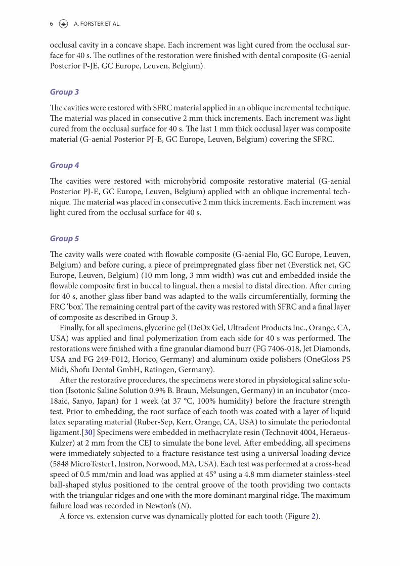
6 A. FORSTER ET AL.
occlusal cavity in a concave shape. Each increment was light cured from the occlusal sur-face for 40 s. The outlines of the restoration were finished with dental composite (G-aenial Posterior P-JE, GC Europe, Leuven, Belgium).
Group 3
The cavities were restored with SFRC material applied in an oblique incremental technique. The material was placed in consecutive 2 mm thick increments. Each increment was light cured from the occlusal surface for 40 s. The last 1 mm thick occlusal layer was composite material (G-aenial Posterior PJ-E, GC Europe, Leuven, Belgium) covering the SFRC.
Group 4
The cavities were restored with microhybrid composite restorative material (G-aenial Posterior PJ-E, GC Europe, Leuven, Belgium) applied with an oblique incremental tech-nique. The material was placed in consecutive 2 mm thick increments. Each increment was light cured from the occlusal surface for 40 s.
Group 5
The cavity walls were coated with flowable composite (G-aenial Flo, GC Europe, Leuven, Belgium) and before curing, a piece of preimpregnated glass fiber net (Everstick net, GC Europe, Leuven, Belgium) (10 mm long, 3 mm width) was cut and embedded inside the flowable composite first in buccal to lingual, then a mesial to distal direction. After curing for 40 s, another glass fiber band was adapted to the walls circumferentially, forming the FRC ‘box’. The remaining central part of the cavity was restored with SFRC and a final layer of composite as described in Group 3.
Finally, for all specimens, glycerine gel (DeOx Gel, Ultradent Products Inc., Orange, CA, USA) was applied and final polymerization from each side for 40 s was performed. The restorations were finished with a fine granular diamond burr (FG 7406-018, Jet Diamonds, USA and FG 249-F012, Horico, Germany) and aluminum oxide polishers (OneGloss PS Midi, Shofu Dental GmbH, Ratingen, Germany).
After the restorative procedures, the specimens were stored in physiological saline solu-tion (Isotonic Saline Solution 0.9% B. Braun, Melsungen, Germany) in an incubator (mco-18aic, Sanyo, Japan) for 1 week (at 37 °C, 100% humidity) before the fracture strength test. Prior to embedding, the root surface of each tooth was coated with a layer of liquid latex separating material (Ruber-Sep, Kerr, Orange, CA, USA) to simulate the periodontal ligament.[30] Specimens were embedded in methacrylate resin (Technovit 4004, Heraeus-Kulzer) at 2 mm from the CEJ to simulate the bone level. After embedding, all specimens were immediately subjected to a fracture resistance test using a universal loading device (5848 MicroTester1, Instron, Norwood, MA, USA). Each test was performed at a cross-head speed of 0.5 mm/min and load was applied at 45° using a 4.8 mm diameter stainless-steel ball-shaped stylus positioned to the central groove of the tooth providing two contacts with the triangular ridges and one with the more dominant marginal ridge. The maximum failure load was recorded in Newton’s (N).
A force vs. extension curve was dynamically plotted for each tooth (Figure 2).
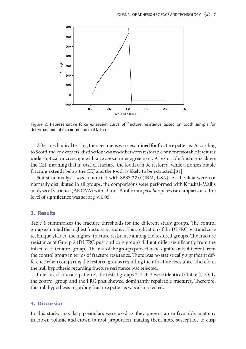
JOURNAL OF ADHESION SCIENCE AND TECHNOLOGY 7
After mechanical testing, the specimens were examined for fracture patterns. According to Scotti and co-workers, distinction was made between restorable or nonrestorable fractures under optical microscope with a two-examiner agreement. A restorable fracture is above the CEJ, meaning that in case of fracture, the tooth can be restored, while a nonrestorable fracture extends below the CEJ and the tooth is likely to be extracted.[31]
Statistical analysis was conducted with SPSS 22.0 (IBM, USA). As the data were not normally distributed in all groups, the comparisons were performed with Kruskal–Wallis analysis of variance (ANOVA) with Dunn–Bonferroni post hoc pairwise comparisons. The level of significance was set at p < 0.05.
3. Results
Table 1 summarizes the fracture thresholds for the different study groups. The control group exhibited the highest fracture resistance. The application of the DLFRC post and core technique yielded the highest fracture resistance among the restored groups. The fracture resistance of Group 2 (DLFRC post and core group) did not differ significantly from the intact teeth (control group). The rest of the groups proved to be significantly different from the control group in terms of fracture resistance. There was no statistically significant dif-ference when comparing the restored groups regarding their fracture resistance. Therefore, the null hypothesis regarding fracture resistance was rejected.
In terms of fracture patterns, the tested groups 2, 3, 4, 5 were identical (Table 2). Only the control group and the FRC post showed dominantly repairable fractures. Therefore, the null hypothesis regarding fracture patterns was also rejected.
4. Discussion
In this study, maxillary premolars were used as they present an unfavorable anatomy in crown volume and crown to root proportion, making them more susceptible to cusp
Figure 2. Representative force extension curve of fracture resistance tested on tooth sample for determination of maximum force of failure.
8 A. FORSTER ET AL.
fractures than other posterior teeth when submitted to occlusal load application.[10] An oblique load (45° to the long axis of the tooth) was applied to the occlusal incline of the buccal cusp using a rounded loading tip, intended to simulate normal working side occlusal contacts.[3]
ETT demonstrate an increased risk of fracture, and therefore, an acceptable restoration, in addition to aesthetic considerations, should not only restore function, but should also aim to preserve and reinforce the remaining dental structures thus reducing the occurrence of catastrophic failures and increasing the longevity of the restoration.[32–35] However, there seems to be little evidence to guide the clinician toward the best restorative solution if only the access cavity needs restoring in a premolar tooth.[4,9]
According to several authors, posterior teeth with an endodontic access preparation and no other structural loss may succeed with a conservative bonded restoration.[8,36,37] In the present study, the specimen restored with composite restorations applied with the oblique layering technique (Group 4) showed the lowest fracture resistance values among the restored groups; however, this was not statistically significantly lower compared to the other restored groups. These findings are in accordance with some studies showing that restored root-filled maxillary premolars without a fiber post showed similar fracture resistance to those when an FRC post was placed.[38–40] This could be attributed to the minimal invasive access cavity preparation in this study, leading to still acceptable ‘inter-nal splinting’ with adhesively bonded composite restorations. Also, the inconsistency in the findings of different FRC post-related studies may be attributed to the difference in postspace preparation procedures. Preparing the root canal to receive a post with pilot drills and removing radicular dentine on purpose, might lead to cracks and defects that can concentrate stress and increase the possibility of tooth fracture.[41] For this reason, a minimal invasive postspace preparation was performed in this investigation, leading to the preservation of the individual, irregular root canal anatomy and shape. Because of this the inserted, smaller rigid FRC post could not fill out the root canal entirely, possibly leading
Table 1. Fracture thresholds of studied groups and the significance of their difference compared to the control group.
Notes: Group 1: glass fibre- reinforced post; Group 2: direct-layered glass fiber-reinforced composite core; Group 3: SFRC applied by an oblique incremental technique; Group 4: obliquely layered conventional composite and Group 5: torsion box with FRC net. As there was no significant difference among the restored groups in this respect, significances are shown as compared to the controls only.
Group Sig. compared to control (p, post hoc) N Minimum (N) Maximum (N) Mean (N) SDControl – 12 605.85 1205.83 922.34 189.21Group 1 0.005 12 208.28 802.61 501.30 186.65Group 2 1.000 12 352.85 1171.19 727.98 287.37Group 3 0.009 12 123.59 865.93 511.61 225.20Group 4 0.005 12 216.67 748.44 456.24 189.75Group 5 0.023 12 303.64 682.83 536.35 126.41
Table 2. Fracture patterns.
Notes: The numbers indicate relative frequencies (n = 12 in each group). Group 1: glass fibre- reinforced post; Group 2: direct-layered glass fiber-reinforced composite core; Group 3: SFRC applied by an oblique incremental technique; Group 4: obliquely layered conventional composite and Group 5: torsion box with FRC net.
Control Group 1 Group 2 Group 3 Group 4 Group 5Reparable 0.66 0.58 0.33 0.33 0.33 0.33Irreparable 0.33 0.42 0.66 0.66 0.66 0.66

JOURNAL OF ADHESION SCIENCE AND TECHNOLOGY 9
to greater amount of luting composite in the prepared space. This could account for the inferior findings in the present study regarding the post-restored group as Group 1 was not statistically superior to the other restored groups.
SFRC is a dental restorative composite resin [29,42,43] intended to be used in high stress bearing areas as a stress-breaker restorative material. Mechanical testing has shown major improvements in the load-bearing capacity, the flexural strength and also the fracture resistance of SFRC in comparison with conventional particulate filler composite resin. [44–48] In the current investigation, the group restored with obliquely layered SFRC (Group 3) reached higher fracture resistance figures compared to the ones restored with conventional composite (Group 4) or even the conventional FRC post group (Group 1); however, the difference was statistically not significant. This has been previously described in molar teeth with MOD cavities, where the SFRC-restored groups yielded better results than the conventional composite restored ones, yet the difference was not significant.[49]
While in the SFRC material the E-glass fibers are randomly oriented and possess iso-tropic features, leading to possible reinforcement in multiple directions, the E-glass fibers in the bidirectional pre-impregnated fiber-reinforced net (everStick Net) show orthotropic properties, therefore reinforcing the structure in two directions.[50,51] The threefold usage of FRC net (FRC box) together with the SFRC-restorative composite is aiming to recon-struct the integrity and strength of the opposing cavity walls. The values of the FRC box restorations showed an increase compared to Groups 1, 3 and 4, this technique could also not emulate the values measured in case of sound premolar teeth. However, in this study, there seems to be a clear tendency of increasing fracture resistance values toward the use of individualized FRC materials compared to conventional techniques (composite restoration or FRC postplacement). This observation is in accordance with the findings of Bijelic et al. [52]. The inferior, though not statistically different results of the FRC box group (Group 5) compared to the DLFRC post and core group (Group 2) can be explained by the coronal position of the FRC box, which can only keep the coronal part of the tooth together but does not reinforce the cervical and root part of the tooth.
Prefabricated FRC posts suffer from two main shortcomings in clinical settings: Insufficient bonding of the interfaces [53–55] and the fact that the post position is in the neutral axis of the root canal. Direct layering of SFRC into the root canal is intended to solve these drawbacks. Seyam et al. and other authors showed that a transparent post can transfer the light and aid the polymerization of composite resin in the root canal,[56–58] enabling layering in the hollow root canal space. However, there are investigations to oppose this statement [59,60]. The DLFRC post and core technique (Group 2) produced the highest frac-ture resistance values among the restored groups in the present study. These results seem to be in accordance with Garoushi et al. showing that the thicker the applied SFRC restoration the greater the fracture resistance is [61]. Although the numbers produced by Group 2 were not significantly higher than the rest of the restored groups, a positive tendency could be visible with the utilization of SFRC materials. Moreover, there was no statistically significant difference between the Group 2 and the intact teeth. This result suggests a move toward a biomimetic restorative concept. It has to be noted that the reported advantages come at the price of increased application time and technically more demanding clinical procedure as compared to Group 1. Development of materials, instruments and light curing equipment specifically for such purposes could be promising and could resolve the main shortcomings of the DLFRC post and core method as described in this investigation.
10 A. FORSTER ET AL.
Regarding fracture pattern, the tested groups were identical with dominantly unfavora-ble, irreparable fractures. Only the control group and Group 1 presented a shift toward favorable, repairable fractures. Therefore, the null hypothesis regarding fracture pattern was rejected. The DLFRC post and core technique according to the findings of this inves-tigation might hold the potential of reinforcing the root and particularly the pericervical area, which is highly beneficial when shear forces are also present (e.g. 45° loading). The DLFRC post and core concept theoretically could present a possibility to compensate for most of the known weaknesses of the presently accepted endo-restorative options with a not complicated, clinically feasible and reproducible methodology. Given these facts and the promising results in the current investigation, it is recommended to conduct further investigations particularly applying artificial ageing, cyclic loading until fracture, different cavity extensions and tooth groups in vitro.
The limitations of this investigation are the limited number of specimens, which makes it much more unlikely to exhibit statistical significance. It is also a limitation that maxi-mal fracture resistance testing only indicates a certain physical property of the structure not giving information of the possibly clinically more relevant reaction to cyclic loading and ageing. Given the mentioned shortcomings the results and the novel method have to be implemented with caution. Therefore, the proposed techniques should require future testing in more clinically realistic situations. The proposed investigation could be deemed as a possible first, still critical step toward the development of clinically relevant future endo-restorative methods.
5. Conclusions
Within the limitations of this investigation, it can be concluded that natural teeth exhibit significantly higher fracture resistance then the endodontically treated and restored pre-molar teeth except for the DLFRC post and core restored group which did not show sta-tistically significant difference compared to the control group. FRC posts exhibited more favorable fracture patterns then the other restored groups examined. The direct layered short fiber-reinforced post and core is a promising alternative to the accepted restorations of endodontically treated teeth, and as such should be further investigated.
AcknowledgementsThe authors wish to thank Balázs P. Szabó PhD for his support in the fracture strength measurements. We also thank dean Prof. Katalin Nagy and dean Kinga Turzó PhD for their support in realizing this project.
Disclosure statementNo potential conflict of interest was reported by the authors.
ORCIDAndrás Forster http://orcid.org/0000-0002-3495-7105Márk Fráter http://orcid.org/0000-0002-0365-1613

JOURNAL OF ADHESION SCIENCE AND TECHNOLOGY 11
References [1] Cecchin D, de Almeida JF, Gomes BP, et al. Effect of chlorhexidine and ethanol on the durability
of the adhesion of the fiber post relined with resin composite to the root canal. J Endod. 2011;37:678–683.
[2] Dietschi D, Duc O, Krejci I, et al. Biomechanical considerations for the restoration of endodontically treated teeth: a systematic review of the literature – part 1. Composition and micro- and macrostructure alterations. Quintessence Int. 2007;38:733–743.
[3] Taha NA, Palamara JE, Messer HH. Fracture strength and fracture patterns of root filled teeth restored with direct resin restorations. J Dent. 2011;39:527–535.
[4] Mohammadi N, Kahnamoii MA, Yeganeh PK, et al. Effect of fiber post and cusp coverage on fracture resistance of endodontically treated maxillary premolars directly restored with composite resin. J Endod. 2009;35:1428–1432.
[5] Balkaya MC, Birdal IS. Effect of resin-based materials on fracture resistance of endodontically treated thin-walled teeth. J Prosthet Dent. 2013;109:296–303.
[6] Rocca GT, Krejci I. Crown and post-free adhesive restorations for endodontically treated posterior teeth: from direct composite to endocrowns. Eur J Esthet Dent. 2013;8:156–179.
[7] Tay FR, Pashley DH. Monoblocks in root canals: a hypothetical or a tangible goal. J Endod. 2007;33:391–398.
[8] Reeh ES, Messer HH, Douglas WH. Reduction in tooth stiffness as a result of endodontic and restorative procedures. J Endod. 1989;15:512–516.
[9] Wu Y, Cathro P, Marino V. Fracture resistance and pattern of the upper premolars with obturated canals and restored endodontic occlusal access cavities. J Biomed Res. 2010;24:474–478.
[10] Soares PV, Santos-Filho PC, Martins LR, et al. Influence of restorative technique on the biomechanical behavior of endodontically treated maxillary premolars. Part I: fracture resistance and fracture mode. J Prosthet Dent. 2008;99:30–37.
[11] Seow LL, Toh CG, Wilson NH. Strain measurements and fracture resistance of endodontically treated premolars restored with all-ceramic restorations. J Dent. 2015;43:126–132.
[12] Mondelli RF, Barbosa WF, Mondelli J, et al. Fracture strength of weakened human premolars restored with amalgam with and without cusp coverage. Am J Dent. 1998;11:181–184.
[13] Stappert CF, Att W, Gerds T, et al. Fracture resistance of different partial-coverage ceramic molar restorations: an in vitro investigation. J Am Dent Assoc. 2006;137:514–522.
[14] Scotti N, Scansetti M, Rota R, et al. The effect of the post length and cusp coverage on the cycling and static load of endodontically treated maxillary premolars. Clin Oral Invest. 2011;15:923–929.
[15] Bitter K, Noetzel J, Stamm O, et al. Randomized clinical trial comparing the effects of post placement on failure rate of postendodontic restorations: preliminary results of a mean period of 32 months. J Endod. 2009;35:1477–1482.
[16] Nothdurft FP, Seidel E, Gebhart F, et al. The fracture behavior of premolar teeth with class II cavities restored by both direct composite restorations and endodontic post systems. J Dent. 2008;36:444–449.
[17] Qualtrough AJ, Mannocci F. Tooth-colored post systems: a review. Oper Dent. 2003;28:86–91.[18] Soares CJ, Soares PV, de Freitas Santos-Filho PC, et al. The influence of cavity design and
glass fiber posts on biomechanical behavior of endodontically treated premolars. J Endod. 2008;34:1015–1019.
[19] Stockton L, Lavelle CL, Suzuki M. Are posts mandatory for the restoration of endodontically treated teeth? Dent Traumatol. 1998;14:59–63.
[20] Trope M, Maltz DO, Tronstad L. Resistance to fracture of restored endodontically treated teeth. Dent Traumatol. 1985;1:108–111.
[21] Zicari F, Van Meerbeek B, Scotti R, et al. Effect of fiber post length and adhesive strategy on fracture resistance of endodontically treated teeth after fatigue loading. J Dent. 2012;40:312–321.
[22] Meyenberg K. The ideal restoration of endodontically treated teeth–structural and esthetic considerations: a review of the literature and clinical guidelines for the restorative clinician. Eur J Esthet Dent. 2013;8:238–268.
12 A. FORSTER ET AL.
[23] Paolone G, Saracinelli M, Devoto W, et al. Esthetic direct restorations in endodontically treated anterior teeth. Eur J Esthet Dent. 2013;8:44–67.
[24] Maceri F, Martignoni M, Vairo G. Mechanical behaviour of endodontic restorations with multiple prefabricated posts: a finite-element approach. J Biomech. 2007;40:2386–2398.
[25] Le Bell-Ronnlof AM, Lassila LV, Kangasniemi I, et al. Load-bearing capacity of human incisor restored with various fiber-reinforced composite posts. Dent Mater. 2011;27:e107–e115.
[26] Garoushi S, Vallittu PK, Lassila LV. Direct restoration of severely damaged incisors using short fiber-reinforced composite resin. J Dent. 2007;35:731–736.
[27] Frater M. The restorative use of fibre-reinforced materials in the posterior region [PhD thesis]. Szeged, Hungary: University of Szeged; 2015.
[28] Frater M, Forster A, Jantyik A, et al. In vitro fracture resistance of premolar teeth restored with fibre-reinforced composite posts using a single or a multi-post technique. Aust Endod J; 2016. doi: 10.1111/aej.12150. [Epub ahead of print]
[29] Schneider SW. A comparison of canal preparations in straight and curved root canals. Oral Sur. Oral Med Oral Pathol. 1971;32:271–275.
[30] Magne P, Paranhos MP, Schlichting LH. Influence of material selection on the risk of inlay fracture during pre-cementation functional occlusal tapping. Dent Mater. 2011;27:109–113.
[31] Scotti N, Coero Borga FA, Alovisi M, et al. Is fracture resistance of endodontically treated mandibular molars restored with indirect onlay composite restorations influenced by fiber post insertion? J Dent. 2012;40:814–820.
[32] Batalha-Silva S, de Andrada MA, Maia HP, et al. Fatigue resistance and crack propensity of large MOD composite resin restorations: direct versus CAD/CAM inlays. Dent Mater. 2013;29:324–331.
[33] Dejak B, Mlotkowski A. Finite element analysis of strength and adhesion of cast posts compared to glass fiber-reinforced composite resin posts in anterior teeth. J Prosthet Dent. 2011;105:115–126.
[34] Torbjorner A, Fransson B. Biomechanical aspects of prosthetic treatment of structurally compromised teeth. Int J Prosthodont. 2004;17:135–141.
[35] Torres-Sanchez C, Montoya-Salazar V, Cordoba P, et al. Fracture resistance of endodontically treated teeth restored with glass fiber reinforced posts and cast gold post and cores cemented with three cements. J Prosthet Dent. 2013;110:127–133.
[36] Scotti N, Rota R, Scansetti M, et al. Influence of adhesive techniques on fracture resistance of endodontically treated premolars with various residual wall thicknesses. J Prosthet Dent. 2013;110:376–382.
[37] Steele A, Johnson BR. In vitro fracture strength of endodontically treated premolars. J Endod. 1999;25:6–8.
[38] Yamada Y, Tsubota Y, Fukushima S. Effect of restoration method on fracture resistance of endodontically treated maxillary premolars. Int J Prosthodont. 2004;17:94–98.
[39] Krejci I, Duc O, Dietschi D, et al. Marginal adaptation, retention and fracture resistance of adhesive composite restorations on devital teeth with and without posts. Oper Dent. 2003;28:127–135.
[40] Fokkinga WA, Le Bell AM, Kreulen CM, et al. Ex vivo fracture resistance of direct resin composite complete crowns with and without posts on maxillary premolars. Int Endod J. 2005;38:230–237.
[41] Siso SH, Hurmuzlu F, Turgut M, et al. Fracture resistance of the buccal cusps of root filled maxillary premolar teeth restored with various techniques. Int Endod J. 2007;40:161–168.
[42] Cailleteau JG, Rieger MR, Akin JE. A comparison of intracanal stresses in a post-restored tooth utilizing the finite element method. J Endod. 1992;18:540–544.
[43] Garoushi S, Vallittu PK, Lassila LV. Short glass fiber reinforced restorative composite resin with semi-inter penetrating polymer network matrix. Dent Mater. 2007;23:1356–1362.
[44] Garoushi S, Vallittu PK, Watts DC, et al. Polymerization shrinkage of experimental short glass fiber-reinforced composite with semi-inter penetrating polymer network matrix. Dent Mater. 2008;24:211–215.
[45] Garoushi S, Vallittu PK, Lassila LV. Fracture toughness, compressive strength and load-bearing capacity of short glass fiber-reinforced composite resin. Chin J Dent Res. 2011;14:15–19.

JOURNAL OF ADHESION SCIENCE AND TECHNOLOGY 13
[46] Vallittu PK. Flexural properties of acrylic resin polymers reinforced with unidirectional and woven glass fibers. J Prosthet Dent. 1999;81:318–326.
[47] Lassila LV, Nohrstrom T, Vallittu PK. The influence of short-term water storage on the flexural properties of unidirectional glass fiber-reinforced composites. Biomaterials. 2002;23:2221–2229.
[48] Turkaslan S, Bagis B, Akan E, et al. Fracture strengths of chair-side-generated veneers cemented with glass fibers. Niger J Clin Pract. 2015;18:240–246.
[49] Frater M, Forster A, Kereszturi M, et al. In vitro fracture resistance of molar teeth restored with a short fiber-reinforced composite material. J Dent. 2014;42:1143–1150.
[50] Fennis WM, Tezvergil A, Kuijs RH, et al. In vitro fracture resistance of fiber reinforced cusp-replacing composite restorations. Dent Mater. 2005;21:565–572.
[51] Basaran EG, Ayna E, Vallittu PK, et al. Load bearing capacity of fiber-reinforced and unreinforced composite resin CAD/CAM-fabricated fixed dental prostheses. J Prosthet Dent. 2013;109:88–94.
[52] Bijelic J, Garoushi S, Vallittu PK, et al. Short fiber reinforced composite in restoring severely damaged incisors. Acta Odontol Scand. 2013;71:1221–1231.
[53] Bell AM, Lassila LV, Kangasniemi I, et al. Bonding of fiber-reinforced composite post to root canal dentin. J Dent. 2005;33:533–539.
[54] Belli S, Eraslan O, Eskitascioglu G, et al. Monoblocks in root canals: a finite elemental stress analysis study. Int Endod J. 2011;44:817–826.
[55] Chieruzzi M, Pagano S, Pennacchi M, et al. Compressive and flexural behaviour of fiber reinforced endodontic posts. J Dent. 2012;40:968–978.
[56] Seyam RS, Mobarak EH. Reinforcement of teeth with simulated coronal fracture and immature weakened roots using resin composite cured by a modified layering technique. Oper Dent. 2014;39:E128–E136.
[57] Galhano GA, de Melo RM, Barbosa SH, et al. Evaluation of light transmission through translucent and opaque posts. Oper Dent. 2008;33:321–324.
[58] Aksornmuang J, Nakajima M, Panyayong W, et al. Effects of photocuring strategy on bonding of dual-cure one-step self-etch adhesive to root canal dentin. Dent Mater J. 2009;28:133–141.
[59] Goracci C, Corciolani G, Vichi A, et al. Light-transmitting ability of marketed fiber posts. J Dent Res. 2008;87:1122–1126.
[60] Faria e Silva AL, Arias VG, Soares LE, et al. Influence of fiber-post translucency on the degree of conversion of a dual-cured resin cement. J Endod. 2007;33:303–305.
[61] Garoushi S, Mangoush E, Vallittu M, et al. Short fiber reinforced composite: a new alternative for direct only restorations. Open Dent J. 2013;7:181–185.
ORIGINAL RESEARCH
In vitro fracture resistance of premolar teeth restored with fibre-reinforced composite posts using a single or a multi-posttechniqueM!ark Fr!ater, DDS1,*; Andr!as Forster, DDS1; !Ad!am Jantyik, DDS1; G!abor Braunitzer, DDS, PhD2; Katalin Nagy, DDS,PhD
2; and Simone Grandini, DDS, PhD3
1 Department of Operative and Esthetic Dentistry, Faculty of Dentistry, University of Szeged, Szeged, Hungary
2 Department of Oral Surgery, Faculty of Dentistry, University of Szeged, Szeged, Hungary
3 Department of Endodontics and Restorative Dentistry, Universita degli Studi di Siena, Siena, Italy
Keywords
bicuspid, fibreglass reinforced polymers, multi-
post, single post, tooth endodontically treated.
Correspondence
Dr. M!ark Fr!ater, Faculty of Dentistry, University
of Szeged, Tisza Lajos k€or!ut 64, Szeged 6722,
Hungary. Email: [email protected]
doi: 10.1111/aej.12150
Abstract
The purpose of this study was to evaluate the reinforcing effect of fibre-rein-
forced composites (FRC) applied in premolar teeth with different techniques
and minimally invasive post space preparation. Fifty extracted and endodonti-
cally treated premolar teeth were used. The teeth were divided into five groups
(n = 10) depending on the restorative technique (Groups 1–5). Group 1: one
single conventional post, Group 2: one main conventional and one collateral
post, Group 3: one elastic post, Group 4: one main elastic and one collateral
post, and Group 5: individual post formed of elastic posts. After cementation
and core build-up, the specimens were submitted to static fracture resistance
test. Fracture thresholds and fracture patterns were measured and evaluated.
Group 4 showed the highest average fracture resistance among the tested
groups. The multi-post techniques (group 2 and 4) exhibited statistically
higher fracture resistance compared to group 1. Regarding fracture patterns,
there was no statistically significant difference between the tested groups.
Within the limitations of this study, the application of multiple elastic or con-
ventional FRC posts or a single elastic post in the same root canal is beneficial
in terms of fracture resistance compared to a single conventional FRC post.
The elasticity or the number of posts did not influence the fracture patterns.
Introduction
Endodontically treated teeth (ETT) are structurally differ-
ent from non-restored vital teeth, and they require speci-
fic restorative treatment (1). The differences include
reduced moisture and dentin fracture resistance and
decreased proprioception (2). According to Dietschi et al.
(3), the consequences of these changes are negligible.
The major issues with ETT seem to be the coronal
destruction derived from caries, fractures of previous
restorations, dentin loss due to the removal of the roof of
the pulp chamber (4), and the weakening of the peri-cer-
vical dentin during access preparation (5). As a result of
the compromised structural integrity, an increased frac-
ture tendency during normal function is notable (6).
Thus, in most ETT, the use of intraradicular posts is rec-
ommended to promote the retention of the final restora-
tion and to biomechanically reinforce the remaining
tooth structure (7). However, it has to be stated that if
ETT still have substantial amount of remaining sound
tooth structure, posts are not improving longevity of ETT
but bear substantial risks when placing them (8,9). In an
attempt to address the problem of compromised struc-
tural integrity in ETT, the development and use of fibre-
reinforced composite (FRC) root canal posts have
increased rapidly over the last 10 years (10).
Recent studies have shown that post space preparation
weakens the remaining tooth structure further, thus
paradoxically, the conventionally accepted process of
strengthening the tooth may cause further increase in
root fracture risk (11). This emphasises the importance of
trying to preserve the original anatomy of the root canal
© 2016 Australian Society of Endodontology Inc 1
Aust Endod J 2016
and minimising dentin loss throughout the endo-restora-
tive treatment. This leads to unique and irregular root
canal forms in several cases, in which one FRC post can-
not provide adequate mechanical friction on its own.
Utilising multiple posts to treat wide, irregular endodon-
tic cavities has already been proposed when restoring
ETT (12). However, the use of this technique is limited
when applying a minimally invasive approach in post
space preparation.
To overcome the difficulties that irregular root canal
forms pose, an elastic FRC post (everStick POST, GC Eur-
ope, Leuven) was introduced to the market in 2011. This
post is individually adaptable and its bonding and flexu-
ral properties (flexural strength: 1145 MPa, Young-mod-
ulus: 15 GPa) (13) appear to be superior to commercially
available, prefabricated FRC posts (10).
The goal of the present in vitro study was to determine
and compare the fracture resistance and fracture patterns
of endodontically treated premolar teeth restored with
different FRC posts in different configurations. The null
hypotheses were the following: (1) The fracture resistance
of the teeth restored with single or multiple posts would
not be different. (2) The application of more elastic posts
would not result in more favourable fracture patterns.
Materials and methods
Fifty upper premolar teeth extracted for periodontal or
orthodontic reasons were selected for this study. The
inclusion criteria were absence of caries or root cracks, no
previous endodontic treatment, no posts or crowns, no
resorption and the absence of lateral canals. Further-
more, buccolingual and mesiodistal radiographs of all
teeth were taken and examined to evaluate root integrity
and the number of canals present. To standardise proce-
dures and materials, all teeth used in this study had one
root canal in each root with a curvature of less than 5°,evaluated by Schneider’s technique (14), and root length
of 15 ! 1 mm and rather similar mesiodistal and buccol-
ingual dimensions (!10%) were selected.
The procedures of this study were approved by the
Regional Ethics Committee for Human Medical Biological
Research (University of Szeged, Hungary).
The freshly extracted teeth were immediately placed in
5.25% NaOCl for 5 min and then stored in 0.9% saline
solution at room temperature. The teeth were used
within 6 months after the extraction. During specimen
preparation, the soft tissue covering of the root surface
was removed with hand scalers.
Before root canal treatment, all crowns were sectioned
at the level of the cementoenamel junction (CEJ) per-
pendicular to the longitudinal axis, using a slow-speed,
water-cooled diamond disc.
At the beginning of the root canal treatment, the work-
ing length was established using a direct method, by sub-
tracting 1 mm from the actual root length determined by
introducing a no. 10 K-file (Maillefer-Dentsply, Bal-
laigues, Switzerland) until it was visible through the api-
cal foramen. A crown down technique was used for
instrumentation with Gates Glidden (Union Broach,
York, PA) #2 to #4 drills and then the canals were instru-
mented using rotary ProTaper files (Dentsply, Maillefer,
Ballaigues, Switzerland). The series of the ProTaper sys-
tem (S1, S2, F1, F2, F3) was used for the preparation at
the working length.
Irrigation was performed after every change of instru-
ment with 2 ml of 2.5% NaOCl solution and the canal
space was filled with irrigant during the instrumentation
phase. A root canal lubricant (Glyde, Dentsply-Maillefer,
Konstanz, Germany) was only used during the shaping of
the coronal third. After shaping and cleaning, the roots
were dried with 96% alcohol and paper points. Root
canal filling was performed by matched-single-cone
obturation with a master cone (F3 gutta-percha, Maille-
fer-Dentsply) matching the final instrument used for
preparation and sealer (AH plus; Dentsply De Trey
GmbH, Konstanz, Germany). The root access was tem-
porarily filled with Clearfil SE Bond and Clearfil AP-X
(Kuraray, Tokyo, Japan). The same composite was
applied to the apical part of the root in order to prevent
leakage through the apex. The teeth were then stored in
an incubator (mco-18aic, Sanyo, Moriguchi, Osaka,
Japan) for 1 week (at 37 °C, 100% relative humidity).
After 1 week of incubation, post space was prepared
in the root portions of the teeth with a depth of
10 mm, as measured from the CEJ on the buccal
aspect of the tooth, but no post space preparation drill
was used so that the individual anatomy could be pre-
served. Only the root canal filling was removed with
size 3 Gates Glidden burs and ISO standard Hedstrom
files, leaving a minimum apical seal of 4–6 mm of
gutta-percha in the canal.
For the restorations, two different types of FRC posts
were used: a prefabricated, conventional FRC post (0,8
GC Fiber Post, GC Europe, Leuven, Belgium) and an elas-
tic FRC post (0,9 EverStick POST, GC Europe, Leuven,
Belgium).
The conventional translucent FRC posts of 0.8 mm
diameter (Fiber Post) were tried in and cleaned with alco-
hol afterwards. The posts did not receive any surface
treatment. The elastic FRC posts were handled according
to the manufacturer’s instructions, with sterile tweezers.
Regardless of the exact type, the main posts were placed
in a way that 5.0 mm was left above the level of decoro-
nation, and 10.0 mm was inserted into the root canal.
This way, a uniform 15.0 mm fibre length was achieved.
© 2016 Australian Society of Endodontology Inc2
Single versus multi-post techniques M. Fr!ater et al.
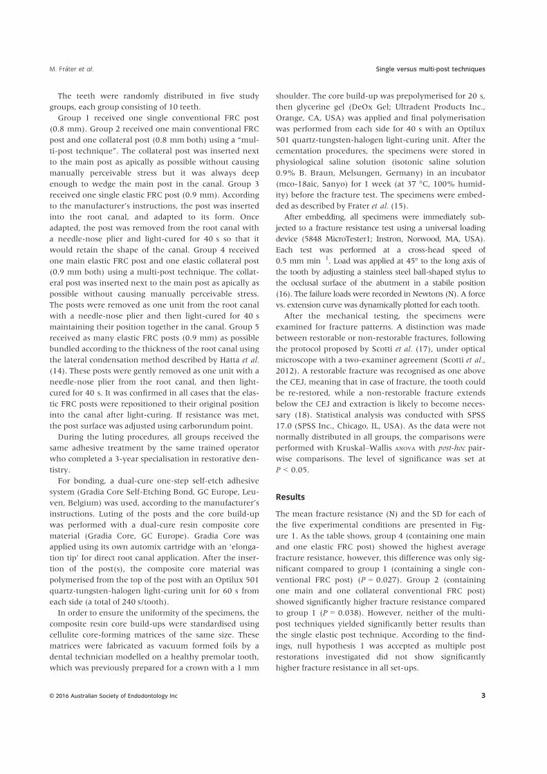
The teeth were randomly distributed in five study
groups, each group consisting of 10 teeth.
Group 1 received one single conventional FRC post
(0.8 mm). Group 2 received one main conventional FRC
post and one collateral post (0.8 mm both) using a “mul-
ti-post technique”. The collateral post was inserted next
to the main post as apically as possible without causing
manually perceivable stress but it was always deep
enough to wedge the main post in the canal. Group 3
received one single elastic FRC post (0.9 mm). According
to the manufacturer’s instructions, the post was inserted
into the root canal, and adapted to its form. Once
adapted, the post was removed from the root canal with
a needle-nose plier and light-cured for 40 s so that it
would retain the shape of the canal. Group 4 received
one main elastic FRC post and one elastic collateral post
(0.9 mm both) using a multi-post technique. The collat-
eral post was inserted next to the main post as apically as
possible without causing manually perceivable stress.
The posts were removed as one unit from the root canal
with a needle-nose plier and then light-cured for 40 s
maintaining their position together in the canal. Group 5
received as many elastic FRC posts (0.9 mm) as possible
bundled according to the thickness of the root canal using
the lateral condensation method described by Hatta et al.
(14). These posts were gently removed as one unit with a
needle-nose plier from the root canal, and then light-
cured for 40 s. It was confirmed in all cases that the elas-
tic FRC posts were repositioned to their original position
into the canal after light-curing. If resistance was met,
the post surface was adjusted using carborundum point.
During the luting procedures, all groups received the
same adhesive treatment by the same trained operator
who completed a 3-year specialisation in restorative den-
tistry.
For bonding, a dual-cure one-step self-etch adhesive
system (Gradia Core Self-Etching Bond, GC Europe, Leu-
ven, Belgium) was used, according to the manufacturer’s
instructions. Luting of the posts and the core build-up
was performed with a dual-cure resin composite core
material (Gradia Core, GC Europe). Gradia Core was
applied using its own automix cartridge with an ‘elonga-
tion tip’ for direct root canal application. After the inser-
tion of the post(s), the composite core material was
polymerised from the top of the post with an Optilux 501
quartz-tungsten-halogen light-curing unit for 60 s from
each side (a total of 240 s/tooth).
In order to ensure the uniformity of the specimens, the
composite resin core build-ups were standardised using
cellulite core-forming matrices of the same size. These
matrices were fabricated as vacuum formed foils by a
dental technician modelled on a healthy premolar tooth,
which was previously prepared for a crown with a 1 mm
shoulder. The core build-up was prepolymerised for 20 s,
then glycerine gel (DeOx Gel; Ultradent Products Inc.,
Orange, CA, USA) was applied and final polymerisation
was performed from each side for 40 s with an Optilux
501 quartz-tungsten-halogen light-curing unit. After the
cementation procedures, the specimens were stored in
physiological saline solution (isotonic saline solution
0.9% B. Braun, Melsungen, Germany) in an incubator
(mco-18aic, Sanyo) for 1 week (at 37 °C, 100% humid-
ity) before the fracture test. The specimens were embed-
ded as described by Frater et al. (15).
After embedding, all specimens were immediately sub-
jected to a fracture resistance test using a universal loading
device (5848 MicroTester1; Instron, Norwood, MA, USA).
Each test was performed at a cross-head speed of
0.5 mm min"1. Load was applied at 45° to the long axis of
the tooth by adjusting a stainless steel ball-shaped stylus to
the occlusal surface of the abutment in a stabile position
(16). The failure loads were recorded in Newtons (N). A force
vs. extension curve was dynamically plotted for each tooth.
After the mechanical testing, the specimens were
examined for fracture patterns. A distinction was made
between restorable or non-restorable fractures, following
the protocol proposed by Scotti et al. (17), under optical
microscope with a two-examiner agreement (Scotti et al.,
2012). A restorable fracture was recognised as one above
the CEJ, meaning that in case of fracture, the tooth could
be re-restored, while a non-restorable fracture extends
below the CEJ and extraction is likely to become neces-
sary (18). Statistical analysis was conducted with SPSS
17.0 (SPSS Inc., Chicago, IL, USA). As the data were not
normally distributed in all groups, the comparisons were
performed with Kruskal–Wallis ANOVA with post-hoc pair-
wise comparisons. The level of significance was set at
P < 0.05.
Results
The mean fracture resistance (N) and the SD for each of
the five experimental conditions are presented in Fig-
ure 1. As the table shows, group 4 (containing one main
and one elastic FRC post) showed the highest average
fracture resistance, however, this difference was only sig-
nificant compared to group 1 (containing a single con-
ventional FRC post) (P = 0.027). Group 2 (containing
one main and one collateral conventional FRC post)
showed significantly higher fracture resistance compared
to group 1 (P = 0.038). However, neither of the multi-
post techniques yielded significantly better results than
the single elastic post technique. According to the find-
ings, null hypothesis 1 was accepted as multiple post
restorations investigated did not show significantly
higher fracture resistance in all set-ups.
© 2016 Australian Society of Endodontology Inc 3
M. Fr!ater et al. Single versus multi-post techniques
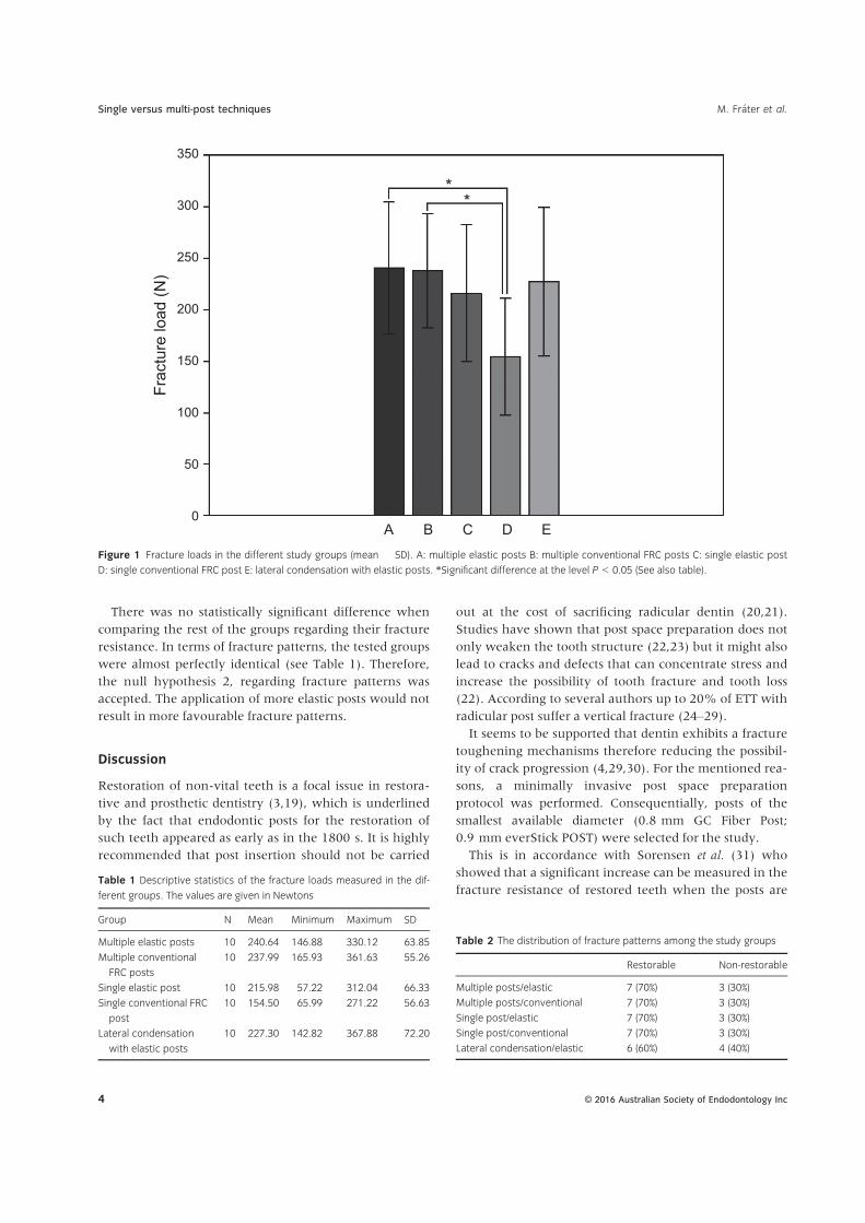
There was no statistically significant difference when
comparing the rest of the groups regarding their fracture
resistance. In terms of fracture patterns, the tested groups
were almost perfectly identical (see Table 1). Therefore,
the null hypothesis 2, regarding fracture patterns was
accepted. The application of more elastic posts would not
result in more favourable fracture patterns.
Discussion
Restoration of non-vital teeth is a focal issue in restora-
tive and prosthetic dentistry (3,19), which is underlined
by the fact that endodontic posts for the restoration of
such teeth appeared as early as in the 1800 s. It is highly
recommended that post insertion should not be carried
out at the cost of sacrificing radicular dentin (20,21).
Studies have shown that post space preparation does not
only weaken the tooth structure (22,23) but it might also
lead to cracks and defects that can concentrate stress and
increase the possibility of tooth fracture and tooth loss
(22). According to several authors up to 20% of ETT with
radicular post suffer a vertical fracture (24–29).It seems to be supported that dentin exhibits a fracture
toughening mechanisms therefore reducing the possibil-
ity of crack progression (4,29,30). For the mentioned rea-
sons, a minimally invasive post space preparation
protocol was performed. Consequentially, posts of the
smallest available diameter (0.8 mm GC Fiber Post;
0.9 mm everStick POST) were selected for the study.
This is in accordance with Sorensen et al. (31) who
showed that a significant increase can be measured in the
fracture resistance of restored teeth when the posts are
350
300
250
200
150
100
50
0A B C D E
Frac
ture
load
(N)
**
Figure 1 Fracture loads in the different study groups (mean ! SD). A: multiple elastic posts B: multiple conventional FRC posts C: single elastic post
D: single conventional FRC post E: lateral condensation with elastic posts. *Significant difference at the level P < 0.05 (See also table).
Table 1 Descriptive statistics of the fracture loads measured in the dif-
ferent groups. The values are given in Newtons
Group N Mean Minimum Maximum SD
Multiple elastic posts 10 240.64 146.88 330.12 63.85
Multiple conventional
FRC posts
10 237.99 165.93 361.63 55.26
Single elastic post 10 215.98 57.22 312.04 66.33
Single conventional FRC
post
10 154.50 65.99 271.22 56.63
Lateral condensation
with elastic posts
10 227.30 142.82 367.88 72.20
Table 2 The distribution of fracture patterns among the study groups
Restorable Non-restorable
Multiple posts/elastic 7 (70%) 3 (30%)
Multiple posts/conventional 7 (70%) 3 (30%)
Single post/elastic 7 (70%) 3 (30%)
Single post/conventional 7 (70%) 3 (30%)
Lateral condensation/elastic 6 (60%) 4 (40%)
© 2016 Australian Society of Endodontology Inc4
Single versus multi-post techniques M. Fr!ater et al.
adapted closely to the canal walls. According to Maceri
et al. (12), a multi-post technique may not only lead to
better adaptation but will also possibly reduce pull out
risk and induce restoration durability to long-term cyclic
loading. Therefore, applying multiple posts in the same
canal (multi-post technique) or using an individual post
is aiming to achieve a better fit to the individual, pre-
served root canal anatomy possibly enhancing long-term
clinical prognosis.
Through the use of a multi-post technique utilising
small diameter posts, the operator is also able to fill large
and irregular root cavities more efficiently than with a
single, centrally positioned post (12).
The results of this study appear to favour the use of
multiple posts in the same root canal. Both multi-post
techniques (conventional FRC (group 2) and elastic FRC
(group 4)) yielded significantly higher fracture resistance
than the single post conventional FRC restoration (group
1). It seems supported by the findings that the elasticity
of the post is not particularly important when applying a
multi-post technique, as there was no statistical differ-
ence between the conventional and elastic multi-post
techniques in terms of the fracture resistance of the
restorations. It is interesting to note that neither of the
two multi-post techniques yielded significantly different
fracture resistance from the single elastic post technique.
The use of a minimally invasive post space preparation
therefore may explain the non-significance between the
results when using a single elastic post and a multi-post
technique as even a single elastic post can achieve a good
fit, thus adequate stability in case of a preserved, rela-
tively irregular root canal cross section. Therefore, the
null hypothesis regarding fracture resistance was partially
rejected (only for non-elastic FRC posts). A possible
explanation is that the elasticity (and thus better adapt-
ability) of the particular type of elastic post used in this
study was enough to make up for the disadvantages of
using a single post only. However, the limitations of this
investigation cannot lead to this conclusion, so further
studies on the adaptive properties of the elastic FRC posts
are necessary.
The rationale behind experimenting with custom (indi-
vidual) post techniques in spite of the advantages of the
application of multiple posts is twofold: one of the rea-
sons is that the problem of the insertion of geometrically
uniform and symmetric single FRC post(s) into a root
canal of irregularly changing diameter along its length is
still not completely resolved. The other reason is that the
amount of luting cement should be minimised, and the
multi-post techniques, by nature, do not fully meet this
requirement.
There are several methods to fabricate individual root
canal posts (1,14,32,33). In this study, the “lateral conden-
sation method” of Hatta and colleagues was used (14). It is
suggested that posts fabricated with this technique might
contain a higher volume of FRC, which, in turn, may result
in higher load-bearing capacity and a greater stability to the
restoration. In the present study, the individual posts
(group 5) yielded better results than restoration with a sin-
gle FRC post (groups 1 and 3). However, the difference did
not reach the level of statistical significance. These results
are in agreement with those of Hatta et al. (14) and Le Bell-
Ronnlof et al. (34) in this respect.
Based on the present results, the fracture resistances
yielded by the individual post technique and the two
multi-post techniques were not significantly different.
The reason might be the minimal invasive preparation of
the root canal, which can limit the number of posts inser-
table to approximately the same depth thus making the
cement-glass fibre ratio of the single-post techniques rel-
atively similar to that of the multiple post techniques.
This phenomena leads to the conclusion that most likely
the preservation of tooth structure is the key influencing
factor in term of mechanical behaviour of ETT as sup-
ported by Wandscher et al. (16).
Fracture of the post(s) or the restored tooth itself are
among the most common failures of ETT restoration
(28,29). In the present study, no difference was found
between the study groups regarding the fracture pattern.
As for that matter, the results are observably uniform
(see Table 2). Thus, the null hypothesis regarding frac-
ture patterns was accepted. This contradicts the findings
of Maceri et al. (12) who conclude that multi-post tech-
niques would reduce the risk of irreversible root frac-
tures. Nevertheless, it has to be noted that teeth
examined by Maceri and co-workers have had a ferrule,
and were restored with a crown making a fundamentally
different set-up compared to that of the current investi-
gation.
In the present investigation, single-root upper premo-
lars were tested because they have been shown to be
more susceptible to root fractures when submitted to
occlusal loading after endodontic treatment (11). Frac-
ture toughness tests, with their limitations, provide an
indication of the load-bearing capacity of restorations in
simulated clinical situations (35). To better represent a
clinical scenario, the techniques used in this study could
be expanded to include artificial ageing and cyclic loading
experiments. The use of a crown would also achieve this
aim and may change the failure patterns of the conven-
tional FRC posts by protecting the luting agent around
these posts from delamination and degradation (36,37).
The importance of crowns in relation to the retention of
posts may also be highlighted (38,39).
According to Nam et al. (2), conventional fibre posts do
not improve the fracture resistance of teeth without a
© 2016 Australian Society of Endodontology Inc 5
M. Fr!ater et al. Single versus multi-post techniques
dental ferrule. This may not be the case when teeth are
restored using the techniques described in this paper.
There are potential clinical benefits to investigating these
new restorations in further lab-based studies. The deci-
sion to not use crowns in this study was based on the
observations of a number of authors where subtle differ-
ences in post behaviour and performance may be masked
when teeth are definitively restored (18,40–43). Despitethese recommendations, the lack of a crown makes it
impossible to extrapolate the results of this investigation
to a clinical situation and there is a need for further
experiments to bridge this gap.
The tested specimens received an oblique load (45
degree to the long axis of the tooth) which appears to be
the worst-case scenario in terms of the fracture resistance
of ETT as described by Wandscher et al. (16). Applying
this angle of force to teeth without a dental ferrule placed
significant stress on the cervical aspect of the restored
tooth (44) and heavy shear forces on the post/luting
agent/radicular dentine interfaces. This should represent
a worst-case occlusal loading scenario for these teeth and
acid-test the integrity of the tested restorations and tooth
structure.
The data demonstrate low fracture loads across the
samples which can be attributed to the compromised
tooth structure (no ferrule), unfavourable loading forces
and a lack of crown restoration. This highlights the
importance of these three factors in the clinical success of
restored, endodontically treated teeth and the impor-
tance of further investigations to achieve meaningful
information about the best way to restore these teeth
clinically.
Conclusions
Within the limitations of this in vitro study, single rooted
premolars restored in the absence of ferrule show signifi-
cantly higher fracture resistance when a multi-post tech-
nique is used, compared to utilising a single conventional
FRC post.
No statistically significant difference was observed
between using conventional or elastic FRC posts for mul-
tiple post restorations.
The teeth restored with a single elastic FRC post exhib-
ited significantly higher fracture resistance than those
restored with a single conventional FRC post.
Fracture patterns were similarly favourable in all
groups. Multi-post techniques are superior to single-
post techniques in terms of achieved fracture resis-
tance, regardless of the type of posts. Single canal
teeth restored with multiple posts achieved superior
fracture resistance to teeth restored with single, con-
ventional FRC posts.
References
1. Cecchin D, de Almeida JF, Gomes BP, Zaia AA, Ferraz CC.
Influence of chlorhexidine and ethanol on the bond
strength and durability of the adhesion of the fiber posts
to root dentin using a total etching adhesive system.
J Endod 2011; 37: 1310–5.2. Nam SH, Chang HS, Min KS, Lee Y, Cho HW, Bae JM.
Effect of the number of residual walls on fracture resis-
tances, failure patterns, and photoelasticity of simulated
premolars restored with or without fiber-reinforced com-
posite posts. J Endod 2010; 36: 297–301.3. Dietschi D, Duc O, Krejci I, Sadan A. Biomechanical con-
siderations for the restoration of endodontically treated
teeth: a systematic review of the literature–Part 1. Compo-
sition and micro- and macrostructure alterations. Quintes-
sence Int 2007; 38: 733–43.4. Al-Omiri MK, Mahmoud AA, Rayyan MR, Abu-Hammad
O. Fracture resistance of teeth restored with post-retained
restorations: an overview. J Endod 2010; 36: 1439–49.5. Clark D, Khademi J. Modern molar endodontic access and
directed dentin conservation. Dent Clin North Am 2010;
54: 249–73.6. Schwartz RS, Robbins JW. Post placement and restoration
of endodontically treated teeth: a literature review.
J Endod 2004; 30: 289–301.7. Cecchin D, Farina AP, Guerreiro CA, Carlini-Junior B.
Fracture resistance of roots prosthetically restored with
intra-radicular posts of different lengths. J Oral Rehabil
2010; 37: 116–22.8. Fokkinga WA, Kreulen CM, Bronkhorst EM, Creugers
NH. Up to 17-year controlled clinical study on post-and-
cores and covering crowns. J Dent 2007; 35: 778–86.9. Mangold JT, Kern M. Influence of glass-fiber posts on the
fracture resistance and failure pattern of endodontically
treated premolars with varying substance loss: an in vitro
study. J Prosthet Dent 2011; 105: 387–93.10. Bell AM, Lassila LV, Kangasniemi I, Vallittu PK. Bonding
of fibre-reinforced composite post to root canal dentin.
J Dent 2005; 33: 533–9.11. Zicari F, Van Meerbeek B, Scotti R, Naert I. Effect of fibre
post length and adhesive strategy on fracture resistance of
endodontically treated teeth after fatigue loading. J Dent
2012; 40: 312–21.12. Maceri F, Martignoni M, Vairo G. Mechanical behaviour
of endodontic restorations with multiple prefabricated
posts: a finite-element approach. J Biomech 2007; 40:
2386–98.13. Lassila LV, Tanner J, Le Bell AM, Narva K, Vallittu PK.
Flexural properties of fiber reinforced root canal posts.
Dent Mater 2004; 20: 29–36.14. Hatta M, Shinya A, Vallittu PK, Shinya A, Lassila LV. High
volume individual fibre post versus low volume fibre post:
the fracture load of the restored tooth. J Dent 2011; 39:
65–71.
© 2016 Australian Society of Endodontology Inc6
Single versus multi-post techniques M. Fr!ater et al.
15. Frater M, Forster A, Kereszturi M, Braunitzer G, Nagy K.
In vitro fracture resistance of molar teeth restored with a
short fibre-reinforced composite material. J Dent 2014;
42: 1143–50.16. Wandscher VF, Bergoli CD, Limberger IF, Ardenghi TM,
Valandro LF. Preliminary results of the survival and
fracture load of roots restored with intracanal posts:
weakened vs nonweakened roots. Oper Dent 2014; 39:
541–55.17. Scotti N, Coero Borga FA, Alovisi M, Rota R, Pasqualini D,
Berutti E. Is fracture resistance of endodontically treated
mandibular molars restored with indirect onlay composite
restorations influenced by fibre post insertion? Journal of
Dentistry 2012; 40: 814–20.18. Batalha-Silva S, de Andrada MA, Maia HP, Magne P. Fati-
gue resistance and crack propensity of large MOD compo-
site resin restorations: direct versus CAD/CAM inlays.
Dental Materials 2013; 29: 324–31.19. Dietschi D, Argente A, Krejci I, Mandikos M. In vitro per-
formance of Class I and II composite restorations: a litera-
ture review on nondestructive laboratory trials–part II.Oper Dent 2013; 38: E182–200.
20. Meyenberg K. The ideal restoration of endodontically
treated teeth – structural and esthetic considerations: a
review of the literature and clinical guidelines for the
restorative clinician. Eur J Esthet Dent 2013; 8:
238–68.21. Paolone G, Saracinelli M, Devoto W, Putignano A.
Esthetic direct restorations in endodontically treated ante-
rior teeth. Eur J Esthet Dent 2013; 8: 44–67.22. Bolhuis HPB, De Gee AJ, Feilzer AJ, Davidson CL. Frac-
ture strength of different core build-up designs. Am J Dent
2001; 14: 286–90.23. Manning KE, Yu DC, Yu HC, Kwan EW. Factors to con-
sider for predictable post and core build-ups of endodonti-
cally treated teeth. Part I: Basic theoretical concepts.
Journal 1995; 61: 685–8, 90, 93-5.24. Fuss Z, Lustig J, Katz A, Tamse A. An evaluation of
endodontically treated vertical root fractured teeth: impact
of operative procedures. J Endod 2001; 27: 46–8.25. Ferrari M, Vichi A, Garcia-Godoy F. Clinical evaluation of
fiber-reinforced epoxy resin posts and cast post and cores.
Am J Dent 2000; 13: 15B–8B.26. Butz F, Lennon AM, Heydecke G, Strub JR. Survival rate
and fracture strength of endodontically treated maxillary
incisors with moderate defects restored with different
post-and-core systems: an in vitro study. Int J Prosthodont
2001; 14: 58–64.27. Lertchirakarn V, Palamara JE, Messer HH. Patterns of ver-
tical root fracture: factors affecting stress distribution in
the root canal. J Endod 2003; 29: 523–8.28. Fuss Z, Lustig J, Tamse A. Prevalence of vertical root frac-
tures in extracted endodontically treated teeth. Int Endod
J 1999; 32: 283–6.
29. Tamse A, Fuss Z, Lustig J, Kaplavi J. An evaluation of
endodontically treated vertically fractured teeth. J Endod
1999; 25: 506–8.30. Kishen A, Asundi A. Experimental investigation on the
role of water in the mechanical behavior of structural den-
tine. J Biomed Mater Res, Part A 2005; 73: 192–200.31. Sorensen JA, Engelman MJ. Effect of post adaptation on
fracture resistance of endodontically treated teeth. J Pros-
thet Dent 1990; 64: 419–24.32. Eskitascioglu G, Belli S, Kalkan M. Evaluation of two post
core systems using two different methods (fracture
strength test and a finite elemental stress analysis).
J Endod 2002; 28: 629–33.33. NewmanMP, Yaman P, Dennison J, Rafter M, Billy E. Frac-
ture resistance of endodontically treated teeth restored with
composite posts. J Prosthet Dent 2003; 89: 360–7.34. Le Bell-Ronnlof AM, Lassila LV, Kangasniemi I, Vallittu
PK. Load-bearing capacity of human incisor restored with
various fiber-reinforced composite posts. Dent Mater
2011; 27: e107–15.35. Ku CW, Park SW, Yang HS. Comparison of the fracture
strengths of metal-ceramic crowns and three ceromer
crowns. J Prosthet Dent 2002; 88: 170–5.36. Mannocci F, Sherriff M, Watson TF. Three-point bending
test of fiber posts. J Endod 2001; 27: 758–61.37. Chieruzzi M, Pagano S, Pennacchi M, Lombardo G,
D’Errico P, Kenny JM. Compressive and flexural beha-
viour of fibre reinforced endodontic posts. J Dent 2012;
40: 968–78.38. Goracci C, Fabianelli A, Sadek FT, Papacchini F, Tay FR, Fer-
rari M. The contribution of friction to the dislocation resis-
tance of bonded fiber posts. J Endod 2005; 31: 608–12.39. Turner CH. Post-retained crown failure: a survey. Dent
Update 1982; 9: 221, 4-6, 8-9 passim.
40. Heydecke G, PetersMC. The restoration of endodontically
treated, single-rooted teethwith cast or direct posts and
cores: a systematic review. J Prosthet Dent 2002; 87: 380–6.41. Ambica K, Mahendran K, Talwar S, Verma M, Padmini G,
Periasamy R. Comparative evaluation of fracture resis-
tance under static and fatigue loading of endodontically
treated teeth restored with carbon fiber posts, glass fiber
posts, and an experimental dentin post system: an in vitro
study. J Endod 2013; 39: 96–100.42. Ozcan M, Valandro LF. Fracture strength of endodonti-
cally-treated teeth restored with post and cores and com-
posite cores only. Oper Dent 2009; 34: 429–36.43. Dietschi D, Duc O, Krejci I, Sadan A. Biomechanical con-
siderations for the restoration of endodontically treated
teeth: a systematic review of the literature, Part II (Evalua-
tion of fatigue behavior, interfaces, and in vivo studies).
Quintessence Int 2008; 39: 117–29.44. Meira JB, Esposito CO, Quitero MF et al. Elastic modulus
of posts and the risk of root fracture. Dent Traumatol
2009; 25: 394–8.
© 2016 Australian Society of Endodontology Inc 7
M. Fr!ater et al. Single versus multi-post techniques