methylglyoxal activates the target of rapamycin complex 2 ... · tetrazolium solution kit for...
TRANSCRIPT
TitleMethylglyoxal activates the target of rapamycin complex 2-protein kinase C signaling pathway in Saccharomycescerevisiae.
Author(s) Nomura, Wataru; Inoue, Yoshiharu
Citation Molecular and cellular biology (2015), 35(7): 1269-1280
Issue Date 2015-04
URL http://hdl.handle.net/2433/198443
Right Copyright © American Society for Microbiology, Mol CellBiol 35:1269‒1280. doi:10.1128/MCB.01118-14
Type Journal Article
Textversion publisher
Kyoto University
Methylglyoxal Activates the Target of Rapamycin Complex 2-ProteinKinase C Signaling Pathway in Saccharomyces cerevisiae
Wataru Nomura, Yoshiharu Inoue
Laboratory of Molecular Microbiology, Division of Applied Life Sciences, Graduate School of Agriculture, Kyoto University, Uji, Kyoto, Japan
Methylglyoxal is a typical 2-oxoaldehyde derived from glycolysis. We show here that methylglyoxal activates the Pkc1-Mpk1 mi-togen-activated protein (MAP) kinase cascade in a target of rapamycin complex 2 (TORC2)-dependent manner in the buddingyeast Saccharomyces cerevisiae. We demonstrate that TORC2 phosphorylates Pkc1 at Thr1125 and Ser1143. Methylglyoxal en-hanced the phosphorylation of Pkc1 at Ser1143, which transmitted the signal to the downstream Mpk1 MAP kinase cascade. Wefound that the phosphorylation status of Pkc1T1125 affected the phosphorylation of Pkc1 at Ser1143, in addition to its protein lev-els. Methylglyoxal activated mammalian TORC2 signaling, which, in turn, phosphorylated Akt at Ser473. Our results suggest thatmethylglyoxal is a conserved initiator of TORC2 signaling among eukaryotes.
All biological activities in organisms must be accompanied byan ample supply of nutrients, which warrants energy produc-
tion to meet the demand for biochemical reactions in cells. There-fore, the machinery for sensing and transducing nutritional con-ditions inside and outside the cell plays crucial roles. Nutrientsand hormones are pivotal signal initiation molecules that regulatecellular energy metabolism in animals. Although nutrients are es-sential for living cells, several adverse by-products, such as reactiveoxygen species (ROS), are formed during energy metabolism (1).ROS are inevitably formed by oxygen respiration in accordancewith ATP production and cause oxidative stress, which damagescellular components (2).
We have been focusing on methylglyoxal (MG) and its role as ametabolic stressor. MG (CH3COCHO) is a typical 2-oxoaldehyde,the main source of which in the cell is glycolysis, a ubiquitousenergy-producing pathway (3, 4). Since MG is highly reactive be-cause of its two carbonyl groups in one molecule, it reacts withDNA/RNA and proteins to make adducts that change their func-tions, and this change of function sometimes leads to severaldiseases, such as diabetes and Alzheimer’s disease (5–7). MG andother carbonyl compounds produce various carbonyl compound-modified proteins through a process that is sometimes referred toas carbonyl stress. Carbohydrate-derived carbonyl compoundsproduce advanced glycation end products (AGEs) via spontane-ous reactions with the amino groups of proteins and carbonylcompounds. However, even though the reactivity of MG with pro-teins is �20,000-fold higher than that of glucose, the formation ofAGEs has been shown to occur on a time scale of days, weeks, orlonger (8, 9). The harmful effects of MG on cellular functions havebeen attributed to MG-derived AGEs (4–9). We previously re-ported that MG causes acute cellular responses in yeasts (10, 11).For example, the exogenous addition of MG activated yeast AP-1-like transcription factors (Yap1 in Saccharomyces cerevisiae andPap1 in Schizosaccharomyces pombe) through the reversible mod-ification of their critical Cys residues within 45 min (10, 11). MGwas also found to enhance the influx of extracellular Ca2� into thecell, thereby activating the Ca2�/calcineurin system in S. cerevisiae(12). In the present study, we found that MG enhances the targetof rapamycin (TOR) complex 2 (TORC2)-protein kinase C (Pkc1)signaling pathway in S. cerevisiae.
The TOR signaling network is evolutionally conserved among
eukaryotes and is involved in diverse cellular activities (13, 14).TOR is a Ser/Thr kinase that forms two distinct protein kinasecomplexes, TORC1 and TORC2 (15). The TOR complex phos-phorylates protein kinases involved in the AGC (named initiallyfor cyclic AMP- and cyclic GMP-dependent protein kinases andprotein kinase C) kinase family (16). S. cerevisiae TORC1 has beenshown to phosphorylate Sch9, whereas TORC2 phosphorylatesYpk1 and Ypk2 (17–19), both of which belong to the AGC kinasefamily. A previous study demonstrated that TOR complexes phos-phorylate conserved Thr/Ser residues within the turn motif (TM)and hydrophobic motif (HM) of AGC kinase (16). PKC1 is theonly gene that encodes protein kinase C in S. cerevisiae (20, 21),and Pkc1 structurally belongs to the AGC kinase family. Thr/Serresidues in the TM and HM were also found to be conserved inPkc1 (Thr1125 in TM and Ser1143 in HM). Genetic interactionsbetween PKC1 and some genes (TOR2, AVO1, and AVO3), theproducts of which are involved in the assembly of TORC2, havebeen reported previously (15, 22–24); however, it still remainsunclear whether Pkc1 is a direct target of TORC2 (25).
TORC1 is involved in events concerning cell growth, such asprotein synthesis, while TORC2 plays a role in actin organization(14). In the present study, we demonstrate that the organization ofactin is perturbed by an MG treatment, thereby inhibiting thepolarized cell growth of S. cerevisiae. Since TORC2 is involved inactin organization, we investigated the relationship between MGand TORC2 signaling. We show that Thr1125 and Ser1143 in Pkc1are phosphorylated by TORC2 both in vivo and in vitro. We also
Received 2 September 2014 Returned for modification 28 September 2014Accepted 18 January 2015
Accepted manuscript posted online 26 January 2015
Citation Nomura W, Inoue Y. 2015. Methylglyoxal activates the target ofrapamycin complex 2-protein kinase C signaling pathway in Saccharomycescerevisiae. Mol Cell Biol 35:1269 –1280. doi:10.1128/MCB.01118-14.
Address correspondence to Yoshiharu Inoue, [email protected].
Supplemental material for this article may be found at http://dx.doi.org/10.1128/MCB.01118-14.
Copyright © 2015, American Society for Microbiology. All Rights Reserved.
doi:10.1128/MCB.01118-14
April 2015 Volume 35 Number 7 mcb.asm.org 1269Molecular and Cellular Biology
on June 16, 2015 by KY
OT
O U
NIV
ER
SIT
Yhttp://m
cb.asm.org/
Dow
nloaded from

found that MG enhances TORC2-Pkc1 signaling, and the down-stream Mpk1 mitogen-activated protein (MAP) kinase cascadewas subsequently activated following the treatment of yeast cellswith MG. Furthermore, we show that MG activates TORC2 sig-naling in mammalian cells. A physiological trigger that has beenshown to activate TORC2 signaling is insulin (and insulin-likegrowth factor) (26–28); however, lower eukaryotes, such as yeast,do not have a hormonal system. Therefore, it remains unclearwhether the ubiquitous physiological molecules that activateTORC2 exist among eukaryotes. Our results suggest that MG is acommon initiator of TORC2 signaling.
MATERIALS AND METHODSMedia. The media used were synthetic dextrose (SD) medium (2% glu-cose, 0.67% yeast nitrogen base without amino acids) and synthetic com-plete (SC)-Gal medium (2% galactose, 3% glycerol, 0.67% yeast nitrogenbase without amino acids). Appropriate amino acids and bases wereadded to SD or SC-Gal medium as necessary. Cells were cultured at 28°Cunless otherwise stated.
Strains. The yeast strains and PCR primers used are summarized inTables S1 and S2 in the supplemental material. The deletion allele of MID2with KanMX or his5� was amplified by PCR with primer sets mid2-F andmid2-R from S. cerevisiae BY4741-based deletion mutants (29). The cor-responding loci of S. cerevisiae YPH250 were disrupted using PCR prod-ucts. To construct wsc1� and mpk1� mutants, the wsc1�::CgLEU2 alleleof YOC2573 (30) and the mpk1�::HIS3 allele of TNP46 (31) were ampli-fied by PCR with primers WSC1F and WSC1R and primers MPK1FSalIand MPK1REcoRI, respectively. The disruption of MKK1, MKK2, andBCK1 was performed as described previously (32).
Plasmids. The plasmids used are summarized in Table S3 in the sup-plemental material. Details for the construction of plasmids are describedin the supplemental material.
Actin staining. Cells were cultured in SD medium until the A610
reached 0.3 to 0.5. MG was then added. Cells were fixed with formalde-hyde (final concentration, 4%) for 1 h. After fixation, the cells were har-vested, washed twice with phosphate-buffered saline (PBS; pH 7.4), andsuspended in 30 �l of PBS. Rhodamine-phalloidin (Molecular Probes)was added to the cell suspension to a final concentration of 33 units/ml(1.1 �M), and the cell suspension was then incubated at 4°C in the darkovernight. Cells were collected by centrifugation and washed twice withPBS, and the distribution of actin was observed using a fluorescence mi-croscope.
Immunoprecipitation. Cells were cultured in SD medium until theA610 reached 0.5. Cells were then collected, washed with a 0.85% NaClsolution, and suspended in lysis buffer B (50 mM Tris-HCl [pH 7.5], 150mM NaCl, 0.1 mM EDTA, 0.5% Tween 20) containing 2.5 mM phenyl-methylsulfonyl fluoride, 50 mM NaF, and protease inhibitor cocktail (Na-calai Tesque). Cells were disrupted with glass beads using a Beads Smash12 cell disrupter (Wakenyaku), and cell homogenates were centrifuged at700 � g for 10 min at 4°C to remove cell debris. The protein concentra-tions in the cell extracts were determined using a DC protein assay (Bio-Rad Laboratories). The Avo3-13myc, Pkc1 tagged with a 3� hemaggluti-nin tag (Pkc1-3HA), or Pkc1 tagged with a 3� FLAG tag (Pkc1-3FLAG)protein was immunoprecipitated from the cell extracts (1.5 or 1 mg pro-tein) by incubation with anti-c-myc antibodies coupled with agarose resin(Nacalai Tesque), anti-HA antibodies coupled with agarose resin (MBL),or anti-FLAG antibody-conjugated resin (Sigma) for 2 h at 4°C in lysisbuffer B. After being incubated, the agarose resins were precipitated bycentrifugation, washed four times with lysis buffer B, and suspended inSDS-PAGE sample buffer. SDS-PAGE was then performed, followed byWestern blotting.
Bacterial expression and purification of Ypk2. Escherichia coliBL21(DE3) cells carrying pET-15b-Ypk2 were grown in LB medium con-taining ampicillin at 37°C until the A610 reached 0.3, and isopropyl-�-D-
thiogalactopyranoside was added to a final concentration of 0.2 mM. Cellswere incubated at 25°C overnight and then disrupted by sonication at 4°Cin 20 mM sodium phosphate buffer (pH 7.4) containing 0.5 mM NaCland 10 mM imidazole. Cell homogenates were centrifuged to remove celldebris, cell extracts were applied to a HisTrapTM HP column (GE Health-care) equilibrated with sonication buffer, and the column was washedwith the same buffer. The proteins that were adsorbed were eluted with 20mM sodium phosphate buffer (pH 7.4) containing 0.5 mM NaCl and 250mM imidazole. Fractions containing Ypk2 were dialyzed against 20 mMHEPES-KOH buffer (pH 7.4) containing 150 mM NaCl and 2 mM dithio-threitol.
In vitro protein kinase assay. An in vitro protein kinase assay forTORC2 was performed as described previously (17), with some modifi-cations. Briefly, TORC2 was immunopurified using myc-tagged Avo3(Avo3-myc) instead of HA-tagged Tor2 (HA-Tor2), which was used in aprevious study (17). Immunopurified TORC2 was incubated with 5 �g ofPkc1 peptides (for wild-type [WT] Pkc1 [Pkc1WT], APPTLTPLPSVLTTSQQEEFRGFSFMPDDL; for Pkc1 with the T1125A and S1143A mutations[Pkc1T1125A/S1143A], APPTLAPLPSVLTTSQQEEFRGFAFMPDDL) or 4�g of recombinant Ypk2 protein. Synthetic Pkc1 peptides were producedby the GenScript Corporation (Piscataway, NJ). The reaction was initiatedby adding [�-32P]ATP (10 �Ci). After being incubated for 30 min at 30°C,the reaction was terminated by the addition of SDS-PAGE sample buffer,and the samples were then incubated for 5 min at 65°C. Samples weresubjected to Tricine-SDS-PAGE (33) or SDS-PAGE, and phosphorylatedpeptides were detected by autoradiography.
Western blotting of Mpk1. Cells were collected and washed with lysisbuffer A (50 mM Tris-HCl [pH 7.5], 150 mM NaCl, 5 mM EDTA, 1%Nonidet P-40, 1 mM sodium pyrophosphate, 1 mM phenylmethylsulfo-nyl fluoride, 1 mM sodium orthovanadate, 20 mM NaF, 2 �g each ofpepstatin A and leupeptin per ml), and cell pellets were frozen with liquidnitrogen. Cell pellets were suspended in lysis buffer A and agitated withglass beads using a Beads Smash 21 cell disrupter (Wakenyaku). Cell ho-mogenates were centrifuged for 5 min at 15,000 � g and 4°C, and theprotein concentration in clear supernatants was determined using the DCprotein assay (Bio-Rad Laboratories). Samples were subjected to SDS-PAGE, and the separated proteins were transferred to a polyvinylidenedifluoride membrane (Millipore). The blots were incubated with appro-priate dilutions of the primary antibodies (anti-phospho-p44/42 MAPkinase [Thr202/Tyr204; Cell Signaling] and anti-Mpk1 [yC-20; catalognumber sc-6803; Santa Cruz Biotechnology]). Immunoreactive bandswere visualized with a 5-bromo-4-chloro-3-indolylphosphate–nitrobluetetrazolium solution kit for alkaline phosphatase (Nacalai Tesque) or Im-mobilon Western chemiluminescent horseradish peroxidase (HRP) sub-strate (Millipore) using a LAS-4000 mini-imaging system (Fujifilm).
Western blotting of Pkc1. Total protein extracts were prepared usingthe trichloroacetic acid extraction method (12). The protein concentra-tions of the samples were determined using an RC DC protein assay kit(Bio-Rad Laboratories). The antibodies used for Western blotting wereanti-Pkc1 antibodies.
Anti-Pkc1, anti-p-T1125, and anti-p-S1143 antibodies. Anti-Pkc1polyclonal antibodies were raised by immunizing rabbits with a peptidecorresponding to amino acid residues 470 to 488 in Pkc1. Antisera wereused to detect Pkc1 in Western blots. Anti-phospho-Pkc1 polyclonal an-tibodies were raised by immunizing rabbits with Pkc1 containing a phos-phorylated Thr1125 peptide (p-T1125) in the turn motif [NH2-C�APPTL(pT)PLPSVLTTSQQEE-COOH] or a phosphorylated Ser1143 peptide(p-S1143) in the hydrophobic motif [NH2-C�EFRGF(pS)FMPD-COOH]. Antisera were loaded onto a column packed with the resin conju-gated with each of the phosphorylated peptides. After the adsorbed frac-tions were recovered, they were passed through a column packed with theresin conjugated with each of the unphosphorylated peptides. The frac-tions were recovered and used as anti-phospho-Pkc1 at Thr1125 (anti-p-T1125) or anti-phospho-Pkc1 at Ser1143 (anti-p-S1143) antibodies.
Nomura and Inoue
1270 mcb.asm.org April 2015 Volume 35 Number 7Molecular and Cellular Biology
on June 16, 2015 by KY
OT
O U
NIV
ER
SIT
Yhttp://m
cb.asm.org/
Dow
nloaded from
Culture conditions for mammalian cells. Mouse 3T3-L1 cells weremaintained in maintenance medium (10% fetal bovine serum and 10mg/ml penicillin-streptomycin in Dulbecco modified Eagle medium[DMEM]) at 37°C with 5% CO2. Preadipocytes were differentiated toadipocytes by the established protocol (34) as briefly described below.3T3-L1 preadipocytes were grown for 2 days until postconfluence, andmedium was switched to a differentiation medium consisting of the main-tenance medium supplemented with 0.25 �M dexamethasone, 10 �g/mlinsulin, and 0.5 mM 3-isobutyl-1-methylxanthine. After 48 h, the me-dium was replaced with a postdifferentiation medium consisting of themaintenance medium supplemented with 5 �g/ml insulin. Medium wasexchanged every 2 days with a fresh postdifferentiation medium until thecells were ready for experimentation.
3T3-L1 adipocytes were incubated in the maintenance medium for 20h in DMEM for Western blotting and then starved of serum for 5 h inDMEM. Cells were lysed with SDS-PAGE sample buffer, and the proteinconcentrations of the samples were determined using the RC DC pro-tein assay kit (Bio-Rad Laboratories). The antibodies used for Westernblotting were anti-Akt antibodies (Cell Signaling) to detect the bulkprotein levels of Akt, anti-phospho-Akt (Ser473) antibodies (Cell Sig-naling) to detect the phosphorylation of Ser473 in the HM of Akt, andanti-phospho-PKC/�II (Thr638/641) antibodies (Cell Signaling) todetect the phosphorylation of Thr450 in the TM of Akt (35). Immuno-reactive bands were visualized with a kit (Immobilon Western chemi-
luminescent HRP substrate; Millipore) and an LAS-4000 mini-imag-ing system (Fujifilm).
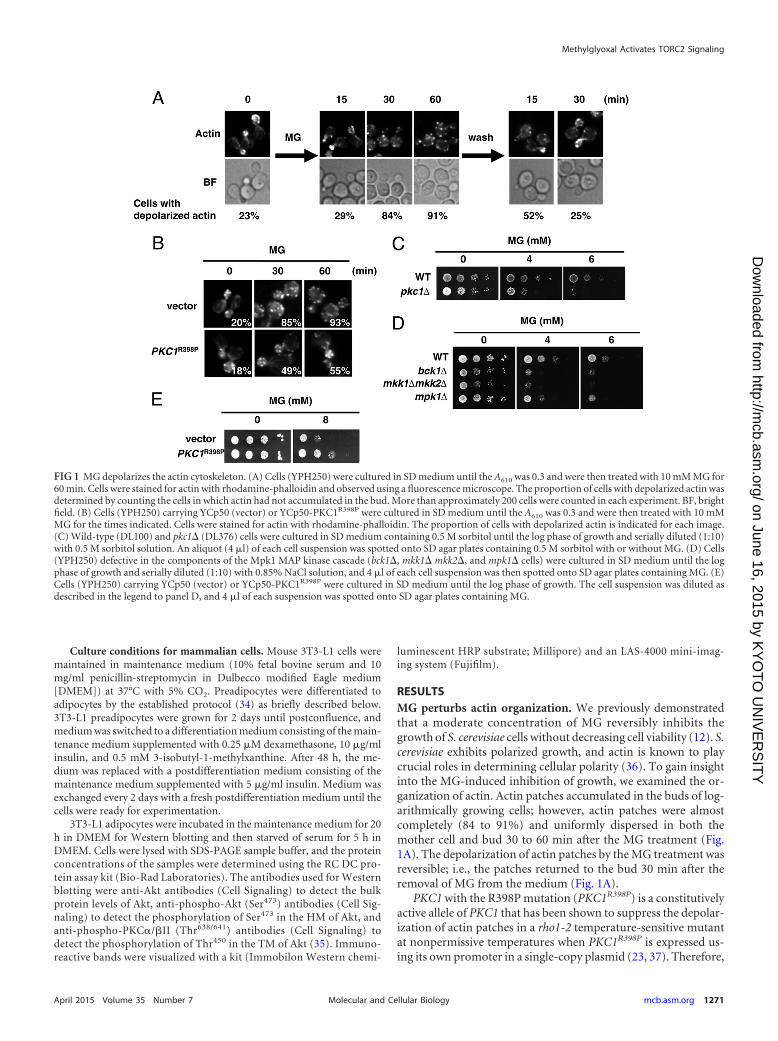
RESULTSMG perturbs actin organization. We previously demonstratedthat a moderate concentration of MG reversibly inhibits thegrowth of S. cerevisiae cells without decreasing cell viability (12). S.cerevisiae exhibits polarized growth, and actin is known to playcrucial roles in determining cellular polarity (36). To gain insightinto the MG-induced inhibition of growth, we examined the or-ganization of actin. Actin patches accumulated in the buds of log-arithmically growing cells; however, actin patches were almostcompletely (84 to 91%) and uniformly dispersed in both themother cell and bud 30 to 60 min after the MG treatment (Fig.1A). The depolarization of actin patches by the MG treatment wasreversible; i.e., the patches returned to the bud 30 min after theremoval of MG from the medium (Fig. 1A).
PKC1 with the R398P mutation (PKC1R398P) is a constitutivelyactive allele of PKC1 that has been shown to suppress the depolar-ization of actin patches in a rho1-2 temperature-sensitive mutantat nonpermissive temperatures when PKC1R398P is expressed us-ing its own promoter in a single-copy plasmid (23, 37). Therefore,
FIG 1 MG depolarizes the actin cytoskeleton. (A) Cells (YPH250) were cultured in SD medium until the A610 was 0.3 and were then treated with 10 mM MG for60 min. Cells were stained for actin with rhodamine-phalloidin and observed using a fluorescence microscope. The proportion of cells with depolarized actin wasdetermined by counting the cells in which actin had not accumulated in the bud. More than approximately 200 cells were counted in each experiment. BF, brightfield. (B) Cells (YPH250) carrying YCp50 (vector) or YCp50-PKC1R398P were cultured in SD medium until the A610 was 0.3 and were then treated with 10 mMMG for the times indicated. Cells were stained for actin with rhodamine-phalloidin. The proportion of cells with depolarized actin is indicated for each image.(C) Wild-type (DL100) and pkc1� (DL376) cells were cultured in SD medium containing 0.5 M sorbitol until the log phase of growth and serially diluted (1:10)with 0.5 M sorbitol solution. An aliquot (4 �l) of each cell suspension was spotted onto SD agar plates containing 0.5 M sorbitol with or without MG. (D) Cells(YPH250) defective in the components of the Mpk1 MAP kinase cascade (bck1�, mkk1� mkk2�, and mpk1� cells) were cultured in SD medium until the logphase of growth and serially diluted (1:10) with 0.85% NaCl solution, and 4 �l of each cell suspension was then spotted onto SD agar plates containing MG. (E)Cells (YPH250) carrying YCp50 (vector) or YCp50-PKC1R398P were cultured in SD medium until the log phase of growth. The cell suspension was diluted asdescribed in the legend to panel D, and 4 �l of each suspension was spotted onto SD agar plates containing MG.
Methylglyoxal Activates TORC2 Signaling
April 2015 Volume 35 Number 7 mcb.asm.org 1271Molecular and Cellular Biology
on June 16, 2015 by KY
OT
O U
NIV
ER
SIT
Yhttp://m
cb.asm.org/
Dow
nloaded from
we investigated whether the depolarization of actin patches in thepresence of MG is alleviated by the expression of PKC1R398P. Asshown in Fig. 1B, Pkc1R398P partially repressed the depolarizationof actin patches in cells treated with MG.
Pkc1-Mpk1 MAP kinase cascade mutants are sensitive toMG. The organization of actin was previously shown to be regu-lated by the Pkc1-Mpk1 MAP kinase cascade (38, 39). To verifywhether MG influences the growth of mutants defective in thePkc1-Mpk1 MAP kinase cascade, we conducted spot assays on SDagar plates containing MG. pkc1� cells are not able to grow with-out sorbitol (38) because synthesis of the cell wall is defective inthis mutant and cell lysis occurs without an osmoprotectant.Therefore, spot assays were performed using SD agar plates con-taining sorbitol. As shown in Fig. 1C, the pkc1� mutant exhibitedsusceptibility to MG. Similarly, bck1�, mkk1� mkk2�, and mpk1�mutants, defective in the MAP kinase cascade, also showed suscep-tibility to MG (Fig. 1D). Wild-type cells displayed an increased toler-ance toward MG when PKC1R398P was expressed from its own pro-moter using a single-copy plasmid (Fig. 1E).
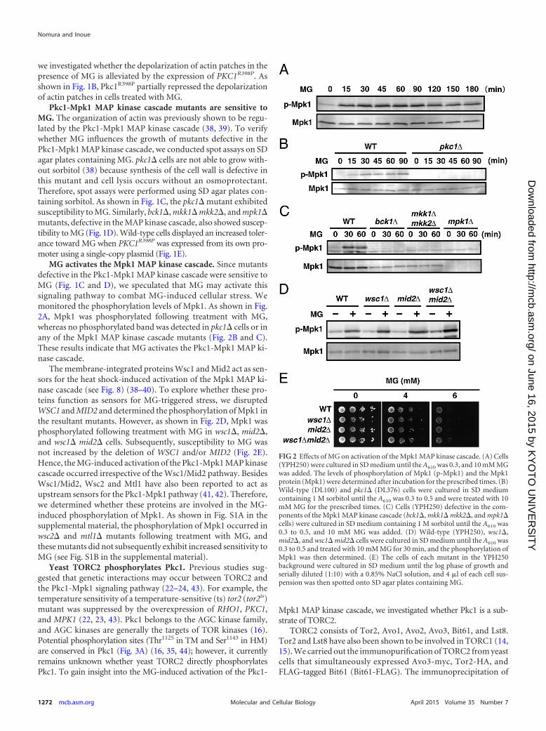
MG activates the Mpk1 MAP kinase cascade. Since mutantsdefective in the Pkc1-Mpk1 MAP kinase cascade were sensitive toMG (Fig. 1C and D), we speculated that MG may activate thissignaling pathway to combat MG-induced cellular stress. Wemonitored the phosphorylation levels of Mpk1. As shown in Fig.2A, Mpk1 was phosphorylated following treatment with MG,whereas no phosphorylated band was detected in pkc1� cells or inany of the Mpk1 MAP kinase cascade mutants (Fig. 2B and C).These results indicate that MG activates the Pkc1-Mpk1 MAP ki-nase cascade.
The membrane-integrated proteins Wsc1 and Mid2 act as sen-sors for the heat shock-induced activation of the Mpk1 MAP ki-nase cascade (see Fig. 8) (38–40). To explore whether these pro-teins function as sensors for MG-triggered stress, we disruptedWSC1 and MID2 and determined the phosphorylation of Mpk1 inthe resultant mutants. However, as shown in Fig. 2D, Mpk1 wasphosphorylated following treatment with MG in wsc1�, mid2�,and wsc1� mid2� cells. Subsequently, susceptibility to MG wasnot increased by the deletion of WSC1 and/or MID2 (Fig. 2E).Hence, the MG-induced activation of the Pkc1-Mpk1 MAP kinasecascade occurred irrespective of the Wsc1/Mid2 pathway. BesidesWsc1/Mid2, Wsc2 and Mtl1 have also been reported to act asupstream sensors for the Pkc1-Mpk1 pathway (41, 42). Therefore,we determined whether these proteins are involved in the MG-induced phosphorylation of Mpk1. As shown in Fig. S1A in thesupplemental material, the phosphorylation of Mpk1 occurred inwsc2� and mtl1� mutants following treatment with MG, andthese mutants did not subsequently exhibit increased sensitivity toMG (see Fig. S1B in the supplemental material).
Yeast TORC2 phosphorylates Pkc1. Previous studies sug-gested that genetic interactions may occur between TORC2 andthe Pkc1-Mpk1 signaling pathway (22–24, 43). For example, thetemperature sensitivity of a temperature-sensitive (ts) tor2 (tor2ts)mutant was suppressed by the overexpression of RHO1, PKC1,and MPK1 (22, 23, 43). Pkc1 belongs to the AGC kinase family,and AGC kinases are generally the targets of TOR kinases (16).Potential phosphorylation sites (Thr1125 in TM and Ser1143 in HM)are conserved in Pkc1 (Fig. 3A) (16, 35, 44); however, it currentlyremains unknown whether yeast TORC2 directly phosphorylatesPkc1. To gain insight into the MG-induced activation of the Pkc1-
Mpk1 MAP kinase cascade, we investigated whether Pkc1 is a sub-strate of TORC2.
TORC2 consists of Tor2, Avo1, Avo2, Avo3, Bit61, and Lst8.Tor2 and Lst8 have also been shown to be involved in TORC1 (14,15). We carried out the immunopurification of TORC2 from yeastcells that simultaneously expressed Avo3-myc, Tor2-HA, andFLAG-tagged Bit61 (Bit61-FLAG). The immunoprecipitation of
FIG 2 Effects of MG on activation of the Mpk1 MAP kinase cascade. (A) Cells(YPH250) were cultured in SD medium until the A610 was 0.3, and 10 mM MGwas added. The levels of phosphorylation of Mpk1 (p-Mpk1) and the Mpk1protein (Mpk1) were determined after incubation for the prescribed times. (B)Wild-type (DL100) and pkc1� (DL376) cells were cultured in SD mediumcontaining 1 M sorbitol until the A610 was 0.3 to 0.5 and were treated with 10mM MG for the prescribed times. (C) Cells (YPH250) defective in the com-ponents of the Mpk1 MAP kinase cascade (bck1�, mkk1� mkk2�, and mpk1�cells) were cultured in SD medium containing 1 M sorbitol until the A610 was0.3 to 0.5, and 10 mM MG was added. (D) Wild-type (YPH250), wsc1�,mid2�, and wsc1� mid2� cells were cultured in SD medium until the A610 was0.3 to 0.5 and treated with 10 mM MG for 30 min, and the phosphorylation ofMpk1 was then determined. (E) The cells of each mutant in the YPH250background were cultured in SD medium until the log phase of growth andserially diluted (1:10) with a 0.85% NaCl solution, and 4 �l of each cell sus-pension was then spotted onto SD agar plates containing MG.
Nomura and Inoue
1272 mcb.asm.org April 2015 Volume 35 Number 7Molecular and Cellular Biology
on June 16, 2015 by KY
OT
O U
NIV
ER
SIT
Yhttp://m
cb.asm.org/
Dow
nloaded from
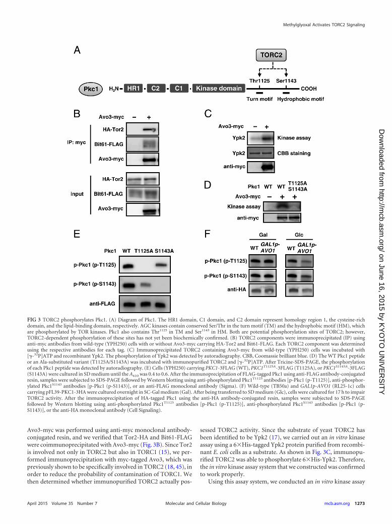
Avo3-myc was performed using anti-myc monoclonal antibody-conjugated resin, and we verified that Tor2-HA and Bit61-FLAGwere coimmunoprecipitated with Avo3-myc (Fig. 3B). Since Tor2is involved not only in TORC2 but also in TORC1 (15), we per-formed immunoprecipitation with myc-tagged Avo3, which waspreviously shown to be specifically involved in TORC2 (18, 45), inorder to reduce the probability of contamination of TORC1. Wethen determined whether immunopurified TORC2 actually pos-
sessed TORC2 activity. Since the substrate of yeast TORC2 hasbeen identified to be Ypk2 (17), we carried out an in vitro kinaseassay using a 6�His-tagged Ypk2 protein purified from recombi-nant E. coli cells as a substrate. As shown in Fig. 3C, immunopu-rified TORC2 was able to phosphorylate 6�His-Ypk2. Therefore,the in vitro kinase assay system that we constructed was confirmedto work properly.
Using this assay system, we conducted an in vitro kinase assay
FIG 3 TORC2 phosphorylates Pkc1. (A) Diagram of Pkc1. The HR1 domain, C1 domain, and C2 domain represent homology region 1, the cysteine-richdomain, and the lipid-binding domain, respectively. AGC kinases contain conserved Ser/Thr in the turn motif (TM) and the hydrophobic motif (HM), whichare phosphorylated by TOR kinases. Pkc1 also contains Thr1125 in TM and Ser1143 in HM. Both are potential phosphorylation sites of TORC2; however,TORC2-dependent phosphorylation of these sites has not yet been biochemically confirmed. (B) TORC2 components were immunoprecipitated (IP) usinganti-myc antibodies from wild-type (YPH250) cells with or without Avo3-myc carrying HA-Tor2 and Bit61-FLAG. Each TORC2 component was determinedusing the respective antibodies for each tag. (C) Immunoprecipitated TORC2 containing Avo3-myc from wild-type (YPH250) cells was incubated with[�-32P]ATP and recombinant Ypk2. The phosphorylation of Ypk2 was detected by autoradiography. CBB, Coomassie brilliant blue. (D) The WT Pkc1 peptideor an Ala-substituted variant (T1125A/S1143A) was incubated with immunopurified TORC2 and [�-32P]ATP. After Tricine-SDS-PAGE, the phosphorylationof each Pkc1 peptide was detected by autoradiography. (E) Cells (YPH250) carrying PKC1-3FLAG (WT), PKC1T1125A-3FLAG (T1125A), or PKC1S1143A-3FLAG(S1143A) were cultured in SD medium until the A610 was 0.4 to 0.6. After the immunoprecipitation of FLAG-tagged Pkc1 using anti-FLAG antibody-conjugatedresin, samples were subjected to SDS-PAGE followed by Western blotting using anti-phosphorylated Pkc1T1125 antibodies [p-Pkc1 (p-T1125)], anti-phosphor-ylated Pkc1S1143 antibodies [p-Pkc1 (p-S1143)], or an anti-FLAG monoclonal antibody (Sigma). (F) Wild-type (TB50a) and GAL1p-AVO1 (RL25-1c) cellscarrying pFL39-PKC1-3HA were cultured overnight in SC-Gal medium (Gal). After being transferred to SD medium (Glc), cells were cultured for 17 h to impairTORC2 activity. After the immunoprecipitation of HA-tagged Pkc1 using the anti-HA antibody-conjugated resin, samples were subjected to SDS-PAGEfollowed by Western blotting using anti-phosphorylated Pkc1T1125 antibodies [p-Pkc1 (p-T1125)], anti-phosphorylated Pkc1S1143 antibodies [p-Pkc1 (p-S1143)], or the anti-HA monoclonal antibody (Cell Signaling).
Methylglyoxal Activates TORC2 Signaling
April 2015 Volume 35 Number 7 mcb.asm.org 1273Molecular and Cellular Biology
on June 16, 2015 by KY
OT
O U
NIV
ER
SIT
Yhttp://m
cb.asm.org/
Dow
nloaded from
to determine whether Pkc1 is phosphorylated by TORC2. Asshown in Fig. 3D, the wild-type Pkc1 peptide (the C-terminalregion of Pkc1 [amino acid residues 1120 to 1149] includingThr1125, Ser1143, and some other Ser and Thr residues) was phos-phorylated, while the Pkc1T1125A/S1143A peptide, an Ala-substi-tuted variant of Thr1125 and Ser1143, was not. In addition, TORC2containing kinase-dead Tor2 did not phosphorylate the Pkc1WT
peptide (see Fig. S2 in the supplemental material). These resultsindicate that TORC2 phosphorylates Pkc1 in vitro.
We then raised anti-phospho-Pkc1T1125 and anti-phospho-Pkc1S1143 antibodies to examine whether Pkc1 is a target ofTORC2 in yeast cells. To verify the specificity of these antibodies,Pkc1 variants with Ala substitutions at Thr1125 (Pkc1T1125A) andSer1143 (Pkc1S1143A) were constructed. As shown in Fig. 3E, eachantibody specifically crossed with the respective phosphorylationsite. To determine whether these amino acid residues are phos-phorylated by TORC2, we used a strain carrying the AVO1 gene,which encodes an essential component of TORC2 and which isdriven by the GAL1 promoter (45), and shifted the cell from agalactose medium to a glucose medium to stop the expression ofAVO1, which impaired TORC2 activity. The impairment ofTORC2 activity in yeast cells in the glucose medium was verifiedby observing the depolarization of actin (see Fig. S3 in the supple-mental material). As shown in Fig. 3F, Thr1125 and Ser1143 werephosphorylated in the basal state in both wild-type and GAL1p-AVO1 cells in the galactose medium; however, the phosphoryla-tion levels of these amino acids decreased in GAL1p-AVO1 cells inthe glucose medium. These results indicate that Pkc1 is phosphor-ylated in a TORC2-dependent manner in yeast cells.
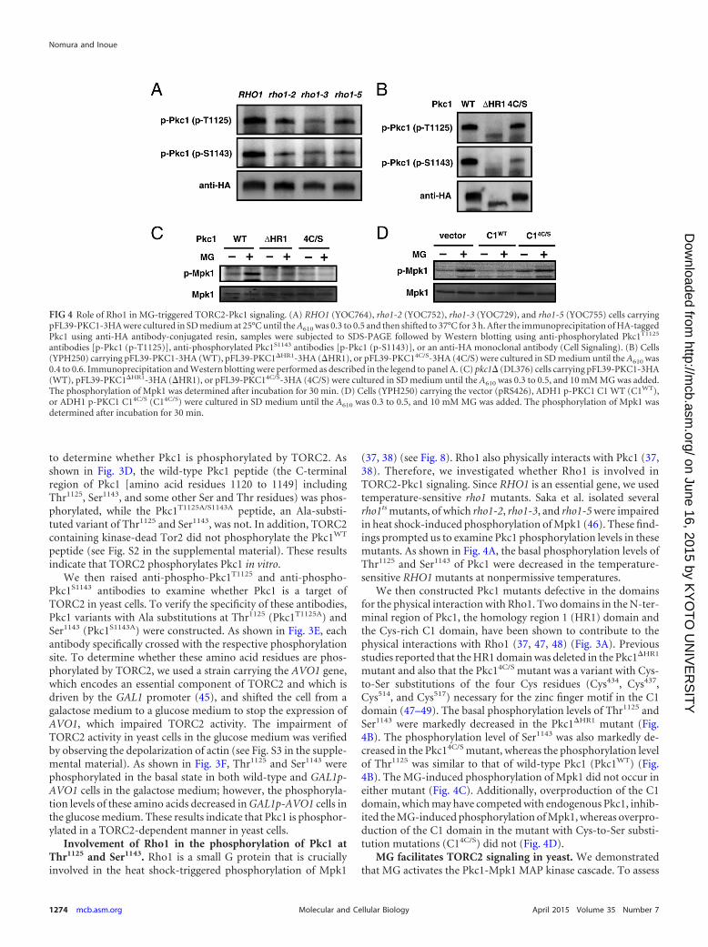
Involvement of Rho1 in the phosphorylation of Pkc1 atThr1125 and Ser1143. Rho1 is a small G protein that is cruciallyinvolved in the heat shock-triggered phosphorylation of Mpk1
(37, 38) (see Fig. 8). Rho1 also physically interacts with Pkc1 (37,38). Therefore, we investigated whether Rho1 is involved inTORC2-Pkc1 signaling. Since RHO1 is an essential gene, we usedtemperature-sensitive rho1 mutants. Saka et al. isolated severalrho1ts mutants, of which rho1-2, rho1-3, and rho1-5 were impairedin heat shock-induced phosphorylation of Mpk1 (46). These find-ings prompted us to examine Pkc1 phosphorylation levels in thesemutants. As shown in Fig. 4A, the basal phosphorylation levels ofThr1125 and Ser1143 of Pkc1 were decreased in the temperature-sensitive RHO1 mutants at nonpermissive temperatures.
We then constructed Pkc1 mutants defective in the domainsfor the physical interaction with Rho1. Two domains in the N-ter-minal region of Pkc1, the homology region 1 (HR1) domain andthe Cys-rich C1 domain, have been shown to contribute to thephysical interactions with Rho1 (37, 47, 48) (Fig. 3A). Previousstudies reported that the HR1 domain was deleted in the Pkc1�HR1
mutant and also that the Pkc14C/S mutant was a variant with Cys-to-Ser substitutions of the four Cys residues (Cys434, Cys437,Cys514, and Cys517) necessary for the zinc finger motif in the C1domain (47–49). The basal phosphorylation levels of Thr1125 andSer1143 were markedly decreased in the Pkc1�HR1 mutant (Fig.4B). The phosphorylation level of Ser1143 was also markedly de-creased in the Pkc14C/S mutant, whereas the phosphorylation levelof Thr1125 was similar to that of wild-type Pkc1 (Pkc1WT) (Fig.4B). The MG-induced phosphorylation of Mpk1 did not occur ineither mutant (Fig. 4C). Additionally, overproduction of the C1domain, which may have competed with endogenous Pkc1, inhib-ited the MG-induced phosphorylation of Mpk1, whereas overpro-duction of the C1 domain in the mutant with Cys-to-Ser substi-tution mutations (C14C/S) did not (Fig. 4D).
MG facilitates TORC2 signaling in yeast. We demonstratedthat MG activates the Pkc1-Mpk1 MAP kinase cascade. To assess
FIG 4 Role of Rho1 in MG-triggered TORC2-Pkc1 signaling. (A) RHO1 (YOC764), rho1-2 (YOC752), rho1-3 (YOC729), and rho1-5 (YOC755) cells carryingpFL39-PKC1-3HA were cultured in SD medium at 25°C until the A610 was 0.3 to 0.5 and then shifted to 37°C for 3 h. After the immunoprecipitation of HA-taggedPkc1 using anti-HA antibody-conjugated resin, samples were subjected to SDS-PAGE followed by Western blotting using anti-phosphorylated Pkc1T1125
antibodies [p-Pkc1 (p-T1125)], anti-phosphorylated Pkc1S1143 antibodies [p-Pkc1 (p-S1143)], or an anti-HA monoclonal antibody (Cell Signaling). (B) Cells(YPH250) carrying pFL39-PKC1-3HA (WT), pFL39-PKC1�HR1-3HA (�HR1), or pFL39-PKC14C/S-3HA (4C/S) were cultured in SD medium until the A610 was0.4 to 0.6. Immunoprecipitation and Western blotting were performed as described in the legend to panel A. (C) pkc1� (DL376) cells carrying pFL39-PKC1-3HA(WT), pFL39-PKC1�HR1-3HA (�HR1), or pFL39-PKC14C/S-3HA (4C/S) were cultured in SD medium until the A610 was 0.3 to 0.5, and 10 mM MG was added.The phosphorylation of Mpk1 was determined after incubation for 30 min. (D) Cells (YPH250) carrying the vector (pRS426), ADH1 p-PKC1 C1 WT (C1WT),or ADH1 p-PKC1 C14C/S (C14C/S) were cultured in SD medium until the A610 was 0.3 to 0.5, and 10 mM MG was added. The phosphorylation of Mpk1 wasdetermined after incubation for 30 min.
Nomura and Inoue
1274 mcb.asm.org April 2015 Volume 35 Number 7Molecular and Cellular Biology
on June 16, 2015 by KY
OT
O U
NIV
ER
SIT
Yhttp://m
cb.asm.org/
Dow
nloaded from

whether TORC2 is involved in this signaling pathway triggered byMG, we first monitored the phosphorylation of Mpk1 using ator2-21ts mutant. Although Tor2 was shown to be involved in bothTORC1 and TORC2, the tor2-21ts allele was specifically defectivein TORC2 function (22). The MG-induced phosphorylation ofMpk1 was observed at 28°C in tor2-21ts cells, whereas its level wasgreatly reduced at a nonpermissive temperature (see Fig. S4A inthe supplemental material). The MG-induced phosphorylation ofMpk1 also rarely occurred in cells defective in TORC2 activity(Fig. 5A), which was achieved by transferring cells carrying theessential components of TORC2 (AVO1 or AVO3) with the GAL1promoter from the galactose medium to the glucose medium. The
same result was obtained when cells with GAL1 promoter-drivenTOR2 were used (Fig. 5A). We confirmed that TORC2 functionwas impaired under these conditions by observing the depolariza-tion of actin (see Fig. S4B in the supplemental material).
Since Thr1125 and Ser1143 in Pkc1 were the direct phosphoryla-tion sites of TORC2 (Fig. 3), we investigated whether these aminoacids are necessary for the MG-induced phosphorylation ofMpk1. As shown in Fig. 5B, Mpk1 was slightly phosphorylated inPKC1S1143A cells but not in PKC1T1125A or PKC1T1125A/S1143A cellsfollowing treatment with MG. These results indicate that Thr1125
and Ser1143 in Pkc1 are involved in the MG-induced activation ofthe Mpk1 MAP kinase cascade.
FIG 5 MG functions as an initiator of the TORC2-Pkc1 signaling pathway. (A) GAL1p-TOR2 (JM340), GAL1p-AVO1 (RL25-1c), and GAL1p-AVO3 (RS61-5b)cells were cultured overnight in SC-Gal medium. After being transferred to SD medium, cells were cultured for 16 h (GAL1p-TOR2), 9 h (GAL1p-AVO1), or 10h (GAL1p-AVO3) to impair TORC2 activity and were then treated with 10 mM MG for 30 min. (B) pkc1� (DL376) cells carrying YEp352-PKC1 (pFR22),YEp352-PKC1T1125A, YEp352-PKC1S1143A (pFR74), or YEp352-PKC1T1125A/S1143A were cultured in SD medium until the A610 was 0.3 to 0.5 and were thentreated with 10 mM MG for the prescribed times. (C) Cells (YPH250) carrying pFL39-PKC1-3HA were cultured in SD medium until the A610 was 0.4 to 0.6, and10 mM MG was added. After the immunoprecipitation of HA-tagged Pkc1 using the anti-HA-antibody-conjugated resin, samples were subjected to SDS-PAGEfollowed by Western blotting using anti-phosphorylated Pkc1T1125 antibodies [p-Pkc1 (p-T1125)] or an anti-HA monoclonal antibody (Cell Signaling). (D)Cells (YPH250) carrying YCplac111-PKC1-3HA were cultured in SD medium. Immunoprecipitation and Western blotting were performed as described in thelegend to panel C, except that anti-phosphorylated Pkc1S1143 antibodies [p-Pkc1 (p-S1143)] were used. (E) Wild-type (TB50a) cells and GAL1p-AVO1 (RL25-1c)cells carrying YCplac111-PKC1-3HA were cultured overnight in SC-Gal medium. After being transferred to SD medium, cells were cultured for 15 h to impairTORC2 activity and were then treated with 10 mM MG for 60 min. Immunoprecipitation and Western blotting were performed as described in the legend topanel C. (F) pkc1� (DL376) cells carrying YEp352-PKC1 (pFR22), YEp352-PKC1T1125A, or YEp352-PKC1S1143A (pFR74) were cultured in SD medium until theA610 was 0.4 to 0.6. DL376 cells carrying the vector (YEp352) were cultured in SD medium containing 1 M sorbitol. Cell extracts were subjected to SDS-PAGEfollowed by Western blotting to determine the protein levels of Pkc1 using anti-Pkc1 antibodies. As the loading control, Pgk1 protein levels were determinedusing an anti-Pgk1 monoclonal antibody (Molecular Probes). The asterisk indicates a nonspecific band.
Methylglyoxal Activates TORC2 Signaling
April 2015 Volume 35 Number 7 mcb.asm.org 1275Molecular and Cellular Biology
on June 16, 2015 by KY
OT
O U
NIV
ER
SIT
Yhttp://m
cb.asm.org/
Dow
nloaded from
We next determined whether MG enhances the phosphoryla-tion of Pkc1. As shown in Fig. 5C, the phosphorylation levels ofThr1125 in the TM of Pkc1 remained unchanged following treat-ment with MG. Meanwhile, the phosphorylation levels of Ser1143
in HM were increased by MG treatment (Fig. 5D). To examinewhether MG directly affects the activity of TORC2 to increase thephosphorylation level of Pck1 at Ser1143, we added MG in a mix-ture of the in vitro kinase assay. However, the phosphorylationlevel of Pkc1Ser1143 was substantially unchanged in the presence ofMG (see Fig. S2 in the supplemental material). Next, to verify thatthe increase in the phosphorylation level of Pkc1 at Ser1143 inMG-treated cells is dependent on TORC2, cells carrying the AVO1gene driven by the GAL1 promoter were shifted from the galactosemedium to the glucose medium in order to reduce the activity ofTORC2 (see Fig. S5 in the supplemental material). These cellswere then treated with MG. As shown in Fig. 5E, the phosphory-lation levels of Ser1143 in TORC2-impaired cells were not in-creased by treatment with MG. These results imply that MG mayhave facilitated TORC2 signaling, thereby increasing the phos-phorylation level of Pkc1 at Ser1143, which transmitted the signaldownstream to the Mpk1 MAP kinase cascade.
The phosphorylation status of Pkc1 at Thr1125 affects its pro-tein levels. Although Thr1125 in the TM of Pkc1 did not respond toMG (Fig. 5C), the basal phosphorylation level of this site appearedto be necessary for transmitting the signal to the Mpk1 MAP ki-nase cascade (Fig. 5B). However, it currently remains unclear whythe phosphorylation of Mpk1 did not occur in Pkc1T1125A cellsfollowing treatment with MG, in spite of Ser1143 being intact. Pre-vious studies reported that the phosphorylation of Thr450 in theTM of Akt secured its stability in mammalian cells (35, 50). Toverify whether the replacement of Thr1125 in TM by Ala affects thestability of Pkc1, whereby the MG-triggered signal is not ade-quately transmitted to the Mpk1 MAP kinase cascade, we deter-mined Pkc1 protein levels. We raised anti-Pkc1 antibodies tomonitor Pkc1 protein levels. Although some nonspecific bandsappeared in the blots, the Pkc1 antibodies raised were able todetect Pkc1 proteins in cell extracts by Western blotting (see Fig.S6 in the supplemental material). As shown in Fig. 5F, the steady-state levels of the Pkc1T1125A protein were markedly lower thanthose of the Pkc1WT protein. This may be one of the reasons whythe MG-induced phosphorylation of Mpk1 did not occur inPkc1T1125A cells (Fig. 5B).
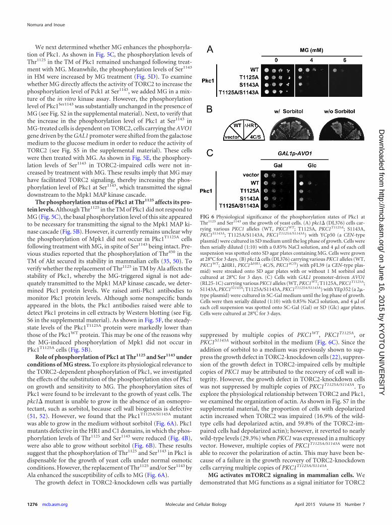
Role of phosphorylation of Pkc1 at Thr1125 and Ser1143 underconditions of MG stress. To explore its physiological relevance tothe TORC2-dependent phosphorylation of Pkc1, we investigatedthe effects of the substitution of the phosphorylation sites of Pkc1on growth and sensitivity to MG. The phosphorylation sites ofPkc1 were found to be irrelevant to the growth of yeast cells. Thepkc1� mutant is unable to grow in the absence of an osmopro-tectant, such as sorbitol, because cell wall biogenesis is defective(51, 52). However, we found that the Pkc1T1125A/S1143A mutantwas able to grow in the medium without sorbitol (Fig. 6A). Pkc1mutants defective in the HR1 and C1 domains, in which the phos-phorylation levels of Thr1125 and Ser1143 were reduced (Fig. 4B),were also able to grow without sorbitol (Fig. 6B). These resultssuggest that the phosphorylation of Thr1125 and Ser1143 in Pkc1 isdispensable for the growth of yeast cells under normal osmoticconditions. However, the replacement of Thr1125 and/or Ser1143 byAla enhanced the susceptibility of cells to MG (Fig. 6A).
The growth defect in TORC2-knockdown cells was partially
suppressed by multiple copies of PKC1WT, PKC1T1125A, orPKC1S1143A without sorbitol in the medium (Fig. 6C). Since theaddition of sorbitol to a medium was previously shown to sup-press the growth defect in TORC2-knockdown cells (22), suppres-sion of the growth defect in TORC2-impaired cells by multiplecopies of PKC1 may be attributed to the recovery of cell wall in-tegrity. However, the growth defect in TORC2-knockdown cellswas not suppressed by multiple copies of PKC1T1125A/S1143A. Toexplore the physiological relationship between TORC2 and Pkc1,we examined the organization of actin. As shown in Fig. S7 in thesupplemental material, the proportion of cells with depolarizedactin increased when TORC2 was impaired (16.9% of the wild-type cells had depolarized actin, and 59.8% of the TORC2-im-paired cells had depolarized actin); however, it reverted to nearlywild-type levels (29.3%) when PKC1 was expressed in a multicopyvector. However, multiple copies of PKC1T1125A/S1143A were notable to recover the polarization of actin. This may have been be-cause of a failure in the growth recovery of TORC2-knockdowncells carrying multiple copies of PKC1T1125A/S1143A.
MG activates mTORC2 signaling in mammalian cells. Wedemonstrated that MG functions as a signal initiator for TORC2
FIG 6 Physiological significance of the phosphorylation states of Pkc1 atThr1125 and Ser1143 on the growth of yeast cells. (A) pkc1� (DL376) cells car-rying various PKC1 alleles (WT, PKC1WT; T1125A, PKC1T1125A; S1143A,PKC1S1143A; T1125A/S1143A, PKC1T1125A/S1143A) with YCp50 (a CEN-typeplasmid) were cultured in SD medium until the log phase of growth. Cells werethen serially diluted (1:10) with a 0.85% NaCl solution, and 4 �l of each cellsuspension was spotted onto SD agar plates containing MG. Cells were grownat 28°C for 3 days. (B) pkc1� cells (DL376) carrying various PKC1 alleles (WT,PKC1WT; �HR1, PKC1�HR1; 4C/S, PKC14C/S) with pFL39 (a CEN-type plas-mid) were streaked onto SD agar plates with or without 1 M sorbitol andcultured at 28°C for 3 days. (C) Cells with GAL1 promoter-driven AVO1(RL25-1C) carrying various PKC1 alleles (WT, PKC1WT; T1125A, PKC1T1125A;S1143A, PKC1S1143A; T1125A/S1143A, PKC1T1125A/S1143A) with YEp352 (a 2�-type plasmid) were cultured in SC-Gal medium until the log phase of growth.Cells were then serially diluted (1:10) with 0.85% NaCl solution, and 4 �l ofeach cell suspension was spotted onto SC-Gal (Gal) or SD (Glc) agar plates.Cells were cultured at 28°C for 3 days.
Nomura and Inoue
1276 mcb.asm.org April 2015 Volume 35 Number 7Molecular and Cellular Biology
on June 16, 2015 by KY
OT
O U
NIV
ER
SIT
Yhttp://m
cb.asm.org/
Dow
nloaded from
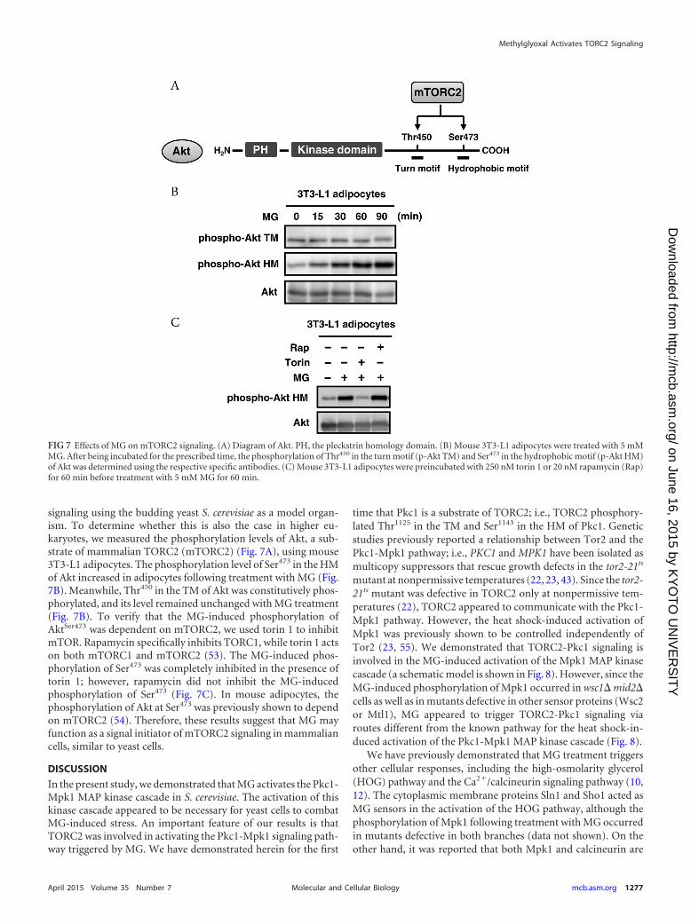
signaling using the budding yeast S. cerevisiae as a model organ-ism. To determine whether this is also the case in higher eu-karyotes, we measured the phosphorylation levels of Akt, a sub-strate of mammalian TORC2 (mTORC2) (Fig. 7A), using mouse3T3-L1 adipocytes. The phosphorylation level of Ser473 in the HMof Akt increased in adipocytes following treatment with MG (Fig.7B). Meanwhile, Thr450 in the TM of Akt was constitutively phos-phorylated, and its level remained unchanged with MG treatment(Fig. 7B). To verify that the MG-induced phosphorylation ofAktSer473 was dependent on mTORC2, we used torin 1 to inhibitmTOR. Rapamycin specifically inhibits TORC1, while torin 1 actson both mTORC1 and mTORC2 (53). The MG-induced phos-phorylation of Ser473 was completely inhibited in the presence oftorin 1; however, rapamycin did not inhibit the MG-inducedphosphorylation of Ser473 (Fig. 7C). In mouse adipocytes, thephosphorylation of Akt at Ser473 was previously shown to dependon mTORC2 (54). Therefore, these results suggest that MG mayfunction as a signal initiator of mTORC2 signaling in mammaliancells, similar to yeast cells.
DISCUSSION
In the present study, we demonstrated that MG activates the Pkc1-Mpk1 MAP kinase cascade in S. cerevisiae. The activation of thiskinase cascade appeared to be necessary for yeast cells to combatMG-induced stress. An important feature of our results is thatTORC2 was involved in activating the Pkc1-Mpk1 signaling path-way triggered by MG. We have demonstrated herein for the first
time that Pkc1 is a substrate of TORC2; i.e., TORC2 phosphory-lated Thr1125 in the TM and Ser1143 in the HM of Pkc1. Geneticstudies previously reported a relationship between Tor2 and thePkc1-Mpk1 pathway; i.e., PKC1 and MPK1 have been isolated asmulticopy suppressors that rescue growth defects in the tor2-21ts
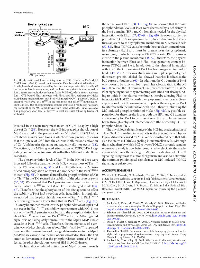
mutant at nonpermissive temperatures (22, 23, 43). Since the tor2-21ts mutant was defective in TORC2 only at nonpermissive tem-peratures (22), TORC2 appeared to communicate with the Pkc1-Mpk1 pathway. However, the heat shock-induced activation ofMpk1 was previously shown to be controlled independently ofTor2 (23, 55). We demonstrated that TORC2-Pkc1 signaling isinvolved in the MG-induced activation of the Mpk1 MAP kinasecascade (a schematic model is shown in Fig. 8). However, since theMG-induced phosphorylation of Mpk1 occurred in wsc1� mid2�cells as well as in mutants defective in other sensor proteins (Wsc2or Mtl1), MG appeared to trigger TORC2-Pkc1 signaling viaroutes different from the known pathway for the heat shock-in-duced activation of the Pkc1-Mpk1 MAP kinase cascade (Fig. 8).
We have previously demonstrated that MG treatment triggersother cellular responses, including the high-osmolarity glycerol(HOG) pathway and the Ca2�/calcineurin signaling pathway (10,12). The cytoplasmic membrane proteins Sln1 and Sho1 acted asMG sensors in the activation of the HOG pathway, although thephosphorylation of Mpk1 following treatment with MG occurredin mutants defective in both branches (data not shown). On theother hand, it was reported that both Mpk1 and calcineurin are
FIG 7 Effects of MG on mTORC2 signaling. (A) Diagram of Akt. PH, the pleckstrin homology domain. (B) Mouse 3T3-L1 adipocytes were treated with 5 mMMG. After being incubated for the prescribed time, the phosphorylation of Thr450 in the turn motif (p-Akt TM) and Ser473 in the hydrophobic motif (p-Akt HM)of Akt was determined using the respective specific antibodies. (C) Mouse 3T3-L1 adipocytes were preincubated with 250 nM torin 1 or 20 nM rapamycin (Rap)for 60 min before treatment with 5 mM MG for 60 min.
Methylglyoxal Activates TORC2 Signaling
April 2015 Volume 35 Number 7 mcb.asm.org 1277Molecular and Cellular Biology
on June 16, 2015 by KY
OT
O U
NIV
ER
SIT
Yhttp://m
cb.asm.org/
Dow
nloaded from
involved in the regulatory mechanism of G2/M delay by a highdose of Ca2� (56). However, the MG-induced phosphorylation ofMpk1 occurred in the presence of the Ca2� chelator EGTA (datanot shown) under conditions in which we have previously shownthat the uptake of Ca2� into the cell was inhibited and activationof Ca2�/calcineurin signaling subsequently did not occur (12).Collectively, the MG-triggered stimulation of TORC2-Pkc1 sig-naling does not seem to cross talk with other MG-induced cellularresponses.
The phosphorylation levels of Ser1143 in the HM of Pkc1 wereincreased following treatment with MG, whereas those of Thr1125
in the TM were not (Fig. 5C and D). Nevertheless, the MG-in-duced phosphorylation of Mpk1 did not occur in the Pkc1T1125A
mutant (Fig. 5B). In mammalian cells, the phosphorylation of Aktat Thr450 in the TM secured the stability of the Akt protein per se(35, 50). We showed that Pkc1 protein levels were markedly de-creased when Thr1125 in the TM of Pkc1 was changed to Ala (Fig.5F). Therefore, the phosphorylation of this site appears to affectthe stability of Pkc1 in S. cerevisiae cells. In addition, intriguingly,we noticed that the phosphorylation level of Ser1143 in Pkc1T1125A
cells was significantly lower than that in Pkc1WT cells (Fig. 3E).This may be another reason why the phosphorylation of Mpk1 didnot occur in Pkc1T1125A cells following treatment with MG. Sincenot only the Pkc1 protein levels but also the phosphorylation lev-els of Sre1143 were lower in Pkc1T1125A cells, the MG-triggeredsignal was not adequately transmitted to the Mpk1 MAP kinasecascade in Pkc1T1125A cells. Therefore, the maintenance of a cer-tain level of phosphorylation at both Thr1125 and Ser1143 appearedto secure the transmittance of the signal downstream to the Mpk1MAP kinase cascade. To the best of our knowledge, this is the firststudy to demonstrate that the phosphorylation status of TM af-fected the phosphorylation levels of HM in AGC kinases.
The heat shock-induced activation of Mpk1 occurs through
the activation of Rho1 (38, 39) (Fig. 8). We showed that the basalphosphorylation levels of Pkc1 were decreased by a deficiency inthe Pkc1 domains (HR1 and C1 domains) needed for the physicalinteraction with Rho1 (37, 47–49) (Fig. 4B). Previous studies re-ported that TORC2 was predominantly located in punctate struc-tures adjacent to the cytoplasmic membrane in S. cerevisiae cells(57, 58). Since TORC2 exists beneath the cytoplasmic membrane,its substrate (Pkc1) also must be present near the cytoplasmicmembrane, in which the enzyme (TORC2) exists. Rho1 is associ-ated with the plasma membrane (38, 59); therefore, the physicalinteraction between Rho1 and Pkc1 may guarantee contact be-tween TORC2 and Pkc1. In addition to the physical interactionwith Rho1, the C1 domain of Pkc1 has been suggested to bind tolipids (49, 55). A previous study using multiple copies of greenfluorescent protein-labeled Pkc1 showed that Pkc1 localized to thebud cortex or bud neck (60). In addition, the C1 domain of Pkc1was shown to be sufficient for its peripheral localization in the cell(60); therefore, the C1 domain of Pkc1 may contribute to TORC2-Pkc1 signaling not only by interacting with Rho1 but also by bind-ing to lipids in the plasma membrane, thereby allowing Pkc1 tointeract with TORC2 beneath the cytoplasmic membrane. Over-expression of the C1 domain may compete with endogenous Pkc1to interfere with the interaction with Rho1, thereby inhibiting theMG-induced phosphorylation of Mpk1 (Fig. 4D). A possible ex-planation for these results is that both the HR1 and C1 domainsare necessary for Pkc1 to be present near the cytoplasmic mem-brane through a physical interaction with Rho1, whereby TORC2phosphorylates Pkc1.
The physiological significance of the MG-induced activation ofTORC2-Pkc1 signaling in yeast cells is the prevention of physio-logical disorders caused by MG. We demonstrated here that MGalso facilitates mTORC2 signaling in mammalian cells. Althoughthe mechanism by which MG activates TORC2 currently remainsunknown, a study is now being conducted to elucidate the mech-anism underlying the sensing of MG and facilitation of TORC2signaling using yeast as a model organism and also to determinethe common physiological significance of MG-induced TORC2signaling in eukaryotes.
ACKNOWLEDGMENTS
We thank T. Kawada, N. Takahashi, T. Goto, Y. Kim, S. Izawa, and K.Maeta for their technical support and helpful discussions. We are gratefulto M. N. Hall, D. E. Levin, T. Miyakawa, J. Thorner, Y. Ohya, J. J. Heinisch,M. Y. Chen, M. S. Cyert, J. R. Broach, K. Irie, and the National Bio-Resource Project (NBRP) of MEXT, Japan, for providing the plasmidsand yeast strains.
REFERENCES1. Rochette L, Zeller M, Cottin Y, Vergely C. 2014. Diabetes, oxidative
stress and therapeutic strategies. Biochim Biophys Acta 1840:2709 –2729.http://dx.doi.org/10.1016/j.bbagen.2014.05.017.
2. Schieber M, Chandel NS. 2014. ROS function in redox signaling andoxidative stress. Curr Biol 24:R453–R462. http://dx.doi.org/10.1016/j.cub.2014.03.034.
3. Inoue Y, Maeta K, Nomura W. 2011. Glyoxalase system in yeasts: struc-ture, function, and physiology. Semin Cell Dev Biol 22:278 –284. http://dx.doi.org/10.1016/j.semcdb.2011.02.002.
4. Thornalley PJ. 2008. Protein and nucleotide damage by glyoxal and meth-ylglyoxal in physiological systems—role in ageing and disease. DrugMetabol Drug Interact 23:125–150.
5. Rabbani N, Thornalley PJ. 2011. Glyoxalase in diabetes, obesity andrelated disorders. Semin Cell Dev Biol 22:309 –317. http://dx.doi.org/10.1016/j.semcdb.2011.02.015.
FIG 8 Schematic model for the integration of TORC2 into the Pkc1-Mpk1MAP kinase (MAPK) cascade in S. cerevisiae. Details are described in the text.Briefly, heat shock stress is sensed by the stress sensor proteins Wsc1 and Mid2on the cytoplasmic membrane, and the heat shock signal is transmitted toRom2 (guanine-nucleotide exchange factor for Rho1), which in turn activatesRho1. GTP-bound Rho1 interacts with Pkc1, and Pkc1 activates the Mpk1MAP kinase cascade (the so-called cell wall integrity [CWI] pathway). TORC2phosphorylates Pkc1 at Thr1125 in the turn motif and at Ser1143 in the hydro-phobic motif. The phosphorylation of these amino acid residues is necessaryfor transmitting the MG signal downstream to the Mpk1 MAP kinase cascade.The phosphorylation level of Ser1143 in Pkc1 increases following treatmentwith MG.
Nomura and Inoue
1278 mcb.asm.org April 2015 Volume 35 Number 7Molecular and Cellular Biology
on June 16, 2015 by KY
OT
O U
NIV
ER
SIT
Yhttp://m
cb.asm.org/
Dow
nloaded from
6. Matafome P, Sena C, Seiça R. 2013. Methylglyoxal, obesity, and diabetes.Endocrine 43:472– 484. http://dx.doi.org/10.1007/s12020-012-9795-8.
7. Angeloni C, Zambonin L, Hrelia S. 2014. Role of methylglyoxal inAlzheimer’s disease. Biomed Res Int 2014:238485. http://dx.doi.org/10.1155/2014/238485.
8. Zoccali C, Mallamaci F, Tripepi G. 2000. AGEs and carbonyl stress: potentialpathogenetic factors of long-term uraemic complications. Nephrol DialTransplant 15:7–11. http://dx.doi.org/10.1093/ndt/15.suppl_1.7.
9. Thornalley PJ. 2005. Dicarbonyl intermediates in the Maillard reaction.Ann N Y Acad Sci 1043:111–117. http://dx.doi.org/10.1196/annals.1333.014.
10. Maeta K, Izawa S, Okazaki S, Kuge S, Inoue Y. 2004. Activity of the Yap1transcription factor in Saccharomyces cerevisiae is modulated by methylg-lyoxal, a metabolite derived from glycolysis. Mol Cell Biol 24:8753– 8764.http://dx.doi.org/10.1128/MCB.24.19.8753-8764.2004.
11. Takatsume Y, Izawa S, Inoue Y. 2006. Methylglyoxal as a signal initiatorfor activation of the stress-activated protein kinase cascade in the fissionyeast Schizosaccharomyces pombe. J Biol Chem 281:9086 –9092. http://dx.doi.org/10.1074/jbc.M511037200.
12. Maeta K, Izawa S, Inoue Y. 2005. Methylglyoxal, a metabolite derivedfrom glycolysis, functions as a signal initiator of the high osmolarity glyc-erol-mitogen-activated protein kinase cascade and calcineurin/Crz1-mediated pathway in Saccharomyces cerevisiae. J Biol Chem 280:253–260.http://dx.doi.org/10.1074/jbc.M408061200.
13. Laplante M, Sabatini DM. 2012. mTOR signaling in growth control anddisease. Cell 149:274 –293. http://dx.doi.org/10.1016/j.cell.2012.03.017.
14. Loewith R, Hall MN. 2011. Target of rapamycin (TOR) in nutrient sig-naling and growth control. Genetics 189:1177–1201. http://dx.doi.org/10.1534/genetics.111.133363.
15. Loewith R, Jacinto E, Wullschleger S, Lorberg A, Crespo JL, BonenfantD, Oppliger W, Jenoe P, Hall MN. 2002. Two TOR complexes, only oneof which is rapamycin sensitive, have distinct roles in cell growth control.Mol Cell 10:457– 468. http://dx.doi.org/10.1016/S1097-2765(02)00636-6.
16. Jacinto E, Lorberg A. 2008. TOR regulation of AGC kinases in yeast andmammals. Biochem J 410:19 –37. http://dx.doi.org/10.1042/BJ20071518.
17. Kamada Y, Fujioka Y, Suzuki NN, Inagaki F, Wullschleger S, LoewithR, Hall MN, Ohsumi Y. 2005. Tor2 directly phosphorylates the AGCkinase Ypk2 to regulate actin polarization. Mol Cell Biol 25:7239 –7248.http://dx.doi.org/10.1128/MCB.25.16.7239-7248.2005.
18. Niles BJ, Mogri H, Hill A, Vlahakis A, Powers T. 2012. Plasma mem-brane recruitment and activation of the AGC kinase Ypk1 is mediated bytarget of rapamycin complex 2 (TORC2) and its effector proteins Slm1and Slm2. Proc Natl Acad Sci U S A 109:1536 –1541. http://dx.doi.org/10.1073/pnas.1117563109.
19. Urban J, Soulard A, Huber A, Lippman S, Mukhopadhyay D, DelocheO, Wanke V, Anrather D, Ammerer G, Riezman H, Broach JR, DeVirgilio C, Hall MN, Loewith R. 2007. Sch9 is a major target of TORC1in Saccharomyces cerevisiae. Mol Cell 26:663– 674. http://dx.doi.org/10.1016/j.molcel.2007.04.020.
20. Antonsson B, Montessuit S, Friedli L, Payton MA, Paravicini G. 1994.Protein kinase C in yeast. Characteristics of the Saccharomyces cerevisiaePKC1 gene product. J Biol Chem 269:16821–16828.
21. Watanabe M, Chen CY, Levin DE. 1994. Saccharomyces cerevisiae PKC1encodes a protein kinase C (PKC) homolog with a substrate specificitysimilar to that of mammalian PKC. J Biol Chem 269:16829 –16836.
22. Helliwell SB, Howald I, Barbet N, Hall MN. 1998. TOR2 is part of tworelated signaling pathways coordinating cell growth in Saccharomycescerevisiae. Genetics 148:99 –112.
23. Helliwell SB, Schmidt A, Ohya Y, Hall MN. 1998. The Rho1 effector Pkc1,but not Bni1, mediates signalling from Tor2 to the actin cytoskeleton. CurrBiol 8:1211–1214. http://dx.doi.org/10.1016/S0960-9822(07)00511-8.
24. Ho HL, Shiau YS, Chen MY. 2005. Saccharomyces cerevisiae TSC11/AVO3 participates in regulating cell integrity and functionally interactswith components of the Tor2 complex. Curr Genet 47:273–288. http://dx.doi.org/10.1007/s00294-005-0570-8.
25. Cybulski N, Hall MN. 2009. TOR complex 2: a signaling pathway of itsown. Trends Biochem Sci 34:620 – 627. http://dx.doi.org/10.1016/j.tibs.2009.09.004.
26. Dazert E, Hall MN. 2011. mTOR signaling in disease. Curr Opin Cell Biol23:744 –755. http://dx.doi.org/10.1016/j.ceb.2011.09.003.
27. Zoncu R, Efeyan A, Sabatini DM. 2011. mTOR: from growth signalintegration to cancer, diabetes and ageing. Nat Rev Mol Cell Biol 12:21–35. http://dx.doi.org/10.1038/nrm3025.
28. Sarbassov DD, Guertin DA, Ali SM, Sabatini DM. 2005. Phosphoryla-tion and regulation of Akt/PKB by the rictor-mTOR complex. Science307:1098 –1101. http://dx.doi.org/10.1126/science.1106148.
29. Gueldener U, Heinisch J, Koehler GJ, Voss D, Hegemann JH. 2002. Asecond set of loxP marker cassettes for Cre-mediated multiple gene knock-outs in budding yeast. Nucleic Acids Res 30:e23. http://dx.doi.org/10.1093/nar/30.6.e23.
30. Sekiya-Kawasaki M, Abe M, Saka A, Watanabe D, Kono K, Minemura-Asakawa M, Ishihara S, Watanabe T, Ohya Y. 2002. Dissection ofupstream regulatory components of the Rho1p effector, 1,3-�-glucan syn-thase, in Saccharomyces cerevisiae. Genetics 162:663– 676.
31. Nakamura T, Ohmoto T, Hirata D, Tsuchiya E, Miyakawa T. 1996.Genetic evidence for the functional redundancy of the calcineurin- andMpk1-mediated pathways in the regulation of cellular events importantfor growth in Saccharomyces cerevisiae. Mol Gen Genet 251:211–219. http://dx.doi.org/10.1007/BF02172920.
32. Irie K, Takase M, Lee KS, Levin DE, Araki H, Matsumoto K, Oshima Y.1993. MKK1 and MKK2, which encode Saccharomyces cerevisiae mitogen-activated protein kinase-kinase homologs, function in the pathway medi-ated by protein kinase C. Mol Cell Biol 13:3076 –3083.
33. Schägger H. 2006. Tricine-SDS-PAGE. Nat Protoc 1:16 –22. http://dx.doi.org/10.1038/nprot.2006.4.
34. Goto T, Lee JY, Teraminami A, Kim YI, Hirai S, Uemura T, Inoue H,Takahashi N, Kawada T. 2011. Activation of peroxisome proliferator-activated receptor-alpha stimulates both differentiation and fatty acid ox-idation in adipocytes. J Lipid Res 52:873– 884. http://dx.doi.org/10.1194/jlr.M011320.
35. Facchinetti V, Ouyang W, Wei H, Soto N, Lazorchak A, Gould C,Lowry C, Newton AC, Mao Y, Miao RQ, Sessa WC, Qin J, Zhang P, SuB, Jacinto E. 2008. The mammalian target of rapamycin complex 2 con-trols folding and stability of Akt and protein kinase C. EMBO J 27:1932–1943. http://dx.doi.org/10.1038/emboj.2008.120.
36. Pruyne D, Bretscher A. 2000. Polarization of cell growth in yeast. J CellSci 113(Pt 4):571–585.
37. Nonaka H, Tanaka K, Hirano H, Fujiwara T, Kohno H, Umikawa M,Mino A, Takai Y. 1995. A downstream target of RHO1 small GTP-binding protein is PKC1, a homolog of protein kinase C, which leads toactivation of the MAP kinase cascade in Saccharomyces cerevisiae. EMBO J14:5931–5938.
38. Levin DE. 2011. Regulation of cell wall biogenesis in Saccharomyces cerevi-siae: the cell wall integrity signaling pathway. Genetics 189:1145–1175.http://dx.doi.org/10.1534/genetics.111.128264.
39. Chen RE, Thorner J. 2007. Function and regulation in MAPK signalingpathways: lessons learned from the yeast Saccharomyces cerevisiae. BiochimBiophys Acta 1773:1311–1340. http://dx.doi.org/10.1016/j.bbamcr.2007.05.003.
40. Philip B, Levin DE. 2001. Wsc1 and Mid2 are cell surface sensors for cellwall integrity signaling that act through Rom2, a guanine nucleotide ex-change factor for Rho1. Mol Cell Biol 21:271–280. http://dx.doi.org/10.1128/MCB.21.1.271-280.2001.
41. Wilk S, Wittland J, Thywissen A, Schmitz HP, Heinisch JJ. 2010. Ablock of endocytosis of the yeast cell wall integrity sensors Wsc1 and Wsc2results in reduced fitness in vivo. Mol Genet Genomics 284:217–229. http://dx.doi.org/10.1007/s00438-010-0563-2.
42. Jin C, Parshin AV, Daly I, Strich R, Cooper KF. 2013. The cell wallsensors Mtl1, Wsc1, and Mid2 are required for stress-induced nuclear tocytoplasmic translocation of cyclin C and programmed cell death in yeast.Oxid Med Cell Longev 2013:320823. http://dx.doi.org/10.1155/2013/320823.
43. Schmidt A, Bickle M, Beck T, Hall MN. 1997. The yeast phosphatidyl-inositol kinase homolog TOR2 activates RHO1 and RHO2 via the ex-change factor ROM2. Cell 88:531–542. http://dx.doi.org/10.1016/S0092-8674(00)81893-0.
44. Roelants FM, Torrance PD, Thorner J. 2004. Differential roles of PDK1-and PDK2-phosphorylation sites in the yeast AGC kinases Ypk1, Pkc1and Sch9. Microbiology 150:3289 –3304. http://dx.doi.org/10.1099/mic.0.27286-0.
45. Wullschleger S, Loewith R, Oppliger W, Hall MN. 2005. Molecularorganization of target of rapamycin complex 2. J Biol Chem 280:30697–30704. http://dx.doi.org/10.1074/jbc.M505553200.
46. Saka A, Abe M, Okano H, Minemura M, Qadota H, Utsugi T, Mino A,Tanaka K, Takai Y, Ohya Y. 2001. Complementing yeast rho1 mutation
Methylglyoxal Activates TORC2 Signaling
April 2015 Volume 35 Number 7 mcb.asm.org 1279Molecular and Cellular Biology
on June 16, 2015 by KY
OT
O U
NIV
ER
SIT
Yhttp://m
cb.asm.org/
Dow
nloaded from
groups with distinct functional defects. J Biol Chem 276:46165– 46171.http://dx.doi.org/10.1074/jbc.M103805200.
47. Schmitz HP, Lorberg A, Heinisch JJ. 2002. Regulation of yeast proteinkinase C activity by interaction with the small GTPase Rho1p through itsamino-terminal HR1 domain. Mol Microbiol 44:829 – 840. http://dx.doi.org/10.1046/j.1365-2958.2002.02925.x.
48. Schmitz HP, Jöckel J, Block C, Heinisch JJ. 2001. Domain shuffling as atool for investigation of protein function: substitution of the cysteine-richregion of Raf kinase and PKC eta for that of yeast Pkc1p. J Mol Biol311:1–7. http://dx.doi.org/10.1006/jmbi.2001.4848.
49. Jacoby JJ, Schmitz HP, Heinisch JJ. 1997. Mutants affected in the puta-tive diacylglycerol binding site of yeast protein kinase C. FEBS Lett 417:219 –222. http://dx.doi.org/10.1016/S0014-5793(97)01287-8.
50. Ikenoue T, Inoki K, Yang Q, Zhou X, Guan KL. 2008. Essential functionof TORC2 in PKC and Akt turn motif phosphorylation, maturation andsignalling. EMBO J 27:1919 –1931. http://dx.doi.org/10.1038/emboj.2008.119.
51. Levin DE, Bartlett-Heubusch E. 1992. Mutants in the S. cerevisiae PKC1gene display a cell cycle-specific osmotic stability defect. J Cell Biol 116:1221–1229. http://dx.doi.org/10.1083/jcb.116.5.1221.
52. Paravicini G, Cooper M, Friedli L, Smith DJ, Carpentier JL, Klig LS,Payton MA. 1992. The osmotic integrity of the yeast cell requires a func-tional PKC1 gene product. Mol Cell Biol 12:4896 – 4905.
53. Thoreen CC, Kang SA, Chang JW, Liu Q, Zhang J, Gao Y, Reichling LJ,Sim T, Sabatini DM, Gray NS. 2009. An ATP-competitive mammaliantarget of rapamycin inhibitor reveals rapamycin-resistant functions of
mTORC1. J Biol Chem 284:8023– 8032. http://dx.doi.org/10.1074/jbc.M900301200.
54. Hresko RC, Mueckler M. 2005. mTOR · RICTOR is the Ser473 kinase forAkt/protein kinase B in 3T3-L1 adipocytes. J Biol Chem 280:40406 –40416. http://dx.doi.org/10.1074/jbc.M508361200.
55. Levin DE. 2005. Cell wall integrity signaling in Saccharomyces cerevisiae.Microbiol Mol Biol Rev 69:262–291. http://dx.doi.org/10.1128/MMBR.69.2.262-291.2005.
56. Mizunuma M, Hirata D, Miyahara K, Tsuchiya E, Miyakawa T. 1998.Role of calcineurin and Mpk1 in regulating the onset of mitosis in buddingyeast. Nature 392:303–306. http://dx.doi.org/10.1038/32695.
57. Berchtold D, Walther TC. 2009. TORC2 plasma membrane localizationis essential for cell viability and restricted to a distinct domain. Mol BiolCell 20:1565–1575. http://dx.doi.org/10.1091/mbc.E08-10-1001.
58. Sturgill TW, Cohen A, Diefenbacher M, Trautwein M, Martin DE, HallMN. 2008. TOR1 and TOR2 have distinct locations in live cells. EukaryotCell 7:1819 –1830. http://dx.doi.org/10.1128/EC.00088-08.
59. Inoue SB, Qadota H, Arisawa M, Watanabe T, Ohya Y. 1999.Prenylation of Rho1p is required for activation of yeast 1,3-�-glucansynthase. J Biol Chem 274:38119 –38124. http://dx.doi.org/10.1074/jbc.274.53.38119.
60. Denis V, Cyert MS. 2005. Molecular analysis reveals localization of Sac-charomyces cerevisiae protein kinase C to sites of polarized growth andPkc1p targeting to the nucleus and mitotic spindle. Eukaryot Cell 4:36 –45. http://dx.doi.org/10.1128/EC.4.1.36-45.2005.
Nomura and Inoue
1280 mcb.asm.org April 2015 Volume 35 Number 7Molecular and Cellular Biology
on June 16, 2015 by KY
OT
O U
NIV
ER
SIT
Yhttp://m
cb.asm.org/
Dow
nloaded from