mesenchymal stem cells and 3d-osteoconductive ... - chai lab
TRANSCRIPT
1
Mesenchymal stem cells and 3D-osteoconductive scaffold regenerate calvarial bone in
critical size defects in swine
Zoe M. Johnson1, Yuan Yuan1, Xiangjia Li2,3, Tea Jashashvili4, Michael Jamieson5, Mark Urata6,
Yong Chen3, and Yang Chai1,*
1. Center for Craniofacial Molecular Biology, Herman Ostrow School of Dentistry, University of Southern California, Los Angeles, CA, USA.2. Department of Aerospace and Mechanical Engineering, School for Engineering of Matter, Transport and Energy, Arizona State University, Tempe, AZ, USA.3. Viterbi School of Engineering, University of Southern California, Los Angeles, CA, USA.4. Molecular Imaging Core, University of Southern California, Los Angeles, CA, USA.5. Ottawa Hospital Research Institute, Ottawa, Canada6. Division of Plastic and Maxillofacial Surgery, Children's Hospital Los Angeles, Los Angeles, CA, USA.
Author ContributionsZoe M. Johnson: Conception and design, collection and assembly of data, data analysis and interpretation, manuscript writingYuan Yuan: Conception and design, collection and assembly of data, data analysis and interpretation, manuscript writingXiangjia Li: Conception and design, collection and assembly of data, manuscript writingTea Jashashvili: Collection of data, data analysis and interpretationMichael Jamieson: Conception and design, data interpretationMark Urata: Conception and design, data interpretationYong Chen: Conception and design, data analysis and interpretation, manuscript writingYang Chai: Conception and design, data analysis and interpretation, manuscript writing
*Corresponding author:Yang ChaiUniversity ProfessorGeorge and MaryLou Boone Chair in Craniofacial BiologyCenter for Craniofacial Molecular BiologyUniversity of Southern California2250 Alcazar Street – CSA 103Los Angeles, CA 90033Phone number: [email protected]
Page 1 of 50
2
This work was supported by the National Institute of Dental and Craniofacial Research National Institute of Health - Center for Dental, Oral and Craniofacial Tissue and Organ Regeneration (C-DOCTOR) U24 DE026914; U24 DE029463 and Alfred Mann Institute (AMI) at the University of Southern California.
Keywords: Critical size defect (CSD); Dental pulp neural crest cell (DPNCC); Bone marrow aspirate (BMA); Mesenchymal stem cells (MSCs); Hydroxyapatite tricalcium phosphate (HA/TCP)
ABSTRACT Craniofacial bones protect vital organs, perform important physiological functions, and shape facial identity. Critical-size defects (CSDs) in calvarial bones, which will not heal spontaneously, are caused by trauma, congenital defects, or tumor resections. They pose a great challenge for patients and physicians, and significantly compromise quality of life. Currently, calvarial CSDs are treated either by allogenic or autologous grafts, metal or other synthetic plates that are associated with considerable complications. While previous studies have explored tissue regeneration for calvarial defects, most have been done in small animal models with limited translational value. Here we define a swine calvarial CSD model and show a novel approach to regenerate high-quality bone in these defects by combining mesenchymal stem cells (MSCs) with a 3D-printed osteoconductive HA/TCP scaffold. Specifically, we have compared the performance of dental pulp neural crest MSCs (DPNCCs) to bone marrow aspirate (BMA) combined with a 3D-printed HA/TCP scaffold to regenerate bone in a calvarial CSD (>7.0 cm2). Both DPNCCs and BMA loaded onto the 3D-printed osteoconductive scaffold support the regeneration of calvarial bone with density, compression strength, and trabecular structures similar to native bone. Our study demonstrates a novel application of an original scaffold design combined with DPNCCs or BMA to support regeneration of high-quality bone in a newly defined and clinically relevant swine calvarial CSD model. This discovery may have important impact on bone regeneration beyond the craniofacial region and will ultimately benefit patients who suffer from debilitating CSDs.
Page 2 of 50
3
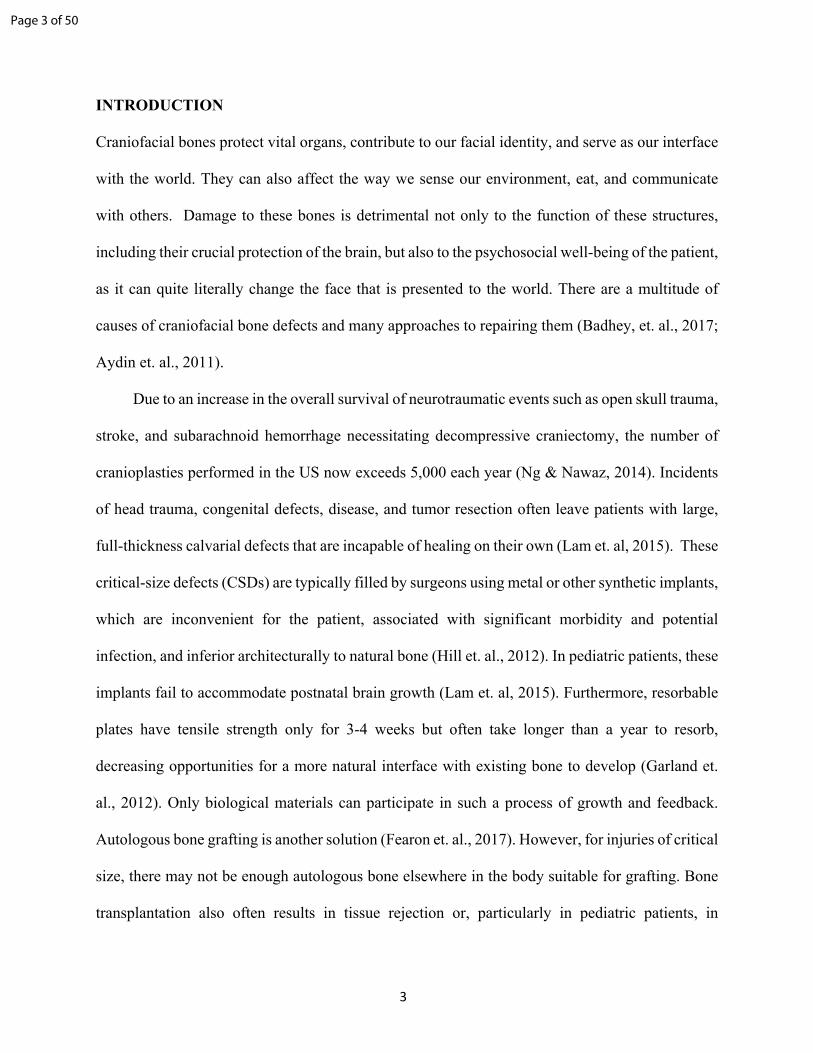
INTRODUCTION
Craniofacial bones protect vital organs, contribute to our facial identity, and serve as our interface
with the world. They can also affect the way we sense our environment, eat, and communicate
with others. Damage to these bones is detrimental not only to the function of these structures,
including their crucial protection of the brain, but also to the psychosocial well-being of the patient,
as it can quite literally change the face that is presented to the world. There are a multitude of
causes of craniofacial bone defects and many approaches to repairing them (Badhey, et. al., 2017;
Aydin et. al., 2011).
Due to an increase in the overall survival of neurotraumatic events such as open skull trauma,
stroke, and subarachnoid hemorrhage necessitating decompressive craniectomy, the number of
cranioplasties performed in the US now exceeds 5,000 each year (Ng & Nawaz, 2014). Incidents
of head trauma, congenital defects, disease, and tumor resection often leave patients with large,
full-thickness calvarial defects that are incapable of healing on their own (Lam et. al, 2015). These
critical-size defects (CSDs) are typically filled by surgeons using metal or other synthetic implants,
which are inconvenient for the patient, associated with significant morbidity and potential
infection, and inferior architecturally to natural bone (Hill et. al., 2012). In pediatric patients, these
implants fail to accommodate postnatal brain growth (Lam et. al, 2015). Furthermore, resorbable
plates have tensile strength only for 3-4 weeks but often take longer than a year to resorb,
decreasing opportunities for a more natural interface with existing bone to develop (Garland et.
al., 2012). Only biological materials can participate in such a process of growth and feedback.
Autologous bone grafting is another solution (Fearon et. al., 2017). However, for injuries of critical
size, there may not be enough autologous bone elsewhere in the body suitable for grafting. Bone
transplantation also often results in tissue rejection or, particularly in pediatric patients, in
Page 3 of 50
4
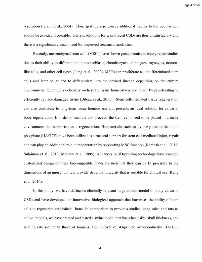
resorption (Grant et al., 2004). Bone grafting also causes additional trauma to the body which
should be avoided if possible. Current solutions for craniofacial CSDs are thus unsatisfactory and
there is a significant clinical need for improved treatment modalities.
Recently, mesenchymal stem cells (MSCs) have shown great promise in injury repair studies
due to their ability to differentiate into osteoblasts, chondrocytes, adipocytes, myocytes, neuron-
like cells, and other cell types (Jiang et al., 2002). MSCs can proliferate as undifferentiated stem
cells and later be guided to differentiate into the desired lineage depending on the culture
environment. Stem cells delicately orchestrate tissue homeostasis and repair by proliferating to
efficiently replace damaged tissue (Biteau et al., 2011). Stem cell-mediated tissue regeneration
can also contribute to long-term tissue homeostasis and presents an ideal solution for calvarial
bone regeneration. In order to mediate this process, the stem cells need to be placed in a niche
environment that supports tissue regeneration. Biomaterials such as hydroxyapatite/tricalcium
phosphate (HA/TCP) have been utilized as structural support for stem cell-mediated injury repair
and can play an additional role in regeneration by supporting MSC function (Ramesh et al., 2018;
Sulaiman et al., 2013; Mauney et al. 2005). Advances in 3D-printing technology have enabled
customized design of these biocompatible materials such that they can be fit precisely to the
dimensions of an injury, but few provide structural integrity that is suitable for clinical use (Kang
et al. 2016).
In this study, we have defined a clinically relevant large animal model to study calvarial
CSDs and have developed an innovative, biological approach that harnesses the ability of stem
cells to regenerate craniofacial bone. In comparison to previous studies using mice and rats as
animal models, we have created and tested a swine model that has a head size, skull thickness, and
healing rate similar to those of humans. Our innovative 3D-printed osteoconductive HA/TCP
Page 4 of 50

5
scaffold with structural integrity in combination with autologous neural crest or bone marrow
MSCs promotes full healing of CSDs in the swine calvaria. Significantly, the regenerated bone
forms a smooth calvarial surface with both fully functional and aesthetically satisfying results.
Moreover, we show that the regenerated bone is of high quality that is comparable to their
surrounding native bones. Importantly, we achieved these successful results with bone marrow
aspirate (BMA), which can be easily obtained from a patient for autologous use and needs minimal
manipulation, saving substantial time, resources, and avoiding possible complications from in vitro
MSC expansion. This discovery represents an innovative, MSC-mediated tissue regeneration
approach in a model that lays the groundwork for improving care for human patients with calvarial
CSDs and has the potential to fill an unmet clinical need.
Page 5 of 50
6
MATERIALS AND METHODS
Yorkshire farm pigs at 10-12 weeks of age were used to investigate stem-cell-mediated calvarial
bone regeneration. All animal procedures were performed in accordance with federal regulations
and with approval from the Institutional Animal Care and Use Committee (IACUC) at the
University of Southern California.
Harvesting of swine dental pulp cells
Each pig (10-12 weeks of age) was anesthetized by veterinary staff and placed on its stomach. The
inside of the mouth was wiped sequentially with a germicide and ethanol to prevent infection. The
right mandibular incisor was extracted and the remaining gap was closed with discontinuous,
absorbable vicryl monofilament sutures in size 3-0. Dental pulp neural crest mesenchymal stem
cells (DPNCCs) were harvested from the incisor and expanded in cell culture as described below.
Culture of swine dental pulp cells
After extraction of the incisor, the outside of the tooth was thoroughly scrubbed with gauze soaked
in bleach for two minutes to eliminate oral microbes. The tooth was cut open using sterile
dissection scissors or a diamond burr. The dental pulp was removed using blunt, sterile forceps.
The dental pulp was then finely chopped using sterile scissors and digested with a 0.2% solution
of Collagenase Type I (Washington Biochemical Corporation, Table 1) diluted with Alpha-MEM
(Gibco, Table 1) in a water bath at 37oC for 1.5 hours. The dental pulp-containing Collagenase
solution was then strained for single cells using a 70 μm cell strainer into a 50mL conical tube.
The remaining solution was centrifuged at 1440 rpm for 5 minutes at 21 oC and the supernatant
Page 6 of 50
7
was discarded. The cell pellet was resuspended in culture media and plated at 1 x 10⁶ cells per 15
cm tissue culture dish. Cells were incubated at 37oC and 5% CO2.
Cells were cultured at low density to form single-cell colonies, and were passaged once
reaching 65-70% subconfluence. To passage, media was removed from the culture dishes and the
cells were washed with DPBS (Gibco, Table 1). After removal of DPBS, cells were detached from
the plates using TryplE Express with Phenol Red (Gibco, Table 1) for 7 minutes at 37oC and 5%
CO2. The cells and enzyme solution were immediately transferred to a conical tube with an equal
volume of culture media to deactivate the enzyme. This solution was used for cell counting as
described below, then centrifuged. The supernatant was discarded and the cell pellet was
resuspended in culture media. With each passage, 1 x 10⁶ cells were plated into 15 cm dishes. This
process was repeated 2-3 times over 14 days before transplantation into the calvarial defect. Cells
were counted using a BioRad TC20 Automated Cell Counter. Ten μL of suspended cells were
loaded into the cartridge for cell counting. For transplantation, three 10 μL suspensions were
counted and averaged. For each animal, 3-4 x 10⁶ autologous cells were transplanted into the CSD
site (see below) with HA/TCP particles or an HA/TCP 3D-printed scaffold.
Cells for the heat-inactivated DPNCC group were prepared and passaged using the same
method as living DPNCCs. At the time of the last passage for transplantation, the cells were again
detached from plastic plates using TriplE for 7 minutes at 37oC and 5% CO2 and transferred to a
conical tube. An equal volume of media was added to this solution before the cells were counted
using a BioRad TC20 Automated Cell Counter as described above. Cells were centrifuged at 1440
rpm for 5 minutes, the supernatant was discarded, and the remaining cell pellet was resuspended
in 1mL of culture media. The resuspension was transferred to a sterile centrifuge tube and heated
to 85oC for 20 minutes. There were no live cells following the heat treatment.
Page 7 of 50

8
With the remaining living cells not used for transplantation to support calvarial bone
regeneration, the ability of the DPNCCs to differentiate into adipocytes, chondrocytes, and
osteoblasts was verified through culture in adipogenic, chondrogenic, and osteogenic culture
media, respectively (Gibco, Table 1). Adipogenic differentiation was evaluated using Oil Red O
staining for lipid droplets. Chondrogenic differentiation was evaluated using Alcian blue staining
for cartilage formation following condensation of cultured DPNCCs (Chung et. al, 2009).
Osteogenic differentiation was evaluated using Alizarin red staining for bone formation.
Application of DPNCCs and HA/TCP nanoparticles or 3D-printed HA/TCP scaffold to swine
calvarial CSD
Cells were washed with DPBS and dissociated from the culture dish as described for passaging
above. For each calvarial CSD (3.0 cm in diameter), 3-4 x 10⁶ autologous cells were transplanted
into the defect site. For experimental groups using HA/TCP nanoparticles, the cells were
resuspended in 3 mL culture media. Cells were placed on ice and transported to the operating room
in a styrofoam container. During surgery, the 3 mL suspension was mixed with 5 g HA/TCP sterile
nanoparticles (Sigma-Aldrich) in a sterile bowl until a paste-like consistency was reached. The
DPNCC-HA/TCP paste was then transplanted into the CSD using a micro-spoon or chisel. The
paste was manipulated to uniformly fill the defect and, at the surface, to follow the contours of the
surrounding parietal bone.
For groups using an HA/TCP 3D-printed scaffold, cells were resuspended in 0.5-1 mL
culture media. Cells were placed on ice and transported to the operating room in a styrofoam
container. During surgery, the 3D-printed HA/TCP scaffold was first sized and placed into the
defect (3.0 cm in diameter circular defect). Any excess scaffold was shaved off using a scalpel or
Page 8 of 50
9
forceps. After placement of the scaffold, the cell suspension was evenly loaded onto the entire
scaffold using a 1mL syringe without a needle.
Bone marrow aspirate collection in swine
In order to compare the ability of bone marrow aspirate (BMA) to DPNCCs in supporting calvarial
bone regeneration, we harvested bone marrow from the swine tibia. After anesthesia by veterinary
staff, the animal was placed in lateral recumbency. The tibia was shaved and cleaned three times
sequentially with germicidal scrub and 70% ethanol. A bone punch was used on the medial aspect
of the proximal tibia, where the bone is not covered by muscle (Swindle et. al, 2009) (Fig. 2G, H).
This was confirmed by palpating the area. Once the needle penetrated the bone cortex, 4-8 mL
bone marrow was collected using a syringe. For the 3 cm defect, 4 mL bone marrow was
centrifuged at 2400 rpm for 9 minutes using the Harvest SmartPrep System. The serum
supernatant was discarded and the remaining cells were loaded into the 3D-printed HA/TCP
scaffold using a 6 mL syringe without a needle.
Culture and differentiation of swine bone marrow MSCs
Swine bone marrow was collected from tibial crest into tubes containing an anticoagulant. The
bone marrow was divided into 10 ml aliquots, further diluted with PBS to 30 ml and mixed well.
Then 10 ml Ficoll‐Paque Plus was slowly added to the bottom of the tube, followed by
centrifugation at 20 °C for 30 min at 400 × g with slow acceleration. The middle layer was
collected into a new tube containing 25 ml PBS. This new tube was centrifuged at room
temperature for 7 min at 350 × g and the supernatant was removed. The cell pellet was
resuspended with MSC culture medium and the cells were plated in a 10 cm culture dish. The
Page 9 of 50

10
medium was changed once every 3 days, and colonies of MSCs were visible after 7–10 days
(Feyen, 2016).
Creating the CSD in swine
Each pig was anesthetized by the veterinary staff and placed on its stomach. The cranial surface
was shaved and then cleaned sequentially with Betadine and 70% ethanol. A scalpel was used to
make a 7-8 cm incision in the skin, about 1 cm left of the midline. A periosteal elevator was used
to lift the periosteum from the calvarial bone. Once the underlying bone was exposed and
surrounding skin and periosteum were retracted, the CSD of 3 cm in diameter was measured and
marked on the bone. An oscillating saw was used to cut into the bone. When approaching the deep
portion of the bone, a hammer and chisel were used to create the full-thickness defect so as not to
damage the dura or brain below. Once the bone was removed, HA/TCP nanoparticles were mixed
with cells and loaded into the defect site. For experimental groups using the 3D-printed HA/TCP
scaffold, the HA/TCP scaffold was sized and loaded into the CSD first, then cells were injected
into the holes of the scaffold using a syringe. Control CSDs were left empty after removal of the
bone. The incision was then closed in layers, with the periosteum and loose connective tissue
closed first and the superficial skin sutured second. Simple interrupted ties were made using 3-0
vicryl absorbable sutures (deep layer) and 0 nylon non-absorbable sutures (superficial layer). The
defect was allowed to heal over 8 or 12 weeks or 6 months.
Dissection and collection of swine calvaria
Eight or twelve weeks or six months after implantation, each animal was first anesthetized then
euthanized by overdose of pentobarbital. The animal was then decapitated. A scalpel was used to
Page 10 of 50

11
make an incision in the skin along the midline of the calvaria, and a periosteal elevator was used
to expose the underlying bone over the defect area. All soft tissue was separated from the surface
of the bone using a scalpel and periosteal elevator. The sample was fixed in 10% formalin
overnight, then placed in 1x PBS solution for CT imaging of the entire skull and calvarial surface.
Once the whole skull was imaged, the calvaria surrounding the defect (about 6 x 6 cm) was
dissected using a cordless reciprocating DeWalt saw with wood-cutting blades. The sample was
again fixed in 10% formalin overnight, then placed in DPBS for further microCT imaging.
Development of the 3D-printed osteoconductive scaffold
HA/TCP scaffold design
The scaffold was designed based on the dimensions of a swine CSD extracted from CT scanning
(Fig. 1E). Specifically, a 3D digital model of the injured swine calvarial bone was generated using
Avizo software, and a digital model of the craniofacial defect was generated based on the contours
of the CSD. This determined the overall shape and dimensions of the scaffold. Next, computer-
aided design software was used to design micro-cell structures in the central portion of the scaffold
(Li et. al., 2020). A set of digital design tools were developed and used for scaffold construction,
enabling design of a scaffold with desired porosity and mechanical strength (Song et. al., 2017).
Lattice structure scaffolds of pore size 4 mm were transplanted into the swine calvaria in
combination with dental pulp- or bone marrow-derived MSCs. The compressive strength of each
scaffold was simulated using Comsol MultiPhysics (Fig. 3C).
HA/TCP printing material preparation
Page 11 of 50
12
Hydroxyapatite (HA) powder with a particle size of 10 μm was purchased from Sigma-Aldrich
(CAS#: 1306-06-5). Tricalcium phosphate (TCP) with a particle size of 4 μm was also purchased
from Sigma-Aldrich (CAS#: 12167-74-7). The photo-curable liquid polymer WaxCast
(purchased from MakerJuice LABS) was used as a polymer binder. First, HA/TCP slurry (30
wt%) was prepared. HA (15 wt%) and TCP (15 wt%) were poured into the liquid photopolymer
resin (WaxCast) sequentially, and the HA/TCP mixture was ball-milled with a rotational speed
of 200 rpm for 40 mins. After that, the HA/TCP powders were homogenously distributed inside
the photocurable resin and the HA/TCP slurry was degassed under a vacuum prior to the printing
process.
3D printing and post-processing of HA/TCP scaffold
A mask image projection-based slurry printing (MIP-SP) process was developed to enable
fabrication of HA/TCP microparticles, as described by Li et. al. (2019). The digital model of the
3D craniofacial scaffold was first imported into the software we developed, and a set of 2D mask
images was obtained by slicing the digital model with 75 m layer thickness. This layer thickness 𝜇
was determined by the cure depth of the HA/TCP slurry. During the printing process, the
photocurable polymer mixed with HA/TCP particles selectively underwent photopolymerization,
which was activated by the radiation of ultraviolet light with a specially designed pattern. The
cured HA/TCP slurry was deposited layer by layer (100 m thickness, to allow for shrinkage) to 𝜇
form the 3D shape. To remove the polymer and improve mechanical performance, the scaffold
was debinded in a tube furnace (MTI Corp) at a temperature higher than 650 oC for 180 minutes.
After this debinding process, the scaffold was allowed to cool to room temperature naturally. Next,
to fuse the ceramic particles together, the scaffold underwent a sintering process. A tube furnace
Page 12 of 50

13
(MTI Corp) was used to heat the scaffold to 1150 oC for another three hours. The finalized HA/TCP
scaffold was then cooled to room temperature. Scaffolds were individually autoclaved before
surgical use.
Cell viability analysis
DPNCCs and BMA were independently seeded at 2.5×10⁴ per well in a 48-well plate and
allowed to rest for 24 hours for attachment. Then, 3D-printed HA/TCP scaffolds were co-
cultured with the cells to test their effect on cell proliferation. Cells were harvested at 24 and 72
hours and quantified using the BioRad Cell Counter as described above.
MicroCT imaging, density, and trabecular bone analysis of calvarial samples
MicroCT analysis was performed using a SCANCO μCT50 (Scanco V1.28) at the University of
Southern California Molecular Imaging Center. The microCT images were acquired from swine 8
or 12 weeks or 6 months after calvarial injury with the x-ray source at 70 kVp and 250 μA. The
data were collected at a resolution of 12.3 μm. Reconstruction in 3D was achieved using Avizo
7.1 (Visualization Sciences Group). The background noise from these segmentations and bones
outside the scope of this study, such as the neck vertebrae, were manually removed using Avizo’s
editor tools (Threshold, Contrast, Cropping).
Compression testing of regenerated bone and native bone
Compression testing was performed using INSTRON 5944 Universal Testing Systems up to 2 kN
(450 lbf) force capacity. A 50 mm compression plate was used as the stationary stage. The
compressive arm was made of a 1 cm diameter probe held by a 3-jaw chuck (INSTRON CAT#:
Page 13 of 50

14
2830-036). To test compressive and mechanical strength, each sample was placed on the stage
such that when the compressive arm lowered, a specific location on the bone was compressed. We
tested 3 locations within the defect area (with or without bone regeneration) and 3 locations on the
surrounding native bone for each sample. The compressive arm moved at a rate of 0.2 cm/s. For
each location, the force applied by the arm and the displacement of the arm were measured. The
end-of-test criteria were: (i) the force that needed to be applied exceeded the capacity of the system,
(ii) a 40% drop in force was observed, or (iii) the moving arm met the stationary stage.
Decalcification and histological analysis of swine calvaria
After imaging, samples were decalcified using 20x sample volume of Leica Decalcifier I reagent.
Samples were placed on a shaker and monitored every week for 3 months. The solution was
changed every 2 weeks. Once pliable enough, the sample was cut down into smaller pieces to
reduce the decalcification and dehydration time. Decalcification progress was examined using
microCT analysis to verify presence or absence of calcified bone. Once fully decalcified, samples
were dehydrated using increasingly concentrated solutions of ethanol (30%, 50%, 70%, 80%, 90%,
100%) then Xylene, a solution of 50:50 Xylene:paraffin, and paraffin. The samples remained in
each solution for 12 hours then fixed in paraffin. SupaMega Stainless Steel Base Molds (Electron
Microscopy Sciences, Supplier #:62354-60) were used to embed these samples. Paraffin-
embedded samples were sectioned on a microtome with slices of 10-25m thickness. Sections
were plated on SupaMega slides and stained with hematoxylin and eosin (H&E) as previously
described (Fischer et. al., 2006). Slides were imaged using a Keyence BZ-X710 fluorescence
microscope in brightfield setting.
Page 14 of 50
15
RESULTS
Creating a critical size defect model in the swine calvaria
In swine, the definition of a critical-sized defect (CSD) in the calvaria has not yet been fully
established in the literature. More generally, a CSD is an injury that will not heal completely over
the lifetime of the animal, or more conservatively, over the time course of the study in question, if
no intervention is made (Park et al., 2016). It has been reported that a defect with 10 mm width
and 10 mm thickness in swine craniofacial bone meets this criterion (Li et al., 2015). We decided
accordingly to create a round, full-thickness defect with a diameter of 3 cm (area >7 cm2) in the
Yorkshire farm pig calvaria (Fig. 1A-E). MicroCT imaging showed minimal bone regeneration at
the edges of the defect and no bone tissue at the center of defect at either 8 or 12 weeks post-injury
(Fig. 1F, G). Histological analysis confirmed that only soft tissue was found in the defect at 12
weeks post-surgery (Fig. 1J-L). Spanning each defect there was a layer of soft tissue approximately
0.5 mm thick (Fig. 1J), leaving minimal protection for the brain in this area. The 3 cm diameter
defects in control swine without cells or HA/TCP failed to heal over the course of 8 weeks (Fig.
1F, H), and we confirmed that a defect of this size would also fail to heal within 12 weeks (Fig.
1G, I). Therefore, we have established a swine calvarial CSD model as a defect of 3 cm in diameter,
which will not normally heal within 12 weeks following surgical removal of the calvarial bone.
Comparison of dental pulp neural crest MSCs and bone marrow aspirate
In the last decade, mesenchymal stem cells (MSCs) have been widely used for regenerative
therapies due to their ability to maintain stemness and to differentiate into many cell types,
including but not limited to osteocytes, chondrocytes, and adipocytes. Previous studies have shown
that in combination with HA/TCP, cranial neural crest-derived MSCs regenerate mainly dense
Page 15 of 50

16
cortical bone while bone marrow-derived MSCs regenerate trabecular bone with marrow space
(Chung et. al., 2009). We hypothesized that cranial neural crest-derived MSCs would regenerate
the dense craniofacial bone necessary for our calvarial injury model but that bone marrow aspirate
may also be adequate to support calvarial bone regeneration. Clinically, autologous dental-derived
MSCs can be obtained from dental pulp, which is limited in supply, and must be expanded in vitro
before transplantation. Bone marrow aspirate (BMA) is more commonly used for bone
regeneration clinically, does not need to be expanded in culture, and is relatively more abundant
while still being relatively simple to collect from the patient. In our study, we tested the
regenerative potential of dental pulp neural crest MSCs (DPNCCs) from the dental pulp and
compared it to that of BMA when each of these cell sources were used in combination with our
3D-printed HA/TCP scaffold in the swine calvarial CSD model. Our comparison is intended to
inform future clinical practice of stem cells mediated bone regeneration in CSDs.
DPNCCs collected from the swine lower incisor (Fig. 2A-C) were cultured at low density in
order to promote single cell colony formation. Within 24 – 48 hours of plating, cells attached to
the bottom of the plastic dish. Cells maintained a rounded shape until fully attached to the plate.
Once attached, cells began to elongate and spread (Fig. 2D), and their morphology remained
consistent for the remainder of the culture period. Every 4-5 days, cells reached approximately
70% confluence and were passaged. After two weeks, DPNCCs were transplanted into the
calvarial defect to support bone regeneration.
In a comparison group, bone marrow was aspirated from the swine tibia while the animal
was anesthetized for the creation of the calvarial CSD (Fig. 2G, H). The aspirate was centrifuged
and remaining cells were transplanted into the calvarial defect.
Page 16 of 50

17
To test the stem cell properties of the DPNCCs and BMA, both were separately cultured and
differentiated toward different cell fates. DPNCCs and cells derived from BMA were successfully
differentiated into chondrocytes, as confirmed with Alcian blue staining (Fig. 2E, J), and also
successfully differentiated into osteocytes, as confirmed with Alizarin red staining (Fig. 2F, K). In
addition, BMA-derived cells were able to differentiate into adipocytes (Fig. 2I). Collectively, these
experiments demonstrate the stemness of these DPNCCs and BMA cells.
DPNCCs with HA/TCP nanoparticles regenerate bone in full-thickness calvarial CSDs
To test the regenerative potential of DPNCCs with HA/TCP, we treated 3 cm diameter calvarial
CSDs in swine with 4 x 10⁶ DPNCCs + 5 g HA/TCP nanoparticles and compared them to controls
(unfilled CSDs) after 8 weeks. We observed a significant difference in bone regeneration between
control swine (n=3) and those which received HA/TCP and DPNCCs (n=3). Calvarial bone CSDs
treated with HA/TCP particles and DPNCCs showed complete calvarial bone regeneration and
normal bone density when compared to the native calvarial bone (n=3, with 100% success rate)
(SFig. 1A, B). In the control group, however, there was no bone formation within the CSD (Fig.
1F, H) in the swine after eight weeks.
To test the functional significance of DPNCCs in supporting calvarial bone regeneration in
swine, we tested the ability of fibroblasts or heat-inactivated DPNCCs combined with HA/TCP
particles to support calvarial bone regeneration. Autologous gingival fibroblasts (3-4 x 10⁶)
combined with HA/TCP particles were placed in the calvarial defect site (n=3) following the same
procedure. Eight weeks later, we did not observe any calvarial bone regeneration within the CSD
with CT analysis (SFig. 1F, G). As with the defects with neither cells nor HA/TCP, we observed
a layer of soft tissue filling the CSD site but there was no bone formation. Similarly, heat-
Page 17 of 50

18
inactivated DPNCCs transplanted with HA/TCP nanoparticles failed to support bone regeneration
to heal the CSDs, as there was no new calvarial bone regeneration observed via microCT 8 weeks
after surgery (n=3) (SFig. 1K, L).
Taken together, this series of experiments demonstrated that living MSCs combined with
HA/TCP can successfully support calvarial bone regeneration, whereas fibroblasts or heat-
inactivated MSCs delivered with HA/TCP cannot, nor can HA/TCP alone. Despite the successful
regeneration of calvarial bone in a CSD using DPNCCs and HA/TCP, the possibility remained
that the regenerated calvarial bone could be aesthetically improved with a smoother surface (SFig.
1A). We hypothesized that our osteoconductive 3D-printed scaffold would assist in the uniform
distribution of stem cells in the defect, supporting the proper formation of calvarial bone structure
and producing a natural bony surface.
Optimization of the 3D-printed osteoconductive scaffold
To improve the surface anatomy of regenerated calvarial bone, we designed and fabricated
a 3D-printed biodegradable scaffold based on a CT scan of a calvarial CSD in an adult farm pig.
The scaffold was designed with a lattice structure to support transplanted MSCs (Fig. 3A-D). This
lattice design allowed for easy cell loading, while providing adequate protection of the brain tissue
under the CSD. The details of the 3D-printing process developed to fabricate the HA/TCP scaffold
have been described previously (Li et. al., 2019; Song et. al., 2017). The top surface of the designed
scaffold was a circle of 3.8 cm diameter and it tapered down to a bottom surface with 3.5cm
diameter (Fig. 3B). The bottom was concave and closely matched the curvature of the inner skull
surface. In addition, the 3D-printed scaffold was designed to be filled with cells via 4 mm holes.
The final scaffold had a disc-like shape of the desired dimensions so that it fit precisely into the
Page 18 of 50
19
swine calvarial CSD (Fig. 3E). The compressive strength of the scaffold, as simulated using
Comsol MultiPhysics Mechanical software, enabled it to maintain its shape during handling and
after surgery (Fig. 3C).
To test how DPNCCs or BMA combined with 3D-printed HA/TCP scaffold may support
calvarial bone regeneration in a CSD, we created a calvarial CSD 3 cm in diameter as described
above. The 3D-printed HA/TCP scaffold was gently placed into the defect and loaded with
DPNCCs or BMA via a syringe. The surgical site was then sutured closed. A cell viability study
showed that each cell type was compatible with the scaffold in vitro and thus suggested it would
be safe for use in vivo (Fig. 3F, G).
MSCs combined with 3D-printed osteoconductive scaffold improve the quality of
regenerated calvarial bone
To test the potential of MSCs delivered with 3D-printed HA/TCP scaffold for stem cell-
mediated bone regeneration in the swine calvaria, we generated a CSD (3 cm in diameter) in the
swine calvaria as outlined above. Following transplantation of the 3D-printed HA/TCP scaffold
with DPNCCs (3-4 x 10⁶ autologous DPNCCs) into the calvarial CSD, we monitored the bone
regeneration at 8 (n=6) and 12 (n=3) weeks. At 8 weeks, the injury had healed throughout the
entire CSD (Fig. 4E, F). The bone was thickest at the edges of the defect, suggesting that it began
to regenerate from the edges and progressed inward. The regenerated bone appeared continuous
with the native bone based on surface appearance and CT analyses (Fig. 4E, F). Histological
staining showed stripes of cortical bone between areas of trabeculation, with more trabecular
structures at the edge of the defect and thick, dense stripes of new bone at the center of the defect
(Fig. 4G, H). Newly regenerated bone surrounded the scaffold material and formed a bone network
Page 19 of 50

20
around the remaining HA/TCP material (Fig. 4G). At 8 weeks post-surgery, density analysis
showed that the regenerated bone was significantly less dense than the surrounding native bone
(p=0.0137, Fig. 6A). To assess the quality of the newly regenerated calvarial bone, we
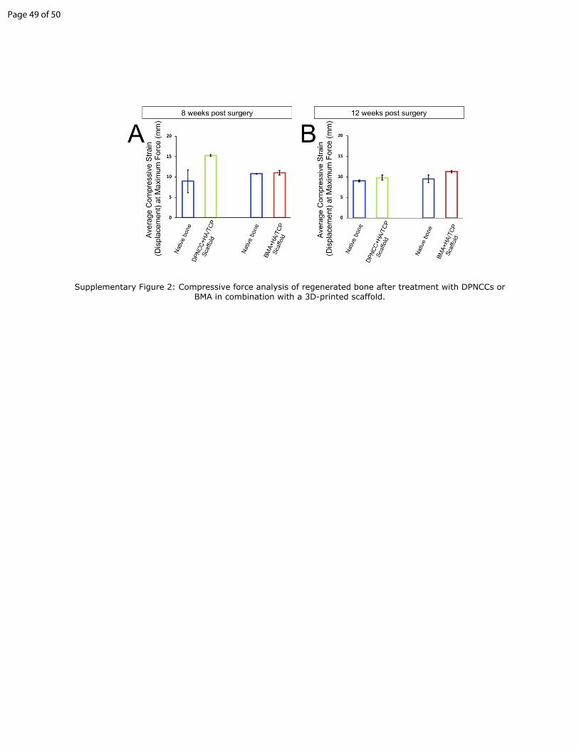
conducted compression tests. The maximum force that was able to be applied to bone regenerated
using DPNCCs and HA/TCP matched that of the surrounding native bone. Furthermore, the
displacement of the compressive arm at maximum force was the same between native and
regenerated bone. In the control group, the CSD area showed no resistance to the compression
force, indicating that DPNCCs and our HA/TCP scaffold successfully supported calvarial bone
regeneration in comparison to the control group eight weeks post-surgery (SFig. 2A). No defects
in the dura or brain were observed under the CSD area when treated with DPNCCs and scaffold,
suggesting that the scaffold protected the vital structures (n=9). No adverse effects to the animals
were observed from the surgical procedure or treatment with the DPNCCs and scaffold (n=9).
There was no ectopic bone nor abnormal tumor-like tissue formation in any of the calvarial CSDs
treated with DPNCCs and 3D-printed HA/TCP scaffold after 8 or 12 weeks.
At 12 weeks post-injury (n=3), the surface of the newly regenerated bone appeared to be
smoother than at 8 weeks post-injury (Fig. 4I). The regenerated bone formed a continuous
connection with the native bone and showed uniform bone formation throughout the CSD (Fig. 4I,
J). The HA/TCP scaffold underwent further degradation, but some HA/TCP remained. The
regenerated bone formed a complex network resembling the endogenous bone structure (Fig. 4K,
L). Histological H&E staining showed new, dense, cortical bone surrounding the remaining
HA/TCP scaffold (Fig. 4K). The mean trabecular number of the regenerated bone was not
significantly different from that of the native bone at 12 weeks post-surgery (Fig. 6D). The
trabecular spacing and thickness of the newly formed bone indicated that the regenerated bone in
Page 20 of 50

21
the CSD had begun remodeling as the ones in the native calvarial bone. The density of the
regenerated bone at 12 weeks post-surgery was comparable to the native bone, as indicated by CT
analysis (Fig. 6B). Compression testing of the regenerated bone from DPNCC + 3D-printed
HA/TCP scaffold samples showed comparable strength to the native bone (SFig. 2B). During
compression testing, cracking of the samples never occurred at the junction between the newly
formed and native bone.
Bone marrow aspirate and 3D-printed scaffold in calvarial bone regeneration to repair CSDs
To test the potential of BMA to regenerate calvarial bone in swine, we collected 4 mL of
bone marrow from the swine tibial crest. After centrifuging, the remaining BMA containing MSCs
was transplanted into a 3D-printed scaffold and subsequently placed into a 3 cm diameter defect
in the calvarial bone CSD (n=8).
As with DPNCCs, the BMA was able to regenerate bone in this CSD in the presence of the
3D-printed HA/TCP scaffold. At 8 weeks, the bone defect was healed throughout, and similar to
the DPNCC-treated group (n=4). CT imaging analyses suggested that the new bone growth began
at the edges of the defect and progressed toward the center (Fig. 5A, B). During the study, no
animals experienced any complications or infections related to the calvarial surgery, bone marrow
retrieval site, or treatment with BMA and HA/TCP scaffold. Upon dissection, the defect site was
smooth to the touch. Upon visual inspection, the newly formed bone resembled the surrounding
calvarial bone in color and texture. Density of the regenerated bone was not significantly different
from that of the native bone (Fig. 6A). MicroCT analysis showed that a trabecular network formed
around the remaining scaffold and integrated with the surrounding native bone (Fig. 5B).
Trabecular structure analysis showed that the regenerated bone had similar trabecular thickness
Page 21 of 50

22
with increased trabecular number and decreased trabecular spacing as compared to the native
calvarial bone at 8 weeks post-surgery (Fig. 6C). Histological analysis of these samples confirmed
the complex trabecular network shown in the microCT images (Fig. 5C, D). The regenerated bone
was dense and cortical, as indicated in our H&E staining at 8 weeks post-surgery (Fig. 5C, D).
Some HA/TCP material persisted within the regenerated calvarial bone. During compression
testing, the regenerated bone performed similarly to the native bone (SFig. 2A). None of the
samples broke at the junction between native and regenerated calvarial bone.
During dissection of the calvaria samples of 12 weeks post-surgery, we observed less
remaining HA/TCP scaffold material as compared to the samples at 8 weeks post-surgery (n=4)
(Fig. 5F). The boundary of the original defect was undetectable both upon visual inspection and
in microCT images (Fig. 5E, F). The bone density and compressive strength of the newly
regenerated calvarial bone were comparable to those of the native bone (Fig. 6B). Furthermore,
these samples never broke at the junction of new and regenerated bone during compressive testing.
At the center of the original CSD, the regenerated bone appeared to be thinner than at the periphery
of the defect, suggesting that new calvarial bone formation initiated from the edge of the CSD
(Fig. 5E, F). Our histological analysis indicated that there was dense calvarial bone regeneration
throughout the CSD (Fig. 5G, H). The remaining HA/TCP scaffold was surrounded by newly
formed cortical bone (Fig. 5G).
Eight and twelve weeks post-surgery, CT imaging revealed that remnants of the scaffold
persisted in the healed defect in both DPNCC and BMA groups (Fig. 4F, J; Fig. 5B, F).
Interestingly, in the scaffold-only groups, nearly the entire scaffold remained, leaving the surface
uneven (Fig. 4A, B). This indicates that the transplanted cells play an important role in scaffold
biodegradation and may integrate the components of HA/TCP to regenerate bone in the CSD
Page 22 of 50

23
defect in the calvaria. Histological analysis of the scaffold-only groups showed growth of large
strips of soft tissue accompanied by small strips of bone, suggesting the necessity of adding cells
to facilitate proper bone regeneration (Fig. 4C, D).
Page 23 of 50
24
DISCUSSION
This study represents a novel approach to addressing a long-standing clinical need, namely a
biological solution to repair critical-sized bone defects in the calvaria. Current therapies for large
calvarial bone defects include metal and plastic implants, as well as autologous bone grafts when
sufficient bone is available. There are many limitations to these therapies, particularly in the case
of non-resorbable plates, which provide a purely mechanical solution to protect the brain and
improve aesthetics. Metal and plastic implants are prone to infection, are uncomfortable for the
patient, and do not accommodate for brain and skull growth in pediatric patients. Since these
implants are not biological, they cannot integrate fully with the native bone, nor can they contribute
to tissue homeostasis. Autologous bone grafts cause further harm to the patient at the harvest site
and often do not yield enough bone to heal a defect of critical size. Our strategy for regenerating
calvarial bone provides several distinct advantages over current clinical practice, owing to the
ability of the newly regenerated bone to integrate with existing bone and participate in maintaining
tissue homeostasis. Importantly for pediatric indications, calvarial bone regenerated using our
approach will be able to grow with the skull and accommodate crucial future brain growth.
Skull injury repair has primarily been investigated in small animal models such as rats (Mardas et.
al., 2002; Spicer et. al., 2012; Bae et al., 2018) and rabbits (Maliha et. al., 2019; Young et. al.,
2008; Kim et al., 2018). These studies have made important contributions to the development of
strategies for calvarial bone regeneration. However, rodent calvaria are very different from that of
humans, and a large animal CSD model is ultimately necessary for enabling clinical translation.
Some recent studies have tested how cell-based approaches can support calvarial bone
regeneration in full-sized and miniature swine (Wehrhan et al., 2011; Cao et al., 2015; Li et al.,
Page 24 of 50
25
2015; Rubessa et al., 2017). However, none of these studies have tested how autologous stem cells
combined with a 3D-printed osteoconductive scaffold can support calvarial bone regeneration in
such a large CSD (>7.0 cm2) in swine. In developing and testing novel treatment modalities for
calvarial defects, the size of the defect matters greatly; large defects are the most challenging cases
for achieving successful bone regeneration. Building on our previous study (Park et. al., 2016), we
therefore sought to establish a calvarial CSD model in swine, and to further investigate how MSCs
combined with osteoconductive biomaterials can support calvarial bone regeneration in this model.
We selected swine (Yorkshire farm pigs) as a large animal model because their bone morphology
and anatomy are similar to those of humans, as are their healing and bone remodeling processes
(Thorwarth et al., 2005; Rubessa et al., 2017). Furthermore, the head of a full-sized farm pig is
larger than that of a human, making it possible to create a large CSD similar to those seen in
patients in order to mimic clinical conditions faithfully (Weickenmeier et al., 2018). The
establishment of this calvarial bone CSD model in swine will serve as an important platform for
future studies to test different biomaterials in combination with stem or progenitor cells in calvarial
bone regeneration.
To develop a regenerative therapy for calvarial CSDs that will produce superior outcomes relative
to the current standards of care, we used dental pulp-derived mesenchymal stem cells (MSCs) and
compared it to bone marrow aspirate (BMA), which also contains MSCs, when used in
combination with our 3D-printed HA/TCP scaffold. MSCs are ideally suited for tissue
regeneration, as they are unique in their ability to self-renew while maintaining their stemness.
These cells are multipotent with the capacity to differentiate into chondrocytes, adipocytes,
osteocytes, and a variety of other cell types under the right conditions (Langer and Vacanti, 1993;
Page 25 of 50
26
Mao et al., 2006). Our previous study has shown that cranial neural crest-derived MSCs as well as
long bone marrow MSCs combined with HA/TCP particles can support bone regeneration (Chang
2009). Here we tested the ability of MSCs derived from the cranial neural crest and long bone
marrow aspirate to regenerate bone to repair a CSD in a large animal model. Although dental pulp
MSCs delivered with HA/TCP particles support calvarial bone regeneration in a CSD, the surface
of the regenerated bone is less than ideal, which motivated us to develop a 3D-printed
osteoconductive scaffold to achieve a better outcome in calvarial bone regeneration.
The osteoconductive, 3D-printed scaffold we developed for calvarial bone regeneration in a CSD
provides a suitable environment that protects the brain during calvarial bone regeneration,
promotes osteogenesis and maintains cell viability while holding the cells in place. Although
HA/TCP has been used to support bone regeneration, the engineering of our 3D-printed HA/TCP
scaffold is an innovative development (Rubessa et al., 2017). Our HA/TCP scaffold is custom-
designed based on the CSD. The lattice design and pore size make it easy to load cells during the
surgical procedure. The HA/TCP scaffold is biocompatible, osteoconductive, undergoes
degradation following bone regeneration, and has the ability to confine cells in place to achieve an
aesthetic surface profile of the newly regenerated calvarial bone. It also facilitates seamless
integration of the newly regenerated and existing calvarial bone, resulting in biological repair of a
calvarial CSD. We have demonstrated that our therapy yields quality cortical bone of sound
structure that is well integrated with the native bone, and moreover, that the new tissue is capable
of cellular turnover and remodeling.
Page 26 of 50
27
MSCs are crucial in supporting calvarial bone regeneration (Stockmann et al., 2012; Cao et al.,
2015). In this study, we have shown that HA/TCP alone, heat-inactivated MSCs, and fibroblasts
are insufficient to support bone regeneration in the swine calvarial CSD model; live MSCs appear
to be required for complete bone regeneration over the time period of our study. Crucially, when
delivered on our 3D-printed osteoconductive scaffold, both DPNCCs and BMA were able to
regenerate quality bone in the swine calvaria. The bone regenerated using BMA + HA/TCP
scaffold appears to have more trabecular structure than bone regenerated using DPNCCs,
consistent with our previous findings (Chung et. al., 2009). Craniofacial bones differ from long
bones in that they develop through a process of intramembranous ossification instead of
endochondral, and they contain significantly less marrow space than long bones. Owing to the
latter quality, the bones of the face and skull are densely cortical, in contrast to the spongy,
trabecular quality of long bones. Due to its crucial function of protecting the brain and sensory
organs, the strength and quality of regenerated craniofacial bone must be considered carefully.
Importantly, our density analysis indicates that the trabecular bone regenerated using BMA when
combined with our HA/TCP scaffold is comparable to the native bone in its mechanical properties,
suggesting that it would be both dense and strong enough to protect the brain. The capacity of both
neural crest- and BMA-derived cells to regenerate cortical bone when combined with our HA/TCP
scaffold in the calvaria may indicate environment-dependent differentiation of MSCs. For future
clinical practice, either DPNCCs or BMA could be suitable for use with our scaffold to regenerate
calvarial bone.
One of the most clinically important aspects of the present work lies in our finding that BMA can
support calvarial CSD repair when combined with our 3D-printed scaffold. Using minimally
Page 27 of 50

28
manipulated BMA directly after harvest to support calvarial bone regeneration has several distinct
advantages. First, it obviates the need for expansion of the cells, which is costly and time-
consuming. Second, it reduces the potential for contamination or loss of stemness of the MSCs
due to their expansion in cell culture. Currently, the advantage may lie with BMA over neural
crest-derived stem cells for these reasons. However, previous results and the current study indicate
that if a source of “off-the-shelf” allogeneic cranial neural crest-derived MSCs were to become
available, they could represent an excellent option.
In summary, we have established a large animal calvarial CSD model with high pre-clinical value.
Using this model we have successfully tested a novel approach for skull bone regeneration that
combines a 3D-printed osteoconductive scaffold with dental pulp MSCs or BMA. In comparison
to the current treatment for patients with CSDs in the skull, our approach will offer patients a safe
and effective biological solution to restore skull form and function, and to improve the quality of
life for patients.
Page 28 of 50
29
ACKNOWLEDGEMENTS
We thank Bridget Samuels for critical reading of the manuscript. This work was supported by the National Institute of Dental and Craniofacial Research National Institute of Health - Center for Dental, Oral and Craniofacial Tissue and Organ Regeneration (C-DOCTOR) U24 DE026914; U24 DE029463 and Alfred Mann Institute (AMI) at the University of Southern California.
CONFLICT OF INTEREST
The authors declare that there is no conflict of interest.
Page 29 of 50
30
REFERENCES
1 Badhey A, Kadakia S, Mourad M et al. Calvarial Reconstruction. Semin Plast Surg.
2017;31(4):222–226. doi:10.1055/s-0037-1606557
2 Aydin S, Kucukyuruk B, Abuzayed B et al. Cranioplasty: Review of materials and
techniques. J Neurosci Rural Pract. 2011;2(2):162-167. doi:10.4103/0976-3147.83584
3 Ng ZY, Nawaz I. Computer-designed PEEK implants: a peek into the future of cranioplasty? J
Craniofac Surg. 2014 Jan;25(1):e55-58. doi: 10.1097/SCS.0b013e3182a2f7b6. PubMed PMID:
24406603.
4 Lam S, Kuether J, Fong A et al. Cranioplasty for large-sized calvarial defects in the pediatric
population: a review. Craniomaxillofac Trauma Reconstr. 2015 Jun;8(2):159-170. doi:
10.1055/s-0034-1395880. Epub 2014 Nov 20. PMID: 26000090; PMCID: PMC4428737.
5 Hill CS, Luoma AM, Wilson SR et al. Titanium cranioplasty and the prediction of
complications. Br J Neurosurg. 2012 Dec;26(6):832-837. doi:
10.3109/02688697.2012.692839. Epub 2012 Jun 18. Review. PubMed PMID: 22702389
6 Garland CB, Pomerantz JH. Regenerative strategies for craniofacial disorders. Front
Physiol. 2012;3:453. doi: 10.3389/fphys.2012.00453. eCollection 2012. PubMed PMID:
23248598; PubMed Central PMCID: PMC3521957
Page 30 of 50
31
7 Fearon JA, Griner D, Ditthakasem K et al. Autogenous Bone Reconstruction of Large
Secondary Skull Defects. Plast Reconstr Surg. 2017 Feb;139(2):427-438. doi:
10.1097/PRS.0000000000002941. Review. PubMed PMID: 28125534
8 Grant GA, Jolley M, Ellenbogen RG et al. Failure of autologous bone-assisted cranioplasty
following decompressive craniectomy in children and adolescents. J Neurosurg. 2004 Feb;100(2
Suppl Pediatrics):163-168. doi: 10.3171/ped.2004.100.2.0163. PMID: 14758944.
9 Jiang Y, Jahagirdar BN, Reinhardt RL et al. "Pluripotency of mesenchymal stem cells derived
from adult marrow". Nature. 2002;418 (6893): 41–49. doi:10.1038/nature00870.
PMID 12077603.
10 Biteau B, Hochmuth CE, Jasper H. Maintaining tissue homeostasis: dynamic control of
somatic stem cell activity. Cell stem cell. 2011;9(5):402-411. doi:10.1016/j.stem.2011.10.004.
11 Ramesh N, Moratti SC, Dias GJ. Hydroxyapatite-polymer biocomposites for bone
regeneration: A review of current trends. Journal of biomedical materials research.
2018;106(5):2046-2057. doi:10.1002/jbm.b.33950
12 Sulaiman SB, Keong TK, Cheng CH et al. Tricalcium phosphate/hydroxyapatite (TCP-HA)
bone scaffold as potential candidate for the formation of tissue engineered bone. Indian J Med
Res. 2013 Jun;137(6):1093-1101. PMID: 23852290; PMCID: PMC3734714.
Page 31 of 50

32
13 Mauney JR, Volloch V, Kaplan DL. Role of adult mesenchymal stem cells in bone tissue
engineering applications: current status and future prospects. Tissue Eng. 2005 May-Jun;11(5-
6):787-802. doi: 10.1089/ten.2005.11.787. PMID: 15998219.
14 Kang, Hyun-Wook, Sang Jin Lee, In Kap Ko, Carlos Kengla, James J. Yoo, and Anthony
Atala. "A 3D bioprinting system to produce human-scale tissue constructs with structural
integrity." Nature Biotechnology 34, no. 3 (2016): 312-319.
15 Feyen DA, van den Akker F, Noort W et al. Isolation of Pig Bone Marrow-Derived
Mesenchymal Stem Cells. Methods Mol Biol. 2016;1416:225-32. doi: 10.1007/978-1-4939-
3584-0_12. PMID: 27236674.
16 Li X, Yuan Y, Liu L et al. “3D Printing of Hydroxyapatite/Tricalcium Phosphate (HA/TCP)
Scaffold with Hierarchical Porous Structure for Bone Regeneration.” Bio-Design and
Manufacturing, (2019) pp1-15 DOI:10.1007/s42242-019-00056-5
17 Song X, Chen Z, Lei L et al. “Piezoelectric Component Fabrication Using Projection
Stereolithography of Barium Titanate Ceramic Suspensions.” Rapid Prototyping Journal, 23, 1,
2017.
18 Swindle MM. “Bone Marrow Access in Swine.” Sinclair Bio Resources. 2009. Retrieved
from http://www.sinclairresearch.com/assets/sites/2/Bone-Marrow-Access.pdf
Page 32 of 50

33
19 Fischer AH, Jacobson KA, Rose J et al. Hematoxylin and eosin staining of tissue and cell
sections. CSH Protoc. 2008 May 1;2008:pdb.prot4986. doi: 10.1101/pdb.prot4986. PubMed
PMID: 21356829.
20 Li Y, Chen SK, Li L et al. Bone defect animal models for testing efficacy of bone substitute
biomaterials. Journal of Orthopaedic Translation. 2015 July;Volume 3, Issue 3, 95-104.
https://doi.org/10.1016/j.jot.2015.05.002
21 Chung IH, Yamaza T, Zhao H et al. Stem cell property of postmigratory cranial neural crest
cells and their utility in alveolar bone regeneration and tooth development. Stem Cells. 2009
Apr;27(4):866-877. doi: 10.1002/stem.2. PubMed PMID: 19350689; PubMed Central PMCID:
PMC2896558.
22 Pepla E, Besharat LK, Palaia G et al. Nano-hydroxyapatite and its applications in preventive,
restorative and regenerative dentistry: a review of literature. Ann Stomatol (Roma).
2014;5(3):108–114. Published 2014 Nov 20.
23 Park S, Zhao H, Urata M et al. Sutures Possess Strong Regenerative Capacity for Calvarial
Bone Injury. Stem Cells Dev. 2016 Dec 1;25(23):1801-1807. Epub 2016 Oct 24. PubMed PMID:
27762665; PubMed Central PMCID: PMC5124738.
Page 33 of 50

34
24 Liu Y et al. “Mesenchymal stem cell-based tissue regeneration is governed by recipient T
lymphocytes via IFN-γ and TNF-α.” Nature medicine vol. 17,12 1594-601. 20 Nov. 2011,
doi:10.1038/nm.2542
25 Song X, Chen Y, Lee TW et al. “Ceramic Fabrication Using Mask-image-projection-based
Stereolithography Integrated with Tape-casting.” SME Journal of Manufacturing Processes, Vol.
20, pp. 456-464, 2015.
26 Langer R, Vacanti J. Advances in tissue engineering. J Pediatr Surg. 2016 Jan;51(1):8-12.
doi: 10.1016/j.jpedsurg.2015.10.022. Epub 2015 Nov 10. PMID: 26711689; PMCID:
PMC4733916.
27 Mao JJ, Giannobile WV, Helms JA et al. Craniofacial tissue engineering by stem cells. J Dent
Res. 2006 Nov;85(11):966-979. doi: 10.1177/154405910608501101. PMID: 17062735; PMCID:
PMC2571078.
28 Stockmann P, Park J, von Wilmowsky C et al. Guided bone regeneration in pig calvarial bone
defects using autologous mesenchymal stem/progenitor cells - a comparison of different tissue
sources. J Craniomaxillofac Surg. 2012 Jun;40(4):310-320. doi: 10.1016/j.jcms.2011.05.004.
Epub 2011 Jun 30. PMID: 21723141.
29 Mardas N, Kostopoulos L, Karring T. Bone and suture regeneration in calvarial defects by e-
PTFE-membranes and demineralized bone matrix and the impact on calvarial growth: an
Page 34 of 50
35
experimental study in the rat. J Craniofac Surg. 2002 May;13(3):453-462; discussion 462-464.
PubMed PMID: 12040218.
30 Spicer PP, Kretlow JD, Young S. et al. Evaluation of bone regeneration using the rat critical
size calvarial defect. Nature protocols. 2012;7(10), 1918–1929.
https://doi.org/10.1038/nprot.2012.113
31 Maliha SG, Lopez CD, Coelho PG et al. Bone Tissue Engineering in the Growing Calvarium
Using Dipyridamole-Coated 3D Printed Bioceramic Scaffolds: Construct Optimization and
Effects to Cranial Suture Patency. Plast Reconstr Surg. 2019 Nov 19;. doi:
10.1097/PRS.0000000000006483. [Epub ahead of print] PubMed PMID: 31790079.
32 Young S, Bashoura AG, Borden T et al. Development and characterization of a rabbit
alveolar bone nonhealing defect model. J Biomed Mater Res A. 2008 Jul;86(1):182-194. doi:
10.1002/jbm.a.31639. PubMed PMID: 17969052.
33 Chang S, Hsu Y, Wang YJ et al. Fabrication of pre-determined shape of bone segment with
collagen-hydroxyapatite scaffold and autogenous platelet-rich plasma. J Mater Sci: Mater
Med20, 23–31 (2009). https://doi.org/10.1007/s10856-008-3507-1
34 Rubessa M, Polkoff K, Bionaz M et al. Use of Pig as a Model for Mesenchymal Stem Cell
Therapies for Bone Regeneration. Anim Biotechnol. 2017 Oct 2;28(4):275-287. doi:
10.1080/10495398.2017.1279169. Epub 2017 Mar 7. PMID: 28267421.
Page 35 of 50
36
35 Weickenmeier J, Kurt M, Ozkaya E et al. Magnetic resonance elastography of the brain: A
comparison between pigs and humans. J Mech Behav Biomed Mater. 2018 Jan;77:702-710. doi:
10.1016/j.jmbbm.2017.08.029. Epub 2017 Aug 26. PMID: 28919161.
36 Thorwarth M, Schultze-Mosgau S, Kessler P et al. Bone regeneration in osseous defects using
a resorbable nanoparticular hydroxyapatite. J Oral Maxillofac Surg. 2005 Nov;63(11):1626-33.
doi: 10.1016/j.joms.2005.06.010. PMID: 16243180.
37 Wehrhan F, Amann K, Molenberg A et al. PEG matrix enables cell-mediated local BMP-2
gene delivery and increased bone formation in a porcine critical size defect model of craniofacial
bone regeneration. Clin Oral Implants Res. 2012 Jul;23(7):805-13. doi: 10.1111/j.1600-
0501.2011.02223.x. Epub 2011 Dec 12. PMID: 22151397.
38 Cao Y, Xiong J, Mei S et al. Aspirin promotes bone marrow mesenchymal stem cell-based
calvarial bone regeneration in mini swine. Stem Cell Res Ther. 2015 Oct 31;6:210. doi:
10.1186/s13287-015-0200-4. PMID: 26519141; PMCID: PMC4628405.
39 Bornstein MM, Heynen G, Bosshardt DD et al. Effect of two bioabsorbable barrier
membranes on bone regeneration of standardized defects in calvarial bone: a comparative
histomorphometric study in pigs. J Periodontol. 2009 Aug;80(8):1289-99. doi:
10.1902/jop.2009.090075. PMID: 19656029.
Page 36 of 50

37
40 Hubbell, J. Biomaterials in Tissue Engineering. Nat Biotechnol 13, 565–576 (1995).
https://doi.org/10.1038/nbt0695-565
Page 37 of 50

38
FIGURE LEGENDS
Figure 1: Generating a critical-sized defect in the swine calvaria.
Schematic drawing of defect placement (A) on the swine calvarial surface. Schematic drawing in
coronal view of (B) normal calvarial bone, periosteum, skin, and dura; (C) retraction of the skin
and periosteum to expose bone to create 3 cm defect; (D) skin and periosteum closure over bone.
(E) Gross anatomy image of empty defect at time of surgery showing exposed dura. MicroCT
showing defect at (F, H) 8 weeks post-surgery and (G, I) 12 weeks post-surgery. (F, G) Inserts
show transverse microCT through defect. Red dashed lines (H, I) indicate boundary of defect
site. (J-L) Histological sections of defect at 12 weeks post-surgery. Boxed areas in J are shown
enlarged in K and L. Scale bars: F-G, 10 mm; H-I, 5mm; J, 1 mm; K-L, 200 μm.
Figure 2: Harvesting and characterization of dental pulp-derived neural crest cells. (A)
Schematic drawing of swine mandible. Red arrow indicates location of extracted swine incisor.
(B) Swine incisor post-extraction and (C) dental pulp removed from incisor. (D) Single-cell colony
of undifferentiated DPNCCs, 48 hrs after extraction and plating. (E) Alcian blue staining of
DPNCCs, 3 weeks post-plating. (F) Alizarin red staining of DPNCCs, one week post-plating. (G)
Photo of swine tibia at time of bone marrow aspiration, indicating the tibial crest landmark and
site of aspiration. (H) Schematic drawing of tibial bone with aspiration site. (I) Oil Red O staining
of BMA, one week post-plating. (J) Alcian blue staining of BMA culture, 3 weeks post-plating.
(K) Alizarin red staining of BMA culture, one week post-plating. Scale bars: B-C, 10 mm; D-F, I,
200 μm; J-K, 5 mm.
Page 38 of 50

39
Figure 3: Design and fabrication of HA/TCP scaffold using 3D printing and light-curing. (A)
Digital model of the custom designed scaffold showing dimensions; (B) digital model of the lattice
design 3D-printed HA/TCP scaffold and measurements from the side view; (C) simulation of
HA/TCP scaffold under compression using Comsol MultiPhysics; (D) image of final scaffold
product; (E) Gross anatomy photo of scaffold loaded with cells in the 3 cm defect at time of
surgery. Cell compatibility studies for (F) DPNCC and (G) BMA culture with HA/TCP scaffold.
Scale bars, 5 mm.
Figure 4: Dental pulp-derived MSCs and 3D-printed scaffold regenerate dense, strong bone
in the swine calvarial CSD.
MicroCT of CSD filled with (A, B) HA/TCP scaffold alone; (E, F) HA/TCP scaffold with
DPNCCs imaged 8 weeks post-surgery; (I, J) HA/TCP scaffold with DPNCCs 12 weeks post-
surgery. (C-E, H-J, M-O) Histological staining of defect filled with HA/TCP scaffold (C, D)
alone or in combination with DPNCCs at (G, H) 8 weeks post-surgery and (K, L) 12 weeks post-
surgery. Black and red dashed lines indicate original site of injury. Scale bars: A-B, F-G, K-L, 10
mm; C-E, H-J, M-O, 1 mm.
Figure 5: Bone marrow aspirate and 3D-printed scaffold regenerate dense, strong bone in a
swine calvarial CSD.
MicroCT of critical size defect filled with HA/TCP scaffold of 4mm pores with bone marrow
aspirate (A, B) imaged 8 weeks post-surgery and (E, F) 12 weeks post-surgery. (C, D, G, H)
Page 39 of 50

40
H&E staining of defect filled with BMA + HA/TCP scaffold (C, D) 8 weeks and (G, H) 12
weeks post-surgery. Black and red dashed lines indicate original site of injury. Scale bars: A-B,
E-F, 5 mm; C, G, 1 mm; D, H 0.5mm.
Figure 6: Density and trabecular analysis of calvarial bone regenerated by DPNCCs and
BMA in combination with a 3D-printed HA/TCP scaffold.
Density analysis comparing native bone to DPNCC- and BMA-mediated regenerated bone at (A)
8 weeks and (B) 12 weeks post-surgery. Comparison of trabecular number in native bone to
DPNCC- and BMA-mediated regenerated bone at (C) 8 weeks and (D) 12 weeks post-surgery.
Scale bars, 5 mm.
Supplementary Figure 1: CNC-derived MSCs combined with HA/TCP particles promote
bone regeneration in a calvarial CSD.
(A-F) MicroCT of CSDs filled with HA/TCP particles and dental pulp neural crest cells at 8
weeks post-surgery. (A inset) Defect filled with HA/TCP particles and DPNCCs at time of
surgery. (B, E) MicroCT of CSD filled with HA/TCP particles and fibroblasts at 8 weeks post-
surgery. (C, F) MicroCT of CSD filled with HA/TCP particles and heat-inactivated DPNCCs at 8
weeks post-surgery. (B and C insets) Transverse microCT through defects shown in B and C.
Black dashed lines in A-C and red dashed lines in D-F indicate boundaries of original defects.
Scale bars: A-C, 10 mm; D-F 5 mm.
Supplementary Figure 2: Compressive force analysis of regenerated bone after treatment
with DPNCCs or BMA in combination with a 3D-printed scaffold.
Page 40 of 50

41
Comparison of compressive load strain between native bone and DPNCC- and BMA-mediated
regenerated bone (A) 8 weeks post-surgery and (B) 12 weeks post-surgery.
Page 41 of 50
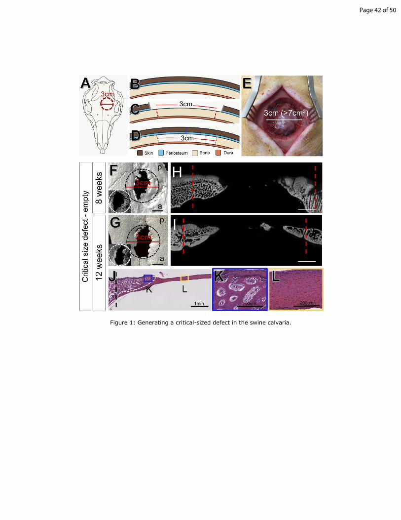
Figure 1: Generating a critical-sized defect in the swine calvaria.
Page 42 of 50

Figure 2: Harvesting and characterization of dental pulp-derived neural crest cells.
Page 43 of 50

Figure 3: Design and fabrication of HA/TCP scaffold using 3D printing and light-curing.
Page 44 of 50

Figure 4: Dental pulp-derived MSCs and 3D-printed scaffold regenerate dense, strong bone in the swine calvarial CSD.
Page 45 of 50

Figure 5: Bone marrow aspirate and 3D-printed scaffold regenerate dense, strong bone in a swine calvarial CSD.
Page 46 of 50
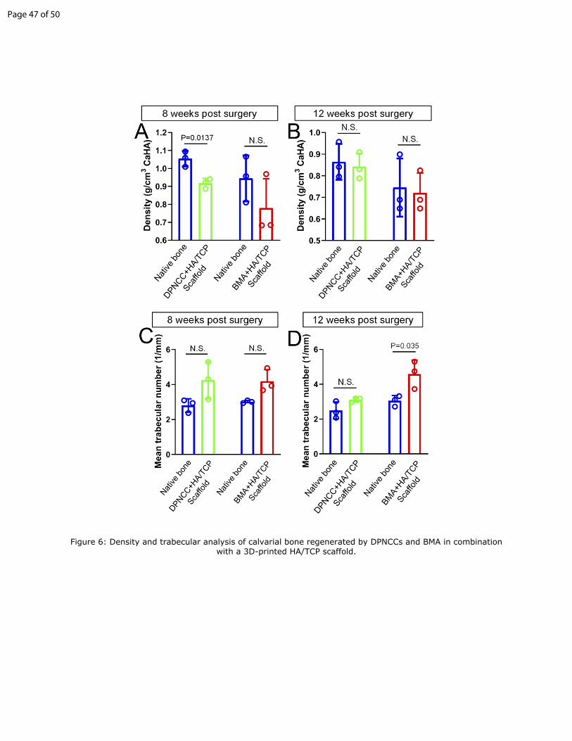
Figure 6: Density and trabecular analysis of calvarial bone regenerated by DPNCCs and BMA in combination with a 3D-printed HA/TCP scaffold.
Page 47 of 50

Page 48 of 50
Supplementary Figure 2: Compressive force analysis of regenerated bone after treatment with DPNCCs or BMA in combination with a 3D-printed scaffold.
Page 49 of 50

Table 1: Cell culture reagents.Reagent Name Company CAT/REF# LOT#
Collagenase Type I Washington Biochemical Corporation
CAT#: LS004194 LOT#: 45D15720
DPBS, no calcium, no magnesium
Gibco CAT#: 14190250
Alpha-MEM Gibco REF#: 12571-063 LOT#: 2044318TryplE Express with Phenol Red
Gibco REF#: 12605-010 LOT#: 1897328
Adipogenic Differentiation Media
Gibco REF#: A10410-01 LOT#: 1925522
Chondrogenic differentiation Media
Gibco REF#: A10069-01 LOT#: 1929138
Osteogenic Differentiation Media
Gibco REF#: A10069-01 LOT#: 1929138
Page 50 of 50