mesenchymal stromal cells attenuate sevoflurane-induced … · yanyong cheng†, yunfeng jiang†,...
TRANSCRIPT

RESEARCH ARTICLE Open Access
Mesenchymal stromal cells attenuatesevoflurane-induced apoptosis in humanneuroglioma H4 cellsYanyong Cheng†, Yunfeng Jiang†, Lei Zhang, Jiayi Wang, Dongdong Chai, Rong Hu, Chunzhu Li, Yu Sun*
and Hong Jiang*
Abstract
Background: Inhalation of sevoflurane can induce neuronal apoptosis, cognitive impairment and abnormalbehaviors. Bone marrow mesenchymal stem cells (MSCs) can secret neurotrophic factors and cytokines to protectfrom oxidative stress-related neuronal apoptosis. However, whether MSCs can protect from sevoflurane-inducedneuronal apoptosis and the potential mechanisms are unclear.
Methods: A non-contact co-culture of MSCs with human neuroglioma H4 cells (H4 cells) was built. H4 cells wereco-cultured with MSCs or without MSCs (control) for 24 h. The co-cultured H4 cells were exposed to 4% sevofluranefor 6 h. The levels of caspase-3, reactive oxygen species (ROS), adenosine triphosphate (ATP), and the release ofcytochrome C were determined by Western blot and fluorescence assay.
Results: Sevoflurane exposure significantly elevated the levels of cleaved caspase 3 and Bax in H4 cells. However,these phenomena were significantly offset by the co-culture with MSCs in H4 cells. Co-culture with MSCs before,but not after, sevoflurane exposure, significantly attenuated sevoflurane-induced ROS production in H4 cells. MSCsprevented sevoflurane-mediated release of cytochrome C from the mitochondria and production of ATP in H4 cells.
Conclusions: Our study indicated that soluble factors secreted by MSCs attenuated the sevoflurane-inducedoxidative stress and apoptosis of neuronal cells by preserving their mitochondrial function.
Keywords: Sevoflurane, Mesenchymal stem cells, Reactive oxygen species, Apoptosis, mitochondrial dysfunction
BackgroundSevoflurane is an inhaled anesthetic for both adult andpediatric anesthesia frequently used in clinical practice,and has characteristics of low blood:gas coefficient, rapidonset and recovery. However, sevoflurane exposure caninduce severe neurological side effects, such as cognitiveimpairment and abnormal behaviors, like autism spectrumdisorder [1–5]. Our previous studies have shown that suchadverse effects of sevoflurane may be associated with thedysfunction of the LIMK1-signaling pathway and down-regulation of circulating insulin-like growth factor 1 in de-veloping and aged rats [6, 7]. Furthermore, sevoflurane
exposure is prone to development of cognitive dysfunctionin rodents and monkeys [3, 8, 9]. Similarly, a prospectiverandomized parallel-group study shows that inhalation ofsevoflurane can promote the progression of amnestic mildcognitive impairment [10]. Moreover, sevoflurane expos-ure can change neurocognitive development and brainstructure, leading to an increased risk of cognitive dys-function in pediatric patients [2, 11–13]. However, themechanisms underlying the neurotoxicity of sevofluraneagainst neuronal cells have not been clarified.Sevoflurane can stimulate reactive oxygen species
(ROS) production and induce neurodegeneration andsynaptic loss, including mitochondria dysfunction [14,15]. Indeed, antioxidant and inhibitors for nicotinamideadenine dinucleotide phosphate oxidase (NADPH) canprotect against long-term memory impairment andneuronal apoptosis in rodents by reducing superoxide
* Correspondence: [email protected]; [email protected]†Yanyong Cheng and Yunfeng Jiang contributed equally to this work.Department of Anesthesiology, Shanghai Ninth People’s Hospital Affiliated toShanghai Jiao Tong University School of Medicine, Center for SpecialtyStrategy Research of Shanghai Jiao Tong University China HospitalDevelopment Institute, 639 Zhizaoju Road, Shanghai 200011, China
© The Author(s). 2018 Open Access This article is distributed under the terms of the Creative Commons Attribution 4.0International License (http://creativecommons.org/licenses/by/4.0/), which permits unrestricted use, distribution, andreproduction in any medium, provided you give appropriate credit to the original author(s) and the source, provide a link tothe Creative Commons license, and indicate if changes were made. The Creative Commons Public Domain Dedication waiver(http://creativecommons.org/publicdomain/zero/1.0/) applies to the data made available in this article, unless otherwise stated.
Cheng et al. BMC Anesthesiology (2018) 18:84 https://doi.org/10.1186/s12871-018-0553-1

levels [16]. Hence, antioxidants and inhibition of ROSproduction can potentially inhibit sevoflurane-relatedneuronal toxicity.Recent studies have demonstrated that mesenchymal
stem cells (MSCs) can secrete neurotrophic factors, che-mokines and cytokines, which have potent neuroprotec-tive effects [17–20]. Infusion with MSCs improvesischemia reperfusion-induced neuronal injury in rodents[21, 22]. Previous clinical trials have demonstrated thatMSC-based therapies are safe and effective for somediseases [23, 24]. Accordingly, we hypothesize thatsoluble factors secreted by MSCs can protect fromsevoflurane-induced oxidative stress and neuronal apop-tosis by preserving the mitochondrial function.Glial cells regulate the synapse development and neur-
onal dysfunction [25], such as Alexander disease thatmanifests leukodystrophy and intellectual disability. Hu-man neuroglioma H4 cells (H4 cells) share some similar-ities with glial cells in vitro and have been widely used forstudies of neuronal impairment and apoptosis [26–28].In this study, we employed a transwell-based co-culturesystem to determine the impact of soluble factors se-creted by MSCs on sevoflurane exposure-induced ROSproduction, apoptosis and mitochondrial dysfunction inH4 cells in vitro.
MethodsCell culturesH4 cells were obtained from central laboratory ofShanghai Ninth People’s Hospital Affiliated to Shang-hai Jiao Tong University School of Medicine and cul-tured in Dulbecco’s modified eagle medium (DMEM,4.5 g/L glucose, Hyclone, USA) containing 10% fetalbovine serum (FBS, Gibco, USA), 100 Units/ml peni-cillin and 100 μg/ml streptomycin (Gibco, USA). Bonemarrow-derived MSCs were isolated from newbornmale Sprague-Dawley rats (SD rats) (6–7 days). Briefly,SD rats were sacrificed and their femurs were dis-sected, followed by flushing with DMEM (1 g/L glu-cose). The isolated MSCs were cultured onto 10-cmplastic dish and exchanged with fresh medium everythree days. The third passage of MSCs was used forthe co-culture system.
Treatment with MSCs before sevoflurane exposure in H4cellsThe effect of pretreatment with MSCs on the productionof ROS and apoptosis of H4 cells was determined by anon-contact co-culture system. MSCs (1 × 10⁴ cells/well,an optimal number determined by preliminary study)were cultured in DMEM (1 g/L glucose) in the upperchambers (6.5 mm inserter, 0.4 μm Polycarbonate Mem-brane, Costar, USA) while H4 cells (1 × 10⁵ cells/well)were cultured in DMEM (4.5 g/L glucose) in the bottom
chambers at 37 °C in an incubator containing 21% O2
and 5% CO2 for 24 h. The upper chambers of the H4cells without MSCs pretreatment group were filled withDMEM (1 g/L glucose) only and served as the control.After the co-culture with MSCs, the upper chamberswere removed and the culture medium was changedwith new medium. The H4 cells in the bottom chamberswere exposed to 4% Sevoflurane or in an incubator for6 h. The levels of intracellular ROS and apoptosis in H4cells were detected at 0 and 24 h post sevoflurane expos-ure. Furthermore, the levels of ROS and apoptosis of H4cells co-cultured with or without MSC pretreatmentwere also detected.To investigate whether the neurotoxicity of sevoflur-
ane was caused by oxidant stress effect, H4 cells cul-tured in the presence of oxidants (Rosup, 100 μM,Beyotime) was regarded as a positive control group;Antioxidant N-acetylcysteine (NAC) (1 mM, Beyotime,China) was used to attenuate the sevoflurane-inducedROS level and apoptosis level. The experimental proto-col is illustrated in Fig. 1.
Treatment with MSCs after sevoflurane exposure in H4cellsH4 cells were cultured alone for 24 h and exposed to 4%sevoflurane for 6 h. Subsequently, the H4 cells were cul-tured alone or co-cultured with MSCs for 24 h. Thelevels of intracellular ROS in the different groups of cellswere determined by fluorescent microscopy andspectrum.
Detection of ROS generationThe contents of ROS in H4 cells were determined byfluorescent microscopy and spectrum. Briefly, the har-vested H4 cells were treated with 10 mM 2,7-dichloro-fluorescein diacetate (DCFH-DA) at 37 °C for 30 minand after being washed, the fluorescent signals in thedifferent groups of H4 cells were observed under a fluor-escent microscope (Ti-S, Nikon, Japan). In addition, thecontents of fluorescent signals in the H4 cells were de-termined using a fluorometric microplate reader (Syn-ergy H1, BioTek, USA) at 488 nm.
Western blottingThe different groups of H4 cells were lyzed in cell lysisbuffer. After being centrifuged, the concentrations oftotal proteins in cell lysates were determined by bicinch-oninic acid assay. The cells lysates (30 μg/lane) wereseparated by sodium dodecyl sulfate polyacrylamide gelelectrophoresis on 10% or 12% gels and transferred ontopolyvinylidene difluoride membranes. The membraneswere blocked with 5% fat-free dry milk in TBST and in-cubated with primary antibodies against caspase-3(1:1000 dilution, Cell Signaling Technology #9661,
Cheng et al. BMC Anesthesiology (2018) 18:84 Page 2 of 9

Danvers, MA), Bax (1:1000 dilution, Cell SignalingTechnology #2774, Danvers, MA), cytochrome C(1:1000 dilution, Cell Signaling Technology #11940,Danvers, MA), Cox4 (1:1000 dilution, Cell SignalingTechnology #4850, Danvers, MA) and β-actin (1:1000dilution, Cell Signaling Technology #8457, Danvers,MA) at 4 °C overnight. The bound antibodies were de-tected with horseradish peroxidase-conjugated second-ary anti-rabbit IgG (1:1000 dilution, Cell SignalingTechnology #7074, Danvers, MA) and visualized usingthe enhanced chemiluminescence (ECL, Thermo Fisher,USA). The relative levels of target protein to β-actinwere determined by densitometric analysis using theImage J software.In addition, the mitochondrial and cytosol samples were
extracted from the different groups of H4 cells using theCell Mitochondria Isolation Kit (Beyotime, China). Thelevels of cytochrome C in the mitochondria and cytosolsamples were characterized by Western blot using Cox4and β-actin as the internal references, respectively.
Adenosine triphosphate (ATP) measurementThe levels of ATP generated by the different groups ofcells were determined by bioluminescent assay using theATP Determination Kit (Invitrogen, USA), according tothe manufacturer’s instruction. Briefly, the differentgroups of H4 cells were cultured in transwell system over-night and exposed to sevoflurane, followed by culturingfor another 24 h. The H4 cells were harvested and lyzedin cold lysis buffer (100 μl/well), followed by centrifuging.The lysate samples were reacted in triplicate in 100 μl ofthe standard reaction solution prepared freshly with re-agents provided for 15 min and the luminescence (arbi-trary unit) of individual wells was measured at emission of560 nm in a luminometer (Synergy H1, BioTek, USA).The levels of ATP in individual samples were calculated,according to the standard curve established using differentconcentrations of ATP provided.
Statistical analysisData were expressed as mean ± SD from each group (n= 6 per group). Differences among groups were statisti-cally analyzed by one-way ANOVA and post hoc Fisher’sleast significant difference (LSD) using the Prism 5 Soft-ware. A p-value of less than 0.05 was considered statisti-cally significant.
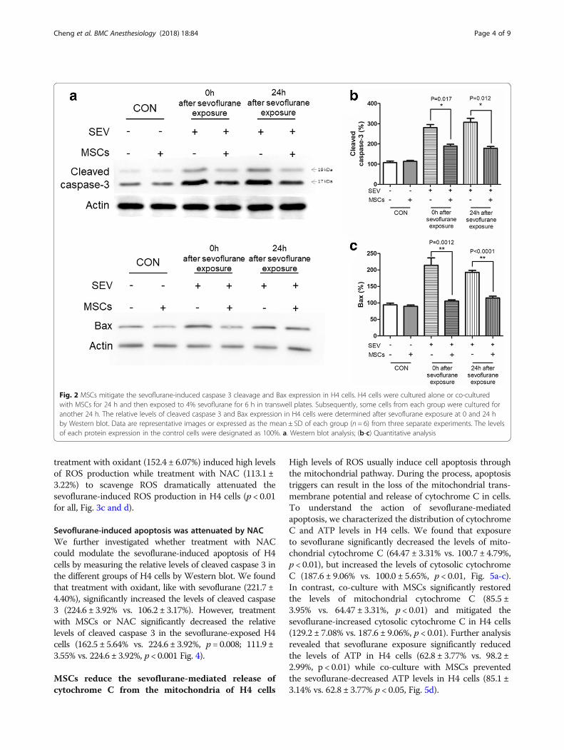
ResultsMSCs mitigate H4 cell apoptosis induced by sevofluraneWe first tested the impact of MSC treatment on thesevoflurane-induced apoptosis in H4 cells by analysis ofthe relative levels of cleaved caspase 3 (281.2 ± 14.93%and 308.0 ± 18.60% vs. 107.5 ± 7.5% at 0 and 24 h, re-spectively) and Bax (214.0 ± 12.90% and 214.0 ± 12.91%vs. 94.3 ± 2.84% at 0 and 24 h, respectively) in H4 cellswithout co-cultured with MSCs (Fig. 2). In contrast,co-cultured of H4 cells with MSCs significantly miti-gated the sevoflurane-elevated cleaved caspase 3 (189.1± 9.30% vs. 281.2 ± 14.93%, p < 0.05; and 179.0 ± 8.49%vs. 308.0 ± 18.60%, p < 0.05) and Bax immediately (0 h,105.0 ± 2.36% vs. 214.0 ± 12.90%, p < 0.01) and 24 h(105.4 ± 2.36% vs. 214.0 ± 12.91%, p < 0.01) after sevoflur-ane exposure in H4 cells in our experimental system(Fig. 2b and c).
MSCs attenuate sevoflurane-induced ROS production inH4 cellsNext, we measured the levels of intracellular ROS in H4cells by DCFH-DA based fluorescent microscopy andspectrum. First, exposure to sevoflurane significantly in-creased the levels of cytoplasmic ROS at both immedi-ately (145.0 ± 6.31% vs. 98.2 ± 3.24%) and further 24 hculture (132.4 ± 4.13% vs. 98.2 ± 3.24%) after sevofluraneexposure while co-culture with MSCs almost abrogatedthe sevoflurane-induced ROS production in H4 cells(110.4 ± 4.90% vs. 145.0 ± 6.31% p < 0.05, or 106.9 ± 3.45%vs. 132.4 ± 4.13% p < 0.01, Fig. 3a and b). Similarly,
Fig. 1 Flowchart of the experimental design
Cheng et al. BMC Anesthesiology (2018) 18:84 Page 3 of 9
treatment with oxidant (152.4 ± 6.07%) induced high levelsof ROS production while treatment with NAC (113.1 ±3.22%) to scavenge ROS dramatically attenuated thesevoflurane-induced ROS production in H4 cells (p < 0.01for all, Fig. 3c and d).
Sevoflurane-induced apoptosis was attenuated by NACWe further investigated whether treatment with NACcould modulate the sevoflurane-induced apoptosis of H4cells by measuring the relative levels of cleaved caspase 3 inthe different groups of H4 cells by Western blot. We foundthat treatment with oxidant, like with sevoflurane (221.7 ±4.40%), significantly increased the levels of cleaved caspase3 (224.6 ± 3.92% vs. 106.2 ± 3.17%). However, treatmentwith MSCs or NAC significantly decreased the relativelevels of cleaved caspase 3 in the sevoflurane-exposed H4cells (162.5 ± 5.64% vs. 224.6 ± 3.92%, p = 0.008; 111.9 ±3.55% vs. 224.6 ± 3.92%, p < 0.001 Fig. 4).
MSCs reduce the sevoflurane-mediated release ofcytochrome C from the mitochondria of H4 cells
High levels of ROS usually induce cell apoptosis throughthe mitochondrial pathway. During the process, apoptosistriggers can result in the loss of the mitochondrial trans-membrane potential and release of cytochrome C in cells.To understand the action of sevoflurane-mediatedapoptosis, we characterized the distribution of cytochromeC and ATP levels in H4 cells. We found that exposureto sevoflurane significantly decreased the levels of mito-chondrial cytochrome C (64.47 ± 3.31% vs. 100.7 ± 4.79%,p < 0.01), but increased the levels of cytosolic cytochromeC (187.6 ± 9.06% vs. 100.0 ± 5.65%, p < 0.01, Fig. 5a-c).In contrast, co-culture with MSCs significantly restoredthe levels of mitochondrial cytochrome C (85.5 ±3.95% vs. 64.47 ± 3.31%, p < 0.01) and mitigated thesevoflurane-increased cytosolic cytochrome C in H4 cells(129.2 ± 7.08% vs. 187.6 ± 9.06%, p < 0.01). Further analysisrevealed that sevoflurane exposure significantly reducedthe levels of ATP in H4 cells (62.8 ± 3.77% vs. 98.2 ±2.99%, p < 0.01) while co-culture with MSCs preventedthe sevoflurane-decreased ATP levels in H4 cells (85.1 ±3.14% vs. 62.8 ± 3.77% p < 0.05, Fig. 5d).
Fig. 2 MSCs mitigate the sevoflurane-induced caspase 3 cleavage and Bax expression in H4 cells. H4 cells were cultured alone or co-culturedwith MSCs for 24 h and then exposed to 4% sevoflurane for 6 h in transwell plates. Subsequently, some cells from each group were cultured foranother 24 h. The relative levels of cleaved caspase 3 and Bax expression in H4 cells were determined after sevoflurane exposure at 0 and 24 hby Western blot. Data are representative images or expressed as the mean ± SD of each group (n = 6) from three separate experiments. The levelsof each protein expression in the control cells were designated as 100%. a. Western blot analysis; (b-c) Quantitative analysis
Cheng et al. BMC Anesthesiology (2018) 18:84 Page 4 of 9

Fig. 3 MSCs attenuate the sevoflurane-induced ROS production in H4 cells. The levels of ROS in the different groups of cells were characterizedafter staining with DCFH-DA by fluorescent microscopy and spectrum. Data are representative images or expressed as the mean ± SD of eachgroup (n = 6) from three separate experiments. a. Microscopy characterization of ROS levels in H4 cells. b. Quantitative analysis. c. NAC scavengesthe sevoflurane-induced ROS in H4 cells. H4 cells were treated with, or without, oxidant or exposed to sevoflurane for 6 h in the presence ofNAC. The levels of intracellular ROS were measured by fluorescent spectrum. d. Quantitative analysis. The levels of ROS in the control cells weredesignated as 100%
Fig. 4 NAC attenuates the sevoflurane-elevated caspase 3 cleavage in H4 cells. H4 cells were treated with vehicle (CON), SEV, Oxidant alone ortogether with MSCs (SEV +MSCs) or SEV + NAC. The relative levels of cleaved caspase 3 in the different groups of cells were determined byWestern blot. Data are representative images or expressed as the mean ± SD of each group (n = 6) from three separate experiments. a. Westernblot analysis. b. Quantitative analysis. The levels of each protein expression in the control cells were designated as 100%
Cheng et al. BMC Anesthesiology (2018) 18:84 Page 5 of 9

Fig. 5 MSCs inhibit the sevoflurane-induced mitochondrial cytochrome C release in H4 cells. The mitochondria and cytosol of the differentgroups of H4 cells were extracted and the relative levels of cytochrome C were determined by Western blot. Furthermore, the levels of ATP inthe different groups of cells were measured. Data are representative images or expressed as the mean ± SD of each group (n = 6) from threeseparate experiments. a. Western blot analysis. b and c. Quantitative analysis. d. The levels of ATP. The levels of each protein expression or ATP inthe control cells were designated as 100%
Fig. 6 Co-culture with MSCs after sevoflurane exposure does not alter the sevoflurane-induced ROS production in H4 cells. H4 cells were culturedfor 24 h and exposed to sevoflurane, followed by co-cultured with MSCs or cultured alone for 24 h. The control H4 cells were cultured alonethroughout the experimental period. The cells were stained with DCFH-DA and the levels of ROS were determined by fluorescent microscopyand spectrum. Data are representative images or expressed as the mean ± SD of each group (n = 6) from three separate experiments. a.Fluorescent microscopy analysis of ROS in H4 cells. b. Fluorescent spectrum analysis of the ROS levels in H4 cells. The levels of ROS in the controlcells were designated as 100%
Cheng et al. BMC Anesthesiology (2018) 18:84 Page 6 of 9

MSCs fail to inhibit the sevoflurane-induced ROSproduction in H4 cells post sevoflurane exposureFinally, we tested whether treatment with MSCs post ex-posure to sevoflurane could inhibit the sevoflurane-inducedROS production in H4 cells. The levels of ROS were 93.7 ±3.28% (CON), 154.0 ± 4.69% (SEV), 150.2 ± 5.55% (Post--treatment), respectively. We found that sevoflurane expos-ure significantly induced high levels of ROS production(154.0 ± 4.69% vs. 93.7 ± 3.28%) and co-culture with MSCsafter sevoflurane exposure did not change the levels ofsevoflurane-induced ROS production in H4 cells (150.2 ±5.55% vs. 154.0 ± 4.69%, p > 0.05, Fig. 6).
DiscussionIn this study, we found that treatment with MSCs be-fore, but not after, sevoflurane exposure attenuated thesevoflurane-induced ROS production and apoptosis inH4 cells, which was abrogated by antioxidant NAC.MSCs prevented the sevoflurane-induced cytochrome Crelease from the mitochondria to the cytoplasm andATP production in H4 cells. These novel data extendedprevious observations [17–22] and indicated that solublefactors secreted by MSCs had potent antioxidant activityagainst oxidative stress-induced apoptosis in H4 cells.These finding may provide new insights into the neur-onal toxicity of sevoflurane. Given that sevoflurane is aninhaled anesthetic used widely in the clinical practice inanesthesia our findings suggest that we should be cau-tious while using sevoflurane.MSCs can secrete neurotrophic factors, cytokines and
other soluble factors that are associated neuroprotectiveactivity [17–20]. In this study, while sevoflurane expos-ure induced ROS production, mitochondrial cytochromeC release and apoptosis in H4 cells co-culture withMSCs before sevoflurane exposure almost completelyprevented the oxidant activity of sevoflurane in H4cells. However, we found that co-culture with MSCsafter sevoflurane exposure failed to mitigate thesevoflurane-induced ROS production in H4 cells. Thesedata suggest that sevoflurane may trigger an oxidativecascade that induces the mitochondrial damages andapoptosis of H4 cells, which may not be easily overcomeby soluble factors from MSCs [29]. A recent study hasshown that infusion of MSCs improves symptoms in pa-tients with Alzheimer’s disease [30]. Further studies arenecessary to identify soluble factors secreted by MSCsand determine the molecular mechanisms underlyingthe action of MSCs.Given that this experiment was performed in transwell
plates the antioxidant effect of MSCs was likely medi-ated by their soluble factors. Indeed, MSCs can secreteneurotrophic factors, cytokines and other extracellularvesicles (EVs). Our findings support the theory that theEVs secreted by MSCs are responsible for their
immunosuppressive activity [31] and the neuronal pro-tection by MSCs may be independent of cell-to-cell con-tact. It is notable that EVs include exosomes, ectosomes,microvesicles, microparticles, apoptotic bodies and otherEV subsets [32]. We are interested in further investigat-ing which type of EV(s) has such potent antioxidant ac-tivity and neuronal protective effect. Given that mostEVs are able to pass through the blood-brain barrier andare relatively stable the identified EVs or soluble factorsmay be valuable for cell-free therapy of oxidativestress-related neuronal degenerative diseases.We recognized that our study has limitations. First, the
MSCs were extracted from newborn SD rats and tested ina human cell line. However, MSCs have the advantage oflow-immunogenicity which make them potentially safe forclinical use. Because of MSC’s immunomodulatory effects,they can be used to repair the damage caused byautoimmune-induced disease and graft-versus-host disease[33]. Autologous and allogenic bone marrow derived MSCshave been shown to be safe for human use [34, 35]. Xeno-geneic bone marrow derived MSCs from human, mouseand rat have been widely used in animal experiments [36–38]. Because of the role of MSC-derived EVs, EVs isolatedfrom conditioned media of MSCs would be used in our fur-ther animal studies. Second, although we used an optimaldose of MSCs in our experiments we did not test the timecourse of the co-culture system in detail. Once MSCs andH4 cells were seeded into their own chambers respectively,the co-culture system was started. After culture for 24 h,the cells should be in the best condition of density and thesevoflurane exposure started. Further studies are needed totest whether co-culture for varying time periods or culturefor a longer period after exposure to sevoflurane has differ-ent effects on sevoflurane-induced apoptosis and ROS pro-duction because co-culture for 24 h or exposure tosevoflurane for 6 h may be not optimal. Third, these resultswere based on cell experiments and still need to be con-firmed in animals.
ConclusionsSevoflurane is the most widely used inhaled anesthetic inclinical practice. In this study, we demonstrated that theexposure to 4% sevoflurane for 6 h induced ROS produc-tion, apoptosis and caspase-3 activation in H4 cells. Fur-thermore, we revealed that the sevoflurane-mediatedapoptosis was mediated by the mitochondrial pathway. Inaddition, we found for the first time that the co-culturesystem with MSCs reduced the toxic effect of sevofluraneon H4 cells. Potentially, our findings may aid in design ofnew therapies for prevention of sevoflurane-induced neur-onal damage.
AbbreviationsATP: Adenosine triphosphate; DCFH-DA: 2,7-dichlorofluorescein diacetate;DMEM: Dulbecco’s modified eagle medium; FBS: Fetal bovine serum; H4
Cheng et al. BMC Anesthesiology (2018) 18:84 Page 7 of 9

cells: Human neuroglioma H4 cells; MSCs: Mesenchymal stem cells; NAC: N-acetylcysteine; ROS: Reactive oxygen species; SD rats: Sprague-Dawley rats
AcknowledgementsWe thank Medjaden for its linguistic assistance during the preparation of thismanuscript.
FundingThis study was supported by the Department of Anesthesiology, ShanghaiNinth People’s Hospital Affiliated to Shanghai Jiao Tong University School ofMedicine.
Availability of data and materialsThe datasets used and/or analyzed during the current study are availablefrom the corresponding author on reasonable request.
Authors’ contributionsYC carried out the studies and drafted the manuscript. YJ carried out thestudies and prepared the manuscript. LZ conceived of the study andparticipated in its design and coordination. JW carried out the studies andcollected the data. DC performed the statistical analysis. RH helped indesigning the study. CL performed the statistical analysis. YS participated inthe design of the study and coordination. HJ participated in the design ofthe study. All authors read and approved the final manuscript.
Ethics approval and consent to participateAll experimental protocols were approved by the Animal Care and Use ofShanghai Ninth People’s Hospital Affiliated to Shanghai Jiao Tong UniversitySchool of Medicine Committee.
Consent for publicationNot applicable.
Competing interestsThe authors declare that they have no competing interests.
Publisher’s NoteSpringer Nature remains neutral with regard to jurisdictional claims inpublished maps and institutional affiliations.
Received: 26 December 2017 Accepted: 27 June 2018
References1. Takaenoki Y, Satoh Y, Araki Y, Kodama M, Yonamine R, Yufune S, Kazama T.
Neonatal exposure to sevoflurane in mice causes deficits in maternalbehavior later in adulthood. Anesthesiology. 2014;120:403–15.
2. Shen X, Dong Y, Xu Z, Wang H, Miao C, Soriano SG, Sun D, Baxter MG,Zhang Y, Xie Z. Selective anesthesia-induced neuroinflammation indeveloping mouse brain and cognitive impairment. Anesthesiology. 2013;118:502–15.
3. Satomoto M, Satoh Y, Terui K, Miyao H, Takishima K, Ito M, Imaki J. Neonatalexposure to sevoflurane induces abnormal social behaviors and deficits infear conditioning in mice. Anesthesiology. 2009;110:628–37.
4. Qiu J, Shi P, Mao W, Zhao Y, Liu W, Wang Y. Effect of apoptosis in neuralstem cells treated with sevoflurane. BMC Anesthesiol. 2015;15:25.
5. Qiao Y, Feng H, Zhao T, Yan H, Zhang H, Zhao X. Postoperative cognitivedysfunction after inhalational anesthesia in elderly patients undergoingmajor surgery: the influence of anesthetic technique, cerebral injury andsystemic inflammation. BMC Anesthesiol. 2015;15:154.
6. Yu Y, Zhang P, Yan J, Sun Y, Wu X, Xi S, Zhang L, Sun Y, Hu R, Jiang H.Sevoflurane induces cognitive impairments via the MiR-27b/LIMK1-signalingpathway in developing rats. Inhal Toxicol. 2016;28:731–8.
7. Jiang J, Lv X, Wu X, Yang Y, Jiang H. Downregulation of circulating insulin-like growth factor 1 contributes to memory impairment in aged mice aftersevoflurane anesthesia. Behav Pharmacol. 2017;28:238–43.
8. Tao G, Zhang J, Zhang L, Dong Y, Yu B, Crosby G, Culley DJ, Zhang Y, Xie Z.Sevoflurane induces tau phosphorylation and glycogen synthase kinase3beta activation in young mice. Anesthesiology. 2014;121:510–27.
9. Liu F, Rainosek SW, Frisch-Daiello JL, Patterson TA, Paule MG, Slikker W Jr,Wang C, Han X. Potential adverse effects of prolonged Sevoflurane
exposure on developing monkey brain: from abnormal lipid metabolism toneuronal damage. Toxicol Sci. 2015;147:562–72.
10. Liu Y, Pan N, Ma Y, Zhang S, Guo W, Li H, Zhou J, Liu G, Gao M. Inhaledsevoflurane may promote progression of amnestic mild cognitiveimpairment: a prospective, randomized parallel-group study. Am J Med Sci.2013;345:355–60.
11. Vutskits L, Xie Z. Lasting impact of general anaesthesia on the brain:mechanisms and relevance. Nat Rev Neurosci. 2016;17:705–17.
12. Backeljauw B, Holland SK, Altaye M, Loepke AW. Cognition and brainstructure following early childhood surgery with anesthesia. Pediatrics. 2015;136:e1–12.
13. Sun L. Early childhood general anaesthesia exposure and neurocognitivedevelopment. Br J Anaesth. 2010;105(Suppl 1):i61–8.
14. Boscolo A, Starr JA, Sanchez V, Lunardi N, DiGruccio MR, Ori C, Erisir A,Trimmer P, Bennett J, Jevtovic-Todorovic V. The abolishment of anesthesia-induced cognitive impairment by timely protection of mitochondria in thedeveloping rat brain: the importance of free oxygen radicals andmitochondrial integrity. Neurobiol Dis. 2012;45:1031–41.
15. Liu B, Gu Y, Xiao H, Lei X, Liang W, Zhang J. Altered metabolomic profilesmay be associated with sevoflurane-induced neurotoxicity in neonatal rats.Neurochem Res. 2015;40:788–99.
16. Sun Z, Satomoto M, Adachi Y, Kinoshita H, Makita K. Inhibiting NADPHoxidase protects against long-term memory impairment induced byneonatal sevoflurane exposure in mice. Br J Anaesth. 2016;117:80–6.
17. Caplan AI, Dennis JE. Mesenchymal stem cells as trophic mediators. J CellBiochem. 2006;98:1076–84.
18. Fu Y, Karbaat L, Wu L, Leijten J, Both SK, Karperien M. Trophic Effects ofMesenchymal Stem Cells in Tissue Regeneration. Tissue Eng Part B Rev.2017;23:515–28.
19. Kim HS, Choi DY, Yun SJ, Choi SM, Kang JW, Jung JW, Hwang D, Kim KP,Kim DW. Proteomic analysis of microvesicles derived from humanmesenchymal stem cells. J Proteome Res. 2012;11:839–49.
20. Koh SH, Noh MY, Cho GW, Kim KS, Kim SH. Erythropoietin increases themotility of human bone marrow-multipotent stromal cells (hBM-MSCs) andenhances the production of neurotrophic factors from hBM-MSCs. StemCells Dev. 2009;18:411–21.
21. Shen LH, Li Y, Chen J, Zacharek A, Gao Q, Kapke A, Lu M, Raginski K, VanguriP, Smith A, et al. Therapeutic benefit of bone marrow stromal cellsadministered 1 month after stroke. J Cereb Blood Flow Metab. 2007;27:6–13.
22. Zhao MZ, Nonoguchi N, Ikeda N, Watanabe T, Furutama D, Miyazawa D,Funakoshi H, Kajimoto Y, Nakamura T, Dezawa M, et al. Novel therapeuticstrategy for stroke in rats by bone marrow stromal cells and ex vivo HGF genetransfer with HSV-1 vector. J Cereb Blood Flow Metab. 2006;26:1176–88.
23. Bordignon C, Carlo-Stella C, Colombo MP, De Vincentiis A, Lanata L, LemoliRM, Locatelli F, Olivieri A, Rondelli D, Zanon P, et al. Cell therapy:achievements and perspectives. Haematologica. 1999;84:1110–49.
24. Si YL, Zhao YL, Hao HJ, Fu XB, Han WD. MSCs: biological characteristics,clinical applications and their outstanding concerns. Ageing Res Rev. 2011;10:93–103.
25. Pfrieger FW. Role of glial cells in the formation and maintenance ofsynapses. Brain Res Rev. 2010;63:39–46.
26. Cheng B, Zhang Y, Wang A, Dong Y, Xie Z. Vitamin C attenuates Isoflurane-induced Caspase-3 activation and cognitive impairment. Mol Neurobiol.2015;52:1580–9.
27. Sun Y, Zhang Y, Cheng B, Dong Y, Pan C, Li T, Xie Z. Glucose may attenuateisoflurane-induced caspase-3 activation in H4 human neuroglioma cells.Anesth Analg. 2014;119:1373–80.
28. Zhang J, Dong Y, Xu Z, Zhang Y, Pan C, McAuliffe S, Ichinose F, Yue Y, LiangW, Xie Z. 2-Deoxy-D-glucose attenuates isoflurane-induced cytotoxicity inan in vitro cell culture model of H4 human neuroglioma cells. AnesthAnalg. 2011;113:1468–75.
29. Griendling KK, FitzGerald GA. Oxidative stress and cardiovascular injury: part I:basic mechanisms and in vivo monitoring of ROS. Circulation. 2003;108:1912–6.
30. de Windt T, Vonk L, Slaper-Cortenbach I, van den Broek M, Nizak R, vanRijen M, de Weger R, Dhert W, Saris D. Allogeneic Mesenchymal stem cellsstimulate cartilage regeneration and are safe for single-stage cartilage repairin humans upon mixture with recycled autologous Chondrons. Stem Cells.2017;35:256–64.
31. Lai RC, Arslan F, Lee MM, Sze NS, Choo A, Chen TS, Salto-Tellez M, TimmersL, Lee CN, El Oakley RM, et al. Exosome secreted by MSC reducesmyocardial ischemia/reperfusion injury. Stem Cell Res. 2010;4:214–22.
Cheng et al. BMC Anesthesiology (2018) 18:84 Page 8 of 9

32. Lotvall J, Hill AF, Hochberg F, Buzas EI, Di Vizio D, Gardiner C, Gho YS,Kurochkin IV, Mathivanan S, Quesenberry P, et al. Minimal experimentalrequirements for definition of extracellular vesicles and their functions: aposition statement from the International Society for Extracellular Vesicles.J Extracell Vesicles. 2014;3:26913.
33. Yang M, Liu L. MHC II gene knockout in tissue engineering may preventimmune rejection of transplants. Med Hypotheses. 2008;70:798–801.
34. Karussis D, Karageorgiou C, Vaknin-Dembinsky A, Gowda-Kurkalli B, GomoriJM, Kassis I, Bulte JW, Petrou P, Ben-Hur T, Abramsky O, et al. Safety andimmunological effects of mesenchymal stem cell transplantation in patientswith multiple sclerosis and amyotrophic lateral sclerosis. Arch Neurol. 2010;67:1187–94.
35. Zhang H, Zeng X, Sun L. Allogenic bone-marrow-derived mesenchymalstem cells transplantation as a novel therapy for systemic lupuserythematosus. Expert Opin Biol Ther. 2010;10:701–9.
36. Costa-Ferro ZS, de Borba Cunha F, de Freitas Souza BS, Leal MM, da SilvaAA, de Bellis Kuhn TI, Forte A, Sekiya EJ, Soares MB, Dos Santos RR.Antiepileptic and neuroprotective effects of human umbilical cord bloodmononuclear cells in a pilocarpine-induced epilepsy model.Cytotechnology. 2014;66:193–9.
37. Li T, Ren G, Kaplan DL, Boison D. Human mesenchymal stem cell graftsengineered to release adenosine reduce chronic seizures in a mouse modelof CA3-selective epileptogenesis. Epilepsy Res. 2009;84:238–41.
38. Venturin GT, Greggio S, Marinowic DR, Zanirati G, Cammarota M, MachadoDC, DaCosta JC. Bone marrow mononuclear cells reduce seizure frequencyand improve cognitive outcome in chronic epileptic rats. Life Sci. 2011;89:229–34.
Cheng et al. BMC Anesthesiology (2018) 18:84 Page 9 of 9