membrane texture induced by specific protein binding and receptor
TRANSCRIPT

Membrane texture induced by specific proteinbinding and receptor clustering: active rolesfor lipids in cellular functionE. B. Watkinsa,b, C. E. Millerb,c, J. Majewskib,1, and T. L. Kuhla,d,e,1
aBiophysics Graduate Group, University of California, Davis, CA 95616; bManuel Lujan Jr. Neutron Scattering Center, Los Alamos National Laboratory,Los Alamos, NM 87545; cStanford Synchrotron Radiation Lightsource, Menlo Park, CA 94025; dDepartment of Biomedical Engineering, University ofCalifornia, Davis, CA 95616; and eDepartment of Chemical Engineering and Material Science, University of California, Davis, CA 95616
Edited* by Jacob N. Israelachvili, University of California, Santa Barbara, CA, and approved February 23, 2011 (received for review October 3, 2010)
Biological membranes are complex, self-organized structures thatdefine boundaries and compartmentalize space in living matter.Composed of a wide variety of lipid and protein molecules, theseresponsive surfaces mediate transmembrane signaling and materi-al transport within the cell and with its environment. It is wellknown that lipid membrane properties change as a function ofcomposition and phase state, and that protein-lipid interactionscan induce changes in the membrane’s properties and biochemicalresponse. Here, molecular level changes in lipid organizationinduced bymultivalent toxin bindingwere investigated using graz-ing incidence X-ray diffraction. Structural changes to lipid mono-layers at the air-water interface and bilayers at the solid-waterinterface were studied before and after specific binding of choleratoxin to membrane embedded receptors. At biologically relevantsurface pressures, protein binding perturbed lipid packing withinmonolayers and bilayers resulting in topological defects andthe emergence of a new orientationally textured lipid phase. Inbilayers this altered lipid order was transmitted from the receptorladen exterior membrane leaflet to the inner leaflet, representing apotential mechanism for lipid mediated outside-in signaling bymultivalent protein binding. It is further hypothesized that cell-surface micro-domains exhibiting this type of lipid order may serveas nucleation sites for vesicle formation in clathrin independentendocytosis of cholera toxin.
X-ray scattering ∣ liquid crystal ∣ supported membrane ∣ganglioside ∣ glycolipid
Interactions between proteins and the cell membrane are anintegral aspect of many biological processes (1). Diverse pro-
tein-lipid complexes exist including transmembrane proteins, per-ipheral membrane proteins, and proteins bound to membraneassociated receptor molecules. The interplay between these bio-logical components is multifaceted: lipids can influence the struc-ture and function of membrane proteins and at the same timemembrane proteins can impact lipid organization (2). In modelsystems lipids are capable of adopting a variety of differentordered states with their phase behavior primarily governed bysteric and van der Waals interactions between neighboring headgroups and alkyl chains. Lipid organization ranges from thetightly packed gel phase to the fluid like liquid ordered (Lo)and liquid disordered (Ld) phases. Lateral heterogeneities withinmodel membranes due to the coexistence of Ld and Lo phaseshave been widely used to study lipid domain formation and asanalogs for lipid rafts (3, 4). In biological systems, lipid raftsare dynamic self-organized membrane microdomains that selec-tively recruit specific proteins and lipids while excluding others(5, 6). Typically enriched in cholesterol, sphingolipids, and glyco-lipids, rafts are characterized by the tighter packing of theirconstituent molecules in a liquid ordered phase (7). Raft micro-domains offer a means to sequester proteins, enhance the localconcentration of raft associated components, as well as alterthe conformation of embedded proteins within the cellular
membrane. Another example of membrane microdomains isthe self-association of glycosphingolipids (GSL) to form a glyco-synapse. Such microdomains are thought to play a role in a widerange of biological functions including cell recognition, adhesion,and signaling (8, 9). For example, in cell adhesion processes GSL-GSL interactions directly influence membrane properties andGSL membrane microdomains have also been shown to modu-late the activity of cytoplasmic protein kinases. A structuralmechanism has not yet been established to explain how GSL mi-crodomains modify cellular activity. In rafts, lipid induced proteinconformation changes can also influence signaling by membraneembedded protein receptors, e.g., ion-channel linked receptors,enzyme linked receptors, and G-protein coupled receptors, asthey transmit signals across the membrane (outside-in signaling)through conformational or chemical changes to the protein’sintracellular domain upon small molecule binding to an extracel-lular domain. These changes may, for example, open a channel,activate enzymatic activity, or induce a signaling cascade thatresults in a cellular response. However, to our knowledge a “lipidonly” mediated structural mechanism for transmembrane signal-ing has not yet been reported. In the work reported here, weexamine if the modulation of lipid membrane by specific proteinbinding can provide a potential mechanism for transmembranesignaling.
Cholera toxin, which selectively binds to ganglioside glycoli-pids, is frequently used as a reporter for membrane rafts andas a tool to investigate protein-lipid interactions (10). The Bsubunit (CTB) is responsible for binding the toxin with highestaffinity to ganglioside GM1 (4.61 × 10⁻¹² M), a cell-surfacereceptor also associated with lipid raft domains (11). Five iden-tical B subunits, each containing one binding site, form a penta-meric ring with a vertical height of 32 Å and a radius of 31 Å(12, 13). Because binding is multivalent and of high affinity,off rates of the toxin are slow and the CTB-GM1 complex is verystable enabling CTB to effectively cross link GM1 receptors in themembrane. Receptor cross linking, in general, may act to stabilizerafts, lead to coalescence of raft domains, and is hypothesized tobe involved in the exploitation of clathrin independent endocy-tosis pathways by multivalent toxins (14). Moreover, limitingthe number of active binding pockets on cholera toxin has beenshown to inhibit endocytosis, presumably due to diminishedreceptor cross linking (15). A mechanistic explanation for thisphenomenon remains unclear.
Author contributions: E.B.W., C.E.M., J.M., and T.L.K. designed research; E.B.W., C.E.M.,J.M., and T.L.K. performed research; E.B.W. and C.E.M. contributed new reagents/analytictools; E.B.W. analyzed data; and E.B.W. and T.L.K. wrote the paper.
The authors declare no conflict of interest.
*This Direct Submission article had a prearranged editor.1To whom correspondence may be addressed. E-mail: [email protected] or [email protected].
This article contains supporting information online at www.pnas.org/lookup/suppl/doi:10.1073/pnas.1014579108/-/DCSupplemental.
www.pnas.org/cgi/doi/10.1073/pnas.1014579108 PNAS Early Edition ∣ 1 of 6
BIOPH
YSICSAND
COMPU
TATIONALBIOLO
GY
Here we report the discovery of a unique lipid phase generatedby multivalent protein binding to raft associated membranereceptors. The packing characteristics of this textured lipid phase(LT) place it intermediate between the well established Lo and gellipid phases. Not restricted to close-packed structures, the LTphase comprises a rich variety of lipid tail tilt orientations includ-ing anisotropic and azimuthally swirled arrangements analogousto those observed in macroscopic hexatic phases of liquid crystals.Specific binding of protein to membrane embedded receptors wasshown to generate the LT phase in model membranes, providing apossible window into otherwise undetectable features of lipidorder within nanoscale domains in the cellular membrane. Wepropose that such orientationally textured domains may havebiological relevance as lipid based signaling platforms and in cel-lular trafficking pathways. Significantly, these altered packingarrangements are transmitted from the receptor laden leafletto the inner leaflet of the membrane providing a means for out-side-in signaling. Further, the LT phase offers a mechanistic ex-planation for nonclathrin mediated endocytosis where alteredlipid packing due to toxin binding serves as a nucleation sitefor vesicle formation.
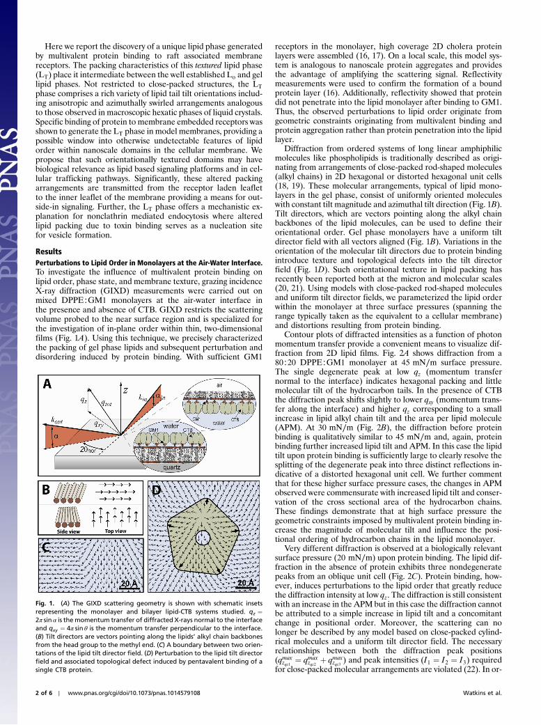
ResultsPerturbations to Lipid Order in Monolayers at the Air-Water Interface.To investigate the influence of multivalent protein binding onlipid order, phase state, and membrane texture, grazing incidenceX-ray diffraction (GIXD) measurements were carried out onmixed DPPE∶GM1 monolayers at the air-water interface inthe presence and absence of CTB. GIXD restricts the scatteringvolume probed to the near surface region and is specialized forthe investigation of in-plane order within thin, two-dimensionalfilms (Fig. 1A). Using this technique, we precisely characterizedthe packing of gel phase lipids and subsequent perturbation anddisordering induced by protein binding. With sufficient GM1
receptors in the monolayer, high coverage 2D cholera proteinlayers were assembled (16, 17). On a local scale, this model sys-tem is analogous to nanoscale protein aggregates and providesthe advantage of amplifying the scattering signal. Reflectivitymeasurements were used to confirm the formation of a boundprotein layer (16). Additionally, reflectivity showed that proteindid not penetrate into the lipid monolayer after binding to GM1.Thus, the observed perturbations to lipid order originate fromgeometric constraints originating from multivalent binding andprotein aggregation rather than protein penetration into the lipidlayer.
Diffraction from ordered systems of long linear amphiphilicmolecules like phospholipids is traditionally described as origi-nating from arrangements of close-packed rod-shaped molecules(alkyl chains) in 2D hexagonal or distorted hexagonal unit cells(18, 19). These molecular arrangements, typical of lipid mono-layers in the gel phase, consist of uniformly oriented moleculeswith constant tilt magnitude and azimuthal tilt direction (Fig. 1B).Tilt directors, which are vectors pointing along the alkyl chainbackbones of the lipid molecules, can be used to define theirorientational order. Gel phase monolayers have a uniform tiltdirector field with all vectors aligned (Fig. 1B). Variations in theorientation of the molecular tilt directors due to protein bindingintroduce texture and topological defects into the tilt directorfield (Fig. 1D). Such orientational texture in lipid packing hasrecently been reported both at the micron and molecular scales(20, 21). Using models with close-packed rod-shaped moleculesand uniform tilt director fields, we parameterized the lipid orderwithin the monolayer at three surface pressures (spanning therange typically taken as the equivalent to a cellular membrane)and distortions resulting from protein binding.
Contour plots of diffracted intensities as a function of photonmomentum transfer provide a convenient means to visualize dif-fraction from 2D lipid films. Fig. 2A shows diffraction from a80∶20 DPPE∶GM1 monolayer at 45 mN∕m surface pressure.The single degenerate peak at low qz (momentum transfernormal to the interface) indicates hexagonal packing and littlemolecular tilt of the hydrocarbon tails. In the presence of CTBthe diffraction peak shifts slightly to lower qxy (momentum trans-fer along the interface) and higher qz corresponding to a smallincrease in lipid alkyl chain tilt and the area per lipid molecule(APM). At 30 mN∕m (Fig. 2B), the diffraction before proteinbinding is qualitatively similar to 45 mN∕m and, again, proteinbinding further increased lipid tilt and APM. In this case the lipidtilt upon protein binding is sufficiently large to clearly resolve thesplitting of the degenerate peak into three distinct reflections in-dicative of a distorted hexagonal unit cell. We further commentthat for these higher surface pressure cases, the changes in APMobserved were commensurate with increased lipid tilt and conser-vation of the cross sectional area of the hydrocarbon chains.These findings demonstrate that at high surface pressure thegeometric constraints imposed by multivalent protein binding in-crease the magnitude of molecular tilt and influence the posi-tional ordering of hydrocarbon chains in the lipid monolayer.
Very different diffraction is observed at a biologically relevantsurface pressure (20 mN∕m) upon protein binding. The lipid dif-fraction in the absence of protein exhibits three nondegeneratepeaks from an oblique unit cell (Fig. 2C). Protein binding, how-ever, induces perturbations to the lipid order that greatly reducethe diffraction intensity at low qz. The diffraction is still consistentwith an increase in the APM but in this case the diffraction cannotbe attributed to a simple increase in lipid tilt and a concomitantchange in positional order. Moreover, the scattering can nolonger be described by any model based on close-packed cylind-rical molecules and a uniform tilt director field. The necessaryrelationships between both the diffraction peak positions(qmax
zqz1 ¼ qmaxzqz2 þ qmax
zqz3 ) and peak intensities (I1 ¼ I2 ¼ I3) requiredfor close-packed molecular arrangements are violated (22). In or-
Fig. 1. (A) The GIXD scattering geometry is shown with schematic insetsrepresenting the monolayer and bilayer lipid-CTB systems studied. qz ¼2π sin α is the momentum transfer of diffracted X-rays normal to the interfaceand qxy ¼ 4π sin θ is the momentum transfer perpendicular to the interface.(B) Tilt directors are vectors pointing along the lipids’ alkyl chain backbonesfrom the head group to the methyl end. (C) A boundary between two orien-tations of the lipid tilt director field. (D) Perturbation to the lipid tilt directorfield and associated topological defect induced by pentavalent binding of asingle CTB protein.
2 of 6 ∣ www.pnas.org/cgi/doi/10.1073/pnas.1014579108 Watkins et al.

der to reduce the low qz intensity and match the measured dif-fraction, topological defects and orientational texture have to beintroduced into the lipid tilt director vector field (Fig. 1D). Thesechanges represent the emergence of a textured lipid phase, LT,specifically induced by multivalent protein binding. The details ofthis phase and modeling are described in the next section.
Modeling GIXD from Textured Phase Monolayers. To describe lipidorder in the textured LT phase, we implemented models that per-turbed the lipid order away from a close-packed gel phase andcalculated the associated diffraction. By loosening the constraintimposed by close packing of lipid alkyl chains, topological defectsand orientational texture could be incorporated into these mod-els. The diffraction pattern of a 20 mN∕m monolayer prior toprotein binding was reproduced with a uniformly oriented tiltdirector field. After protein binding, no close-packed model wasable to reproduce the diffraction pattern. Instead, a textured 2Dliquid crystal smectic-like phase, here specified as the lipid LTphase, with azimuthal perturbations to the molecular tilt directorswas required. The simplest texture, for example, can be generatedfrom a single defect, or disclination, at the domain center yieldingan arrangement of tilt directors analogous to bend domains inliquid crystals (23). Azimuthal angles of the tilt directors weredescribed by the equation ϕ ¼ s tan−1ðy∕xÞ þ ϕ0 where x and yare the lateral coordinates of the molecules, and s is the strengthof the disclination with s ¼ �1 corresponding to the lowest en-ergy symmetric textures. The LT regions have a finite size andthe average length scale associated with these domains is inver-sely related to the width of the Bragg peaks. For a domain radiusof 150 Å (corresponding to the measured Bragg peak FWHM),the scattered intensity at low qz decreased with increasing s untils ≈ 5 (Fig. 3). Increasing the strength of the texture beyond thisvalue did not improve the quality of the fit.
While diffraction from textured domains with a single centerdefect approximated the measured data, protein crystallographyof the CTB-GM1 complex shows that bound GM1 moleculesorient radially outward at an oblique angle from the protein’sbinding pocket (24). Using additional Monte Carlo simulationswe investigated how these geometrical constraints would influ-ence the orientation of tilt directors and lipid order. Simulationsof pentavelent CTB binding to 150 Å lipid domains that take intoaccount the geometric constraints imposed by the structure of thelipid-protein complex yielded highly textured tilt director fieldscontaining multiple topological defects (Fig. 4). Diffraction pat-terns calculated from these lipid orientations were very similar todiffraction from domains with a single s ¼ 3 disclination (Fig. 3).Using this more physically relevant model, the experimentaldiffraction data after CTB binding was best approximated bythe scattering calculated from textured lipid phases with a mole-cular tilt magnitude of 20.0°, and an APM of 44.7 Å2. Comparedto the 20 mN∕m monolayer diffraction before protein binding,the tilt is unchanged and the small 3.7% increase in APM
(43.1 to 44.7 Å2) corresponds to the degree that the lipid mole-cules are nonclose-packed. The modest APM expansion andloosened packing, driven by geometric constraints on the recep-tor molecules due to protein binding, indicates transformation tothe LT phase.
Perturbations to Bilayer Order Induced by Protein Binding. Bilayerstudies allow the impact of multivalent protein binding on bothleaflets to be investigated. GIXD experiments were conducted onDPPE∶GM1 supported bilayers in the presence and absence ofcholera toxin CTAB (Fig. 3 D and E) (†). For simplicity, lateralcorrelations between tilt director orientations (Fig. 4) were notconsidered in these models. Prior to protein binding the lipidshad an APM of 41.3 Å2, were tilted 13.5° from the surface nor-mal, and had a small degree of orientational texture. Moleculartilt directors spanned an azimuthal range with FWHM ¼ 22°resulting in a texture similar to the lipid order previously observedin supported phosphocholine bilayers (21). Following proteinbinding, the lipid APM and tilt increased and the azimuthalvariation increased to span a range with a FWHM ¼ 30°. Thus,the geometric constraints induced by protein binding decreasedlipid packing efficiency and enhanced orientational texture in thebilayer in a manner similar to that observed with monolayers.Importantly, these lipid packing changes were transmitted acrossthe bilayer—from the exterior leaflet containing GM1 receptorsto the inner lipid leaflet. The coupling between the outer and in-ner membrane leaflet is clearly evident from Bragg rod peakwidth analysis. The FWHM in the Bragg rod intensity distributionrelates to the length of the diffracting lipid molecule. Assuming arod-shaped scattering object the length over which the lipids dif-fract is given by the simple equation: FWHM ¼ 2π∕LZ. In thebilayer diffraction (Fig. 3 F and G), the FWHM analysis yieldeda length of about 40 Å, twice the length of a lipid tail, and clearlydemonstrates that the alkyl chains are coupled between the twoleaflets both before and after protein binding. This molecularlevel coupling, which persists in the presence of cholera toxin,allows the perturbations to lipid order in one leaflet to be com-municated to the opposing leaflet. Cross leaflet coupling ofmacrophase separated membrane domains has been previouslyobserved in model and cellular membranes and molecular levelcoupling has recently been reported in gel phase phosphocholinebilayers (21, 25, 26).
DiscussionMembrane Topological Defects and Orientationally Textured LipidPhases. We have observed that multivalent proteins can dramati-cally manipulate their lipid environment upon binding to theirputative cell-surface receptors. The protein does not penetrate
Fig. 2. Grazing incidence diffraction from 80∶20 DPPE∶GM1 monolayers at three surface pressures (A1, B1, and C1) and diffraction from the same systemsfollowing binding of CTB (A2, B2, and C2). At high surface pressures, protein binding causes an increase in the lipid APM that is commensurate with the increasein lipid tilt. Although APM increased after CTB binding at 20 mN∕m, the lipid tilt remained approximately the same. The resulting lipid order was no longerclose packed and exhibited topological defects and texture of the lipid tilt orientations.
†In this case the full toxin was used. In the absence of enzymatic cleavage of the A subunit,the effects on lipid order can be attributed to binding of the B subunit only.
Watkins et al. PNAS Early Edition ∣ 3 of 6
BIOPH
YSICSAND
COMPU
TATIONALBIOLO
GY

the membrane, but imposes geometric constraints which restrictthe position and orientation of bound receptors. At high lipidpacking densities (high surface pressures), the perturbations tolipid order manifest themselves as changes in the lipid APMand tilt magnitude. At lower surface pressures, protein bindingchanges the lipid tilt director field introducing topological defectsand orientational textures. A prerequisite for the formation ofdefects and textures is the relaxation of positional registry awayfrom a close-packed configuration. These structural changescaused by protein-lipid complex formation reflect a phase transi-tion from hexatic to a textured lipid phase, LT, when the surfacepressure is in a biologically relevant regime.
Orientational textures of lipids are analogous to larger lengthscale textures observed in liquid crystal systems and represent adistinct lipid phase state, LT. This LT phase is characterized bymolecular order intermediate between the gel and liquid orderedphases. Adjacent lipids cooperatively self-organize to accommo-date receptors constrained by protein binding causing the emer-gence of textured domains. High surface pressures likely suppressthe phase transition either by changing GM1’s conformationor limiting the number of bound GM1 receptors. It is interestingto note that a natural consequence of the constrained orientationof bound receptors is the localization of topological defects at ornear the position of CTB’s central pore. In our Monte Carlosimulations correlation of the defect position with the center ofthe protein was observed in ∼50% of the cases. Because choleratoxin’s active A subunit attacks the membrane through CTB’spore, a topological defect and associated instability of the lipidpacking in this region of the membrane should enhance translo-cation of the protein across cellular membranes.
The textured, LT, phase and resulting topological defects maybe prevalent in a variety of toxin-receptor membrane complexesfeaturing multivalent binding or nanoscale protein aggregation.For example, preliminary GIXD measurements indicate orienta-tional texture in lipid monolayers following the binding of botu-linum toxin. The crystal structure for this protein also suggeststhat the ligand fits into the receptor’s binding pocket at an obli-que angle (27). Although botulinum only has a single receptorsite, protein aggregation and oligomerization may impose suffi-cient constraints to induce texture formation, similar to pentava-lent cholera toxin binding.
Structural Evidence for a Nonclathrin Mediated Endocytosis Pathway.Cholera toxin’s infection pathway involves transport of the pro-tein across the plasma membrane followed by trafficking to theendoplasmic reticulum. Approximately half of the uptake ofcholera molecules into cells is attributed to caveolar or clathrinmediated pathways and half is associated with GM1 enrichedlipid rafts (28). Raft associated endocytosis is driven by mem-brane dynamics and involves the formation of tubular structureswith a diameter of 40–80 nm and the creation of vesicles as avehicle to transport the protein across the membrane (28). Simi-lar membrane invaginations have been correlated with the uptakeof Shiga toxin, another pentameric protein, and with simian virus40 (SV40) both of which bind to raft associated glycolipids (29,30) In the case of SV40, endocytotic uptake of the capsid wasfound to depend on GM1’s tail structure and was enhanced whenbound to GM1 with long saturated alkyl chains. The mechanismby which toxin binding initiates the formation of invaginationsis unresolved. We propose that the generation of orientationaltexture induced by multivalent binding and protein aggregationinitiates this process. Theoretical work has previously demon-strated that membrane regions incorporating textured domainscan impact local membrane curvature and serve as nucleationsites for the budding of vesicles (31). Our studies show that multi-valent protein binding to membrane receptors in both mono-layers and bilayers generates the textured LT phase. Formationof texture through cooperative lipid rearrangement would beenhanced by long saturated alkyl tails, consistent with findingsthat endocytosis of SV40 is dependent on GM1’s hydrocarbonstructure. Our results are also consistent with the decreasedinternalization of cholera toxin when the number of active bind-
Fig. 4. Real space configurations of lipid tilt directors (vectors along the mo-lecular backbones) exhibiting texture. Top left and center schematics showorientation of tilt directors around an s ¼ 1, ϕ0 ¼ π∕4 and an s ¼ 5,ϕ0 ¼ π∕4 disclination respectively. In the bottom left schematic, ∼10–15CTB proteins are arranged beneath a 150 Å radius nano-domain correspond-ing to the lateral correlation size determined from the FWHM of the BraggPeak. Magnification to the right displays the orientation of tilt directorsobtained from Monte Carlo simulation. Dark arrows represent moleculeswith fixed orientations. A topological defect (source) can be seen near thecentral pore of the top left CTB molecule.
Fig. 3. Grazing incidence diffraction frommonolayers with bound CTB (top)and bilayers before and after CTAB binding (bottom). Diffraction data fromthe monolayer-CTB complex (A) was reproduced by a textured lipid phaseobtained via Monte Carlo simulation (B). (C) Bragg rod profiles extractedfrom (A) by integrating along qxy and fits corresponding to defect texturesof strengths (s ¼ 1, s ¼ 2, s ¼ 3, and s ¼ 5) and the Monte Carlo generateddomain (dark line). For a domain radius of 150 Å (corresponding to the mea-sured Bragg Peak FWHM), the scattered intensity at low qz decreased withincreasing s. The best fit to the data was found for s ≈ 5. In the bilayer caseprior to protein binding (D, F), the Bragg rod FWHM indicates coupling be-tween the membrane leaflets and exhibited limited texture. Following pro-tein binding (E, G), cross leaflet coupling was preserved and the decreasedintensity at low qz reflects an increased degree of texture.
4 of 6 ∣ www.pnas.org/cgi/doi/10.1073/pnas.1014579108 Watkins et al.

ing sites is reduced from 5 to 1–2 (15). Such mutants retain theirability to bind and associate with lipid rafts but cannot cluster orcross link GM1 molecules as effectively. Mechanistically, thesestudies suggest that the formation of textured lipid microdomainsvia multivalent binding and protein aggregation into clusters areimportant in triggering the endocytotic pathway.
A Potential Lipid Mediated Signaling Mechanism. Generation oforientational texture and a distinct LT lipid phase in membranesmay have broad biological implications if perturbations to lipidorder can be appropriated as a signaling mechanism by the cell.Analogous to Lo∕Ld coexistence phases or self-association oforder forming species in lipid rafts, textured lipid phases allowfor new types of lateral heterogeneity in membranes. In addition,orientationally textured domains provide a means for proteinbinding induced changes in lipid order to be spread laterallyby cooperative self-organization of adjacent lipids. Resultingalterations of membrane structure may facilitate raft clusteringand potentially influence protein function (e.g., peripheral ortransmembrane) at distant locations (4). On a more local scale,textured lipid phases are also capable of transmitting informationacross the membrane. We have shown that an extracellularmolecule binding to membrane embedded receptors alters lipidpacking and orientation within the receptor leaflet causing aphase transition to the LT phase and that these changes in pack-ing and orientation are transmitted to the opposing leaflet. Theresulting structural changes do not require either the transloca-tion of a small signaling molecule through the membrane’s hydro-phobic core or the transmission of the signal via conformationalor chemical changes to a transmembrane protein. Rather, if weconsider the bilayer as an analog to a cellular membrane, thealtered lipid order at the apical leaflet induces a change in thepacking of the cytoplasmic leaflet. The passing of structuralchanges across the bilayer represents the possibility for a funda-mentally new, lipid mediated mechanism for transmembranesignal transduction.
Experimental ProceduresMaterials.Lipid monolayers and bilayers were composed of 80∶20mole% of [1, 2-Dipalmitoyl-sn-Glycero-3-Phosphoethanolamine:monosialotetrahexosylganglioside (DPPE:GM1)] from AvantiPolar Lipids. Although limited in physiological relevance tothe exoplasmic leaflet, DPPE satisfies the conditions for diffrac-tion and bears structural similarities to a large variety of saturatedphospholipids, sphingolipids, and ceramides present in cellularmembranes. Previous work has shown that DPPE and GM1 are
miscible and do not phase separate under these conditions. Lipidswere dissolved in chloroform:methanol 90∶10 (∼1 mg∕mL)and deposited on an H2O pH ¼ 8 buffered subphase preparedusing Millipore water with 170 mMNaCl, 1.4 mM Sodium Azide,0.3 mM EDTA, 15 mM Trizma-Base from Sigma. CTAB andsubunit CTB were purchased from Sigma. For monolayer experi-ments, CTB in powder form was dissolved in water and injectedinto the subphase to a final concentration of ∼4 mg∕L. Highreceptor concentration in the monolayer and high protein con-centration in the subphase yielded 2D protein crystals boundto the monolayer with surface coverage ∼50–60% (16). Surfacepressure of the monolayer was adjusted from 20 to 45 mN∕mand the temperature was 23 °C. Bilayer samples were depositedvia Langmuir-Blodgett and Langmuir-Schaffer deposition techni-que at 45 mN∕m and incubated with a 0.1 mg∕mL CTAB solu-tion for a minimum of 5 h before replacement with buffer.
Grazing Incident X-Ray Diffraction. Synchrotron X-ray measure-ments on monolayers were carried out using a temperature con-trolled Langmuir trough mounted on the liquid surfacediffractometer at the BW1 beam line at HASYLAB, DESY(Hamburg, Germany) at a wavelength of λ ¼ 1.304 Å. Sollercollimation yielding a lateral resolution of Δqxy ¼ 0.0084 Å−1
and a one-dimensional position sensitive detector with verticalacceptance 0 < qz < 1.2 Å−1 were used. Bilayer measurementswere conducted on beamline 6-ID at the Advanced PhotonSource (Argonne National Laboratory) at a wavelength of λ ¼0.545 Å and data were collected using a Mar345 image plate.The high X-ray energy used enabled measurements to be per-formed at the solid-liquid interface through a 1 cm thick waterlayer (21, 32). For GIXD experiments, the X-ray beam wasadjusted to strike the surface at an incident angle correspondingto momentum transfer vector qz ¼ 0.85 qc, where qc is the criticalscattering vector for total external reflection from the interface.At this angle an evanescent wave is generated which travels alongthe surface and Bragg scatters from the molecular arrangementsat the interface.
ACKNOWLEDGMENTS. We thank Dr. Kristian Kjaer for help with themonolayer measurements and Dr. Doug Robinson for assistance with thebilayer measurements. This work was supported by the National ScienceFoundation Chemistry Division under award 0957868, the Department ofEnergy (DOE) Office of Basic Energy Sciences supported travel to Argonneunder award DE-FG02-OGER46340 and Los Alamos National Laboratoryunder DOE Contract DE-AC52-06NA25396. T.L.K. thanks the Jeff and DianeChild/Steve Whitaker fund for Distinguished Teaching and Scholarship forfinancial support.
1. White SH, Wimley WC (1999) Membrane protein folding and stability: Physicalprinciples. Annu Rev Bioph Biom 28:319–365.
2. Phillips R, et al. (2009) Emerging roles for lipids in shaping membrane-proteinfunction. Nature 459:379–385.
3. Dietrich C, et al. (2001) Lipid rafts reconstituted in model membranes. Biophys J80:1417–1428.
4. Risselada HJ, Marrink SJ (2008) The molecular face of lipid rafts in model membranes.Proc Natl Acad Sci USA 105:17367–17372.
5. Simons K, Ikonen E (1997) Functional rafts in cell membranes. Nature 387:569–572.6. Edidin M (2003) The state of lipid rafts: from model membranes to cells. Annu Rev
Bioph Biom 32:257–283.7. Lingwood D, Simons K (2010) Lipid rafts as a membrane-organizing principle. Science
327:46–50.8. Hakomori S (2002) The glycosynapse. Proc Natl Acad Sci USA 99:225–232.9. Hakomori SI (2008) Structure and function of glycosphingolipids and sphingolipids:
recollections and future trends. Biochimica et Biophysica Acta 1780:325–346.10. Hammond AT, et al. (2005) Crosslinking a lipid raft component triggers liquid ordered-
liquid disordered phase separation in model plasma membranes. Proc Natl Acad SciUSA 102:6320–6325.
11. Kuziemko GM, Stroh M, Stevens RC (1996) Cholera toxin binding affinity and speci-ficity for gangliosides determined by surface plasmon resonance. Biochemistry35:6375–6384.
12. Zhang RG, et al. (1995) The 3-Dimensional crystal-structure of cholera-toxin. J Mol Biol251:563–573.
13. Zhang RG, et al. (1995) The 2.4 Angstrom crystal-structure of cholera-toxin-B subunitpentamer—choleragenoid. J Mol Biol 251:550–562.
14. Lundmark R, Carlsson SR (2010) Driving membrane curvature in clathrin-dependent
and clathrin-independent endocytosis. Semin Cell Dev Biol 21:363–370.
15. Wolf AA, et al. (2008) Attenuated endocytosis and toxicity of a mutant cholera toxin
with decreased ability to cluster ganglioside GM(1) molecules. Infect Immun
76:1476–1484.
16. Miller CE, et al. (2008) Part I: an X-ray scattering study of cholera toxin penetration and
induced phase transformations in lipid membranes. Biophys J 95:629–640.
17. Miller CE, et al. (2008) Part II: diffraction from two-dimensional cholera toxin crystals
bound to their receptors in a lipid monolayer. Biophys J 95:641–647.
18. Alsnielsen J, et al. (1994) Principles and applications of grazing-incidence X-ray and
neutron-scattering from ordered molecular monolayers at the air-water-interface.
Phys Rep 246:252–313.
19. Kjaer K (1994) Some simple ideas on X-ray reflection and grazing-incidence diffraction
from thin surfactant films. Physica B 198:100–109.
20. Bernchou U, et al. (2009) Texture of lipid bilayer domains. J Am Chem Soc
131:14130 14131.
21. Watkins EB, et al. (2009) Structure and orientational texture of self-organizing lipid
bilayers. Phys Rev Lett 102:238101-1–238101-4.
22. Kaganer VM, et al. (1995) Tilted phases of fatty-acid monolayers. J Chem Phys
102:9412–9422.
23. Ignes-Mullol J, et al. (2007) Spread monolayers: structure, flows and dynamic
self-organization phenomena. Phys Rep 448:163–179.
24. Merritt EA, et al. (1997) Structural studies of receptor binding by cholera toxin
mutants. Protein Sci 6:1516–1528.
Watkins et al. PNAS Early Edition ∣ 5 of 6
BIOPH
YSICSAND
COMPU
TATIONALBIOLO
GY

25. Collins MD, Keller SL (2008) Tuning lipid mixtures to induce or suppress domain
formation across leaflets of unsupported asymmetric bilayers. Proc Natl Acad Sci
USA 105:124–128.
26. Lingwood D, et al. (2008) Plasma membranes are poised for activation of raft
phase coalescence at physiological temperature. Proc Natl Acad Sci USA
105:10005–10010.
27. Stenmark P, et al. (2008) Crystal structure of botulinum neurotoxin type a in complex
with the cell surface co-receptor GT1b—insight into the toxin-neuron interaction.
Plos Pathog 4:e1000129-1–e1000129-10.
28. Kirkham M, et al. (2005) Ultrastructural identification of uncoated caveolin-independent early endocytic vehicles. J Cell Biol 168:465–476.
29. Romer W, et al. (2007) Shiga toxin induces tubular membrane invaginations for itsuptake into cells. Nature 450:670–675.
30. Ewers H, et al. (2010) GM1 structure determines SV40-induced membrane invagina-tion and infection. Nat Cell Biol 12:11–18.
31. Sarasij RC, Rao M (2002) Tilt texture domains on a membrane and chirality inducedbudding. Phys Rev Lett 88:088101-1–088101-4.
32. Miller CE, et al. (2008) Probing the local order of single phospholipidmembranes usinggrazing incidence X-ray diffraction. Phys Rev Lett 1:058103-1–058103-4.
6 of 6 ∣ www.pnas.org/cgi/doi/10.1073/pnas.1014579108 Watkins et al.